Received: 28 March 2021 Revised: 10 May 2021 Accepted: 20 May 2021 DOI: 10.1002/biot.202100165 REVIEW Naming CHO cells for bio-manufacturing: Genome plasticity and variant phenotypes of cell populations in bioreactors question the relevance of old names Maria J. Wurm Florian M. Wurm Life Science Faculty, Swiss Federal Institute of Technology Lausanne [EPFL], Lausanne, Switzerland Correspondence Florian M. Wurm, Life Science Faculty, Swiss Federal Institute of Technology Lausanne [EPFL], Lausanne 1015, Switzerland; Excell- Gene SA, 1870 Monthey, Switzerland. Email: [email protected] Abstract Chinese Hamster Ovary [CHO] cells are the workhorse for production of modern biopharmaceuticals. They are however immortalized cells with a high propensity for genetic change. Judging from published culture records, CHO cell populations have undergone hundreds of population doublings since their origin in the late 1950s. Dif- ferent cell populations were established and named from 1 to 3 decades after their generation, such as CHO-Pro–, CHO-K1, CHO-DG44, CHO-S, CHO-DUK, CHO-DXB- 11 to indicate origin and certain phenotypic features. These names are commonly used in scientific publications still today. This article discusses the relevance of such names. We argue that they provide a false sense of identity. To substantiate this, we provide the long (and poorly recorded) history of CHO cells as well as their highly complex genetics. Finally, we suggest an alternative naming system for CHO cells which pro- vides more relevant information. While the implementation of a new naming conven- tion will require substantial discussions among members of the relevant community, it should improve interpretation and comparability between laboratories. This, in turn will help scientific communities and industrial users to attain and further the full poten- tial of CHO cells. KEYWORDS CHO history, cytogenetics, evolution, identity, name assignment 1 INTRODUCTION Over the last 3 decades CHO cells have become the most popular cell line for production of recombinant proteins for human therapy. In fact, nine of the world’s 15 top selling drugs are protein therapeutics derived from CHO cells—with 76 Billion US Dollars in sales in 2018, [1] and total sales of CHO products today far exceeding 100 Billion US Dollars/year. All CHO cells go back to a poorly described immortalization event that occurred in the late 1950s in an adherent, glass dish-maintained cul- This is an open access article under the terms of the Creative Commons Attribution-NonCommercial-NoDerivs License, which permits use and distribution in any medium, provided the original work is properly cited, the use is non-commercial and no modifications or adaptations are made. © 2021 The Authors. Biotechnology Journal published by Wiley-VCH GmbH ture of the laboratory of Prof. Theodore T. Puck (1916–2005), [2] the founder and director of the Eleanor Roosevelt Institute for Cancer Research in Denver, Colorado, USA. CHO cells had not been envisioned for production or even for lab-based protein expression. Their early popularity was due to the ease of investigations on cytogenetics and associated studies on mam- malian genes. Scientists liked their large chromosomes, visible under light microscopy. In addition, metabolic mutants of cells could be established. [3] Some of these mutants were found to be correlated with Biotechnol. J. 2021;2100165. www.biotechnology-journal.com 1 of 13 https://doi.org/10.1002/biot.202100165

Welcome message from author
This document is posted to help you gain knowledge. Please leave a comment to let me know what you think about it! Share it to your friends and learn new things together.
Transcript
Received: 28March 2021 Revised: 10May 2021 Accepted: 20May 2021
DOI: 10.1002/biot.202100165
R E V I EW
Naming CHO cells for bio-manufacturing: Genome plasticityand variant phenotypes of cell populations in bioreactorsquestion the relevance of old names
Maria J.Wurm FlorianM.Wurm
Life Science Faculty, Swiss Federal Institute
of Technology Lausanne [EPFL], Lausanne,
Switzerland
Correspondence
FlorianM.Wurm, Life ScienceFaculty, Swiss
Federal InstituteofTechnologyLausanne
[EPFL], Lausanne1015, Switzerland; Excell-
GeneSA, 1870Monthey, Switzerland.
Email: [email protected]
Abstract
Chinese Hamster Ovary [CHO] cells are the workhorse for production of modern
biopharmaceuticals. They are however immortalized cells with a high propensity for
genetic change. Judging from published culture records, CHO cell populations have
undergone hundreds of population doublings since their origin in the late 1950s. Dif-
ferent cell populations were established and named from 1 to 3 decades after their
generation, such as CHO-Pro–, CHO-K1, CHO-DG44, CHO-S, CHO-DUK, CHO-DXB-
11 to indicate origin and certain phenotypic features. These names are commonly used
in scientific publications still today. This article discusses the relevance of such names.
We argue that they provide a false sense of identity. To substantiate this, we provide
the long (and poorly recorded) history of CHO cells as well as their highly complex
genetics. Finally, we suggest an alternative naming system for CHO cells which pro-
vides more relevant information. While the implementation of a new naming conven-
tion will require substantial discussions among members of the relevant community,
it should improve interpretation and comparability between laboratories. This, in turn
will help scientific communities and industrial users to attain and further the full poten-
tial of CHO cells.
KEYWORDS
CHOhistory, cytogenetics, evolution, identity, name assignment
1 INTRODUCTION
Over the last 3 decades CHO cells have become the most popular cell
line for production of recombinant proteins for human therapy. In fact,
nineof theworld’s 15 top sellingdrugs areprotein therapeutics derived
fromCHOcells—with 76BillionUSDollars in sales in 2018,[1] and total
sales of CHOproducts today far exceeding 100BillionUSDollars/year.
All CHO cells go back to a poorly described immortalization event that
occurred in the late 1950s in an adherent, glass dish-maintained cul-
This is an open access article under the terms of the Creative Commons Attribution-NonCommercial-NoDerivs License, which permits use and distribution in any
medium, provided the original work is properly cited, the use is non-commercial and nomodifications or adaptations aremade.
© 2021 The Authors. Biotechnology Journal published byWiley-VCHGmbH
ture of the laboratory of Prof. Theodore T. Puck (1916–2005),[2] the
founder and director of the Eleanor Roosevelt Institute for Cancer
Research in Denver, Colorado, USA.
CHO cells had not been envisioned for production or even for
lab-based protein expression. Their early popularity was due to the
ease of investigations on cytogenetics and associated studies on mam-
malian genes. Scientists liked their large chromosomes, visible under
light microscopy. In addition, metabolic mutants of cells could be
established.[3] Someof thesemutantswere found tobe correlatedwith
Biotechnol. J. 2021;2100165. www.biotechnology-journal.com 1 of 13
https://doi.org/10.1002/biot.202100165
2 of 13 WURM ANDWURM
APRT
LDHA
GAA
APRT
LDHA
GAA
APRT
LDHA
GAA
PGM3
PGM3
APRT
LDHA
GAA
A B
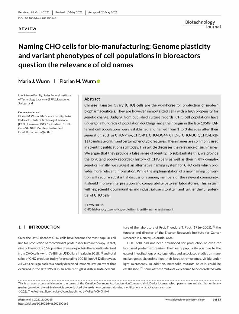
F IGURE 1 Generation of three Z-chromosomes by rearrangements. Sites of several enzyme encoding genes are indicated. APRT, LDHA, GAA,PGM3 indicate approximate gene locations of corresponding enzymes. The images are re-worked figures from,[4] done by C. P.Wurm,Montpellier,France. (A) Inversion of amajor chromosomal fragment within chromosome 3 results in the generation of a new, unusual chromosome Z4. Genelocations are changed accordingly and thus follow the prior determined banding patterns. B) Reciprocal translocation between chromosomes 3and 4 results in the generation of two new, unusual chromosomes Z3 and Z7.Note: The color shading in the image of Giemsa-banded chromosomeswas introduced to better visualize the new arrangement within the emerged Z-chromosomes
recognized chromosomal rearrangements and thus supported ground-
breaking and fundamental studies of genes, their positions on chromo-
somes and their functions in a mammalian in-vitro system. An example
of suchwork is shown in Figure 1,modified from reference.[4] “Giemsa-
bands” allow to identify specific chromosomes (even if similar in size) in
mammals and/or in cells derived from them. The figure demonstrates
structural changes (inversionof a chromosomal fragment anda translo-
cation). Some of the changes correlate with metabolic functions of
the concerned genetic markers. The studies enforced insights gained
by Thomas Morgan Hunt (1866–1945) on linked inheritance of genes
(synteny) when localized closely to each other on a chromosome.
Genomic rearrangements ofDNAoccur frequently inCHOcells and
thus provided an approach to link gene locations with functions. Many
cell lines emerged or were selected for specific phenotypes, some-
times after irradiation or chemicalmutagenesis andwere subsequently
named to differentiate them from each other. Some of these names are
still in use by the biologics producing industry. Such names (shall) typ-
ically indicate the origin of cell populations, but do they provide infor-
mation on their utility?
This review will refer occasionally to the results of karyotyping, to
explain the genetic/genomic evolution of CHO cells. This method is
highly suitable and at timesmore efficient than genome sequencing, to
visualize rapidly genomic modifications in both individual cells and in
populations of CHO cells.
CHO cells show, in comparison to cancer cells, a relatively stable
chromosome number (!) matching globally the diploidHamsterwith 22
chromosomes (11 pairs), albeit with considerable structural instability
of all chromosomes. The overall genome size of these cells is slightly
smaller than that of Chinese Hamster Cricetulus griseus, characterized
by haplodiploidy, losses of genes and trends for rearrangements of
“normal” Hamster and other chromosomes that have already unusual
structures. We quote:[3] “the modal chromosome number is 21, or 20,
in the case of one subclone isolated by Kao and Puck (1968), com-
pared with a diploid number of 22 for the Chinese hamster. Although
CHO cells carry many chromosomes which differ from those of the
diploid Chinese hamster karyotype (Kao and Puck, 1969), the modal
chromosome number of a clone is usually constant over many months
or years.” Thementioned reference for theChineseHamster karyotype
is in.[5]
A body of literature states that immortalized cells are very similar to
cancer cells when comparing their genetic and phenotypic instability,
based on an enormous number of genetically diverse cells in a cancer
patient or in bioreactors.[6,7,8]
This discussion on cell line utility for expression of recombinant
proteins conveys that ancient names provide little information on their
genetic constitution and essentially nothing on their phenotypes for
industrial use. It is misleading that “old” names indicate usefulness
for any specific approach in cloning of genes, in expression yield,
WURM ANDWURM 3 of 13
or—in the context of industrial application—for culturing these cells
in deep tank bioreactors. Even verifying the authenticity of the name
is impossible since a documented history of subcultivations cannot be
provided.
We will revisit some of the profound studies done with these
cells during the 1960–1980. This will allow us to better understand
the genomic plasticity and corresponding phenotypes when cells are
now grown in fixed-bed culture systems, or in stirred or shaken
bioreactors.
2 CHO ORIGIN, IMMORTALIZATION, ANDNAMES
CHO cells were established 1957 as a cell line. For decades derived
cells served as an excellent source for fundamental research in molec-
ular cell genetics. Cell populations were used by laboratories to study
gene numbers, structures and locations of mammalian genes, and for
the purpose to unravel functions and genetic principles of chromo-
somes in mammals. A 1963 paper refers to CHO-pro– (minus) cells,[9]
which appears to be a dominant phenotype of all CHO cells.[10] Thus,
the loss of proline synthesis capacitywas apparently a very early event,
before the distribution and use of derived cell lines. Among the derived
cell populations was the “K1” cell line that was claimed to be derived
from a single cell—a “clone”.[11] Many other auxotrophic mutants of
CHO cells were found and/or generated.[12,13]
The immortalization event of CHO cells, that is, the conversion of
the ovary-isolated primary cells into an adherent cell line, occurred
sometimes during regular subcultivations in sterilized glass flasks or
dishes. At the time, cells were cultured using media containing serum
or major fractions of serum, mostly of bovine origin, at concentrations
of 10–20%. No chemical or physical exposure appeared related to the
immortalization event. It had occurred “spontaneously”. In the 1958
publication,[2] we find “and one (cell line) arising from the ovary of a Chi-
nese Hamster, was selected for long-term cultivation in the complete growth
medium of Table I. These (cells) have now been carried for more than 9
months during which they have undergone a minimum of two generations
per week, or a total of 78 generations, equivalent to 1023 progeny”.
This remarkable statement demonstrates the robustness and ease
of handling of these cells. The 78 generations of the cell population
should also be considered with respect to the genetics. 1023 cells,
equivalent to 350 million tons (1 cell weighs 3.5 x 10⁻⁹ g, the weight
of all mankind today being about 450 million tons), are the product
of error prone DNA/chromosome duplications! A heterogenic cell
population without maintenance of diploidy emerged, even before any
specific names were coined.We have referred to these cells as CHOori
before.[14]
The K1 cell line was established in the late 1960s by Dr. Fa-Ten Kao,
then a Senior Scientist in Puck’s laboratory, as a clonally derived cell
line, from the earlier mentioned CHO-Pro– [minus] cells.[12] “Mutagen-
esis experiments were performed with CHO Pro – and its K1 subclone”.[11]
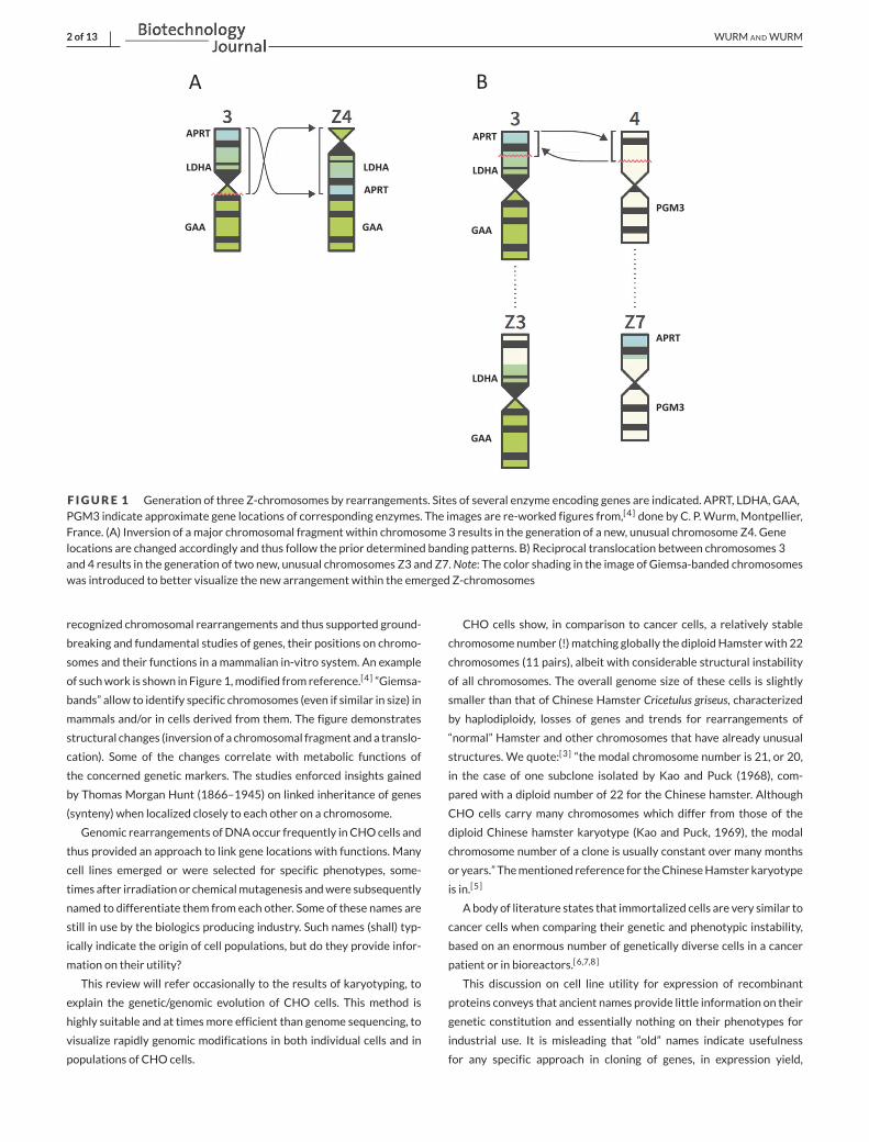
No information is provided how the cloning occurred, but maybe
cells were scraped off or aspirated from an adherent colony with a
pipette—similar to the ones in the image provided in the Kao and Puck
1967 [10] paper (Figure 2).
The “stemnumber” (The stemnumber, equivalent to themodal chro-
mosome number, indicates the total number of chromosomes, deter-
mined as the majority of cells containing that number, quasi-tetraploid
cells are excluded.) of chromosomes in these cells had declined from
22 to 21. CHO Pro– cells were obtained in 1962 by a laboratory in Los
Alamos. Analyzing cells by karyotyping, reported on about a decade
later (1973), indicated that the modal (or stem) chromosome number
was 21 [15]: “Our results demonstrate that only 8 of the 21 chromosomes
are normal when compared with Chinese Hamster chromosomes.” Refer-
ence is made in this paper to the K1 cell line and its’ similarity to the
unnamed cell line’s chromosome structures. The K1 karyotype had
been published earlier (1970), albeit thenwith amuch lower resolution
technique. The K1 cell line was having amodal chromosome number of
20:[11]“In some experiments, the subclone, CHO/Pro–-K1, which possesses
a stemline of only 20 chromosomes, was utilized”. This is in contradiction
to a 1985 publication by Puck indicating a modal chromosome number
of 21.[12] The1968paperbyKaoandPuck[11] also refers to theK1 sub-
clone frequently converting to glycine auxotrophy [dependence on the
addition of glycine].
The periods between published statements on chromosomal num-
bers, specific phenotypes and applied names of cell lines are typically
long (years) and do not provide culture details for these periods.
We bolded “clonally derived cell line” above: Simply cultivating cells
results in drastic genetic changes in populations, clonal or not, and
this has been verified frequently by karyotyping. The susceptibility for
rapid changes of the genome of individual CHO cells at each mitotic
cycle has been discussed in a paper using the term CHO Quasispecies
when referring to clonally derived cell lines.[16] The term “clone”, fre-
quently used, perpetuates an impression for non-experts that such
cells are genetically identical (or near identical).
Having identified a good CHO “clone” actually means that the popu-
lation of cells has maintained to a high degree (>70%?) the transcribed
DNAof interest. This populationmaycontinue todeliver for a fewmore
weeks the productwith yields of 60–100%of the initial productivity. In
a good “clone” themajority of cells have, thus, kept the chromosome or
chromosome fragments containing the gene of interest. The majority
of cells—even if diverse genetically—while transcribing the exogenous
DNAwill also assure growth and overall metabolic robustness. Popula-
tions emerging after cloning are related to each other, a CHO Quasis-
pecies. However, the overall genetic diversity of such clonally derived
cell populations remains and exceeds the genetic diversity of popula-
tions of a biological species.
Most labs, having worked with the early CHO cultures, have been
dismantled and/or handed over to successors. Puck, a highly respected
researcher with many achievements during his career, has passed
away. Prof. Fa-Ten Kao is 86 at the time of writing this text. Our
attempts to identify a laboratory that may still have frozen cell vials
from the 60–70s have not been successful. Prof. Lawrence Chasin,
the originator of the first industry-used cell lines,[17] is maybe the
only one who has kept cells generated first in the 1970s in his freez-
ers. To replenish his stocks of cells, he repeatedly refroze cells from

4 of 13 WURM ANDWURM
F IGURE 2 Copy of Figure 1 in,[10] indicating the requirement the CHOPro– cell line for proline
fresh cultures into small research banks for distribution (personal
communication).
The cells are kept in liquid nitrogen and are grownwith FBS contain-
ing media. Chasin’s DHFR-negative CHO cell line, named CHO DXB-
11, (also sometimes referred to as CHO-DUK) became the host sys-
tem for the first CHO produced pharmaceuticals, such as the Genen-
tech Inc. developedActivaseTissuePlasminogenActivator (TPA) or the
AMGEN Inc. EPOGEN Erythropoeitin (EPO). To obtain recombinant
cells synthetizing these proteins, DHFR-expression vectors were con-
structed that also contained theDNAencoding humanTPAor Erythro-
poeitin. The functional DHFR gene on the vector would thus “repair”
the deficiency in cells and would allow the selection of recombinant
CHO cells co-expressing the DNA for TPA or EPO.
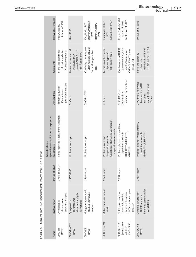
Table 1 provides information on some of the cell lines utilised during
the period of 1960 to 1990, contributing to knowledge on mammalian
genetics, chromosome identity and structures, gene locations, etc.
3 INDUSTRIALLY APPLIED CHO CELL LINES
Names as “CHO-Pro–,“ “CHO-DUK,” “CHODXB11,” “CHO-K1,” “CHO-
DG44,” “CHO-S” falsely imply how to differentiate cells from each
other. At the time of generating these names, all cells were grown in
the presence of serum and in dishes or flasks. The majority of indus-
trially used CHO cell lines are now grown in chemically defined media
in suspension cultures in stirred or shaken bioreactors.[18,19] However,
Prof. RichardG. Ham (1932-2011) grewCHOcells serum-free in 1977,
prior to similar efforts in the early biotech industry.[20] Interesting in
this context is also L. Thompson’s publication, the same year, using the
first suspension cultureofCHOcellswith aMendelian type inheritance
of phenotypes. When plating them as single cells on soft agar he could
reproduce two phenotypes, “arrested dome” and “fried egg”morpholo-
gies of colonies.[21]
Obtaining highly productive recombinant cell populations in large-
scale bioreactors is a complex task. Transfection with optimized
expression vectors for expression of the transgene(s) from strong pro-
moters leads to the selection of clonally derived cell populations with
genome-integrated vector DNA. Frequently now antibiotics are added
to the culture medium, with the corresponding resistance marker on
the vector. This approach can replace the above mentioned DHFR-
or Glutamine Synthetase (GS) selections. When using antibiotics for
selection any CHO line can be used.
3.1 Productivity
The most widely used industrial principles for CHO-based manu-
facturing under suspension culture were developed in South San
Francisco at Genentech Inc., USA. Adherent cells could not produce
sufficient amounts of human Tissue Plasminogen Activator (TPA), for
a successful introduction into the US-American market. The expected
dose of 10 mg/patient was found in clinical trials to be 10 times higher,
WURM ANDWURM 5 of 13
TABLE1
CHOcelllin
esusedinfundam
entalresearchfrom1957to
1990
Nam
eR&Dwork
for
PeriodofR
&D
Modifications
(gen
etic/m
etabolic/specialexp
osures,
treatm
ents)
Derived
from
Commen
tsRelevantreferences
CHO-ori
(1957)
Cytogenetics,
chromosome
structure
analysis
1956-1960s(?)
Nonereported
(spont.im
mortalization)
Primaryculture
of
Ovary
tissue
(outbredham
ster)
Manylabsusedthese
cells
prioran
dafter
K1becam
epopular
Puck,C
iecuira,
Robinson1958
CHO-Pro
minus
(1957)
Cytogenetics,
chromosome
structure
analysis
karyotypes
1957-1980
Prolin
eau
xothroph
CHO-ori
Numerousderived
cell
lines
werethen
nam
edCHOPro
–3,
Pro
–5,andso
on.
Ham
1963
CHO-K1
(1968)
Mutagenesis,m
etab
olic
studies,karyotype
analysis
1968-today
Prolin
eau
xothroph
CHO-Pro
minus
Confusingchromosome
Stem
number
(21/20)
Serum-freegrowth
of
cells
Kao
,Puck
1967
Deaven,Peterson
1973
Ham
ilton,H
am,
1977
CHO-S
(1974)
Mutagenesis,m
etab
olic
stud.
1974-today
Prolin
eau
xothroph
Suspen
siongrowth
Gen
eticsofp
hen
otypevariationof
suspen
sionculture
cells
CHO-ori
Men
delianinheritan
ce
ofp
hen
otypes
of
cells
onsoftagar.
Thompson,Baker
1974
Konradet
al.1977
CHO-D
XB11
(1980)(also
ref.to
as
CHO-D
UK)
DHFRgenestructure,
metab
olic
studies,
geneam
plification,
MTXtreatm
ent,gene
tran
sfer
1980-today
Prolin
e-,glycine-,hyp
oxan
thine-,
thym
idine-au
xotoph
(DHFRminus /DHFRminus )
GHTminus
CHO-K1after
Chem
icalan
d
gamma-rayradiation
Onetran
scribed
DHFR
generemaining,with
singleAAmutation
CHO-D
UKNOTsame
asDX-B11
Chasin,U
rlau
b1980
Kaaset
al.2015
Turilova
etal.2021
CHO-D
G44
(1983)
Gen
omicstructure
of
DHFRsequen
ces
Simplifiedgenetran
sfer
withDHFR
1985-today
Prolin
e-,glycine-,hyp
oxan
thine-,
thym
idine-au
xotoph
(DHFRminus /DHFRminus )
CHO-Pro-3
follo
wing
treatm
entw.M
TX
forgene
amplificationan
d
Y-rays
Note:thepaper
by
Urlau
bet
al.
men
tionsDG41an
d
DG42,butnotDG44
Urlau
bet
al.1983
6 of 13 WURM ANDWURM
resulting in a critical manufacturability problem. The only solution was
to have the producer cell line grow as a suspension culture in large
stirred bioreactors (STRs). Drawing from insights gained with the BHK
cells for Foot and Mouth Disease Virus production,[22,23] the conver-
sion to suspension cell populations for STR based manufacturing was
eventually done—with cells derived from a bank of cells under adher-
ent culture. The term “adaptation” was used for the process towards
suspension culture–wrongly aswewould argue. (This seems academic,
but a selection of genetically different subpopulations with a higher
capacity to grow in suspension culture is the most likely explanation,
that is, the change in phenotype is connected to a suspected modifi-
cation of genotype overall.) The process that delivered the required
product quantity for the US-American market was initially based on
the use of 2000 L and then 10′000 L bioreactors and had a volumetric
yield of 50mg L−1. From today’s perspective, this is a low yield, but one
needs to remember that the optimization of cell culture media compo-
sitions was just starting at the time and a cell density of 2–3× 10⁶ cells
mL−1 was considered excellent for a 7-day batch process. The pro-
duction line was derived from the aforementioned “CHO-DXB11”
cell, which conveniently allowed transfer of the DNA of interest into
the CHO genome by co-transfer with a DHFR expression cassette.
Dr. Chasin had established that one allele of the functional DHFR
gene was missing in his cell line (by loss of a chromosome fragment),
whereas the other had a single nucleotide mutation.[17] To increase
productivity methotrexate (MTX)—an antagonist to DHFR—was used.
This approach, called gene amplification selects for populations of cells
that have increased the copy-number of the DHFR/TPA vector within
CHO chromosomes. The mechanisms for such gene amplifications
have been studied in cancers and cancer-derived cell lines, as well as in
CHO cell lines. They involve drastic chromosomal rearrangements. In
other words, the known genomic malleability of CHO cells was used to
select for subpopulations of cells, emerging spontaneously, to increase
productivity.[24,25]
In optimized fed-batch processes, CHO cells can produce today 3–
10 g L−1 of recombinant antibodies and other molecules, with chem-
ically defined media that maximize viable cell densities. These pro-
cesses apply animal component free media. Research into medium
formulations provided much enriched compositions. Medium formula-
tions are non-published intellectual properties. High yields have been
shownwithout any host cell engineering.[18,26]
3.2 Cloning, subcultivations of cells, geneticbottlenecks, and population dynamics
Regulators request evidence of clonality when producing a thera-
peutic protein—the reasons being debated in the industry.[27] The
topic was discussed again recently by scientists of the Food and
Drug Administration.[28] Importance of clonal derivation was main-
tained, while admitting that “a master cell bank created under even
the strictest conditions possible may never be truly “clonal””. The word
“may” is misleading—the MCB cells will never be clonal in the
sense of its typical use in biology. FW[16] recommended minimiz-
ing growth-restricting conditions. But this was considered by the
FDA authors “incompatible with the nature of modern biopharmaceuti-
cal production”. We do not understand this: Growth-restricting envi-
ronments can be avoided, or at least reduced. This supports mainte-
nance of an averaged phenotype of evendiverse but genetically related
populations.
Clonality is hoped to increase robustness/reliability or reproducibil-
ity of themanufacturing of the product, such as the avoidance of a sec-
ond cell with a different genetic background to be the “unknown cause”
of a mixed and/or unstable population. Insistence of clonal deriva-
tion was not enforced during the 80s and 90s. FW has picked clonal
populations as visually marked colonies of hundreds of adherent cells
from Petri dishes using sterilized cotton swaps or “cloning rings.” Cells
adhering to the cotton or being aspired off were transferred into the
medium within a micro-well. Colonies on a plate could be easily com-
pared in size. Cloning single cells, a standard today, was done thenwith
efficiencies below 10%, even with fetal bovine serum as part of the
medium composition.
Several “cotton-swab” derived cell lines are the source of glob-
ally marketed products still today. The relative genetic and produc-
tion homogeneity of emerged populations and their overall production
stability was mostly based on the purified protein product, assuring
that the manufacturing process delivered a reliable and reproducible
pharmaceutical.[27]
Whatever the starting point of a cell population for production,
likely a suspension of frozen cells in a small vial, cells are exposed to
many steps. All work is also associatedwith the generation of Research
Banks,Master Cell Banks andWorking Cell Banks, derived from short-
term, expanded cultures to be aliquoted into small containers, medium
exchanged and exposure to freeze-thaw cycles.
To estimate the number of these frequently critical cell processing
steps is impossible to judge. Again, cloning does not protect against
population heterogeneity. This will become more transparent in the
next chapter.
4 GENETIC AND PHENOTYPE IMPLICATIONSOF NON-CLONAL AND CLONALLY DERIVED CELLPOPULATIONS
It is likely that 200 or more subcultivations have been executed since
the cells were named during the 1960–1980 period. We wish to put
this into a calculation that starts with 2 × 10⁶ cells. With only 100
subcultivations the astonishing number of 1 × 1096 cells could have
been generated. The necessary DNA replication of a 3 billion base pair
genome will involve numerous modifications of the underlying CHO
genomes. The “normal” mutation rate is 10–8 per locus/cell in mam-
malian systems.[29] In cultures of a suspension mouse cell line, a muta-
tion rate of 1–7 × 10⁻⁷/cell/generation (generation = cell duplication)
was found.[30] With these numbers in mind, and the large number of
duplications in regularmaintenance of cell populations occurring, CHO
cells will accumulate very fast mutations that will affect phenotypes.
Mutation driven selection will lead to evolution of cell populations,
WURM ANDWURM 7 of 13
depending on the various environmental conditions—following the
concepts elegantly and convincingly described by Charles Darwin,[31]
more than 150 years ago. Genome and phenotype affecting changes
will be even more striking when cloning steps are involved that gener-
ate “genetic bottlenecks” inwhich a single cell gives rise to an emerging
new population.
4.1 Complex cytogenetics of cloned andnon-cloned cell populations
T. C. Hsu [1917–2003] published 1961 a landmark review article
“Chromosomal Evolution in Cell Populations”[32] in which he stated
that “cells grown in-vitro change from their original genetic composition to
a heteroploid condition”. Recent (2021) karyotyping of CHO cells veri-
fied this statement, while drawing also important insights from kary-
otyping of CHO cells over decades.[33] Next generation genome DNA
sequencing techniqueson cancer cells andother immortalized cell lines
revealed that some genomic changes can be dramatic, even “catas-
trophic”, resulting in breaks into multiple fragments of chromosomes
(chromothripsis). However, the majority of chromosomemodifications
inmany cancer cells and cell lines result in “simple” translocations, frag-
ment inversions, multiplications, losses of fragments and entire chro-
mosomes, losses of heterozygosity, polyploidizations. HeLa cells may
be an extreme example: These cells, the first established cell line in his-
tory, show these modifications, including chromothripsis. More than
60′000 papers have been published usingHeLa cells.[34] The cellswereobtained and established as a culture by the treating doctor, G. O. Gey
(1899–1970) of Mrs. Henrietta Lacks. She passed away in 1951 in Bal-
timore at the age 31 due to complications from amalignant cervix car-
cinoma that had invaded the entire body and resulted in an acute kid-
ney failure.[35] The cells were instrumental in establishing cell culture
technologies as they are used still today and with tremendous bene-
fits for biology andmedicine. A recent genome sequence analysis[36,37]
of HeLa populations showed hyper-triploidy on average, with some
genomic fragments having a ploidy of four or higher and presenting
20 highly aberrant and rearranged chromosomes, four of which hav-
ing been the subject of chromothripsis. Also, theHeLa genome exhibits
significant stretches of homozygosity, explainable only by losses of
the heterozygote partner chromosomes or fragments thereof. In addi-
tion, there is a plethora of single nucleotide variants, indels (“short”
DNA- fragment—50 base pair to 10′000 base pair—deletions and
insertions) and copy-number changes, includingmore than 2800 struc-
tural variations, dominated by large deletion events. The numbers are
staggering—approximately 4.5 million single nucleotide variations and
about 500′000 indels!A genome sequenceofCHOcellswas published2011: “The genomic
sequence of the Chinese hamster ovary [CHO]-K1 cell line.”[38] We
have serious reservation for the dual use of “THE” in the title of
that paper. The article seems to imply that there is such a thing as
one genomic sequence and one CHO-K1 cell line. In the article the
term “ancestral” was used, probably referring to the fact that the cells
were grown in simple culture media with 10% Fetal Bovine Serum
(FBS) and were obtained from the American Type Culture Collections
(ATCC).
The published genome sequence represented 21 scaffolds as non-
interacting DNA blocks, interpreted as 21 chromosomes. An image
of size-ranked chromosomes was shown. As a reminder, the 1973
karyotype paper [15] showed also a distribution of chromosome num-
bers per cell. They ranged from 19 to 44 [1 pseudo tetraploid cell],
when analyzing 50 metaphase spreads. Thirty eight of these show
the chromosome stem number of 21, six cells have 20 chromosomes
and four have 22! The CHO K1 genome sequence paper does not
provide chromosome-based sequence data and gives little informa-
tion on differences from the sequence composition of the 11 chro-
mosome identities of the Hamster. The paper did not comment on
indels and single nucleotide variations, nor does it discuss the level of
ploidy and homo- and heterozygosity. Also, the presence of endoge-
nous retroviral sequences—a major concern for products made with
immortalized cells—giving rise to A- and C-type retrovirus like parti-
cles is not mentioned. The relative scarcity of such particles in CHO
cells and the mostly non-functionality of corresponding sequences in
their genome,[39] in comparison to mouse-derived cell hosts, was one
of the reasons why CHO cells were considered a safe(r) host system
for human therapeutic protein production.
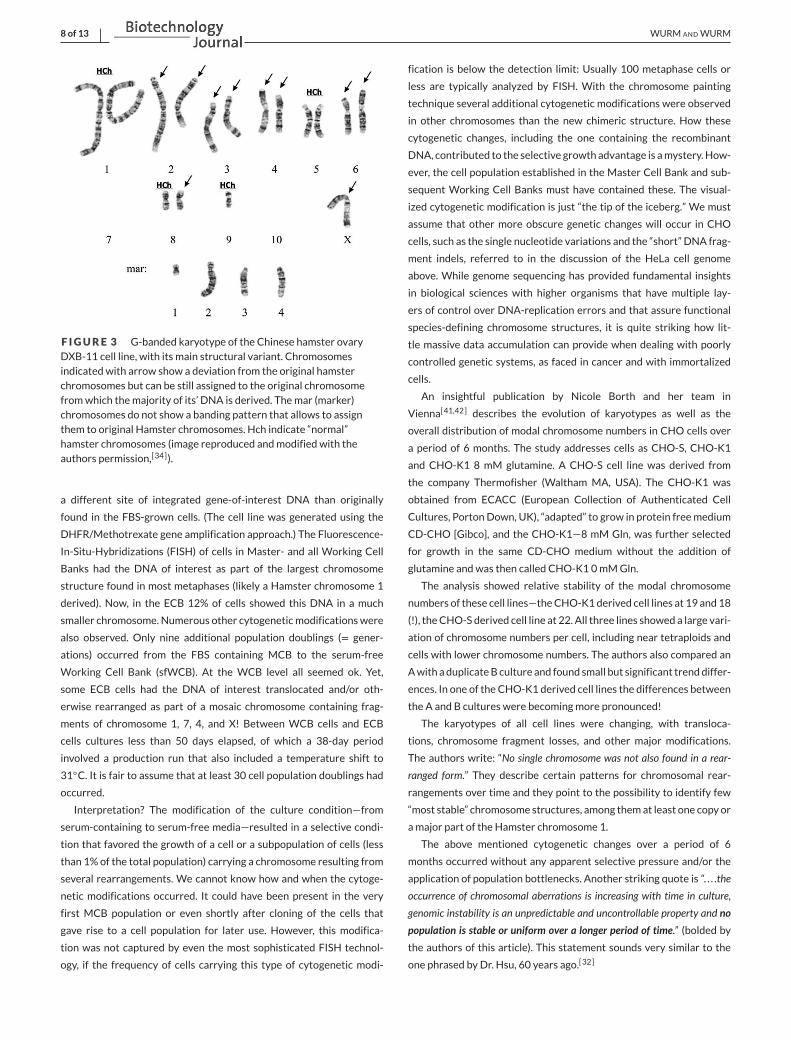
Highly useful insights gained by karyotyping of a CHO DXB-11 cell
line and comparison with other CHO karyotypes were just recently
published.[33] In this cell line, delivered to Russian Cell Culture Col-
lection in 1984, only 17% of cells appear to have a karyotype that
would be accepted as the main structural variant, that is, repeatedly
occurring structural (chromosome) features in cells. This main struc-
tural variant [MSV] shows six normal hamster chromosomes (pairs of
the Hamster chromosomes 1 and 5, and a single representative of
the Hamster chromosomes 8 and 9) and 14 structurally rearranged
chromosomes (i.e., their origin can be deducted from the banding pat-
tern), including four marker chromosomes (see Figure 3). Eighty three
percent of cells—based on an analysis of 112 karyotypes—displayed
clonal and nonclonal additional structural rearrangements of chromo-
somes. The authors also reveal that a prior assumed equivalence of
CHODUK and CHODXB-11 cannot be verified. Apparently, a naming
confusion occurred, since the here shown karyotype differs from the
published CHODUK karyotype. This example of an “old” cell line, ana-
lyzed recently, shows quite strikingly how misguided identity assign-
ments can be and how relatively small the numbers of cells can be in
a population that appear to be characterizable by a defined genotype.
Recombinant CHO cells used in pharmaceutical manufacture
can show similarly confusing complexities when considering their
karyotypes.[40] TheMerck (Darmstadt) groupused chromosomepaint-
ing techniques to identify origin and structures of the chromosomes
of a cloned (!) cell line producing a therapeutic protein. The pro-
ducer cell line was initially grown with FBS under adherent culture
and a Master Cell bank was generated. A fixed-bed bioreactor was
used to manufacture a biopharmaceutical. Eventually the company
decided to “adapt” the cell line of FBS -free processes. Strikingly, cells
derived from an extended cell bank (ECB) made with cells from a
Post-Production Cell bank (PPCB) showed a 12% subpopulation with
8 of 13 WURM ANDWURM
F IGURE 3 G-banded karyotype of the Chinese hamster ovaryDXB-11 cell line, with its main structural variant. Chromosomesindicated with arrow show a deviation from the original hamsterchromosomes but can be still assigned to the original chromosomefromwhich themajority of its’ DNA is derived. Themar (marker)chromosomes do not show a banding pattern that allows to assignthem to original Hamster chromosomes. Hch indicate “normal”hamster chromosomes (image reproduced andmodified with theauthors permission,[34]).
a different site of integrated gene-of-interest DNA than originally
found in the FBS-grown cells. (The cell line was generated using the
DHFR/Methotrexate gene amplification approach.) The Fluorescence-
In-Situ-Hybridizations (FISH) of cells in Master- and all Working Cell
Banks had the DNA of interest as part of the largest chromosome
structure found in most metaphases (likely a Hamster chromosome 1
derived). Now, in the ECB 12% of cells showed this DNA in a much
smaller chromosome. Numerous other cytogeneticmodificationswere
also observed. Only nine additional population doublings (= gener-
ations) occurred from the FBS containing MCB to the serum-free
Working Cell Bank (sfWCB). At the WCB level all seemed ok. Yet,
some ECB cells had the DNA of interest translocated and/or oth-
erwise rearranged as part of a mosaic chromosome containing frag-
ments of chromosome 1, 7, 4, and X! Between WCB cells and ECB
cells cultures less than 50 days elapsed, of which a 38-day period
involved a production run that also included a temperature shift to
31◦C. It is fair to assume that at least 30 cell population doublings had
occurred.
Interpretation? The modification of the culture condition—from
serum-containing to serum-free media—resulted in a selective condi-
tion that favored the growth of a cell or a subpopulation of cells (less
than 1% of the total population) carrying a chromosome resulting from
several rearrangements. We cannot know how and when the cytoge-
netic modifications occurred. It could have been present in the very
first MCB population or even shortly after cloning of the cells that
gave rise to a cell population for later use. However, this modifica-
tion was not captured by even the most sophisticated FISH technol-
ogy, if the frequency of cells carrying this type of cytogenetic modi-
fication is below the detection limit: Usually 100 metaphase cells or
less are typically analyzed by FISH. With the chromosome painting
technique several additional cytogenetic modifications were observed
in other chromosomes than the new chimeric structure. How these
cytogenetic changes, including the one containing the recombinant
DNA, contributed to the selective growth advantage is amystery.How-
ever, the cell population established in the Master Cell Bank and sub-
sequent Working Cell Banks must have contained these. The visual-
ized cytogenetic modification is just “the tip of the iceberg.” We must
assume that other more obscure genetic changes will occur in CHO
cells, such as the single nucleotide variations and the “short” DNA frag-
ment indels, referred to in the discussion of the HeLa cell genome
above. While genome sequencing has provided fundamental insights
in biological sciences with higher organisms that have multiple lay-
ers of control over DNA-replication errors and that assure functional
species-defining chromosome structures, it is quite striking how lit-
tle massive data accumulation can provide when dealing with poorly
controlled genetic systems, as faced in cancer and with immortalized
cells.
An insightful publication by Nicole Borth and her team in
Vienna[41,42] describes the evolution of karyotypes as well as the
overall distribution of modal chromosome numbers in CHO cells over
a period of 6 months. The study addresses cells as CHO-S, CHO-K1
and CHO-K1 8 mM glutamine. A CHO-S cell line was derived from
the company Thermofisher (Waltham MA, USA). The CHO-K1 was
obtained from ECACC (European Collection of Authenticated Cell
Cultures, PortonDown, UK), “adapted” to grow in protein freemedium
CD-CHO [Gibco], and the CHO-K1—8 mM Gln, was further selected
for growth in the same CD-CHO medium without the addition of
glutamine andwas then called CHO-K1 0mMGln.
The analysis showed relative stability of the modal chromosome
numbers of these cell lines—theCHO-K1derived cell lines at 19 and18
(!), theCHO-S derived cell line at 22. All three lines showed a large vari-
ation of chromosome numbers per cell, including near tetraploids and
cells with lower chromosome numbers. The authors also compared an
Awith aduplicateB culture and found small but significant trenddiffer-
ences. In one of the CHO-K1 derived cell lines the differences between
the A and B cultures were becomingmore pronounced!
The karyotypes of all cell lines were changing, with transloca-
tions, chromosome fragment losses, and other major modifications.
The authors write: “No single chromosome was not also found in a rear-
ranged form.” They describe certain patterns for chromosomal rear-
rangements over time and they point to the possibility to identify few
“most stable” chromosome structures, among themat least one copy or
amajor part of the Hamster chromosome 1.
The above mentioned cytogenetic changes over a period of 6
months occurred without any apparent selective pressure and/or the
application of population bottlenecks. Another striking quote is “. . . .the
occurrence of chromosomal aberrations is increasing with time in culture,
genomic instability is an unpredictable and uncontrollable property and no
population is stable or uniform over a longer period of time.” (bolded by
the authors of this article). This statement sounds very similar to the
one phrased by Dr. Hsu, 60 years ago.[32]
WURM ANDWURM 9 of 13
In conclusion, readers will understand now that CHO cells cannot
be defined under the rules of the more stringent genetics of animal
and plants species. In CHO cells, evolution of genomes is a constant
phenomenon and at times, with drastic results and occurs within time
frames of typical laboratory cultures. It seems that the success of CHO
cells in manufacturing may be at least in part a result of the capacity of
CHO populations to approach and achieve fitness for bioreactor envi-
ronments relatively fast.
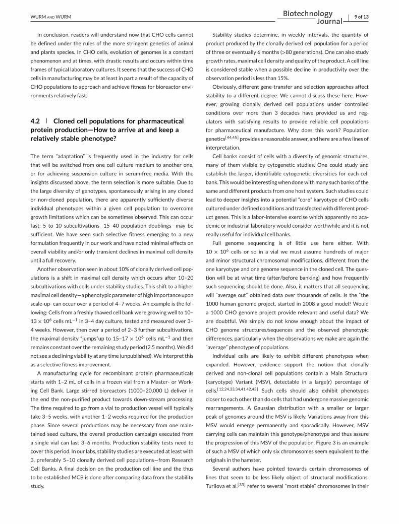
4.2 Cloned cell populations for pharmaceuticalprotein production—How to arrive at and keep arelatively stable phenotype?
The term “adaptation” is frequently used in the industry for cells
that will be switched from one cell culture medium to another one,
or for achieving suspension culture in serum-free media. With the
insights discussed above, the term selection is more suitable. Due to
the large diversity of genotypes, spontaneously arising in any cloned
or non-cloned population, there are apparently sufficiently diverse
individual phenotypes within a given cell population to overcome
growth limitations which can be sometimes observed. This can occur
fast: 5 to 10 subcultivations -15–40 population doublings—may be
sufficient. We have seen such selective fitness emerging to a new
formulation frequently in our work and have noted minimal effects on
overall viability and/or only transient declines in maximal cell density
until a full recovery.
Another observation seen in about 10% of clonally derived cell pop-
ulations is a shift in maximal cell density which occurs after 10–20
subcultivations with cells under stability studies. This shift to a higher
maximal cell density—aphenotypic parameter of high importanceupon
scale-up- can occur over a period of 4–7 weeks. An example is the fol-
lowing: Cells from a freshly thawed cell bank were growing well to 10–
13 × 106 cells mL−1 in 3–4 day culture, tested and measured over 3–
4 weeks. However, then over a period of 2–3 further subcultivations,
the maximal density “jumps”up to 15–17 × 106 cells mL−1 and then
remains constant over the remaining study period (2.5months).Wedid
not see a declining viability at any time (unpublished).We interpret this
as a selective fitness improvement.
A manufacturing cycle for recombinant protein pharmaceuticals
starts with 1–2 mL of cells in a frozen vial from a Master- or Work-
ing Cell Bank. Large stirred bioreactors (1000–20,000 L) deliver in
the end the non-purified product towards down-stream processing.
The time required to go from a vial to production vessel will typically
take 3–5 weeks, with another 1–2 weeks required for the production
phase. Since several productions may be necessary from one main-
tained seed culture, the overall production campaign executed from
a single vial can last 3–6 months. Production stability tests need to
cover this period. In our labs, stability studies are executed at leastwith
3, preferably 5–10 clonally derived cell populations—from Research
Cell Banks. A final decision on the production cell line and the thus
to be established MCB is done after comparing data from the stability
study.
Stability studies determine, in weekly intervals, the quantity of
product produced by the clonally derived cell population for a period
of three or eventually 6 months (>80 generations). One can also study
growth rates,maximal cell density and quality of the product. A cell line
is considered stable when a possible decline in productivity over the
observation period is less than 15%.
Obviously, different gene-transfer and selection approaches affect
stability to a different degree. We cannot discuss these here. How-
ever, growing clonally derived cell populations under controlled
conditions over more than 3 decades have provided us and reg-
ulators with satisfying results to provide reliable cell populations
for pharmaceutical manufacture. Why does this work? Population
genetics[44,45] provides a reasonable answer, andhere are a few lines of
interpretation.
Cell banks consist of cells with a diversity of genomic structures,
many of them visible by cytogenetic studies. One could study and
establish the larger, identifiable cytogenetic diversities for each cell
bank. Thiswouldbe interestingwhendonewithmany suchbanksof the
same and different products from one host system. Such studies could
lead to deeper insights into a potential “core” karyotype of CHO cells
cultured under defined conditions and transfectedwith different prod-
uct genes. This is a labor-intensive exercise which apparently no aca-
demic or industrial laboratory would consider worthwhile and it is not
really useful for individual cell banks.
Full genome sequencing is of little use here either. With
10 × 106 cells or so in a vial we must assume hundreds of major
and minor structural chromosomal modifications, different from the
one karyotype and one genome sequence in the cloned cell. The ques-
tion will be at what time (after/before banking) and how frequently
such sequencing should be done. Also, it matters that all sequencing
will “average out” obtained data over thousands of cells. Is the “the
1000 human genome project, started in 2008 a good model? Would
a 1000 CHO genome project provide relevant and useful data? We
are doubtful. We simply do not know enough about the impact of
CHO genome structures/sequences and the observed phenotypic
differences, particularly when the observations we make are again the
“average” phenotype of populations.
Individual cells are likely to exhibit different phenotypes when
expanded. However, evidence support the notion that clonally
derived and non-clonal cell populations contain a Main Structural
(karyotype) Variant (MSV), detectable in a large(r) percentage of
cells.[12,24,33,34,41,42,43] Such cells should also exhibit phenotypes
closer to each other than do cells that had undergonemassive genomic
rearrangements. A Gaussian distribution with a smaller or larger
peak of genomes around the MSV is likely. Variations away from this
MSV would emerge permanently and sporadically. However, MSV
carrying cells can maintain this genotype/phenotype and thus assure
the progression of this MSV of the population. Figure 3 is an example
of such a MSV of which only six chromosomes seem equivalent to the
originals in the hamster.
Several authors have pointed towards certain chromosomes of
lines that seem to be less likely object of structural modifications.
Turilova et al.[33] refer to several “most stable” chromosomes in their
10 of 13 WURM ANDWURM
cultures of DXB-11 cells, one being the single “authentic” chromosome
9, and other chromosomes that were identified as derived from the
X-chromosome and from the chromosomes 2, 4 and 6. Interestingly,
these latter structures represent rearranged chromosomes, clearly
different from the original hamster homolog. Thus, it is fair to assume
that certain authentic and non-authentic hamster chromosomes in
a population have a (slightly?) higher probability to be maintained as
part of a MSV. However, it would be too simplified to conclude that
authentic hamster chromosomes have a higher probability for being
maintained unaltered, as indicated by publications of the Borth group
in Vienna.[41,42]
The observed phenotype of a given cell population is the result of all
genotypes. Among this, one shouldnot underestimate the role of short-
lived cells that emerge and die in a population in this context. Even in a
“100% viable” culture, cells die permanently—we just do not see them
in cell counting methods. These cells deliver into the medium compo-
nents that may support the growth of others. Other strange phenom-
ena occur, observed in hybridoma cell cultures. One example is this: A
larger than typical cell engages into cell division, resulting “almost” in
two daughter cells. This is shown in a time-lapsed video recording of
cells under a microscope. However, within 45 min, further separations
into 5 cell-like structures occur, to then fuse back within 40 min first
to three and then to two separate cells,[46] see also.[14] One wonders
whatmay have happened to the genome of the first cell during the sub-
sequent events.
Thus, genetic and phenotypic stability of a population of cells is to
be understood as averaged from diversity. If there are enough cells in
the population that are, in terms of their physiology, a best fit to the
environment to which they are exposed to, then this phenotype could
turn out to be relatively stable, as long as this environment is kept.
An interesting paper provided opportunities of the diversity of clon-
ally derived cell populations for the generation of recombinant pro-
teins that require specific metabolic activities for obtaining acceptable
quality of the protein of interest. This work demonstrates the utility of
leveraging diversity toward delivering specific performance character-
istics of cells.[47] Heterogeneity in suspension cultures ofCHO-K1cells
was revealed by single-cell transcriptome analyses, indicating diversity
due to epigenetic influences, but also revealed mitochondrial genome
variation and heteroplasmy in cells.[48]
A note on DHFR/methotrexate (MTX) based cell lines use in indus-
try: In the early years of our industry the DHFR/MTX approach to
high yielding CHO cell lines was dominant,[24] later joined by a simi-
lar approach using glutamate synthetase (GS) and methylsulfoximine
(MSX).[49] Other selective agents, such as antibiotics, are also used
today, including their corresponding selective markers that convey
resistance after transfer with Zn-finger nucleases[50,51] and CRISPR-
Cas mediated gene transfers.[52] Together with modified expression
vector cassettes andadditional elements in plasmids, these approaches
enhance targeting the gene of interest sequences into open chromatin.
However, with single genome targets into specific chromosomal sites,
one should take thehighprobability ofmodifiedgenomesby rearrange-
ments into consideration.
5 NAMING OF CHOCELLS—RECOMMENDATIONS AND FINAL REMARK
Clonal or not, CHO cells have such a high-level propensity towards
larger cytogenetic and other less detectable genetic changes that a
name given 30–50 years ago makes little sense. This is even more so
when there is no documented history available that traces back the
various modes of culture and media used for a given cell line. While
we are fully aware that a widely accepted nomenclature is very diffi-
cult to change, we wish to initiate at least a discussion on this topic.
Such naming modification—at least for the cell lines used for indus-
trial purposes—would make their use, their analysis by modern pro-
teomics, transcriptomics and genomics and their improvement by tar-
geted and selectedmodificationsmuchmore efficient.We recommend
therefor establishing names of cells based on definable and relevant
phenotypic features.Well establishedmethods, such as DNA sequenc-
ing with properly designed primers and/or other methods, such as iso-
enzyme analysis[53] allows to assure a given cell line is in fact CHO
derived.
Thus, the basic name “CHO” is entirely sufficient. A suspensionCHO
cell line grown in chemically definedmediumshould,whenpassedon to
another user for industrial or research purposes, have this phenotypic
description somehow captured in its’ name. There is nothing wrong to
identify the company or laboratory which has done most of the work.
It seems that some cell lines established by commercial companies
(“Lonza cell line”) have at least in part embraced this idea. If genetic
modifications are involved (“knock-in, knock-out”) they should be part
of it. Examples could be “Lonza CHO CDM GSminus 2002″ “HorizonCHO (animal component-free) ACF DHFRminus 2010″. The chemically
defined medium (CDM) can refer to the specific medium applied, and
the year indicates the timing of the deposition of a bank. The number of
subcultivations under use of thementionedmediumwould be useful as
well: “Lonza CHOProCHO523 2002″.Realistically, we are fully aware that these or any other suggestions
will take time tobecomepossibly acceptedby the interestedCHOman-
ufacturing community. ESACT (European Society of Animal Cell Tech-
nology) or the Cell Culture Engineering meetings (USA) could be fora
for such discussions. Also, a discussion should be initiated with the
ATTC or ECACC. If a new naming standard could be implemented it
would provide more clarity, both on the actual derivation of cells and
their probability to be useful for protein manufacturing.
In concluding: No text written in early in 2021 can capture all his-
toric and technical details that gives a complete and profound under-
standing on where the cells in our labs came from. We have dealt
with them cumulatively now for more than 60 years and remain sur-
prised how little we know. We are also impressed how efficient these
cells can be modulated in their behavior in bioreactors if one takes
the time and studies their physiology under production conditions
carefully.
We hoped to bring a bit more clarity to the very obscure history
of these cells and also highlight those points and perceptions that are
clearlymisleading. And thus, whether or not one assumes that the cells
WURM ANDWURM 11 of 13
in my lab are “K1” derived or “S” derived, does not matter really. What
really counts is the handling of cells under well-defined conditions and
observing the spectrum of opportunities these cells and their unique
subpopulations provide. CHO cells have taken a dominating lead in
manufacturing of high-value protein therapeutics in bioreactors and it
is doubtful that any other host system will ever achieve the productiv-
ities and product qualities seen with these cells, providing products of
unsurpassed purity and safety for millions of patients.
ACKNOWLEDGMENTS
The authors thankDrs. Victoria Turilova and Tatiana Yakovleva, as well
as Dr. Joeri Kint, for reviewing of the paper and for useful suggestions
for improvements on interpretation of discussed data available in the
literature.
CONFLICT OF INTEREST
The authors are founders and managers of ExcellGene SA, a service
company for the Pharma and Biotech Industry. Both have had long
careers in academic institutions and the observations and conclusions
made onCHOcells are derived fromboth employment activities over a
period of 40 (FW) and 25 years (MW), respectively.
DEDICATION
To the memory of Professor Fritz Anders (1919-1999), genetics
teacher of FW, an early pioneer in oncogene- and tumor suppressor-
gene biology at the University of Giessen, Germany.
AUTHOR CONTRIBUTIONS
CASRAI CRediT Taxonomy: authors’ contribution(s) to the submitted
manuscript are attributed as follows: CRediT Taxonomy, Maria Wurm,
conceptualization-equal, investigation-equal, supervision-equal,
writing-, original draft-equal, florian wurm, conceptualization-equal,
investigation-equal, supervision-equal, writing-original draft-equal.
DATA AVAILABILITY STATEMENT
All data discussed are publicly available.
ORCID
FlorianM.Wurm https://orcid.org/0000-0003-1114-4520
REFERENCES
1. https://www.pharmacompass.com/radio-compass-blog/top-drugs-
and-pharmaceutical-companies-of-2018-by-revenues
2. Puck, T. T., Ciecuira, S. J., & Robinson, A. (1958). Genetics of somatic
mammalian cells. Journal of Experimental Medicine, 108(6), 945–956.3. Thompson, L. H., & Baker R.M. (1974). Isolation ofmutants of cultured
mammalian cells.Methods Cell Biology, 6, 209–281.4. Siciliano,M. J., Stallings, R. L., & Adair, G.M. (1985). The geneticmap of
the Chinese hamster and the genetic consequences of chromosomal
rearrangements in CHO cells. In: Gottesman, M. M. [ed]Molecular CellGenetics, JohnWiley and Sons. 95–135.
5. Kao, F. T., & Puck, T. T. (1969). Genetics of somatic mammalian cells.
IX. Quantitation of mutagenesis by physical and chemical agents. Jour-nal of Cellular Physiology, 74(3), 245–258. https://doi.org/10.1002/jcp.1040740305
6. Yao, Y., &Dai,W. (2014). Genomic instability and cancer. Journal of Car-cinogenesis Mutagenesis, 5, 1000165. https://doi.org/10.4172/2157-2518.1000165
7. Negrini, S. Gorgoulis V. G., & Halazonetis T. D. (2010). Genomic
instability—An evolving hallmark of cancer. Nature Review MolecularCell Biology, 11, 220–228. [PubMed: 20177397.]
8. Kloor, M., von Knebel, & Doeberitz, M. (2016). The immune biology of
microsatellite-unstable cancer. Trends in Cancer 2(3), 121–133.9. Ham, R. G. (1963). An improved nutrient solution for diploid Chinese
Hamster and human cell lines. Experimental Cell Research, 29, 515–526.
10. Kao, F. T., & Puck, T. T. (1967). Genetics of somatic mammalian cells.
iv. Properties of Chinese hamster cell mutants with respect to the
requirement for proline.Genetics 55, 513–524.11. Kao, F. T., & Puck, T. T. (1968). Genetics of somaticmammalian cells, VII.
Induction and isolationof nutritionalmutants inChinesehamster cells.
Proceedings of TheNational Academy of Sciences USA,60(4), 1275–1281.12. Puck, T. T. (1985). Development of the Chinese Hamster Ovary cell. In:
Gottesman, M.M. [ed] Molecular Cell Genetics, John Wiley and Sons.
37–64.
13. Chasin, L. A., Feldman, A., Konstam, M., & Urlaub, G. (1974) Rever-
sion of a Chinese Hamster cell auxotrophic mutant. Proceedings of theNational Academy of Sciences, 71(3), 718–722.
14. Wurm, F. M., & WurmM. J. (2017). Cloning of CHO cells, productivity
and genetic stability—A discussion. Processes 5[2] 20. https://doi.org/
10.3390/pr5020020.
15. Deaven, L. L., & Petersen, D. F. (1973). The chromosomes of CHO, an
aneuploid Chinese hamster cell line: G-band, C-band, and autoradio-
graphic analyses. Chromosoma, 41, 129–144.16. Wurm, F. M. (2013). CHO quasi-species—Implication for manufacturing
processes. Processes, 1, 296–311.17. Urlaub, G., & Chasin, L. A. (1980). Isolation of Chinese Hamster cell
mutants lacking dihydrofolate reductase activity. Proceedings of TheNational Academy of Science, 77, 4216–4220.
18. Wurm, F.M. (2004). Productionof recombinant protein therapeutics in
cultivatedmammalian cells.Nature Biotechnology, 22(11), 1393–1398.19. Arathoon, W. R., & Birch, J. R. (1986). Large-scale cell culture in
biotechnology. Science 232(4756) 1390–1395.20. Hamilton,W.G., &HamR.G. (1977). Clonal growthofChinesehamster
cell lines in protein-freemedia. In Vitro, 13, 537–547.21. Konrad, M. W., Storrie, B., Glaser, D. A., & Thompson, L. H. (1977).
Clonal variation in colony morphology and growth of CHO cells cul-
tured on agar. Cell, 10, 305–312.22. Capstick, P. B., Telling, R. C., Chapman, W. G., & Steward, D. L. (1962).
Growth of a cloned strain of hamster kidney cells in suspended cul-
tures and their susceptibility to the virus of foot-and-mouth disease.
Nature, 195, 1163–1164.23. Capstick, P. B., Garland, J. J., Chapman, W. G., & Masters, R. C. (1965).
Production of foot-and-mouth disease virus antigen from bhk21 clone
13 cells grown and infected in deep suspension cultures. Nature, 205,1135–1136.
24. Wurm, F. M., & Petropoulos, C. J. (1994). Plasmid integration, amplifi-
cation and cytogenetics in CHO cells: Questions and comments. Bio-logicals, 22, 95–102.
25. Pallavicini, M. G., DeTeresa, P. S., Rosette, C. Gray, J. W., &Wurm, F. M.
(1990). Effects of methotrexate on transfected DNA stability in mam-
malian cells.Molecular Cell Biology, 10(1), 401–404.26. Huang, Y.-M., Hu,W., Rustandi, E., Chang, K., Yusuf-Makagiansar, H., &
Ryll, T. (2010). Maximizing productivity of CHO cell-based fed-batch
culture using chemically defined media conditions and typical manu-
facturing equipment. Biotechnology Progress, 26, 1400–1410.27. Frye, C., Deshpande, R., Estes, S., Francissen, K., Joly, J., Lubiniecki, A.,
Munro, T., Russel, R.,Wang, T., & Anderson, K. (2016). Industry view on
the relative importance of “clonality” of biopharmaceutical-producing
cell lines. Biologicals, 44, 117–122.
12 of 13 WURM ANDWURM
28. Welch, J. T., & ArdenN. S. (2019). Considering “clonality”: A regulatory
perspective on the importance of the clonal derivation of mammalian
cell banks in biopharmaceutical development. Biologicals, 62, 16–21.29. Tomlinson, I. P. M, Novelli, M. R., & BodmerW. F. (1996). The mutation
rate and cancer. Proceedings of the National Academy of Science of theUSA, 93, 14800–14803.
30. Boesen, J. J. B,Niericker,M. J., Dieteren,N., & Simons, J.W. I.M. (1993).
How variable is an spontaneous mutation rate in cultured mammalian
cells?Mutation Research, 307, 121–129.31. Charles D. (1854).On the Origin of Species. JohnMurray, London, Albe-
marle Street 1859.
32. Hsu, T. C. (1961). Chromosomal evolution in cell populations Interna-
tional Review of Cytology, 12, 69–161.33. Turilova, V. I., Goryachaya, T. S., & Yakovleva, T. K. (2021)Chinese
hamster ovary cell line DXB-11: Chromosomal instability and kary-
otype heterogeneity.Molecular Cytogenetics, 14(1), 11. https://doi.org/10.1186/s13039-021-00528-3.
34. Berhanu, A. N. (2013). Reflection on Henrietta lacks? Legacy. Journalof Biosafety & Health Education, 1, 106. https://doi.org/10.4172/2232-0893.1000106.
35. Skloot, R. (2010). The Immortal Life of Henrietta Lacks. CrownPublishingGroup.
36. Landry, J., Pyl, P. T., Rausch, T., Tekkedil, M. M., Stütz, A. M., Jauch, A.,
Aiyar, R. S., Pau, G., Delhomme, N., & Gagneur, J. (2013). The genomic
and transcriptomic landscape of a HeLa cell line. G3 Genes GenomesGenetics, 3, 1213–1224.
37. Mittelman, D., & Wilson, J. H. (2013). The fractured genome of HeLa
cells.Genome Biology, 14, 111.38. Xu, X., Nagarajan, H., Lewis, N. E., Pan, S., Cai, Z., Liu, X., Chen, W.,
Xie, M., Wang, W., Hammond, S., Andersen, M. R., Neff, N., Passarelli,
B., Koh, W., Fan, H. C., Wang, J., Gui, Y., Lee, K. H., Betenbaugh, M. J.,
Quake, S. R., Famili, I., Palsson, B. O., Wang, J. (2011). The genomic
sequence of the Chinese hamster ovary (CHO)-K1 cell line. NatureBiotechnology, 29, 735–741.
39. Anderson, K. P., Lie, Y. S. Low,M. L.Williams, S. R., Fennie, E. H. Nguyen,
T. P., &Wurm, F.M. (1990). Presence and transcription of intracisternal
A-particle-related sequences in CHO cells. Journal of Virology, 64(5),2021–2032.
40. Roullier, Y., Kleuser, B., Toso, E., Palinsky, W, Rossi, M., Rossatto, P.,
Barberio, D., & Broly, H. (2015). Reciprocal translocation observed in
end-of-production cells of a commercial CHO-Based Process. Journalof Pharmaceutical Science and Technology, 69(4), 540–552.
41. Vcelar, S., Jadhv, V., Melcher, M., Auer, N., Hrdina, A., Sagmeister, R.,
Heffner, K., Puklowiski, A., Betenbaugh, M., Wenger, T., Leisch, F., Bau-
mann, M., & Borth, N. (2017). Karyotype variation of CHO host cell
lines over time in culture characterized by chromosome counting and
chromosome painting. Biotechnology Bioengineering, 115(1), 165–173.
42. Weinguny, M, Klanert, G., Eisenhut, P., Johnson, A., Ivansson, D., Löv-
gren, A., & Borth, N. (2020). Directed evolution approach to enhance
efficiency and speed of outgrowth during single cell subcloning of
Chinese Hamster Ovary cells. Computational and Structural Biotech-nology Journal, 18, 1320–1329. https://doi.org/10.1016/j.csbj.2020.05.020.
43. Clincke, M.-F., Mölleryd, C., Samani, P. K., Lindskog, E., Fäldt, E., Walsh,
K., & Chotteau, V. (2013). Very high density of ChineseHamsterOvary
cells in perfusion by alternating tangential flow or tangential flow fil-
tration in WAVE bioreactor™—Part II: Applications for antibody pro-
duction and cryopreservation. Biotechnology Progress, 29(3):767–777.44. Okasha, S. (2016). Population Genetics The Stanford Encyclopedia of Phi-
losophy [Winter 2016 Edition], Edward N. Zalta [ed.], https://plato.
stanford.edu/archives/win2016/entries/population-genetics.
45. Wilke, C. O. (2005). Quasispecies theory in the context of popula-
tion genetics. BMC Evolutionary Biology, 5, 44. https://doi.org/10.1186/1471-2148-5-44.
46. Jordan, M. (1983). Die Rolle von Serum bei der hydrodynamischenBelastung von tierischen Zellen im Bioreaktor—Möglichkeiten der Serum-Reduction, PhD thesis, ETH Zürich.
47. O’Callaghan, P. M., Berthelot, M. E., Young, R. J., Graham, J. W. A.,
Racher, A. J., & Aldana, D. (2015). Diversity in host clone performance
within aChineseHamsterOvary cell line.Biotechnology Progress, 31(5),1186–1200, https://doi.org/10.1002/btpr.2097.
48. Ogata, N., Nishimura, A., Matsuda, T., Kubota, M., & Omasa, T. (2020).
Single-cell transcriptome analyses reveal heterogeneity in suspension
cultures and clonal markers of CHO-K1 cells. Biotechnology Bioengi-neering, 118, 944—951. https://doi.org/10.1002/bit.27624
49. Cockett, M. I., Bebbington, C. R., & Yarranton, G. T. (1990). High
level expression of tissue inhibitor of metalloproteinases in Chinese
Hamster Ovary cells using glutamine synthetase gene amplification.
Biotechnology, 8, 662–667.50. Porteus, M. H. (2005). Mammalian gene targeting with designed zinc
finger nucleases.Molecular Therapy, 13(2), 438–446.51. Sigma-Aldrich White paper: Accelerating cell line and process devel-
opment. https://www.sigmaaldrich.com/content/dam/sigma-
aldrich/docs/Sigma-Aldrich/General_Information/3/chozn-ucoe-
white-paper-mk.pdf.
52. Zhao, M., Wang, J., Luo, M., Luo, H., Zhao, M., Han, L., Zhang, M., Yang,
H., Xie, Y., Jiang, H., Feng, L., Lu, H., & Zhu, J. (2018). Rapid development
of stable transgene CHO cell lines by CRISPR/Cas9-mediated site-
specific integration intoC12orf35.AppliedMicrobiology andBiotechnol-ogy, 102(14), 6105–6117.
53. Steube, K. G., Gunicke, D., & Drexler, H. G. (1995). Isoenzyme analysis
as a rapid method for the examination of the species identity of cell
cultures. In Vitro Cellular and Developmental Biology—Animal, 31, 115–119.

WURM ANDWURM 13 of 13
AUTHOR BIOGRAPHY
Maria J. Wurm (56),
formerly Maria João
De Jesus, gradu-
ated with a degree
in Environmental
Engineering from
the University of
Lisbon and obtained
her PhD at the Swiss
Federal Institute
of Technology in
Lausanne (EPFL), Switzerland (1995). Subsequently, she joined
the Laboratory of Cellular Biotechnology as a senior researcher.
During her tenure at the EPFL, Maria invented several innovative
approaches to the suspension culture of animal cells and how
to improve their performance in protein production and in gene
transfer. Some of these inventions are now widely used in the
industry. In 2002,Maria joined as a first and co-founding employee
the company ExcellGene SA, Monthey, Switzerland. She hired
and trained a high-quality team and managed all activities related
to the manufacturing of recombinant protein and virus-vector-
based pharmaceuticals made in animal cells in bioreactors. With
increasing responsibilities and growth of the company, Maria
joined the Board of Directors of ExcellGene in 2006. Maria was
promoted to Chief Operating Officer in 2009 and accepted
the position of CEO in 2017. Maria is a frequent invitee to
international conferences to speak on process sciences with mam-
malian cells and has published over 40 highly referenced papers.
Florian M. Wurm, 70, trained as Biologist/Molecular Geneticist
(University of Giessen, PhD 1980). His career involved academic
and industrial work: Behringwerke AG, Marburg (1981–1984),
Harvard Medical School, Boston (1984–1985), Genentech Inc,
San Francisco (1986–1995), Swiss Federal Institute of Technology,
Lausanne (EPFL), (1995–2015), ExcellGene SA, Monthey, Suisse
(2001-today). He contributed to the generation of several high-
value therapeutic products (cumulatively now sold formulti-billion
Dollars/year globally), such as Herceptin®–an anti-breast cancer
antibody, Pulmozyme®-for cystic fibrosis and Tenecteplase®–a
highly potent thrombolytic agent. His research activities covered
Process Sciences, from DNA to large-scale manufacturing, using
recombinant animal cells (CHO,HEKandothers) in bioreactors. He
is/was the founder (2001), CSO (2002-today, CEO (2015–2016),
and CSO/Chairman (since 2017) of ExcellGene SA, a family-owned
service company for the Pharmaceutical Industry. He retired from
the EPFL in 2015. Florian is member and was Chairman of the
European Society of Animal Cell Technology (ESACT). He has pub-
lished, with co-workers, more than 230 highly referenced papers
and filed more than 30 patents covering aspects of expression
and manufacture of clinical proteins using mammalian cells in
bioreactors.
How to cite this article: Wurm,M. J., &Wurm, F. M. (2021).
Naming CHO cells for bio-manufacturing: Genome plasticity
and variant phenotypes of cell populations in bioreactors
question the relevance of old names. Biotechnol. J, e2100165.
https://doi.org/10.1002/biot.202100165
Related Documents