N 2 -Methylation of Guanosine at Position 10 in tRNA Is Catalyzed by a THUMP Domain-containing, S-Adenosylmethionine-dependent Methyltransferase, Conserved in Archaea and Eukaryota* □ S Received for publication, April 7, 2004, and in revised form, May 28, 2004 Published, JBC Papers in Press, June 20, 2004, DOI 10.1074/jbc.M403845200 Jean Armengaud‡§, Jaunius Urbonavic ius¶, Bernard Fernandez‡, Guylaine Chaussinand‡, Janusz M. Bujnicki**‡‡, and Henri Grosjean¶ From the ‡Commissariat a ` l’Energie Atomique VALRHO, DSV-DIEP-SBTN, Service de Biochimie Post-ge ´nomique & Toxicologie Nucle ´aire, F-30207 Bagnols-sur-Ce `ze, France, ¶Laboratoire d’Enzymologie et Biochimie Structurales, CNRS, Bld. 34, 1 Avenue de la Terrasse, F-91198 Gif-sur-Yvette, France, and **International Institute of Molecular and Cell Biology, Trojdena 4, Warsaw 02-109, Poland In sequenced genomes, genes belonging to the cluster of orthologous group COG1041 are exclusively, and al- most ubiquitously, found in Eukaryota and Archaea but never in Bacteria. The corresponding gene products exhibit a characteristic Rossmann fold, S-adenosyl- methionine-dependent methyltransferase domain in the C terminus and a predicted RNA-binding THUMP (thio- uridine synthases, RNA methyltransferases, and pseudo- uridine synthases) domain in the N terminus. Recombi- nant PAB1283 protein from the archaeon Pyrococcus abyssi GE5, a member of COG1041, was purified and shown to behave as a monomeric 39-kDa entity. This protein (EC 2.1.1.32), now renamed Pab Trm-G10, which is extremely thermostable, forms a 1:1 complex with tRNA and catalyzes the adenosylmethionine-dependent meth- ylation of the exocyclic amino group (N 2 ) of guanosine located at position 10. Depending on the experimental conditions used, as well as the tRNA substrate tested, the enzymatic reaction leads to the formation of either N 2 -monomethyl (m 2 G) or N 2 -dimethylguanosine (m 2 2 G). Interestingly, Pab Trm-G10 exhibits different domain or- ganization and different catalytic site architecture from another, earlier characterized, tRNA-dimethyltrans- ferase from Pyrococcus furiosus ( Pfu Trm-G26, also known as Pfu Trm1, a member of COG1867) that catalyzes an identical two-step dimethylation of guanosine but at po- sition 26 in tRNAs and is also conserved among all se- quenced Eukaryota and Archaea. The co-occurrence of these two guanosine dimethyltransferases in both Ar- chaea and Eukaryota but not in Bacteria is a hallmark of distinct tRNAs maturation strategies between these do- mains of life. The considerable effort of the last 5 years in sequencing entire genomes from the three domains of life (Bacteria, Ar- chaea, and Eukaryota (1)) has made possible the establishment of a list of clusters of orthologous groups (COGs) 1 of unknown function (2– 4) (available on the World Wide Web at www. ncbi.nlm.nih.gov/COG/). This continuously updated list allows functional annotation of newly sequenced genomes and ge- nome-wide evolutionary analyses. Among these COGs, some appear to have great importance, since they are present in most, if not all, members of one or several domains of life. Distinction can be made between those conserved in all three domains (thus defining a universal subset of proteins probably present in LUCA, the last universal common ancestor of the extant life forms), those specific to a single domain, and those shared by two of the three domains of life. Several comparative studies on characterized proteins, aimed at distinguishing or- ganisms of the three biological domains, have indicated that many, although not all, proteins involved in metabolic path- ways of Archaea resemble more closely their bacterial than eukaryotic counterparts, whereas proteins involved in the or- ganization or processing of genetic information (structures of ribosome and chromatin, translation, transcription, replica- tion, and DNA repair) suggest a closer relationship between Archaea and Eukaryota (5– 8). A series of 32 COGs of unknown function have been identified that are common to Eukaryota and Archaea but not found in Bacteria (9). These groups were named PACEs (for proteins from Archaea conserved in Eu- karyota (see, on the World Wide Web, www-archbac.u-psud.fr/ projects/pace/paceproteins.html). Interestingly, for some of these proteins, roles in transcription, translation, or replication processes have been postulated (9). For example, polypeptides from the PACE08 group (COG1369) have been identified as one of the subunits of ribonuclease P (RNase P), a ribonucleopro- tein complex involved in 5-end tRNA maturation (10). The SSO0175 and MJ0051 archaeal proteins, as well as the Ybl057c protein from Saccharomyces cerevisiae (all from the PACE07 group), have been shown to have peptidyl-tRNA hydrolase activity and are thus involved in recycling peptidyl-tRNA molecules prematurely dissociated from the mRNA template (11, 12). Among proteins of the PACE groups characterized so far, only those belonging to PACE 11 have been shown not to be related to organization or processing of cellular information. Two of us recently reported that the product of the Pyrococcus * This work was supported in part by a grant from the Centre Na- tional de la Recherche Scientifique (Geomex program, 2002–2004) (to H. G.). The costs of publication of this article were defrayed in part by the payment of page charges. This article must therefore be hereby marked “advertisement” in accordance with 18 U.S.C. Section 1734 solely to indicate this fact. □ S The on-line version of this article (available at http://www.jbc.org) contains two additional figures. § To whom correspondence should be addressed. Tel.: 33-4-66-79-68- 02; Fax: 33-4-66-79-19-05; E-mail: [email protected]. Supported by a postdoctoral FEBS fellowship. ‡‡ Supported by the EMBO/HHMI Young Investigator Program and by a fellowship from the Foundation for Polish Science. 1 The abbreviations used are: COG, cluster of orthologous group; AdoMet, S-adenosylmethionine; DTT, dithiothreitol; HEPPS, N-(2-hy- droxyethyl)piperazine-N-(3-propanesulfonic acid); MTase, methyltrans- ferase; PSI-BLAST, position-specific iterated BLAST; TLC, two-dimen- sional thin layer cellulose; MOPS, 4-morpholinepropanesulfonic acid; bis- tris, 2-[bis(2-hydroxyethyl)amino]-2-(hydroxymethyl)propane-1,3-diol. THE JOURNAL OF BIOLOGICAL CHEMISTRY Vol. 279, No. 35, Issue of August 27, pp. 37142–37152, 2004 © 2004 by The American Society for Biochemistry and Molecular Biology, Inc. Printed in U.S.A. This paper is available on line at http://www.jbc.org 37142 by guest on December 31, 2019 http://www.jbc.org/ Downloaded from

Welcome message from author
This document is posted to help you gain knowledge. Please leave a comment to let me know what you think about it! Share it to your friends and learn new things together.
Transcript
N2-Methylation of Guanosine at Position 10 in tRNA Is Catalyzedby a THUMP Domain-containing, S-Adenosylmethionine-dependentMethyltransferase, Conserved in Archaea and Eukaryota*□S
Received for publication, April 7, 2004, and in revised form, May 28, 2004Published, JBC Papers in Press, June 20, 2004, DOI 10.1074/jbc.M403845200
Jean Armengaud‡§, Jaunius Urbonavic� ius¶�, Bernard Fernandez‡, Guylaine Chaussinand‡,Janusz M. Bujnicki**‡‡, and Henri Grosjean¶
From the ‡Commissariat a l’Energie Atomique VALRHO, DSV-DIEP-SBTN, Service de Biochimie Post-genomique &Toxicologie Nucleaire, F-30207 Bagnols-sur-Ceze, France, ¶Laboratoire d’Enzymologie et Biochimie Structurales,CNRS, Bld. 34, 1 Avenue de la Terrasse, F-91198 Gif-sur-Yvette, France, and **International Institute of Molecularand Cell Biology, Trojdena 4, Warsaw 02-109, Poland
In sequenced genomes, genes belonging to the clusterof orthologous group COG1041 are exclusively, and al-most ubiquitously, found in Eukaryota and Archaea butnever in Bacteria. The corresponding gene productsexhibit a characteristic Rossmann fold, S-adenosyl-methionine-dependent methyltransferase domain in theC terminus and a predicted RNA-binding THUMP (thio-uridine synthases, RNA methyltransferases, and pseudo-uridine synthases) domain in the N terminus. Recombi-nant PAB1283 protein from the archaeon Pyrococcusabyssi GE5, a member of COG1041, was purified andshown to behave as a monomeric 39-kDa entity. Thisprotein (EC 2.1.1.32), now renamed PabTrm-G10, which isextremely thermostable, forms a 1:1 complex with tRNAand catalyzes the adenosylmethionine-dependent meth-ylation of the exocyclic amino group (N2) of guanosinelocated at position 10. Depending on the experimentalconditions used, as well as the tRNA substrate tested,the enzymatic reaction leads to the formation of eitherN2-monomethyl (m2G) or N2-dimethylguanosine (m2
2G).Interestingly, PabTrm-G10 exhibits different domain or-ganization and different catalytic site architecture fromanother, earlier characterized, tRNA-dimethyltrans-ferase from Pyrococcus furiosus (PfuTrm-G26, also knownas PfuTrm1, a member of COG1867) that catalyzes anidentical two-step dimethylation of guanosine but at po-sition 26 in tRNAs and is also conserved among all se-quenced Eukaryota and Archaea. The co-occurrence ofthese two guanosine dimethyltransferases in both Ar-chaea and Eukaryota but not in Bacteria is a hallmark ofdistinct tRNAs maturation strategies between these do-mains of life.
The considerable effort of the last 5 years in sequencingentire genomes from the three domains of life (Bacteria, Ar-chaea, and Eukaryota (1)) has made possible the establishment
of a list of clusters of orthologous groups (COGs)1 of unknownfunction (2–4) (available on the World Wide Web at www.ncbi.nlm.nih.gov/COG/). This continuously updated list allowsfunctional annotation of newly sequenced genomes and ge-nome-wide evolutionary analyses. Among these COGs, someappear to have great importance, since they are present inmost, if not all, members of one or several domains of life.Distinction can be made between those conserved in all threedomains (thus defining a universal subset of proteins probablypresent in LUCA, the last universal common ancestor of theextant life forms), those specific to a single domain, and thoseshared by two of the three domains of life. Several comparativestudies on characterized proteins, aimed at distinguishing or-ganisms of the three biological domains, have indicated thatmany, although not all, proteins involved in metabolic path-ways of Archaea resemble more closely their bacterial thaneukaryotic counterparts, whereas proteins involved in the or-ganization or processing of genetic information (structures ofribosome and chromatin, translation, transcription, replica-tion, and DNA repair) suggest a closer relationship betweenArchaea and Eukaryota (5–8). A series of 32 COGs of unknownfunction have been identified that are common to Eukaryotaand Archaea but not found in Bacteria (9). These groups werenamed PACEs (for proteins from Archaea conserved in Eu-karyota (see, on the World Wide Web, www-archbac.u-psud.fr/projects/pace/paceproteins.html). Interestingly, for some ofthese proteins, roles in transcription, translation, or replicationprocesses have been postulated (9). For example, polypeptidesfrom the PACE08 group (COG1369) have been identified as oneof the subunits of ribonuclease P (RNase P), a ribonucleopro-tein complex involved in 5�-end tRNA maturation (10). TheSSO0175 and MJ0051 archaeal proteins, as well as the Ybl057cprotein from Saccharomyces cerevisiae (all from the PACE07group), have been shown to have peptidyl-tRNA hydrolaseactivity and are thus involved in recycling peptidyl-tRNAmolecules prematurely dissociated from the mRNA template(11, 12).
Among proteins of the PACE groups characterized so far,only those belonging to PACE 11 have been shown not to berelated to organization or processing of cellular information.Two of us recently reported that the product of the Pyrococcus
* This work was supported in part by a grant from the Centre Na-tional de la Recherche Scientifique (Geomex program, 2002–2004) (toH. G.). The costs of publication of this article were defrayed in part bythe payment of page charges. This article must therefore be herebymarked “advertisement” in accordance with 18 U.S.C. Section 1734solely to indicate this fact.
□S The on-line version of this article (available at http://www.jbc.org)contains two additional figures.
§ To whom correspondence should be addressed. Tel.: 33-4-66-79-68-02; Fax: 33-4-66-79-19-05; E-mail: [email protected].
� Supported by a postdoctoral FEBS fellowship.‡‡ Supported by the EMBO/HHMI Young Investigator Program and
by a fellowship from the Foundation for Polish Science.
1 The abbreviations used are: COG, cluster of orthologous group;AdoMet, S-adenosylmethionine; DTT, dithiothreitol; HEPPS, N-(2-hy-droxyethyl)piperazine-N�-(3-propanesulfonic acid); MTase, methyltrans-ferase; PSI-BLAST, position-specific iterated BLAST; TLC, two-dimen-sional thin layer cellulose; MOPS, 4-morpholinepropanesulfonic acid; bis-tris, 2-[bis(2-hydroxyethyl)amino]-2-(hydroxymethyl)propane-1,3-diol.
THE JOURNAL OF BIOLOGICAL CHEMISTRY Vol. 279, No. 35, Issue of August 27, pp. 37142–37152, 2004© 2004 by The American Society for Biochemistry and Molecular Biology, Inc. Printed in U.S.A.
This paper is available on line at http://www.jbc.org37142
by guest on Decem
ber 31, 2019http://w
ww
.jbc.org/D
ownloaded from
abyssi GE5 PAB0944 gene (COG1019) corresponds to a singledomain polypeptide involved in the fourth step of coenzymeA biosynthesis (phosphopantetheine adenylyltransferase),whereas its human ortholog is fused to another domain, de-phosphocoenzyme A kinase, which is responsible for catalyzingthe last step of the coenzyme A biosynthetic pathway (13). Allof these examples clearly indicate that PACE proteins havefundamental cellular functions and that several of them areobviously related to RNA metabolism.
In the present study, we focus our attention on PAB1283from the hyperthermophilic archaeon P. abyssi GE5. This pro-tein belongs to the PACE18 group (9) and is classified as amember of the COG1041 group (2). A search of the NCBIconserved domain data base (available on the World Wide Webat www.ncbi.nlm.nih.gov/Structure/cdd/cdd.shtml) (14) indi-cates that all sequences from COG1041 encompass twodomains: a putative AdoMet-dependent methyltransferase(MTase) domain (pfam01170) at the C terminus and a so-calledTHUMP (thiouridine synthases, RNA methyltransferases, andpseudouridine synthases) domain (pfam02926 (15)) of about110 amino acid residues at the N terminus.
The reactions catalyzed by MTases are very diverse and cantransfer the AdoMet methyl group onto either nitrogen, oxy-gen, or carbon atoms in DNA, RNA, proteins, lipids, or smallmolecules such as catechol (reviewed in Ref. 16). The THUMPdomain has been identified in the ThiI enzyme catalyzing theformation of 4-thiouridine in bacterial tRNAs as well as inseveral other putative RNA modification enzymes, such asMTases and pseudouridine synthases (see Ref. 15 and refer-ences therein). For this reason, it is expected to be an ancientRNA-binding domain. Therefore, we hypothesized thatCOG1041 proteins are as yet uncharacterized RNA MTases forwhich the target nucleoside within RNA molecules has still tobe discovered.
Most stable cellular RNAs (tRNAs, rRNAs, and small nu-clear (small nucleolar)RNAs) contain many post-transcription-ally modified nucleosides. To date, at least 96 different chem-ically distinct modified nucleosides have been identified in themany naturally occurring RNA analyzed (see, on the WorldWide Web, medstat.med.utah.edu/RNAmods/). Among them,ribose and/or base methylations are among the most frequentlyencountered (17) (for a review, see Ref. 18). They have beenshown to play a role in stabilization, structural folding, protec-tion from endonucleases, and molecular recognition of the func-tional RNAs by numerous proteins as well as in RNA-RNAinteractions (for a review, see many chapters in Ref. 19).
To verify that proteins belonging to COG1041 are AdoMet-dependent RNA MTases, we purified to homogeneity and char-acterized a recombinant form of the PAB1283 gene productfrom the hyperthermophilic archaeon P. abyssi. We identifiedin vitro both the reaction catalyzed by the purified enzyme andthe target nucleotide within its RNA substrates. Our resultsdemonstrate for the first time that a THUMP-containingMTase is associated with a tRNA modification function andthat the PAB1283 gene product corresponds to a site-specifictRNA: m2
2G dimethyltransferase acting on the N2-exocyclicamine group of guanosine at position 10 in tRNA.
MATERIALS AND METHODS
Construction of an N-terminal His6-tagged PAB1283-overexpressingPlasmid—A construct was designed in order to get overexpression of aslightly modified version of P. abyssi PAB1283 open reading frame. TheMet-ATG located at 1592098 (numbering refers to whole P. abyssigenomic sequence), and not the upstream Leu-TTG codon that genomeannotators mentioned in the data base, was taken as the most probableinitiator codon. This was the choice because TTG codons are rarely usedas initiators in Archaea and also because the resulting N-terminalPAB1283 sequence aligns better with those of the other archaeal ho-
mologs. Thirteen amino acid residues (MRGSHHHHHHGMAS) wereintroduced with the N-terminal His6 tag. Two synthetic oligonucleotideprimers were designed in order to amplify the PAB1283 gene from P.abyssi total DNA: oAD34 (5�-catatggctagcATGTTCTACGTTGAAATC-CTAGGTTTG-3�) that contains two engineered restriction sites (NdeIand NheI) and oAD35 (5�-ggatcctcaTCACCTCGCC TCCATTATGTAG-AAG-3�) that contains an engineered BamHI site just after the stopcodon. Nucleotides in lowercase type were not present in the originalcoding sequence. PCR performed with Pwo polymerase (Roche AppliedScience) gave a 1.0-kb homogeneous product that was cloned intopCRScript-cam (Stratagene), resulting in plasmid pSBTN-AB89. The994-bp insert from pSBTN-AB89 was removed by digestion with NheIand BamHI and ligated with T4-DNA ligase with plasmid pSBTN-AB23,2 a derivative of pCR T7/NT-topo (Invitrogen) containing a T7promoter and His6 tag, previously digested with compatible endonucle-ases (NheI and BglII). The resulting plasmid was sequenced in order toascertain the integrity of the nucleotide sequence and was namedpSBTN-AC18.
Purification of Recombinant PAB1283 Protein—Overexpression ofthe PAB1283 construct was achieved with the Escherichia coliRosetta(DE3)pLysS strain (Novagen) transformed with pSBTN-AC18.Large scale liquid cultures were set up at 30 °C and induced with 1 mM
isopropyl-�-D-thiogalactopyranoside as described earlier (13). Cells (63g of wet material) were resuspended in 300 ml of cold 50 mM Tris/HClbuffer (pH 8.3 at 20 °C) and disrupted by means of a BasicZ celldisrupter (Constant Systems Ltd.) operated at 900 bars. The cold cellextract was then centrifuged at 20,000 � g for 20 min at 10 °C to removecellular debris and aggregated proteins. PAB1283 was purified from 45ml of this cell extract (corresponding to 7 g of wet cells). The sample wassubjected to a 20-min heat treatment at 65 °C and centrifuged at20,000 � g for 15 min at 10 °C. The resulting 40-ml clear supernatantwas diluted with an equal volume of 50 mM Tris/HCl buffer (pH 8.3)containing 100 mM KCl and 50 mM imidazole (buffer A). The sample wasapplied at room temperature onto a 5-ml HiTrap chelating HP column(Amersham Biosciences) at a flow rate of 0.6 ml/min. After a 5-columnvolume wash with buffer A at a flow rate of 1 ml/min, the N-terminalHis6-tagged PAB1283 protein was eluted over a 15-ml linear gradientcomprising 50–300 mM imidazole. The major peak, which eluted fromthe column at about 150 mM imidazole, was desalted by gel filtration ata flow rate of 1.5 ml/min on an Amersham Biosciences XK26/40 columncontaining 175 ml of packed G25SF gel (Amersham Biosciences) previ-ously equilibrated with 50 mM Tris/HCl buffer (pH 8.3) containing 50mM NaCl. The protein was further purified by chromatography eitheron MonoQ or hydroxyapatite columns. In the first case, samples wereinjected at a 0.5 ml/min flow rate onto a 1-ml MonoQ HR5/5 column(Amersham Biosciences) previously equilibrated with buffer A, and thebound proteins were eluted over a 40-ml linear gradient comprising50–550 mM NaCl. The proteins, which eluted between 100 and 150 mM
NaCl, were then dialyzed against 50 mM Tris/HCl buffer (pH 8.3)containing 20 mM NaCl and stored at �80 °C. Alternatively, sampleswere first dialyzed against 100 mM NaH2PO4/Na2HPO4 buffer at pH 7.2containing 300 mM NaCl and 2 mM DTT (buffer H1) and then injectedat a 0.5 ml/min flow rate onto an HR10/10 column (Amersham Bio-sciences) packed with 7 ml of hydroxyapatite Bio-gel HT (Bio-Rad) andpreviously equilibrated with buffer H1. Proteins were resolved with a3-column volume linear gradient from 100 to 500 mM NaH2PO4/Na2HPO4 and then dialyzed against 50 mM Tris/HCl buffer (pH 8.3)containing 20 mM NaCl. Protein concentrations were determined by UVspectrophotometry at 280 nm using the molar absorption coefficient of29,300 M�1 cm�1 obtained from calculation of the amino acid composi-tion of the recombinant protein (20) (available on the World Wide Webat www.expasy.org/tools/protparam.html).
Estimation of Quaternary Structure of Purified Recombinant Enzymeby Gel Filtration—The native molecular mass of PAB1283 protein wasfirst estimated by gel filtration chromatography on a Superdex 200 gelpacked into a HR10/30 column (Amersham Biosciences) with a final bedvolume of 24 ml. The column was equilibrated at room temperature ata flow rate of 0.5 ml/min with 50 mM Tris/HCl buffer, pH 8.3, containing50 mM NaCl and eluted with the same buffer. A second, more accurateestimation was made on a Superdex75 gel packed into a HR10/30column (Amersham Biosciences) with a final bed volume of 24 ml. Thiscolumn was equilibrated at room temperature at a flow rate of 0.75ml/min with 25 mM sodium phosphate buffer, pH 7.2, containing 5 mM
MgCl2 and 2 mM DTT. Samples containing the purified recombinantPAB1283 protein (2.3 and 3.7 nmol, respectively) were chromato-
2 J. Armengaud and V. Chaumont, unpublished data.
PabTrm-G10 Dimethyltransferase 37143
by guest on Decem
ber 31, 2019http://w
ww
.jbc.org/D
ownloaded from

graphed on Superdex200 and Superdex75 columns. Complex formationbetween PAB1283 protein and various samples of tRNAs (bulk yeasttRNAs, bulk E. coli tRNAs, or purified yeast tRNAAsp) were evaluatedby comparing the elution profiles of tRNAs and protein alone with thoseof tRNA-protein complexes on a Superdex75 gel filtration column.Transfer RNAs from both yeast and E. coli were obtained from Sigma,whereas purified yeast tRNAAsp and purified 74-nucleotide RNA thatincludes part of helix 22 and helices 34 and 35 from E. coli 23 S rRNA(described in Ref. 21) were generous gifts of Dr. Anne Theobald-Ditriech(CNRS-IBMC, Strasbourg, France) and Dr. Dominique Fourmy (CNRS-ICSN, Gif-sur-Yvette, France), respectively. Before use, the RNAs wereprecipitated with ethanol and resuspended in 10 mM Tris/HCl buffer,pH 7.4, containing 10 mM MgCl2 and 150 mM NaCl.
Differential Scanning Calorimetry—The transition temperature, Tm,of purified PAB1283 was determined with a high sensitivity differentialscanning microcalorimeter, VP-DSC (MicroCal). The reference bufferwas 30 mM HEPPS/NaOH, pH 8.0, containing 50 mM NaCl and 5 mM
MgCl2. Prior to the calorimetric analysis, the sample was dialyzed atroom temperature against this reference buffer. Recombinant His6-tagged PAB1283 protein was analyzed at a protein concentration of 5.3�M. Calorimetric scans against the reference buffer were carried out in0.5 ml of fixed-in-place cells. A self-contained pressurizing system(0–45 p.s.i.) allowed the scanning of solutions above their boiling point.Scans were recorded between 30 and 110 °C with a heating scan rate of1.0 °C/min.
tRNA Methylation Assays—To determine the tRNA (G10) MTaseactivity of PAB1283, various 32P-radiolabeled tRNA transcripts wereused as substrates. They were obtained by in vitro transcription with T7polymerase (Promega) of linearized plasmids harboring appropriatesynthetic tRNA genes using �-32P-labeled GTP, UTP, or CTP (Amer-sham Biosciences). Both preparation and purification of resulting tran-scripts on urea gels have been described elsewhere (22). For testingenzyme activity, an aliquot (a few nmol) of purified recombinantPAB1283 protein in 2.5 �l of 25 mM sodium phosphate buffer (pH 7.2)containing 1 mM MgCl2, 10% glycerol, and 1 mg/ml serum albumin wasadded to 22.5 �l of the standard reaction mixture consisting in 25 mM
sodium phosphate buffer (pH 7.2), 5 mM MgCl2, 2 mM DTT, 40 �g/mlpolyuridylic acid (Sigma), 80 �M AdoMet (Sigma), with enough 32P-radiolabeled tRNA to obtain maximum incorporation of the methylgroup from AdoMet to the tRNA substrate after 1 h of incubation at50 °C. AdoMet stock solutions were made at 20 mM in hydrochloric acid,pH 2.0, and stored at �70 °C. Prior to the addition to the reactionmixture, the solutions of tRNA substrates contained in siliconized Ep-pendorf tubes were renatured by a 3-min incubation in a water bath at75 °C followed by gradual cooling to room temperature. After incuba-tion, the reaction was stopped by adding 200 �l of cold 0.3 M sodiumacetate (pH 5.3) immediately followed by the addition of an equalvolume of phenol/chloroform (24:1). Denatured proteins were then re-moved by centrifugation at 13,000 � g for 3 min at room temperature,and nucleic acids present in the upper phase were precipitated withethanol, collected by centrifugation, washed once with 70% ethanol,dried, and finally completely digested into 5�-monophosphate nucleo-sides by overnight incubation at 37 °C with an excess (0.4 �g) of nucle-ase P1 (Roche Applied Science) in 10 �l of 50 mM ammonium acetate/acetic acid buffer at pH 5.3. Alternatively, complete digestion of driedRNA samples into 3�-phosphate nucleosides was performed by over-night incubation at 37 °C with 10 �l of 50 mM ammonium acetate/aceticacid buffer at pH 4.6 containing a home-made mixture of RNase T1 andRNase T2 (23). The resulting hydrolysates were then analyzed bytwo-dimensional thin layer chromatography on 10 � 10-cm celluloseplates as described elsewhere together with the necessary referencemaps (23). Localization of radioactive spots on the thin layer plates andevaluation of their relative radioactivity were performed after exposureof the plates to a PhosphorImager screen followed by scanning with aSTORM instrument (Amersham Biosciences). Using this approach, theoptimal experimental conditions described above for testing tRNAmethylation were obtained by varying the nature of the buffering sys-tem, the nature and concentration of monovalent salt and Mg2� ions,and the protein and tRNA stabilizers such as bovine serum albuminand polyuridilic acid. Maximum activity of PAB1283 protein was ob-tained in 25 mM sodium phosphate buffer (pH 7.2) containing 5 mM
MgCl2, 40 �g/ml polyuridylic acid, and 100 �g/ml of bovine serumalbumin. For testing the incorporation of the tritiated methyl group intotRNA, the same reaction mixture as described above was used, exceptthat unlabeled bulk E. coli tRNA (Sigma) and methyl-3H-labeledAdoMet (15 Ci/mmol; Amersham Biosciences) were used as substrateand methyl donor, respectively. Evaluation of tritium incorporation into
tRNA was measured after trichloroacetic acid precipitation of the nu-cleic acid and filtration on a Millipore membrane.
RESULTS
COG1041 Proteins Are Almost Ubiquitous in Archaea andEukaryota—A search of the nonredundant protein data basefrom NCBI using the PSI-BLAST tool (24) and PAB1283 se-quence as query revealed a set of 20 full-length closely relatedproteins (hits reported in the first iteration with E values below10�14 and sequence identity above 30%), all from Archaea.Among more distantly related proteins, eukaryotic sequenceswere detected in the second iteration, such as AAQ10284 fromHomo sapiens (gi33329797) and Yol124c from S. cerevisiae(gi6324448). Reciprocal data base searches yielded 41 pre-sumed orthologs from Eukaryota and Archaea grouped inCOG1041. No close homologs (E value below 10�12 in thesecond iteration) with full-length sequence matching toPAB1283 were found among Bacteria, except closely relatedproteins from Bacillus anthracis A2012 and Bacillus cereusATCC14579. However, reciprocal BLAST searches revealed nofurther close bacterial homologs of these proteins, suggestingthat COG1041 members are not generic to Bacteria and mighthave been introduced to this domain by horizontal genetransfer.
Remarkably, another related sequence, Vng2242c, was de-tected in the archaeon Halobacterium sp. NRC-1 but matchedonly the C-terminal part of all of the other COG1041 sequences.After analysis of the corresponding nucleic acid sequence(NC_002607), we found that a 322-amino acid polypeptide ex-hibiting significant similarity to all COG1041 proteins alongthe whole sequence would be encoded by the reverse strandfrom a GTG codon at 1,669,809 (and not from the ATG reportedby genome annotators at 1,669,326). Except for the early diver-gent eukaryon Encephalitozoon cuniculi that has an exception-ally small genome, all of the sequenced Archaea and Eukaryotawere found to have at least one PAB1283 homolog.
Fig. 1 shows the results of an NCBI conserved domain searchwith PAB1283 as the query (14). All of the sequences fromCOG1041 encompass two conserved domains: a THUMP do-main (15) in the N terminus (pfam 02926) and an MTasedomain in the C terminus (pfam 01170). Fig. 1 also presents asequence alignment of the COG1041 C-terminal regions from aselection of 10 representative organisms, revealing a pattern ofconserved motifs characteristic of an AdoMet-dependentMTase. Both the AdoMet-binding and the catalytic residues arelocated within a single domain with an �/� Rossmann-like fold.Assuming that the THUMP domain is primarily involved insubstrate binding (15), the linear arrangement of structuralelements seen in the COG1041 proteins makes them membersof the � class of Rossmann fold MTases (16, 25).
On the basis of comparisons with other MTases (26, 27),three Asp residues in PAB1283 are probably crucial to cofactorbinding (Fig. 1). Asp185, Asp208, and Asp235 are predicted tocoordinate the methionine moiety via a water molecule (28), the2�- and 3�-hydroxyl groups of the ribose moiety, and the N6
and/or N3 atoms of the adenine moiety, respectively. The pre-dicted substrate-binding pocket comprises motifs IV, VI, VIII,and X. The highly conserved consensus pentapeptide sequenceDPPYG (including Asp254) from the �4 region (highlighted inred in Fig. 1) comprises a typical motif IV signature, whencompared with the consensus (D/N/S)PP(F/Y/W/H) patternidentified in a subset of MTases that methylate exocyclicamines of adenosine, cytidine, or guanosine (29). Asp254 isexpected to coordinate the methylatable exocyclic amino group,whereas Tyr257 stabilizes the target base by stacking interac-tions (30, 31). In motif VIII (not shown in Fig. 1), an invariantArg (Arg321 in PAB1283) is predicted to participate in the
PabTrm-G10 Dimethyltransferase37144
by guest on Decem
ber 31, 2019http://w
ww
.jbc.org/D
ownloaded from

binding of the substrate, possibly via direct contacts with themethylated base.
The phylogenetic tree generated from the aligned sequences(data not shown) allows sorting of the MTases belonging toCOG1041 into two main groups, depending on their archaeal oreukaryal origin. It is noteworthy that, unlike other Archaea,the pyrococci (abyssi, furiosus, and horikoshi) encode twoparalogous subfamilies (32): PAB1283 (this work), PF0400, andPH0338 on the one hand and PAB1719, PF1027, and PH0997on the other. Proteins from the second group are unique mem-bers in that they exhibit a degenerated motif IV signature, withthe “classical” DPPY tetrapeptide substituted by an unusualsequence EPYM. A similar replacement (EPPL or GCLT in-stead of the DPPY motif IV) has already been reported inBAB13453 and Kar4 lineages of proteins related to the MTA70subunit of human mRNA:m6A MTase (33). Moreover,PAB1719-related proteins contain a unique insertion of about10 amino acids in motif VI. These deviations from the con-served pattern suggest that they may have evolved some newfunction not present in the “orthodox” members of theCOG1041 family.
The Purified Recombinant PAB1283 Is Hyperthermostable—To characterize the putative MTase activity of COG1041 mem-bers, PAB1283 from P. abyssi was produced as an N-terminalHis6-tagged form in E. coli. This polypeptide has an apparentmolecular mass of �38–39 kDa (Fig. 2, lane 2). However, onlya fraction of PAB1283 was soluble (Fig. 2, compare lanes 1 and3) but remained in this state even when the cell extract washeated to 85 °C. A three-step purification protocol was devel-oped to purify the soluble form. It includes a 20-min heattreatment at 65 °C that eliminated a large proportion of E. coliproteins (Fig. 2, lane 4), followed by a metal ion affinity chro-matography (Fig. 2, lanes 5 and 6) and an ion exchange chro-matography on either a MonoQ or a hydroxyapatite column(Fig. 2, lanes 7–9). This last step was required to removeresidual protein contaminants and most of nucleic acids that
bound strongly to the recombinant protein. Fig. 2 (lanes 8 and9) shows the high degree of purity obtained (�98%). Yields of�0.35 and 0.45 mg of pure protein per liter of culture wereobtained by the MonoQ and the hydroxyapatite procedures,respectively.
The transition temperature of His-tagged PAB1283 was in-vestigated by differential scanning microcalorimetry. Thermalunfolding of PAB1283 protein was found to be an irreversibleprocess. The differential calorimetric melting curve (Fig. 3)indicates a maximum centered at 102 °C, corresponding to theapparent midpoint melting temperature (Tm). Noticeable un-folding of the protein (�5%) was detected above 86 °C. Al-though slightly modified by a His6 tag in the N terminus and inan environment quite different from the normal cellular condi-tions (34), this protein originating from a hyperthermophilicmicroorganism is therefore highly thermostable.
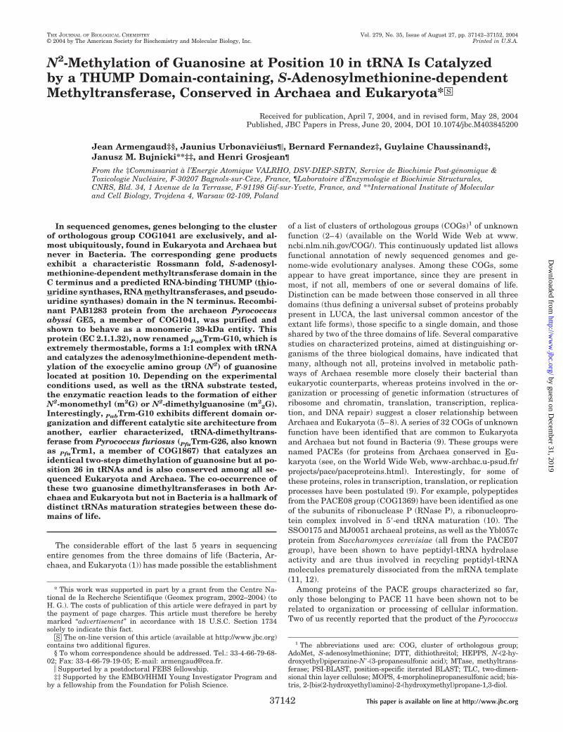
The Monomeric PAB1283 Protein Interacts with RNAs—Thenative molecular mass of PAB1283 was determined by sizeexclusion chromatography. Fig. 4A shows the elution profile ofthe purified enzyme obtained with a Superdex75 column. Re-sults indicate that the enzyme behaves as a monomeric proteinwith an apparent molecular mass of 40 kDa. This result issupported by the PAB1283 melting profile (Fig. 3), in that mostmultimeric proteins would be expected to show oligomer disso-ciation prior to subunit unfolding (35), whereas here a singlewell defined melting peak was recorded. When chromatogra-phy was performed on a Superdex200 and with a less purifiedenzyme pool obtained after the immobilized metal ion adsorp-tion chromatography and before the hydroxyapatite purifica-tion step, an additional minor peak was visible at a position ofthe elution profile that corresponds to a mean molecular massof about 70 kDa. Since the ratio A260/A280 of this minor peakwas 1.60 instead of 0.57 for the main protein pool, the presenceof small nucleic acids complexed to the enzyme was suspected.
Since the THUMP domain has been predicted to be a RNA-binding domain (15), we checked whether the purified
FIG. 1. General domain organization of COG1041 sequences. Archaeal and eukaryal homologs were obtained from public data bases(available on the World Wide Web at www.ncbi.nlm.nih.gov/) by PSI-BLAST searches (24). Domains were identified by the NCBI conserved domainsearch tool (available on the World Wide Web at www.ncbi.nlm.nih.gov). Multiple alignments were performed by ClustalW and manually refined.Predictions of secondary structure elements of the PAB1283 sequence were made through the Psipred Web-interfaced software (available on theWorld Wide Web at bioinf.cs.ucl.ac.uk/psipred/). Predicted �-sheets and �-helices are represented by arrows and pipes, respectively. Amino acidnumbering refers to PAB1283 sequence. Motifs I, II, IV, and VI, corresponding to AdoMet binding regions, are marked above the residues in whichthey appear. The asterisks mark sets of residues that are not shown for clarity. Strictly conserved residues are in red, and residues with greaterthan 70% identity or strictly of the same type are in blue. Accession numbers are indicated beside each organism name. Based on phylogeneticanalysis (data not shown), 10 representative sequences were selected from the 42 COG1041 members. The first four sequences are from Eukaryota,whereas the last six sequences are from Archaea. The Vng2242c sequence from Halobacterium sp. NRC-1, labeled Q9HN57*, has been numberedaccording to the new annotation proposed under “Results.”
PabTrm-G10 Dimethyltransferase 37145
by guest on Decem
ber 31, 2019http://w
ww
.jbc.org/D
ownloaded from

PAB1283 protein could interact with small nucleic acids suchas transfer RNAs. After incubation of the enzyme with yeastbulk (unfractionated) tRNAs, the mixture was subjected to gelfiltration on Superdex75, and the elution profile was comparedwith those obtained under identical conditions with the proteinor bulk tRNAs alone. As shown in Fig. 4A, the results suggesta strong interaction between the enzyme and tRNAs, since anew peak of compound eluting faster than either the enzyme ortRNAs alone was visible. Identical results were obtained whenE. coli total tRNAs were mixed with the PAB1283 protein (datanot shown). If a stoichiometric amount of purified tRNAPhe orpurified tRNAAsp (both from yeast) was mixed with thePAB1283 enzyme, a 1:1 stable complex was clearly visible (Fig.
4B). When PAB1283 and a 23 S rRNA fragment of approxi-mately the same size (74 nucleotides) as a tRNA molecule (21)were mixed and subjected to gel filtration, a complex was alsoseen but eluted over a much larger fraction (Fig. 4C), revealingthe existence of a 1:1 complex but of lower affinity. Theseresults suggest that the substrate of PAB1283 is more likely tobe tRNA than rRNA.
Identification of tRNA Substrates and Type of Methylation—The ubiquitous occurrence of orthologous genes (COG1041)corresponding to MTases present in both Archaea and Eu-karyota but not in Bacteria (9), together with the observationthat PAB1283 forms strong complexes with bulk or purifiedtRNAs (see above), prompted us to test whether tRNAs aresubstrates for methylation and to examine which of the knownmethylated nucleotides in archaeal and eukaryal tRNAs maybe catalyzed by the newly identified putative MTase. Table Ilists the positions and types of methylation that are known tooccur simultaneously in tRNAs from both archaeon Haloferaxvolcanii (41 sequences known (36, 37)) and eukaryon S. cerevi-siae (33 sequences known (38); see, on the World Wide Web,www.tRNA.uni-bayreuth.de/) but absent from any of the manybacterial tRNAs sequenced so far (49 sequences known (38); seealso Refs. 39–41). Data indicate that, of the many modifiednucleosides normally present in tRNAs, only N2-methyl-guanosine, N2,N2-dimethylguanosine, and C5-methylcytidinepresent at positions 10, 26, 40, 48, and/or 49 of a large numberof tRNAs fulfill the above criteria. Formation of m2G and/orm2
2G at position 26 was shown to be catalyzed by a single geneproduct (TRM1) in S. cerevisiae (42), Caenorhabditis elegans(43), H. sapiens (44), and Pyrococcus furiosus (45, 46), whereasformation of m5C at positions 40, 48, and/or 49 in both S.cerevisiae and P. abyssi is catalyzed by the multisite-specifictRNA:m5C MTase Trm4p (47).3 Therefore, only m2G and m2
2Gat position 10 remain unassigned to any characterized tRNAMTases from Archaea and Eukaryota.
When bulk E. coli tRNAs (the majority of them have G10,and none contain methylated G10) and bulk tRNAs from yeast(most of them have m2G10) were incubated in vitro at 50 °Cwith tritiated AdoMet and purified PAB1283, substantial
3 S. Auxilien and H. Grosjean, unpublished results.
FIG. 2. SDS-PAGE analysis of expression and purification of recombinant PAB1283. SDS-PAGE was performed on a 4–12% gradientNovex bis-tris acrylamide gel (Invitrogen) with MOPS buffer. Polyacrylamide gels were stained either with BlueSafe Coomassie solution (lanes 1–4and 9) or silver nitrate staining kit (lanes 5–8), both from Invitrogen. Lane M, SeeBlue Plus 2 molecular weight markers (Invitrogen); lane 1,cell-free extract of E. coli Rosetta(DE3)(pLysS)(pSBTN-AC18) uninduced (1.5 �l); lane 2, cell-free extract of E. coli Rosetta(DE3)(pLysS)(pSBTN-AC18) isopropyl-�-D-thiogalactopyranoside-induced (1.5 �l); lane 3, soluble proteins from cell-free extract (1.5 �l); lane 4, soluble proteins after the20-min heat treatment at 65 °C (10 �l); lane 5, proteins eluted from the immobilized metal ion adsorption chromatographic column (2 �g); lane 6,proteins eluted from the G25 desalting column (2 �g); lanes 7 and 8, MonoQ eluate (1 �g); lane 9, hydroxyapatite eluate (2 �g). Bands correspondingto recombinant PAB1283 polypeptide are indicated with the arrow.
FIG. 3. Evaluation of PAB1283 thermostability by differentialscanning calorimetry. Thermogram of PAB1283 was recorded at ascan rate of 1 °C/min. The heat capacity (Cp) maximum correspondingto the Tm value is indicated. Measurements were carried out at pH 8.0(at 20 °C) with 30 mM HEPPS/NaOH rather than Tris/HCl buffer. Thelatter is not adapted for such measurements due to its temperaturedependence (�pH/�T � �0.028 units/°C). Measurements were per-formed in fixed-in-place cells with a self-containing pressurizing sys-tem, in which temperatures up to 130 °C could be reached.
PabTrm-G10 Dimethyltransferase37146
by guest on Decem
ber 31, 2019http://w
ww
.jbc.org/D
ownloaded from
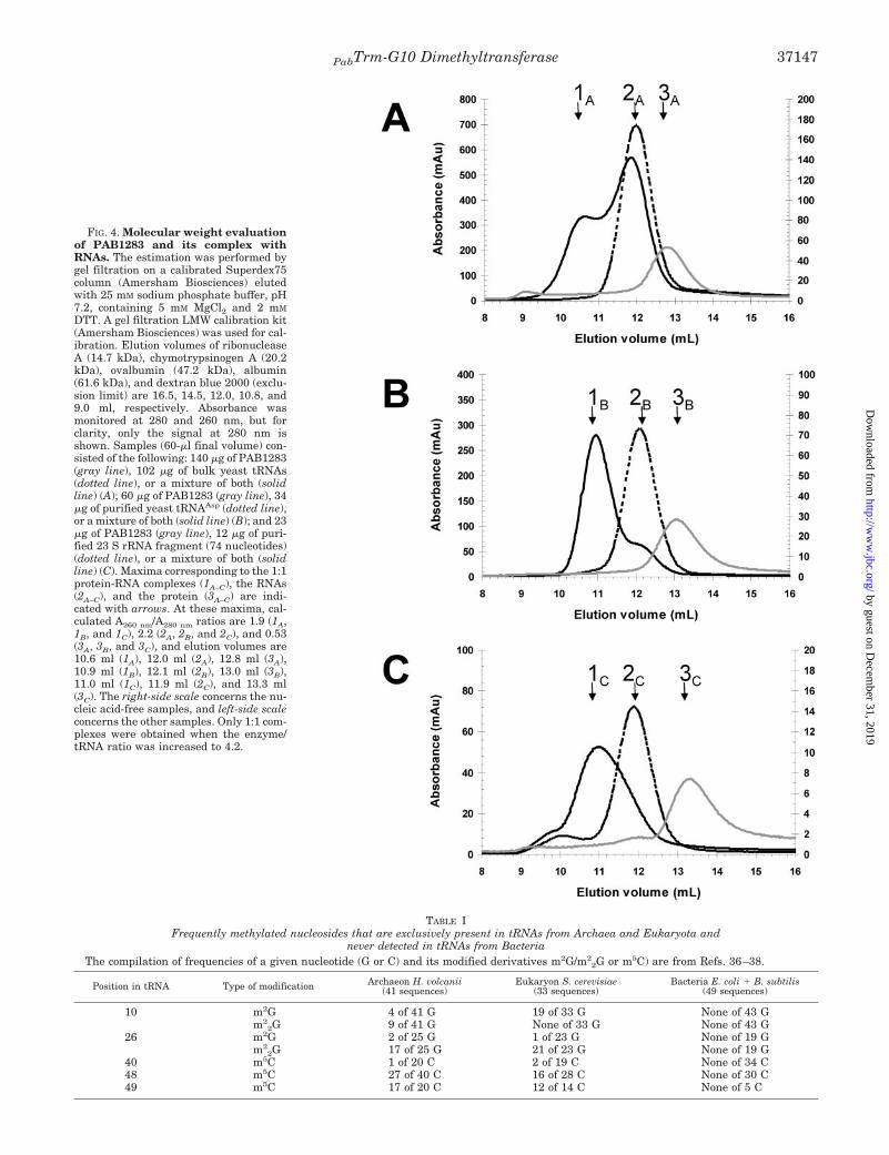
FIG. 4. Molecular weight evaluationof PAB1283 and its complex withRNAs. The estimation was performed bygel filtration on a calibrated Superdex75column (Amersham Biosciences) elutedwith 25 mM sodium phosphate buffer, pH7.2, containing 5 mM MgCl2 and 2 mM
DTT. A gel filtration LMW calibration kit(Amersham Biosciences) was used for cal-ibration. Elution volumes of ribonucleaseA (14.7 kDa), chymotrypsinogen A (20.2kDa), ovalbumin (47.2 kDa), albumin(61.6 kDa), and dextran blue 2000 (exclu-sion limit) are 16.5, 14.5, 12.0, 10.8, and9.0 ml, respectively. Absorbance wasmonitored at 280 and 260 nm, but forclarity, only the signal at 280 nm isshown. Samples (60-�l final volume) con-sisted of the following: 140 �g of PAB1283(gray line), 102 �g of bulk yeast tRNAs(dotted line), or a mixture of both (solidline) (A); 60 �g of PAB1283 (gray line), 34�g of purified yeast tRNAAsp (dotted line),or a mixture of both (solid line) (B); and 23�g of PAB1283 (gray line), 12 �g of puri-fied 23 S rRNA fragment (74 nucleotides)(dotted line), or a mixture of both (solidline) (C). Maxima corresponding to the 1:1protein-RNA complexes (1A–C), the RNAs(2A–C), and the protein (3A–C) are indi-cated with arrows. At these maxima, cal-culated A260 nm/A280 nm ratios are 1.9 (1A,1B, and 1C), 2.2 (2A, 2B, and 2C), and 0.53(3A, 3B, and 3C), and elution volumes are10.6 ml (1A), 12.0 ml (2A), 12.8 ml (3A),10.9 ml (1B), 12.1 ml (2B), 13.0 ml (3B),11.0 ml (1C), 11.9 ml (2C), and 13.3 ml(3C). The right-side scale concerns the nu-cleic acid-free samples, and left-side scaleconcerns the other samples. Only 1:1 com-plexes were obtained when the enzyme/tRNA ratio was increased to 4.2.
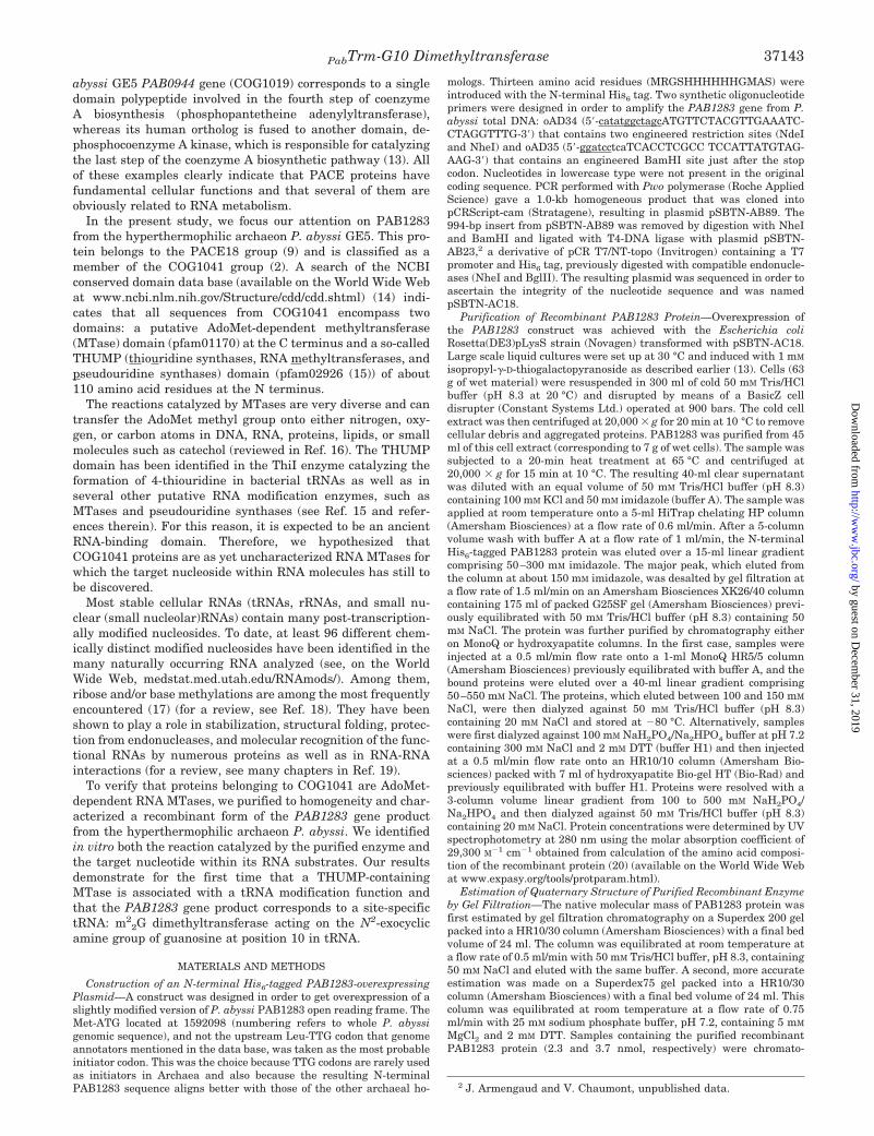
TABLE IFrequently methylated nucleosides that are exclusively present in tRNAs from Archaea and Eukaryota and
never detected in tRNAs from BacteriaThe compilation of frequencies of a given nucleotide (G or C) and its modified derivatives m2G/m2
2G or m5C) are from Refs. 36–38.
Position in tRNA Type of modification Archaeon H. volcanii(41 sequences)
Eukaryon S. cerevisiae(33 sequences)
Bacteria E. coli � B. subtilis(49 sequences)
10 m2G 4 of 41 G 19 of 33 G None of 43 Gm2
2G 9 of 41 G None of 33 G None of 43 G26 m2G 2 of 25 G 1 of 23 G None of 19 G
m22G 17 of 25 G 21 of 23 G None of 19 G
40 m5C 1 of 20 C 2 of 19 C None of 34 C48 m5C 27 of 40 C 16 of 28 C None of 30 C49 m5C 17 of 20 C 12 of 14 C None of 5 C
PabTrm-G10 Dimethyltransferase 37147
by guest on Decem
ber 31, 2019http://w
ww
.jbc.org/D
ownloaded from
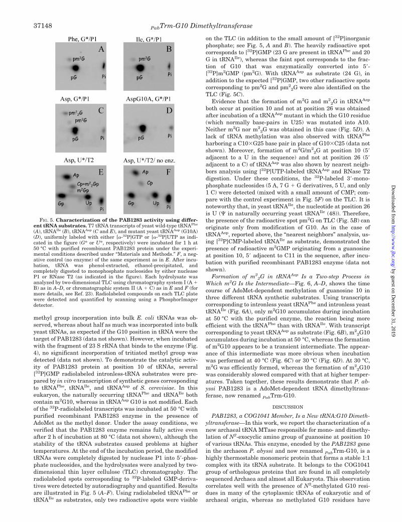
methyl group incorporation into bulk E. coli tRNAs was ob-served, whereas about half as much was incorporated into bulkyeast tRNAs, as expected if the G10 position in tRNA were thetarget of PAB1283 (data not shown). However, when incubatedwith the fragment of 23 S rRNA that binds to the enzyme (Fig.4), no significant incorporation of tritiated methyl group wasdetected (data not shown). To demonstrate the catalytic activ-ity of PAB1283 protein at position 10 of tRNAs, several[32P]GMP radiolabeled intronless-tRNA substrates were pre-pared by in vitro transcription of synthetic genes correspondingto tRNAPhe, tRNAIle, and tRNAAsp of S. cerevisiae. In thiseukaryon, the naturally occurring tRNAPhe and tRNAIle bothcontain m2G10, whereas in tRNAAsp G10 is not modified. Eachof the 32P-radiolabeled transcripts was incubated at 50 °C withpurified recombinant PAB1283 enzyme in the presence ofAdoMet as the methyl donor. Under the assay conditions, weverified that the PAB1283 enzyme remains fully active evenafter 2 h of incubation at 80 °C (data not shown), although thestability of the tRNA substrates caused problems at highertemperatures. At the end of the incubation period, the modifiedtRNAs were completely digested by nuclease P1 into 5�-phos-phate nucleosides, and the hydrolysates were analyzed by two-dimensional thin layer cellulose (TLC) chromatography. Theradiolabeled spots corresponding to 32P-labeled GMP-deriva-tives were detected by autoradiography and quantified. Resultsare illustrated in Fig. 5 (A–F). Using radiolabeled tRNAPhe ortRNAIle as substrates, only two radioactive spots were visible
on the TLC (in addition to the small amount of [32P]inorganicphosphate; see Fig. 5, A and B). The heavily radioactive spotcorresponds to [32P]GMP (23 G are present in tRNAPhe and 20G in tRNAIle), whereas the faint spot corresponds to the frac-tion of G10 that was enzymatically converted into 5�-[32P]m2GMP (pm2G). With tRNAAsp as substrate (24 G), inaddition to the expected [32P]GMP, two other radioactive spotscorresponding to pm2G and pm2
2G were also identified on theTLC (Fig. 5C).
Evidence that the formation of m2G and m22G in tRNAAsp
both occur at position 10 and not at position 26 was obtainedafter incubation of a tRNAAsp mutant in which the G10 residue(which normally base-pairs in U25) was mutated into A10.Neither m2G nor m2
2G was obtained in this case (Fig. 5D). Alack of tRNA methylation was also observed with tRNAPhe
harboring a C10�G25 base pair in place of G10�C25 (data notshown). Moreover, formation of m2G/m2
2G at position 10 (5�adjacent to a U in the sequence) and not at position 26 (5�adjacent to a C) of tRNAAsp was also shown by nearest neigh-bors analysis using [32P]UTP-labeled tRNAAsp and RNase T2digestion. Under these conditions, the 32P-labeled 3�-mono-phosphate nucleosides (5 A, 7 G � G derivatives, 5 U, and only1 C) were detected (mixed with a small amount of CMP; com-pare with the control experiment in Fig. 5F) on the TLC. It isnoteworthy that, in yeast tRNAIle, the nucleotide at position 26is U ( in naturally occurring yeast tRNAIle (48)). Therefore,the presence of the radioactive spot pm2G on TLC (Fig. 5B) canoriginate only from modification of G10. As in the case oftRNAAsp, reported above, the “nearest neighbors” analysis, us-ing [32P]CMP-labeled tRNAIle as substrate, demonstrated thepresence of radioactive m2GMP originating from a guanosineat position 10, 5� adjacent to C11 in the sequence, after incu-bation with purified recombinant PAB1283 enzyme (data notshown).
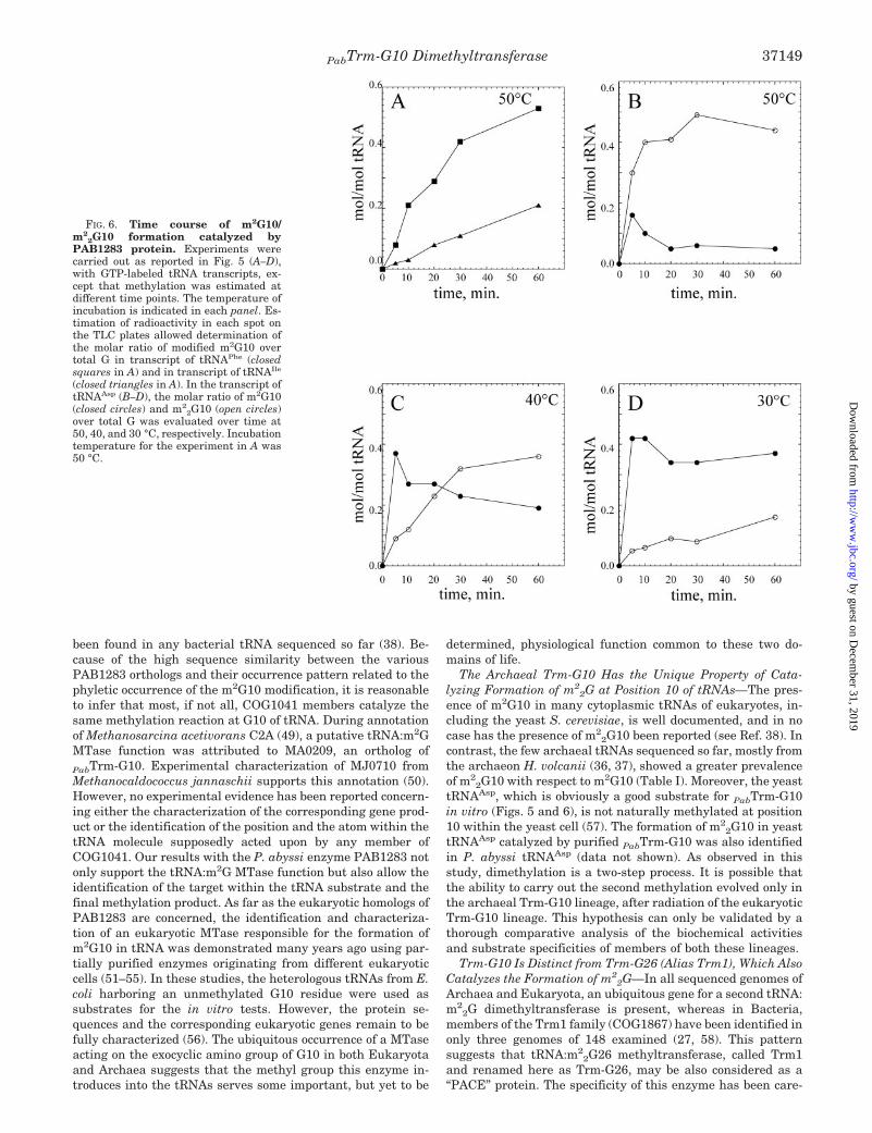
Formation of m22G in tRNAAsp Is a Two-step Process in
Which m2G Is the Intermediate—Fig. 6, A–D, shows the timecourse of AdoMet-dependent methylation of guanosine 10 inthree different tRNA synthetic substrates. Using transcriptscorresponding to intronless yeast tRNAPhe and intronless yeasttRNAIle (Fig. 6A), only m2G10 accumulates during incubationat 50 °C with the purified enzyme, the reaction being moreefficient with the tRNAPhe than with tRNAIle. With transcriptcorresponding to yeast tRNAAsp as substrate (Fig. 6B), m2
2G10accumulates during incubation at 50 °C, whereas the formationof m2G10 appears to be a transient intermediate. The appear-ance of this intermediate was more obvious when incubationwas performed at 40 °C (Fig. 6C) or 30 °C (Fig. 6D). At 30 °C,m2G was efficiently formed, whereas the formation of m2
2G10was considerably slowed compared with that at higher temper-atures. Taken together, these results demonstrate that P. ab-yssi PAB1283 is a AdoMet-dependent tRNA dimethyltrans-ferase, now renamed PabTrm-G10.
DISCUSSION
PAB1283, a COG1041 Member, Is a New tRNA:G10 Dimeth-yltransferase—In this work, we report the characterization of anew archaeal tRNA MTase responsible for mono- and dimethy-lation of N2-exocyclic amino group of guanosine at position 10of various tRNAs. This enzyme, encoded by the PAB1283 genein the archaeon P. abyssi and now renamed PabTrm-G10, is ahighly thermostable monomeric protein that forms a stable 1:1complex with its tRNA substrate. It belongs to the COG1041group of orthologous proteins that are found in all completelysequenced Archaea and almost all Eukaryota. This observationcorrelates well with the presence of N2-methylated G10 resi-dues in many of the cytoplasmic tRNAs of eukaryotic and ofarchaeal origin, whereas no methylated G10 residues have
FIG. 5. Characterization of the PAB1283 activity using differ-ent tRNA substrates. T7 tRNA transcripts of yeast wild-type tRNAPhe
(A), tRNAIle (B), tRNAAsp (C and E), and mutant yeast tRNAAsp (G10A)(D), uniformly labeled with either [�-32P]GTP or [�-32P]UTP as indi-cated in the figure (G* or U*, respectively) were incubated for 1 h at50 °C with purified recombinant PAB1283 protein under the experi-mental conditions described under “Materials and Methods.” F, a neg-ative control (no enzyme) of the same experiment as in E. After incu-bation, tRNA was phenol-extracted, ethanol-precipitated, andcompletely digested to monophosphate nucleosides by either nucleaseP1 or RNase T2 (as indicated in the figure). Each hydrolysate wasanalyzed by two-dimensional TLC using chromatography system I (A �B) as in A–D, or chromatographic system II (A � C) as in E and F (formore details, see Ref. 23). Radiolabeled compounds on each TLC platewere detected and quantified by scanning using a PhosphorImagerdetector.
PabTrm-G10 Dimethyltransferase37148
by guest on Decem
ber 31, 2019http://w
ww
.jbc.org/D
ownloaded from
been found in any bacterial tRNA sequenced so far (38). Be-cause of the high sequence similarity between the variousPAB1283 orthologs and their occurrence pattern related to thephyletic occurrence of the m2G10 modification, it is reasonableto infer that most, if not all, COG1041 members catalyze thesame methylation reaction at G10 of tRNA. During annotationof Methanosarcina acetivorans C2A (49), a putative tRNA:m2GMTase function was attributed to MA0209, an ortholog ofPabTrm-G10. Experimental characterization of MJ0710 fromMethanocaldococcus jannaschii supports this annotation (50).However, no experimental evidence has been reported concern-ing either the characterization of the corresponding gene prod-uct or the identification of the position and the atom within thetRNA molecule supposedly acted upon by any member ofCOG1041. Our results with the P. abyssi enzyme PAB1283 notonly support the tRNA:m2G MTase function but also allow theidentification of the target within the tRNA substrate and thefinal methylation product. As far as the eukaryotic homologs ofPAB1283 are concerned, the identification and characteriza-tion of an eukaryotic MTase responsible for the formation ofm2G10 in tRNA was demonstrated many years ago using par-tially purified enzymes originating from different eukaryoticcells (51–55). In these studies, the heterologous tRNAs from E.coli harboring an unmethylated G10 residue were used assubstrates for the in vitro tests. However, the protein se-quences and the corresponding eukaryotic genes remain to befully characterized (56). The ubiquitous occurrence of a MTaseacting on the exocyclic amino group of G10 in both Eukaryotaand Archaea suggests that the methyl group this enzyme in-troduces into the tRNAs serves some important, but yet to be
determined, physiological function common to these two do-mains of life.
The Archaeal Trm-G10 Has the Unique Property of Cata-lyzing Formation of m2
2G at Position 10 of tRNAs—The pres-ence of m2G10 in many cytoplasmic tRNAs of eukaryotes, in-cluding the yeast S. cerevisiae, is well documented, and in nocase has the presence of m2
2G10 been reported (see Ref. 38). Incontrast, the few archaeal tRNAs sequenced so far, mostly fromthe archaeon H. volcanii (36, 37), showed a greater prevalenceof m2
2G10 with respect to m2G10 (Table I). Moreover, the yeasttRNAAsp, which is obviously a good substrate for PabTrm-G10in vitro (Figs. 5 and 6), is not naturally methylated at position10 within the yeast cell (57). The formation of m2
2G10 in yeasttRNAAsp catalyzed by purified PabTrm-G10 was also identifiedin P. abyssi tRNAAsp (data not shown). As observed in thisstudy, dimethylation is a two-step process. It is possible thatthe ability to carry out the second methylation evolved only inthe archaeal Trm-G10 lineage, after radiation of the eukaryoticTrm-G10 lineage. This hypothesis can only be validated by athorough comparative analysis of the biochemical activitiesand substrate specificities of members of both these lineages.
Trm-G10 Is Distinct from Trm-G26 (Alias Trm1), Which AlsoCatalyzes the Formation of m2
2G—In all sequenced genomes ofArchaea and Eukaryota, an ubiquitous gene for a second tRNA:m2
2G dimethyltransferase is present, whereas in Bacteria,members of the Trm1 family (COG1867) have been identified inonly three genomes of 148 examined (27, 58). This patternsuggests that tRNA:m2
2G26 methyltransferase, called Trm1and renamed here as Trm-G26, may be also considered as a“PACE” protein. The specificity of this enzyme has been care-
FIG. 6. Time course of m2G10/m2
2G10 formation catalyzed byPAB1283 protein. Experiments werecarried out as reported in Fig. 5 (A–D),with GTP-labeled tRNA transcripts, ex-cept that methylation was estimated atdifferent time points. The temperature ofincubation is indicated in each panel. Es-timation of radioactivity in each spot onthe TLC plates allowed determination ofthe molar ratio of modified m2G10 overtotal G in transcript of tRNAPhe (closedsquares in A) and in transcript of tRNAIle
(closed triangles in A). In the transcript oftRNAAsp (B–D), the molar ratio of m2G10(closed circles) and m2
2G10 (open circles)over total G was evaluated over time at50, 40, and 30 °C, respectively. Incubationtemperature for the experiment in A was50 °C.
PabTrm-G10 Dimethyltransferase 37149
by guest on Decem
ber 31, 2019http://w
ww
.jbc.org/D
ownloaded from

fully tested with different orthologs from several organisms(43, 44, 52, 55, 59–63). This enzyme catalyzes the site-specificformation of m2G and of m2
2G at position 26 by a mechanismvery similar to that reported in this paper for Trm-G10 (46, 60)(reviewed in Ref. 64). It is important to note that the targets
G10 and G26 are very close to each other within the three-dimensional structure of the tRNA (in fact they stack on eachother; see Fig. 7, A and B). However, the exocyclic N2 atoms ofG10 and G26 point in different directions (Fig. 7C). Therefore,it may be that the two enzymes have to approach the tRNA
FIG. 8. Comparison of the AdoMet-binding and active sites of PabTrm-G10 (PAB1283) and PabTrm-G26 (PAB2092). A, superposition ofmodeled structures of the catalytic domain of both enzymes. Protein models were built based on the sequence-structure alignments generated bythe GeneSilico protein structure prediction metaserver (77). The 1dus.pdb structure was selected as the best scoring template, and the models werebuilt according to the “FRankenstein’s monster” protocol (78). Both models passed the evaluation procedure implemented in Verify3D (79). Thebackbone of PAB1283 (amino acids 161–329) and PAB2092 (amino acids 27–238) is shown in blue and magenta, respectively. The N and C terminiare labeled. Two insertions in PAB2092 (amino acids 100–114 and 173–199) were omitted from the model. The ligand-binding residues are shownand labeled (PAB1283/PAB2092); identical residues (four Asp residues from motifs I, II, III, and IV) are shown in cyan, and different residues (frommotifs IV and VIII) are shown in yellow. AdoMet is shown in white, and guanosine is shown in red. B, sequence alignment of the common MTasemotifs, depicting strong differences in all residues except those participating in cofactor binding. Residues shown in A are indicated in red.
FIG. 7. Reactions catalyzed by two distinct tRNA:guanosine dimethyltransferases in tRNAs. Shown on a three-dimensional structureof yeast tRNAAsp (2tra in the Protein Data Bank) is the location of guanosines (yellow) at positions 10 and 26 (A, whole view; B, zoomed view; C,rotated view). Note that, in naturally occurring yeast tRNAAsp, guanosines at positions 10 and 26 are unmodified. As indicated by the arrows, theexocyclic N2 atoms of G10 (in red), which would be modified by PabTrm-G10 (this work), and G26 (in green), which would be modified by PabTrm-G26(PAB2092) based on data obtained on its closely related ortholog, PfuTrm-G26 (also known as PabTrm1 (41, 42)), point in different directions.
PabTrm-G10 Dimethyltransferase37150
by guest on Decem
ber 31, 2019http://w
ww
.jbc.org/D
ownloaded from
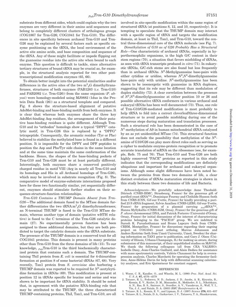
substrate from different sides, which could explain why the twoenzymes are very different in their amino acid sequences andbelong to completely different clusters of orthologous groups(COG1867 for Trm-G26; COG1041 for Trm-G10). The differ-ences in site specificity between archaeal Trm-G10 and Trm-G26 may be explained by several factors such as specific en-zyme positioning on the tRNA, the local environment of theactive site amino acids, and base composition and sequence ofthe tRNA. Any of these might facilitate or impede flipping outthe guanosine residue into the active site when bound to eachenzyme. This question is difficult to tackle, since alternativetertiary structures of bound tRNA may be crucial as, for exam-ple, in the structural analysis reported for two other post-transcriptional modification enzymes (65, 66).
To obtain better insight into the potential similarities and/ordifferences in the active sites of the two m2
2G dimethyltrans-ferases, structures of both enzymes (PAB1283 (i.e. Trm-G10)and PAB2092 (i.e. Trm-G26)) from the same organism (P. ab-yssi) were homology-modeled using MJ0882 (1dus in the Pro-tein Data Bank (26)) as a structural template and compared.Fig. 8 shows the structure-based alignment of putativeAdoMet-binding and base-binding motifs from both enzymes. Itis clear that whereas both enzymes share the three keyAdoMet-binding Asp residues, the arrangement of their puta-tive base-binding residues is quite different. Although Trm-G10 and its “orthodox” homologs show a typical “DPPY” cata-lytic motif, in Trm-G26 this is replaced by a “DPFG”tetrapeptide. Consequently, the aromatic residue (Tyr or Phe)believed to stabilize the methylated base is found in a differentposition. It is impossible for the DPPY and DPF peptides toposition the Asp and Phe/Tyr side chains in the same locationand at the same time maintain the same conformation of thebackbone. Hence, the shapes of the base-binding pockets ofTrm-G10 and Trm-G26 must be at least partially different.Interestingly, both enzymes share a conserved positivelycharged residue in motif VIII (Arg in nearly all Trm-G10 andits homologs and His in all Archaeal homologs of Trm-G26),which may be involved in substrate recognition (Fig. 8). Thecomparative model of enzyme-substrate interactions proposedhere for these two functionally similar, yet sequentially differ-ent, enzymes should stimulate further studies on their se-quence-structure-function relationships.
Trm-G10 Contains a THUMP Domain Absent from Trm-G26—The additional domain fused to the MTase domain fur-ther differentiates the two tRNA:m2
2G dimethyltransferases;an N-terminal THUMP is fused to the Trm-G10 MTase do-main, whereas another type of domain (putative wHTH rela-tive) is fused to the C terminus of the Trm-G26 catalytic do-main (27). No experimentally proven function has beenassigned to these additional domains, but they are both pre-dicted to target the catalytic domain onto the tRNA substrate.The presence of the THUMP domain is a conserved trait for allCOG1041 proteins. This domain is present in many proteinsother than Trm-G10 from the three domains of life (15). To ourknowledge, PabTrm-G10 is the third biochemically character-ized protein that contains such a domain. The THUMP-con-taining ThiI protein from E. coli is essential for 4-thiouridineformation at position 8 of some bacterial tRNAs (67, 68). Veryrecently, Tan1 protein from S. cerevisiae also harboring aTHUMP domain was reported to be required for N4-acetylcyti-dine formation in tRNAs (69). This modification is present atposition 12 in tRNAs specific for leucine and serine and wasshown to be important for tRNA stability. It should be notedthat, in agreement with the putative RNA-binding role thatmay be attributed to the THUMP, the three characterizedTHUMP-containing proteins, ThiI, Tan1, and Trm-G10, are all
involved in site-specific modification within the same region ofstructured tRNA (at positions 8, 12, and 10, respectively). It istempting to speculate that the THUMP domain may interactwith a specific region of tRNA and targets the modificationdomain, at least in ThiI, Tan1, and Trm-G10, toward the cen-tral three-dimensional core of the tRNA molecule (see Fig. 7).
Dimethylation of G10 as of G26 Probably Has a StructuralRole—One characteristic of archaeal tRNAs, especially in hy-perthermophilic organisms, is the high G/C content in theirstem regions (70), a situation that favors misfolding of tRNAs,as seen with tRNA transcripts produced in vitro (71). In eukary-otic tRNAs, G/C-rich stems are also found but less frequentlythan in archaeal tRNAs. N2-Methylguanosine base-pairs witheither cytidine or uridine, whereas N2,N2-dimethylguanosinebase-pairs only with uridine. N2-methylguanosine has beenshown to be isoenergetic with guanosine in RNA duplexes,suggesting that its role may be different than modulation ofduplex stability (72). A clear correlation between the presenceof N2,N2-dimethylguanosine at either position 10 or 26 andpossible alternative tRNA conformers in various archaeal andeukaryal tRNAs has been well documented (73). Thus, one roleof Trm-G10/G26-mediated modification is most probably tohelp tRNA to fold into a correct cloverleaf/three-dimensionalstructure or to avoid possible misfolding during one of thenumerous steps during maturation and translation processes.Such a structural role has been demonstrated in the case ofN1-methylation of A9 in human mitochondrial tRNA catalyzedby an as yet unidentified MTase (74). This structural functiondoes not exclude the possibility that methylation of the N2-amine of G10/G26 can play more direct roles such as serving asa cipher to modulate enzyme-protein recognition or to promoteaccurate translation of mRNA on the ribosomes (see, for exam-ple, Refs. 75 and 76). That Trm-G10 and Trm-G26 are bothhighly conserved “PACE” proteins as reported in this studyindicates that the corresponding modifications are definitelyubiquitous and important for archaeal and eukaryal organ-isms. Although some slight differences have been noted be-tween the proteins from these two domains of life, a cleardistinct evolution of tRNAs maturation strategies is shown inthis study between these two domains of life and Bacteria.
Acknowledgments—We gratefully acknowledge Anne Theobald-Ditriech (CNRS-IBMC, Strasbourg, France) for the generous gift ofpurified yeast tRNAAsp, Dominique Fourmy and Clothilde Husson (bothfrom CNRS-ICSN, Gif-sur-Yvette, France) for kindly providing a puri-fied 23 S rRNA fragment, Sylvie Auxilien (CNRS-LEBS, Gif-sur-Yvette,France) for preparation of a plasmid carrying PabtRNAAsp,Hannu Myllykallio (CNRS-IGM, Orsay, France) for the generous gift ofP. abyssi chromosomal DNA, and Patrick Forterre (Universite d’Orsay,Orsay, France) for initial discussions of the interest of characterizingproteins belonging to the “PACE18” group. We are indebted toSuresh K. Purushothaman and Bruno Lapeyre (both from CNRS-CRBM, Montpellier, France) for discussions regarding their ongoingproject on COG1041 yeast ortholog, Marcus Johansson andAnders Bystrom (both from Umeå University, Umeå, Sweden) for shar-ing information on TAN1 prior to publication, and Gary J. Olsen (Uni-versity of Illinois, Urbana-Champaign, IL) for making us aware, duringsubmission of this manuscript, of their unpublished studies on MJ0710.We thank the following colleagues (all from CEA VALRHO):Isabelle Dany, Jean-Charles Gaillard, and Alain Dedieu for assistancewith mass spectrometry controls, Valerie Chaumont for help in overex-pression analysis, Charles Marchetti for operating the fermenter facil-ities, Anne-Helene Davin for help with differential scanning calorime-try analyses, and Eric Quemeneur for constant support.
REFERENCES
1. Woese, C. R., Kandler, O., and Wheelis, M. L. (1990) Proc. Natl. Acad. Sci.U. S. A. 87, 4576–4579
2. Tatusov, R. L., Fedorova, N. D., Jackson, J. D., Jacobs, A. R., Kiryutin, B.,Koonin, E. V., Krylov, D. M., Mazumder, R., Mekhedov, S. L., Nikolskaya,A. N., Rao, B. S., Smirnov, S., Sverdlov, A. V., Vasudevan, S., Wolf, Y. I.,Yin, J. J., and Natale, D. A. (2003) BMC Bioinformatics 4, 41
3. Tatusov, R. L., Koonin, E. V., and Lipman, D. J. (1997) Science 278, 631–6374. Tatusov, R. L., Natale, D. A., Garkavtsev, I. V., Tatusova, T. A., Shankavaram,
PabTrm-G10 Dimethyltransferase 37151
by guest on Decem
ber 31, 2019http://w
ww
.jbc.org/D
ownloaded from
U. T., Rao, B. S., Kiryutin, B., Galperin, M. Y., Fedorova, N. D., Koonin,E. V., and Lipman, D. J. (2001) Nucleic Acids Res. 29, 22–28
5. Dennis, P. P. (1997) Cell 89, 1007–10106. Olsen, G. J., and Woese, C. R. (1997) Cell 89, 991–9947. Makarova, K. S., and Koonin, E. V. (2003) Genome Biol. 4, 1158. Terns, M. P., and Terns, R. M. (2002) Gene. Expr. 10, 17–399. Matte-Tailliez, O., Zivanovic, Y., and Forterre, P. (2000) Trends Genet. 16,
533–53610. Hartmann, E., and Hartmann, R. K. (2003) Trends Genet. 19, 561–56911. Fromant, M., Ferri-Fioni, M. L., Plateau, P., and Blanquet, S. (2003) Nucleic
Acids Res. 31, 3227–323512. Rosas-Sandoval, G., Ambrogelly, A., Rinehart, J., Wei, D., Cruz-Vera, L. R.,
Graham, D. E., Stetter, K. O., Guarneros, G., and Soll, D. (2002) Proc. Natl.Acad. Sci. U. S. A. 99, 16707–16712
13. Armengaud, J., Fernandez, B., Chaumont, V., Rollin-Genetet, F., Finet, S.,Marchetti, C., Myllykallio, H., Vidaud, C., Pellequer, J. L., Gribaldo, S.,Forterre, P., and Gans, P. (2003) J. Biol. Chem. 278, 31078–31087
14. Marchler-Bauer, A., Anderson, J. B., DeWeese-Scott, C., Fedorova, N. D., Geer,L. Y., He, S., Hurwitz, D. I., Jackson, J. D., Jacobs, A. R., Lanczycki, C. J.,Liebert, C. A., Liu, C., Madej, T., Marchler-Bauer, A., Marchler, G. H.,Mazumder, R., Nikolskaya, A. N., Rao, B. S., Panchenko, A. R., Shoemaker,B. A., Simonyan, V., Song, J. S., Thiessen, P. A., Vasudevan, S., Wang, Y.,Yamashita, R. A., Yin, J. J., and Bryant, S. H. (2003) Nucleic Acids Res. 31,383–387
15. Aravind, L., and Koonin, E. V. (2001) Trends Biochem. Sci 26, 215–21716. Fauman,E.B.,Blumenthal,R.M.,andCheng,X.(1999)inS-Adenosylmethionine-
dependent Methyltransferases: Structures and Functions (Cheng, X., andBlumenthal, R. M., eds) pp. 1–38, World Scientific Publishing, Singapore
17. Rozenski, J., Crain, P. F., and McCloskey, J. A. (1999) Nucleic Acids Res. 27,196–197
18. Hopper, A. K., and Phizicky, E. M. (2003) Genes Dev. 17, 162–18019. Grosjean, H., and Benne, R. (1998) Modification and Editing of RNA
(Grosjean, H., and Benne, R., eds) American Society for Microbiology Press,Washington, D. C.
20. Gill, S. C., and von Hippel, P. H. (1989) Anal. Biochem. 182, 319–32621. Lebars, I., Yoshizawa, S., Stenholm, A. R., Guittet, E., Douthwaite, S., and
Fourmy, D. (2003) EMBO J. 22, 183–19222. Grosjean, H., Droogmans, L., Giege, R., and Uhlenbeck, O. C. (1990) Biochim.
Biophys. Acta 1050, 267–27323. Grosjean, H., Keith, G., and Droogmans, L. (2004) Methods Mol. Biol. 265,
357–39224. Altschul, S. F., Madden, T. L., Schaffer, A. A., Zhang, J., Zhang, Z., Miller, W.,
and Lipman, D. J. (1997) Nucleic Acids Res. 25, 3389–340225. Cheng, X. (1995) Curr. Opin. Struct. Biol. 5, 4–1026. Bujnicki, J. M., and Rychlewski, L. (2002) BMC Bioinformatics 3, 1027. Bujnicki, J. M., Leach, R. A., Debski, J., and Rychlewski, L. (2002) J. Mol.
Microbiol. Biotechnol. 4, 405–41528. Bugl, H., Fauman, E. B., Staker, B. L., Zheng, F., Kushner, S. R., Saper, M. A.,
Bardwell, J. C., and Jakob, U. (2000) Mol. Cell 6, 349–36029. Bujnicki, J. M. (2000) FASEB J. 14, 2365–236830. Gong, W., O’Gara, M., Blumenthal, R. M., and Cheng, X. (1997) Nucleic Acids
Res. 25, 2702–271531. Goedecke, K., Pignot, M., Goody, R. S., Scheidig, A. J., and Weinhold, E. (2001)
Nat. Struct. Biol. 8, 121–12532. Fitch, W. M. (2000) Trends Genet. 16, 227–23133. Bujnicki, J. M., Feder, M., Radlinska, M., and Blumenthal, R. M. (2002) J. Mol.
Evol. 55, 431–44434. Scholz, S., Sonnenbichler, J., Schafer, W., and Hensel, R. (1992) FEBS Lett.
306, 239–24235. Backmann, J., and Schafer, G. (2001) Methods Enzymol. 334, 328–34236. Gupta, R. (1984) J. Biol. Chem. 259, 9461–947137. Gupta, R. (1986) System. Appl. Microbiol. 7, 102–10538. Sprinzl, M., Horn, C., Brown, M., Ioudovitch, A., and Steinberg, S. (1998)
Nucleic Acids Res. 26, 148–15339. Limbach, P. A., Crain, P. F., and McCloskey, J. A. (1994) Nucleic Acids Res. 22,
2183–219640. Grosjean, H., Sprinzl, M., and Steinberg, S. (1995) Biochimie (Paris) 77,
139–14141. Motorin, Y., and Grosjean, H. (2004) Encyclopedia of Life Sciences, p. A3866,
MacMillan Reference Ltd., Basingstoke (Hampshire), England42. Ellis, S. R., Morales, M. J., Li, J. M., Hopper, A. K., and Martin, N. C. (1986)
J. Biol. Chem. 261, 9703–970943. Liu, J., Zhou, G. Q., and Straby, K. B. (1999) Gene (Amst.) 226, 73–8144. Liu, J., and Straby, K. B. (2000) Nucleic Acids Res. 28, 3445–345145. Constantinesco, F., Benachenhou, N., Motorin, Y., and Grosjean, H. (1998)
Nucleic Acids Res. 26, 3753–376146. Constantinesco, F., Motorin, Y., and Grosjean, H. (1999) J. Mol. Biol. 291,
375–39247. Motorin, Y., and Grosjean, H. (1999) RNA 5, 1105–111848. Simos, G., Tekotte, H., Grosjean, H., Segref, A., Sharma, K., Tollervey, D., and
Hurt, E. C. (1996) EMBO J. 15, 2270–228449. Galagan, J. E., Nusbaum, C., Roy, A., Endrizzi, M. G., Macdonald, P.,
FitzHugh, W., Calvo, S., Engels, R., Smirnov, S., Atnoor, D., Brown, A.,Allen, N., Naylor, J., Stange-Thomann, N., DeArellano, K., Johnson, R.,Linton, L., McEwan, P., McKernan, K., Talamas, J., Tirrell, A., Ye, W.,Zimmer, A., Barber, R. D., Cann, I., Graham, D. E., Grahame, D. A., Guss,A. M., Hedderich, R., Ingram-Smith, C., Kuettner, H. C., Krzycki, J. A.,Leigh, J. A., Li, W., Liu, J., Mukhopadhyay, B., Reeve, J. N., Smith, K.,Springer, T. A., Umayam, L. A., White, O., White, R. H., de Macario, E. C.,Ferry, J. G., Jarrell, K. F., Jing, H., Macario, A. J. L., Paulsen, I., Pritchett,M., Sowers, K. R., Swanson, R. V., Zinder, S. H., Lander, E., Metcalf, W. W.,and Birren, B. (2002) Genome Res. 12, 532–542
50. Graham, D. E. (2000) Archaeal Gene Identification. Ph.D. thesis, University ofIllinois at Urbana-Champaign
51. Kraus, J., and Staehelin, M. (1974) Nucleic Acids Res. 1, 1455–147852. Glick, J. M., Averyhart, V. M., and Leboy, P. S. (1978) Biochim. Biophys. Acta
518, 158–17153. Izzo, P., and Gantt, R. (1977) Biochemistry 16, 3576–358154. Taylor, M. J., and Gantt, R. (1979) Biochemistry 18, 5253–525855. Sindhuphak, T., Hellman, U., and Svensson, I. (1985) Biochim. Biophys. Acta
824, 66–7356. Bujnicki, J. M., Droogmans, L., Grosjean, H., Purushothaman, S. K., and
Lapeyre, B. (2004) Nucleic Acids Mol. Biol. 15, 139–16857. Gangloff, J., Keith, G., Ebel, J. P., and Dirheimer, G. (1972) Biochim. Biophys.
Acta 259, 210–22258. Takeda, H., Hori, H., and Endo, Y. (2002) Nucleic Acids Res. Suppl. 2, 229–23059. Liu, J., and Straby, K. B. (1998) Nucleic Acids Res. 26, 5102–510860. Reinhart, M. P., Lewis, J. M., and Leboy, P. S. (1986) Nucleic Acids Res. 14,
1131–114861. Kraus, J., and Staehelin, M. (1974) Nucleic Acids Res. 1, 1479–149662. Edqvist, J., Grosjean, H., and Straby, K. B. (1992) Nucleic Acids Res. 20,
6575–658163. Edqvist, J., Blomqvist, K., Straby, K. B., and Grosjean, H. (1994) Biochemistry
33, 9546–955164. Edqvist, J., Straby, K. B., and Grosjean, H. (1995) Biochimie (Paris) 77, 54–6165. Hoang, C., and Ferre-D’Amare, A. R. (2001) Cell 107, 929–93966. Ishitani, R., Nureki, O., Nameki, N., Okada, N., Nishimura, S., and Yokoyama,
S. (2003) Cell 113, 383–39467. Palenchar, P. M., Buck, C. J., Cheng, H., Larson, T. J., and Mueller, E. G.
(2000) J. Biol. Chem. 275, 8283–828668. Mueller, E. G., Palenchar, P. M., and Buck, C. J. (2001) J. Biol. Chem. 276,
33588–3359569. Johansson, M. J., and Bystrom, A. S. (2004) RNA 10, 712–71970. Marck, C., and Grosjean, H. (2002) RNA 8, 1189–123271. Uhlenbeck, O. C. (1995) RNA 1, 4–672. Rife, J. P., Cheng, C. S., Moore, P. B., and Strobel, S. A. (1998) Nucleic Acids
Res. 26, 3640–364473. Steinberg, S., and Cedergren, R. (1995) RNA 1, 886–89174. Helm, M., Brule, H., Degoul, F., Cepanec, C., Leroux, J. P., Giege, R., and
Florentz, C. (1998) Nucleic Acids Res. 26, 1636–164375. Roe, B., Michael, M., and Dudock, B. (1973) Nat. New Biol. 246, 135–13876. Niederberger, C., Graub, R., Costa, A., Desgres, J., and Schweingruber, M. E.
(1999) FEBS Lett. 464, 67–7077. Kurowski, M. A., and Bujnicki, J. M. (2003) Nucleic Acids Res. 31, 3305–330778. Kosinski, J., Cymerman, I. A., Feder, M., Kurowski, M. A., Sasin, J. M., and
Bujnicki, J. M. (2003) Proteins 53, 369–37979. Luthy, R., Bowie, J. U., and Eisenberg, D. (1992) Nature 356, 83–85
PabTrm-G10 Dimethyltransferase37152
by guest on Decem
ber 31, 2019http://w
ww
.jbc.org/D
ownloaded from
Janusz M. Bujnicki and Henri GrosjeanJean Armengaud, Jaunius Urbonavicius, Bernard Fernandez, Guylaine Chaussinand,
Conserved in Archaea and Eukaryota-Adenosylmethionine-dependent Methyltransferase,SDomain-containing,
-Methylation of Guanosine at Position 10 in tRNA Is Catalyzed by a THUMP2N
doi: 10.1074/jbc.M403845200 originally published online June 20, 20042004, 279:37142-37152.J. Biol. Chem.
10.1074/jbc.M403845200Access the most updated version of this article at doi:
Alerts:
When a correction for this article is posted•
When this article is cited•
to choose from all of JBC's e-mail alertsClick here
Supplemental material:
http://www.jbc.org/content/suppl/2004/07/08/M403845200.DC1
http://www.jbc.org/content/279/35/37142.full.html#ref-list-1
This article cites 75 references, 14 of which can be accessed free at
by guest on Decem
ber 31, 2019http://w
ww
.jbc.org/D
ownloaded from
Related Documents


