Title: N-terminal chimeras with signal sequences enhance the functional expression and alter the subcellular localization of heterologous membrane-bound inorganic pyrophosphatases in yeast Running Title: Enhanced expression of H + -PPases in yeast Authors: Rocío DRAKE, Aurelio SERRANO 1 and José R. PÉREZ-CASTIÑEIRA Instituto de Bioquímica Vegetal y Fotosíntesis, Universidad de Sevilla-CSIC, Avda. Americo Vespucio, 49, 41092 Sevilla, Spain 1 To whom correspondence should be addressed (Phone: +-34-954489525. Fax: +-34- 954460065; Email: [email protected]) Abbreviations used: ACMA, 9-amino-6-chloro-2-methoxy acridine; CCCP, cyanide m- chlorophenylhydrazone; DAPI, 4',6-diamidino-2-phenylindole; FM4-64, N-(3- triethylammoniumpropyl)-4-(p-diethylaminophenyl-hexatrienyl) pyridinium dibromide; GFP, green fluorescent protein; H + -PPase, proton-translocating inorganic pyrophosphatase; ORF, open reading frame; PP i , inorganic pyrophosphate; sPPase, soluble inorganic pyrophosphatase; PCR, polymerase chain reaction; yEGFP, yeast enhanced green fluorescent protein.

Welcome message from author
This document is posted to help you gain knowledge. Please leave a comment to let me know what you think about it! Share it to your friends and learn new things together.
Transcript

Title: N-terminal chimeras with signal sequences enhance the functional expression and alter the subcellular localization of heterologous membrane-bound inorganic pyrophosphatases in yeast Running Title: Enhanced expression of H+-PPases in yeast Authors: Rocío DRAKE, Aurelio SERRANO1 and José R. PÉREZ-CASTIÑEIRA Instituto de Bioquímica Vegetal y Fotosíntesis, Universidad de Sevilla-CSIC, Avda. Americo Vespucio, 49, 41092 Sevilla, Spain
1 To whom correspondence should be addressed (Phone: +-34-954489525. Fax: +-34-954460065; Email: [email protected]) Abbreviations used: ACMA, 9-amino-6-chloro-2-methoxy acridine; CCCP, cyanide m-chlorophenylhydrazone; DAPI, 4',6-diamidino-2-phenylindole; FM4-64, N-(3-triethylammoniumpropyl)-4-(p-diethylaminophenyl-hexatrienyl) pyridinium dibromide; GFP, green fluorescent protein; H+-PPase, proton-translocating inorganic pyrophosphatase; ORF, open reading frame; PPi, inorganic pyrophosphate; sPPase, soluble inorganic pyrophosphatase; PCR, polymerase chain reaction; yEGFP, yeast enhanced green fluorescent protein.
Aurelio
Texto escrito a máquina
Aurelio
Texto escrito a máquina
Biochem J. 2010 Feb 9;426(2):147-57. doi: 10.1042/BJ20091491

Abstract Expression of heterologous multispanning membrane proteins in Saccharomyces cerevisiae is a difficult task. Quite often, the use of multicopy plasmids where the foreign gene is under the control of a strong promoter does not guarantee efficient production of the corresponding protein. Here, we show that the expression level and/or subcellular localization in S. cerevisiae of an heterologous type of multispanning membrane protein, the H+-translocating inorganic pyrophosphatase (H+-PPase), can be changed by fusing it with various suitable N-terminal signal sequences. Chimeric proteins were constructed by adding the putative N-terminal extra domain of Trypanosoma cruzi H+-PPase or the bona fide signal sequence of S. cerevisiae invertase Suc2p to H+-PPase polypeptides of different organisms (from bacteria to plants) and expressed in a yeast conditional mutant deficient in its cytosolic PPi hydrolysis activity when grown on glucose. Chimeric constructs not only substantially enhanced H+-PPase expression levels in transformed mutant cells but also allowed functional complementation in those cases in which native H+-PPase failed to accomplish it. Activity assays and Western blot analyses further demonstrated the occurrence of most H+-PPase in internal membrane fractions of these cells. The addition of N-terminal signal sequences to the vacuolar H+-PPase AVP1 from the plant Arabidopsis thaliana -a protein efficiently expressed in yeast in its natural form- alters the subcellular distribution of the chimeras suggesting further progression along the secretory sorting pathways, as shown by density gradient ultracentrifugation and in vivo fluorescence microscopy of the corresponding GFP-H+-PPase fusion proteins.
Key words: chimeric proteins, green fluorescent protein, heterologous expression, N-terminal signal sequences, proton-translocating inorganic pyrophosphatase, Saccharomyces cerevisiae.
INTRODUCTION Heterologous expression may pose important difficulties, especially in the case of multispanning membrane proteins, whose overexpression can be toxic to the host cell. The use of appropriate promoters of known properties allows the control of transcription; however, quite often, high levels of messenger RNA do not imply high amounts of the desired protein. This suggests that, besides transcription, other processes such as translation, protein targeting or degradation may also be crucial in order to obtain high levels of functional foreign proteins in a given system. Eukaryotic cells have an intracellular membrane network that allows them to accommodate additional membrane proteins [1, 2]; therefore, they are frequently utilized in order to express heterologous multispanning membrane proteins.
In eukaryotes, membrane proteins are targeted to specific cell compartments or to the plasma membrane by the endomembrane system, or secretory pathway, that includes the endoplasmic reticulum (ER), Golgi complex, the vacuole and other transport vesicles, and by the endocytic pathway that eventually internalizes them to the endosomes [1]. Native H+-ATPases have been used among other membrane proteins to investigate the vesicular transport machinery in the budding yeast Saccharomyces cerevisiae [2, 3], in which heterologous membrane proteins are usually delivered by default to the ER. Nascent proteins in eukaryotic cells must pass stringent quality controls designed to optimize their synthesis, folding and, when necessary, post-translational modifications [4, 5]. It is not unusual that proteins expressed in eukaryotic systems fail to pass these controls being diverted to proteolytic compartments, resulting in low or, indeed, no expression. In the case of S. cerevisiae, one of the more advantageous systems for the heterologous expression of membrane proteins [6], these problems have been sometimes overcome by utilizing mutants defective in their vacuolar proteases [7]; however, even the latter may not be sufficient in some cases [8]. Alternatively, construction of chimeric proteins with modified N-terminal domains has also been reported to improve heterologous expression of the integral membrane protein SR Ca2+-ATPase in yeast [9]. The latter suggests that the N-terminus might be crucial to determine the fate of membrane proteins in this system.
Here, we show that the expression in S. cerevisiae of several bacterial and eukaryotic membrane-bound proton-translocating inorganic pyrophosphatases (H+-PPases), a type of membrane-embedded proteins naturally absent in yeast, can be improved by attaching different signal peptides to their N-termini. Naturally occurring H+-PPases are located in plasma membrane vesicles and acidocalcisomes of prokaryotes and acidic single-membrane organelles (acidocalcisomes, lysosomes, vacuoles) of certain eukaryotes: protists and plants [10-13], although a plasma membrane localization was also reported for plant-like K+-stimulated H+-PPase of some parasitic protists [14]. In this work, chimeric H+-PPase proteins were expressed in YPC3, a novel version of a yeast conditional mutant in its essential cytosolic pyrophosphatase (Ipp1p), which is unable to grow on glucose [15]. In most cases, expression levels of the chimeras were higher than those of their natural counterparts; this usually improved the ability of the different H+-PPases to support growth of YPC3 on glucose by functionally complementing Ipp1p. In the case of A. thaliana vacuolar H+-PPase AVP1, a protein already expressed efficiently in yeast [15, 16], both expression levels and membrane-associated PPase activity values of the chimeras were similar to those of the native protein, but density gradient ultracentrifugation showed altered subcellular distributions, suggesting different progressions along the secretory pathway. In order to further investigate this effect, GFP was fused to AVP1, and two different N-terminal signal sequences were attached to the
resulting chimeric polypeptide. Fluorescence microscopy analysis of yeast cells expressing these chimeras showed that N-terminal domains substantially modify the subcellular distribution of the heterologously-expressed H+-PPase proteins.
EXPERIMENTAL Generation of YPC3 strain YPC3 mutant strain was obtained from S. cerevisiae haploid strain W303-1A (MATa, ade2-1 can1-100 his3-11,15 leu2-3,112 trp1-1, ura3-1) by inserting the yeast HIS3 cassette from plasmid pUG34 (http://mips.helmholtz-muenchen.de/proj/yeast/info/tools/hegemann/ gfp.html) followed by the galactokinase (GAL1) promoter between the promoter and the coding sequence of the gene that codes for the cytosolic inorganic pyrophosphatase (IPP1) using the single-step transplacement procedure [17]. For this purpose, an appropriate linear DNA fragment was obtained as follows: first, the coding sequence of IPP1, along with a 390 bp-long region upstream the putative start codon [18], was amplified from genomic DNA by high fidelity PCR using TaqPlus DNA polymerase (Stratagene) and ligated into p-GEM-T plasmid (Promega), following the manufacturers' instructions. Then, two DNA fragments containing the yeast HIS3 cassette (1.7 kb long) and the GAL1 promoter (0.75 kb long), respectively, were sequentially introduced at the EcoRV site located 21 bases upstream the starting codon of IPP1 [18] by standard cloning techniques [19]. This yielded plasmid pYPC3 that was digested with ApaI and SacI and, then, introduced in S. cerevisiae haploid strain W303-1A, by the LiOAc-PEG method [20]. Transformants were selected by growing cells on 2% agar plates made in galactose-containing culture medium [21] without histidine. The insertion of the construction was checked by PCR performed with genomic DNA of one of the transformants unable to grow on glucose (YPC3). Multiple sequences alignments and signal peptide predictions Multiple amino acid sequence alignments of selected eukaryotic and prokaryotic H+-PPases were done using the Clustal X v.1.8 program [22] and corrected manually. Sequence data from public databases and unfinished genome projects were obtained by similarity searches using BLAST algorithms against websites of the National Center of Bioinformatics (NCBI), USA (http://www.ncbi.nih.gov/PMGifs/Genomes/allorg.html), the Joint Genomic Institute (JGI), USA (http://spider.jgi-psf.org/JGI_microbial/html/), the Sanger Institute, UK (http://www.sanger.ac.uk/Projects/), or the Institute for Genomic Research (TIGR), USA (http://www.tigr.org/tdb/mdb/mdb.html). N-terminal extension analyses of different H+-PPases for prediction of signal peptides were performed with the SignalP 3.0 [23] and SIG-Pred programs, available at http://www.cbs.dtu.dk/services/SignalP/ and http://bmbpcu36.leeds.ac.uk/prot_analysis/Signal.html, respectively. Plasmid constructions Several derivatives of the URA3-containing E. coli/S. cerevisiae shuttle plasmid pRS699 [24] were obtained in order to express different proteins in YPC3. As a first step, an artificial SpeI site was introduced by PCR downstream the XhoI site located between the PMA1 promoter and terminator of pRS699. This allowed directional cloning in the resulting plasmid (named pRS699 b). The coding sequences of the H+-PPase (TcVP) the parasitic protist (kinetoplastid) Trypanosoma cruzi strain Y (accession number AF159881) and the S. cerevisiae gene for secreted invertase precursor SUC2 (accession number AAFW02000123) were introduced into pRS699b following the same strategy: high fidelity PCR amplification (from cDNA or genomic DNA, respectively) with primers containing artificial SalI and SpeI

sites, digestion of the amplified fragments with these enzymes and ligation to pRS699b cleaved with XhoI and SpeI. These constructions were subsequently utilized as templates in order to obtain the PMA1 promoter followed by the sequences coding for the N-terminal domains containing the signal sequences of TcVP and Suc2p by PCR (Table S1). An artificial EcoRI site at the 5'-end and two consecutive SalI and SpeI sites at the 3'-end of each of the amplified DNA fragments were introduced, so that they could be inserted in plasmid pRS699b by utilizing the EcoRI and SpeI sites of the latter. This yielded plasmids pTC and pINV. Plasmids pHisTC and pHisINV were obtained by the same procedure except that a sequence coding for the epitope MGRGSH6 was introduced at the 5’-ends of TgVP and SUC2 in their amplification reaction. Finally, the gene coding for yeast-enhanced GFP (yEGFP) was obtained by PCR using plasmid pUG34 (see above) as a template and introducing artificial XhoI and SalI sites at the 5'-end and 3'-end, respectively. The resulting DNA fragment was ligated in the right sense at the unique XhoI site of pRS699b and SalI sites of pTC and pINV, respectively. This yielded plasmids pGFP, pTcGFP and pInvGFP. All these plasmids were sequenced to rule out possible errors produced by the PCR reactions.
The coding sequences of different H+-PPases were amplified by PCR from genomic DNA preparations or cDNA clones using primers with appropriate artificial restriction sites and inserted between the unique XhoI/SalI and SpeI sites of plasmids pRS699b, pTC and pINV. AVP1 (accession number A38230) was also introduced in plasmids pHisTc, pHisINV, pGFP, pTcGFP and pInvGFP. Another version of AVP1 containing the sequence coding for epitope MGRGSH6 at the 5'-end, obtained by PCR, was inserted in plasmid pRS699b, yielding pHisAVP1. All plasmids utilized for subsequent experiments are listed in Table S2.
Transformation and growth of transformed YPC3 cells S. cerevisiae mutant strain YPC3 was transformed with the plasmids shown in Table 2 as described [20]. YPC3 cells were initially grown at 30 °C in galactose-containing synthetic medium devoid of histidine. Transformants were selected by growing cells on 2% agar plates in culture medium without histidine and uracil. Complementation studies of the cytosolic inorganic pyrophosphatase by the H+-PPases were performed by pre-inoculating 2 ml of galactose-containing selective medium with transformed cells from the plates and growing overnight at 30 ºC with agitation (200 r.p.m.). The following day, 2 ml of glucose-containing selective medium were inoculated with 20 μl of cells grown on galactose and allowed to grow as above. This treatment is necessary to bring down the pyrophosphatase activity associated to Ipp1p [15]. After growth on glucose, serial 10-fold dilutions of the cultures were made in sterile water and 10 μl drops of each dilution were spotted onto YPD and YPGal agar plates. Plates were grown at 30 ºC for 5-6 days. Isolation of yeast membranes and sucrose gradient fractionation Preparation of membrane fractions from YPC3 cells transformed with the different plasmids were obtained by a modification of a method previously described [24]: yeast colonies were collected from a plate and liquid-grown up to stationary phase in galactose-containing selective medium; then, 4 ml of stationary cultures were used to inoculate 400 ml of YPD. After overnight growth, cells were sedimented by centrifugation at 700 g for 10 min, washed thoroughly with water, resuspended in 5 ml of ice-cold buffer A (25 mM Tris-HCl, pH 8, 10% glycerol, 4 mM β-mercaptoethanol, 2 mM DTT, 2 mM EDTA, 10 mM MgCl2, 1 mM benzamidine, 2 mM ε-aminocaproic acid, 1 mM PMSF), and homogenized by vigorous shaking with glass beads. The homogenate was diluted up to 25 ml with buffer B (10 mM Tris-HCl, pH 7.6, 10% glycerol, 2 mM DTT, 1 mM EDTA) and centrifuged for 10 min at
700 g to remove beads and debris. The resulting supernatant was centrifuged for 20 min at 20,000 g. The “high density membranes” (HDM) pellet thus obtained was homogenized in stripping buffer [25] and sedimented (20 min at 20,000 g), whereas the supernatant was further centrifuged for 30 minutes at 120,000 g thus yielding a “low density membranes” (LDM) pellet. This pellet was also taken up and homogenized in stripping buffer and centrifuged for 20 minutes at 120,000 g. Finally, both HDM and LDM pellets were washed separately with buffer B and centrifuged (20 min, 120,000 g). The final pellets were resuspended and homogenized in 1 ml of buffer B.
HDM preparations obtained from mutant YPC3 expressing the different versions of AVP1 were applied onto continuous 20-50% (w/w) sucrose gradients made by diffusion [24]. After an overnight centrifugation (12 h.) at 100,000 g, fractions of approximately 0.6 ml were collected and distributed in microfuge tubes using a peristaltic pump. Pyrophosphatase activity and H+-translocation assays, Western blot and total protein estimation Pyrophosphatase activity and proton-translocation assays, and Western blots were performed as described elsewhere [26-28]. Proton-translocation was assayed by recording the quenching of 9-amino-6-chloro-2-methoxy acridine (ACMA). This has been shown to be the probe of choice for the measurement of relatively small ∆pH gradients [29]. For immunodetection of H+-PPases, two primary antibodies were utilized: one, raised against the synthetic peptide HKAAVIGDTIGDPLKC, a consensus H+-PPase sequence [16], was obtained from SIGMA (St. Louis, MI, USA) and routinely utilized at 1:1000 dillution. The other was a polyclonal antibody raised against the purified H+-PPase from the marine bacterium Thermotoga maritima [30]. This antibody was affinity-purified as described elsewhere [31] and utilized at 1:100 dillution. For studies about the processing of N-terminal ends of chimeric proteins, a monoclonal antibody against the epitope RGSH4 (QIAGEN, Germantown, MD, USA) was utilized. For localization of different membrane fractions in gradients the following antibodies against marker proteins were used: anti-Pma1p (polyclonal, generously provided by Prof. R. Serrano), anti-Vma1p, anti-Vph1p and anti-Dpm1p (monoclonals, purchased from Molecular Probes, Eugene, OR, USA). Total protein was estimated by the Bradford method [32] with ovoalbumin as a standard. In situ fluorescence microscopy methods YPC3 cells transformed with plasmids pGFPAVP1, pTcGFPAVP1 and pInvGFPAVP1 and grown in YPD were fixed with 70% ethanol and stained with DAPI as described elsewhere [33]. FM4-64 (Molecular Probes), a vital lipophilic stain that shows red fluorescence when bound to membranes of acidic internal compartments, was used with Texas Red filters for co-localization studies with GFP-tagged H+-PPase [34]. Images were taken with a fully automated Leica DM6000B microscope (Leica Microsystems, Wetzlar, Germany) with a 100x objective and equipped with a cooled CCD camera (ORCA-AG, Hamamatsu Photonics, Hamamatsu, Japan) with a Z-optical spacing of 0.2 µm.

RESULTS The H+-PPase from the kinetoplastid protist Trypanosoma cruzi efficiently complements Ipp1p and has a putative N-terminal signal peptide sequence A yeast mutant bearing an insertion of the HIS3 cassette and the GAL1 promoter between the promoter and the coding sequence of gene IPP1 was generated. The chromosomal insertion was checked by PCR, thus, a fragment of approximately 3.9 kb was amplified using genomic DNA of a selected transformant (YPC3) as template whereas a 1.3 kb long fragment was obtained in the case of strain W303-1A (Fig. 1). Analysis by Northern blot and activity assays performed with YPC3 cells grown up to stationary phase on galactose and, then, transferred to glucose for further 12 hours showed that IPP1 was transcribed only in the presence of galactose (not shown), thereby demonstrating that the only functional copy of this gene in YPC3 was under the control of the GAL1 promoter.
Transformation of YPC3 with a plasmid bearing the coding sequence of T. cruzi H+-PPase (TcVP) under the control of constitutive PMA1 promoter allowed the mutant to recover the capacity to grow efficiently on glucose (Fig. 2A). Activity assays further showed K+-stimulated PPase activity in membrane preparations of the former cells and no activity in cytosolic fractions (not shown). A detailed comparative analysis of the deduced amino acid sequence of this protein revealed an extra N-terminal domain with respect to the majority of H+-PPases from other sources, such as those well studied ones from the plant Arabidopsis thaliana or the proteobacterium Rhodospirillum rubrum (RVP). The H+-PPases from other parasitic protists like the kinetoplastids Trypanosoma brucei (TbVP) and Leishmania major (LmVP) or the apicomplexan Toxoplasma gondii (TgVP1) also showed extra N-terminal domains of diverse length (Fig. 2B); however, none of these proteins supported YPC3 growth on glucose (not shown). Interestingly, whereas the three analyzed kinetoplastid sequences exhibit definite N-terminal signal sequences in the range of 20 to 24 amino acids including a putative transmembrane region as predicted by the SignalP and SIG-Pred methods, the T. gondii H+-PPase precursor holds a longer N-terminal extension with a not so well defined bipartite structure (see Fig. 2B). No signal sequences were predicted for the other H+-PPases from both prokaryotes and eukaryotes organisms analyzed. It should be noted that the H+-PPase of T. cruzi has been previously identified by immunochemical methods in the Golgi apparatus and, to a lesser extent, in plasma membrane vesicles of this parasitic protist [14].
Addition of signal sequences at the N-terminus of several H+-PPases increases their expression levels and/or their capacity to functionally complement Ipp1p Constructs composed of the 5’-end region of TcVP (its predicted 24 amino acids long signal peptide, plus 4 residues after the putative cleavage site) (see Table S1) followed by the coding sequences of different genes for H+-PPases were obtained and inserted in the cloning site of a multicopy shuttle plasmid under the control of a constitutive promoter. Similar constructions made with the 5´-region of yeast gene SUC2, encoding the 19 amino acids long signal sequence of secreted invertase (see Table S1), were also tested for comparison. YPC3 was transformed with the different plasmids thus obtained (Table S2) and growth of the transformants on glucose was checked. Two main scenarios were observed: (1) several H+-PPases could not functionally substitute for Ipp1p originally but gained this ability when an appropriate N-terminal signal domain was added, and (2) A. thaliana AVP1 could complement Ipp1p in its original form but this capacity was clearly enhanced when fused with any of the N-terminal domains tested. In one case, that of LmVP, the resulting chimera showed expression levels significantly higher than the natural protein; however, it could not
functionally substitute for Ipp1p (not shown). Figure 3 shows the results obtained with the K+-independent H+-PPase from the
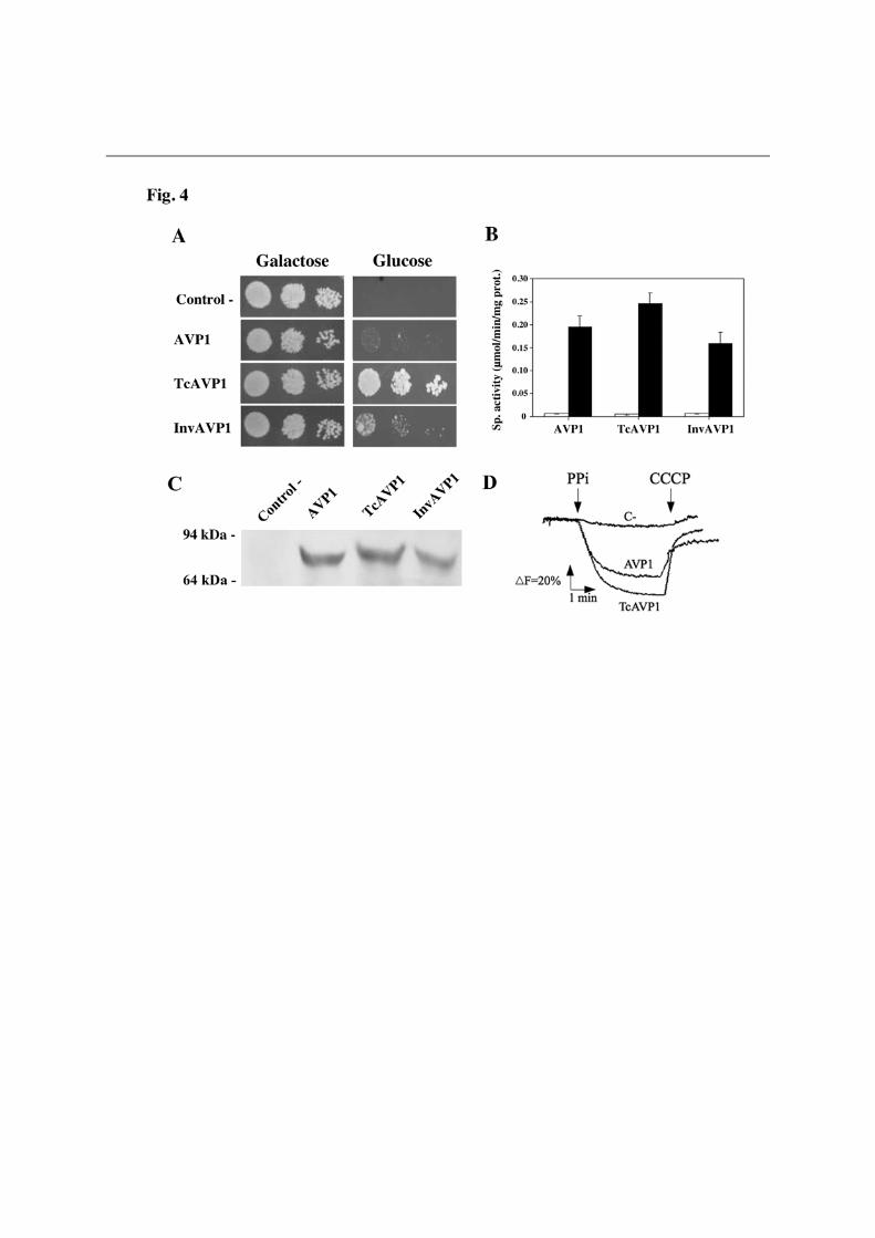
photosynthetic proteobacterium R. rubrum (RVP). Transformation of YPC3 with plasmids pRVP and pTcRVP did not allow the mutant to grow on glucose; however, cells transformed with plasmid pInvRVP efficiently grew on this carbon source. Assays of PPase activity and Western blot analysis further showed the presence of significant amounts of a K+-independent H+-PPase with an apparent molecular mass (ca. 60 kDa) smaller than that of other H+-PPases in membrane fractions of the latter cells. Interestingly, InvRVP was by far the most effectively membrane targeted protein (see Fig. 3C). Similar results were obtained with another prokaryotic K+-independent H+-PPase, from the actinobacterium Streptomyces coelicolor (not shown). In the case of the K+-dependent H+-PPase TgVP1 of the apicomplexan protist T. gondii, only YPC3 cells transformed with plasmid pTcTgVP, encoding a fusion protein with the putative signal sequence of T. cruzi H+-PPase, could recover the capacity to grow on glucose. PPi hydrolysis activity assays and Western blots consistently demonstrated the presence of a K+-dependent PPase in membranes of these cells; however, yeast cells transformed with plasmid pInvTgVP, encoding a fusion protein with the signal sequence of yeast Suc2p, also expressed the protein (Fig. S1). YPC3 cells transformed with plasmid pTcAVP1 could grow on glucose more efficiently than those transformed with plasmids pAVP1 or pInvAVP1; however, activity assays and Western blot analysis showed no significant differences in membrane-associated activity or protein levels (Fig. 4). In all cases, H+-translocation activities of membrane preparations were proportional to the PPi hydrolysis activities tested (Figs. 3D, S1D and 4D).
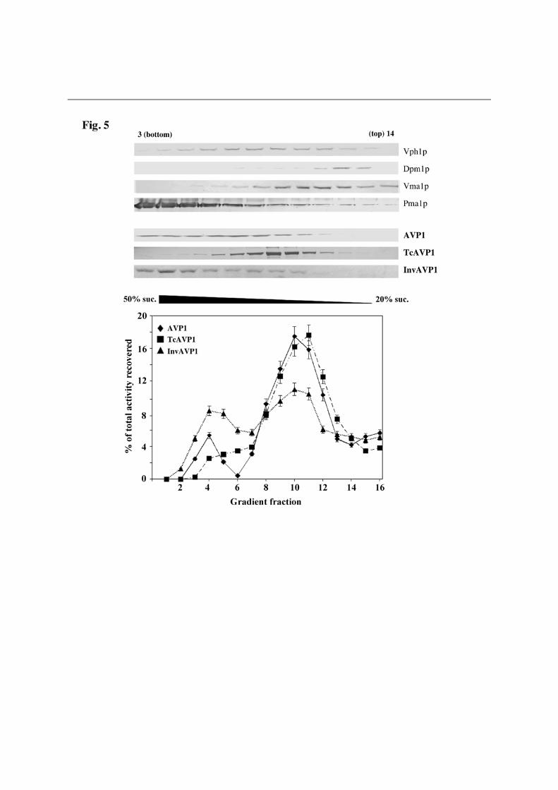
The N-terminal signal sequences from TcVP and Suc2p are processed when the chimeras with AVP1 are expressed in YPC3 In order to check whether the N-terminal signal sequences of TcVP and Suc2p were effectively processed during delivery and membrane sorting of the chimeric proteins, YPC3 cells were transformed with plasmids pHisAVP1, pHisTcAVP1 and pHisInvAVP1 (Table S2). Immunoblots of membrane preparations obtained from the different transformants using an antibody against the epitope RGSH4 recognised a polypeptide only in the case of cells transformed with pHisAVP1; however, the antibody against a highly conserved H+-PPase sequence identified polypeptides of the right size (ca. 70 kDa) in all cases (Fig. S2). Activity assays data were similar to those shown in Fig. 4. These results clearly indicate that the N-terminal domain of TcVP holds a bona fide signal sequence that, like the homologous signal peptide of Suc2p, is correctly processed in yeast. To our knowledge, this is the first direct evidence reported for protein processing and sorting of a H+-PPase, in an heterologous system in this case. An N-terminal fusion of TcVP with the RGSH4 domain that is functionally expressed in YPC3 seems to be processed as well, but unfortunately the chimeric protein is not recognized by the H+-PPase antibodies used in this study (data not shown). Natural AVP1 and its derived N-terminal chimeras TcAVP1 and InvAVP1 exhibit different subcellular distributions in yeast cells Fractionation by sucrose gradient ultracentrifugation of high density membranes obtained from yeast cells transformed with plasmids pAVP1, encoding natural plant vacuolar AVP1, and pTcAVP1 and pInvAVP1, encoding derived N-terminal chimeras with signal sequences, showed significant differences in the subcellular distributions of H+-PPase protein and PPi hydrolytic activity. In the case of pAVP1, activity was preferentially associated to internal membranes (Golgi complex/vacuoles), with some activity associated to the plasma
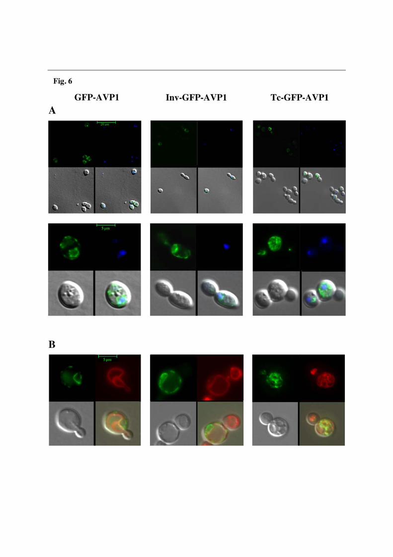
membrane. In cells transformed with pInvAVP1, an analogous distribution was found, although plasma membrane-associated activity was clearly higher. In contrast to that, membrane preparations from cells transformed with TcAVP1 showed virtually all the PPase activity to be associated to ER/Golgi/vacuolar membranes (Fig. 5). Western blot analyses showed AVP1 to be evenly distributed between gradient fractions 3 and 12 (being fraction 1 the bottom of the gradient), whereas TcAVP1 appeared preferentially in fractions 8-12 (Golgi complex/vacuole). Strikingly, in the case of InvAVP1 most of the protein appeared associated to the plasma membrane (fractions 3-5) (Fig. 5). In situ fluorescence microscopy confirms subcellular distribution results obtained with sucrose density gradients The question of the subcellular distribution of the different versions of AVP1 was further investigated by fluorescence microscopy of YPC3 cells. Transformants with plasmids pGFPAVP1 and pInvGFPAVP1, encoding respectively a GFP-AVP1 fusion protein without and with the Suc2p signal sequence, showed a very characteristic pattern of green fluorescence around the nucleus (cisternae vesicles of the ER) and in the plasma membrane. On the contrary, green fluorescence in cells transformed with plasmid pTcGFPAVP1 appeared exclusively associated to the internal membrane network, showing a spread spotted distribution throughout the YPC3 cells (Fig. 6A). Labelling of the different transformed yeast cells with FM4-64 clearly shows that GFP-AVP1 and InvGFP-AVP1 do not co-localize with the vacuolar membrane; by contrast, TcGFP-AVP1 seems to partially co-localize with a high number of small acidic vesicles (Fig. 6B). DISCUSSION
A S. cerevisiae haploid strain (YPC3), whose gene coding for the essential cytosolic soluble PPase (IPP1) is under the control of the glucose-repressable galactokinase (GAL1) promoter, has been generated by the single-step transplacement procedure. This approach resulted in the insertion of the yeast HIS3 marker gene followed by the GAL1 promoter between the promoter and the coding sequence of IPP1. Consequently, the conditional mutant strain generated YPC3 cannot grow in the presence of glucose because Ipp1p, essential for growth, is not expressed under these conditions. YPC3 is more convenient than YPC1, a mutant with the same phenotype previously described [15], to check functional complementation of Ipp1p due to its higher genomic stability.
The H+-PPase of the kinetoplastid protist T. cruzi (TcVP) has a putative N-terminal extension with the hallmarks of type I membrane protein signal sequences, as determined by in silico studies: a hydrophobic core, which presumably comprises a transmembrane (TM) segment, with two flanking groups of charged residues and a putative peptidase cleavage site at the C-terminus of the signal peptide [35]. The observation that the growth of YPC3 on glucose was efficiently supported by TcVP prompted us to investigate the possible effects that this domain (absent in many other proteins of this class) might have on the heterologous expression in yeast of other H+-PPases available in our laboratory. Comparative studies of these effects with those obtained with chimeras made with the N-terminal domain of a S. cerevisiae protein were also performed. Suc2p N-terminal signal peptide was chosen for two main reasons: (a) the initial steps of multispanning membrane proteins translation are analogous to those of secreted proteins synthesis [36], (b) this signal peptide had been studied in significant detail and utilized to efficiently translocate polypeptides across the ER membrane [37-39].
Plasmids bearing sequences coding for either of these N-terminal peptides followed by those of RVP and TgVP1 significantly increased the expression levels of the corresponding proteins in yeast. This improvement was high enough to functionally complement Ipp1p. Isolation of membrane fractions, assaying of membrane-associated PPi-hydrolysis activity as well as Western blot analyses revealed that expression levels of the chimeric proteins were higher than those of their original counterparts. It should be noted, however, that the achieved targeting efficiencies varied significantly between chimeric H+-PPases from different sources, e.g., N-terminal fusions with the Suc2p signal sequence dramatically enhanced protein and activity levels of prokaryotic H+-PPases, whereas both Tc- and Inv- fusions showed similar effectiveness with their eukaryotic counterparts. On the other hand, it was previously reported that the full-length TgVP1 gene cannot be expressed in vacuolar protease-deficient S. cerevisiae strains unless its N-terminal domain is removed [8]. This could be explained by assuming that the latter is not recognized by the yeast signal recognition particle (SRP) and, consequently, the polypeptide synthesis would be stopped at the initial steps of translation. Addition of the TcVP N-terminal signal sequence to TgVP1 would allow this recognition and, therefore, the expression of the chimeric protein in our non-defficient vacuolar protease yeast strain. The fact that a signal peptide from a trypanosomatid protist such as T. cruzi may be efficiently recognised by S. cerevisiae SRP supports the idea that components of the SRP pathway and salient features of the molecular mechanism of SRP-dependent protein targeting are conserved among different organisms [40]. Altogether, these results show that addition of appropriate N-terminal signal domains to multispanning membrane proteins can enhance their heterologous expression in yeast, presumably by increasing the targeting of the proteins to the ER, a proposal that is consistent with the results obtained with fluorescence microscopy of GFP-AVP1 fusions (see below). However, it is necessary to try different combinations of N-termini and coding sequences in order to optimize expression, as it is not possible to know before hand whether a given chimeric polypeptide will be effectively expressed in this system; that is, a trial-and-error approach is still needed. In any case, we have developed a useful set of plasmids where the desired ORF can be inserted downstream the sequences of several N-terminal signal peptides yielding different chimeras whose expression levels can be readily tested in S. cerevisiae.
AVP1 turned out to be a different case: the ability of this protein to functionally complement Ipp1p was significantly improved by attaching TcVP N-terminal domain to its N-terminus, although only a slight improvement was obtained with Suc2p; however, similar membrane-associated PPi hydrolytic activity and protein levels were observed for both chimerae. By utilizing an antibody against the epitope RGSH4, it was shown that both TcVP and Suc2p N-terminal peptides added were processed when attached to AVP1. This may not be significant in the case of Suc2p; however, it strongly suggests that the N-terminus of TcVP is fully recognisable by S. cerevisiae type I signal peptidases [41]. This idea was further supported by the fact that identical results were obtained with other yeast strains, such as W303-1A and the vacuolar protease-deficient mutant BJ5457 [7] (data not shown). N-terminal signal peptides have been shown to occur in all organisms studied to date and share some conserved general features (see Fig. 2), consequently, they have been extensively used in order to improve expression of foreign soluble proteins in yeast with varied results, revealing that not all signal sequences are functionally equivalent. Thus, species-specificity of signal peptide utilization has been demonstrated not only between divergent organisms. i. e., mammalian and yeast [42] but also between closely related species [43]. This seems to be also the case for heterologous membrane proteins (mostly transporters and receptors) expressed in yeast, although the information available is much scantier. Here, we report the
first direct evidence obtained so far for protein precursor processing of a proton pump, in this case, a member of the H+-PPase family. On the other hand, it was important to check this point as we aim at optimizing production of these membrane proteins in yeast with minimal changes with respect to the natural polypeptide.
Results with TgVP I and AVP1 demonstrate that, although a threshold of PPase activity is required to complement Ipp1p, this may not be enough and some other biologically relevant features, such as subcellular distribution, are important. The possibility that AVP1 and its derived chimerae, TcAVP1 and InvAVP1, might have different locations within the cell was studied by sucrose density gradient ultracentrifugation. Previous separation of the yeast membrane preparation in “high density” (HDM) and “low density” (LDM) fractions was essential to achieve the desired resolution in the gradients. Fractionation of HDM indicated that AVP1 was rather evenly distributed at different membranes, although activity was maximal at internal membranes (ER/Golgi complex/vacuole). The chimera TcAVP1 and its associated activity seemed to be preferentially located in internal membranes, showing a rather homogeneous intracellular distribution; however, in the case of InvAVP1 a striking situation was observed: although most of the polypeptide was found at the plasma membrane, membrane-bound K+-stimulated PPi hydrolytic activity was higher in internal membranes. This result suggests two important points: (a) the N-terminus of Suc2p can preferentially target membrane-bound proteins to the plasma membrane, and (b) the lipid composition of the membrane where InvAVP1 is located may significantly modulate its activity. Thus, according to the results obtained with AVP1 and InvAVP1, the plasma membrane does not seem to provide an optimal lipid environment for this protein, naturally occurring in plant vacuolar membrane (tonoplast) [11, 12]. The modulation of activity by lipids has been reported for other proton pumps such as the plant plasma membrane H+-ATPase [44], the vacuolar H+-ATPase [27] and the H+-PPase from Trypanosoma cruzi [14]
Density gradient ultracentrifugation presents certain limitations at resolving subcellular fractions in yeast [24]; therefore, these studies were completed by in situ fluorescence microscopy studies using protein fusions with the green fluorescent protein. Initially, immunocytochemical localization with antibodies currently available in our laboratory was attempted without success. Then, fusion of GFP at the C-terminus of AVP1 [45] was carried out, but this yielded a polypeptide with no activity. This is in agreement with previous studies indicating that the C-terminus is essential for both the oligomerization and functionality of the H+-PPase proteins in the membrane [46]. Consequently, a fusion protein in which the yEGFP was attached at the N-terminus was constructed. The resulting polypeptide was efficiently expressed in YPC3 as demonstrated by its capacity to complement Ipp1p and the high levels of K+-dependent membrane-associated PPi hydrolytic activity found; actually, GFP-AVP1 showed higher expression levels than AVP1 (not shown). The presence of GFP at the N-terminus of some membrane proteins has been previously reported to enhance their expression levels in heterologous systems [47]. This effect is currently being investigated for other H+-PPases available in our laboratory.
Fluorescence microscopy showed a pattern of GFP-AVP1 distribution strikingly similar to that previously obtained for AVP1 by immunocytochemical localization: the protein is mainly associated to ER-derived perinuclear membranes (cisternae vesicles) with a significant fraction located at the plasma membrane and small prevacuolar vesicles [15]. This similarity was further confirmed by sucrose gradient fractionation (not shown). Therefore, the presence of GFP at the N-terminus does not seem to alter the targeting of AVP1 in yeast. Further addition of TcVP N-terminal signal domain resulted in major changes in green fluorescence distribution, which remains associated to internal membranes. On the other
hand, addition of Suc2p signal peptide resulted in a subcellular distribution similar to that of GFP-AVP1. Co-localization of TcGFP-AVP1 with the acidic vesicles membranes further shows that this chimera is effectively targeted in a functional active form to specific internal compartments. Altogether, fluorescence microscopy results confirmed those obtained by sucrose density gradient fractionation and strengthen the idea that the presence of an appropriate N-terminal signal peptide dramatically alters the intracellular targeting and allows further trafficking along the biosynthetic secretory pathway of heterologously expressed H+-PPases in S. cerevisiae.
As a final conclusion, it is important to point out that the strategy followed in this work can be utilized in concert to others found in the literature aimed at optimizing heterologous expression of membrane proteins in yeast. Actually, the expression levels of several of the chimeric proteins described here can be further increased if they are expressed in vacuolar-deficient yeast mutants [7]. All the strategies described here are already under way in our laboratory in order to get the high amounts of protein required for structural studies, such as crystallization and 2D/3D structure determination. On the other hand, results obtained with AVP1 further support that the N-terminal signal sequences alter the subcellular localization of a membrane-embedded protein. This capacity could also be utilized to preferentially target proteins to specific organelles and/or membranes within the cell, which may be crucial when performing studies of functional complementation.

REFERENCES 1 Sommer, T. and Wolf, D. H. (1997) Endoplasmic reticulum degradation: reverse protein flow of no return. FASEB J. 11, 1227-1233 2 Luo, W. and Chang, A. (1997) Novel genes involved in endosomal traffic in yeast revealed by suppression of a targeting-defective plasma membrane ATPase mutant. J. Cell Biol. 138, 731-746 3 Ferreira, T., Mason, A. B., Pypaert, M., Allen, K. E. and Slayman, C. W. (2002) Quality control in the yeast secretory pathway: a misfolded PMA1 H+-ATPase reveals two checkpoints. J. Biol. Chem. 277, 21027-21040 4 Griffith, D. A., Delipala, C., Leadsham, J., Jarvis, S. M. and Oesterhelt, D. (2003) A novel yeast expression system for the overproduction of quality-controlled membrane proteins. FEBS Lett. 553, 45-50 5 Ellgaard, L. and Helenius, A. (2003) Quality control in the endoplasmic reticulum. Nat Rev Mol. Cell. Biol. 4, 181-191 6 Newstead, S., Kim, H., von Heijne, G., Iwata, S. and Drew, D. (2007) High-throughput fluorescent-based optimization of eukaryotic membrane protein overexpression and purification in Saccharomyces cerevisiae. Proc. Natl. Acad. Sci. U S A. 104, 13936-13941 7 Jones, E. W. (1991) Tackling the protease problem in Saccharomyces cerevisiae. Methods Enzymol. 194, 428-453 8 Drozdowicz, Y. M., Shaw, M., Nishi, M., Striepen, B., Liwinski, H. A., Roos, D. S. and Rea, P. A. (2003) Isolation and characterization of TgVP1, a type I vacuolar H+-translocating pyrophosphatase from Toxoplasma gondii. The dynamics of its subcellular localization and the cellular effects of a diphosphonate inhibitor. J. Biol. Chem. 278, 1075-1085 9 Reis, E. M., Kurtenbach, E., Ferreira, A. R., Biselli, P. J., Slayman, C. W. and Verjovski-Almeida, S. (1999) N-terminal chimeric constructs improve the expression of sarcoplasmic reticulum Ca(2+)-ATPase in yeast. Biochim. Biophys. Acta. 1461, 83-95 10 Docampo, R., de Souza, W., Miranda, K., Rohloff, P. and Moreno, S. N. (2005) Acidocalcisomes - conserved from bacteria to man. Nat. Rev. Microbiol. 3, 251-261 11 Drozdowicz, Y. M. and Rea, P. A. (2001) Vacuolar H(+) pyrophosphatases: from the evolutionary backwaters into the mainstream. Trends Plant Sci. 6, 206-211 12 Maeshima, M. (2001) TONOPLAST TRANSPORTERS: Organization and Function. Annu. Rev. Plant Physiol. Plant Mol. Biol. 52, 469-497 13 Pérez-Castiñeira, J. R., Gómez-García, R., López-Marqués, R. L., Losada, M. and Serrano, A. (2001) Enzymatic systems of inorganic pyrophosphate bioenergetics in photosynthetic and heterotrophic protists: remnants or metabolic cornerstones? Int. Microbiol. 4, 135-142 14 Martínez, R., Wang, Y., Benaim, G., Benchimol, M., de Souza, W., Scott, D. A. and Docampo, R. (2002) A proton pumping pyrophosphatase in the Golgi apparatus and plasma membrane vesicles of Trypanosoma cruzi. Mol Biochem Parasitol. 120, 205-213 15 Pérez-Castiñeira, J. R., López-Marqués, R. L., Villalba, J. M., Losada, M. and Serrano, A. (2002) Functional complementation of yeast cytosolic pyrophosphatase by
bacterial and plant H+-translocating pyrophosphatases. Proc. Natl. Acad. Sci. U S A. 99, 15914-15919 16 Kim, E. J., Zhen, R. G. and Rea, P. A. (1994) Heterologous expression of plant vacuolar pyrophosphatase in yeast demonstrates sufficiency of the substrate-binding subunit for proton transport. Proc. Natl. Acad. Sci. U S A. 91, 6128-6132 17 Rothstein, R. J. (1983) One-step gene disruption in yeast. Methods Enzymol. 101, 202-211 18 Kolakowski, L. F., Jr., Schloesser, M. and Cooperman, B. S. (1988) Cloning, molecular characterization and chromosome localization of the inorganic pyrophosphatase (PPA) gene from S. cerevisiae. Nucleic Acids Res. 16, 10441-10452 19 Sambrook, J. and Russell, D. (2001) Molecular Cloning: A Laboratory Manual. Cold Spring Harbor Laboratory Press, Cold Spring Harbor, New York 20 Schiestl, R. H. and Gietz, R. D. (1989) High efficiency transformation of intact yeast cells using single stranded nucleic acids as a carrier. Curr. Genet. 16, 339-346 21 Treco, D. A. and Lundblad, V. (2001) Preparation of yeast media. Curr. Protoc. Mol. Biol. Chapter 13, Unit13 11 22 Thompson, J. D., Gibson, T. J., Plewniak, F., Jeanmougin, F. and Higgins, D. G. (1997) The CLUSTAL_X windows interface: flexible strategies for multiple sequence alignment aided by quality analysis tools. Nucleic Acids Res. 25, 4876-4882 23 Emanuelsson, O., Brunak, S., von Heijne, G. and Nielsen, H. (2007) Locating proteins in the cell using TargetP, SignalP and related tools. Nat. Protoc. 2, 953-971 24 Serrano, R. and Villalba, J. M. (1995) Expression and localization of plant membrane proteins in Saccharomyces. Methods Cell. Biol. 50, 481-496 25 Lenoir, G., Menguy, T., Corre, F., Montigny, C., Pedersen, P. A., Thines, D., le Maire, M. and Falson, P. (2002) Overproduction in yeast and rapid and efficient purification of the rabbit SERCA1a Ca(2+)-ATPase. Biochim. Biophys. Acta. 1560, 67-83 26 Rathbun, W. B. and Betlach, M. V. (1969) Estimation of enzymically produced orthophosphate in the presence of cysteine and adenosine triphosphate. Anal. Biochem. 28, 436-445 27 Pérez-Castiñeira, J. R. and Apps, D. K. (1990) Vacuolar H(+)-ATPase of adrenal secretory granules. Rapid partial purification and reconstitution into proteoliposomes. Biochem. J. 271, 127-131 28 Valverde, F., Losada, M. and Serrano, A. (1997) Functional complementation of an Escherichia coli gap mutant supports an amphibolic role for NAD(P)-dependent glyceraldehyde-3-phosphate dehydrogenase of Synechocystis sp. strain PCC 6803. J. Bacteriol. 179, 4513-4522 29 Casadio, R. (1991) Measurements of transmembrane pH differences of low extents in bacterial chromatophores. A study with the fluorescent probe 9-amino, 6-chloro, 2-methoxyacridine. Eur. Biophys. J. 19, 189-201 30 López-Marqués, R. L., Pérez-Castiñeira, J. R., Buch-Pedersen, M. J., Marco, S., Rigaud, J. L., Palmgren, M. G. and Serrano, A. (2005) Large-scale purification of the proton pumping pyrophosphatase from Thermotoga maritima: a "Hot-Solve" method for isolation of recombinant thermophilic membrane proteins. Biochim. Biophys. Acta. 1716, 69-76

31 Pringle, J. R., Adams, A. E., Drubin, D. G. and Haarer, B. K. (1991) Immunofluorescence methods for yeast. Methods Enzymol. 194, 565-602 32 Bradford, M. M. (1976) A rapid and sensitive method for the quantitation of microgram quantities of protein utilizing the principle of protein-dye binding. Anal. Biochem. 72, 248-254 33 Rodrigues, F., van Hemert, M., Steensma, H. Y., Corte-Real, M. and Leao, C. (2001) Red fluorescent protein (DsRed) as a reporter in Saccharomyces cerevisiae. J. Bacteriol. 183, 3791-3794 34 Conibear, E. and Stevens, T. H. (2002) Studying yeast vacuoles. Methods Enzymol. 351, 408-432 35 Harley, C. A. and Tipper, D. J. (1996) The role of charged residues in determining transmembrane protein insertion orientation in yeast. J. Biol. Chem. 271, 24625-24633 36 Cheng, Z. and Gilmore, R. (2006) Slow translocon gating causes cytosolic exposure of transmembrane and lumenal domains during membrane protein integration. Nat. Struct. Mol. Biol. 13, 930-936 37 Kaiser, C. A. and Botstein, D. (1986) Secretion-defective mutations in the signal sequence for Saccharomyces cerevisiae invertase. Mol. Cell. Biol. 6, 2382-2391 38 Ngsee, J. K., Hansen, W., Walter, P. and Smith, M. (1989) Cassette mutagenic analysis of the yeast invertase signal peptide: effects on protein translocation. Mol. Cell. Biol. 9, 3400-3410 39 Rothe, C. and Lehle, L. (1998) Sorting of invertase signal peptide mutants in yeast dependent and independent on the signal-recognition particle. Eur. J. Biochem. 252, 16-24 40 Keenan, R. J., Freymann, D. M., Stroud, R. M. and Walter, P. (2001) The signal recognition particle. Annu. Rev. Biochem. 70, 755-775 41 Dalbey, R. E., Lively, M. O., Bron, S. and van Dijl, J. M. (1997) The chemistry and enzymology of the type I signal peptidases. Protein Sci. 6, 1129-1138 42 Bird, P., Gething, M. J. and Sambrook, J. (1987) Translocation in yeast and mammalian cells: not all signals are functionally equivalent. J. Cell Biol. 105, 2905-2914 43 Al-Qahtani, A., Teilhet, M. and Mensa-Wilmot (1998) Species specificity in endoplasmic reticulum signal peptide utilization revealed by proteins from Trypanosoma brucei and Leishmania. Biochem. J. 331, 521-529 44 Kasamo, K. (2003) Regulation of plasma membrane H+-ATPase activity by the membrane environment. J. Plant Res. 116, 517–523 45 Drew, D., Newstead, S., Sonoda, Y., Kim, H., von Heijne, G. and Iwata, S. (2008) GFP-based optimization scheme for the overexpression and purification of eukaryotic membrane proteins in Saccharomyces cerevisiae. Nat. Protoc. 3, 784-798 46 Mimura, H., Nakanishi, Y. and Maeshima, M. (2005) Oligomerization of H(+)-pyrophosphatase and its structural and functional consequences. Biochim. Biophys. Acta. 1708, 393-403 47 Wagner, S., Bader, M. L., Drew, D. and de Gier, J. W. (2006) Rationalizing membrane protein overexpression. Trends Biotechnol. 24, 364-371 48 Bernsel, A., Viklund, H., Falk, J., Lindahl, E., von Heijne, G. and Elofsson, A. (2008) Prediction of membrane-protein topology from first principles. Proc. Natl. Acad. Sci. U S A. 105, 7177-7181
AUTHOR CONTRIBUTION R. Drake carried out most of the experimental work, except generation of YPC3 and fluorescence microscopy. J. R. Pérez-Castiñeira supervised R. Drake’s work, designed the study, supervised data collection, generated YPC3 mutant, performed microscopy work and drafted the manuscript. A. Serrano initially conceived the study, did the in silico analyses, co-supervised the experimental work and data collection and contributed to the manuscript draft. ACKNOWLEDGEMENTS
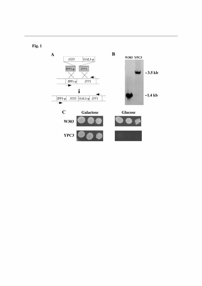
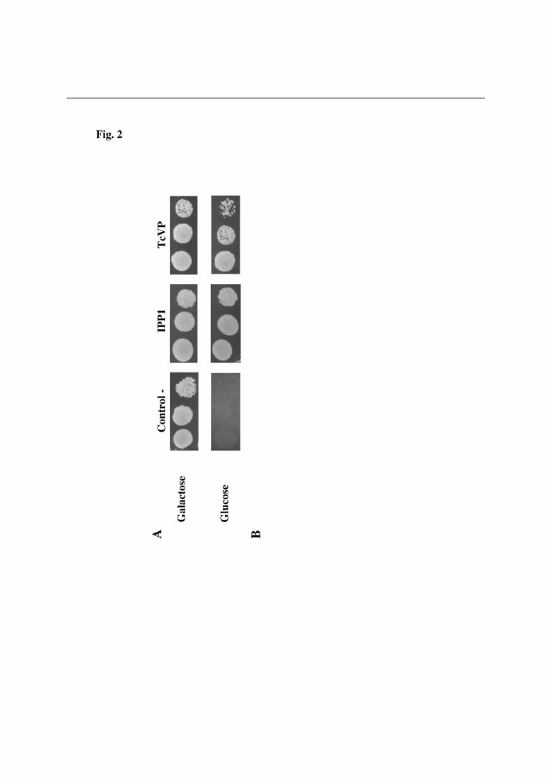
We gratefully acknowledge Dr. Rosa L. López-Marqués (Univ. of Copenhagen, Denmark), Prof. Andrés Aguilera (Univ. of Seville, Spain), Profs. Silvia Moreno and Roberto Docampo (Univ. of Georgia, Athens, GA, USA) for generously providing antibody against T. maritima H+-PPase, plasmid pUG34 and the cDNA clones of T. gondii and T. cruzi H+-PPases, respectively. We also thank Dr. Alicia Orea for excellent technical assistance with fluorescence microscopy. FUNDING This work was supported by research grants from the Spanish Ministerio de Ciencia e Innovación (BMC2007-61887) and Regional Andalusian Goverment (PAIDI group BIO-261). A part of funds came from the FEDER program of the EU. LEGENDS OF FIGURES Figure 1. Generation of YPC3, a new yeast mutant whose essential cytosolic inorganic pyrophosphatase Ipp1p is under the control of the galactokinase (GAL1) promoter. (A) The gene coding for the cytosolic pyrophosphatase of yeast haploid strain W303-1A was disrupted by insertion of a linear DNA fragment comprising the yeast HIS3 cassette as a selection marker followed by the galactokinase (GAL1) gene promoter. Arrows represent the oligonucleotides utilized as PCR primers. (B) Analysis by PCR of the insertion, genomic DNA obtained from strains YPC3 and W303-1A were utilized as templates and oligonucleotides corresponding to the promoter region and the end of the coding sequence of IPP1 as primers. (C) Phenotypic characterization of YPC3 by checking its growth on glucose as compared with the yeast strain utilized for its generation. See main text for details. Figure 2. Functional complementation of the cytosolic pyrophosphatase Ipp1p of S. cerevisiae by the H+-PPase from the kinetoplastid protist Trypanosoma cruzi (A) and
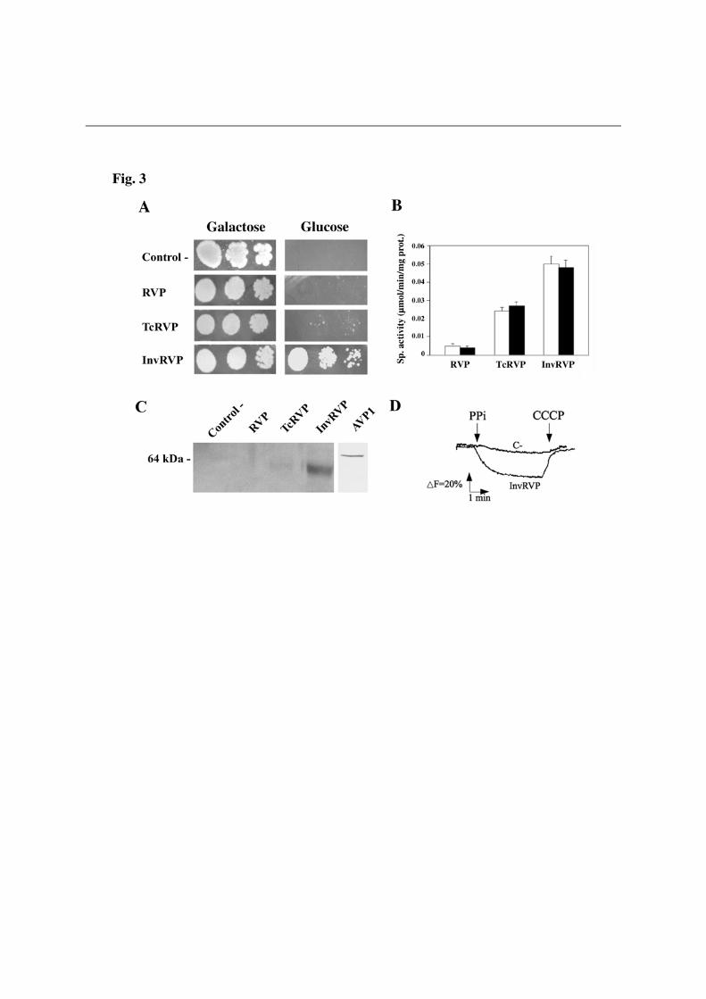
multiple sequences alignment of the N-terminal regions of H+-PPase orthologs from selected eukaryotic (bold) and prokaryotic (plain) organisms (B). N-terminal extensions having the general features of signal peptides, namely, a hydrophobic core and a positively charged N-terminal domain, and predicted as such by the SignalP and SIG-Pred programs are shown boxed and the corresponding putative cleavage sites indicated by vertical arrowheads. Positively charged (K,R) and hydrophobic (A,V,L,I,W,F,M) amino acid residues in the putative signal sequences are represented in italics and in boldface with grey background, respectively. Note the bipartite signal peptide with two putative cleavage sites predicted for the H+-PPase of the apicomplexan protist Toxoplasma gondii, for which the N-terminal residues of truncated constructs used by Drozdowicz et al. [8] for expression in yeast are indicated by folded arrows. Approximate locations of the transmembrane segment (TMS) characteristic of signal leader peptides and the first one (TMS1) in all H+-PPases are also shown [48]. The protein sequences used are (accession numbers in parentheses): Trypanosoma cruzi (AAF80381); Trypanosoma brucei (AAK95376); Leshmania major (CAJ08309); T. gondii TgVP1 (AAK38077); Plasmodium falciparum PfVP1 (AAD17215); Arabidopsis thaliana AVP1 (A38230); Oryza sativa (BAA08232); Thermotoga maritima (D72049); Rhodospirillum rubrum (AAC38615); Methanosarcina mazei (AAM22543); Pyrobaculum aerophilum (AAF01029); Streptomyces coelicolor (T36668), and Chlorobium tepidum (contig g12, NC_002932). The unpublished H+-PPase sequences from the microalga Chlorella sp. NC64A were identified by their JGI genome project code numbers. Figure 3. Drop tests (A), membrane-associated PPi hydrolysis activity assays (B), Western blot analysis (C) and traces of ACMA fluorescence quenching by membrane preparations (D) of yeast mutant strain YPC3 transformed with plasmids pRS699b, pRVP, pTcRVP and pInvRVP. Experiments were performed with three independent clones taken from each of the transformant plates. (A) Typical drop test. Activity values shown in (B) are means + S.E. (n=3) using LDM preparations obtained from YPC3 cells transformed with pRS699b as reference White and black bars represent activities in the absence and presence of potassium chloride, respectively. (C) Immunodetection of low density membrane (LDM) preparations as described in Experimental Procedures. Approx. 70 µg of protein were loaded per lane. (D) Typical trace of ACMA fluorescence quenching obtained with LDM preparations of YPC3 transformed with plasmid pInvRVP. No quenching was observed in the case of RVP whereas, in the case of TcRVP, an intermediate trace between those of C- and InvRVP was obtained (not shown). Figure 4. Drop tests (A), membrane-associated PPi hydrolysis activity assays (B), Western blot analysis (C) and traces of ACMA fluorescence quenching by membrane preparations (D) of yeast mutant strain YPC3 transformed with plasmids pRS699b, pAVP1, pTcAVP1 and pInvAVP1. Same conditions as in Fig. 3 except that similar traces to that of AVP1 were routinely obtained with LDM preparation of YPC3 cells transformed with plasmid pInvAVP1.
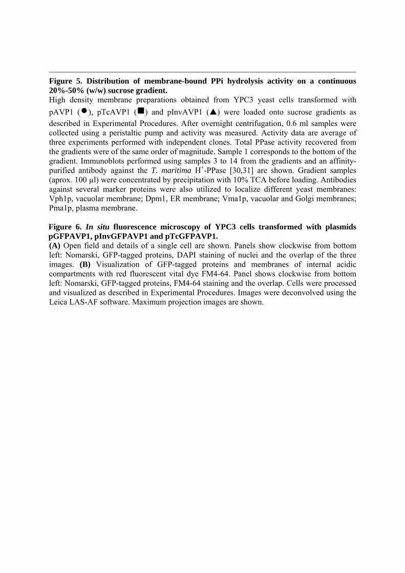
Figure 5. Distribution of membrane-bound PPi hydrolysis activity on a continuous 20%-50% (w/w) sucrose gradient. High density membrane preparations obtained from YPC3 yeast cells transformed with
pAVP1 (), pTcAVP1 () and pInvAVP1 () were loaded onto sucrose gradients as
described in Experimental Procedures. After overnight centrifugation, 0.6 ml samples were collected using a peristaltic pump and activity was measured. Activity data are average of three experiments performed with independent clones. Total PPase activity recovered from the gradients were of the same order of magnitude. Sample 1 corresponds to the bottom of the gradient. Immunoblots performed using samples 3 to 14 from the gradients and an affinity-purified antibody against the T. maritima H+-PPase [30,31] are shown. Gradient samples (aprox. 100 µl) were concentrated by precipitation with 10% TCA before loading. Antibodies against several marker proteins were also utilized to localize different yeast membranes: Vph1p, vacuolar membrane; Dpm1, ER membrane; Vma1p, vacuolar and Golgi membranes; Pma1p, plasma membrane. Figure 6. In situ fluorescence microscopy of YPC3 cells transformed with plasmids pGFPAVP1, pInvGFPAVP1 and pTcGFPAVP1. (A) Open field and details of a single cell are shown. Panels show clockwise from bottom left: Nomarski, GFP-tagged proteins, DAPI staining of nuclei and the overlap of the three images. (B) Visualization of GFP-tagged proteins and membranes of internal acidic compartments with red fluorescent vital dye FM4-64. Panel shows clockwise from bottom left: Nomarski, GFP-tagged proteins, FM4-64 staining and the overlap. Cells were processed and visualized as described in Experimental Procedures. Images were deconvolved using the Leica LAS-AF software. Maximum projection images are shown.
Supplementary information section
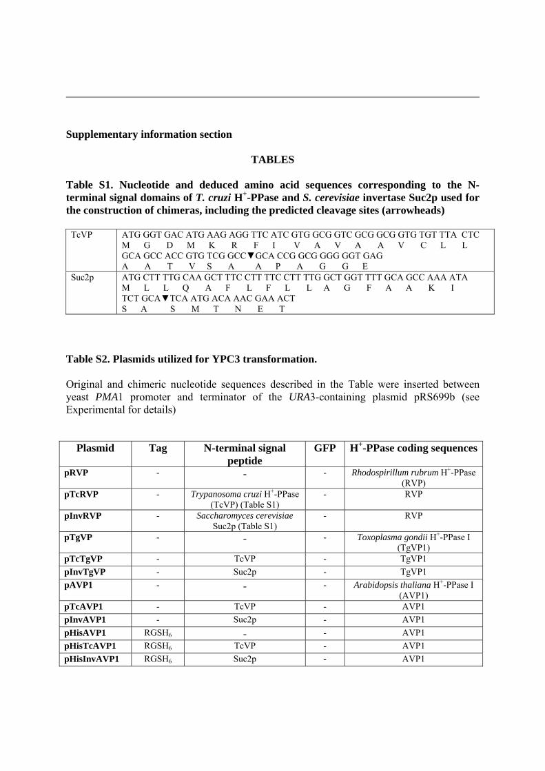
TABLES Table S1. Nucleotide and deduced amino acid sequences corresponding to the N-terminal signal domains of T. cruzi H+-PPase and S. cerevisiae invertase Suc2p used for the construction of chimeras, including the predicted cleavage sites (arrowheads)
TcVP ATG GGT GAC ATG AAG AGG TTC ATC GTG GCG GTC GCG GCG GTG TGT TTA CTC M G D M K R F I V A V A A V C L L GCA GCC ACC GTG TCG GCC▼GCA CCG GCG GGG GGT GAG A A T V S A A P A G G E
Suc2p ATG CTT TTG CAA GCT TTC CTT TTC CTT TTG GCT GGT TTT GCA GCC AAA ATA M L L Q A F L F L L A G F A A K I TCT GCA▼TCA ATG ACA AAC GAA ACT S A S M T N E T
Table S2. Plasmids utilized for YPC3 transformation. Original and chimeric nucleotide sequences described in the Table were inserted between yeast PMA1 promoter and terminator of the URA3-containing plasmid pRS699b (see Experimental for details)
Plasmid Tag N-terminal signal peptide
GFP H+-PPase coding sequences
pRVP - - - Rhodospirillum rubrum H+-PPase (RVP)
pTcRVP - Trypanosoma cruzi H+-PPase (TcVP) (Table S1)
- RVP
pInvRVP - Saccharomyces cerevisiae Suc2p (Table S1)
- RVP
pTgVP - - - Toxoplasma gondii H+-PPase I (TgVP1)
pTcTgVP - TcVP - TgVP1
pInvTgVP - Suc2p - TgVP1
pAVP1 - - - Arabidopsis thaliana H+-PPase I (AVP1)
pTcAVP1 - TcVP - AVP1
pInvAVP1 - Suc2p - AVP1
pHisAVP1 RGSH6 - - AVP1
pHisTcAVP1 RGSH6 TcVP - AVP1
pHisInvAVP1 RGSH6 Suc2p - AVP1
pGFPAVP1 - - Yes AVP1
pTcGFPAVP1 - TcVP Yes AVP1
pInvGFPAVP1 - Suc2p Yes AVP1
SUPPLEMENTARY FIGURES LEGENDS
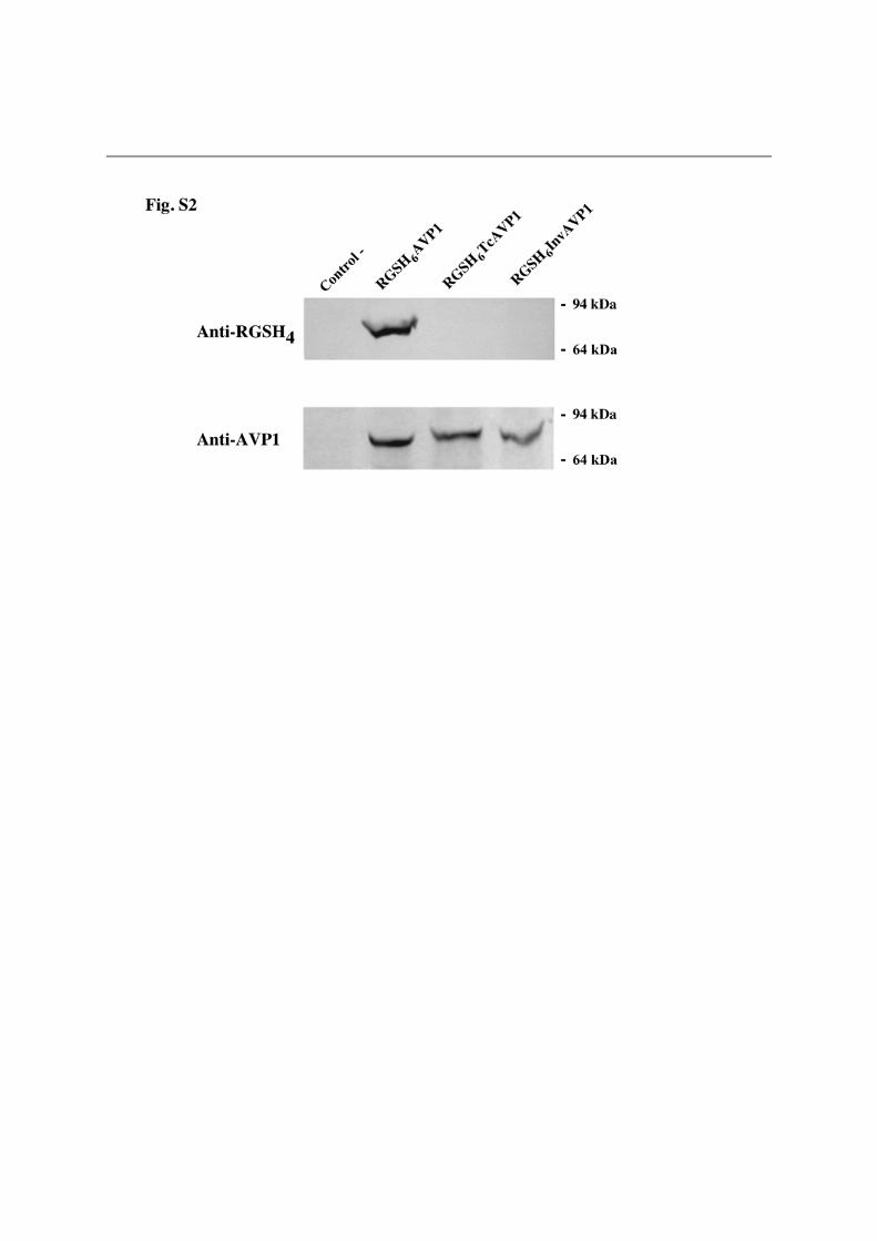
Figure S1. Drop tests (A), membrane-associated PPi hydrolysis activity assays (B), Western blot analysis (C) and traces of ACMA fluorescence quenching by membrane preparations (D) of yeast mutant strain YPC3 transformed with plasmids pRS699b, pTgVP, pTcTgVP, pInvTgVP. Same conditions as in Fig. 3 except that in the case of InvTgVP1 a fluorescence quenching trace similar to that of TcTgVP1 was obtained. No quenching was observed for TgVP1 (not shown). Note that the scale of fluorescence changes is ten times smaller than those shown in Figures 3 and 5. Figure S2. Western blot analyses of LDM preparations obtained from YPC3 cells transformed with plasmids pRS699b, pHisAVP1, pHisTcAVP1 and pHisInvAVP1. Results obtained with a monoclonal antibody (used at 1:2000 dilution) against the epitope RGSH4 (upper panel) and against a highly conserved sequence of H+-PPases [16] are shown. The latter antibody was used at a 1:1000 dilution. About 70 μg of protein were loaded in each track.

Related Documents