This Provisional PDF corresponds to the article as it appeared upon acceptance. Fully formatted PDF and full text (HTML) versions will be made available soon. N-arachidonoyl glycine, an abundant endogenous lipid, potently drives directed cellular migration through GPR18, the putative abnormal cannabidiol receptor. BMC Neuroscience 2010, 11:44 doi:10.1186/1471-2202-11-44 Douglas McHugh ([email protected]) Sherry S-J Hu ([email protected]) Neta Rimmerman ([email protected]) Ana Juknat ([email protected]) Zvi Vogil ([email protected]) J MICHAEL Walker ([email protected]) Heather B Bradshaw ([email protected]) ISSN 1471-2202 Article type Research article Submission date 12 March 2010 Acceptance date 26 March 2010 Publication date 26 March 2010 Article URL http://www.biomedcentral.com/1471-2202/11/44 Like all articles in BMC journals, this peer-reviewed article was published immediately upon acceptance. It can be downloaded, printed and distributed freely for any purposes (see copyright notice below). Articles in BMC journals are listed in PubMed and archived at PubMed Central. For information about publishing your research in BMC journals or any BioMed Central journal, go to http://www.biomedcentral.com/info/authors/ BMC Neuroscience © 2010 McHugh et al. , licensee BioMed Central Ltd. This is an open access article distributed under the terms of the Creative Commons Attribution License ( http://creativecommons.org/licenses/by/2.0), which permits unrestricted use, distribution, and reproduction in any medium, provided the original work is properly cited.

Welcome message from author
This document is posted to help you gain knowledge. Please leave a comment to let me know what you think about it! Share it to your friends and learn new things together.
Transcript
This Provisional PDF corresponds to the article as it appeared upon acceptance. Fully formattedPDF and full text (HTML) versions will be made available soon.
N-arachidonoyl glycine, an abundant endogenous lipid, potently drives directedcellular migration through GPR18, the putative abnormal cannabidiol receptor.
BMC Neuroscience 2010, 11:44 doi:10.1186/1471-2202-11-44
Douglas McHugh ([email protected])Sherry S-J Hu ([email protected])
Neta Rimmerman ([email protected])Ana Juknat ([email protected])
Zvi Vogil ([email protected])J MICHAEL Walker ([email protected])
Heather B Bradshaw ([email protected])
ISSN 1471-2202
Article type Research article
Submission date 12 March 2010
Acceptance date 26 March 2010
Publication date 26 March 2010
Article URL http://www.biomedcentral.com/1471-2202/11/44
Like all articles in BMC journals, this peer-reviewed article was published immediately uponacceptance. It can be downloaded, printed and distributed freely for any purposes (see copyright
notice below).
Articles in BMC journals are listed in PubMed and archived at PubMed Central.
For information about publishing your research in BMC journals or any BioMed Central journal, go to
http://www.biomedcentral.com/info/authors/
BMC Neuroscience
© 2010 McHugh et al. , licensee BioMed Central Ltd.This is an open access article distributed under the terms of the Creative Commons Attribution License (http://creativecommons.org/licenses/by/2.0),
which permits unrestricted use, distribution, and reproduction in any medium, provided the original work is properly cited.

1
N-arachidonoyl glycine, an abundant endogenous lipid, potently
drives directed cellular migration through GPR18, the putative
abnormal cannabidiol receptor.
Douglas McHugh1, Sherry S-J Hu
2, Neta Rimmerman
3, Ana Juknat
4, Zvi
Vogel3,4
, J Michael Walker2, and Heather B Bradshaw
1§.
1The Department of Psychological and Brain Sciences, 1101 East 10th Street,
Indiana University, Bloomington, IN 47405, USA.
2The Gill Center for Biomolecular Science, 1101 East 10th Street, Indiana
University, Bloomington, IN 47405, USA.
3The Neurobiology Department, Weizmann Institute of Science, Rehovot, Israel.
4The Adelson Center for the Biology of Addictive Diseases, Sackler Faculty of
Medicine, Tel Aviv University, Israel.
§Corresponding author
Email addresses:
3
Abstract
Background
Microglia provide continuous immune surveillance of the CNS and upon
activation rapidly change phenotype to express receptors that respond to
chemoattractants during CNS damage or infection. These activated microglia
undergo directed migration towards affected tissue. Importantly, the molecular
species of chemoattractant encountered determines if microglia respond with pro-
or anti-inflammatory behaviour, yet the signaling molecules that trigger migration
remain poorly understood. The endogenous cannabinoid system regulates
microglial migration via CB2 receptors and an as yet unidentified GPCR termed
the ‘abnormal cannabidiol’ (Abn-CBD) receptor. Abn-CBD is a synthetic isomer
of the phytocannabinoid cannabidiol (CBD) and is inactive at CB1 or CB2
receptors, but functions as a selective agonist at this Gi/o-coupled GPCR.
N-arachidonoyl glycine (NAGly) is an endogenous metabolite of the
endocannabinoid anandamide and acts as an efficacious agonist at GPR18. Here,
we investigate the relationship between NAGly, Abn-CBD, the unidentified
‘Abn-CBD’ receptor, GPR18, and BV-2 microglial migration.
Results
4
Using Boyden chamber migration experiments, yellow tetrazolium (MTT)
conversion, In-cell Western, qPCR and immunocytochemistry we show that
NAGly, at sub-nanomolar concentrations, and Abn-CBD potently drive cellular
migration in both BV-2 microglia and HEK293-GPR18 transfected cells, but
neither induce migration in HEK-GPR55 or non-transfected HEK293 wildtype
cells. Migration effects are blocked or attenuated in both systems by the
‘Abn-CBD’ receptor antagonist O-1918, and low efficacy agonists
N-arachidonoyl-serine and cannabidiol. NAGly promotes proliferation and
activation of MAP kinases in BV-2 microglia and HEK293-GPR18 cells at low
nanomolar concentrations – cellular responses correlated with microglial
migration. Additionally, BV-2 cells show GPR18 immunocytochemical staining
and abundant GPR18 mRNA. qPCR demonstrates that primary microglia,
likewise, express abundant amounts of GPR18 mRNA.
Conclusions
NAGly is the most effective lipid recruiter of BV-2 microglia currently reported
and its effects mimic those of Abn-CBD. The data generated from this study
supports the hypothesis that GPR18 is the previously unidentified ‘Abn-CBD’
receptor. The marked potency of NAGly acting on GPR18 to elicit directed
migration, proliferation and perhaps other MAPK-dependent phenomena
advances our understanding of the lipid-based signaling mechanisms employed by
5
the CNS to actively recruit microglia to sites of interest. It offers a novel research
avenue for developing therapeutics to elicit a self-renewing population of
neuroregenerative microglia, or alternatively, to prevent the accumulation of
misdirected, pro-inflammatory microglia which contribute to and exacerbate
neurodegenerative disease.
Background
In normal brain, microglia possess a characteristic ramified morphology which
facilitates continuous immune surveillance [1-2]. When the CNS is damaged or
infected, microglia undergo a phenotypic shift, altering their shape and expressing
receptors that recognize endogenous and exogenous chemoattractants [3].
Receptor-initiated signaling cascades enable microglia to execute rapid, directed
migration towards affected tissue [4]. Depending on the molecular species
encountered, altered gene expression further adjusts the microglial phenotype
towards pro- or anti-inflammatory [5-6]. Directed microglial migration is a major
CNS defense and provides for homeostatic maintenance and tissue repair.
Dysregulation of migration and phenotype leads to excessive pro-inflammatory
and cytotoxic responses implicated in several neurodegenerative diseases,
including multiple sclerosis and Alzheimer’s disease [7-11]. Despite their
6
importance, the mechanisms controlling microglial migration and phenotype
remain poorly understood.
Endogenous cannabinoid signaling regulates microglial migration via CB2
receptors and an unidentified GPCR, the ‘abnormal cannabidiol’ (Abn-CBD)
receptor [12-13] (a.k.a. the ‘endothelial anandamide’ receptor or CBx). The
pharmacology of endogenous and phytocannabinoids is complex; well
documented pharmacological evidence supports multiple cannabinoid receptor
subtypes. Two have been cloned, CB1 and CB2, whereas others discriminated
using pharmacological and genetic tools remain to be identified at the molecular
level [14-17]. The ‘Abn-CBD’ receptor is the most prominent of these receptors
and has been implicated in endothelium-dependent vasodilation in isolated
resistance vessels, haemodynamic responses and modulation of microglial,
endothelial and glioma cell migration [12-13, 15-16, 18-21]. Its defining
characteristics are: activation by two synthetic isomers of cannabidiol (CBD),
Abn-CBD and O-1602, which are inactive at CB1 and CB2 [15-16, 18]. Other
agonists include anandamide (AEA) and 2-arachidonoyl glycerol (2-AG), but not
palmitoyl ethanolamide (PEA) [13, 18, 22]. CBD and N-arachidonoyl serine
(ARA-S) are very low efficacy agonists behaving as partial agonists/antagonists
depending on receptor expression levels; whereas another CBD analogue,
O-1918, and rimonabant act as antagonists, although rimonabant does so only
moderately [13, 17, 19, 23]. The receptor is Gi/o-coupled and its
7
activation stimulates p44/42 mitogen-activated protein kinase (MAPK) [13, 19].
N-arachidonoyl glycine (NAGly) is an endogenous metabolite of AEA, differing
by the oxidation state of the carbon β to the amido nitrogen – a modification that
drastically reduces its activity at CB1 and CB2 [24]. A wealth of data
demonstrates that NAGly triggers antinociceptive and anti-inflammatory activities
[25]. Several parallel pathways have been described for its synthesis [25], it is
hydrolyzed by fatty acid amide hydrolase (FAAH) [25], and is a high affinity
ligand for Gi/o-coupled GPR18 [26] and a partial agonist of Gq/11-coupled GPR92
receptors [27].
Here, using the immortalized primary microglial cell line (BV-2) [28], which
have been shown to retain most of the morphological, phenotypical and functional
properties described for freshly isolated active microglial cells [13, 28], we
investigate the hypothesis that NAGly and Abn-CBD regulate microglial
migration through GPR18; identifying GPR18 as the unknown ‘Abn-CBD’
receptor. We demonstrate that NAGly is the most potent pro-migratory lipid for
BV-2 microglia described to date and its effects mimic those of Abn-CBD at the
‘Abn-CBD’ receptor. Our data support the hypothesis that GPR18 is the
‘Abn-CBD’ receptor and suggest that NAGly is a primary means for initiating
directed microglial migration in the CNS.
8
Results
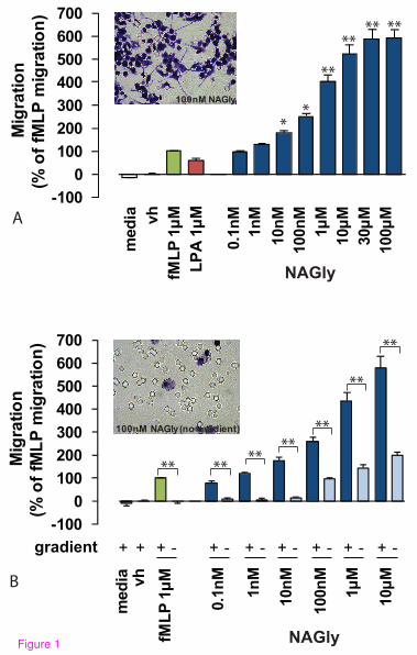
NAGly potently induces directed microglial migration
Directed microglial migration and phenotypic modifications are known to be
stimulated by factors including bacterial peptides, lysophospholipids and
endocannabinoids [13, 29-30]. Therefore, we compared NAGly-induced BV-2
microglial migration with N-formyl-methionine-leucine-phenylalanine (fMLP)
and archidonoyl lysophophatidic acid (LPA), chemotactic ligands released under
conditions of brain injury or infection [31-32]. NAGly potently induced
concentration-dependent migration, and elicited a response twice that produced by
1 µM fMLP or LPA at NAGly concentrations of 0.17 nM and 0.08 nM,
respectively (Figure 1A).
Chemotaxis (directed migration) is the process whereby cells sense soluble
molecules and purposely advance along a concentration gradient to their source.
This is in contrast to chemokinesis (stimulated random motion), where cells
experience spontaneous cytoskeletal polymerization which prompts
indiscriminate meandering. Checkerboard analysis offers a means to differentiate
migratory behaviour between chemotaxis and chemokinesis, and is based on
disrupting the concentration gradient of a pro-migratory ligand. Indiscriminate
cell migration across a filter membrane will remain unaffected by the
9
absence of a concentration gradient. Whereas directed cell migration is prevented
by the absence of the guidance cue derived from the concentration gradient.
Checkerboard analysis of NAGly revealed BV-2 microglia exhibit chemotaxis,
and purposely advance towards the source of NAGly in a directed manner
(Figures 1A & 1B). A low basal level of chemokinesis was observed, which is
the case with all established chemoattractants (Figure 1B).
As NAGly undergoes hydrolysis via FAAH to form AA and glycine [25], both of
which are signaling molecules in their own right, we investigated whether the
NAGly-induced response was due to its metabolism to either of these products.
1 µM NAGly produced a migratory response (% of fMLP migration) of 435.9% ±
36.9% compared to 22.4% ± 2.9% for AA, and -15.4% ± 5.4% for glycine; these
values are significantly different (P<0.001 compared to 1 µM NAGly; one-way
ANOVA; n =3). This indicates that neither AA nor glycine can account for the
migratory response produced in BV-2 microglial cells by NAGly.
In 2003, Walter et al described endocannabinoid system involvement in recruiting
microglia toward dying neurons: pathological stimulation of neurons and
microglia led to a dramatic and selective increase in 2-AG production which
triggered microglial migration by engaging CB2 and ‘Abn-CBD’ receptors [13].
Therefore, we next compared NAGly-induced migration to various
endocannabinoids, endogenous lipids and compounds relevant to ‘Abn-CBD’
10
receptor pharmacology to compare the potency of each of these compounds to
induce migration (Figures 2A & 2B). A response double that of 1 µM fMLP was
elicited by the following concentrations of ligand: 0.17 nM NAGly, 0.27 nM
O-1602, 5.2 nM 2-AG, 13.1 nM Abn-CBD and 123 nM AEA (Figure 2A). PEA
(the endogenous AEA analogue), palmitoyl glycine (PALGly; the endogenous
NAGly analogue), and L-α-lysophosphatidylinositol (LPI) all exerted a minimal,
concentration-independent stimulation of migration. The mean migration
achieved for these compounds across a concentration range of 0.1 nM – 10 µM
being 12.5% ± 3.5% (PEA; n = 3), 16.9% ± 3.9% (PALGly; n = 3), and 23.5% ±
4.7% (LPI; n = 3). Thus, NAGly potently induced concentration-dependent
migration of BV 2 microglia, and was more efficacious than previously described
cannabinoid ligands (Fig 2A). A ~50-fold greater concentration of 2-AG than
NAGly was required to reach the half-maximal response of 2-AG; and in terms of
AEA, a ~1000-fold greater concentration of AEA than NAGly was required to
reach the half-maximal response of AEA.
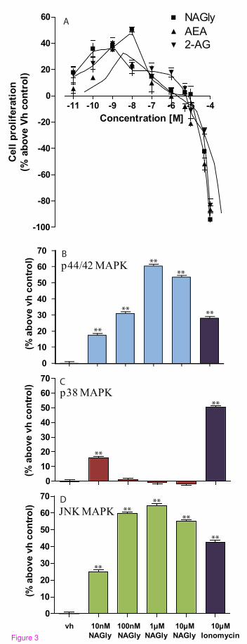
Microglia in the adult CNS derive chiefly from a self-renewing population or
rarely are replenished from adult bone marrow [31]. As they invade an injured
region of the CNS, microglia can enter the cell cycle and proliferate via mitosis
[5], e.g. elevated numbers of microglia are found in brains of patients with
multiple sclerosis [33], Alzheimer’s disease [34] and HIV [35]. The reduction of
tetrazolium salts is widely accepted as a reliable way to examine
11
cell proliferation. In the MTT reduction technique, the yellow tetrazolium 3-(4,5-
dimethyl-thiazoyl-2-yl)-2,5-diphenyltetrazolium bromide (MTT) is reduced by
metabolically active cells, in part by the action of dehydrogenase enzymes, to
generate reducing equivalents such as NADH and NADPH. The resulting
intracellular purple formazan dye can be solubilised and quantified by
spectrophotometric means. Using this means to quantify cell proliferation in
response to NAGly, AEA and 2-AG, we found that NAGly increased the
population of BV-2 microglia at picomolar to low nanomolar concentrations after
24 hours (Figure 3A). The rank order of potency was NAGly > 2-AG > AEA at
stimulating BV-2 cell proliferation: a ~ 50% increase was achieved by 10 nM
NAGly, which was significantly greater than the ~ 24% and ~ 21% seen with
10 nM AEA and 2-AG, respectively (P < 0.01; one-way ANOVA; n = 3) (Figure
3A). Decreased cell viability was observed for all three compounds at
concentrations greater than 1 µM. Carrier et al had previously shown that 2-AG,
but not AEA, exerted a M-CSF (macrophage-colony stimulating factor)
dependent proliferative effect on rat RTMGL1 microglia via CB2 receptors [36].
They observed ~ 30% increase with 300 nM 2-AG 24 hours after treatment, and
this was accompanied by an increase in active p44/42 MAPK (a.k.a. ERK1/2).
MAPKs respond to extracellular stimuli/mitogens and regulate activities such as
cell proliferation, differentiation, motility, and death. As migration is an
activated-MAPK-dependent phenomenon and ‘Abn-CBD’ receptors have been
shown to induce p44/42 MAPK phosphorylation [13, 19], we
12
investigated the effect of NAGly on p44/42, p38 and JNK MAPK enzymes using
In-Cell Western assays (Figures 3B, 3C & 3D). NAGly induced a marked
concentration-dependent phosphorylation of p44/42 and JNK MAPK (Figure 3B
& 3D), reflecting activation of these kinases, whereas, p38 MAPK was only
significantly activated by 10 nM NAGly (Figure 3C). Our findings extend those
of Carrier et al, showing that NAGly, 2-AG and AEA independently induce BV-2
microglial mitosis, with NAGly being the most potent of the three. Given the
association between cell migration and proliferation, and that both are
MAPK-dependent, ‘Abn-CBD’ receptor-activated phosphorylation of p44/42 and
JNK MAPK in response to NAGly likely underlies the migratory and proliferative
phenomena in BV-2 microglia.
In summary, the rank order of chemotactic potency published by Walter (2003)
for BV-2 microglia was largely reproduced here. They found 2-AG > AEA >
Abn-CBD at inducing migration, while PEA caused a weak concentration-
independent response [13]. Here we report, NAGly > O-1602 > 2-AG >
Abn-CBD > AEA, and the effects of PEA, LPI and PALGly were weak and
concentration-independent (Figure 2A). NAGly is the most potent pro-migratory
lipid for BV-2 microglia cells described to date, triggering directed migration and
proliferation via MAPK activation (Figures 1, 2 & 3).
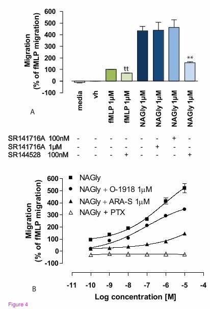
NAGly acts via a Gi/o-coupled GPCR
13
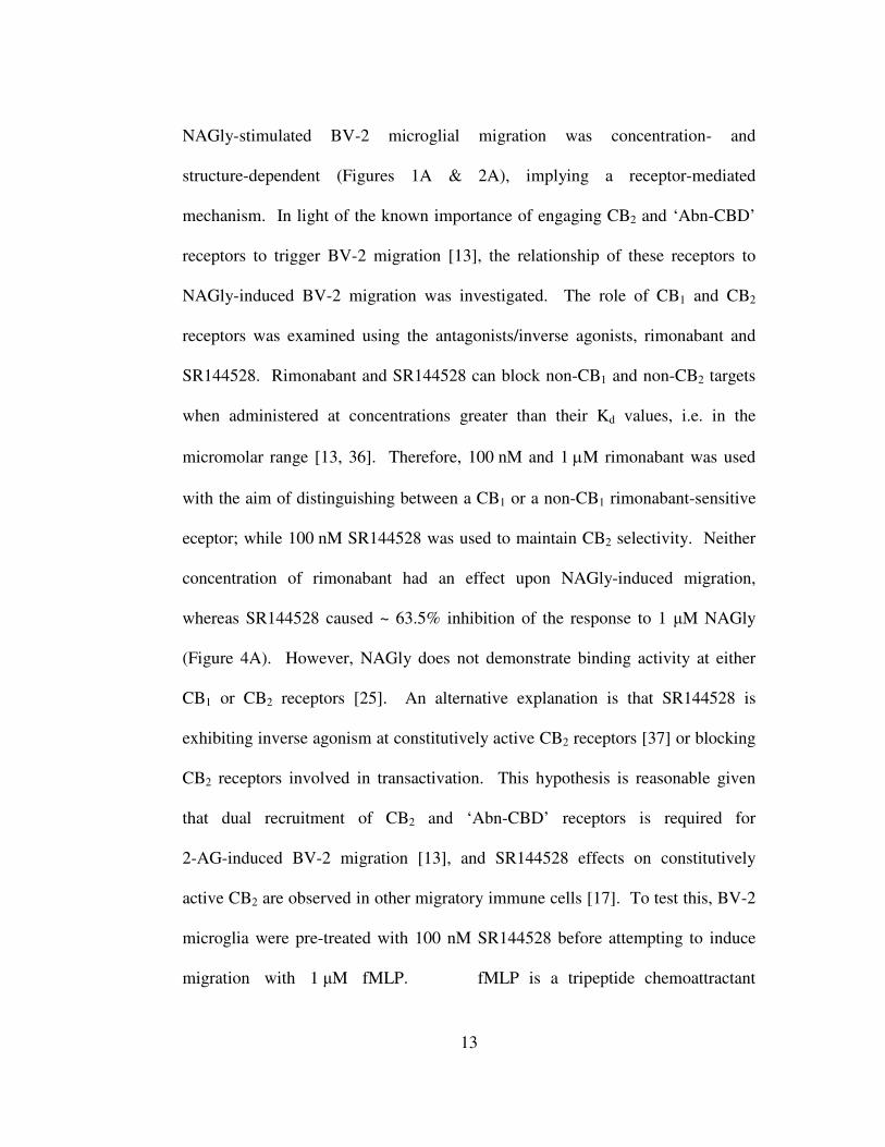
NAGly-stimulated BV-2 microglial migration was concentration- and
structure-dependent (Figures 1A & 2A), implying a receptor-mediated
mechanism. In light of the known importance of engaging CB2 and ‘Abn-CBD’
receptors to trigger BV-2 migration [13], the relationship of these receptors to
NAGly-induced BV-2 migration was investigated. The role of CB1 and CB2
receptors was examined using the antagonists/inverse agonists, rimonabant and
SR144528. Rimonabant and SR144528 can block non-CB1 and non-CB2 targets
when administered at concentrations greater than their Kd values, i.e. in the
micromolar range [13, 36]. Therefore, 100 nM and 1 µM rimonabant was used
with the aim of distinguishing between a CB1 or a non-CB1 rimonabant-sensitive
eceptor; while 100 nM SR144528 was used to maintain CB2 selectivity. Neither
concentration of rimonabant had an effect upon NAGly-induced migration,
whereas SR144528 caused ~ 63.5% inhibition of the response to 1 µM NAGly
(Figure 4A). However, NAGly does not demonstrate binding activity at either
CB1 or CB2 receptors [25]. An alternative explanation is that SR144528 is
exhibiting inverse agonism at constitutively active CB2 receptors [37] or blocking
CB2 receptors involved in transactivation. This hypothesis is reasonable given
that dual recruitment of CB2 and ‘Abn-CBD’ receptors is required for
2-AG-induced BV-2 migration [13], and SR144528 effects on constitutively
active CB2 are observed in other migratory immune cells [17]. To test this, BV-2
microglia were pre-treated with 100 nM SR144528 before attempting to induce
migration with 1 µM fMLP. fMLP is a tripeptide chemoattractant
14
released from both bacteria and damaged mitochondria [38-39], and activates two
formyl peptide receptors, designated FPR and FPRL-1 [40]. SR144528 caused ~
32.2% inhibition of the response to 1 µM fMLP (Figure 4A). The estimated
percentage viability ± 100 nM SR144528 was 97.3 ± 0.62 and 97.1 ± 0.67 %,
respectively; these values were not significantly different (P > 0.05; Student’s
unpaired t-test; n = 3), excluding cell death as a factor. Additionally, in
subsequent experiments with HEK293 cells, which do not express CB2 receptors,
we found that 100 nM SR144528 had no effect on HEK293 cells stably
transfected with GPR18 induced in response to 1 µM NAGly; the migration being
497 ± 8 cells and 501 ± 11 cells in the presence and absence of SR144528
respectively (P > 0.05; Student’s unpaired t-test; n = 3). These data instead infer a
role for tonic CB2 signaling or transactivation in the migratory mechanism.
Interactions among GPCRs are complex [41] and they have a propensity to
experience cross-talk when co-expressed, e.g. receptor dimerization or
heterologous desensitization. Thus, CB2 may cross-modulate with fMLP
receptors and the Gi/o receptor targeted by NAGly to regulate migration in BV-2
microglia. In summary, there is no evidence of a role for CB1, which is consistent
with the low levels of CB1 gene product previously observed in BV-2 microglia
[42]. While CB2 is demonstrably involved in BV-2 migration, it remains
questionable that NAGly is signaling directly via CB2 receptors.
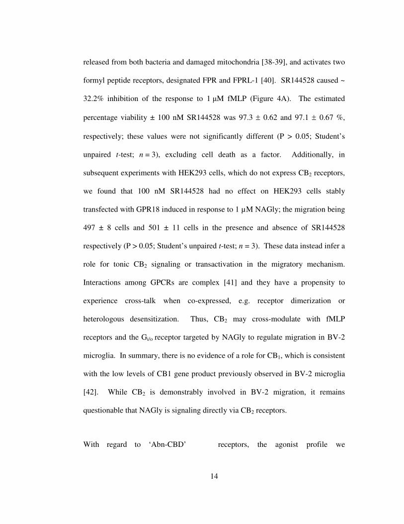
With regard to ‘Abn-CBD’ receptors, the agonist profile we
15
observed with BV-2 migration was consistent with that known for this novel
receptor, i.e. Abn-CBD, AEA, 2-AG and O-1602 stimulate migration (Figure 2A)
[ 13, 15-16, 18, 22, 43]. However, 1 µM rimonabant failed to attenuated the
NAGly response despite a reported IC50 value of 600 nM toward ‘Abn-CBD’
receptors (Figure 4A) [18]. Whether or not total block with 1 µM rimonabant
should be expected in this circumstance would depend on the affinity of NAGly
for the receptor, the concentration of NAGly employed and the number of ‘Abn-
CBD’ receptors that need to be activated to see signaling; information that is not
yet available. As a consequence, we further probed the role of ‘Abn-CBD’
receptors by investigating the antagonistic effects of ARA-S and O-1918 on
NAGly- and fMLP-induced migration. In the presence of 1 µM ARA-S or 1 µM
O-1918, the migration induced by NAGly was significantly attenuated (Figure
4B), whereas the migration in response to fMLP remained unaffected; 100.0% ±
3.5% (1 µM fMLP alone), 101.2% ± 2.9% (1 µM fMLP + 1 µM ARA-S), 100.8%
± 3.14% (1 µM fMLP + 1 µM O-1918). These values were not significantly
different, P > 0.05; one-way ANOVA; n = 3. Likewise, neither ARA-S nor
O-1918 had any effect on basal BV-2 cell migration; 0.0% ± 2.1% (Vh alone),
0.3% ± 1.9% (Vh + 1 µM ARA-S), 0.2% ± 2.3% (Vh + 1 µM O-1918). Similarly,
these values were not significantly different, P > 0.05; one-way ANOVA; n = 3.
Since NAGly activates Gi/o-coupled GPR18 and Gq/11-coupled GPR92 [26-27], we
investigated the effect of pertussis toxin (PTX) on the NAGly
16
migratory response. PTX pre-treatment abolished the migration to NAGly
(Figure 4B), without affecting cell viability. Using the trypan blue exclusion
method, the estimated percentage viability of cells pre-treated for 24 hours was
not different (± 1 µg/ml PTX was 97.8 ± 0.47 and 97.4 ± 0.51 %, respectively;
these values were not significantly different, P > 0.05; Student’s unpaired t-test;
n = 3). Taken together these data indicate Gi/o GPCR involvement, and support
the hypothesis that NAGly is acting via the ‘Abn-CBD’ GPCR to induce BV-2
microglial migration.
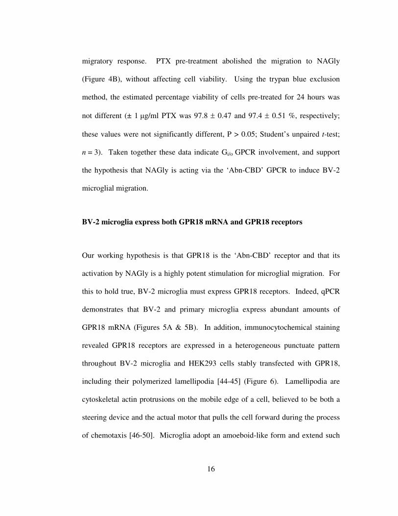
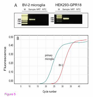
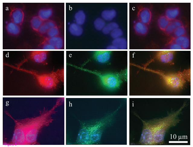
BV-2 microglia express both GPR18 mRNA and GPR18 receptors
Our working hypothesis is that GPR18 is the ‘Abn-CBD’ receptor and that its
activation by NAGly is a highly potent stimulation for microglial migration. For
this to hold true, BV-2 microglia must express GPR18 receptors. Indeed, qPCR
demonstrates that BV-2 and primary microglia express abundant amounts of
GPR18 mRNA (Figures 5A & 5B). In addition, immunocytochemical staining
revealed GPR18 receptors are expressed in a heterogeneous punctuate pattern
throughout BV-2 microglia and HEK293 cells stably transfected with GPR18,
including their polymerized lamellipodia [44-45] (Figure 6). Lamellipodia are
cytoskeletal actin protrusions on the mobile edge of a cell, believed to be both a
steering device and the actual motor that pulls the cell forward during the process
of chemotaxis [46-50]. Microglia adopt an amoeboid-like form and extend such
17
motile lamellipodia, in order to achieve directed migration, enabling them to
move toward relevant CNS locations and affect appropriate responses [51-54].
These data support our hypothesis that GPR18 mediates NAGly-induced directed
migration of microglia.
Overexpression of GPR18 affects directed migration induced by NAGly and
Abn-CBD
To further examine the hypothesis that NAGly is acting through GPR18 to
mediate its migratory effects in BV-2 microglia, and in light of there being no
known GPR18 antagonists, we modelled BV-2 microglial migratory observations
using wildtype or HEK293 cells stably transfected with HA11-tagged GPR18
(HEK293-GPR18). NAGly elicited a concentration-dependent migratory
response in HEK293-GPR18 but not wildtype cells (Figures 7A & 7B), with an
Emax similar to BV-2 microglia. 24 h pre-treatment with PTX abolished the
response to 1 µM NAGly, the mean number of cells migrated with and without
PTX pre-treatment was 505 ± 11 and 2 ± 1 respectively; these values were
significantly different (P > 0.05; Student’s unpaired t-test; n = 3). 1 µM NAGly-
induced migration was also significantly attenuated in the presence of 1 µM
O-1918 or 1 µM ARA-S (Figure 7C). Abn-CBD and O-1602 also induced
migration in HEK293-GPR18 cells, with O-1602 being more potent than
Abn-CBD (Figure 7C); both responses were significantly inhibited in the presence
18
of 1 µM ARA-S or 1 µM O-1918 (Figure 7C), which again is in agreement with
the BV-2 microglial data. CBD is known to behave as a partial agonist/antagonist
of ‘Abn-CBD’ receptors depending on receptor expression levels [13, 17]. 1 µM
NAGly-induced migration of both BV-2 microglia and HEK293-GPR18 receptors
was also significantly attenuated in the presence of 1 µM CBD (Figure 7D). The
NAGly-induced p44/42 MAPK activation observed with BV-2 microglia too was
reproduced in HEK293-GPR18 cells (Figure 7E).
Several publications have suggested the orphan receptor GPR55 interacts with
certain cannabinoid ligands, including Abn-CBD and O-1602 [55]. While this
proposition remains a contentious one, BV-2 microglia do express GPR55 mRNA
[42]. Therefore we explored whether NAGly, Abn-CBD, O-1602 or LPI
stimulate migration in HEK293 cells stably transfected with HA11-tagged GPR55
(HEK293-GPR55). All four of these compounds produced a weak,
concentration-independent migratory response in HEK293-GPR55 cells (Figure
7F) that was irreconcilable with the NAGly, Abn-CBD and O-1602 effects on
BV-2 migration.
Discussion
‘Abn-CBD’ receptors have primarily been characterized in vascular tissue and
microglia. Studies investigating the vasodilatory effects of AEA in CB1/CB2
19
knockout mice led to the postulation of the ‘Abn-CBD’ receptor as a novel
endothelial cannabinoid target for which AEA, Abn-CBD and O-1602 were
agonists that induced relaxation of the whole mesenteric arterial system [18].
Subsequent investigations have elaborated that multiple signaling pathways
underlie the hemodynamic effects elicited by AEA, and involve CB1, TRPV1,
‘Abn-CBD’ receptors and perhaps another distinct endothelium-independent
Abn-CBD/O-1602-sensitive target [22, 56-57]. The specifics vary according to
particular location in the vascular network and the preparation under scrutiny, e.g.
aorta vs mesenteric artery segments, endothelium-intact vs endothelium-denuded
vessels [18-19, 22, 58]. In 2003, well-executed studies with primary and BV-2
microglia reproduced the pharmacology of the endothelial ‘Abn-CBD’ receptor,
revealing its expression and significant migratory role in microglia [13].
As a whole the cannabinoid field has eagerly awaited developments that will
clarify the molecular identity of the ‘Abn-CBD’ receptor. Our analyses
demonstrate that NAGly and Abn-CBD regulate cellular migration through
GPR18, and we propose this GPCR is the unidentified ‘Abn-CBD’ receptor.
Multiple lines of evidence substantiate this hypothesis: NAGly, at sub-nanomolar
concentrations, together with the ‘Abn-CBD’ receptor agonists Abn-CBD and
O-1602 [15-16, 18], potently drives cellular migration in both BV-2 microglial
and HEK293-GPR18 transfected cells, but not in HEK293-GPR55 or
non-transfected HEK293 cells. O-1602 was ~ 50 times more effective
20
than Abn-CBD at inducing migration in BV-2 microglia. This is in keeping with
the work of Jarái et al which first characterized ‘Abn-CBD’ receptors in the rat
mesenteric bed, where O-1602 was ~ 80 times more potent than Abn-CBD at
causing vasodilation [18]. ‘Abn-CBD’ receptors couple via Gi/o proteins [13];
here, PTX pre-treatment to uncouple such Gi/o proteins prevented the migratory
response to NAGly in BV-2 and HEK293-GPR18 cells. The NAGly-,
Abn-CBD-, and O-1602-induced migration was blocked or attenuated in BV-2 or
HEK293-GPR18 cells by the ‘Abn-CBD’ receptor antagonist O-1918, and low
efficacy agonists ARA-S and CBD. NAGly promotes proliferation and activation
of MAPK enzymes at low nanomolar concentrations in BV-2 cells and
HEK293-GPR18 cells, demonstrating cellular responses correlated with
microglial migration and previous ‘Abn-CBD’ receptor activity on p44/42 MAPK
[13, 19]. Finally, BV-2 microglia show heterogeneous GPR18
immunocytochemical staining, including the polymerized actin-containing
lamellipodia that permit motile cells to achieve directed migration, and abundant
GPR18 mRNA. qPCR demonstrates that primary microglia, likewise, express
abundant amounts of GPR18 mRNA.
Both the academic community and pharmaceutical industry are engaged in
intensive research of the endogenous cannabinoid signaling system, focussing on
its potential therapeutic exploitation regarding mental illness, neuropathic and
inflammatory pain, obesity, osteoporosis, nicotine addiction,
21
cardiovascular disorders, and liver disease. Therefore, our recognition of GPR18
as the unidentified ‘Abn-CBD’ receptor has far-reaching implications. Firstly,
hitherto unrecognized GPR18-mediated effects by cannabinoid ligands,
particularly those that were previously classified as CB1- or CB2-receptor-
selective, may have resulted in the misinterpretation of the role of those receptors
in various systems. Secondly, our present definition and understanding of the
endogenous cannabinoid signaling system will have to be expanded given the
recognition of GPR18 as the ‘Abn-CBD’ receptor and that its endogenous ligand,
NAGly, is a metabolic product of AEA [25]. Thirdly, elucidation of GPR18’s
other physiological roles will further reveal the molecular mechanisms
responsible for the detrimental and medicinal effects of cannabis constituents.
Lastly, GPR18-selective ligands will make available novel therapeutic routes
targeting a broad spectrum of pathophysiologies.
With specific regard to the CNS, microglia represent a major cellular component
of the brain, constituting a widely distributed network of immunoprotective cells
[59-60]. During the last decades, it has become clear that the roles traditionally
ascribed to microglia, i.e. to dispose of dead cells and debris and to mediate brain
inflammatory states, are only a fraction of a much wider repertoire of functions
spanning from brain development to aging and neuropathology [61-62]. Such
functions are necessarily reliant upon the complex signaling systems subserving
the reciprocal communication that occurs between neurons and

22
microglia [63]. Indeed, the loss of specific communication between damaged
neurons and microglia is viewed as responsible for the turning of microglia to a
hyperactivated state, which allows them to escape neuronal control and to give
rise to persistent inflammation, resulting in exacerbation of neuropathology [60].
Conclusions
The marked potency of NAGly acting on GPR18 to elicit directed migration,
proliferation and perhaps other MAPK-dependent phenomena advances our
understanding of the lipid-based signaling mechanisms employed by the CNS to
actively recruit microglia to sites of interest. It offers a novel research avenue for
developing therapeutics to elicit a self-renewing population of neuroregenerative
microglia, or alternatively, to prevent the accumulation of misdirected, pro-
inflammatory microglia which contribute to and exacerbate neurodegenerative
disease.
Methods
Cells in culture
The mouse microglial cell line BV-2 (a gift from Dr. N. Stella; University of
Washington, Seattle), which was originally generated by immortalizing primary
23
microglia (Blasi et al., 1990), were grown in high glucose DMEM (Gibco, USA)
supplemented with FBS (10%), penicillin (100 units/ml), streptomycin
(100 µg/ml), and passaged every 4-5 days for a maximum of 30 passages.
HEK293 wildtype (ATCC, USA), HEK293 cells stably transfected with HA11-
tagged GPR55 (HEK293-GPR55; previously generated [64]) and HEK293 cells
stably transfected with HA11-tagged GPR18 (HEK293-GPR18; generated for this
study), were grown in Eagle’s MEM (Gibco, USA) supplemented with FBS
(10%), penicillin (100 units/ml), streptomycin (100 µg/ml) and L-glutamine
(0.292 mg/ml), and passaged every 4-5 days for a maximum of 30 passages.
HEK293 cells were transfected with 2 µg of HA11-tagged hGPR18 plasmid using
Lipofectamine and Plus reagents (Invitrogen, USA) in a 6-well plate using
standard molecular biological techniques [65]. G418-resistant colonies were used
as a positive control to validate the specificity of the hGPR18-CT purified
antibody.
In order to obtain primary microglia for the RNA extraction studies, mixed glial
cells were isolated from dissociated cerebral cortex of newborn (P0-P1)
C57BL/6J mice as previously described [66]. The cell suspension was prepared in
culture medium for glial cells [DMEM supplemented with 10% FCS, L-glutamine
(1 mM), sodium pyruvate (1 mM), penicillin (100 U/ml), and streptomycin
(100 mg/ml)] and cultured at 37°C/5% CO2 in 75-cm2 Falcon tissue-culture
flasks, coated with polyD-lysine (PDL). Half of the medium was

24
changed after the first day and every second day thereafter, for a total culture time
of 10-14 days. Microglia were shaken off the primary mixed brain glial cell
cultures (150 rpm for 4-6h at 37°C), with maximal yields between days 10 and
14. Cells were seeded onto PDL-pretreated 60mm plates and grown in culture
medium for microglia [RPMI medium supplemented with 10% FCS, L-glutamine
(1 mM), sodium piruvate (1 mM), penicillin (100 U/ml) and streptomycin (100
mg/ml). The cells were allowed to adhere to the PDL-coated plate (30 min,
37°C/5% CO2) and the nonadherent cells were rinsed off. After 48h microglial
cells are ready to be used for experiments.
Test compounds
Appropriate stock concentrations of the compounds tested in this study were
prepared in 100% DMSO, before being serially diluted to achieve the desired
final working concentrations, each containing 0.1% DMSO as vehicle.
Migration assay
In vitro cell migration assays were performed using a modified 96-well Boyden
Chamber and PVP-free polycarbonate filters with 10 µm diameter pores
(Neuroprobe Inc., USA), which can be discerned as the clear unstained circles in
the photographed filters of figures 1 & 6. The upper wells of the Boyden chamber
25
were filled with 50 µl of suspension of 1x10⁶ cells ml
-1 in serum-free DMEM,
before incubation with a 5% CO2 atmosphere at 37°C for 3 hours. 1 µM fMLP
acted as positive control. Following incubation, non-migrated cells were then
removed before fixation and staining with Diff-Quik stain set. Finally, the filter
was sectioned and mounted onto microscope slides and the migrated cells counted
in ten non-overlapping fields (x40 magnification) with a light microscope by
multiple scorers blinded to experimental conditions. For inhibition of induced
migration, cells were pre-incubated with antagonist for 30 min at 37ºC in a water
bath before loading into the upper wells, the lower wells contained the equivalent
concentration of antagonist and test compound to ensure that the only
concentration gradient present is that generated by the test compound as they
diffuse through the pores in the filter.
Cell proliferation assay
BV-2 cells were plated in 96-well plates at a seeding density of 1x10⁴ cells per
well overnight in media containing 1% FBS. The media was then changed to
fresh media containing 1% FBS and the appropriate concentrations of test
compound, then incubated for 24 hours. Cell density was assessed with the
3-(4,5-dimethyl-thiazoyl-2-yl)-2,5-diphenyltetrazolium bromide (MTT) formazan
dye conversion assay (ATCC, USA) according to the manufacturer’s instructions
and measured at 570 nm with a SpectraMax M5 spectrophotometer
26
(Molecular Devices, USA).
In-Cell Western assay
An In-Cell Western assay was employed to simultaneously detect both the
phosphorylated MAPK protein and normalize for total MAPK protein. The
following primary antibodies were used to detect endogenous levels of the
relevant total MAPK and phosphorylated MAPK: p44/42 MAPK rabbit pAb, and
phospho-p44/42 MAPK mouse mAb (#4695 and #9106; Cell Signaling
Technology, USA); p38 MAPK rabbit pAb, and phosphor-p38 MAPK mouse
mAb (#9212 and #9216; Cell Signaling Technology, USA); and SAPK/JNK
MAPK rabbit pAb, and phospho-SAPK/JNK MAPK mAb (#9252 and #9255;
Cell Signaling Technology, USA).
BV-2 cells were plated into 96-well plates coated with 1 µg ml-1
poly-L-lysine
and treated with vehicle (Vh) (0.1% DMSO) or NAGly (10 nM – 10 µM) for
3 hours. Ionomycin (10 µM) treatment in the final 5 min was used as a positive
control. Upon completion of the drug treatments, an In-cell Western assay was
conducted: the 96-well plates were immediately placed on ice, the media removed
and cells fixed with 100 µl/well of 3.7% formaldehyde in PBS for 15 min. The
96-well plates were then removed from the ice and allowed to warm up to room
temperature over 30 min. The formaldehyde solution was replaced by 100 µl/well

27
of ice-cold methanol and the plate kept at -20°C for 20 min. The cells were
washed with 200 µl of 0.1% Triton X-100 in PBS with gentle shaking for 5 min at
room temperature, the wash solution was removed before adding fresh 0.1%
Triton X-100 and repeating for a total of 5 times. Following the final wash, cells
were blocked with 150 µl of Odyssey blocking buffer (Li-Cor, USA) with
moderate shaking for 90 min at 20°C. Primary antibody pairs (e.g. p44/42 and
phospho-p44/42 MAPK) were diluted in Odyssey blocking buffer 1:200 and the
plate was then incubated overnight with moderate shaking at 4°C. Primary
antibody solution or Odyssey blocking buffer was then removed from all wells
before they were washed with 0.1% Tween-20 in PBS with moderate shaking for
5 min at room temperature, this was repeated for a total of 5 times. Fluorescently
labelled secondary antibodies (Odyssey 926-32211 goat anti-rabbit 800 nm
antibody; 926-32220 goat anti-mouse 680 nm antibody; Li-Cor, USA) were
diluted in Odyssey blocking buffer 1:800 containing 0.2% Tween-20. The
secondary antibody solution was added to all wells and incubated in the dark for
90 min at room temperature. Secondary antibody solution was removed and the
wells then washed with 0.1% Tween-20 in PBS with moderate shaking for 5 min
at room temperature for a total of 5 times, while protecting from light. The final
wash solution was removed and discarded. The plate was then scanned using the
Li-Cor Odyssey Infrared Imaging System (Li-Cor, USA), using both 700 and
800 nm channels, a resolution of 42 µm, quality set to high, an intensity of 5, and
focal offset of 4 mm.
28
Using the Odyssey application software, changes in MAPK activation were
determined by calculating the mean background fluorescence from all non-
primary antibody containing control wells, for both 700 and 800 nm channels.
Background fluorescence was subtracted from the fluorescence measured in
primary antibody containing wells, for both the 700 and 800 nm channels. The
relative intensity of phospho-MAPK fluorescence was normalized against the
relative intensity of fluorescence measured for total-MAPK. Finally the %
response of all test compounds relative to vehicle was determined.
Isolation of total RNA and real-time quantitative PCR (qPCR)
RNA was extracted from BV-2, HEK293-GPR18 transfected cells, and primary
microglial cells using the RNAqueous® small scale phenol-free total RNA
isolation kit (Applied Biosystems, USA) and RNA samples (2 µg) were reverse
transcribed using the SuperScript II™ Reverse Transcription Kit (Invitrogen,
USA).
Expression of GPR18 mRNA in BV-2 and primary microglia was determined by
RT-qPCR, using B2-MG as a normalizing gene, as previously described [67].
Normal, mock reversed transcribed samples (NRT), and no template controls
(NTC; total mix without cDNA) were run for each of the examined mRNAs.
RT-qPCR reactions were subjected to an initial HotStar Taq (Qiagen, USA) DNA
29
polymerase activation step (15 min at 95ºC), followed by 40 cycles each
consisting of 15 s at 94ºC, 30 s at 60ºC and 30s at 72ºC. Fluorescence was
measured at the end of each elongation step. Data were analyzed using the Rotor-
gene software (Corbett Research, Australia) and a threshold cycle value Ct was
calculated from the exponential phase of each RT-qPCR sample. Amounts of
mRNA were calculated and expressed in relative units of SYBR Green
fluorescence. PCR products were analyzed on a 2 % agarose gel with ethidium
bromide.
Expression of GPR18 in BV-2 microglia and HEK293-GPR18 cells was also
determined by PCR using oligonucleotide primers based on the sequence of the
Mus musculus G protein-coupled receptor 18 (GPR18) mRNA (GenBank
Accession No. NM_182806.1) and B2-MG mRNA (GenBank Accession No.
NM_009735). The primer sequences used were forward,
TGAAGCCCAAGGTCAAGGAGAAGT and reverse, TTCATGAGGAA
GGTGGTGAAGGCT (amplicon 163 bp) for the GPR18 and forward,
ATGGGAAGCCGAACATACTG and reverse, CAGTCTCAGTGGGGGTGAAT
(amplicon of 176 bp) for B2-MG. PCR reactions were subjected to an initial
HotStart Taq DNA polymerase activation step of 95°C for 7 minutes, followed by
40 cycles of 94°C for 30 seconds, 55°C for 30 seconds, and 72°C for 30 seconds.
PCR products were analyzed on a 2% agarose gel with ethidium bromide. Single
bands corresponding to 163 bp for the GPR18 amplicon and 176 bp for
30
the B2-MG amplicon were recorded.
GPR18 antibody generation
A GST fusion protein expression construct was produced by inserting the DNA
coding for a C-terminal 29-aa peptide
(YRNYLRSMRRKSFRSGSLRSLSNINSEML) from human G-protein-coupled
receptor (hGPR18) into a pGEX-3X vector at the BamH I and EcoR I restriction
sites. The fusion protein was purified from BL21 E. coli lysates on a glutathione
Sepharose column and was injected into two rabbits to generate antisera (Cocalico
Biologicals, USA) using standard approaches [68]. The antiserum was purified in
two steps, first by exclusion on a GST column and then by binding to and elution
from an affinity column made with the injected GST fusion protein.
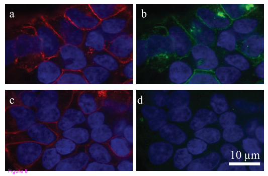
Immunocytochemistry
The GPR18 antibody generated for this study recognizes hGPR18 receptors stably
expressed in HEK293-GPR18 cells (Figure 8) and endogenous GPR18 in BV-2
microglia (Figure 6). Cells were fixed with paraformaldehyde, blocked, and
stained as follows: polyclonal rabbit anti-C-terminal GPR18 (1:150) (generated
for this study) and Texas Red-conjugated phalloidin (1:40; Molecular Probes,
Eugene, OR). Secondary IgG antibodies were FITC-conjugated donkey anti-
31
rabbit (1:150; Jackson ImmunoResearch, USA). Images were acquired with a
Nikon Eclipse TE2000-E confocal microscope (Nikon, USA).
Analysis of data
For BV-2 microglia, the mean number of cells migrated in response to test
compounds was normalized against the mean number of migrated cells elicited by
1 µM fMLP (0.1% DMSO). The number of migrated cells under vehicle only
conditions (0.1 % DMSO) was subtracted.
For HEK293 wildtype and HEK293-GPR18 transfected cells, simply the mean
number of cells migrated above vehicle only conditions was used. All data are
expressed as means ± s.e.mean and n = number of independent experiments.
Statistical analyses were performed with GraphPad Prism 4.
Concentration-response curves were generated using a sigmoidal dose-response
(variable slope) curve-fitting process, except for that representing BV-2 cell
proliferation where a simple point-to-point curve fit was employed instead.
List of abbreviations
Abn-CBD, abnormal cannabidiol; ANOVA, analysis of variance; AA,
arachidonic acid; AEA, N-arachidonoyl ethanolamine; 2-AG, 2-arachidonoyl
32
glycerol; ARA-S, N-arachidonoyl-L-serine; B2-MG, beta-microglobulin; CBD,
cannabidiol; CBN, cannabinol; CB1, cannabinoid receptor 1; CB2, cannabinoid
receptor 2; CNS, central nervous system; DMEM, Dulbecco’s Minimum Essential
Medium; DMSO, dimethyl sulphoxide; ERK1/2, extracellular signal-regulated
kinase 1/2; FAAH, fatty acid amide hydrolase; FBS, fetal bovine serum; fMLP,
N-formyl-methionine-leucine-phenylalanine; GPCR, G protein-coupled receptor;
LPA, arachidonoyl lysophosphatidic acid; LPI, L-α-lysophophatidylinositol;
MAPK, mitogen-activated protein kinase; M-CSF, macrophage-colony
stimulating factor; MTT 3-(4,5-dimethyl-thiazoyl-2-yl)-2,5-diphenyltetrazolium
bromide; NAGly, N-arachidonoyl glycine; NRT, normal, mock reverse
transcribed samples; NTC, total mix without cDNA; PTX, pertussis toxin;
O-1602, trans-4-[3-methyl-6-(1-methylethenyl)-2-cyclohexen-1-yl]-5-methyl-1,3-
benzenediol; O-1918, 1,3-dimethoxy-5-methyl-2-[(1R,6R)-3-methyl-6-(1-
methylethenyl)-2-cyclohexen-1-yl)-benzene; PALGly, palmitoylglycine; PEA
palmitoyl ethanolamine; Rimonabant (a.k.a. SR141716A), N-(piperidin-1-yl)-5-
(4-chlorophenyl)-1-(2,4-dichlorophenyl)-4-methyl-1H-pyrazole-3-carboximide
hydrochloride; RT-PCR, reverse transcriptase polymerase chain reaction;
SR144528, 5-(4-chloro-3-methylphenyl)-1-[(4-methylphenyl)methyl]-N—
[(1S,4R,6S)-1,5,5-trimethyl-6bicyclo[2.2.1]heptanyl]pyrazole-3-carboxamide.
Competing interests
33
The authors declare no conflict of interest.
Author’s contribution
DM performed the cell culture procedures, cell migration studies, cell
proliferation experiments, In-Cell Western assays and immunocytochemistry
imaging; design and coordination of the studies; data interpretation; statistical
analyses; and manuscript preparation. NR PCR studies. AJ PCR studies. SSH
generated the hGPR18 antibody; immunocytochemistry imaging. ZV PCR
studies. JWM initial study design. HBB design and coordination of the studies,
and manuscript preparation. All authors read and approved the final manuscript.
Acknowledgements
We dedicate this manuscript to J. Michael Walker (1950–2008) whose love of
science and generosity was an inspiration to us all. This work was supported by
NIH DA018224 and DA011322, and the Weizmann Institute. NIH and the
Weizmann Institute had no role in the study design; in the collection, analysis and
interpretation of data; in the writing of the manuscript; or in the decision to submit
the manuscript for publication. DM, HBB and JMW were supported by
DA018224; SS-JJ was supported by DA011322; and NR, AJ and ZV were
supported by the Weizmann Institute. The manuscript preparation

34
was funded by NIH DA018224.
35
References
1. Fetler L, Amigorena S: NEUROSCIENCE: Brain Under Surveillance: The
Microglia Patrol. Science 2005, 309:392-393.
2. Raivich G: Like cops on the beat: the active role of resting microglia. Trends
Neurosci 2005, 28:571-573.
3. Block ML, Zecca L, Hong J-S: Microglia-mediated neurotoxicity: uncovering
the molecular mechanisms. Nat Rev Neurosci 2007, 8:57-69.
4. Trapp BD, et al: Evidence for synaptic stripping by cortical microglia. Glia
55:360-368.
5. Garden GA, Moller T: Microglia biology in health and disease. J Neuroimmune
Pharmacol 2006, 1:127-137.
6. Gordon S: Alternative activation of macrophages. Nat Rev Immunol 2003,
3:23-35.
7. van Rossum D, Hanisch U-K: Microglia. Metab Brain Dis 2004, 19:393-411.
8. Carson MJ: Microglia as liaisons between the immune and central nervous
systems: functional implications for multiple sclerosis. Glia 2002, 40:218-
231.

36
9. Eikelenboom P, Bate C, Van Gool WA, Hoozemans JJ, Rozemuller JM, Veerhuis
R, Williams A: Neuroinflammation in Alzheimer's disease and prion
disease. Glia 2002, 40:232-239.
10. Streit WJ: Microglia as neuroprotective, immunocompetent cells of the CNS.
Glia 2002, 40:133-139.
11. Danton GH, Dietrich WD: Inflammatory mechanisms after ischemia and
stroke. J Neuropathol Exp Neurol 2003, 62:127-136.
12. Franklin A, Stella N: Arachidonylcyclopropylamide increases microglial cell
migration through cannabinoid CB2 and abnormal-cannabidiol-sensitive
receptors. Eur J Pharmacol 2003, 474:195-198.
13. Walter L, Franklin A, Witting A, Wade C, Xie Y, Kunos G, Mackie K, Stella N:
Nonpsychotropic Cannabinoid Receptors Regulate Microglial Cell
Migration. J Neurosci 2003, 23:1398-1405.
14. Brown AJ: Novel cannabinoid receptors. Br J Pharmacol 2007, 152:567-575.
15. Begg M, Pacher P, Bátkai S, Osei-Hyiaman D, Offertáler L, Mo FM, Liu J,
Kunos G: Evidence for novel cannabinoid receptors. Pharmacol Ther 2005,
106:133-145.
16. Mackie K, Stella N: Cannabinoid receptors and endocannabinoids: Evidence for
new players. AAPS J 2006, 8:E298-E306.
17. McHugh D, Tanner C, Mechoulam R, Pertwee RG,
37
Ross RA: Inhibition of human neutrophil chemotaxis by endogenous
cannabinoids and phytocannabinoids: Evidence for a site distinct from
CB1 and CB2. Mol Pharmacol 2008, 73:441-450.
18. Járai Z, Wagner JA, Varga Kr, Lake KD, Compton DR, Martin BR, Zimmer AM,
Bonner TI, Buckley NE, Mezey E et al: Cannabinoid-induced mesenteric
vasodilation through an endothelial site distinct from CB1 or CB2
receptors. Proc Natl Acad Sci USA 1999, 96:14136-14141.
19. Offertáler L, Mo F-M, Batkai S, Liu J, Begg M, Razdan RK, Martin BR, Bukoski
RD, Kunos G: Selective Ligands and Cellular Effectors of a G Protein-
Coupled Endothelial Cannabinoid Receptor. Mol Pharmacol 2003, 63:699-
705.
20. Mo FM, Offertáler L, Kunos G: Atypical cannabinoid stimulates endothelial
cell migration via a Gi/Go-coupled receptor distinct from CB1, CB2 or
EDG-1. Eur J Pharmacol 2004, 489:21-27.
21. Vaccani A, Massi P, Colombo A, Rubino T, Palolaro D: Cannabidiol inhibits
human glioma cell migration through a cannabinoid receptor-
independent mechanism. Eur J Pharmacol 489:21-27.
22. Wagner JA, Varga K, Jarai Z, Kunos G: Mesenteric Vasodilation Mediated by
Endothelial Anandamide Receptors. Hypertension 1999, 33:429-434.
23. Milman G, Maor Y, Abu-Lafi S, Horowitz M, Gallily R, Batkai S, Mo F-M,
Offertaler L, Pacher P, Kunos G et al: N-arachidonoyl l-serine, an
endocannabinoid-like brain constituent with vasodilatory properties.
38
Proc Natl Acad Sci USA 2006, 103:2428-2433.
24. Sheskin T, Hanus L, Slager J, Vogel Z, Mechoulam R: Structural Requirements
for Binding of Anandamide-Type Compounds to the Brain Cannabinoid
Receptor. J Med Chem 1997, 40:659-667.
25. Bradshaw H, Rimmerman N, Hu S, Benton V, Stuart J, Masuda K, Cravatt B,
O'Dell D, Walker JM: The endocannabinoid anandamide is a precursor
for the signaling lipid N-arachidonoyl glycine by two distinct pathways.
BMC Biochem 2009, 10:14.
26. Kohno M, Hasegawa H, Inoue A, Muraoka M, Miyazaki T, Oka K, Yasukawa M:
Identification of N-arachidonylglycine as the endogenous ligand for
orphan G-protein-coupled receptor GPR18. Biochem Biophyl Res Comm
2006, 347:827-832.
27. Oh DY, Yoon JM, Moon MJ, Hwang J-I, Choe H, Lee JY, Kim JI, Kim S, Rhim
H, O'Dell DK et al: Identification of Farnesyl Pyrophosphate and N-
Arachidonylglycine as Endogenous Ligands for GPR92. J Biol Chem 2008,
283:21054-21064.
28. Blasi E, Barluzzi R, Bocchini V, Mazzolla R, Bistoni F: Immortalization of
murine microglial cells by a v-raf/v-myc carrying retrovirus. J
Neuroimmunol 1990, 27:229-237.
29. Lorton D, Schaller J, Lala A, De Nardin E: Chemotactic-like receptors and Aβ
peptide induced responses in Alzheimer's Disease. Neurobiol Aging 2000,
21:463-473.
39
30. Schilling T, Stock C, Schwab A, Eder C: Functional importance of Ca2+
-
activated K+ channels for lysophosphatidic acid-induced microglial
migration. Eur J Neurosci 2004, 19:1469-1474.
31. Gao X, Hu X, Qian L, Yang S, Zhang W, Zhang D, Wu X, Fraser A, Wilson B,
Flood PM et al: Formyl-methionyl-leucyl-phenylalanine-induced
dopaminergic neurotoxicity via microglial activation: a mediator between
peripheral infection and neurodegeneration? Environ Health Perspect
2008, 116:593-598.
32. Puntambekar SS, Doose JM, Carson MJ: Microglia: A CNS-Specific Tissue
Macrophage. In: Central Nervous System Diseases and Inflammation.
Springer NY; 2008: 1-12.
33. Schönrock, Kuhlmann, Adler, Bitsch, Brück: Identification of glial cell
proliferation in early multiple sclerosis lesions. Neuropathol Appl
Neurobiol 1998, 24:320-330.
34. Mackenzie IRA, Hao C, Munoz DG: Role of Microglia in Senile Plaque
Formation. Neurobiol Aging 1995, 16:797-804.
35. Gendelman H, Lipton S, Tardieu M, Bukrinsky M, Nottet H: The
neuropathogenesis of HIV-1 infection. J Leukoc Biol 1994, 56:389-398.
36. Carrier EJ, Kearn CS, Barkmeier AJ, Breese NM, Yang W, Nithipatikom K,
Pfister SL, Campbell WB, Hillard CJ: Cultured Rat Microglial Cells
Synthesize the Endocannabinoid 2-Arachidonylglycerol, Which Increases
Proliferation via a CB2 Receptor-Dependent Mechanism. Mol Pharmacol

40
2004, 65:999-1007.
37. Rinaldi-Carmona M, Barth F, Millan J, Derocq JM, Casellas P, Congy C, Oustric
D, Sarran M, Bouaboula M, Calandra B et al: SR144528, the first potent and
selective antagonist of the CB2 cannabinoid receptor. J Pharmacol Exp
Ther 1998, 284:644-650.
38. Carp H: Mitochondrial N-formylmethionyl proteins as chemoattractants for
neutrophils. J Exp Med 1982, 155:264-275.
39. Chadwick VS, Mellor DM, Myers DB, Selden AC, Keshavarzian A, Broom MF,
Hobson CH: Production of Peptides Inducing Chemotaxis and Lysosomal
Enzyme Release in Human Neutrophils by Intestinal Bacteria in Vitro
and in Vivo. Scand J Gastroenterol 1988, 23:121 - 128.
40. Hartt JK, Barish G, Murphy PM, Gao J-L: N-formylpeptides Induce Two
Distinct Concentration Optima for Mouse Neutrophil Chemotaxis by
Differential Interaction with Two N-formylpeptide Receptor (FPR)
Subtypes: Molecular Characterization of FPR2, a Second Mouse
Neutrophil FPR. J Exp Med 1999, 190:741-748.
41. Snyderman R, Uhing RJ: Chemoattractant stimulus-response coupling. In:
Inflammation: Basic Principles and Clinical Correlates. Raven Press Lts.
NY; 1999, pp421-439.
42. Pietr M et al: Differential changes in GPR55 during microglial cell activation.
FEBS let 583: 2071-2076.
41
43. Showalter V, Compton D, Martin B, Abood M: Evaluation of binding in a
transfected cell line expressing a peripheral cannabinoid receptor (CB2):
identification of cannabinoid receptor subtype selective ligands. J
Pharmacol Exp Ther 1996, 278:989-999.
44. Mitchison TJ, Cramer LP: Actin-Based Cell Motility and Cell Locomotion. Cell
1996, 84:371-379.
45. Watanabe N, Mitchison TJ: Single-Molecule Speckle Analysis of Actin
Filament Turnover in Lamellipodia. Science 2002, 295:1083-1086.
46. Alberts, Bruce, et al: Molecular Biology of the Cell. (Fourth Edition). Garland
Science, Taylor & Francis Group. New York, pp 908, 931, 973-975.
47. Sanchez-Madrid F, del Pozo MA: Leukocyte polarization in cell migration and
immune interactions. EMBO J 1999, 18:501-511.
48. Bokoch GM: Regulation of innate immunity by Rho GTPases. Trends Cell
Biol 2005, 15:163-171.
49. Xu J, Wang F, Van Keymeulen A, Herzmark P, Straight A, Kelly K, Takuwa Y,
Sugimoto N, Mitchison T, Bourne HR: Divergent signals and cytoskeletal
assemblies regulate self-organizing polarity in neutrophils. Cell 2003,
114:201-214.
50. Dong X, Mo Z, Bokoch G, Guo C, Li Z, Wu D: P-Rex1 is a primary Rac2
guanine nucleotide exchange factor in mouse neutrophils. Curr Biol 2005,
15:1874-1879.
42
51. Kreutzberg GW: Microglia: a sensor for pathological events in the CNS.
Trends Neurosci 1996, 19:312-318.
52. Bruce-Keller AJ: Microglial-neuronal interactions in synaptic damage and
recovery. J Neurosci Res 1999, 58:191-201.
53. Becher B, Prat A, Antel JP: Brain-immune connection: immuno-regulatory
properties of CNS-resident cells. Glia 2000, 29:293-304.
54. Stence N, Waite M, Dailey ME: Dynamics of microglial activation: a confocal
time-lapse analysis in hippocampal slices. Glia 2001, 33:256-266.
55. Ross RA: The enigmatic pharmacology of GPR55. Trends Pharmacol Sci 2009,
30:156-163.
56. Zygmunt PM, Hogestatt ED: Role of potassium channels in endothelium-
dependent relaxation resistant to nitroarginine in the rat hepatic artery.
Br J Pharmacol 1996, 117:1600-1606.
57. Gebremedhin D, Lange AR, Campbell WB, Hillard CJ, Harder DR: Cannabinoid
CB1 receptor of cat cerebral arterial muscle functions to inhibit L-type
Ca2+ channel current. Am J Physiol Heart Circ Physiol 1999, 276:H2085-
2093.
58. Mukhopadhyay S, Chapnick BM, Howlett AC: Anandamide-induced
vasorelaxation in rabbit aortic rings has two components: G protein
dependent and independent. Am J Physiol Heart Circ Physiol 2002,
282:H2046-2054.

43
59. Vaughan D. W., Peters A: Neuroglial cells in the cerebral cortex of rats from
young adult to old age: an electron microscopy study. J. Neurocytol. 1974,
3: 405-429.
60. Banati R: Neuropathological imaging: in vivo detection of glial activation as a
measure of disease and adaptive change in the brain. Brit. Med. Bul. 2003,
65: 121-131.
61. Innocenti G. M., Clarke S., Koppell H: Transitory macrophages in the white
matter of the developing visual cortex. II. Development and relations with
axonal pathways. Dev. Brain Res. 1983, 11: 55-66.
62. Marin-Teva J. L., Dusart I., Colin C., Gervais A., van Rooijen N., Mallat M:
Microglia promote the death of developing purkinje cells. Neuron. 2004,
41: 535-547.
63. Polazzi E., Contestabile A: Reciprocal interactions between microglia and
neurons: from survival to neuropathology. Rev. Neurosci. 2004, 13: 221-
242
64. Lauckner JE, Jensen JB, Chen HY, Lu HC, Hille B, Mackie K: GPR55 is a
cannabinoid receptor that increases intracellular calcium and inhibits M
current. Proc Natl Acad Sci USA 2008, 105:2699-2704.
65. Daigle TL, Kearn CS, Mackie K: Rapid CB1 cannabinoid receptor
desensitization defines the time course of ERK1/2 MAP kinase signaling.
Neuropharmacology 2008, 54:36-44.
44
66. Juknat AA, Kotler ML, Quaglino A, Carrillo NM, Hevor T: Necrotic cell death
induced by delta-aminolevulinic acid in mouse astrocytes. Protective role
of melatonin and other antioxidants. J Pineal Res 2003, 35:1-11.
67. Butovsky O, Landa G, Kunis G, Ziv Y, Avidan H, Greenberg N, Schwartz A,
Smirnov I, Pollack A, Jung S et al: Induction and blockage of
oligodendrogenesis by differently activated microglia in an animal model
of multiple sclerosis. J Clin Invest 2006, 116:905-915.
68. Bodor AL, Katona I, Nyiri G, Mackie K, Ledent C, Hajos N, Freund TF:
Endocannabinoid signaling in rat somatosensory cortex: laminar
differences and involvement of specific interneuron types. J Neurosci
2005, 25:6845-6856.
45
Figure Legends
Figure 1: NAGly-induced directed BV-2 microglial migration. (A) BV-2
microglial migration in response to basal conditions; vh (0.1% DMSO); 1 µM
fMLP; 1 µM LPA; 0.1 nM – 300 µM NAGly. * = P<0.05, ** = P<0.01 compared
to 1 µM fMLP; one-way ANOVA; n = 8. Insert is a filter photograph of one
random field of view at x40 magnification indicating the migration produced by
100 nM NAGly. The 10 µm diameter pores can be discerned as the clear
unstained circles. (B) BV-2 microglial migration in response to
basal conditions; vh (0.1% DMSO); 1 µM fMLP ± concentration gradient; 0.1 nM
– 10 µM NAGly ± concentration gradient. ** = P<0.01 compared to the
corresponding concentration gradient; Student’s unpaired t-test; n = 3. Insert is a
filter photograph of one random field of view at x40 magnification indicating the
migration produced by 100 nM NAGly in the absence of a concentration gradient.
Figure 2: NAGly-induced BV-2 microglial migration is concentration- and
structure-dependent. (A) BV-2 microglial migration in response to 0.1 nM –
10 µM concentrations of NAGly; AEA; 2-AG; PEA; PALGly; Abn-CBD;
O-1602; LPI; n = 3. (B) Filter photographs of one random field of view at x40
magnification indicating the migration produced by 10 µM concentrations of
46
NAGly, O-1602, 2-AG, Abn-CBD, AEA and LPI.
Figure 3: NAGly-induced BV-2 cell proliferation and MAPK enzyme
activation. (A) BV-2 microglial proliferation in response to 0.01 nM – 100 µM
concentrations of NAGly; AEA; 2-AG; n = 3. (B) p44/42 MAPK activation in
BV-2 microglia in response to vh (0.1% DMSO) for 3 hours; 10 nM – 10 µM
NAGly for 3 hours; 10 µM Ionomycin for 5 min. ** = P<0.01 compared to vh;
one-way ANOVA; n = 3. (C) p38 MAPK activation in BV-2 microglia in
response to vh (0.1% DMSO) for 3 hours; 10 nM – 10 µM NAGly for 3 hours;
10 µM Ionomycin for 5 min. ** = P<0.01 compared to vh; one-way ANOVA; n =
3. (D) JNK MAPK activation in BV-2 microglia in response to vh (0.1% DMSO)
for 3 hours; 10 nM – 10 µM NAGly for 3 hours; 10 µM Ionomycin for 5 min.
** = P<0.01 compared to vh; one-way ANOVA; n = 3.
Figure 4: NAGly-induced BV-2 microglial migration is Gi/o-receptor
mediated and can be antagonized. (A) BV-2 microglial migration in response
to basal conditions; vh (0.1% DMSO); 1 µM fMLP; 1 µM fMLP + 100 nM
SR144528; 1 µM NAGly; 1 µM NAGly + 1 µM SR141716A; 1 µM NAGly + 100
nM SR141716A; 1 µM NAGly + 100 nM SR144528;. ** = P<0.01 compared to
1 µM NAGly; ŧŧ = P<0.01 compared to 1 µM fMLP; Student’s unpaired t-test; n =
3. (B) BV-2 microglial migration in response to 0.1 nM – 10 µM NAGly ± 1 µM
O-1918, ± 1 µM ARA-S, or ± 24 h pre-treatment with 1 µg/ml PTX ; n = 3.

47
Figure 5: BV-2 microglia express GPR18 mRNA and GPR18 receptors. (A)
Gel electrophoresis of BV-2 microglia and HEK293-GPR18 RT-qPCR products.
RT-qPCR products were collected from the RT-qPCR run, loading buffer was
added to the samples, and samples were run on a 2% agarose gel. No template
control (NTC) and a control without reverse transcription (NRT) were used as
controls. (B) Representative qPCR amplification curves showing the different
amounts of mRNAs for GPR18 in primary microglia and BV-2 cells; n = 3.
Figure 6: BV-2 microglia and HEK293-GPR18 transfected, but not HEK293
wildtype, cells express GPR18. Immunofluorescent confocal microscopy was
conducted using an antibody against the GPR18 C-terminus (1:150; green),
phalloidin to label actin (1:40; red), and DAPI (1.5 µg/ml) to label the nucleus
(blue). a, HEK293 wildtype with DAPI and phalloidin. b, HEK293 wildtype with
GPR18 antibody and phalloidin. c, HEK293 wildtype with GPR18 antibody,
DAPI, and phalloidin. d, HEK293-GPR18 transfected with DAPI and phalloidin.
e, HEK293-GPR18 transfected with GPR18 antibody and phalloidin. f, HEK293-
GPR18 transfected with GPR18 antibody, DAPI, and phalloidin. g, BV-2
microglia with DAPI and phalloidin. h, BV-2 microglia with GPR18 antibody and
phalloidin. i, BV-2 microglia with GPR18 antibody, DAPI, and phalloidin.
Figure 7: NAGly-induced migration of HEK293-GPR18 and HEK293-
GPR55 cells. (A) HEK293 wildtype and HEK293-GPR18 transfected cell
48
migration in response to 0.1 nM – 10 µM NAGly; n = 3. (B) Filter photographs
of one random field of view at x40 magnification indicating the migration
produced by 10 µM concentrations of NAGly in HEK293-GPR18 and HEK293
wildtype cells. The 10 µm diameter pores can be discerned as the clear unstained
circles. (C) HEK293-GPR18 cell migration in response to Vh (0.1% DMSO);
1 µM NAGly; 1 µM NAGly ± 1 µM O-1918; 1 µM NAGly ± 1 µM ARA-S. *** =
P<0.001 compared to 1 µM NAGly; one-way ANOVA; n = 3. HEK293-GPR18
cell migration in response to 1 µM Abn-CBD; 1 µM Abn-CBD ± 1 µM O-1918;
1 µM Abn-CBD ± 1 µM ARA-S; ŧŧŧ = P<0.001 compared to 1 µM Abn-CBD; n =
3. HEK293-GPR18 cell migration in response to 1 µM O-1602; 1 µM O-1602 ±
1 µM O-1918; 1 µM O-1602 ± 1 µM ARA-S. §§§ = P<0.001 compared to 1 µM
O-1602; one-way ANOVA; n = 3. (D) BV-2 microglia and HEK293-GPR18 cell
migration in response to Vh (0.1% DMSO); 1 µM NAGly; 1 µM NAGly = 1 µM
CBD; *** = P<0.001 compared to 1 µM NAGly; one-way ANOVA; n = 3. (E)
p44/42 MAPK activation in HEK293-GPR18 cells in response to vh (0.1%
DMSO) for 3 hours; 10 nM – 10 µM NAGly for 3 hours; 10 µM Ionomycin for
5 min. ** = P<0.01 compared to vh; one-way ANOVA; n = 3. (F) HEK293-
GPR55 cell migration in response to 0.1 nM – 10 µM concentrations of LPI,
Abn-CBD, NAGly, O-1602 and Vh (0.1% DMSO); n = 3.
Figure 8: GPR18 antibody recognizes hGPR18 receptors stably expressed in
HEK293-GPR18 cells. Immunofluorescent confocal
49
microscopy was conducted using HEK293 cells stably transfected with HA11-
tagged hGPR18 and HA11 (1:500) and GPR18 (1:500) antibodies. a, HA11
antibody (detected with Texas Red secondary; red) staining shows HA11-
hGPR18 transfected cells. b, hGPR18 antibody (detected with FITC secondary;
green) staining of the same cells identified with the HA11 antibody. c, HA11
antibody (detected with Texas Red secondary; red) staining shows HA11-
hGPR18 transfected cells. d, hGPR18 antibody staining (detected with FITC
secondary; green) was blocked when the GPR18 antibody was co-incubated with
immunizing protein (30 µg/ml) for 1 hour before administration. DAPI (1.5
µg/ml; blue) was used to label the nucleus of all HEK293-GPR18 cells.
0.1
nM
1n
M
10n
M
100n
M
1µ
M
10µ
M
NAGly
med
ia vh
fML
P1µ
M
-100
0
100
200
300
400
500
600
700
Mig
rati
on
(% o
f fM
LP
mig
rati
on
)
gradient + + + -+ - + - + - + - + - + -
B
** ****
**
**
**
**
100nM NAGly (no gradient)
med
ia vh
fML
P 1
µM
LP
A 1
µM
-100
0
100
200
300
400
500
600
700M
igra
tio
n
(% o
f fM
LP
mig
rati
on
)
A
**
**
**** **
0.1
nM
1n
M
10n
M
100n
M
1µ
M
10µ
M
30µ
M
100µ
M
NAGly
100nM NAGly
Figure 1

Figure 2
0
10
20
30
40
50
60
70
(% a
bo
ve
vh
co
ntr
ol)
**
**
**
**
**
B
p44/42 MAPK
0
10
20
30
40
50
60
70
**
**
C
(% a
bo
ve
vh
co
ntr
ol)
p38 MAPK
0
10
20
30
40
50
60
70
vh 10nM
NAGly
100nM
NAGly
1µM
NAGly
10µM
NAGly
10µM
Ionomycin
**
****
**
**
D
(% a
bo
ve
vh
co
ntr
ol) JNK MAPK
-11 -10 -9 -8 -7 -6 -5 -4
-100
-80
-60
-40
-20
0
20
40
60 NAGly
AEA
2-AG
Concentration [M]
Cell
pro
life
rati
on
(% a
bo
ve V
h c
on
tro
l)A
Figure 3
med
ia vh
fML
P1µ
M
fML
P 1
µM
NA
Gly
1µ
M
NA
Gly
1µ
M
NA
Gly
1µ
M
NA
Gly
1µ
M-100
0
100
200
300
400
500
600
Mig
rati
on
(% o
f fM
LP
mig
rati
on
)
SR141716A 100nM
SR141716A 1µMSR144528 100nM
- - - - - - + -
- - - - - + - -- - - + - - - +
A
**
ŧŧ
B-11 -10 -9 -8 -7 -6 -5 -4
-100
0
100
200
300
400
500
600 NAGly
NAGly + ARA-S 1oM
NAGly + O-1918 1oM
NAGly + PTX
Log concentration [M]
Mig
rati
on
(% o
f fM
LP
mig
rati
on
)
Figure 4
HEK293-GPR18M Sample NRT NTC
BV-2 microgliaM Sample NRT NTC
A F
luorescence
0.40
0.30
0.20
0.10
0.00
Cycle number
5 10 15 20 25 30 35 40
primary
microglia
BV-2
0.50B
Figure 5
a b c
fed
g h i
10 µmFigure 6

/33 /32 /; /: /9 /8 /7
2322422522622722822922 JGM4;5"yknfv{rg
JGM4;5/IRT3:
PCIn{"Nqi"]O_
Ogc
p"pw
odg
t"qh"e
gnnu
/33 /32 /; /: /9 /8 /7
2
322
422
522
622
722
822NRKCdp/EDFPCIn{Q/3824Xj
Nqi"]O_
Ogcp"pw
odg
t"qh"e
gnnu
JGM4;5/IRT3:"egnn"okitcvkqpvqyctfu"32-O"""PCIn{
JGM4;5""yknfv{rg""egnn"okitcvkqpvqyctfu"32-O"""PCIn{
3-O"Cdp/EDF
3-O"Cdp/EDF-"Q/3;3:
3-O"Cdp/EDF-"CTC/U
3-O "Q/3824
3-O "Q/3824-"Q/3;3:
3-O"Q/3824-"CTC/U
2
322
422
522
622
722
822
Ogc
p"pw
odg
t"qh"e
gnnu
3- O"PCIn{
3-O"PCIn{
-"Q/3;3:
3-O"PCIn{-" CTC/U
ゅゅゅゅゅゅ
よよよよよよ
Xj"
2
322
422
522
622
722
822
Ogc
p"%"qh"egnnu"o
kitcvgf
DX/4"oketqinkc JGM4;5/IRT3:"egnnu"
Xj -""""""" Î Î -""""""" Î Î3"-O"PCIn{ Î -"""""""-"""""""""""""""Î -""""""""-3"-O"EDF"""""""""""""Î Î -"""""""""""""""Î Î -
ゅゅゅ ゅゅゅ
2
32
42
52
62
72
82
r66164
"OCR
M"cevkxcvkqp
*'"cdq
xg"xjeq
pvtqn+
xj 32pOPCIn{
322pOPCIn{
3-OPCIn{
32-OPCIn{
32-OKqpqo{ekp
Figure 7
a b
dc
10 µm
Figure 8
Related Documents


