International Rehabilitation Medicine Association Myofascial Pain Syndrome Due to Trigger Points David G. Simons, M.D. IRMA Monograph Series Number 1 November 1987

Welcome message from author
This document is posted to help you gain knowledge. Please leave a comment to let me know what you think about it! Share it to your friends and learn new things together.
Transcript
International Rehabilitation Medicine Association
Myofascial Pain Syndrome
Due to Trigger Points
David G. Simons, M.D.
IRMA Monograph Series Number 1 November 1987
2
INTERNATIONAL REHABILITATION MEDICINE ASSOCIATION
President
Tyrone M. Reyes, M.D. Santo
Tomas University Hospital
Espana Street
Manila, Philippines
Past President
Herman J. Flax, M.D., F.A.C.P.
153 Cruz Street
Apartment 2A
San Juan, Puerto Rico 00901
Honorary Secretary
Martin Grabois, M.D.
IRMA Administrative Office Baylor College of Medicine
1333 Moursund Avenue, Rm. A221 Houston, Texas 77030, USA
For additional copies, contact: GEBAUER COMPANY 9410 St. Catherine Avenue
Cleveland, Ohio 44104
Telephone Toll Free 1-800-321-9348

3
MYOFASCIAL PAIN SYNDROME DUE TO TRIGGER POINTS
By David G. Simons, M.D.
Monograph Series Number 1 of the International Rehabilitation Medicine Association
in cooperation with Gebauer Company
4 INTRODUCTION 21 Supinator 4 DEFINITIONS 21 Extensores Digitorum and Carpi Radialis 4 INCIDENCE 21 Flexores Digitorum
21 Interossei of Hand 5 PATHOPHYSIOLOGY 23 TRUNK AND BACK PAIN 5 SENSITIZATION OF NERVES AT THE 23 Pectoralis Major and Minor
TRIGGER POINT 23 Serratus Anterior 5 REFERRED PAIN 23 Serratus Posterior Superior 6 PALPABLE BAND 23 Quadratus Lumborum 6 Shortened Sarcomeres 25 Thoracolumbar Paraspinal Muscles 8 Local Twitch Response 25 Abdominal Muscles 8 METABOLIC DISTRESS 25 LOWER EXTREMITY PAIN
CD
CD
00 Experimental Evidence Value
of Stretch Weakness and Fatiguability
25 25 25
Gluteus Maximus Gluteus Medius Gluteus Minimus
O C
D C
D DIAGNOSIS
HISTORY AND PAIN PATTERNS EXAMINATION
27 27 27
Piriformis Adductores Longus and Brevis Quadriceps Femoris
10 LABORATORY FINDINGS 27 27
Biceps Femoris Gastrocnemius
10 DIFFERENTIAL DIAGNOSIS 27 Soleus
11 FIBROSITIS/FIBROMYALGIA 29 Tibialis Anterior 14 ARTICULAR DYSFUNCTION 29 Peroneus Longus and Brevis 14 COMMON PAIN DIAGNOSES 29 Extensores Digitorum and Hallucis Longus 29 Interossei of the Foot 15 TREATMENT 29 PATIENT EDUCATION 15 STRETCH AND SPRAY 30 OTHER STRETCH TECHNIQUES 15 HEAD AND NECK PAIN 30 Post-isometric Relaxation 17 Upper and Lower Trapezius 30 Ischemic Compression 17 Sternocleidomastoid 30 Massage 17 Masseter and Temporalis 30 INJECTION AND STRETCH 17 Lateral Pterygoid 31 PERPETUATING FACTORS 17 Splenii 31 Mechanical Perpetuating Factors 17 Posterior Cervical Muscles 31 Anatomic variations 19 Suboccipital Muscles 31 Seated postural stress 19 SHOULDER AND UPPER EXTREMITY PAIN 31 Standing postural stress 19 Scaleni 31 Vocational stress 19 Levator Scapulae 33 Systemic Perpetuating Factors 19 Deltoid 33 Enzyme dysfunction 19 Infraspinatus 34 Metabolic and endocrine dysfunction 19 Supraspinatus 35 Chronic infection and infestation 19 21 21
Latissimus Dorsi Subscapularis Biceps Brachii
35 36
Posttraumatic hyperirritability syndrome Psychological stress
21 Brachialis 36 PROGNOSIS 21 Triceps Brachii
REPRINTED FROM: ■ Simons DG: Myofascial pain syndrome due to trigger
points, Chapter 45. Rehabilitation Medicine edited by Joseph
Goodgold. C.V. Mosby Co., St. Louis, 1988 (pp. 686-723).
4
INTRODUCTION
This monograph will interest anyone who sees patients complaining of the remarkably common myofascial pain that originates in muscle. Pain and tenderness are char-acteristically referred from myofascial trigger points (TPs) that are located in muscle remote from the site of the pain. This is confusing to the patient and misleading to the prac-titioner. Despite its cryptic origin, referred pain from TPs can be devastatingly severe. Fortunately, pain due to myo-fascial TPs can be identifiable by careful history and skill-ful physical examination; it is quickly responsive to physical medical management in the absence of serious perpetuating factors.
We all owe Janet G. Travell, M.D. an enormous debt of gratitude for her life-long dedication to our understanding of myofascial TPs.
134 The recent surge of research interest
in the elucidation of muscle pain syndromes is now reducing the confusion and doubts surrounding the pathophysiology of TPs. Many of these studies are con-sidered here.
Skeletal muscle is the largest organ of the body. It makes up nearly half of body weight. Muscles are the motors of the body. They work with and against the ubiq-uitous spring of gravity. Together with the cartilage, liga-ments, and intervertebral discs, they serve as the body's mechanical shock absorbers. Each one of the approxi-mately 500 skeletal muscles is subject to acute and chronic strain. Each muscle can develop myofascial TPs and has its own characteristic pattern of referred
1 TT 1 ̂ 4 133,134
Acute cases of a single-muscle myofascial pain syn-drome (MPS) can often be treated readily and effectively when the specific muscle harboring the TP responsible for the pain is promptly recognized. Prompt resolution of an acute single-muscle MPS prevents the needless persis-tence of disabling pain. Perpetuating factors can increase irritability of muscles, leading to the propagation of TPs and increasing the distribution and severity of pain. This progression leads, in time, to the complex disaster, chronic pain.
51
DEFINITIONS
A myofascial TP is defined as "a hyperirritable spot, usually within a taut band of skeletal muscle or in the muscle's fascia, that is painful on compression and that can give rise to characteristic referred pain, tenderness, and autonomic phenomena."*
The term myofascial pain syndrome is used here either with a specific or a collective meaning. A single-muscle MPS refers to the signs and symptoms caused by active TPs in one specific muscle. Generically, MPS as used in the title, refers to the diagnosis and the signs and symp-toms associated with one or many single-muscle myofa-scial pain syndromes due to TPs.
Trigger points in other tissues such as skin, fat pads, tendons, joint capsules, ligaments and periosteum are not considered myofascial. These TPs in other tissues appar-ently do not produce referred pain patterns that are as consistent and characteristic of specific sites of origin, as are the patterns from TPs in muscles. The referred ten-derness and autonomic phenomena associated with myo-fascial TPs are also an important and common source of confusion.
*From Travell JG and Simons DG: "Myofascial Pain and Dysfunction:
The Trigger Point Manual," Williams & Wilkins, Baltimore, 1983, pg. 3.
Through the years many different terms have been used to describe the specific myofascial pain syndromes gen-erated by TPs in muscles throughout the body. Previous literature has been extensively reviewed for muscle pain syndromes by Simons
105 and for fibrositis by Reynolds.
93
Confusion developed over the past century because suc-cessive authors recognized different, often overlapping, aspects of pain due to myofascial TPs and sometimes included features of other conditions. Many authors used general terms applicable to the whole body, such as fibro-sitis (which has accrued multiple meanings through the years),
93 fibromyalgia,
144 muscular rheumatism (used in
Europe for nearly a century),80
nonarticular rheumatism,24
myogeloses (muscle gelling),5463
Muskelharten (muscle hardenings) in Germany, interstitial myofibrositis in America, myalgia or myalgic spots in England,
40 and
osteochrondrosis in Russ ia .8 8
Other authors used terms applicable to one region of the body without noting its muscular origin or its common-ality with other parts of the body. Examples include: occip-ital neuralgia, tendinitis, tennis elbow,
16 chest wall
syndrome, 3 scapulocostal syndrome,
78 lumbago,
56'69
and sciatica.
125 Each of these terms may be used to identify at
least two conditions, one of which is often MPS due to TPs.
INCIDENCE
A meaningful interpretation of incidence must distin-guish between active TPs that cause pain, either at rest or in relation to muscular activity, and latent TPs. A latent TP may show all the diagnostic features of an active TP except that it causes pain only when the TP is examined by palpation.
Latent TPs afflict nearly half the population by early adulthood. Among 100 male and 100 female 19 year-old asymptomatic Air Force recruits, Sola and associates
120
found focal tenderness in shoulder-girdle muscles indica-tive of latent TPs in 54% of the women and 45% of the men. Pain referred from the TP to its reference zone was demonstrable in 5% of these subjects.
Recent reports from chronic pain treatment centers showed that myofascial syndromes were the cause of pain in over half of the patients. Among 283 consecutive admis-sions to a comprehensive pain center, the primary organic diagnosis of myofascial syndromes was assigned in 85% of cases.
30 A neurosurgeon and a physiatrist made this
diagnosis independently, based upon physical examina-tion for soft tissue findings as described by Travell.
113133 In
another study,35
the diagnosis was tabulated for 296 patients referred to a dental clinic for chronic head and neck pain of at least 6 months duration. In 164 (55.4%) of these patients, the primary diagnosis was MPS due to active TPs. The pain of another 21% was ascribed to dis-ease of the temporomandibular joint.
35
Acute myofascial pain syndromes due to TPs are rela-tively common in general medical practice. In an internal medicine group practice, 10% of 61 consecutive consul-tation or follow-up patients for all causes had myofascial trigger points that were pr im ar i l y responsible for theirsymptoms. Of those patients presenting with a pain complaint, myofascial TPs caused the pain in nearly one third (31%).
1f7
Many health professionals who have learned how to rec-ognize myofascial syndromes are impressed with how common they are. Only when one looks for them routinely with a skilled examination technique does the true mag-nitude of this source of musculoskeletal pain become apparent. Mounting experimental evidence is now con-
5
firming that most chronic pain and much acute pain for which patients seek relief is referred pain. Thus, the source of the pain is most likely not where the patient complains of pain. To add to the confusion, the site of referred pain often exhibits referred tenderness.
PATHOPHYSIOLOGY
Seven clinical features of MPS due to TPs134
require explanation: [1] the exquisite local tenderness of the TP; [2] the referral of pain, tenderness and autonomic phe-nomena to areas some distance from the TP; [3] the nature of the electrically quiet palpable band associated with a TP in a muscle that exhibits restricted stretch range of motion; [4] the nature of the local twitch response that is uniquely characteristic of a TP in a palpable band; [5] the perpetuation of TPs by only slight compromise of the muscle's energy supply or of its energy enzyme systems, [6] the remarkable therapeutic effect achieved by stretch-ing the involved muscle; and [7] the weakness without atrophy and the increased fatiguability
44 of muscles
afflicted with myofascial TPs.
Clinical and research evidence indicates that the TP phenomenon begins primarily as a neuromuscular (histo-chemical) dysfunction resulting from muscle overload. Active TPs then progress at an unpredictable and variable rate to a dystrophic phase with demonstrable pathological changes.
8'̂134
SENSITIZATION OF NERVES AT THE TRIGGER POINT
[1] The exquisite local tenderness of the TP is well explained by sensitization of the nerve endings of group III and group IV muscle nociceptors. Mense reported in his doctoral thesis
77 on the muscle nociceptors in mammalian
(cat) muscles that nociception (response to stimuli of tissue-damaging intensity) is mediated by group III, small myelinated (A-delta) fibers and by group IV, unmyelinated (C) fibers. He found little response to algogenic sub-stances from the larger myelinated group I and II fibers serving muscle s p i n d l e s and musculotendinous receptors.
76 Sensitization is clearly one mechanism
responsible for the tenderness and pain associated with tissue injury and inflammatory processes.
86 Sensitization
of an afferent nerve, such as a C-fiber polymodal nocicep-tor, causes the nerve to respond at a reduced threshold, to increase its response to a given stimulus, and thus, sen-sitization may induce spontaneous firing in a nerve that was not spontaneously active.
85 Substances which are
known to sensitize tissues include potassium, bradykinin, prostaglandins, histamine, serotonin, substance P and leukotrienes. Mense and coinvestigators
76 found that the
group III and IV muscle nociceptors are most responsive to bradykinin
33 and less responsive successively to sero-
tonin, histamine and potassium in that order.3159
These small fibers are also responsive to prostaglandin
77 and
essentially unresponsive to the metabolic products phos-phate and lactate.
60 A clinical study by Frost
38specifically
implicates prostaglandin as a sensitizing agent in TPs. He found that injecting a prostaglandin inhibitor, diclofenac, into myofascial TPs provided more relief than lidocaine. The role of leukotrienes as a sensitizing agent is controversial,
136 and substance P appears very unlikely to
be a major sensitizing agent in TPs.
Awad1 biopsied a tender nodular area in muscles (tra-
pezius, triceps brachii or quadriceps femoris) of 10 sub-jects. Electron microscopic examination showed discharging mast cells and large clusters of blood plate-
lets, each of which is the source of a sensitizing agent, histamine and serotonin, respectively.
REFERRED PAIN
[2] Sensitized group III and IV nociceptor muscle affe-rents would also be capable of generating nerve action potentials that are misinterpreted by the brain and pro-jected as referred pain and tenderness. Neural input from this source may also account for referred autonomic phe-nomena such as coryza, scleral injection and tearing caused by TPs in the sternocleidomastoid muscle. The nerves that mediate local pain at the TP may or may not be the same nerves that initiate referred phenomena.
Appreciation of the ubiquitousness of referred pain is critical to the successful diagnosis and management of myofascial pain syndromes. The source of the pain is rarely where the patient feels pain.
At least four physiological mechanisms are known that can explain referred pain from TPs: 1) convergence-projection, 2) convergence-facilitation, 3) peripheral branching of primary afferent nociceptors, and 4) activity of sympathetic nerves. The first two and to some extent the fourth mechanisms would depend on central nervous system pathways.
When pain is referred by the first mechanism, convergence-projection, a single cell in the spinal cord receives nociceptive (pain) input via nerves from an inter-nal organ and via other nerves from the skin and/or mus-cle. The brain has no way to distinguish whether the nociceptive signal originates from the somatic structure or from the visceral organ. According to this mechanism, the brain would interpret any such messages as coming from the skin or muscle nerves rather than from the internal organ. Convergence of visceral nociceptive fibers and skin and/or muscle nociceptive fibers onto pain projection neurons in the thoracic spinal cord has proven to be the rule in cats
32A and monkeys.
81. The TP activity in the muscle
would correspond to the visceral pain input and would be perceived as coming from the nerves supplying the skin and subcutaneous tissues of the reference zone.
Clinically, both the convergence-projection and the axon branching mechanisms explain how blocking the zone of referred pain with a local anesthetic could have no effect on the perception of pain originating from a visceral or muscular source. Convergence-projection is the rule, not the exception, for mammalian visceral nociceptors and is very common f o r mammalian m u s c l e nociceptors.
3*'
81
Many sensory nerves have a resting background activ-ity that is greatly exceeded when responding to a noxious (tissue-damaging) stimulus. When pain is referred by the second mechanism, convergence-facilitation, the effect of this background signal from the reference zone on the ascending (spinothalamic tract) neuron is greatly enhanced (facilitated) by the augmented activity arriving from a visceral source (or from a TP). Clinically, when pain is mediated by the convergence-facilitation mechanism, blocking the sensory pathways from the reference zone with cold or other local anesthetic would be expected to provide relief for the duration of the anesthesia.
Third, with axon branching of one sensory nerve to sep-arate parts of the body, the brain could easily misinterpret the source. Impulses actually originating from a nerve ending in one part of the body can be misinterpreted as coming from the other part. Evidence for peripheral branching of unmyelinated nerves has been observed in anatomical studies as high as root level in spinal nerves.
6
Sympathetic nerves may mediate referred pain originat-
ing in TPs by releasing substances that sensitize primary afferent endings in the region of referred pain.
90 Alterna-
tively, sympathetic activity may cause pain by restricted blood flow in vessels that nourish the sensory nerve fiber itself.
95
Experiments have demonstrated that anesthetizing the reference zone sometimes provides relief, and sometimes does not.
134 Apparently several of these mechanisms
cause clinical referred pain.
Torebjork and associates127
demonstrated clearly that action potentials in nearly one third of the nociceptive median nerve fascicles supplying muscles distal to the elbow generate the perception of referred pain. Experi-mental subjects felt pain proximally in the arm and chest wall. This experiment did not identify which model of referred pain applied.
There is now adequate basis to explain the ubiquitious-ness of referred pain from muscle. The pertinent question remaining is, "Which one or ones of several mechanisms is responsible for the patient's referred pain?"
PALPABLE BAND.
The palpable taut band is characteristic of myofascial TPs and is very helpful in the identification of a TP when examining superficial muscles. The absence of electrical activity in a taut band in resting muscle restricts possible mechanisms to ones that do not involve the usual excitation-contraction mechanism mediated by action potentials. This eliminates muscle spasm of central origin as a mechanism.
The palpable characteristics of the taut band are best explained by shortening, in the region of the TP, of the sarcomeres of the muscle fibers comprising the taut band.
112 The local twitch response (LTR) is uniquely char-
acteristic of the taut band associated with a myofascial TP. The LTR is not known to occur under any other circum-stances and, therefore, is a valuable objective clinical identifier of myofascial TPs. The nature of the palpable band will be discussed under the headings Shortened sar-comeres and Local twitch response.
Shortened sarcomeres. [3] The ropy sensation pro-duced by rubbing the tip of the palpating finger across the muscle fibers of a palpable taut band at the TP can be explained by contracture (shortening of the sarcomeres without electrical activity).
Palpation of the muscle reveals increased muscle ten-sion due to tautness of the palpable band.
6183 This
increased muscle tension has been a prominent feature in past descriptions of muscular rheumatism and myogelosis. Early in this century the increased consis-tency was identified as a fibrositic "nodule",
126 later as a
ropy band.61
Some authors described both nodules and
Most patients with myofascial TPs show ropiness; occasionally one encounters a more circum-scribed nodular sensation on palpation.
Motor neurons supplying muscles in the reference zone show increased spontaneous background activity and increased excitability during voluntary activity. This can be considered a form of spasm. In addition, other muscles, whose function parallels that of the afflicted muscle, are likely to exhibit protective splinting (spasm) that is also measurable as electromyographic activity.
To compensate for the shortened sarcomeres at the TP, the sarcomeres distant from the TP near the musculoten-dinous junction would become longer than the average length sarcomeres in a normal fiber, as illustrated in Fig. 1. Considering the serial arrangement of sarcomeres in one muscle fiber, and the marked change in sarcomere strength with change in length, normal muscle function depends strongly on all sarcomeres remaining the same length throughout the length of a fiber. Involvement of sar-comeres through a limited distance could explain the sen-sation of a nodule instead of a band. Both clinical and histological evidence suggests shortening of sarcomeres in the region of the TP (Fig. 1).
Clinically, the patient experiences pain whenever ten-sion on the fibers of the taut band is increased: 1) by passive stretch beyond the slack position of the muscle, 2) by strong voluntary contraction of that muscle, and 3) by pressure applied to the TP area. Attempts to rapidly stretch the muscle passively or actively to its full range of motion result in so much pain that the individual finds it intolerable.
UNIFORM SARCOMERE LENGTH
Figure 1. Schematic of sarcomeres that are of equal length in normal muscle fibers as compared with the likely distribution of unequal sarcomere lengths in the fibers of a palpable taut band passing through a trigger point. Short ened sarcomeres in the region of the trigger point would increase the tension in the fascicles of the taut band and restrict the stretch range of motion of the muscle.
99,100
ropiness.
7
In a recent study,
19 the tender tense areas in the mus-
cles of 26 myofascial pain patients were massaged 30-45 minutes for 10 treatments. Among the 21 patients who responded to massage, the plasma myoglobin concentra-tion more than doubled 2 hours following the first mas-sage. This myoglobin release progressively subsided with subsequent treatments along with progressive relief of pain, resolution of the local induration and reduction in local tenderness.
All 13 patients in a preceding study responded.18
These results indicate that massage caused leakage of myoglo-bin from the muscle fibers and that the palpable tense-ness of the muscle involved the contractile elements of muscle fibers, not just connective tissue elements.
Sarcomere shortening also explains why leaving the muscle in a shortened position for a prolonged period (e.g. while sleeping at night) may convert a latent TP to an active TP. Patients not infrequently report initial awareness of a severe single-muscle MPS on awakening in the morn-ing.
Additional clinical evidence for shortened sarcomeres is seen when an LTR is elicited by snapping palpation of a TP. The greatest movement through the skin is seen along the line of taut band fibers at a distance from the TP where the fibers approach the musculotendinous attachment. This movement would be in the region of lengthened sar-comeres that have the greatest potential for change in length (Fig. 1).
Several histological observations support the presence of increased fiber tension and shortened sarcomeres in the taut band of a TP. However, in each study there is ambiguity whether the findings apply to tender points of fibrositis/fibromyalgia, to myofascial trigger points or to both. Under electron microscopy,
55 biopsies of tender
points in the upper trapezius muscle in 11 out of 12 fibromyalgia patients showed papillary projections of the sarcolemmal membrane at locations corresponding to Z bands. Two of the 11 patients with papillary projections also had narrowing of the I band, which raises the possi-bility that the projections were caused by the sarcomeres of hypercontracted segments.
Fassbender24
examined tender areas in the muscles of patients with non-articular rheumatism by electron micros-copy. He reported fibers with "moth-eaten" I bands, which appeared as degeneration of the actin close to the Z band. These disrupted actin filaments may reflect degeneration due to prolonged unrelieved mechanical tension on the sarcomeres; they may reflect disintegration of the sarco-meres due to metabolic distress, or they may reflect both processes.
Contraction knots seen by light microscopy may be part of the TP process. A knot involves one muscle fiber and appears as severe maximal shortening of 100 or so adja-cent sarcomeres with compensatory elongation of the sar-comeres on either side. Occasionally only an empty sarcolemmal tube remains on either side of the contracted sarcomeres; the contractile elements have torn loose. This phenomenon was clearly described and illustrated by Simons.
111 In a 1960 study of 77 biopsies of involved mus-
cles, the muscles most severely afflicted with muscular rheumatism showed these "knotty distentions", hyper-chromicity and emptying of the sarcolemmal sheaths of muscle fibers.
79 An earlier study by Glogowski and
Wallrath39
of 24 muscle biopsies is accompanied by illus-trations that include these contraction knots in "myogelo-tic" muscles.
It is difficult to determine the relative in vivo sarcomere length from histological techniques because all fixation techniques are prone to cause muscle contraction with sarcomere shortening.
Two well-known physiological mechanisms could account for the shortening of sarcomeres without electro-genie activity (physiological contracture). McArdle's dis-ease serves as a model for one mechanism and rigor mortis for the other. The McArdle's disease model appears more likely. Employing it, the following hypothesis explains the clinical phenomena associated with myofa-scial TPs.
Contraction of striated skeletal muscle depends on forceful interaction between actin and myosin filaments. The contraction process is normally activated by ionic cal-cium that is released from the sarcoplastic reticulum in response to an action potential. Contractile activity per-sists until the calcium is returned to the sarcoplastic reti-culum. The calcium pump that returns the calcium to the sarcoplasm reticulum is driven by the high energy phos-phate, adenosine triphosphate (ATP).
48
Absence of phosphorylase (McArdles disease) or phos-phofructokinase (Tarui's disease)
97 results in the clinical
symptoms of painful muscle contracture with exercise. This contracture is remarkable for the absence of electro-genie activity. In McArdle's disease, the temporary con-tracture of the muscle fibers is attributed to depletion of ATP in the sarcoplasmic reticulum compartment, causing failure of its calcium pump and loss of calcium uptake. This ATP depletion must be specific to the sarcoplasmic reticulum compartment because there is no generalized depletion of ATP in the diseased muscle either at rest or in the contractured state.
96 A comparable deficit of ATP in
the sarcoplasmic compartment because of the energy cri-sis in the region of a TP might produce a similar localized contracture.
Rupture of the sarcoplasmic reticulum due to stress overload of the muscle could release calcium with no immediate mechanism for recovering it. The calcium would initiate an uncontrolled localized contracture of the muscle, comparable to that of McArdle's disease.
9697
Such localized severe shortening in a group of muscle fibers can be expected to cut off local circulation of the capillaries in the TP zone just as strong voluntary contrac-tion produces severe ischemia of an entire muscle. If the local ischemia were to prevent restoration of ATP to the sarcoplasmic reticular compartment and the muscle fiber contracture were to continue to consume large amounts of energy, the ATP-dependent calcium pump of the sarcopla-smic reticulum would still be unable to recover ionized calcium after the rupture repaired itself.
This mechanism explains why sustained voluntary con-traction, especially in the shortened position, or too fre-quent repetitive contraction without adequate intervening rest periods is likely to convert latent TPs to active TPs and to perpetuate active TPs. The energy crisis also explains the more rapid onset of fatigue in muscles afflicted with active TPs compared with muscles that are free of them.
The other model of contracture without electrogenic activity is rigor mortis. After a myosin head locks into posi-tion onto actin, it is released only by ATP. In the absence of ATP, the cross bridges are fixed in place and the muscle becomes stiff.
Either contracture mechanism might account for the observation by Schade
101 that in 4 patients following
death, their palpable bands remained palpable until the bands were indistinguishable from surrounding fibers stiff-ened by rigor mortis.
8
Local twitch response. [4] The local twitch response
(LTR) is a transient contraction of essentially only those muscle fibers in a taut band associated with a TR The LTR may be seen as a transient twitch or dimpling of the skin near the musculotendinous attachment of the fibers, or, during injection, it may be felt through the skin with the examining hand. The LTR is clearly demonstrable electromyographically.
34 It is valuable clinically to confirm
the presence of a myofascial TR Studies to date20
'49
do not resolve to what extent this response is propagated from the TP via the muscle fibers in the taut band and to what extent the response is mediated through a central nervous system reflex arc. There is experimental evidence for both mechanisms.
49 The clinical observation of pro-
jected LTRs (when palpation of a taut band in one muscle elicits an LTR in a different but nearby muscle) strongly implicates a spinal reflex mechanism in those responses.
METABOLIC DISTRESS.
[5] The TP is a region of metabolic distress that is already deficient in energy. The metabolic dysfunction could account for the local generation of sensitizing agents. Further clinical compromise of the muscle's energy supply or energy enzyme systems would aggra-vate the metabolic distress reinforcing the TP dysfunction.
Several lines of experimental evidence, both specific and nonspecific, point to the TP as a region of metabolic distress due to the combination of increased energy demand and impairment of oxygen and energy supply, probably because of locally restricted circulation. This combination could produce a self-sustaining cycle (Fig. 2).
Any compromise of muscle energy pathways appears to sensitize a muscle to the development of TPs and to aggravate and perpetuate existing TPs. Clinically, compro-mises of this kind include vitamin inadequacies (B.,, B6, B12, folic acid), anemia and inadequate thyroid function.
Experimental evidence. Nonspecific evidence for met-abolic distress is found in a recent light microscopic biopsy study of 77 muscles from 57 patients with primary fibromyalgia by Bengtsson and associates.
9 They com-
pared these patient biopsies with 17 control biopsies from 9 healthy subjects. Forty one of the 77 biopsies were taken
from the upper trapezius muscle. Thirty one of those 41 trapezius biopsies were taken from a tender point (local pain on compression). Often these biopsy sites were iden-tified as "trigger points" (radiation of pain on compres-sion). Nearly half of the patient biopsies showed significant pathological changes, most conspicuous of which were ragged red fibers and moth-eaten fibers. Nei-ther of these changes are seen in most normal skeletal muscles. However, the trapezius muscle showed changes in both patient and control biopsies. Ragged red fibers are commonly seen in mitochondrial myopathies, muscles that also are suffering from metabolic compromise; both ragged red fibers and moth-eaten fibers can be induced by experimental hypoxia.
9
Biopsies of upper trapezius muscles in patients with soft-tissue rheumatism that were studied by electron microscopy showed swollen capillary organelles and sick mitochondria and were interpreted as indicating hypoxia and disturbed metabolism.
24
An earlier light microscopic study of 77 biopsies from mostly upper trapezius muscles in patients with muscular rheumatism identified four groups based on the presence or absence of palpable changes in the muscle and the severity of symptoms.
79 The authors identified no histo-
logical abnormality in the tender muscles of those who had pain and no palpable findings (fibrositis ?). Among those with no pain complaint but with tender palpable hardenings in the muscle (latent TPs?), the authors noted consistent microscopic fat accumulation, "fat dusting" which was attributed to an oxygen deficit. Muscles of patients with palpable findings and serious pain complaint (active TPs?) showed non-specific dystrophic pathological changes, but the "fat dusting" was not always present. The same authors
79 also identified a disproportionate def-
icit of aldolase compared with lactic acid in the 59 biopsies studied histochemically. This finding was interpreted as a failure of oxidative disposal of lactic acid, again due to hypoxia. A subsequent study of 33 additional biopsies strengthened the above findings.
80
Experimental studies specifically of TPs also point to increased metabolic activity in the presence of impaired circulation. The temperature of a TP measured with a nee-dle thermocouple by Travell
129 was greater than surround-
Figure 2. Schematic of a cycle of events that could maintain sarcomere shortening. The process would begin with release of ionized calcium from ruptured sarcoplasmic reticulum. Vigorous contractile activity increases local metabolic'demand. Vigorous local sarcomere shortening compromises local circulation producing anemic hypoxia, which could compromise the adenosine triphosphate (ATP) energy supply of the sarcoplasmic reticular compartment. The resulting failed calcium pump of the sarcoplasmic reticulum (SR) would leave the ionized calcium free to maintain the spontaneous contractile activity.
9
ing muscle. A radioisotope study reported by Poplianskii et al
87 indicated slowing of perfusion in the region of the
muscular lesion. These observations are consistent with the TP being a source of more metabolic heat than sur-rounding muscle and/or a local reduction of heat removal by decreased blood perfusion.
Lund and associates70
recently measured oxygenation directly in the subcutaneous tissue and at 8 points on the surface of the muscle overlying "trigger points" in trape-zius and brachioradialis muscles using an oxygen elec-trode. The total mean subcutaneous oxygen pressure in 7 patients was 45 mm Hg, which was significantly lower (p > 0.01) than the 65 mm observed in 6 controls. The surface of the muscle overlying 14 TPs in 10 patients pro-duced abnormal oxygen tests, indicating subnormal oxy-genation probably due to disturbed vascular control, as compared with normal results in 7 of 8 normal control subjects. The muscle oxygenation was abnormally low in the region of TPs in these fibromyalgia patients. The authors identified a TP as an area of such intense pain on compression that the patient often jumped and that pro-duced radiation of pain.
70 Palpable changes were not con-
sidered.
In a companion biopsy study, Bengtsson and associates
8 found a significant decrease in high energy
phosphates coupled with an increase in low energy phos-phate and creatine. This strong evidence of energy deple-tion was found in biopsies of the trapezius muscles of 15 patients when compared with samples of nonpainful ante-rior tibial muscles in 6 patients and with samples of the trapezius muscle from 8 healthy controls. Together these last two studies strongly confirm the previous evidence that a TP (or tender point?) is a region of metabolic dis-tress.
In shortened sarcomeres where ionized calcium is still present, the actin and myosin filaments continue to inter-act and consume energy as long as ATP is available. How-ever, if the sarcomere is fully stretched, few if any of the myosin heads can reach active sites on the actin fila-ments; contractile metabolic activity would cease and the vicious cycle (Fig. 2) would be broken.
Value of stretch. [6] Stretching the contractured sarco-meres to their full length would be difficult but immediately therapeutic because the utilization of ATP would cease, the contractile tension would be released and normal met-abolic equilibium return. If the metabolic stress generated sensitizing agents such as prostaglandins that were responsible for the hyperirritability of the TP, normalization of metabolism would remove the source of sensitization and hence eliminate local tenderness and referred phe-nomena. Some prostaglandins have half-lives on the order of seconds, or less, and would disappear rapidly with normalization of metabolic activity in the region of the TP.
The metabolic distress described above could explain what caused the severe dystrophic changes observed in patients who had the most severe pain and dysfunction in the study by Miehlke and associates.
79
The need to equalize the length of individual sarco-meres throughout the entire length of each muscle fiber also explains why athletes find muscle stretching exer-cises so valuable before and after sporting events. This need also explains the importance of following any myo-fascial therapeutic procedure with full range of motion to both the totally lengthened and totally shortened positions to reestablish normal muscle function.
Weakness and Fatiguability. [7] The increased fatigua-bility and weakness observed in patients with TPs may be due to the reduced circulation and hypoxia observed in afflicted muscles.
70
Weakness and increased fatiguability of the adductor pollicis muscle were demonstrated in patients with fibrom-yalgia (and many TPs). The changes were interpreted as being central in origin.
7 This weakness could also be due
to inhibition of a reflex nature that was initiated by afferent impulses from an active TP.
DIAGNOSIS
Each one of the individual myofascial pain syndromes is caused by TPs in a specific muscle.
134 The symptoms and
signs are strongly muscle-oriented. In the absence of diagnostic laboratory and imaging tests, the recognition of a myofascial syndrome depends on a history that identi-fies referred pain patterns and a physical examination that includes palpation of the muscles for myofascial TPs. One must look for TPs to find them.
HISTORY AND PAIN PATTERNS
The recognition and management of acute single-muscle syndromes can be remarkably simple. Successful management of chronic multiple-muscle pain syndromes complicated by perpetuating factors intertwined with pain behaviors is challenging and time-consuming. Specific details of the mechanical stresses associated with the acute initial onset of myofascial pain helps greatly to iden-tify the muscles that are most likely involved. In motor vehicle accidents, the direction of impact provides guide-lines as to which muscles are most likely to have devel-oped active TPs.
3 In order to distinguish acute from
insidious onset, one can ask the patient, "Can you remember the day that you were first aware of the pain?" If the patient remembers the event that initiated the pain, then it is important to find out exactly what the patient was doing: what position or movement and what stress or trauma were associated with the onset.
In the absence of perpetuating factors and with normal daily activities that stretch the muscle, active TPs tend to revert to latent TPs. Latent TPs do not cause clinical pain complaints, but may have all the other diagnostic signs of an active TP.
The pain and tenderness referred by a TP are usually projected to a distance, much as pressure on the trigger of a gun causes the bullet to impact elsewhere. The pain is usually aching (dull or intense) and variable from hour to hour and day to day. Pain intensity is often strongly related to posture and muscular activity. Pain experienced only with movement indicates a lesser degree of TP irritability; pain at rest indicates more severe involvement. It is not unusual for patients to suffer activation of a latent TP for several days or weeks with gradual spontaneous recovery followed by subsequent recurrence. Increasingly frequent recurrences with progressively greater severity and more widespread and severe myofascial pain, whether of acute or insidious onset, strongly suggest serious perpetuating factors that require resolution.
The referred pain pattern is usually the key to diagnosis. A precise drawing that includes all of the patient's pain patterns is essential. Each area of pain should be delin-eated by the patient with one finger on the body, and should be drawn by the examiner; the drawing is then corrected or confirmed by the patient. When pain involves several parts of the body, it is useful to number the pain areas in the sequence of their appearance, distinguishing
10
which pains are experienced together and which occur at different times in association with different movements and positions. The known pain pattern of each muscle (see Treatment Section below) is then applied in reverse to identify which muscle or muscles are most likely to be causing the patient's pain. The importance of obtaining a complete and accurate pain drawing at the initial as well as subsequent patient v i s i t s cannot be over-emphasized.
134
EXAMINATION
Overall patient examination concentrates on the obser-vation of antalgic movements and postures and the iden-tification of restricted range of motion.
Restricted stretch range of motion is identified by noting protective and substitute movements and by screening tests. An involved muscle may cause pain both when pas-sively stretched and when voluntarily contracted,
71 espe-
cially in the fully shortened position. Range is painfully restricted in the direction of stretch.
71 Merely holding an
involved muscle in the shortened position, and especially contracting it when shortened, are likely to further activate its TPs.
Testing for strength reveals a "rachety" or "break-away" weakness that may reflect conscious or uncon-scious limitation of effort to avoid pain. If the test produces pain, the severity and location of the pain is important. Pain may be local, may be referred from active TPs in the muscles being tested, or may arise in remote muscles that stabilize the movement.
Examination of a muscle suspected of harboring active TPs begins by palpation with the finger tip rubbed gently across the long axis of muscle fibers in the region of the suspected TP. Successive palpations along the taut band identify the most sensitive spot, which is the TP. Pressure at the TP causes a "jump sign", with grimacing and/or vocalization of the patient. Eliciting a local twitch response of the taut band by rapid snapping palpation of the TP confirms the presence of a TP, most likely an active one. Occasionally, an additional remote twitch response may appear simultaneously in a taut band of a nearby but ana-tomically independent muscle.
Quantification of the sensitivity of a TP is now possible using recently-developed algometers.
28'29
'5253
' Algo-metry is one effective way to impress the patient with the exquisite sensitivity of the TP area. Patients assume that the examiner is pressing harder on the TP, not that the TP is more sensitive. Pressure threshold measurements help to document the extent and severity of TP involvement and to quantify the progress achieved by treatment. The patient's pain symptoms may be relieved with only partial elimination of the abnormal TP sensitivity; a latent TP remains.
The final confirmation of the TP source of pain to both the patient and clinician is reproduction of the patient's pain complaint by pressure on the TP. Identification of a TP pain syndrome sometimes can be so simple as recog-nizing the pain pattern, placing a finger directly on the predictably sensitive TP and reproducing the patient's pain. However, simply finding one TP that reproduces the pain does not eliminate the possibility of active TPs in other muscles that refer pain to the same area.
LABORATORY FINDINGS
At this time, no laboratory or imaging test is diagnostic of myofascial pain syndromes due to TPs. However, many systemic perpetuating factors are identified by laboratory abnormalities. When perpetuating factors are present,
their identification and resolution are essential for lasting pain relief. Two new imaging tests, thermography and magnetic resonance imaging are promising.
Thermograms are obtainable using electronic radio-metry or using films of liquid crystals. Recent advances in infrared radiation (electronic) thermography with computer analysis make it a powerful new tool for the visualization of cutaneous reflex phenomena characteristic of myofascial TPs. The less expensive contact sheets of liquid crystals have many limitations that make reliable interpretation dif-ficult.
Either thermogram technique measures the tempera-ture of the skin surface only to a depth of a few milimeters; it effectively measures changes in the circulation of blood within, but not beneath, the skin. Sympathetic nervous system activity is usually the endogenous cause of these changes. A thermographic picture is similar in meaning to changes in skin resistance and sweat production, but electronic radiation thermography is superior to these other measures in convenience and in spatial and tempo-ral resolution.
At this point, electronic thermography alone is not suf-ficient to make the diagnosis of myofascial TPs. However, it appears to be an effective way to document myofascial TPs that have been identified by history and physical examination. Early myofascial thermographic studies noted that myofascial pain is associated with disk-shaped hot spots that are 5-10 cm in diameter and are located over the TP.
25 Whether this spot is actually over the
referred pain zone rather than the TP is unclear from the literature to date. Some papers avoid this issue;
145
another paper indicated that a reduced pressure threshold reading at the hot spot proved it is a TP.
26 However, the
local tenderness at the hot spot could be referred tender-ness in the pain reference zone rather than tenderness of the TP itself. Other papers specifically relate the hot spot to the area of pain complaint,
25'27
-139
which is usually the zone of referred pain, not the location of the TP. The referred pain zone has been variously referred to as hot,
27
hot or cold25
and as cold.134
Failure to clearly distinguish whether observed thermal changes are located over the TP itself or over its referred pain zones is a potential source of much confusion in the interpretation of thermo-graphic changes due to TPs.
With sufficient resolution, magnetic resonance imaging has promising potential for imaging changes in the phos-phorus (ATP) concentration in the vicinity of active myofa-scial TPs.
DIFFERENTIAL DIAGNOSIS
Referred pain of muscular origin can readily be con-fused with pain of neurological or rheumatic/inflammatory origin. In addition, one must consider pain of skeletal, vascular, tumor, or psychogenic origin. Pain of muscular origin characteristically waxes and wanes in relation to posture and muscular activity. This pain frequently relates to the use of one specific muscle group. Other sources of pain are usually not so closely related to muscular func-tion.
Pain that begins at the moment of the initiating stress is likely to be due to a fracture, ligamentous sprain, or bruised muscle. Pain that develops after an interval of many minutes or hours is more likely to be due to newly activated myofascial TPs. However, reactivation of latent TPs may cause pain immediately.
Myofascial pain due to TPs is one of three common musculoskeletal dysfunctions that are frequently over-

11
looked and deserve serious attention. The other two are fibrositis/fibromyalgia and articular dysfunction. At this time, none of the three conditions has a diagnostic labo-ratory or imaging test; they depend on diagnosis by history and physical examination alone. All three diagnoses are likely to be missed on routine conventional examination. In each case, the examiner must know precisely what to look for, must know how to look for it and must be considering the diagnosis.
Pain of neurological origin is likely to be associated with neurological deficits such as loss of or change in sensa-tion, electrodiagnostic abnormalities, and deficits that match a peripheral nerve or root distribution. The above does not apply to "central" pain of central nervous system origin.
The signs and symptoms of multiple joint involvement help greatly to identify rheumatic articular disease. Fibro-sitis/fibromyalgia as identified by rheumatologists will be discussed separately below. Inflammatory conditions such as bursitis and tendinitis can present symptoms that are easily confused with those of myofascial TPs.
As opposed to well-recognized skeletal conditions that show clearly on radiographs and computerized tomogra-phy scans, the articular dysfunctions that require mobili-zation or manipulation for restoration of normal joint function are considered controversial by many physicians. A recent study
58 reveals that although many patients with
musculoskeletal backache do seek aid from those who practice joint mobilization, the patients are most likely to experience only temporary relief. This compilation of the experience of 492 backache patients with various health care providers was conducted by a patient and gives some insight into where patients go for help and how much help they receive. Of those studied, 86% saw a chiropractor and 87%, an orthopedist. For both providers,
1/4 of the
patients experienced moderate or dramatic long term relief. However, 28% seeing the chiropractors, but only 9% of those seeing an orthopedic surgeon also experi-enced short term relief. The chiropractor was reported as ineffective by 33%, and 61% found the orthopedist inef-fective.
Interestingly, although only 6% of those studied58
went to a physiatrist,
1/& of them experienced dramatic long term
relief and over half received moderate long-term help. Only 7% found this approach ineffective. Lewit
67 empha-
sizes that a significant number of patients experience last-ing relief only if both the joint dysfunction and the muscle dysfunction due to TPs are relieved. Each type of dysfunc-tion requires a different examination and a different emphasis in treatment techniques.
Pain of vascular origin is likely to have a stocking-glove distribution or be pulsatile, synchronous with the heart beat. Tumors generally produce pain through direct mechanical pressure, or indirectly through pressure on nerves.
Purely psychogenic pain is rare.51
Anxiety and frustra-tion facilitates the development and perpetuation of myo-fascial TPs and intensifies the suffering caused by the pain;
89 psychological stress, in turn, is augmented by the
uncertainties and limitations imposed by persistent pain, the cause of which is obscure and which responds poorly to the efforts of health care providers.
FIBROSITIS/FIBROMYALGIA
An MPS is distinguished from fibrositis/fibromyalgia by the presence of TPs. A TP is a focal lesion in a muscle that
occurs equally often in men and women.120134
The pres-ence of perpetuating factors blurs the distinction between the syndromes.
A recent two-day symposium summarized current con-cepts of fibrositis/fibromyalgia.
11 A short monograph is
also available that summarizes both fibrositis and MPS.13
Their relationship is addressed in a current text on soft tissue rheumatic pain.
104
Much evidence indicates that fibrositis/fibromyalgia is a systemic disease of unknown origin with a 5:1 preponder-ance of females
141 for which only supportive treatment,
aimed at factors that modify the condition, is available to date.
11(pp1518)13 Its systemic nature is substantiated by
widespread bilateral pain,141
subcutaneous IgG deposits at the dermal-epidermal junction
14'1522
and muscle pathology not specific to tender points or TPs.
5 To distin-
guish between fibrositis/fibromyalgia and MPS, at this time, it is essential to distinguish between tender points and TPs.
109 Only TPs have palpable taut bands with local
twitch responses and TPs are more likely to produce referred pain on palpation. How much more likely has yet to be resolved.
Over the last two decades, rheumatologists generally have adopted a redefinition of fibrositis. In 1981, Smythe, who initiated this redefinition, listed his updated diagnostic criteria for fibrositis: (1) Widespread aching of more than 3 months duration; (2) local tender-ness at 12 or more of 14 specific sites; (3) skin-roll tender-ness over the upper scapular region; and (4) disturbed sleep with morning fatigue and stiffness. Other authors
10141142 have slightly modified these criteria as to the
number of tender points and the associated clinical symptoms. Based on extensive studies, Wolfe requires 7 tender points at 14 prescribed sites to make the diagnosis of fibrositis.
141 In 1982, Yunus and associates
1 intro-
duced the term fibromyalgia to replace the term fibrositis, because the latter is a misnomer with a long history of multiple confusing definitions.
93 They reduced the
required number of tender points to three and further mod-ified the definition for fibromyalgia to include patients with increased tiredness and fatigue, anxiety and/or depres-sion. These patients also sometimes experience increased symptoms when exposed to cold or humid weather, fatigue (physical or mental) and physical inactiv-ity. Characteristic physical findings included normal joints and normal strength, 3 or more tender points, muscle spasm, tender "fibrositic nodules", and erythema over the palpated tender points.
These definitions clearly distinguish fibrositis/fibromyal-gia from myofascial TPs in patients with an acute single-muscle MPS by the history of recent onset and the presence of myofascial TPs in the latter. However, in patients with chronic pain in multiple regions, the distinc-tion between fibrositis/fibromyalgia and a patient who has multiple TPs with perpetuating factors is easily lost unless the taut bands, local twitch responses and reproduction of referred pain patterns of TPs are carefully considered. Clinically, in addition to the referral of pain, the presence of taut bands may be the most useful characteristic to distinguish TPs from tender points.
To date, treatment of fibrositis/fibromyalgia is aimed at educating the patient about the condition and modifying factors that influence severity including sleep disturbance, overuse syndromes, mechanical stress, psychic stress and unnecessary concern about the prognosis.
1113 Man-
agement of a MPS, on the other hand, is aimed at elimi-nation of the cause of the pain (myofascial TPs) and their perpetuating factors.
134 Patients haying either MPS or
fibrositis/fibromyalgia are probably misdiagnosed as hav-

12
TABLE I. COMMON PAIN DIAGNOSES FREQUENTLY UNRECOGNIZED AS
ORIGINATING FROM MYOFASCIAL TRIGGER POINTS IN SPECIFIC MUSCLES.
Common Diagnosis
Tension (Migraine) Headache
Atypical Facial Neuralgia
Myofascial Pain Dysfunction
Earache, normal drum
Occipital Neuralgia
Acute Stiff Neck
Postdural Puncture Headache
Arthritis of Shoulder
Subdeltoid Bursitis
Thoracic Outlet Syndrome
Epicondylitis, "Tennis Elbow"
Angina
Upper Back Pain
Low Back Pain
Appendicitis
Pelvic Pain
Arthritis of Hip (hip pain)
Meralgia Paresthetica
Muscular Origins
Sternocleidomastoid Upper trapezius Posterior cervicals Splenii Temporalis
Sternocleidomastoid (sternal division)
Facial muscles
Lateral pterygoid Masseter
Deep masseter Sternocleidomastoid (clavicular division)
Splenii Multifidus, Semispinalis Suboccipitals
Levator scapulae Sternocleidomastoid Upper trapezius
Posterior cervicals
Infraspinatus
Infraspinatus
Deltoid Supraspinatus
Scaleni Pectoral is Minor
Supinator Wrist extensors Triceps brachii
Pectoral is major, minor Sternalis
Scaleni Levator scapulae Rhomboids Latissimus dorsi Serratus post. sup. Thoracic paraspinals
Quadratus lumborum Thoracolumbar paraspinals Gluteus, max. & med. Rectus abdominis Iliopsoas
Rectus abdominis Iliocostalis
Coccygeus and levator ani
Tensor fasciae latae
Tensor fasciae latae Sartorius
References
42,134(Chap.7) 43,134(Chap.6) 134(Chap.16) 134(Chap.15) 134(Chap.9)
131,134(Chap.7)
134(Chap.13)
134(Chap.11) 134(Chap.8)
134(Chap.8) 134(Chap.7)
41,134(Chap.15) 134(Chap.16) 98,134(Chap.17)
128,134(Chap.19) 134(Chap.7)
134(Chap.6)
50,134(Chap.16)
92,134(Chap.22)
137,134(Chap.22)
134(Chap.28) 134(Chap.21)
43,98,134(Chap.2O) 98,134(Chap.43)
134(Chap.36) 134(Chap.34) 134(Chap.32)
134(Chaps.42,43) 134(Chap.44)
134(Chap.2O) 134(Chap.19) 134(Chap.27) 134(Chap.24) 134(Chap.45) 134(Chap.48)
108,114,121,135(Chap.4) 134(Chap.48) 115,135(Chaps.7&8) 134(Chap.49) 114,135(Chap.5)
103,134(Chap.49) 134(Chap.48)
115,118,135(Chap.6)
135(Chap.12)
135(Chap.12)
13
Common Diagnosis Muscular Origins References
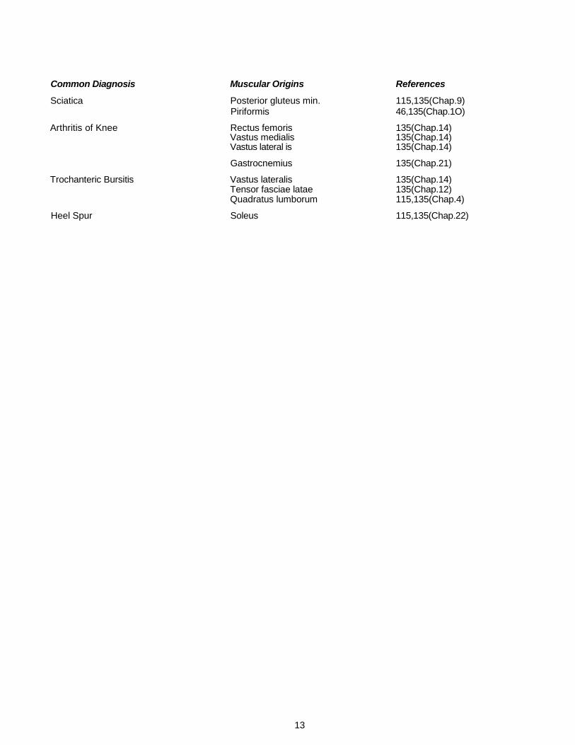
Sciatica Posterior gluteus min. 115,135(Chap.9) Piriformis 46,135(Chap.1O)
Arthritis of Knee Rectus femoris 135(Chap.14) Vastus medialis 135(Chap.14) Vastus lateral is 135(Chap.14)
Gastrocnemius 135(Chap.21)
Trochanteric Bursitis Vastus lateralis 135(Chap.14) Tensor fasciae latae 135(Chap.12) Quadratus lumborum 115,135(Chap.4)
Heel Spur Soleus 115,135(Chap.22)

14
ing the other and many patients are very likely to have both conditions.
ARTICULAR DYSFUNCTION
Articular dysfunction is identified by examining the joint for loss of normal mobility and range of motion not only in its kinesiological planes of voluntary motion, but also for "joint play". This "joint play" is motion not obtainable by voluntary muscular ac t ion .
7 5 The classical techniques
for joint mobilization or manipulation have been well described.
12,21,45,73,82 In addition, Lewit
66,67 emphasizes the
previously-recognized82
importance of releasing muscular tightness in conjunction with joint mobilization. The need for joint mobilization is established by skilled examination of the joint specifically for loss of mobility in all its planes of motion.
COMMON PAIN DIAGNOSES
Many common pain conditions are misdiagnosed because the examiner is not aware of referred pain pat-terns characteristic of myofascial TPs and fails to examine the muscles for them. Some conditions that are commonly misdiagnosed are listed in Table 1.
It is now becoming clear that tension headache is usu-ally due to myofascial TPs
42 and that frontal headache is
probably due to TPs in the clavicular division of the ster-nocleidomastoid muscle (Fig. 3C).
134
Face pain of enigmatic origin is likely to be called atyp-ical facial neuralgia by physicians and commonly identified by dentists as temporomandibular joint dysfunc-tion or myofascial pain dysfunction.The latter is often mistakenly considered to be chiefly psychogenic and behavioral in nature.
134<Chap5) However, this condition fre-
quently is at least partly due to myofascial TPs in the lateral pterygoid (Fig. 3G) or masseter (Fig. 3E) muscles. It is critically important when dealing with chronic masti-catory muscle pain and dysfunction to inactivate TPs in cervical musculature that refers pain to the face.
The sternocleidomastoid (Fig. 3C and D) and upper tra-pezius (Fig. 3A) muscles commonly cause pain referred to the face and secondarily activate and perpetuate TPs in the masticatory muscles which likewise refer pain to the face, and sometimes to the teeth.
Otolaryngologists are frequently deeply frustrated by patients who complain of earache due to referred pain from the deep masseter muscle (Fig. 3E). Many dentists are familiar with this syndrome and manage it well.Pain diagnosed as occipital neuralgia has been demonstrated often to be due to TPs in the posterior neck muscles (Fig. 4A and B).
41
An acute stiff neck is usually of myofascial origin128
in contrast to the neurogenic and/or psychogenic chronic torticollis.
134
What was first assumed to be post-dural puncture headache came from TPs that were discovered in the cervical paraspinal muscles during the post-partum period following a delivery in which spinal block was used.
50
Pain referred to the shoulder from the infraspinatus muscle (Fig. 4G) is likely to be ascribed to arthritis because the pain usually is perceived deep in the shoul-der joint.
9213*
Rather than coming from subdeltoid bursitis, pain and tenderness in the acromial and middle deltoid area of the shoulder is often referred from myofascial TPs.
The myofascial origin of thoracic outlet syndrome is entrapment of the lower trunk (mostly ulnar nerve fibers) of the brachial plexus by taut bands in the anterior and/or middle scalene muscles (Fig. 4C).
A common underlying cause of epicondylitis or "ten-nis elbow", which has many other names including "briefcase elbow", is referred pain and tenderness from TPs in the supinator muscle (Fig. 5F) and also from the wrist extensor muscles (Fig. 5G) near the lateral epi-condyle. In such cases, the lateral epicondyle is tender to thumping palpation. Examination for TPs and referred pain from TPs in these muscles quickly establishes the true origin of the pain. Occasionally, the triceps brachii (Fig. 5E) contributes to "tennis elbow" pain.
Pain mimicking angina, but poorly correlated with the duration and intensity of exercise or activity, may arise from TPs in the pectoralis major and/or minor muscles (Fig. 6C).
The most common source of upper back pain in the region of the upper vertebral border of the scapula is from TPs in the scalene muscles (Fig. 4C), with TPs of the levator scapulae (Fig. 4D) running a close second. The latissimus dorsi (Fig. 5A) may be responsible. When the rhomboids
134 are involved, the large pectoralis major
muscles are often shortened by latent TPs. Even though the pectoral TPs are not causing pain, the shortened pec-toral muscles overload the rhomboids. Serratus posterior superior TPs are covered by the retracted scapula and usually produce a pain deep in the chest (Fig. 6F). Upper thoracic paraspinal muscles are common offenders; TPs in their superficial layers are easily identified by palpation.
The most common muscular source of low back pain is TPs in the quadratus lumborum muscle (Fig. 6G). The iliopsoas is often involved in conjunction with the qua-dratus lumborum, and occasionally is involved by itself. Paraspinal muscles (Figs. 6H, 7C and D) frequently harbor TPs and, except for the deep multifidi and rotatores, are readily identified by palpation. The gluteus maximus and gluteus medius TPs commonly refer pain to the region of the buttock and sacrum. Pain referred from the rectus abdominis is distinctively horizontal and generally at either the mid-thoracic or low lumbar level corresponding to TPs in the upper and lower ends of the rectus abdo-minis muscle.
Many patients who were found to have a normal appen-dix at surgery for appendicitis may have had a misdiag-nosed but treatable myofascial pain syndrome of the rectus abdominis (Fig. 7H) or iliocostalis (Fig. 7A) muscles.
103
A muscular source of enigmatic pelvic pain may be located by intrarectal palpation of the coccygeus, levator ani, obturator internus and sphyncter ani muscles for TP tenderness and taut b a n d s .
5 8
Pain simulating arthritis of the hip is referred by the tensor fasciae latae muscle
135 deeply into the hip joint,
comparable to the pain referred by the infraspinatus mus-cle into the shoulder joint; patients with either arthritis of the hip or pain referred from the tensor fasciae latae mus-cle find weight bearing painful.
The neurological pain of meralgia paresthetics may be due to entrapment of the lateral femoral cutaneous nerve by involved sartorius or tensor fasciae latae muscles.
The pain commonly attributed to sciatica is rarely due to a demonstrable neuropathy of the sciatic nerve. This pain is much more likely to be referred from TPs in the posterior section of the gluteus minimus (Fig. 8B) or in the piriformis (Fig. 8C) muscles.
115

15
The pain of arthritis of the knee or knee pain from
known hip disease is mimicked by active myofascial TPs in the rectus femoris (Fig. 8E) and vastus medialis (Fig. 8G) muscles, which cause knee pain in the region of the patella; TPs in the upper end of the vastus lateralis (Fig. 81) and the gastrocnemius (Fig. 9B) muscles refer pain to the back of the knee.
The pain and tenderness of trochanteric bursitis is emulated by TPs in the vastus lateralis (Fig. 8H) or tensor fasciae latae
135 muscles.
When there is a heel spur, the spur is easily misiden-tified as the source of heel pain that actually is referred from TPs in the soleus muscle (Fig. 9A). In this case, the asymptomatic contralateral side often has an equally large and innocuous heel spur.
One simple way of confirming that myofascial TPs are the cause of the patient's symptoms is to inactivate the TPs and relieve the pain and tenderness.
TREATMENT
Single-muscle myofascial pain syndromes can be refreshingly simple to manage, whereas complex chronic myofascial syndromes driven by severe perpetuating fac-tors can be enormously difficult to resolve. The latter will be discussed below under Perpetuating Factors. Uncom-plicated single-muscle syndromes may persist but are nonprogressive. If the muscle is relieved of strain for sev-eral days, the TPs may revert from active to latent, reliev-ing pain.
The key to treatment of an acute MPS is the identifica-tion of the specific muscles harboring the active TPs. The muscles needing therapy are identified by a detailed his-tory of the onset of pain, knowledge of myofascial referred pain patterns, and confirmation of the location of active TPs responsible for the pain by examining the muscles. Palpation with one finger tip is used to look for the tender TP with its taut band and local twitch response. Modifica-tion of the patient's sensation in the pain reference zone by pressure on the TP provides diagnostic confirmation.
Lasting success in treatment depends on education of the patient and on inactivation of the responsible TPs by stretch and spray of the involved muscles and/or injection of the TPs. These techniques are used to restore full range of motion with pain-free function. Often the most difficult part of treatment is locating and correcting perpet-uating factors. It is critically important to teach the patient a home self-stretch program
6 that is tailored to prevent
and manage recurrences. The patient must learn how to safely use, not abuse, the involved muscles.
134
Specific myofascial TP therapies include stretch and spray, postisometric relaxation, injection, specific kinds of massage, and ultrasound or electrical stimulation applied to the TP. Since stretch and spray is the initial treatment of choice, the principles underlying its use are identified first and then its application to individual muscles is presented with each muscle's pain pattern.
STRETCH AND SPRAY
The stretch and spray method of treatment is one of the simplest, quickest and least painful ways to resolve a single-muscle MPS; it is frequently used immediately after TP injection to ensure inactivation of all TPs in that mus-cle. It is also valuable for complex cases where many muscles in a region of the body are involved.
107 Since
muscles within one functional group interact strongly, this technique is useful to release several closely related mus-cles at one time.
The purpose of stretch and spray is to inactivate the trigger point(s) by restoring the muscle to its full stretch range of motion with minimal discomfort and without excit-ing reflex spasm. Voluntary relaxation of the muscle being stretched is essential. The alarming cutaneous stimula-tion generated in the reference zone by the vapocoolant spray helps to block reflex spasm and pain,
130 permitting
gradual passive stretch of the muscle and inactivation of the TP mechanism.
A jet stream of vapocoolant spray is applied to the skin in one-directional parallel sweeps. If the skin is cold to the touch, it is too cold for application of vapocoolant. Exces-sive cooling evidenced by frosting of the skin is avoided. Fluori-Methane®* is preferred to ethyl chloride because the latter is a potentially lethal general anesthetic, is flam-mable, explosive and colder than desirable. Parallel sweeps of Fluori-Methane®* spray are applied slowly at 10 cm (4 in)/sec over the entire length of the muscle in the direction of and including the referred pain zone. The bot-tle is held about 45 cm (18 in) from the skin in order to permit the room-temperature vapocoolant in the bottle to cool as it passes through the air before it hits the skin. The stream of spray is most effective if it impacts the skin at an angle of about 30°.
134
Complete relaxation of the muscle to be stretched is essential. To obtain this relaxation, the patient should be positioned comfortably with all limbs and the back well supported. Initially, one or two sweeps of spray should precede stretch to inhibit the pain and stretch reflexes. Then, during each sweep of spray, the operator maintains gentle, smooth, steady tension on the muscle to take up any slack that develops, carefully avoiding force strong enough to produce pain. Jerky and rapid rocking motions activate TPs and should also be avoided.
Stretch is facilitated by asking the patient to slowly take a deep, breath and then slowly exhale through pursed lips, fully exhaling. During this long slow exhalation, the muscles tend to relax and are more easily stretched. Inspi-ration is facilitated by having the patient look up, and exha-lation by having the patient look down down toward the feet.
67 Thus, looking down and exhaling together further
facilitate relaxation. The Lewit stretch technique,68
which will be considered later, may be combined with stretch and spray or used as an alternate method of stretching the muscle without spray. Following stretch and spray the skin should be rewarmed with moist heat by dry hot packs or a wetproof moist heating pad, and then the muscle should be moved through the full active range of motion. A more detailed description of the stretch and spray procedure is available.
134
HEAD AND NECK PAIN
A number of neck muscles including the upper trape-zius, sternocleidomastoid, splenii and suboccipital mus-cles refer pain strongly to the head. These muscles frequently are responsible for headache, especially when it has been diagnosed as "tension" headache or "muscle tension" headache.
42 Masticatory muscles are likely to
cause temporal, maxillary and jaw pain, also earache and toothache; TPs in cutaneous muscles of the head and neck sometimes contribute to facial pain.
134
The primary masticatory muscles for closing the jaw include the masseter, temporalis, medial pterygoid and upper division of the lateral pterygoid; their antagonists are the digastric and lower division of the lateral pterygoid muscles, which primarily open the jaw. When ear pain,
*Gebauer Chemical Co., 9410 St. Catherine Ave., Cleveland, OH 44I04
16
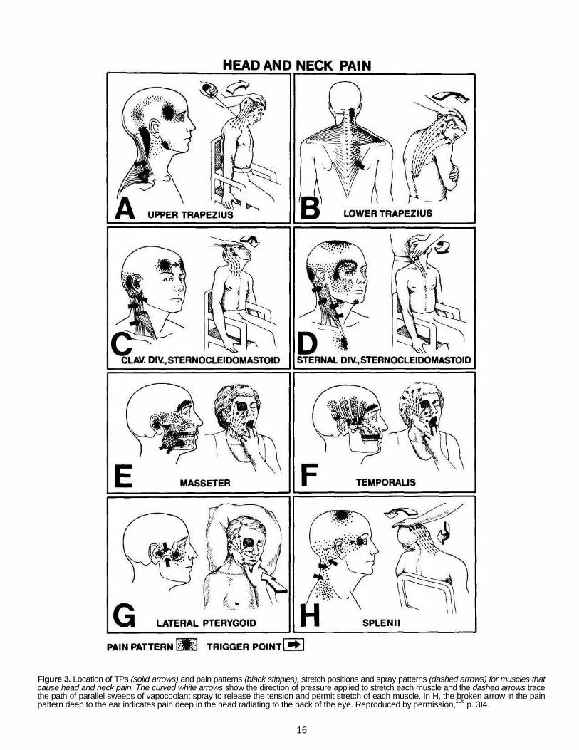
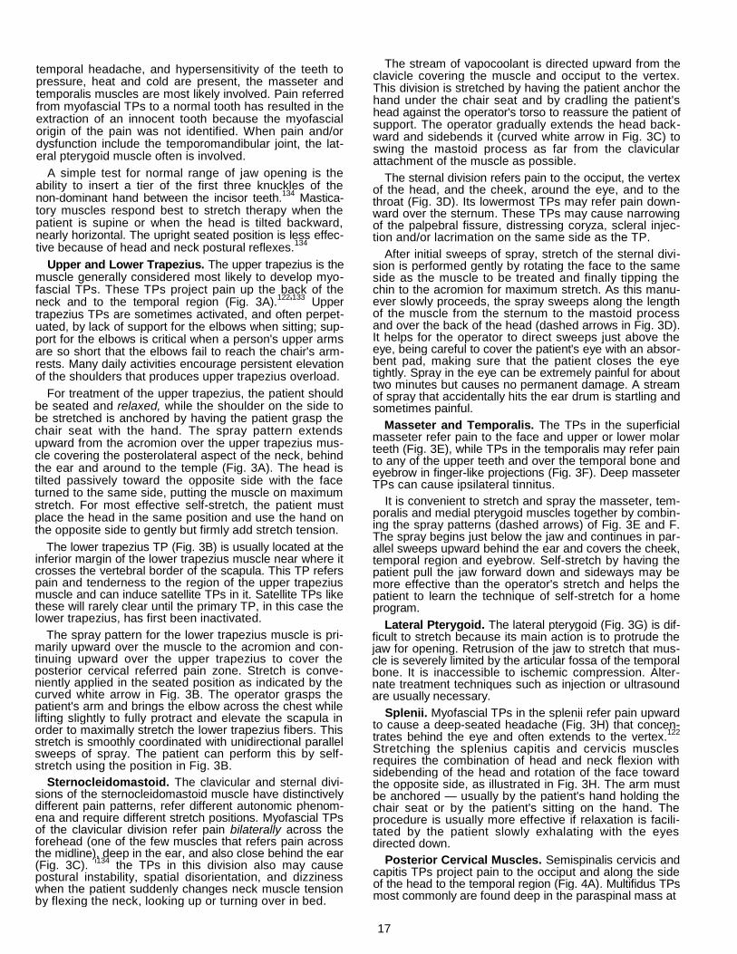
Figure 3. Location of TPs (solid arrows) and pain patterns (black stipples), stretch positions and spray patterns (dashed arrows) for muscles that cause head and neck pain. The curved white arrows show the direction of pressure applied to stretch each muscle and the dashed arrows trace the path of parallel sweeps of vapocoolant spray to release the tension and permit stretch of each muscle. In H, the broken arrow in the pain pattern deep to the ear indicates pain deep in the head radiating to the back of the eye. Reproduced by permission,
106 p. 3I4.
17
temporal headache, and hypersensitivity of the teeth to pressure, heat and cold are present, the masseter and temporalis muscles are most likely involved. Pain referred from myofascial TPs to a normal tooth has resulted in the extraction of an innocent tooth because the myofascial origin of the pain was not identified. When pain and/or dysfunction include the temporomandibular joint, the lat-eral pterygoid muscle often is involved.
A simple test for normal range of jaw opening is the ability to insert a tier of the first three knuckles of the non-dominant hand between the incisor teeth.
134 Mastica-
tory muscles respond best to stretch therapy when the patient is supine or when the head is tilted backward, nearly horizontal. The upright seated position is less effec-tive because of head and neck postural reflexes.
134
Upper and Lower Trapezius. The upper trapezius is the muscle generally considered most likely to develop myo-fascial TPs. These TPs project pain up the back of the neck and to the temporal region (Fig. 3A).
122'133
Upper trapezius TPs are sometimes activated, and often perpet-uated, by lack of support for the elbows when sitting; sup-port for the elbows is critical when a person's upper arms are so short that the elbows fail to reach the chair's arm-rests. Many daily activities encourage persistent elevation of the shoulders that produces upper trapezius overload.
For treatment of the upper trapezius, the patient should be seated and relaxed, while the shoulder on the side to be stretched is anchored by having the patient grasp the chair seat with the hand. The spray pattern extends upward from the acromion over the upper trapezius mus-cle covering the posterolateral aspect of the neck, behind the ear and around to the temple (Fig. 3A). The head is tilted passively toward the opposite side with the face turned to the same side, putting the muscle on maximum stretch. For most effective self-stretch, the patient must place the head in the same position and use the hand on the opposite side to gently but firmly add stretch tension.
The lower trapezius TP (Fig. 3B) is usually located at the inferior margin of the lower trapezius muscle near where it crosses the vertebral border of the scapula. This TP refers pain and tenderness to the region of the upper trapezius muscle and can induce satellite TPs in it. Satellite TPs like these will rarely clear until the primary TP, in this case the lower trapezius, has first been inactivated.
The spray pattern for the lower trapezius muscle is pri-marily upward over the muscle to the acromion and con-tinuing upward over the upper trapezius to cover the posterior cervical referred pain zone. Stretch is conve-niently applied in the seated position as indicated by the curved white arrow in Fig. 3B. The operator grasps the patient's arm and brings the elbow across the chest while lifting slightly to fully protract and elevate the scapula in order to maximally stretch the lower trapezius fibers. This stretch is smoothly coordinated with unidirectional parallel sweeps of spray. The patient can perform this by self-stretch using the position in Fig. 3B.
Sternocleidomastoid. The clavicular and sternal divi-sions of the sternocleidomastoid muscle have distinctively different pain patterns, refer different autonomic phenom-ena and require different stretch positions. Myofascial TPs of the clavicular division refer pain bilaterally across the forehead (one of the few muscles that refers pain across the midline), deep in the ear, and also close behind the ear (Fig. 3C).
i134 the TPs in this division also may cause
postural instability, spatial disorientation, and dizziness when the patient suddenly changes neck muscle tension by flexing the neck, looking up or turning over in bed.
The stream of vapocoolant is directed upward from the clavicle covering the muscle and occiput to the vertex. This division is stretched by having the patient anchor the hand under the chair seat and by cradling the patient's head against the operator's torso to reassure the patient of support. The operator gradually extends the head back-ward and sidebends it (curved white arrow in Fig. 3C) to swing the mastoid process as far from the clavicular attachment of the muscle as possible.
The sternal division refers pain to the occiput, the vertex of the head, and the cheek, around the eye, and to the throat (Fig. 3D). Its lowermost TPs may refer pain down-ward over the sternum. These TPs may cause narrowing of the palpebral fissure, distressing coryza, scleral injec-tion and/or lacrimation on the same side as the TP.
After initial sweeps of spray, stretch of the sternal divi-sion is performed gently by rotating the face to the same side as the muscle to be treated and finally tipping the chin to the acromion for maximum stretch. As this manu-ever slowly proceeds, the spray sweeps along the length of the muscle from the sternum to the mastoid process and over the back of the head (dashed arrows in Fig. 3D). It helps for the operator to direct sweeps just above the eye, being careful to cover the patient's eye with an absor-bent pad, making sure that the patient closes the eye tightly. Spray in the eye can be extremely painful for about two minutes but causes no permanent damage. A stream of spray that accidentally hits the ear drum is startling and sometimes painful.
Masseter and Temporalis. The TPs in the superficial masseter refer pain to the face and upper or lower molar teeth (Fig. 3E), while TPs in the temporalis may refer pain to any of the upper teeth and over the temporal bone and eyebrow in finger-like projections (Fig. 3F). Deep masseter TPs can cause ipsilateral tinnitus.
It is convenient to stretch and spray the masseter, tem-poralis and medial pterygoid muscles together by combin-ing the spray patterns (dashed arrows) of Fig. 3E and F. The spray begins just below the jaw and continues in par-allel sweeps upward behind the ear and covers the cheek, temporal region and eyebrow. Self-stretch by having the patient pull the jaw forward down and sideways may be more effective than the operator's stretch and helps the patient to learn the technique of self-stretch for a home program.
Lateral Pterygoid. The lateral pterygoid (Fig. 3G) is dif-ficult to stretch because its main action is to protrude the jaw for opening. Retrusion of the jaw to stretch that mus-cle is severely limited by the articular fossa of the temporal bone. It is inaccessible to ischemic compression. Alter-nate treatment techniques such as injection or ultrasound are usually necessary.
Splenii. Myofascial TPs in the splenii refer pain upward to cause a deep-seated headache (Fig. 3H) that concen-trates behind the eye and often extends to the vertex.
122
Stretching the splenius capitis and cervicis muscles requires the combination of head and neck flexion with sidebending of the head and rotation of the face toward the opposite side, as illustrated in Fig. 3H. The arm must be anchored — usually by the patient's hand holding the chair seat or by the patient's sitting on the hand. The procedure is usually more effective if relaxation is facili-tated by the patient slowly exhalating with the eyes directed down.
Posterior Cervical Muscles. Semispinalis cervicis and capitis TPs project pain to the occiput and along the side of the head to the temporal region (Fig. 4A). Multifidus TPs most commonly are found deep in the paraspinal mass at
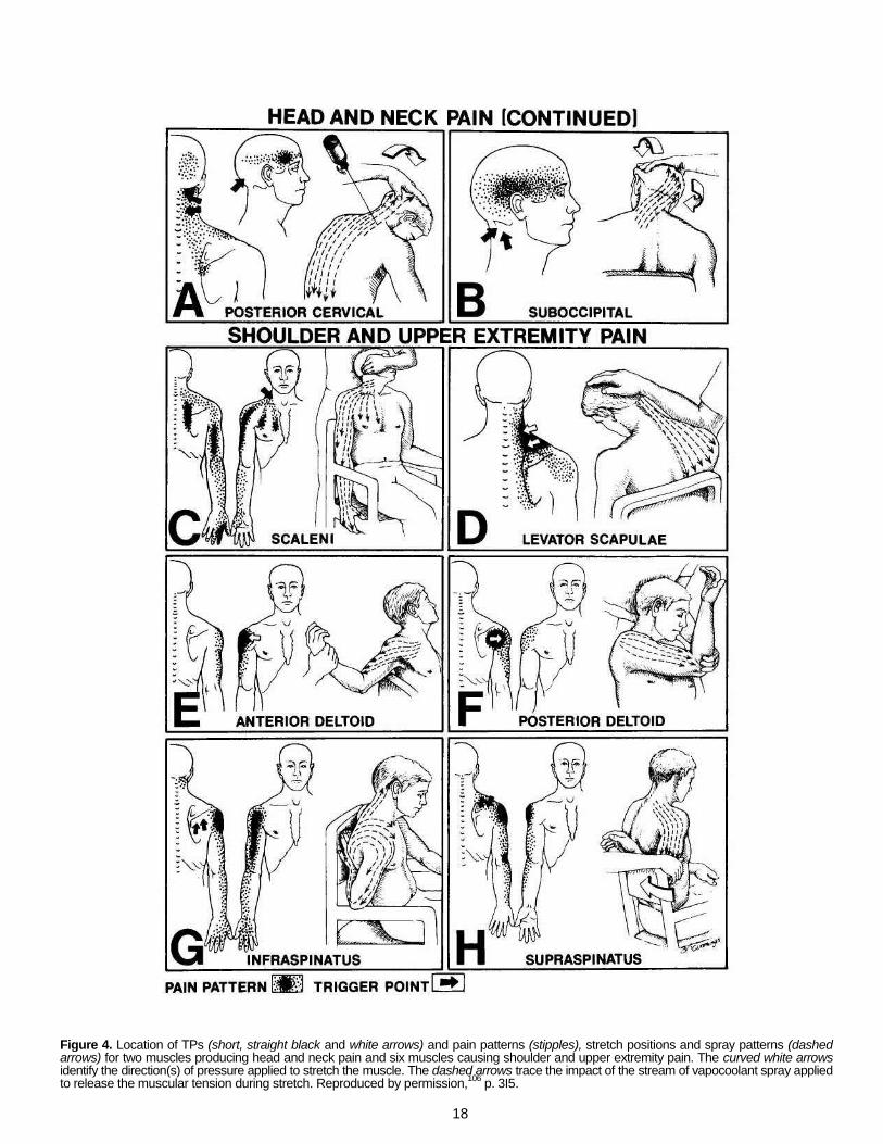
18
Figure 4. Location of TPs (short, straight black and white arrows) and pain patterns (stipples), stretch positions and spray patterns (dashed arrows) for two muscles producing head and neck pain and six muscles causing shoulder and upper extremity pain. The curved white arrows identify the direction(s) of pressure applied to stretch the muscle. The dashed arrows trace the impact of the stream of vapocoolant spray applied to release the muscular tension during stretch. Reproduced by permission,
106 p. 3I5.
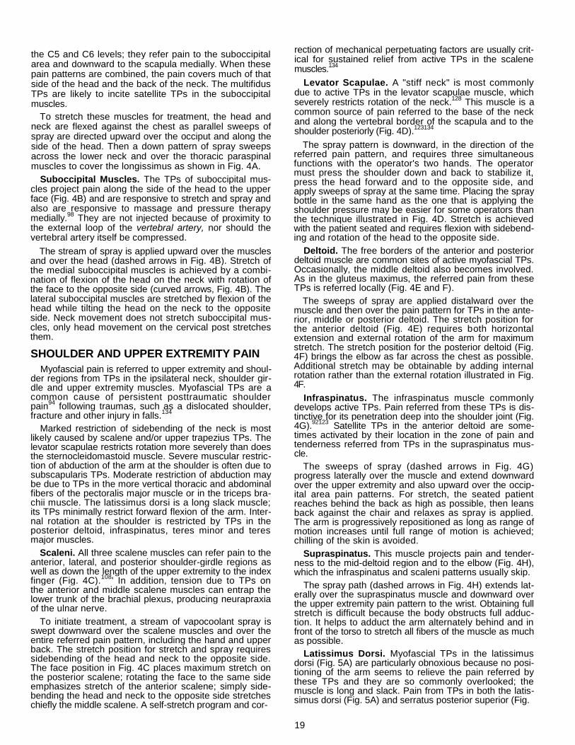
19
the C5 and C6 levels; they refer pain to the suboccipital area and downward to the scapula medially. When these pain patterns are combined, the pain covers much of that side of the head and the back of the neck. The multifidus TPs are likely to incite satellite TPs in the suboccipital muscles.
To stretch these muscles for treatment, the head and neck are flexed against the chest as parallel sweeps of spray are directed upward over the occiput and along the side of the head. Then a down pattern of spray sweeps across the lower neck and over the thoracic paraspinal muscles to cover the longissimus as shown in Fig. 4A.
Suboccipital Muscles. The TPs of suboccipital mus-cles project pain along the side of the head to the upper face (Fig. 4B) and are responsive to stretch and spray and also are responsive to massage and pressure therapy medially.
98 They are not injected because of proximity to
the external loop of the vertebral artery, nor should the vertebral artery itself be compressed.
The stream of spray is applied upward over the muscles and over the head (dashed arrows in Fig. 4B). Stretch of the medial suboccipital muscles is achieved by a combi-nation of flexion of the head on the neck with rotation of the face to the opposite side (curved arrows, Fig. 4B). The lateral suboccipital muscles are stretched by flexion of the head while tilting the head on the neck to the opposite side. Neck movement does not stretch suboccipital mus-cles, only head movement on the cervical post stretches them.
SHOULDER AND UPPER EXTREMITY PAIN
Myofascial pain is referred to upper extremity and shoul-der regions from TPs in the ipsilateral neck, shoulder gir-dle and upper extremity muscles. Myofascial TPs are a common cause of persistent posttraumatic shoulder pain
94 following traumas, such as a dislocated shoulder,
fracture and other injury in falls.134
Marked restriction of sidebending of the neck is most likely caused by scalene and/or upper trapezius TPs. The levator scapulae restricts rotation more severely than does the sternocleidomastoid muscle. Severe muscular restric-tion of abduction of the arm at the shoulder is often due to subscapularis TPs. Moderate restriction of abduction may be due to TPs in the more vertical thoracic and abdominal fibers of the pectoralis major muscle or in the triceps bra-chii muscle. The latissimus dorsi is a long slack muscle; its TPs minimally restrict forward flexion of the arm. Inter-nal rotation at the shoulder is restricted by TPs in the posterior deltoid, infraspinatus, teres minor and teres major muscles.
Scaleni. All three scalene muscles can refer pain to the anterior, lateral, and posterior shoulder-girdle regions as well as down the length of the upper extremity to the index finger (Fig. 4C).
108' In addition, tension due to TPs on
the anterior and middle scalene muscles can entrap the lower trunk of the brachial plexus, producing neurapraxia of the ulnar nerve.
To initiate treatment, a stream of vapocoolant spray is swept downward over the scalene muscles and over the entire referred pain pattern, including the hand and upper back. The stretch position for stretch and spray requires sidebending of the head and neck to the opposite side. The face position in Fig. 4C places maximum stretch on the posterior scalene; rotating the face to the same side emphasizes stretch of the anterior scalene; simply side-bending the head and neck to the opposite side stretches chiefly the middle scalene. A self-stretch program and cor-
rection of mechanical perpetuating factors are usually crit-ical for sustained relief from active TPs in the scalene muscles.
134
Levator Scapulae. A "stiff neck" is most commonly due to active TPs in the levator scapulae muscle, which severely restricts rotation of the neck.
128 This muscle is a
common source of pain referred to the base of the neck and along the vertebral border of the scapula and to the shoulder posteriorly (Fig. 4D).
123134
The spray pattern is downward, in the direction of the referred pain pattern, and requires three simultaneous functions with the operator's two hands. The operator must press the shoulder down and back to stabilize it, press the head forward and to the opposite side, and apply sweeps of spray at the same time. Placing the spray bottle in the same hand as the one that is applying the shoulder pressure may be easier for some operators than the technique illustrated in Fig. 4D. Stretch is achieved with the patient seated and requires flexion with sidebend-ing and rotation of the head to the opposite side.
Deltoid. The free borders of the anterior and posterior deltoid muscle are common sites of active myofascial TPs. Occasionally, the middle deltoid also becomes involved. As in the gluteus maximus, the referred pain from these TPs is referred locally (Fig. 4E and F).
The sweeps of spray are applied distalward over the muscle and then over the pain pattern for TPs in the ante-rior, middle or posterior deltoid. The stretch position for the anterior deltoid (Fig. 4E) requires both horizontal extension and external rotation of the arm for maximum stretch. The stretch position for the posterior deltoid (Fig. 4F) brings the elbow as far across the chest as possible. Additional stretch may be obtainable by adding internal rotation rather than the external rotation illustrated in Fig. 4F.
Infraspinatus. The infraspinatus muscle commonly develops active TPs. Pain referred from these TPs is dis-tinctive for its penetration deep into the shoulder joint (Fig. 4G).
92123 Satellite TPs in the anterior deltoid are some-
times activated by their location in the zone of pain and tenderness referred from TPs in the supraspinatus mus-cle.
The sweeps of spray (dashed arrows in Fig. 4G) progress laterally over the muscle and extend downward over the upper extremity and also upward over the occip-ital area pain patterns. For stretch, the seated patient reaches behind the back as high as possible, then leans back against the chair and relaxes as spray is applied. The arm is progressively repositioned as long as range of motion increases until full range of motion is achieved; chilling of the skin is avoided.
Supraspinatus. This muscle projects pain and tender-ness to the mid-deltoid region and to the elbow (Fig. 4H), which the infraspinatus and scaleni patterns usually skip.
The spray path (dashed arrows in Fig. 4H) extends lat-erally over the supraspinatus muscle and downward over the upper extremity pain pattern to the wrist. Obtaining full stretch is difficult because the body obstructs full adduc-tion. It helps to adduct the arm alternately behind and in front of the torso to stretch all fibers of the muscle as much as possible.
Latissimus Dorsi. Myofascial TPs in the latissimus dorsi (Fig. 5A) are particularly obnoxious because no posi-tioning of the arm seems to relieve the pain referred by these TPs and they are so commonly overlooked; the muscle is long and slack. Pain from TPs in both the latis-simus dorsi (Fig. 5A) and serratus posterior superior (Fig.
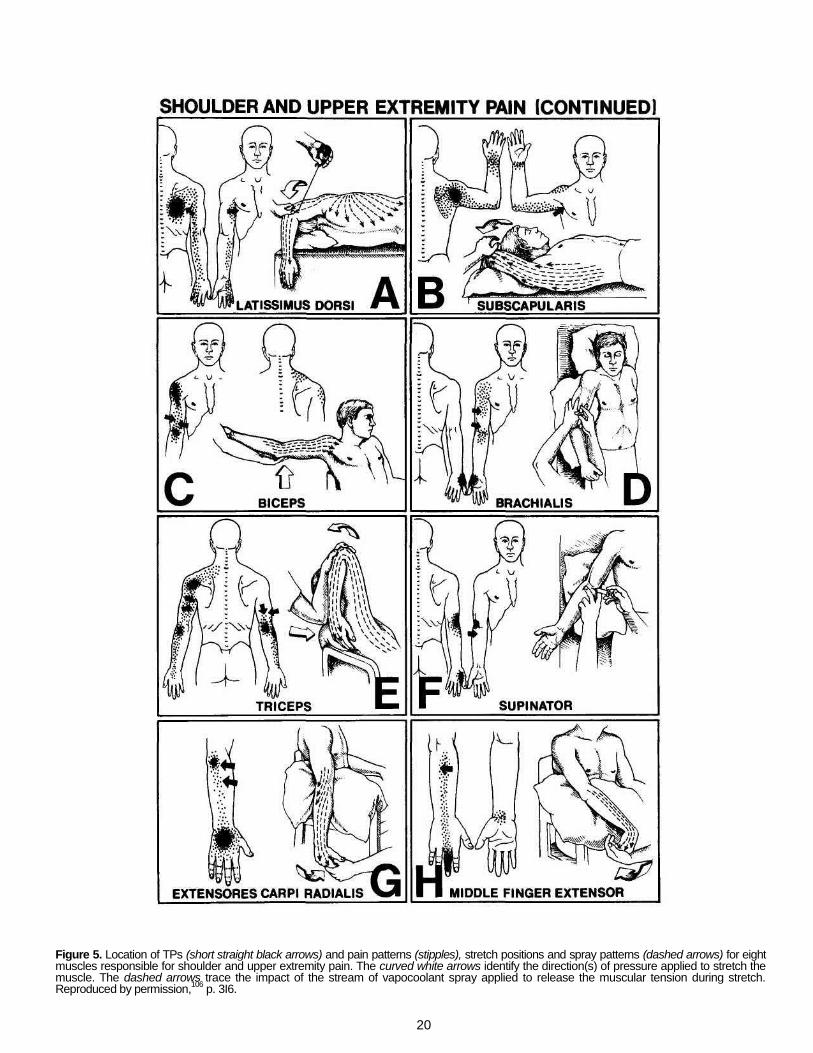
20
Figure 5. Location of TPs (short straight black arrows) and pain patterns (stipples), stretch positions and spray patterns (dashed arrows) for eight muscles responsible for shoulder and upper extremity pain. The curved white arrows identify the direction(s) of pressure applied to stretch the muscle. The dashed arrows trace the impact of the stream of vapocoolant spray applied to release the muscular tension during stretch. Reproduced by permission,
106 p. 3I6.

21
6F) is referred to the scapular area. Pain referred to the posterior scapula from TPs in the serratus posterior supe-rior tends to be more cephalad and feels deep in the chest, sometimes through the chest. This depth is not characteristic of pain referred from the latissimus dorsi.
Because of the complex pain pattern referred by the latissimus dorsi muscle, vapocoolant is directed from the TP in the direction of the pain pattern, covering the entire muscle, including its attachments on the crest of the ilium and to the lumbar and thoracic spine; sweeps of spray also course upward over the upper half of the muscle covering the pain pattern on the dorsum of the upper extremity to the tips of the ulnar fingers (Fig. 5A).
Full stretch can be attained seated, but stretch and spray is usually more satisfactory when the patient is in the supine position. Effective stretch requires full arm flex-ion and external rotation (Fig. 5A) in essentially the same position as for the subscapularis (Fig. 5B). If the subsca-pularis is involved, full stretch of the latissimus dorsi will not be attained until the subscapularis is released.
Subscapularis. A frozen-shoulder syndrome often develops because of TPs (thick black arrow in Fig. 5B) in the subscapularis muscle.
16134 Active TPs in this muscle
severely restrict both abduction and external rotation of the arm. The severely restricted range of motion predis-poses to TPs in numerous other shoulder-girdle muscles, which leads to a frozen-shoulder syndrome. The pain referred from the subscapularis muscle is focused on the back of the shoulder and frequently includes a "band" of pain and tenderness around the wrist. The chief complaint may be this pain and tenderness of the wrist with restricted range of shoulder motion.
Treatment begins with the application of upsweeps of spray covering the side of the chest and axilla as in Fig. 5B, covering all of the scapula including its vertebral bor-der. Sweeps continue until they cover the wrist. The stretch position requires gentle, progressive, abduction and external rotation of the arm at the shoulder.
Biceps Brachii. The biceps referred pain pattern covers the anterior shoulder and sometimes extends to the elbow Fig. 5C). Bicipital tendonitis may require inactivation of ~Ps in the long head of the biceps brachii for prompt, lasting relief.
The stream of vapocoolant is directed upward over the entire length of the muscle in parallel sweeps to include the referred pain pattern at the shoulder. The long head of this muscle crosses two joints and requires both elbow extension and horizontal extension of the arm at the shoul-der for full stretch as in Fig. 5C. The doorway stretch is an important part of the patient's home program.
134
Brachialis. The pain referred from this muscle is remarkable for its strong projection to the base of the thumb (Fig. 5D), to which the supinator (Fig. 5F), brachioradialis and adductor pollicis
116 muscles also
refer pain and tenderness. The stream of spray progresses downward over the muscle to cover the thumb as in Fig. 5D. Stretching the brachialis muscle requires extension of the forearm at the elbow. Brachialis TPs are readily palpated by pushing aside the distal biceps; they respond well to injection.
Triceps Brachii. The three heads of the triceps muscle have five TP locations that refer separate pain patterns.
134
The five patterns are combined in Fig. 5E. The long head is a two joint muscle; TPs in it restrict flexion both at the elbow and at the shoulder. The long head of the triceps is commonly involved with other shoulder-girdle muscles and is a frequently overlooked cause of shoulder dysfunc-
tion. The taut bands of the long head TPs are readily felt by pincer palpation of the belly of the long head just above mid-arm adjacent to the humerus. The muscle is exam-ined for taut bands as the muscle fibers slip between the finger tips that start palpating outward from the groove between the muscle and the humerus.
134
The spray is applied in parallel sweeps over the mus-cle(s) harboring active TPs in the direction of the pain pattern, which may be upward over the arm, or distalward over the forearm to the hand. Full stretch requires simul-taneous flexion at the elbow and at the shoulder (Fig. 5E).
Supinator. For patients with "tennis elbow" or epi-condylitis, TPs in the supinator muscle are often the key to resolution of their distress. This muscle refers pain to the dorsal web space of the thumb and to the region of the lateral epicondyle (Fig. 5F), which becomes tender to fin-ger taps.
The stream of vapocoolant sweeps downward over the muscle, swings back to cover the lateral epicondyle and then proceeds distalward over the dorsum of the forearm and thumb. Stretch is achieved by combined extension at the elbow and pronation of the hand. Injection of the most common TPs in the medial (radial) portion of the muscle is sometimes required (Fig. 5F). The radial nerve passes through a more lateral portion of the muscle and should be avoided when injecting.
134 The finger and wrist extensors,
which form the extensor muscle mass attached to the lat-eral epicondyle, frequently also develop TPs as part of the "tennis elbow" syndrome. These TPs are readily located by palpation of the extensor muscles for taut bands and local twitch responses.
Extensores Digitorum and Carpi Radialis. The hand and finger extensor group of muscles is essential for strong grip. Patients with TPs in these muscles frequently develop a painful hand grip and an impaired sense of grip strength; a cup or a glass unexpectedly drops from their grasp. Active TPs in the extensor carpi radialis refer pain to the lateral epicondyle and dorsum of the hand (Fig. 5G). The TPs in the finger extensor muscles refer pain specif-ically to the dorsal surface of the corresponding finger, as exemplified by the middle finger extensor in Fig. 5H.
The stream of spray covers the dorsal forearm, wrist, hand and fingers. The stretch position of the extensor digitorum muscle requires full flexion of the wrist and fin-gers (Fig. 5H). This positioning also stretches the wrist extensors except for the addition of ulnar or radial devia-tion of the wrist.
Flexores Digitorum. No distinction is made in the dis-tribution of pain referred from the flexores digitorum sublimis and profundus. Pain from their TPs refers to the finger that corresponds to the muscle involved as, for example, the third finger flexor (Fig. 6A). Patients with TPs in this muscle usually feel that the pain is projected to the very tip of the finger, occasionally beyond.
The stream of vapocoolant spray is directed distalward over the forearm flexor surface of the forearm and to the finger tips. Since this muscle produces flexion across mul-tiple joints, all of those joints must be extended simulta-neously for a full stretch (Fig. 6A).
Interossei of Hand. Myofascial TPs in the interossei are not uncommon and those in the dorsal interossei are readily palpable against the metacarpal bones. These TPs refer pain primarily along the side of the digit that corre-sponds to the distal attachment of that interosseus mus-cle. The first dorsal interosseus also projects pain across the hand and into the little finger (Fig. 6B).
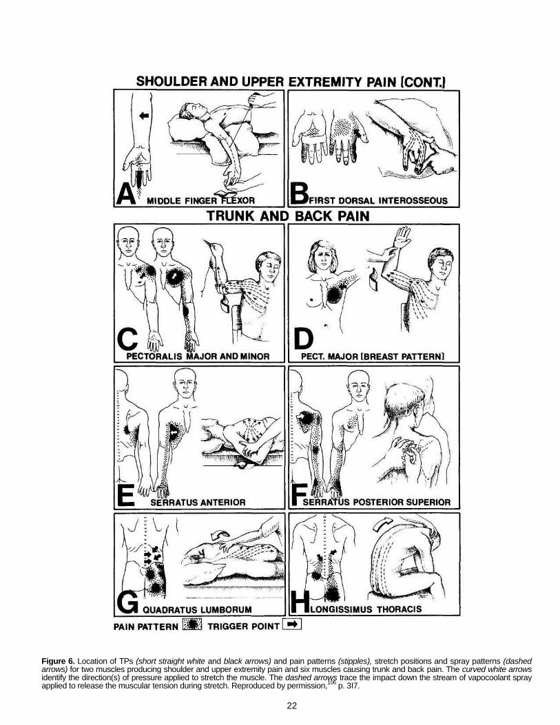
22
Figure 6. Location of TPs (short straight white and black arrows) and pain patterns (stipples), stretch positions and spray patterns (dashed arrows) for two muscles producing shoulder and upper extremity pain and six muscles causing trunk and back pain. The curved white arrows identify the direction(s) of pressure applied to stretch the muscle. The dashed arrows trace the impact down the stream of vapocoolant spray applied to release the muscular tension during stretch. Reproduced by permission,
106 p. 3I7.
23
The stream of vapocoolant spray is applied distalward
from the wrist to the ends of the painful digits while the fingers are separated to stretch the dorsal interosseous muscle that lies between them as in Fig. 6B. Frequently, injection is more satisfactory, because the metacarpopha-langeal joints of many patients have limited lateral mobil-ity.
TRUNK AND BACK PAIN
Myofascial TPs in muscles of the chest and abdomen exhibit viscerosomatic and somatovisceral interactions. Viscerosomatic influence is demonstrated by the satellite TPs that develop in the pectoralis major muscle in the zone of pain referred from an acute myocardial infarction or from recent myocardial ischemia. Similarly, TPs in the external oblique muscle of the abdomen may develop in response to the pain referred to that region by gastroin-testinal ulcer disease.
74
Conversely, an example of somatovisceral interaction is the cardiac arrhythmia associated with the "arrhythmia TP" in the right pectoralis major muscle.
134 These inter-
actions suggest feedback loops within the central nervous system among TPs, visceral structures and their referred pain zones.
Pectoralis Major and Minor. Myofascial TPs in the more horizontal sternal division fibers of the pectoralis major and in the pectoralis minor muscle can refer pain that closely mimics cardiac ischemia (Fig. 6C).
122'134
Trigger points in the lateral, nearly vertical thoracic fibers of the pectoralis major refer pain and tenderness to the breast (Fig. 6D). These TPs may cause a degree of breast hypersensitivity that, in either sex, makes clothing contact with the nipple intolerable.
For both pectoral muscles, the vapocoolant is first directed upward and laterally over the muscle fibers on the chest. The path of the stream continues over the arm to include, for the pectoralis major, the lateral side of the hand (Fig. 6C).
Stretch is applied to the sternal division fibers of the pectoralis major by horizontal extension of the arm and to the pectoralis minor by retraction of the scapula with back-ward traction on the arm (curved white arrow in Fig. 6C). The nearly vertical thoracic fibers along the lateral border of the pectoralis major are stretched more effectively by full abduction and flexion of the arm at the shoulder than by the partial stretch position shown in Fig. 6D. The fully flexed position of the arm also stretches the latissimus dorsi. The application of spray should also include that muscle, as in Fig. 5A, to ensure that it is not restricting full stretch of the pectoralis major.
The head-forward, round-shouldered posture that main-tains the pectoral muscles in the shortened position usu-ally requires correction for lasting inactiyation of pectoral TPs. This poor posture is spontaneously improved without muscle strain by providing comfortable lumbar support with a roll or small pillow whenever the patient sits, and by assumption of the weight-on-toes posture when standing.
134
Serratus Anterior. This muscle is not generally consid-ered a muscle of accessory respiration
1 but active TPs
in it decrease maximum chest expansion and cause the patient to feel short of breath. Pain is referred over the side of the chest and to the lower vertebral border of the scap-ula (Fig. 6E), not far from the focus of pain characteristic of latissimus dorsi TPs (Fig. 5A). Pain from both muscles may extend down the arm to the hand.
The stream of vapocoolant is directed in radial sweeps that start at the TP, cover the muscle and include the entire pain pattern, ensuring coverage of the lower half of the scapula. The serratus anterior is stretched either with the patient supine as illustrated in Figure 6E or seated in an armchair. For a self-stretch, the patient can sit and reach backward with the arm and scapula, using the uninvolved arm to reach behind the body and grasp the humerus of the involved arm distally to assist the passive stretch. In either position, the elbow is progressively moved posteri-orly to strongly retract the scapula.
Serratus Posterior Superior. The pain produced by this muscle can be a tantalizing joker. It is threateningly deep, frequently projecting into and through the chest suggesting visceral disease. It is enigmatic because posi-tioning of the arm or shoulder may have little effect on it. The serratus posterior superior TPs are not palpable unless the scapula is fully protracted, uncovering the mus-cle laterally.
134 These TPs are identified by palpating
through the trapezius and rhomboid muscles against the ribs for their exquisite sensitivity, taut bands and repro-duction of pain pattern. Trigger points in this muscle respond well to ischemic compression. Because these serratus fibers run between the ribs and the spinous pro-cesses, stretch is frequently unsatisfactory, so the TPs may require injection (Fig. 6H). However, one must be very careful to direct the needle nearly parallel to the skin sur-face and not let it penetrate between the ribs, causing a pneumothorax.
Quadratus Lumborum. This muscle is probably the most common source of myogenic back pain,
122'155145
and is all too commonly overlooked.
121 Quadratus lumborum
TPs project pain to the sacroiliac joint, lower buttock and lateral hip regions (Fig. 6G).
123 These TPs are prone to
generate satellite TPs in the posterior gluteus minimus muscle, producing a secondary sciatica-like pain pattern (Fig. 8B) that further misleads the diagnostician. Bilateral quadratus lumborum involvement is likely to produce pain that extends across the sacroiliac region.
A complete examination of the quadratus lumborum muscle requires that the sidelying patient be positioned to provide adequate space for palpation between the 12th rib and the crest of the ilium. The space is realized by elevat-ing the rib cage and dropping the pelvis on that side. This examination position is similar to the stretch position of Fig. 6G, but with the upper leg behind the lower leg and wi th the upper knee rest ing on the examining {ab|e 108,114,1^
To release these TPs, the stream of vapocoolant is directed downward covering all of the lumbar area and all aspects of the buttock, particularly the sacral and sacro-liac joint region. Two stretch positions may be needed to release TPs in all parts of this complex tripje muscle.
135
The leg-forward stretch position in Fig. 6G tilts the pelvis away from the ribs and rotates the pelvis forward with respect to the chest; the leg-back position
108135 places the
upper leg behind the lower leg, reverses the twist on the thoracolumbar region and also stretches the iliopsoas muscle. To ensure that tightness of the iliopsoas muscle is not blocking stretch of the quadratus lumborum, spray is directed downward over the abdomen beside the midline to the inner thigh. This releases iliopsoas muscle tightness.
106
Injection of TPs of this muscle is remarkably effective, but requires careful technique and appreciation of the anatomy involved.
108114135
Quadratus lumborum TPs are often perpetuated by a short leg and/or a small hemipelvis. Both are readily cor-
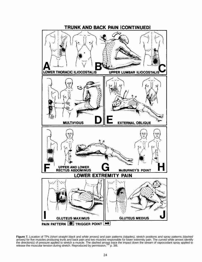
24
Figure 7. Location of TPs (short straight black and white arrows) and pain patterns (stipples), stretch positions and spray patterns (dashed arrows) for five muscles producing trunk and back pain and two muscles responsible for lower extremity pain. The curved white arrows identify the direction(s) of pressure applied to stretch a muscle. The dashed arrows trace the impact down the stream of vapocoolant spray applied to release the muscular tension during stretch. Reproduced by permission,
106 p. 3I8.
25
rectable with lifts, which are often essential for lasting relief of myofascial low back pain.
134'135
Thoracolumbar Paraspinal Muscles. In the mid-thoracic region, the most medial paraspinal muscle group is the spinalis, which lies close to the spinous pro-cesses. Next laterally is the longissimus, which is one of the longest muscles in the body; it covers most of the paraspinal space in the thoracic and lumbar areas. The iliocostalis fibers are the most lateral paraspinal muscles and attach to the ribs. Bilaterally, the deeper paraspinal muscles form an inverted "V"; the deeper they lie the shorter they are and the more diagonal their course. The deepest and shortest rotatores connect adjacent verte-brae at nearly a 45 angle. Therefore, to stretch the deeper fibers one must either mobilize the adjacent vertebrae by manipulative techniques or strongly rotate as well as flex the spine.
Taut bands in the longissimus fibers are readily pal-pated and course parallel to the axis of the spine. Myofa-scial TPs in these bands refer pain distalward, usually at least several segments removed; pain from longissimus TPs may project to the distal buttock (Fig. 6H).
Lower thoracic iliocostalis TPs may refer pain anteriorly to the abdominal wall at approximately the same derma-tomal level, as well as caudally (Fig. 7A). Upper lumbar iliocostalis TPs refer pain lower, to the mid-buttock (Fig. 7C). The deeper multifidus muscles refer pain more locally (Fig. 7D). The short rotatores often refer pain to to the midline at the same segmental level so that the pain pattern alone may not identify on which side these TPs lie.
134
The spray pattern for all thoracolumbar paraspinal mus-cles is caudalward and covers all of the muscle fibers of the involved muscle, which, for the longissimus and ilio-costalis muscles, extends the length of the spine below the TPs and includes the buttocks (Figs. 6H and 7B). Mul-tifidus TPs require a less extensive spray pattern, which is angulated somewhat laterally (Fig. 7D).
The stretch position for the longissimus is simple flex-ion, most effectively performed with the patient seated with the legs spread widely apart, feet on the floor. The arms dangle; the patient must allow the neck, hands and elbows to flex in a relaxed manner as the fingers reach the floor. The spray must also be extended downward to cover the gluteus maximus (Fig. 6H), which, too, is stretched by hip flexion in this position. Stretch of the iliocostalis can be augmented by having the patient rotate the face and chest to the opposite side while flexing the spine.
For stretch of the multifidi the patient rotates toward the same side and flexes the spine at the same time (Fig. 7D). Strong rotation is essential. Injection may be more effec-tive for these deep diagonal muscles. Care must be exer-cised in the thoracic region to direct the needle medially toward the deep rotatores in order to avoid injecting too laterally, between the ribs, and producing a pneumotho-rax.
Abdominal Muscles. Myofascial TPs in an external abdominal oblique muscle may refer pain that is nearby, or the pain may extend into adjacent areas and cross the midline (Fig. 7E).
122'134
Those TPs close to the thoracic or pelvic attachment of the rectus abdominis are likely to refer pain horizontally across the back at approximately the same level as the TP (Fig. 7F). A TP in the lower right quadrant of the rectus abdominis may convincingly simu-late the pain of appendicitis (Fig. 7H). Visceral disease can cause abdominal myofascial TPs and TPs in the abdominal muscles can closely simulate visceral disease, such as duodenal ulcer.
74134
Having the patient tense the abdominal muscles by ele-vating the feet while lying in the supine position easily differentiates a source of pain in the viscera from one in the abdominal wall. Tensing the abdomen augments ten-derness of the musculature and protects the underlying viscera from the pressure of palpation.
118 If the patient
also has a complaint of back pain, one must exercise cau-tion to avoid overstrain with this test. Instead, the exam-iner could elicit contraction of the supine patient's abdominal muscles by resisting the patient's effort to extend downward his or her upper limbs.
The spray pattern for TPs in the abdominal muscles is essentially downward over the entire abdomen including any additional referred pain patterns. Stretch of the abdominal wall muscles is achieved by having the patient lie supine with the hips over the end of the table with the feet resting on a chair seat; a towel or roll is placed under the lumbar spine to hyperextend it (Fig. 7G). The patient then protrudes the abdomen by taking a deep breath, holding it and contracting the diaphragm while relaxing the abdominal wall muscles as parallel sweeps of spray are applied downward over the entire abdomen. A twisting movement is added for the external oblique (Fig. 7E) that is not required for the rectus abdominis (Fig. 7G).
LOWER EXTREMITY PAIN
With a few exceptions, TPs in lower extremity muscles refer pain locally and/or distally. Since the gluteal regions are often identified by patients as part of the low back, gluteal myofascial TPs often contribute to "low back pain" syndromes.
115
Gluteus Maximus. As in the deltoid muscle, TPs in the gluteus maximus are easily palpated for taut bands, usu-ally have eloquent local twitch responses, and produce relatively local referred pain patterns that concentrate along the sacrum and inferior surface of the buttock (Fig. 71).
For treatment, the patient is placed in the sidelying posi-tion, as illustrated in Fig. 71. Vapocoolant spray is directed distally over the buttock to the upper thigh as the thigh is progressively flexed, bringing the knee to the chest. Gluteal TPs are commonly associated with tight hamstring muscles that also must be released (Fig. 81) for return of full function and lasting relief.
Gluteus Medius. The TPs in this muscle (Fig. 7J) refer pain along the crest of the ilium and over the sacrum and may project downward across the buttock to include the upper thigh.
To inactivate these TPs, a stream of vapocoolant spray is directed from the crest of the ilium downward over the buttock and upper half of the thigh in parallel sweeps as illustrated in Fig. 7J. The thigh is flexed 90° and, since this muscle is primarily an abductor, the thigh is progressively adducted as the sweeps of spray are applied. It is often helpful to have an assistant pull backward on the anterior superior iliac spine region to anchor the pelvis for more effective stretch. The Dudley J. Morton foot configuration is a perpetuator of these TPs and should be corrected/
34135
Gluteus Minimus. Myofascial TPs in the anterior and posterior portions of the gluteus minimus muscle have distinctively different pain patterns. The anterior TPs project pain primarily along the lateral thigh that may extend to the lateral ankle and also to the lower buttock (Fig. 8A); posterior gluteus minimus TPs refer pain over a sciatic distribution that concentrates on the buttock, pos-terior upper thigh and upper calf (Fig. 8B). This pattern is suggestive of an S-1 radiculopathy; the patient may have
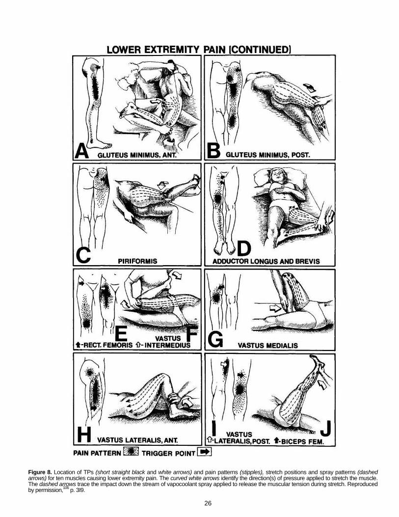
26
Figure 8. Location of TPs (short straight black and white arrows) and pain patterns (stipples), stretch positions and spray patterns (dashed arrows) for ten muscles causing lower extremity pain. The curved white arrows identify the direction(s) of pressure applied to stretch the muscle. The dashed arrows trace the impact down the stream of vapocoolant spray applied to release the muscular tension during stretch. Reproduced by permission,
106 p. 3I9.
27
a combination of both active gluteus minimus TPs and a radiculopathy that perpetuates them.
Sweeps of spray for treatment of the anterior portion of the gluteus minimus are applied along the anterior portion of the muscle belly and downward over the lateral thigh and leg to the ankle, including a detour to cover the lower buttock (Fig. 8A). The stretch position for this muscle requires the patient to be sidelying with progressive exten-sion and adduction of the thigh at the hip. The knee may be bent (Fig. 8A).
The direction of spray for the posterior gluteus minimus is downward from the crest of the ilium over the posterior buttock and thigh to include most of the calf. To stretch this muscle the patient is placed on the edge of the table with the shoulders flat against the table; the thigh being treated is flexed slightly, placed forward over the other leg and moved into adduction (Fig. 8B). Care must be taken to include the gluteus maximus muscle with sweeps of spray to eliminate any restricting TPs in that muscle.
Piriformis. An active piriformis TP restricts the combi-nation of adduction and internal rotation of the thigh at the hip. Trigger point tenderness is palpable both rectally and externally.
1 Piriformis TPs refer pain to the buttock lat-
erally and sometimes medially to the sacrum (Fig. 8C).46
The stream of vapocoolant is applied distally over the lateral and posterior hip and thigh to the knee as the mus-cle is stretched in the sidelying patient. The thigh is flexed at the hip to nearly 90° and internally rotated by elevating the ankle as the knee is lowered gently to increase adduc-tion at the hip, as seen in Fig. 8C.
The gluteus maximus muscle should be included in the spray pattern; it, too, is being stretched and may harbor restricting TPs.
Tension caused by taut bands due to TPs in the piri-formis muscle may entrap the peroneal part or all of the sciatic nerve depending upon anatomical variations of how the nerve passes under or through the muscle.
115
Adductores Longus and Brevis. In addition to the pain pattern represented in Fig. 8D, pain from the adductores longus and brevis may refer upward throughout the groin
116133 and/or downward to the anteromedial aspect of
the thigh just above the knee; referred pain may extend down to the ankle (Fig. 8D). Active TPs in this muscle markedly restrict abduction of the thigh and are a common source of groin and distal anterior thigh pain above the knee. To treat these muscles, the stream of vapocoolant is applied in upward sweeps across the anteromedial thigh and over the inguinal region, and then in downward sweeps across the lower anteromedial thigh, medial knee, and over the pain pattern covering the anteromedial leg to the ankle. As the spray is applied, the muscle is gradually stretched by placing the patient's foot against the oppo-site knee and slowly abducting the thigh at the hip as indicated by the white arrow in Fig. 8D.
Quadriceps Femoris. Rectus femoris TPs are usually located at the proximal end of the muscle near its muscu-lotendinous junction (solid black arrow, Fig. 8E) and refer pain distalward to cover the knee cap and sometimes the slightly distal half of the anterior thigh. The vastus inter-medius TP (small white arrow, Fig. 8E) is deeper than and distal to the rectus femoris TP; it refers intense pain locally and distally over the upper part of the thigh.
The vastus medialis TPs (Fig. 8G) refer pain to the medial aspect of the knee. The distal (more anterior) TPs of the vastus lateralis (solid black arrow in Fig. 8H) are frequently multiple and difficult to eliminate; they refer pain intensely and extensively over the lateral thigh, some-
times including the lateral buttock. The proximal (and more posterior) TPs of the vastus lateralis (small white arrow, Fig. 81) usually refer pain only to the vicinity of the TP.
To release TPs in the rectus femoris muscle, place the patient with the knee flexed and the hip extended in the sidelying position. The stream of vapocoolant is applied distalward from the anterior inferior iliac spine region over the muscle to and including the knee cap (Fig. 8F). For release, TPs in the vastus medialis require distalward sweeps of spray from the hip over the muscle to the knee (Fig. 8G). However, the proximal referral pattern of the vastus lateralis calls for upward sweeps of spray (dashed arrows, Fig. 8H).
Of the four quadriceps muscles, only the rectus femoris crosses two joints, the hip and knee. Therefore, only that muscle requires hip extension as well as knee flexion for full stretch; the three vasti require only knee flexion. The latter are stretched by bringing the heel to the buttock, a position that also stretches the posterior portion of the vastus lateralis muscle.
Biceps Femoris. Pain referred from TPs in this ham-string muscle (solid black arrow, Fig. 81) is projected to the back of the knee and sometimes extends over the proxi-mal calf.
Vapocoolant spray is applied in parallel sweeps distal-ward from the hip over the posterior thigh and knee to include the calf. Stretch can be obtained by slowly per-forming a straight leg raising manuever as vapocoolant is applied (Fig. 8J). Usually, effective stretch is obtained more quickly by initially abducting the thigh at the hip without raising the foot and applying the spray upward over the adductor magnus and inguinal region to release it. This massive adductor muscle is an important extensor of the thigh. The foot is then elevated to bring the thigh from the abducted to the 90° flexed position, applying parallel sweeps of spray over the length of the thigh; the spray should be applied progressively more laterally as more lateral muscles are stretched by gradually moving the flexed thigh into adduction.
135
Gastrocnemius. Myofascial TPs in the superficial, two-joint gastrocnemius muscle are usually found along either the medial or lateral border of the muscle at the level indicated by the solid black arrow in Fig. 9B. These TPs commonly refer pain over the calf and to the instep of the foot.
123' Active TPs in this muscle make walking uphill
painful and commonly cause nocturnal calf cramps.
For treatment, the patient is placed in a prone position with the feet hanging over the end of the treatment table and with the knees straight (Fig. 9B). The stream of spray is directed distally over the back of the thigh downward over the calf and heel covering the sole. The foot is strongly dorsiflexed using the operator's knee for powerful leverage (curved white arrow, Fig. 9B).
The patient is taught to self-stretch this muscle by standing with the involved leg behind the other and mov-ing the pelvis and body forward, bending the forward knee while keeping the knee of the affected leg fully extended and the heel solidly on the floor. Moving the pelvis and body forward dorsiflexes the foot and with the knee straight stretches the gastrocnemius muscle.
Soleus. Myofascial TPs in the second-layer, single-joint soleus muscle frequently are the cause of heel pain and tenderness that is mistakenly attributed to a heel spur (Fig. 9A). Occasionally, TPs in this muscle project pain to the area of the sacroiliac joint on the same s ide .
5 1 3 5
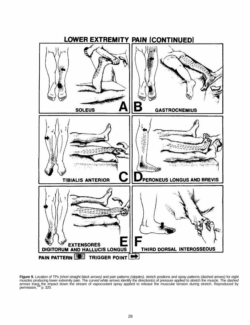
28
Figure 9. Location of TPs (short straight black arrows) and pain patterns (stipples), stretch positions and spray patterns (dashed arrows) for eight muscles producing lower extremity pain. The curved white arrows identify the direction(s) of pressure applied to stretch the muscle. The dashed arrows trace the impact down the stream of vapocoolant spray applied to release the muscular tension during stretch. Reproduced by permission,
106 p. 320.
29
A stream of vapocoolant is directed distally in parallel
sweeps covering the calf, the heel and most of the sole. The stretch position (Fig. 9A) is flexion of the knee with the patient prone. Progressive downward dorsiflexion pres-sure is applied to the ball of the foot to stretch this muscle (curved arrow, Fig. 9A). The patient should be taught self-stretch by standing and progressively bending the knee while keeping the heel on the floor. Progressive dorsifle-xion at the ankle with the knee bent selectively stretches the soleus but not the gastrocnemius muscle. To take up the slack as the tight soleus muscle releases, the patient slowly increases ankle dorsiflexion by further flexing the knee, while keeping the heel firmly on the floor.
Tibialis Anterior. This muscle dorsiflexes and helps to invert the foot. Its TPs occur at the proximal end of the muscle (Fig. 9C). They refer pain downward along the course of the muscle and its tendon to the medial aspect of the foot, concentrating on the great toe.
116123
To release anterior tibial TPs, a stream of vapocoolant is applied distalward over the muscle and its pain pattern on the shin, the dorsum of the ankle and the large toe. To stretch this muscle, the end of the foot is grasped firmly and simultaneously plantar flexed and slowly everted (white curved arrow, Fig. 9E) to pick up the slack as the spray helps to release muscle tension.
Peroneus Longus and Brevis. These adjacent muscles generally have indistinguishable referred pain patterns. The TPs in the peroneus longus are usually a few centi-meters distal to the common peroneal nerve as it passes over the fibula and under the peroneus longus muscle just distal to the fibular head (Fig. 9D).
Referred pain from the peroneal muscles projects behind the lateral malleolus and may extend distally over the lateral aspect of the leg and over the lateral aspect of the dorsum of the foot (Fig. 9D).
Treatment is initiated by application of vapocoolant spray distalward over the lateral aspect of the leg, ankle, and dorsum of the foot to the toes. The foot of the supine patient is strongly dorsiflexed and inverted (curved white arrow, Fig. 9D) to slowly take up the slack that develops as the muscle releases in response to the vapocoolant spray.
The tension produced by taut bands associated with TPs in the peroneus longus can entrap the deep peroneal nerve against the underlying fibula, producing character-istic loss of sensation and a degree of dorsiflexion weak-ness (foot drop). Inactivation of the TPs promptly relieves neurapraxia of the peroneal nerve caused by the taut bands. Trigger points in these muscles are perpetuated by the stress of walking on the "knife-edge" produced by a long 2nd metatarsal bone (D. J. Morton foot configuration).
134
Extensores Digitorum and Hallucis Longus. These TPs, like those of the peroneal muscles, refer pain that includes the dorsum of the foot laterally (Fig. 9E). In addi-tion, this pain may extend part way up the leg or downward to include the toes.
With the patient supine, vapocoolant is applied distally over the length of the muscles from the knee over the anterolateral aspect of the leg, and over the pain reference zones on the ankle, dorsum of the foot and the toes. These toe extensors are stretched together by simulta-neously plantar flexing the foot and all toes, as illustrated in Fig. 9E.
Interossei of the Foot. As in the hand, active TPs in the dorsal interossei of the foot are not rare. These TPs refer pain distally to the side of the toe corresponding to the attachment of the involved muscle (Fig. 9F). However, in
the foot, interosseous TPs are commonly associated with hammer toe and, if not too long-standing, this joint restric-tion can be alleviated by inactivating the TPs. The taut bands are exquisitely tender. The dorsal interosseous TPs are readily palpable against the metatarsal bones and, once localized, easily injected. Stretch and spray is not reliably effective in these muscles. Full stretch may be difficult to achieve.
PATIENT EDUCATION
Characteristically, one stress activates a TP and other factors or conditions perpetuate it. To effectively manage an acute MPS that has persisted and propagated into widespread chronic involvements, one must clearly distin-guish perpetuating factors from the stress(es) that initially activated the TPs.
Acute single-muscle myofascial syndromes are much less likely to recur and become chronic if the patient understands the myofascial origin of the pain. The patient then learns what muscle caused the pain and what move-ments can overload that muscle. Acute onset is often associated with an accident or fall, when specific muscles were overloaded by lengthening contractions used to cushion impact. Sometimes the TPs were initiated by muscle overload stress resulting from a specific move-ment or positioning that the patient should learn to avoid. This is particularly true when the muscle is commonly used in a repetitive movement that would also perpetuate its TPs.
When the patient presents with a clear-cut MPS, the patient must understand that the pain is of muscular origin and not due to nerve damage or bone changes. By having the patient note the exact position of the part of the body being moved before and after stretch, patients can see the increase in range of motion for themselves, can experi-ence the increase in function and can relate these to the reduction in pain. Emphasizing that the treatment that pro-vided relief changed only the muscles and did nothing to the nerves or bones helps the patient to believe that the pain originated in their muscles.
Reproduction of the patient's pain by pressure on a TP convinces both the patient and the operator that the TP is responsible for at least part of the patient's pain com-plaint. Frequently, patients with chronic pain are as con-cerned about its origin and prognosis as with the pain itself. Learning the muscular cause of the pain and how to relieve it is profoundly reassuring.
Not uncommonly, patients will return discouraged because they still have pain and, therefore, feel that their last treatment was of no help. When an accurate detailed drawing is made of their current pain patterns and com-pared with their previous drawing, it becomes apparent that the pattern of pain has changed significantly; the pat-tern of pain produced by those muscles treated previously is gone. These patients are only aware of the fact that they still have pain, but had not noticed precisely where it was located and, therefore, failed to recognize their progress. Many times, a previous pain emerges that comes from less irritable TPs which have been uncovered — like peel-ing off the outer layer of an onion.
Whenever possible, patients are encouraged to con-tinue their accustomed activities but in a manner that avoids overloading vulnerable muscles. Patients learn to use, not abuse their muscles. Correction of an anatomical or postural perpetuating factor frequently requires change in patient behavior; unless patients fully understand the relationship between that perpetuating factor and the
30
cause of their pain, they quickly revert to previous behav-ior and then wonder why the pain has returned.
OTHER STRETCH TECHNIQUES
Muscle stretch can be achieved in several ways: by stretch and spray, which was described above; by post-isometric relaxation; by ischemic compression; and by deep friction massage. Additional modalities can be help-ful. Low-intensity ultrasound applied directly to the TP is valuable when the TP is otherwise inaccessible. Some find electrical stimulation over the TP helpful; high voltage galvanic stimulation is usually effective.
Post-isometric Relaxation. Use of voluntary contrac-tion alternated with passive stretch for releasing tight mus-cles has been identified by many names. Physical therapists frequently refer to contract-relax or rhythmic stabilization. Osteopathic physicians are likely to speak of muscle energy techniques and myofascial release.
Post-isometric muscular relaxation, as described by Lewit and Simons
68 is simple and effective. It combines
nicely with the stretch-and-spray technique and is most valuable when used by the patient for self-treatment at home. The muscle is gently stretched to the onset of resis-tance (take up the slack) and held there isometrically. For the next 3-7 seconds, either the operator or the patient exerts fixed resistance against which the muscle gently contracts in that position at approximately 25% of maximal effort. While the same position is passively maintained, the patient "lets go" (relaxes the contracting muscle). Only after the patient has thoroughly relaxed is the muscle slowly, gently and passively extended, taking up the slack that developed. This contract-relax cycle is repeated three to five times. Full release of tension may not occur until after the second or third cycle. Relaxation is facilitated by having the patient slowly take a full (complete) breath and then slowly exhale through pursed lips, emptying the lungs to maximal exhalation. During exhalation, the patient concentrates on total relaxation of the muscle to be stretched.
68 Additional relaxation and release may be
achieved by downward gaze during exhalation.67
Ischemic Compression. Ischemic compression or "thumb therapy' is non-invasive and effective, but painful.
106 This technique may be applied by the operator
or as self-treatment by the patient. Pressure is applied directly on the spot of greatest tenderness (the TP) with a steady moderately painful (tolerable) pressure. As the pain eases, the pressure is increased to maintain approxi-mately the same level of discomfort. When the TP is no longer painful (after 15 sec to a minute of pressure) the pressure is released and full active range of motion per-formed.
Immediately after release, blanching of the skin is fol-lowed by reactive ftyperemia at the site of pressure. Very likely the same hyperemia is present in the muscle itself and is part of the benefit. Some therapists apply less pres-sure for a shorter time, but with repeated applications on successive days until TP tenderness is obliterated and the referred pain disappears.
Massage. Deep muscle massage (deep friction or strip-ping massage) can effectively inactivate TPs. Stripping massage require^ lubrication of the skin and the applica-tion of firm bilateral thumb pressure by sliding the thumbs slowly, progressively along the length of the taut band. The band is firmly compressed between the thumbs. The slowly progressive "milking" action relieves the sense of induration at the TP after several repetitions and releases the tautness of the palpable band. This technique is pain-ful, but effective in superficial muscles. A confirmation
and extension19
of an earlier study18
found that repeated massage of tender tense areas in the muscles of myofa-scial pain patients effectively relieved their pain and elim-inated the areas of abnormal tenseness in 21 of 26 patients. Elevation of serum myoglobin levels following massage decreased as the amount of remaining indura-tion decreased with successive treatments.
1819
INJECTION AND STRETCH
Treatment by injection of TPs is selected initially when the TPs are inaccessible to stretch therapy or because of mechanical restriction of joint motion. It is used on TPs unresponsive to the foregoing non-invasive methods. Dry needling
65'123
'134
and isotonic saline injection134
also are effective; without a local anesthetic, dry needling is likely to be more painful. Isotonic saline for injection usually contains 0.9% of the preservative and local anesthetic, benzyl alcohol. The flushing effect of the injected fluid on sensitizing agents is probably also important.
Needle penetration of the skin can be painless if the needle is inserted very rapidly with a flick of the wrist after the antiseptic alcohol has dried. Stretching the skin until it is tight reduces skin sensitivity to needle prick. Alternately, the site may be chilled for six to eight seconds with a stream of Fluori-Methane* applied in a figure of eight pat-tern with the cross-over at the point of needle penetration.
134138 The site of injection is chilled just short of
frosting the skin.
The rationale of needle therapy depends primarily on the disruption of the self-sustaining TP mechanism. This approach requires precise localization and penetration of the TP with the needle. Injection of 0.5% procaine in iso-tonic saline without epinephrine reduces the severe, sometimes devastating, pain of TP penetration. Important-ly, it preserves the tenderness to palpation of the remain-ing TPs and thus permits detection of any TPs that were overlooked. The local analgesic effect of the 0.9% con-centration of benzyl alcohol preservative in the saline for injection permits its substitution for procaine in those few patients who are allergic to procaine. Half percent lido-caine also is less desirable than procaine but can be used, if necessary. Long acting local anesthetics are avoided because they obliterate all local TP tenderness and pro-duce muscle necrosis.
134
When injecting muscles next to the ribs, the needle must not penetrate between the ribs, which easily pro-duces a pneumothorax.
To perform an injection, the tender TP and its taut band are localized between the fingers. The needle is then directed precisely into the TP.
4 Contact with the TP is
confirmed by a jump response of the patient and a local twitch response of the taut band in the muscle. Probing should continue until a response has been obtained or the region has been fully explored with the needle. Tender-ness to palpation must be relieved. Pressure is applied during and after injection to insure hemostasis.
Procaine injection is followed by stretch and spray to release any TPS that were missed by the needle. Appli-cation of moist heat by a hot pack or wetproof heating pad for five minutes immediately after injection helps to avoid post-injection soreness that otherwise may last for two or three days. The patient should be warned of this possible soreness; aspirin relieves it. The moist heat is immediately followed by several cycles of active full range of motion in
*From Travell JG and Simons DG: "Myofascial Pain and Dysfunction:
The Trigger Point Manual," Williams & Wilkins, Baltimore, I983, pg. 3.

31
both the shortened and stretch positions of the treated muscles to reestablish normal function. Passive self-stretch range of motion exercises at home are instituted routinely. This stretching helps to maintain the increased range of motion obtained by treatment.
PERPETUATING FACTORS
The presence or absence of perpetuating factors deter-mines the answer to the question, "How long should the effect of specific myofascial therapy last?" In the absence of perpetuating factors, relief should last indefinitely until the TP is reactivated, as in the beginning, by another over-load stress. In the presence of perpetuating factors, relief is temporary; lasting relief depends on eliminating perpet-uating factors
3642 so that the effect of successive therapy is
cumulative.
In a few patients, the muscles become so hyperirritable due to perpetuating factors that any attempt at specific myofascial therapy aggravates the pain. The perpetuating factors must be addressed first. In severe cases, perpet-uating factors are usually multiple.
A significant perpetuating factor may have caused no symptoms before activation of the TPs. A leg length dis-crepancy of 6 mm (
1/4") may have caused no pain or dis-
comfort throughout most of a lifetime; upon activation of quadratus lumborum TPs by another stress, the discrep-ancy becomes a potent perpetuator of thoseTPs.
Perpetuating factors are identified as either mechanical or systemic factors.
Mechanical perpetuating factors are ubiquitous, and systemic perpetuating factors are very common. Elimina-tion of one or several, but not all, of the factors may pro-vide only modest improvement in the therapeutic response until the remaining factors are resolved. The more thoroughly all significant perpetuating factors are managed, the more effective and longer lasting the treat-ment becomes.
Mechanical Perpetuating Factors. The columns in Table 2 list specific mechanical perpetuating stresses jux-taposed beside the muscle or muscles most likely to be affected by each stress. Systemic perpetuating factors may relate to any or all skeletal muscles. Mechanical per-petuating factors include anatomical variations, seated and standing postural stress, life style and vocational stress.
Anatomic Variations: One common anatomic variation is a short leg and/or small hemipelvis,^
34 which must be
corrected by a heel lift and/or butt lift for lasting relief of low back
115 and sometimes head, neck and shoulder pain
due to myofascial TPs.
The relatively common phenomenon of short upper arms is frequently an unrecognized perpetuator of TPs in the shoulder elevator muscles. This variation is corrected by providing elbow rests or pads that modify the furniture to fit the individual.
134
The long 2nd metatarsal, or D.J. Morton foot configura-tion, throws the foot off balance due to the knife-edge effect during toe-off that disturbs gait and overloads lower extremity muscles. This is corrected by inserting a toe pad under the head of the 1st metatarsal bone.
134'
Seated postural stress: This source of stress on the muscles may be induced by a hard smooth mat under an office chair, a chair seat too high for heels to reach the floor, the lack of a firm back support and persistent head-forward posture.
A hard smooth mat, such as plexiglass, makes the office chair with free castors glide readily whenever its occupant changes position or exerts the slightest pressure against the desk. The long toe flexors and intrinsic foot muscles try to grasp the slick floor; the effort overloads and perpetuates TPs in these muscles.
Another seated postural stress is caused by a chair seat too high for that individual's leg length, leaving the heels dangling off the floor. This causes under-thigh compres-sion of the hamstrings and chronic shortening of the soleus muscle. Both effects are perpetuating factors for TPs and can be avoided by providing a suitable footrest (book, pillow or small footstool).
Sitting in a chair with the back unsupported may be caused by a seat: that is too long from front to back, that is flat and provides no lumbar support, that supplies no scapular contact or that has a backrest with inadequate backward angulation. In a seat that is too long from front to back, the calves encounter the front of the seat prevent-ing the buttocks from reaching the backrest. The backrest should be contoured to support a normal lumbar lordosis. This also corrects the head-forward posture by correcting the thoracic kyphosis induced by an abnormally flattened back, thereby balancing the head erect over the shoulders without muscular effort. Scapular contact with the back-rest and backward angulation of the backrest help to carry the weight of the head and shoulders and to stabilize the spine, relieving the quadratus lumborum and paraspinal muscles.
Standing postural stress: The head-forward posture is also induced by weight bearing on the heels and relieved, when standing, by shifting the the center of grav-ity forward onto the balls of the feet, restoring the normal lumbar curve. This spinal posture permits the person to hold the head erect, balanced over the shoulders without muscle strain. It elevates the chest and restores normal postural relations by swinging the scapulae backward to their normal resting position and thus relieving the persis-tent shortening of the pectoral muscles.
134 This improved
posture takes a major load off the posterior cervical mus-cles, which in the head-forward posture must hold the weight of head against the pull of gravity. To many neck and shoulder-girdle muscles, the head-forward posture is a powerful perpetuating factor that requires a major change in patient behavior.
A canted running surface is common on the slanted beach of the seashore or on a curved track. It produces the same effect as a short leg
134 that tilts the pelvis. The
tilt must be compensated by the quadratus lumborum and/or paraspinal muscles, causing a persistent overload that perpetuates TPs in these muscles.
Vocational stress: Sustained shoulder elevation com-monly overloads the upper trapezius and levator scapulae muscles, perpetuating their TPs. Typists and other work-ers using their hands in a relative fixed elevated position are prone to maintain their shoulders in a shrugged posi-tion to help elevate the hands to the level of their work. The work should be lowered or the patient's body raised.
Prolonged arm abduction similarly overloads the supraspinatus and deltoid muscles. Elbow support should be provided. Workers developed myofascial syndrome of the supraspinatus and upper trapezius muscles due to frequent repetitive movements stressing those muscles. When tested, these painfully involved muscles had shorter duration of endurance and more rapid onset of electro-myographic evidence of fatigue than non-painful muscles.
44 The authors related their findings to alteration
in muscle metabolism due to ischemia.

32
TABLE 2. Muscles most likely to be affected by specific mechanical perpetuating factors.
STRESS MUSCLES REFERENCES
Anatomic Variations
Short Leg and/or Small Hemipelvis
Quadratus lumborum T-L Paraspinals Shoulder-girdle and neck-righting Masticatory
134(Chap.4), 135(Chap.4) 134(Chap.48) 134(Chaps.7,19,20) 134(chaps.8-12)
Short Upper Arms Levator scapulae Upper Trapezius Rhomboids
134(Chap.4,19) 134(Chap.6) 134(Chap.27)
Long Second Metatarsal (D.J.Morton foot configuration)
Peroneus longus Vastus medialis Gluteus medius
134(Chap.4), 135(Chap.2O) 135(Chap.14) 135(Chap.8)
Seated Postural Stress
Hard Smooth Mat Long Toe Flexors Foot intrinsics
135(Chap.25) 135(Chap.27)
Heels Dangling Hamstrings Soleus
135(Chap.16) 135(Chap.22)
Back Unsupported no backrest contact no lumbar support, no scapular contact
Quadratus lumborum T-L Paraspinals Pectoral is major Rhomboids
114,135(Chap.4) 134(Chap.48) 134(Chap.42) 134(Chap.27)
Standing Postural Stress
Head-forward Posture Pectoralis major and rhomboids Posterior cervicals
134(Chap.42)
134(Chap.16) Canted Running Surface Quadratus lumborum
Scaleni 114,135(Chap.4) 134(Chap.2O)
Vocational Stress
Shoulder Elevation Upper trapezius Levator scapulae
134(Chap.6) 134(Chap.19)
Arm Abduction Supraspinatus Deltoid
134(Chap.21) 134(Chap.28)
Hand supination Supinator 134(Chap.36)
Grasp Finger extensors Finger flexors
134(Chap.35) 134(Chap.38)
Systemic Factors
Any Muscular Function All muscles 134(Chap.4)
33
Overload of hand supination as when playing tennis or
using a screw driver readily overloads the supinator mus-cle. The symptoms produced by active TPs in the supina-tor muscle are frequently labelled epicondylitis or tennis elbow. The arm should not be fully extended at the elbow when playing tennis, since that eliminates the forceful supinator function of the biceps brachii muscle.
Strong grasp overloads the finger extensors because vigorous extensor function is an essential part of grasp. Active TPs in these muscles tend to cause a painful and weak grip. Items are likely to slip out of the grasp unex-pectedly due to unpredictable reflex inhibition. The supi-nator, the wrist and finger extensor muscles are commonly involved together.
Systemic Perpetuating Factors. Systemic perpetuat-ing factors can aggravate TPs in any muscle and increase the irritability of all skeletal muscles rendering them more vulnerable to the development of secondary and satellite TPs.
134 Systemic factors include enzyme dysfunction,
metabolic and endocrine dysfunction, chronic infection or infestation, and psychological stress.
Correction of a significant perpetuating factor reduces irritability of the muscles, which results in less pain and/or improved responsiveness of the muscles to specific myo-fascial therapy.
Muscle is an energy engine. It converts the energy of a high-energy molecule, adenosine triphosphate (ATP), to mechanical movement by converting it to a lower-energy molecule, adenosine disphosphate (ADP). Understand-ably, anything that interferes with energy metabolism of the muscle would tend to compromise this function and thereby would increase both muscle irritability and sus-ceptibility to TPs.
Enzyme dysfunction: The nutritional inadequacies that most commonly perpetuate myofascial TPs are lack of B-complex vitamins, particularly B.,, B6, B12 and folic acid. The detailed metabolic enzymatic functions and congenital deficiencies of each of these vitamins has been reviewed in detail.
134 Low electrolyte levels, potassium
and calcium may be critical and minerals such as calcium, copper, and iron are essential. Vitamin deficiency is sig-naled by abnormally low laboratory values and by clinical symptoms that are ascribable to a lack of that vitamin. Vitamin inadequacy is a suboptimal level, that may pro-duce only a partial picture of deficiency or simply impair muscle function by increasing its irritability and tendency to develop TPs.
The prevalence of unrecognized vitamin deficiency is remarkably high, especially in hospital patients. Among 120 hospital patients, 88% had abnormally low levels in one or more of 11 vitamins.
4 Despite this high prevalence,
the history of dietary intake was inadequate in only 39%. More than half of the patients were low in two or more vitamins. Serum folate, which was the commonest vitamin deficiency, was low in 45% of these patients. Symptoms of vitamin deficiency was clinically apparent in only 38% of these patients.
4 How many more of these patients had a
vitamin inadequacy was not determined.
Clinical experience shows that the lower the serum value within the lower quartile of the "normal" range, the more likely it is that this degree of vitamin inadequacy contributes significantly to increased muscle irritability, and that it will require correction for lasting relief of that patient's chronic TP pain.
Vitamin dependence is observed in a few babies who are born with severe congenital deficiency of an enzyme that requires that vitamin as its coenzyme. Such defects
require the ingestion of pharmacological megadoses of the vitamin to sustain life; the specific enzyme defects and their chemical recognition have been summarized for each of the B vitamins considered here.
134 However, unex-
plored is the prevalence of milder degrees of such con-genital enzyme deficiencies that could multiply by many times the minimum vitamin requirement of individuals who have become progressively deficient in that enzyme as they reach middle age. The serum vitamin level might appear to be safely in the mid-normal range for these individuals. The wide range of individual variation in the requirements for essential nutrients including vitamins is well established.
140
All of the B complex vitamins and vitamin C are water soluble. They have remarkably low toxicity because an excess is quickly excreted in the urine. This is not the case for the fat soluble vitamins A, D, and E, which readily accumulate to toxic levels. Vitamin A toxicity is not uncom-mon and can be a source of pain.
The common assumption that adequate dietary inges-tion assures an adequate metabolic supply of a vitamin does not consider the many causes of vitamin insufficien-cy. They include not only inadequate ingestion of the vita-min, but also impaired absorption, inadequate utilization, increased metabolic requirement and increased excretion or destruction within the body.
143 In addition, the usual
selection process for individuals who serve as controls to establish normal values does not screen out individuals with marginal insufficiency, including many who show chemical evidence of vitamin deficiency and depletion of stores.
2 One should expect, therefore, that published nor-
mal values are not optimal values. This helps to explain why, clinically, the lower quartile of "normal" is often a zone of inadequacy for muscle metabolism.
A characteristic neurological finding in vitamin B, inad-equacy is increasingly severe loss of vibration sense at progressively more distal sites on the upper and lower extremities. By comparing the time of loss of sensibility at successively more proximal levels following one activation of a long-period tuning fork, one can demonstrate progres-sively lower thresholds of response at successive proximal sites on the extremities.
The recommended daily allowance (RDA) for Thiamine (vitamin B,) is dependent on the daily energy expendi-ture. The requirement for vitamin B-, is greatest when car-bohydrate is the source of energy. It is an essential enzyme for the entry of pyruvate (end-product of anerobic metabolism) into the Krebs cycle for oxydative metabolism (the chief source of energy in muscle). The critical symp-toms of severe thiamine deficiency are recognized as wet beriberi (heart muscle failure) or dry beriberi that includes severe skeletal muscle weakness and serious central and peripheral nervous system dysfunction.
84
Impressively so, pyridoxine (vitamin B6) is a jack of all trades: an essential coenzyme to more than 60 enzymes in human metabolism. It is essential for the metabolism of numerous amino acids including the methionine to cysteine pathway, blockage of which leads to homocysti-nuria. It plays an important conformational or structural role in the enzyme phosphorylase. This enzyme is essen-tial for the release of glucose from glycogen, which is a necessary first step for all anerobic (glycolytic) metabo-lism in skeletal muscle. Pyridoxine is essential to the syn-thesis or metabolism of nearly all neurotransmitters. This correlates with the deterioration of mental function seen in experimental pyridoxine deficiency. It is required for the synthesis of nucleic acids, which are required for messen-ger RNA (ribonucleic acid) and, therefore, essential for normal cell reproduction. It is critical in the synthesis of at
34
least 10 hormones including insul in and growth hormone.
134 This list is by no means complete.
Symptoms of vitamin B6 deficiency include skin symp-toms of dermatitis, glossitis and stomatitis; nervous sys-tem dysfunction that can lead to convulsions and peripheral neuritis; connective tissue swelling (carpal tun-nel syndrome); and severe compromise of erythropoiesis that produces hypochromic anemia in experimental animals.
17 Several classes of drugs are well known to
increase the demand for vitamin B6. They include antitu-bercular drugs, oral contraceptives, the chelating agent penicillamine, anticonvulsants, corticosteroids, and excessive alcohol consumption.
134 Severe depletion of
vitamin B., in chronic alcoholism is well recognized and is not uncommon in the heavy social drinker.
Both thiamine and pyridoxine are widely distributed in nature, but in limited amounts. Vitamin B^ is rapidly destroyed by heat in neutral and alkaline solutions. It is stable in acidic solutions, but only to boiling temperature. Vitamin B1 is quickly leached out of food during washing or boiling. Pyridoxine suffers substantial losses during cooking and is quickly destroyed by ultraviolet light (sun-light) and oxidation (when held on a steam table). Ribofla-vin (Vitamin B2) and ascorbic acid (Vitamin C) are destroyed by fluorescent light.
Both cobalamin (vitamin B12) and folate play an essential role in the synthesis of deoxyribonucleic acid (DNA) that is required for the maturation of erythrocytes and, therefore, for oxygen transport. Vitamin B12 is also essential for fat and carbohydrate metabolism; this may account for its importance to the integrity of the peripheral nervous system.
Both vitamin B12 and folate deficiency characteristically cause megaloblastic anemia, but only vitamin B12 pro-duces serious peripheral nervous system deficits. Many clinicians have been accustomed to basing vitamin ther-apy on the response of a patient's hematopoietic system. If the anemia is caused by vitamin B12 deficiency, but the patient is being treated with folic acid, the hematological picture will revert to normal with no improvement in, or even exacerbation of, the neurological deficits. This approach has lead to permanent neurological damage. For this reason, the Federal Drug Administration limits non-prescription folic acid to 400 micrograms per dose.
There is no need to guess. Laboratory testing for serum vitamin levels of both vitamin B12 and folate are readily available and usually leave no doubt as to what needs correction. Every chronic myofascial pain patient deserves these tests. Vitamin B12 tests err on the high side. The patient's serum level is likely to be lower, not higher, than the reported value.
134
The metabolic interdependence of vitamin B12 and folate produces a reciprocal therapeutic effect. An exam-ple is the methyl folate trap in which folate metabolism is blocked for lack of vitamin B12.
47 Folate may actually accu-
mulate to unexpectedly high serum folate levels. Admin-istration of vitamin B12 can precipitously drop the serum folate level and deplete what initially appeared to be an adequate reserve. A reverse effect may also be seen: a 20 or 30% drop in the serum vitamin B12 level following oral supplementation of badly needed folic acid.
Vitamin B12 is rarely deficient in the diet except in strict vegetarians. This vitamin is synthesized by bacteria and obtainable only in food products that have been contami-nated by or have been affected by bacterial action. Source foods include practically all animal products and some legumes.
47
An adequate serum cobalamin level is dependent on adequate ingestion, on gastric secretion of intrinsic factor, on adequate intestinal absorption (which is compromised by Meal disease), on reabsorption of much of the vitamin B12 that is secreted in the bile, and on adequate amounts of transport transcobalamines in the gut wall. Only if all of these are normal is daily ingestion of 3 - 5 micrograms of cobalamin sufficient.
Conversely, folates are widely distributed in many foods in modest amounts, particularly leafy green vegetables (foliage), and are very readily destroyed by processing and cooking. Folate is highly vulnerable to destruction by heat and oxidation; generally, 50-95% of the folate in food is destroyed in processing and preparation. Folate defi-ciency is the most common vitamin deficiency, especially in the elderly and those eating institutional cafeteria-style meals. Consistently over a period of years, three-fourths of the select group of patients referred to a chronic myofa-scial pain clinic had inadequate levels of vitamin B-,, or folate (within the lower quartile of "normal" or lower).
10
Serum levels of vitamins B1 and B6 are expensive and sometimes difficult to obtain. The vitamin B12 and folate tests are readily obtainable. Supplementation with I-3 mg of folic acid daily, p.o., should bring the folate level to at least the mid-normal range within 2 or 3 weeks. A one mg (I000 ug) daily oral supplement of vitamin B12 will usually restore it to mid-range within 4 to 6 weeks, thus avoiding the necessity for injection. This dose is several hundred times the RDA, but totally innocuous. The administration of either vitamin B12 or folic acid alone can be hazardous because of their reciprocal relationship, unless the serum level of the other is safely above the mid-normal range. One cannot predict one vitamin inadequacy based on another, but the demonstrated inadequacy of one or sev-eral vitamins and temporary response to treatment should increase suspicion. The administration of a balanced B-complex supplement up to ten times RDA (B-50 dosage) should ensure that minor problems of diet, absorption and increased demand are met. Also, adverse interactions caused by excessive administration of one vitamin will be avoided. This philosophy of providing an excess is not applicable to the fat soluble vitamins A, D, and E. Quite the contrary, one source of increased muscular irritability appears to be above-normal serum vitamin A levels. Chronic MPS patients taking a total of more than 30,000 i.u. (including dietary intake) of vitamin A daily should have this serum level tested.
For different reasons, absorbic acid (vitamin C) is important to MPS patients. It is essential for hydroxalation of the amino acids lysine and proline to form the protocol-lagen molecule. Without it, the integrity of the connective tissue is compromised. In the absence of vitamin C to provide the collagen needed for strong vessel walls, the patient experiences marked capillary fragility and easy bruising. Capillary fragility leads to ecchymoses following injection of TPs. Ecchymoses are unsightly and irritating to the muscle.
Smoking markedly increases the oxidation of vitamin C, rapidly depleting it. One should beware of injecting TPs in smokers unless they have taken at least two grams of timed-release vitamin C daily for a minimum of 3 days prior to treatment.
Vitamin C is also of clinical importance to the muscles because 500 mg of timed-release vitamin taken at the time of excessive muscular activity can prevent much poste-xercise muscle soreness and stiffness.
Metabolic and endocrine dysfunction: The metabolic factors of gout, anemia, low electrolyte levels and hypogly
35
cemia increase muscle irritability and symptoms from TPs, as do also the endocrine disturbances of hypometa-bolism and estrogen deficiency.
The monosodium urate crystals of gout are less soluble in the acidic media of injured tissues than in blood, and hence are deposited in areas of tissue injury and meta-bolic distress such as TPs. Patients with a gouty diathesis respond better to treatment when the hyperuricemia is under control, and generally respond better to injection than to stretch and spray. Vitamin C in relatively large amounts (one to four grams per day) is an innocuous and effective uricosuric agent.
57 The hyperirritability of TPs in
the muscles of some patients with serum uric acid levels in the high normal range subsides remarkably with urico-suric therapy.
From the muscle's point of view, anemia of any cause is a serious metabolic problem because the muscle depends on oxygen to sustain oxidative metabolism essential for meeting the bulk of its energy needs.
Abnormally low electrolyte levels of ionized calcium and potassium seriously disturb muscle function and increase muscle irritability, apparently because of their critical roles in the contractile mechanism. Serum ionized calcium is the essential measure. The total calcium that is ordinarily included in a blood chemistry profile correlates poorly with the ionzed calcium.
The occurrence of hypoglycemia would intensify the metabolic distress of the muscle and it clearly aggravates myofascial TPs. Stretch or injection therapy should be deferred in patients while they are hypoglycemic, treat-ment then is likely to aggravate rather than relieve their symptoms. A packet of powdered soluble gelatin prepared as a drink is a handy source of available carbohydrate with enough protein to avoid a subsequent hypoglycemic reac-tion.
Evidence of hypometabolism is found in some treatment-refractory patients with persistent active myofa-scial TPs. Their serum folate levels should be up to at least the mid-normal range; this is important because folate inadequacy can cause symptoms resembling those of low thyroid function and is readily corrected. Confusion arises because laboratory tests of thyroid function are usu-ally low normal and dismissed as being within normal lim-its. These patients have marginally low T3 uptake and low to midrange T4 by radioimmune assay (RIA). In this group, insufficient thyroid function is revealed by a low basal met-abolic rate
124 or a low basal body temperature,
6 by elevation
of the serum cholesterol level and by the response to thyroid supplementation.
The basal temperature is obtained with an ovulation thermometer placed in the axilla daily by the patient for 10 minutes before arising after sleep. Normally, the basal temperature averages more than 36.1 C (97.OF). The far-ther the basal temperature is below this value, the more vulnerable the patient is to hyperirritable TPs in the mus-cles and often to depression. Basal temperature in ovulat-ing women is reached immediately after menses.
Sonkin124
demonstrated that, with thyroid therapy, those patients needing supplemental thyroid consistently recovered their energy and positive outlook on life and had an increased basal metabolic rate with a decreased serum cholesterol value. Their muscles became less vul-nerable to myofascial TPs and were more responsive to s p e c i f i c TP therapy. Travell corroborated these observations.
131
Thyroid supplementation is contraindicated in patients with known cardiac arrhythmias or known myocardial dis-
ease that compromises cardiac reserve. Thyroid medica-tion increases vitamin B1 and estrogen requirements and may increase blood pressure. Overmedication causes symptoms of hyperthyroidism. Adjustment of dosage in these patients is dependent largely on clinical judgement and responses of basal metabolic rate and basal temper-ature. The site of metabolic dysfunction is apparently at the level of intracellular utilization and is poorly reflected in serum hormone levels.
Thyroid supplementation for those patients who meet the criteria described by Sonkin
124 remains controversial
among endocrinologists, but of critical importance to those patients in whom this is a major that is perpetuating severe myofascial pain.
Chronic infection and infestation: Viral disease, bac-terial infection and parasitic infestation can perpetuate MPS.
During a systemic viral illness, including the common cold or attack of "flu", the irritability of myofascial TPs increases markedly. One of the most common sources is an outbreak of Herpes Simplex virus type I; however, nei-ther Herpes Simplex virus type II (genital Herpes) nor Herpes Zoster are known to aggravate MPS. Herpes virus type I can cause the common aphthous mouth ulcer, can-ker sore, or cold sore. It may also appear on the skin of the body or extremities as isolated vesicles filled with clear fluid. Lesions have been reported in the esophagus, and the symptoms of vomiting and diarrhea strongly implicate gastrointestinal involvement comparable to that of the mouth.
No drug is known to cure Herpes Simplex Virus type I, but a multi-pronged attack can greatly reduce the fre-quency and severity of recurrences. A daily dose of 300 -500 mg of niacinamide reinforces mucous membrane resistance. Three tablets (or 1 packet) of viable lactoba-cillis two or three times daily for at least a month helps to reestablish the normal intestinal bacteria, reducing the chance of an intestinal viral outbreak. Local therapy is applied by rubbing an antiviral ointment into the skin or mouth lesions 3 times daily, which accelerates resolution of the lesion.
Persistent bacterial infection tends to exacerbate mus-cle irritability. A chronic infection such as an abcessed tooth, infected sinus or chronic urinary tract infection can be a major perpetuating factor. Chronic sinusitis may arise from both infection and allergy. Normal erythrocyte sedi-mentation rate and C-reactive protein tests help to elimi-nate the possibility of chronic infection.
A parasitic infestation should be considered in travel-ers exposed to conditions of poor sanitation and among active homosexuals, as a likely perpetuator of myofascial pain. The worst offender is the fish tapeworm; next is giar-diasis, and occasionally amoebiasis. The first two tend to impair absorption of or consume vitamin B12 and the amoebae may produce myotoxins that are absorbed. The diagnosis of infestation is investigated by three stool examinations for occult blood, ova and parasites.
Posttraumatic hyperirritability syndrome: The group of myofascial pain patients with posttraumatic hyperirrita-bility syndrome suffer greatly, are poorly understood and are difficult to help. They respond to strong sensory stim-uli much differently than most patients. Following a major impact to the body and/or head, the muscles exhibit marked hyperirritability of TPs and a distressing vulnera-bility to strong.sensory stimuli. The trauma has usually been an automobile accident or fall that was sufficiently severe to have inflicted some degree of damage to the sensory pathways of the central nervous system. These

36
patients describe constant pain that is easily augmented by any strong sensory input including severe pain, a loud noise, vibration, prolonged physical activity and emotional stress. It may take days or weeks to recover from a degree of trauma or noise that to most people would be inconse-quential. From the date of onset, coping with pain has suddenly become the focus of life for these patients who previously paid no particular attention to pain. Their func-tion is impaired by a marked increase in pain and fatigue if they exceed their restricted limit of activity.
One of the distinguishing characteristics of patients with the posttraumatic-hyperirritability syndrome is their loss of tolerance to what are to most people trivial mechanical stresses such as jarring, vibration, loud noises and mild bumps or thumps. Exposure to such a stimulus immedi-ately produces an increase in the pain level. Most distinc-tive, the stimulus also causes a markedly increased sensitivity to subsequent stimuli so they suddenly become much more vulnerable to further aggravation of their mis-ery. This increased arousal of the sensory system sub-sides slowly. It may take hours, days, or weeks— depending on the intensity of the stimulus—for this increased excitability of the sensory system to subside to its previous state. A strong sensory input appears to mod-ulate the excitability of the arousal system. This increased excitability is paralleled by a corresponding increase in irritability of all of that patient's myofascial TPs.
The target area of TPs and pain tends to concentrate in the somatic distribution of the brainstem, cervical cord or lumbosacral cord. A few unfortunate individuals seem to have involvement of several regions. These patients are highly vulnerable to reinjury by additional trauma. It takes much less subsequent impact to exacerbate the whole process as compared to an initial accident.
The most effective treatment approach has been to inactivate all identifiable TPs and to correct perpetuating factors. On occasion, it may be necessary to reset the system by suppressing central nervous system excitability. To date, barbituates have been found most effective.
Psychological stress: It is generally agreed that among chronic pain patients malingering is rare, a few percent at most.
51 Much controversy surrounds the question: "Is the
chronic pain an expression of the patient's psychological dysfunctions or is the pain driving the patient crazy?" Patients who experience a serious chronic MPS that is undiagnosed and untreated are strongly impacted psycho-logically. They are confronted with a severe inescapable pain of unknown origin and of uncertain prognosis that is devastating their vocational, social and private lives. The future is an ominous, impenetrable dark cloud. The ensu-ing depression aggravates the pain and reinforces the uncertainty and sense of hopelessness.
51 The most valu-
able service to these patients is an unambiguous diagno-sis of treatable MPS. They learn self-treatment and
self-management techniques that give them control of the pain, rather than the pain controlling their lives and vic-timizing them.
As a positive prognostic factor for patients in chronic pain rehabilitation programs, the fact that they are still employed is much more important than the absence of litigation.
51 When patients reorient their primary focus of
attention from being productive members of society to being pain patients, they develop a new self-image that shifts from function orientation to sickness orientation. It is of utmost importance to preserve the patient's vocational activity, if at all possible.
PROGNOSIS
Generally, the initial prognosis when seeing a patient with chronic MPS is dependent primarily on the number of perpetuating factors that must be identified and resolved and then on the competence with which the active TPs are located and inactivated.
The disability produced by a latent TP may persist throughout a lifetime. The limitation in range of motion due to a latent myofascial TP has persisted for decades in many individuals. Stretch and spray after that length of time may nevertheless inactivate the TPs and within min-utes lastingly restore normal, pain-free, full range of motion.
132 Following inactivation of clinically active myo-
fascial TPs, the patient experiences pain relief and recov-ery of full range of motion, but careful examination often reveals some residual TP tenderness and a taut band. In this case, an active TP has been converted to a latent TP, but remains a potential locus of reactivation whenever that muscle is subjected to overload or perpetuating factors develop.
It is not clear whether a muscle harboring an active myofascial TP, with the best of treatment, reverts com-pletely to its original normal state. Some TPs appear to resolve completely with specific myofascial therapy, oth-ers clearly remain latent.
Some individuals are more prone to development of TPs than others, and have greater difficulty keeping them inac-tivated. These patients should be more conscientious in their daily stretch program to prevent the muscles from shortening and redeveloping TP tightness. It appears that, for these individuals myofascial TPs accumulate through-out their life; they must learn to correct perpetuating fac-tors and to avoid the overloads that convert latent TPs to active TPs.
Acknowledgement: The author expresses deep indebt-edness to Janet G. Travell, M.D. for her great reservoir of wisdom that she has so generously contributed to this chapter, and to both her and Lois Simons, R.P.T. for review of the manuscript.
37
References
[1] Awad EA: Interstitial myofibrositis: hypothesis of the mechanism.
Arch Phys Med 54:449-453, I973.
[2] Azuma J, Kishi T, Williams RH, Folkers K: Apparent deficiency of vitamin B6 in typical individuals who commonly serve as normal controls. Res Commun Chem Pathol Pharmacol 14:343-348,1976.
[3] Baker BA: The muscle trigger: evidence of overload injury. J Neu-rol Orthop Med Surg 7:35-43, I986.
[4] Baker H, Frank O: Vitamin status in metabolic upsets. World Rev Nutr Diet 9:124-160, I968.
[5] Bartels EM, Danneskiold-Samsoe B: Histological abnormalities in muscle from patients with certain types of fibrositis. Lancet 1:755-757, I986.
[6] Barnes B: Basal temperature versus basal metabolism. JAMA II9:IO72-IO74, I942.
[7] Bengtsson A, Henriksson K-G, Jorfeldt L, Kagedal B, Lennmarken C, Lindstrom F: Primary fibromyalgia, a clinical and laboratory study of 55 patients. Scand J Rheum 15:340-347, I986.
[8] Bengtsson A, Henriksson K-G, Larsson J: Reduced high-energy phosphate levels in painful muscle in patients with primary fibrom-yalgia. Arthritis Rheum 29:817-821, I986.
[9] Bengtsson A, Henriksson K-G, Larsson J: Muscle biopsy in pri-mary fibromyalgia. Scand J Rheumatol 15:1-6, I986.
[10] Bennett RM: Fibrositis: Misnomer for a common rheumatic disor-der. West J Med 134:405-413, I98I.
[11] Bennett RM: The fibrositis/fibromyalgia syndrome: current issues and prospectives. Am J Med 8l(Suppl. 3A) I986.
[12] Bourdillon JF: Spinal Manipulation. Edited by W. Heinemann, Ed. 3. London, New York, Appleton, Century Crofts, I983.
[13] Campbell SM, Bennett RM: Fibrositis. Disease-a-Month 32(11):653-722, I986.
[14] Caro XJ: Immunofluorescent detection of IgG at the dermal-epidermal junction in patients with apparent primary fibrositis syn-drome. Arthritis Rheum 27:1174-1179, 1984.
[15] Caro XJ: Immunofluorescent studies of skin in primary fibrositis syndrome. Am J Med 81(Suppl 3A):43-49, I986.
[16] Cyriax J: Textbook of Orthopaedic Medicine, Ed. 5, Vol 1. Williams & Wilkins, Baltimore, I969 (pp. 312-316).
[17] Danford DE, Munro HN: Water-soluble vitamins: the vitamin B complex and ascorbic acid, Chapter 66. In: The Pharmacological Basis of Therapeutics, edited by A.F. Gilman, L.S. Goodman and A. Gilman, Ed. 6. MacMillan Publishing Co., Inc., New York, I980 (pp. I560-I582).
[18] Danneskiold-Samsoe B, Christiansen E, Lund B, Anderson RB: Regional muscle tension and pain ("fibrositis"): effect of massage on myoglobin in plasma. Scand J Rehab Med 15:17-20, I983.
[19] Danneskiold-Samsoe B, Christiansen E, Anderson RB: Myofa-scial pain and the role of myoglobin. Scand J Rheum 15:154-178, I986.
[20] Dexter JR, Simons DG: Local twitch response in human muscle evoked by palpation and needle penetration of a trigger point. Arch Phys Med Rehabil 62:521, 1981.
[21] Dvorak J, Dvorak V, Schneider W: Manual Medicine, 1984. Berlin, Springer-Verlag, I985.
[22] Enestrom S, Bengtsson A, Lindstrom F: Attachment of IgG to dermal collagen in patients with primary fibromyalgia. [See Bengtsson A: Primary fibromyalgia. Linkoping University Disser-tation 224, 1986]
[23] Epstein SE, Gerber LH, Borer JS: Chest wall syndrome. JAMA 241:2793-2797, I979.
[24] Fassbender HG: Pathology of Rheumatic Diseases. Springer-Verlag, New York, I975. (Chapter 13 Non-articular rheumatism, pp. 303-314).
[25] Fischer AA: Diagnosis and management of chronic pain in phys-ical medicine and rehabilitation, Chapter 8. In: Current Therapy in Physiatry, edited by A.P.Ruskin. Philadelphia, W. B. Saunders, I984(pp. 131-134).
[26] Fischer AA: The present status of neuromuscular thermography. Academy of Neuro-muscular Thermography: Clinical Proceed-ings, Postgraduate Medicine: Custom Communications, March I986, pp. 26-33.
[27] Fischer AA: Correlation between site of pain and "hot spots" on thermogram in lower body. Academy of Neuro-muscular Thermo-graphy: Clinical Proceedings, Postgraduate Medicine: Custom Communications, March I986, p. 99.
[28] Fischer AA: Pressure threshold meter: its use for quantification of tender spots. Arch Phys Med Rehabil 67:836-838, 1986.
[29] Fischer AA: Pressure tolerance over muscles and bones in normal subjects. Arch Phys Med Rehabil 67:406-409, I986.
[30] Fishbain DA, Goldberg M, Meagher BR, Steele R, Rosomoff H: Male and female chronic pain patients categorized by DSM-III psychiatric diagnostic criteria. Pain, 26:181-197, I986.
[31] Fock S, Mense S: Excitatory effects of 5-hydroxytryptamine, his-tamine and potassium ions on muscular group IV afferent units: a comparison with bradykinin. Brain Res 105:459-469, 1976.
[32A] Foreman RD, Blair RW, Weber RN: Viscerosomatic convergence onto T2-T4 spinoreticular, spinoreticular-spinothalamic, and spi-nothalamic tract neurons in the cat. Exp Neurol 85:597-619, I984.
[32B] Foreman RD, Schmidt RF, Willis WD: Effects of mechanical and chemical stimulation of fine muscle afferents upon primate spino-thalamic tract cells. J Physiol 286:215-231, 1979.
[33] Franz M, Mense S: Muscle receptors with group IV afferent fibres responding to application of bradykinin. Brain Res 92:369-383, I975.
[34] Fricton JR, Auvinen MD, Dykstra D, Schiffrman E: Myofascial pain syndrome: electromyographic changes associated with local twitch response. Arch Phys Med Rehabil 66:314-317, I985.
[35] Fricton JR, Kroening R, Haley D, Siegart R: Myofascial pain syn-drome of the head and neck: A review of clinical characteristics of I64 patients. Oral Surg 60:615-623, I985.
[36] Fricton JR, Schiffman LS: The craniomandibular index: validity. In Press, J Prosth Dent I987.
[37] Fricton JR: Unpublished data, I987.
[38] Frost A: Diclofenac versus lidocaine as injection therapy in myo-fascial pain. Scand J Rheumatol 15:153-156, I986.
[39] Glogowski G, Wallraff J: Ein beitrag zur Klinik und Histologie der Muskelhoarten (Myogelosen). Z Orthop 80:237-268, I95I.
[40] Good MG: Rheumatic myalgias. Practitioner 146:167-174, I94I.
[41] Graff-Radford SB, Jaeger B, Reeves JL: Myofascial pain may present clinically as occipital neuralgia. Neurosurg 19(4):610-613, I986.
[42] Graff-Radford SB, Reeves JL, Jaeger B: Management of head and neck pain: effectiveness of altering factors perpetuating myo-fascial pain. Headache 27:186-190, I987.
[43] Grosshandler SL, Stratas NE, Toomey TC, Gray WF: Chronic neck and shoulder pain. Postgrad Med 77:149-158, I985.
[44] Hagberg M, Kvarnstrom S: Muscular endurance and electromyo-graphic fatigue in myofascial shoulder pain. Arch Phys Med Reha-bil 65:522-525, I984.
[45] Haldeman S: Modern Developments in the Principles and Prac-tice of Chiropractic, New York, Appleton-Century-Crofts, 1980.
[46] Hallin RP: Sciatic pain and the piriformis muscle. Postgrad Med 74:69-72, I983.
[47] Hillman RS: Vitamin B12, folic acid, and the treatment of megalo-blastic anemias, Chapter 57. In: The Pharmacological Basis of Therapeutics, edited by A.G. Gilman, L.S. Goodman and A. Gil-man, Ed. 6. MacMillan Publishing Co., Inc., New York, I980 (pp. 1331-1346).
[48] Hoyle G: Muscles and their Neural Control. New York, John Wiley & Sons, I983 (Chapter 6, Excitation-contraction coupling, pp. I93-225).
[49] Hong C-Z, Simons DG, Statham L: Electromyographic analysis of local twitch responses of human extensor digitorum communis muscle during ischemic compression over the arm. Arch Phys Med Rehabil 67:680, I986.

38
[50] Hubbell SL, Thomas M: Postpartum cervical myofascial pain syn-
drome: review of four patients. Obstet Gynecol 65, No. 3 (Supple-ment) 56S-57S, 1985.
[51] Institute of Medicine: Pain and Disability: Clinical, Behavioral and Public Policy Perspectives. National Academy Press, Washington, D.C., I987.
[52] Jaeger B, Reeves JL: Quantification of changes in myofascial trigger point sensitivity with the pressure algometer following pas-sive stretch. Pain 27:203-210, I986.
[53] Jensen K, Anderson HO, Olesen J, Lindblom U: Pressure-pain threshold in human temporal region. Evaluation of a new pressure algometer. Pain 25:313-323, 1986.
[54] Jordan HH: Myogeloses: the significance of pathologic conditions of the musculature in disorders of posture and locomotion. Arch Phys Ther 23:36-54, 1942.
[55] Kalyan-Raman UP, Kalyan-Raman K, Yunus MB, Masi AT: Muscle pathology in primary fibromyalgia syndrome: a light microscopic, histochemical and ultrastructural study. J Rheumatol 11:808-813, I984.
[56] Kelly M: Lumbago and abdominal pain. Med J Aust 1:311-317, I942.
[57] Kelley WN: Gout and other disorders of purine metabolism, Chap-ter 92. In: Harrison's Principles of Internal Medicine, edited by K.J. Isselbacher, R.D. Adams, E. Braunwald, et al., Ed 9. McGraw-Hill, New York, I980 (pp. 479-486).
[58] Klein AC, Sobel D: Backache Relief. New York, Random House, I985.
[59] Kniffki K-D, Mense S, Schmidt RF: Mechanisms of muscle pain: a comparison with cutaneous nociception. In: Sensory Functions of the Skin, edited by Y. Zotterman (Wenner-Gren Vol 27). Oxford, Pergamon Press, I976.
[60] Kniffki K-D, Mense S, Schmidt RF: Responses of Group IV affer-ent units from skeletal muscle to stretch, contraction and chemical stimulation. Exp Brain Res 31:511-522, I978.
[61] Kraft GH, Johnson EW, LeBan MM: The fibrositis syndrome. Arch Phys Med Rehabil 49:155-162, 1968.
[62] Kraus H: Clinical Treatment of Back and Neck Pain. New York, McGraw-Hill, I970.
[63] Lange M: Die Muskelharten (Myogelosen). Munchen, J.F Leh-mann's Verlag, 1931.
[64] Lehninger AL: Biochemistry. Worth, New York, I970 (pp. 383-550).
[65] Lewit K: The needle effect in the relief of myofascial pain. Pain 6:83-90, 1979.
[66] Lewit K: Manipulative Therapy in Rehabilitation of the Motor Sys-tem. London, Boston, Butterworths, 1985.
[67] Lewit K: Postisometric relaxation in combination with other meth-ods of muscular facilitation and inhibition. Manual Med 2:101-104, I986.
[68] Lewit K, Simons DG: Myofascial pain: relief by post-isometric relaxation. Arch Phys Med Rehabil 65:452-456, I984.
[69] Lindstedt F: Zur Kenntnis der Aetiologie und Pathogenese der Lumbago und ahnlicher Ruckenschmerzen. Acta Med Scand 55:248-280, 1921.
[70] Lund N, Bengtsson A, Thorborg P: Muscle tissue oxygen pressure in primary fibromyalgia. Scand J Rheumatol 15:165-173, I986.
[71] Macdonald AJR: Abnormally tender muscle regions and associ-ated painful movements. Pain 8:197-205, 1980.
[72] Maitland GD: Peripheral Manipulation, Ed. 2. London, Boston, Butterworths, 1977.
[73] Maitland GD: Vertebral Manipulation, Ed. 4. (ed. D. A. Brewerton). London, Boston, Butterworths, I977.
[74] Melnick J: Trigger areas and refractory pain in duodenal ulcer. NY St J Med 57:1073-1076, I957.
[75] Mennell JM: Joint Pain. Boston, Little, Brown and Company, I964.
[76] Mense S: Nervous outflow from skeletal muscle following chemi-cal noxious stimulation. J Physiol 267:75-88, I977.
[77] Mense S: Muskelreceptoren mit dunnen markhaltigen und mark-
losen afferenten fasern: receptive Eigenschaften und mogliche Funktion. [Muscle receptors with thin myelinated and unmyelina-ted afferent fibers: receptive characteristics and possible func-tion.] Doctoral Thesis. Physiologisches Institute der Universitat Kiel, (D-2300 Kiel 1), I978.
[78] Michele AA: Scapulocostal syndrome—its mechanism and diag-nosis. NY State J Med 55:2485-2493, I955.
[79] Miehlke K, Schulze G, Eger W: Klinische und exerimentelle Unter-suchungen zum Fibrositissyndrom. Z Rheumaforsch 19:310-330, I960.
[80] Miehlke K, Schulze G: Der sogenannte Muskelrheumatismus. Internist 2:447-453, I96I.
[81 ] Milne RJ, Foreman RD, Giesler GJ, Jr., Willis WD: Convergence of cutaneous and pelvic visceral nociceptive inputs onto primate spi-nothalamic neurons. Pain 11:163-183, 1981.
[82] Mitchell FL, Jr., Moran PS, Pruzzo NA: Evaluation and Treatment Manual of Osteopathic Muscle Energy Procedures. Valley Park, MO, Mitchell, Moran and Pruzzo, Associates, I979.
[83] Muller A. Der Untersuchungsbefund am rheumatisch erkrankten Muskel. Z Klin Med 74:34-73, 1912.
[84] Neal RA, Sauberlich HE: Thiamin, Chapter 6E. In: Modern Nutri-tion in Health and Disease, edited by R.S. Goodhart, M.E. Shils, Ed. 6. Lea & Febiger, Philadelphia, I980 (pp. I9I, I93-I96).
[85] Perl ER: Sensitization of nociceptors and its relation to sensation. In: Advances in Pain Research and Therapy, edited by J. J. Boni-ca, D. Albe-Fessard, Vol. 1. Raven Press, New York, I976 (pp. 1-29) (p. 13).
[86] Perl ER: Unraveling the story of pain. In: Advances in Pain Research and Therapy, Vol. 9, edited by H.L. Fields, et al. Raven Press, New York, I985 (pp. 1-29).
[87] Popelianskii lalu, Zaslavskii ES, Veselovskii VP: [Medicosocial significance, etiology, pathogenesis, and diagnosis of nonarticu-lar disease of soft tissues of the limbs and back.] (Russian) Vopr Revm 3:38-43, I976.
[88] Popelianskii lalu, Bogdanov El, Khabirov FA: [Algesic trigger zones of the gastrocnemius muscle in lumbar osteochondrosis (clinico-pathomorphological and electromyographic analysis)] (Russian). ZH Nevropatol Psikhiatr 84:1055-1061, I984.
[89] Price DD, Harkins SW, Baker C: Sensory-affective relationships among different types of clinical and experimental pain. PAIN 28:297-307, I987.
[90] Procacci P, Francini F, Zoppi M, Maresca M: Cutaneous pain threshold changes after sympathetic block in reflex dystrophies. Pain 1:167-175, I975.
[91] Reeves JL, Jaeger B, Graff-Radford SB: Reliability of the pressure algometer as a measure of myofascial trigger point sensitivity. Pain 24:313-321, I986.
[92] Reynolds MD: Myofascial trigger point syndromes in the practice of rheumatology. Arch Phys Med Rehabil 62:111-114, 1981.
[93] Reynolds MD: The development of the concept of fibrositis. J Hist Med Allied Sci 38:5-35, 1983.
[94] Reynolds MD: Myofascial trigger points in persistent posttrauma-tic shoulder pain. South Med J 77:1277-1280, I984.
[95] Roberts JT: The effect of occlusive arterial diseases of extremities on the blood supply of nerves; experimental and clinical studies on the role of the vasa nervorum. Am Heart J 35:369-392, I948.
[96] Rowland LP, Araki S, Carmel P: Contracture in McArdle's Dis-ease. Arch Neurol 13:541-544, I965.
[97] Rowland LP: Inherited diseases (of muscle and neuromuscular junction) Chapter 538. In: Cecil Textbook of Medicine, edited by B. Wyngaarden and L.H. Smith, Jr. W. B. Saunders Co., Philadel-phia, I985 (p. 2205).
[98] Rubin D: An approach to the management of myofascial trigger point syndromes. Arch Phys Med Rehabil 62:107-110, 1981.
[99] Rueff S: Ein Beitrag zur Frage der Myogelosen. Wien Arch Inn Med 23:139-154, I932.
[100] Ruhmann W. Uber das Wesen der rheumatischen Muskelharte. Dtsch Arch Klin Med 173:625-645, 1932.

39
[101] Schade H. Beitrage zur Umgrenzung und Klarung einer Lehre
von der Erkältung. Z Ges Exp Med 7:275-374, I9I9.
[102] Schultz DR: Occipital neuralgia. J Amer Osteopath Assoc 76:335-
343, I977.
[103] Schwartz RG, Gall NG, Grant AE: Abdominal pain in quadripa-resis: myofascial syndrome as unsuspected cause. Arch Phys Med Rehabil 65:44-46, 1984.
[104] Shoen RP, Moskowitz RW, Goldberg VM: Soft Tissue Rheumatic
Pain, Ed. 2. Lea & Febiger, Philadelphia, I987.
[105] Simons DG: Muscle pain syndromes—Parts I and II. Am J Phys
Med 54:289-311, 1975, and 55:15-42. 1976.
[106] Simons, DG: Myofascial pain syndromes, part of Chapter 11. In:
Medical Rehabilitation, edited by J.V. Basmajian and R.L. Kirby.
Williams & Wilkins, Baltimore, 1984 (pp. 209-215, 313-320).
[107] Simons DG: Myofascial pain syndromes due to trigger points: 1. Principles, diagnosis, and perpetuating factors. Manual Med 1:67-71, I985.
[108] Simons DG: Myofascial pain syndromes due to trigger points: 2. Treatment and single-muscle syndromes. Manual Med 1:72-77, 1985.
[109] Simons DG: Fibrositis/Fibromyalgia: a form of myofascial trigger points? Am J Med 81(Suppl 3A):93-98, I986.
[110] Simons DG: Unpublished data, I987.
[111] Simons DG, Stolov WC: Microscopic features and transient con-traction of palpable bands in canine muscle. Amer J Phys Med 55:65-88, I976.
[112] Simons DG, Travell JG: Myofascial trigger points, a possible explanation. Pain 10:106-109, 1981.
[113] Simons DG, Travell JG: Myofascial origins of low back pain. 1. Principles of diagnosis and treatment. Postgrad Med 73:66-77, I983.
[114] Simons DG, Travell JG: Myofascial origins of low back pain. 2. Torso muscles. Postgrad Med 73:81-92, I983.
[115] Simons DG, Travell JG: Myofascial origins of low back pain. 3. Pelvic and lower extremity muscles. Postgrad Med 73:99-108, I983.
[116] Simons DG, Travell JG: Myofascial pain syndromes, Chapter 2.A.7. In: Textbook of Pain. Edited by P.D. Wall and R. Melzack. Churchill Livingstone, London, 1984 (pp. 263-276).
[117] Skootsky S: Incidence of myofascial pain in an internal medical group practice. Presented to the American Pain Society, Wash-ington, D.C., November 6-9, I986.
[118] Slocumb JC: Neurological factors in chronic pelvic pain: trigger points and the abdominal pelvic pain syndrome. Am J Obstet ' Gynecol 149:536-543, 1984.
[119] Smythe HA: Fibrositis and other diffuse musculoskeletal syn-dromes, Chapter 32. In: Textbook of Rheumatology, edited by W. N. Kelley, E.D. Harris, Jr., S. Ruddy, et al., Vol. 1. W.B. Saunders, Philadelphia, 1981 (pp. 485-493).
[120] Sola AE, Rodenberger ML, Gettys BB: Incidence of hypersensi-tive areas in posterior shoulder muscles. Am J Phys Med 34:585-590, 1955.
[121] Sola AE, Williams RL: Myofascial pain syndromes. Neurology 6:91-95, 1956.
[122] Sola AE: Treatment of myofascial pain syndromes. In: Advances in Pain Research and Therapy, Vol. 7, edited by C.Benedetti et al. Raven Press, New York, I984 (pp. 467-485).
[123] Sola AE: Trigger point therapy, Chapter 47. In: Clinical Procedures in Emergency Medicine, edited by J.R. Roberts and J. R. Hedges. Saunders, Philadelphia, I985.
[124] Sonkin LS: Endocrine disorders, locomotor and temporomandi-bular joint dysfunction, Chapter 6. In: Clinical Management of Head, Neck and TMJ Pain and Dysfunction, edited by H.G. Gelb. Saunders, Philadelphia, I977 (pp. 140-180).
[125] Steindler A: The interpretation of sciatic radiation and the syn-drome of low-back pain. J Bone Jt Surg 22:28-34, I940.
[126] Stockman R: Rheumatism and Arthritis. Edinburgh, W. Green, 1920.
[127] Torebjork HE, Ochoa JL, Schady W: Referred pain from intra-neural stimulation of muscle fascicles in the median nerve. Pain 18:145-156, I984.
[128] Travell J: Rapid relief of acute "stiff neck" by ethyl chloride spray. J Am Med Worn Assoc 4:89-95, 1949.
[129] Travell J: Introductory Comments. In: Connective Tissues: Trans-actions of the Fifth Conference, I954, edited by C. Ragan. Josiah Macy, Jr. Foundation, New York, I954 (pp. 12-22).
[130] Travell J: Myofascial trigger points: clinical view. In: Advances in Pain Research and Therapy, edited by J.J. Bonica and D. Albe-Fessard, Vol. 1. Raven Press, New York, 1976 (pp. 919-926).
[131] Travell J: Identification of myofascial trigger point syndromes: a case of atypical facial neuralgia. Arch Phys Med Rehabil 62:100-106, 1981.
[132] Travell JT: Unpublished data, I986.
[133] Travell J, Rinzler SH: The myofascial genesis of pain. Postgrad Med 11:425-434, 1952.
[134] Travell JG, Simons DG: Myofascial Pain and Dysfunction: The Trigger Point Manual. Williams & Wilkins, Baltimore, MD., 1983.
[135] Travell JG, Simons DG: Myofascial Pain and Dysfunction: The Trigger Point Manual, Vol. II. Baltimore, Williams & Wilkins, in process.
[136] Wall PD: Cancer pain: neurogenic mechanisms. In: Advances in Pain Research and Therapy, Vol 9, edited by H. L. Fields et al. Raven Press, New York, I985 (pp. 575-587).
[137] Weed ND: When shoulder pain isn't bursitis. Postgrad Med 74:97-104, I983.
[138] Weeks VD, Travell J: How to give painless injections. In: AMA Scientific Exhibits. Grune & Stratton, New York, I957 (pp. 3I8-322).
[139] Weinstein G: The diagnosis of trigger points by thermography. Academy of Neuro-muscular Thermography: Clinical Proceed-ings, Postgraduate Medicine: Custom Communications, March I986, pp. 96-98.
[140] Williams RJ: Physicians Handbook of Nutritional Science. Charles C Thomas, Springfield III., I975 (pp. 48, 70-82).
[141] Wolfe F: The clinical syndrome of fibrositis. Am J Med 81(Suppl 3A):7-14, I986.
[142] Wolfe F, Cathey MA: Prevalence of primary and secondary fibro-sitis. J Rheumatol 10:965-968, I983.
[143] Wood B, Breen KJ: Clinical thiamine deficiency in Australia: the size of the problem and approaches to prevention. Med J Aust 1:461-462, 464, 1980.
[144] Yunus M, et al.: Primary fibromyalgia. Am Fam Physician 25:115-121, I982.
[145] Zohn DA: The quadratus lumborum: an unrecognized source of back pain, clinical and thermographic aspects. Orthopaedic Rev 15:87-92, I985.
Related Documents