MyoD, myogenin and proliferating cell nuclear antigen expression in growing Nile tilapia (Oreochromis niloticus L.) Danilo Henrique Aguiar 1,2 , Cla Ł udio Bock 3 , Carlos Roberto Padovani 4 & Maeli Dal Pai-Silva 2 1 Department of Cellular Biology, Institute of Biology, UNICAMP, Campinas, SP, Brazil 2 Department of Morphology, Institute of Biosciences, UNESPand CAUNESP, Botucatu, SP, Brazil 3 Center of Research and Training in Aquaculture, CEPTA, IBAMA, Pirassununga, SP, Brazil 4 Department of Biostatistics, Institute of Biosciences, UNESP, Botucatu, SP, Brazil Correspondence: M D Pai-Silva, Department of Morphology, Institute of Biosciences, UNESP and CAUNESP, Botucatu,18618-000 SP, Brazil. E-mail: [email protected] Abstract In the present study, immunohistochemical and mor- phometric analysis was used to characterize varia- tions in muscle growth performance during muscle ¢bre recruitment and hypertrophy. As in ¢sheries, ¢sh were classi¢ed into four age stages: alevin 35 days (0.65 0.08 g); juvenile 60 days (13.67 1.35 g); adult 90 days (73.18 4.70 g) and adult 190 days (349.76 34.62 g). The number of nuclei expres- sing MyoD and myogenin was similar in alevin, juve- nile and adult 90 days; however, in adult 190 days, the number of nuclei expressing myogenin was high- er than the number expressing MyoD. The number of proliferating cell nuclear antigen-stained nuclei in each stage was higher than MyoD and myogenin staining with peaks in alevin and adult 90 days. These data suggest that growth per se stimulated cel- lular proliferation and nuclei accretion of Nile tilapia muscle ¢bres in alevin, juvenile and adult 90 days. Muscle ¢bre di¡erentiation was more pronounced in adult 190 days. Keywords: skeletal muscle growth, myogenic reg- ulatory factors, Nile tilapia Introduction Fast myotomal muscle comprises at least 60% of body mass in most teleost and is therefore quantita- tively an important tissue in determining the overall energy budget. Skeletal muscle growth in ¢sh occurs by both hypertrophy and hyperplasia from undi¡er- entiated myogenic progenitor cells or myoblasts (Johnston 1999, 2006). In hyperplasia, the undi¡er- entiated myoblasts aggregate to the surface of exist- ing ¢bres, forming myotubes, which then separate, giving rise to new muscle ¢bres. Therefore, when hyperplasia occurs, a mosaic of di¡erent diameter ¢bres (large and small associated ¢bres) is often seen in white muscle (Johnston 1999, 2001). The rate of hypertrophic growth will vary with so- matic growth rate and at di¡erent stages in life (Row- lerson, Mascarello, Radaelli & Veggetti 1995). The persistence of hypertrophic growth throughout life up to the adult stages, even after hyperplastic growth has ceased, has also been described for various ¢sh species including those of commercial interest (Weatherley, Gill & Rogers1980; Stickland1983; Alfei, Maggi, Parvopassu, Bertoncello & de Vita 1989; Kiessling, Storebakken & Asgard 1991). During subsequent hypertrophic growth, the formation of new myo¢brils keeps pace with increasing ¢bre size (Johnston 2001). Myoblast proliferation and di¡erentiation is char- acterized and regulated by the sequential expression of members of the MyoD family of muscle-speci¢c myogenic regulatory transcription factors (MRFs) (Watabe 1999). Primary MRFs (MyoD and myf5) di- rect proliferating myogenic progenitors towards a myogenic lineage, whereas secondary MRFs (myo- genin and Myf6) control myoblast di¡erentiation and fusion, forming myo¢bres (Megeney & Rudnicki 1995; Rudnicki & Jaenish 1995). Aquaculture Research, 2008, 39, 1673^1679 doi: 10.1111/j.1365-2109.2008.02042.x r 2008 Blackwell Munksgaard No claim to original US government works 1673

Welcome message from author
This document is posted to help you gain knowledge. Please leave a comment to let me know what you think about it! Share it to your friends and learn new things together.
Transcript

MyoD, myogenin and proliferating cell nuclear antigen
expression in growing Nile tilapia (Oreochromis
niloticus L.)
Danilo Henrique Aguiar1,2, ClaŁ udio Bock3, Carlos Roberto Padovani4 & Maeli Dal Pai-Silva2
1Department of Cellular Biology, Institute of Biology, UNICAMP, Campinas, SP, Brazil2Department of Morphology, Institute of Biosciences, UNESPand CAUNESP, Botucatu, SP, Brazil3Center of Research andTraining in Aquaculture, CEPTA, IBAMA, Pirassununga, SP, Brazil4Department of Biostatistics, Institute of Biosciences, UNESP, Botucatu, SP, Brazil
Correspondence: M D Pai-Silva, Department of Morphology, Institute of Biosciences, UNESP and CAUNESP, Botucatu, 18618-000 SP,
Brazil. E-mail: [email protected]
Abstract
In the present study, immunohistochemical andmor-phometric analysis was used to characterize varia-tions in muscle growth performance during muscle¢bre recruitment and hypertrophy. As in ¢sheries,¢sh were classi¢ed into four age stages: alevin 35days (0.65 � 0.08 g); juvenile 60 days (13.67 �1.35 g); adult 90 days (73.18 � 4.70 g) and adult 190days (349.76 � 34.62 g).The number of nuclei expres-sing MyoD and myogeninwas similar in alevin, juve-nile and adult 90 days; however, in adult 190 days,the number of nuclei expressing myogeninwas high-er than the number expressing MyoD.The number ofproliferating cell nuclear antigen-stained nuclei ineach stage was higher than MyoD and myogeninstaining with peaks in alevin and adult 90 days.These data suggest that growth per se stimulated cel-lular proliferation and nuclei accretion of Nile tilapiamuscle ¢bres in alevin, juvenile and adult 90 days.Muscle ¢bre di¡erentiation was more pronounced inadult190 days.
Keywords: skeletal muscle growth, myogenic reg-ulatory factors, Nile tilapia
Introduction
Fast myotomal muscle comprises at least 60% ofbody mass in most teleost and is therefore quantita-tively an important tissue in determining the overallenergy budget. Skeletal muscle growth in ¢sh occurs
by both hypertrophy and hyperplasia from undi¡er-entiated myogenic progenitor cells or myoblasts(Johnston 1999, 2006). In hyperplasia, the undi¡er-entiated myoblasts aggregate to the surface of exist-ing ¢bres, forming myotubes, which then separate,giving rise to new muscle ¢bres. Therefore, whenhyperplasia occurs, a mosaic of di¡erent diameter¢bres (large and small associated ¢bres) is often seeninwhite muscle (Johnston1999, 2001).The rate of hypertrophic growthwill vary with so-
matic growth rate and at di¡erent stages in life (Row-lerson, Mascarello, Radaelli & Veggetti 1995). Thepersistence of hypertrophic growth throughout lifeup to the adult stages, even after hyperplastic growthhas ceased, has also been described for various ¢shspecies including those of commercial interest(Weatherley, Gill & Rogers1980; Stickland1983; Alfei,Maggi, Parvopassu, Bertoncello & de Vita 1989;Kiessling, Storebakken & Asgard 1991). Duringsubsequent hypertrophic growth, the formation ofnew myo¢brils keeps pace with increasing ¢bre size(Johnston 2001).Myoblast proliferation and di¡erentiation is char-
acterized and regulated by the sequential expressionof members of the MyoD family of muscle-speci¢cmyogenic regulatory transcription factors (MRFs)(Watabe 1999). Primary MRFs (MyoD and myf5) di-rect proliferating myogenic progenitors towards amyogenic lineage, whereas secondary MRFs (myo-genin and Myf6) control myoblast di¡erentiationand fusion, forming myo¢bres (Megeney & Rudnicki1995; Rudnicki & Jaenish1995).
Aquaculture Research, 2008, 39, 1673^1679 doi:10.1111/j.1365-2109.2008.02042.x
r 2008 Blackwell MunksgaardNo claim to original US government works 1673
The aim of this paper is to show the myogenic reg-ulatory factor expression during ¢bre recruitmentand hypertrophy of muscle growth in Nile tilapia.This should provide information for both develop-ment biologists and those seeking to re¢ne tilapiamusculature for aquaculture.
Materials and methods
Fish
Newly hatched, 48-h-old, Nile tilapia larvae (Oreochro-mis niloticus) were purchased from Aquabel Fisheries,Rola“ ndia, PR, and transported to the Center ofResearch and Training in Aquaculture, CEPTA-IBAMA, Pirassununga, SP. Larvae were initially dis-tributed into three Bernauer aquariums utilized forsexual reversion with a capacity of 250 L each and are-circulating freshwater system (25^27 1C, 14/10h light dark�1). Fish were placed at 2 larvae L�1 ineach aquarium and held for 5 days of adaptation be-fore being fed. After this period, when yolk sacs hadbeen reabsorbed, food was administered with the hor-mone a-methyl-testosterone for sexual reversion.Fish were fed to satiation six times a day (08:00;
10:00; 12:00; 14:00; 16:00 and 18:00 hours) for 30days. The initial body mass and standard length, LT,were 0.010 � 0.001g and 0.93 � 0.031cm (mean �SD, n550) respectively.After sexual reversion, ¢sh were transferred to
4.0m long, 1.80m wide, 1.0m high outside tanks(n510) under natural conditions. Alevins (n5100)were placed in each tank for 160 days until theyreached a commercially feasible weight (350 and400 g). During the experimental period, ¢sh were fedwith commercial food pellets four times a day (09:00,12:00, 14:00 and 17:00 hours) until satiation. Waterquality was monitored daily. The mean morning(09:00 hours) and evening (17:00 hours) water tem-peratures were 26.5 and 27.5 1C respectively; the oxy-gen content was 6.20% and the pHwas around 5.75.
Sacri¢ce and extract of muscle samples
Fish were classi¢ed, as per ¢sheries, into four agestages: alevin 35 days (0.65 � 0.08 g); juvenile 60days (13.67 � 1.35 g); adult 90 days (73.18 � 4.70 g);and adult190 days (349.76 � 34.62 g).In each study period,50 ¢sh from each stage were
anaesthetized with MS-222 (Tricaine Methanensul-phonate) ^ Sigma (St Louis, MO, USA), placed inwater
and sacri¢ced.Weight gain (g) and length (cm) weredetermined.White muscle samples from the epaxialregion, at the dorsal ¢n level (n510) were with-drawn, immersed in n-hexane cooled in liquid nitro-gen (�159 1C) and then stored in a freezer at�80 1C until sectioning. Samples obtained fromother specimens (n510) were ¢xed in 4% parafor-maldehyde as per Bancroft and Stevens (1990) forlater analysis.
Morphometric and immunohistochemicalanalysis
White muscle cross-sections (5^7 mm thick) were ob-tained using a �20 1C cryostat, and then stainedwith haematoxylin^eosin (Bancroft & Stevens1990).This was used to calculate the smaller diameter mus-cle ¢bres (Dubowitz & Brooke1973). The smallest dia-meters were measured from100 white muscle ¢bresfrom ¢ve animals per stage using a Leica Qwin imageanalysis system (Heerbugg, Switzerland). The smal-lest ¢bre diameter was used to avoid any errors thatmight have been caused by the cross-sections notbeing completely true. Muscle ¢bres were later dis-tributed into diameter classes based onVeggetti, Row-lerson, Radaelli, Arrighi and Domeneghini (1999)and Rowlerson et al. (1995).Frozen sections (5^7 mm thick) from the 10 ani-
mals per stage were cut using a cryostat (�20 1C)and mounted on glass slides coated with poly-L-lysine. The sections were air-dried and subjected tothe following immunohistochemical reactions:MyoD and myogenin to evaluate the degree of myo-genic progenitor cell proliferation and di¡erentiationrespectively.First sections were ¢xed in acetone for 10min and
thenwashed three times for 5min in phosphate-buf-fered saline (PBS).The primaryantibody used againstMyoDandmyogenin (SantaCruz Biotechnology, San-ta Cruz, CA, USA) was diluted 1:20 in a solution con-taining 1% (m/v) bovine serum albumin (BSA) inPBS. Sections were incubated overnight at 4 1C. Afterthree washes in PBS, sections were incubated for1hwith biotinylated goat anti-mouse secondary anti-body (IgG; Vector, Orton Southgate, Peterborough,UK), diluted 1:20 for MyoD and myogenin in asolution containing 1% (m/v) BSA in PBS. The sec-tions were again washed three times for 5min inPBS and incubated for 45min in a1:20 dilution withavidin-biotin-peroxidase (ABC; Vector). Peroxidasewas located with diamino benzidine (DAB; Sigma).Sections were washed three times for 2min, the last
MyoD, myogenin and PCNA expression in growing Nile tilapia D H Aguiar et al. Aquaculture Research, 2008, 39, 1673^1679
r 2008 Blackwell Munksgaard1674 No claim to orignial US government works, Aquaculture Research, 39,1673^1679

with1% triton, andweremounted in permount (Hsu,Raine & Fanger1981).Specimens ¢xed in 4% paraformaldehyde were
embedded in Paraplast, and 5^7-mm-thick sectionsfrom the10 animals per stagewere subjected to a pro-liferating cell nuclear antigen (PCNA) immunohisto-chemical reaction, to evaluate the degree of cellproliferation. After depara⁄nization, the primaryPCNAantibody (NCL-PCNA; Novocastra) was diluted1:100 in a solution containing 1% (m/v) BSA in PBS.Sections were incubated overnight at 4 1C. Later, thesections were incubated with secondary antibody(Anti-mouse IgG; Vector), diluted 1:100 in a solutioncontaining1%BSA in PBS, and ¢nally with ABC (Vec-tor), diluted1:20 in PBS. At each solution change, sec-tions were washed three times for 5min each in PBS.Peroxidase was located with DAB (Sigma). Sectionswere then mounted in permount (Hsu et al.1981).The number of nuclei stained for MyoD, myogenin
and PCNAwere identi¢ed and quanti¢ed using a Lei-ca Qwin image analysis system. Immunoreactive cellcounts were performed from three di¡erent1.30mm2
areas at 400� for each stage ¢sh. Only two areaswere counted in alevin because of the small myoto-mal area available in ¢sh of this stage.
Statistical analysis
The following statistical tests were used to comparethe experimental stages: ANOVA for LT and mass;
Goodman test for association between stages and dia-meter distributions (Goodman 1964, 1965); and anon-parametric Kruskal^Wallis test and a Kruskal^Wallis test for nuclei frequency (Norman & Streiner1994; Zar 1999). Di¡erences were considered to besigni¢cant at Po0.05.
Results
Morphology and morphometry
At the end of the experiment, ¢sh from all stagesshowed increased muscle mass and length. Haema-toxylin^eosin stain showed white skeletal musclemaking up most of the muscle mass in all stages; thismuscle was composed of a round or a polygonal mus-cle ¢bre separated by ¢ne septa of connective tissue,the endomysium. Thicker septa of connective tissueseparated the muscle ¢bres into fascicles and consti-tuting the perimysium. Muscle ¢bres were distribu-ted in a mosaic pattern, characterized by ¢bres ofdi¡erent diameters. However, in adult190 days, mus-cle ¢bres had the most uniform diameter pattern,with few small ¢bres (Fig.1).Muscle ¢bre frequency distribution showed the
highest percentage of ¢bres below 8.0 mm diameterin alevin, followed by juvenile; in adult 90 days andadult 190 days, their frequency was lower and simi-lar.The frequencyof ¢bres between 8.0 and16 mmde-creased from alevin to adult190 days. The frequency
(a)
∗
(b)
∗
(d)
∗
(c)
∗
20 µm 20 µm
20 µm20 µm
Figure 1 Nile tilapia (Or-eochromis niloticus) skele-tal muscle. Transversesection of the white mus-cle displaying the associa-tion between small(triangle) and large (aster-isk) ¢bres. Endomysium(double arrows) and peri-mysium (single arrows)(a) alevin, (b) juvenile (c)adult 90 days, (d) adult190 days.
Aquaculture Research, 2008, 39, 1673^1679 MyoD, myogenin and PCNA expression in growing Nile tilapia D H Aguiar et al.
r 2008 Blackwell MunksgaardNo claim to orignial US government works, Aquaculture Research, 39,1673^1679 1675

of16^24 mm ¢bres was higher in alevin and juvenile,and lower in adult 90 days and adult 190 days. Juve-nile had a higher frequency of 24^32 mm ¢bres thanthe other stages. All stages had di¡erent frequenciesfor over 32 mm ¢bres, alevin the smallest and adult190 days the highest (Fig. 2).
Immunohistochemistry
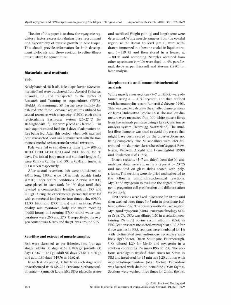
The number of nuclei expressing MyoD and myogen-in was similar in alevin, juvenile and adult 90 days;however, in adult 190 days the number of nuclei ex-pressing myogeninwas higher than those expressingMyoD. The number of PCNA-stained nuclei in eachstage was higher than MyoD and myogenin stainedwith peaks in alevin and adult 90 days (Fig.3). MyoD-and myogenin-positive nuclei in white muscle areillustrated in Fig.4.
Discussion
Most musculature in tilapia is constituted by whitemuscle ¢bre, which is the most commercially impor-
tant (Zhang, Swank & Rome 1996).We were able tocharacterize post-larval muscle growth in Nile tilapiawith reference to hypertrophy and hyperplasiaand protein expression involved in muscle growthmechanisms in the stages essential for commercialpurposes.On comparing the stages studied, we observed an
increase in the frequency of higher diameter muscle¢bres mainly in adult 90 days and adult190 days as-sociated with a gradual decrease in small diameter ¢-bre frequency during growth. This indicated thatmuscle ¢bre recruitment was more evident in earlygrowth (alevin and juvenile), which agrees withValente, Rocha and Gomes (1999) and RowlersonandVeggetti (2001), where, the presence of small dia-meter muscle ¢bres, 20^25 mm, indicated that hyper-plasiawas occurring.Hyperplasia began in the germinal zones of cell
proliferation, near the intermediate layer, and con-sisted of mainly new ¢bres that are responsible forthickening muscle mass in the early developmentstages (Rowlerson & Veggetti 2001; Johnston,Fernandez, Calvo, Vieira, North, Abercromby &
Diameter classes (µm)
0
10
20
30
40
50
60
70
80
90
0-8 8-16 16-24 24-32 > 32
Alevins
Juveniles
90 day adults
190 day adults
Fre
quen
cy (
%)
cb
a a
d
c
b
a
bb
aa
a b
aa
a
b
c
d
Figure 2 Relative frequencydistribution of white muscle ¢brediameters in Nile tilapia (Oreo-chromis niloticus) during growth(letters in the column show sizeclasses with signi¢cant varia-tion, Po0.05).
0
5
10
15
20
25
30
35
40
45
MyoD Myogenin PCNA
Alevin
Juvenile
Adult 90 days
Adult 190 days
b
b ba
aa a
b
c
a
b
a
Figure 3 Frequency of myonu-clei MyoD1, myogenin1 andPCNA1 in thewhite muscle ¢breof Nile tilaŁ pia (Oreochromis niloti-cus). Comparison of the proteinsamong the stages; values withthe same lower case letter werenot signi¢cantly di¡erent.�Po0.001; ~Po0.005. PCNA,proliferating cell nuclear antigen.
MyoD, myogenin and PCNA expression in growing Nile tilapia D H Aguiar et al. Aquaculture Research, 2008, 39, 1673^1679
r 2008 Blackwell Munksgaard1676 No claim to orignial US government works, Aquaculture Research, 39,1673^1679

Garland 2003). This growth event is known as strati-¢ed hyperplasia and occurs in most ¢sh species(Johnston1999). In species of indeterminate growth,strati¢ed hyperplasia is followed by mosaic hyperpla-sia, so called because myotubes form on the surfaceof fast muscle ¢bres, giving rise to a mosaic of ¢brediameters (Johnston 2006). Strati¢ed and mosaic hy-perplasia contribute to an increase in the number ofmuscle ¢bres (Rowlerson et al. 1995; Rowlerson &Veggetti 2001).When hyperplasia occurs, small ¢brescan be seen in between the larger ones, forming amosaic of ¢bres with di¡erent diameters and degrees
of di¡erentiation; this is more common inwhite mus-cle (Johnston 1999; Rowlerson & Veggetti 2001). Ingrowing Nile tilapia, muscle mass thickening in-volves both strati¢ed and mosaic hyperplasia.The proportion of small diameter ¢bres in skeletal
muscle depends on the ¢nal ¢sh size and is a relevantfactor in comparing hyperplasic growth in large-sized species of economical bene¢t (Kiessling et al.1991; Fauconneau, Andre¤ , Chmaitilly, LeŒ Bail, Krieg& Kaushik 1997). Our study showed that alevin andjuvenile muscle growth occurred predominantly bystrati¢ed and mosaic hyperplasia. Adult 90 days andadult 190 days had a higher frequency of 432 mmdiameter ¢bres than alevin and juvenile, showingthat hypertrophy was more intense in adult 90 days’and adult190 days’muscle growth.According to Zimmermanand Lowery (1999), dur-
ing muscle growth, new ¢bre recruitment stopswhen ¢sh reach about 56% of their full-grown size;after this, muscle growth is mainly due to hypertro-phy. Although the adult190 days are not indicative oftheir ¢nal size, our study showed that muscle ¢bre re-cruitment in this growth phase was less than in theother stages (alevin, juvenile and adult 90 days).Skeletal muscle growth in ¢sh is dependent on the
proliferationand di¡erentiation of myogenic progeni-tor or satellite cells that express several transcrip-tional activators; these control the expression ofmuscle-speci¢c genes and contribute to hyperplasiaand hypertrophy. Myogenic precursor cell activity isregulated by di¡erential expression of myogenic reg-ulatory factors (MyoD family) (Watabe1999).In Nile tilapia, nuclei expressing MyoD and myo-
genin were observed throughout growth. The num-ber of nuclei expressing both MyoD and myogeninwas similar in alevin, juvenile and adult 90 days, butin adult190 days the number of myogenin expressingnucleiwas higher than the number expressingMyoD.During early ¢sh development and growth, the in-
tense recruitment of new muscle ¢bres is due to theproliferation of undi¡erentiated myogenic progenitorcells that express primary MRF, MyoD and Myf5(Rescan, Gauvry, Paboeuf & Fauconneau 1994; Me-geney & Rudnicki 1995; Grobet, Royo Martin & Pon-celet 1997;Watabe 1999). In the alevin, juvenile andadult 90 days stages, a large number of MyoD-expres-sing nuclei are related to the intense cell proliferationoccurring in this growth period. These nuclei con-tributed to both hyperplasia and hypertrophy as ob-served bymorphometric analysis (Brooks & Johnston1993). However, as the small diameter ¢bre frequencyin alevin, juvenile was more intense, a large number
(a)
(b)
(c)
20 µm
20 µm
20 µm
Figure 4 Nile tilapia (Oreochromis niloticus) skeletalmuscle. Transverse section of white muscle. (a, b and c):MyoD, myogenin and proliferating cell nuclear antigenpositive nuclei (arrow) respectively.
Aquaculture Research, 2008, 39, 1673^1679 MyoD, myogenin and PCNA expression in growing Nile tilapia D H Aguiar et al.
r 2008 Blackwell MunksgaardNo claim to orignial US government works, Aquaculture Research, 39,1673^1679 1677

of nuclei expressing MyoD can be associated withmyoblast proliferation and intense hyperplasia(Johansen & Overturf 2005) in these earlier develop-ment stages.The presence of equal numbers of nuclei expres-
sing secondary MRF myogenin in alevin, juvenileand adult 90 days shows that in addition to cell pro-liferation, cell di¡erentiation is occurring. During thedi¡erentiation process, nuclei fuse to each other,forming new muscle ¢bres; this was con¢rmed onanalysing muscle ¢bre diameters in these stages.Our ¢ndings con¢rm that during the initial phasesof tilapia growth, myogenic progenitor cell prolifera-tion and di¡erentiation is occurring.The presence of MyoD-positive cells in adult 190
days indicates that myoblast proliferation is occur-ring and that this is involved in muscle ¢bre hyper-trophy, the predominant mechanism in musclegrowth as observed by morphometric analysis. Mus-cle-speci¢c myogenic regulatory transcription factorexpression in adult skeletal muscle may aid proteinsynthesis and degradation, responsible for the main-tenance of muscle phenotype (Mozdziak, Greaser &Schultz1998). Myoblast proliferation may be involvedin nuclear turnover, as 2% of nuclei are recycledweekly in adult skeletal muscle (Schmalbruch &Lewis 2000).Myogenin expression increased in this stage, indi-
cating that di¡erentiation was predominant duringhypertrophyat the same time as a decrease in muscle¢bre recruitment. As gene targeting experimentssuggest that MRF myogenin is involved in myotubefusion and growth (Hasty, Bradley, Morris, Edmond-son, Venuti, Olson & Klein 1993); Black and Olson(1998), hypertrophy can be inferred by analysing theexpression of these MRFs (Johansen & Overturf2005).According to Fauconneau and Paboeuf (2000) and
Brodeur, Peck and Johnston (2002), theoretically, in-creasedMyoD andmyogenin expression during mus-cle growth could also be in response to a stimulus,such as forced swimming or feeding status. However,in Nile tilapia with no external interference, musclegrowth displayed uniformity with myoblast prolif-eration and di¡erentiation until the190th day.In all the stages studied, PCNA expression was
higher than both MyoD and myogenin expression.Proliferating cell nuclearantigen is amarker formyo-genic progenitor cell activation and proliferation(Johnson & Allen 1993; Allen, Sheehan, Taylor,Kendall & Rice 1995). This increase in PCNA expres-sion over MyoD and myogenin in each stage implies
that it regulates myogenic progenitor cell activationand proliferation (Martin & Johnston 2005).These data suggest that growth per se stimulates
cell proliferation and nuclei accretion in the whitemuscle ¢bres of alevin, juvenile and adult 90 daysNile tilapia. Around190 days, in adult190 days, mus-cle ¢bre di¡erentiationwas more pronounced.
Acknowledgments
This work is part of the PhD Thesis that will be pre-sented by DHAtoUniversidade Estadual de Campinas^ UNICAMP. This study was supported by FAPESP,Proc. No.04/13286-0.The researchers of CEPTAweremost helpful during the planning and execution ofexperiments. The colleagues of the Department ofMorphology, Biosciences Institute, carried out valu-able work in the laboratory.
References
Alfei L., Maggi F., Parvopassu F., Bertoncello G. & deVita R.(1989) Post-larval muscle growth in ¢sh: DNA £ow cyto-metric and morphometric analysis. Basic Applied Histo-chemistry 33,147^158.
Allen R.E., Sheehan S.M., Taylor R.G., Kendall T.L. & RiceG.M. (1995) Hepatocyte growth factor activates quiescentskeletal muscle satellite cells in vitro. Journal of CellularPhysiology165,307^312.
Bancroft J.D. & Stevens A. (1990) Theory and Practice of His-tological Techniques, 3rd edn. Churchill Livingstone, NewYork, NY, USA, pp.81^92.
Black B.L. & Olson E.N. (1998) Transcriptional control ofmuscle development by myocyte enhancer factor-2(MEF2) proteins. Annual Review of Cell and DevelopmentalBiology14,167^196.
Brodeur J.C., Peck L.S. & Johnston I.A. (2002) Feeding in-creasesMyoD and PCNAexpression in myogenic progeni-tor cells of Notothenia coriiceps. Journal of Fish Biology 60,1475^1485.
Brooks S. & Johnston I.A. (1993) In£uence of developmentand rearing temperature on the distribution, ultrastruc-ture and myosin sub-unit composition of myotomal mus-cle ¢ber types in the plaice Pleuronectes platessa. MarineBiology117,501^513.
Dubowitz V. & Brooke M. (1973) Muscle Biopsy: A ModernApproach. London, UK: Saunders, 475pp.
Fauconneau B. & Paboeuf G. (2000) E¡ect of fasting and re-feeding on in vitro muscle cell proliferation in rainbowtrout (Onchorhynchus mykiss). Cell and Tissue Research301, 459^463.
Fauconneau B., Andre¤ S., Chmaitilly J., LeŒ Bail P.-Y., Krieg F.& Kaushik S.J. (1997) Control of skeletal muscle ¢bers and
MyoD, myogenin and PCNA expression in growing Nile tilapia D H Aguiar et al. Aquaculture Research, 2008, 39, 1673^1679
r 2008 Blackwell Munksgaard1678 No claim to orignial US government works, Aquaculture Research, 39,1673^1679

adipose cells size in the £esh of rainbow trout. Journal ofFish Biology 50, 296^314.
Goodman L.A. (1964) Simultaneous con¢dence intervals forcontrasts among multinomial populations. Annals ofMathematical Statistics 35,716^725.
Goodman L.A. (1965) On simultaneous con¢dence intervalsfor multinomial proportions.Technometrics 7, 247^254.
Grobet L., RoyoMartin L.J. & Poncelet D. (1997) Adeletion inthe bovine myostatin gene causes the double-muscledphenotype in cattle. Nature Genetics17,71^74.
Hasty P., Bradley A., Morris J.H., Edmondson D.G., VenutiJ.M., Olson E.N. & Klein W.H. (1993) Muscle de¢ciencyand neonatal death in mice with a targeted mutation inmyogenin gene. Nature 364,501^506.
Hsu S.M., Raine L. & Fanger H. (1981) Use of avidin-biotin-peroxidase complex (abc) in immunoperoxidase techni-ques: a comparison between abc and unlabeled antibody(pap) procedures. Journal of Histochemistry and Cytochem-istry 29,577^580.
Johansen K.A. & Overturf K. (2005) Quantitative expressionanalysis of genes a¡ecting muscle growth during devel-opment of rainbow trout (Oncorhynchus mykiss). MarineBiotechnology 7,576^587.
Johnson S.E. & Allen R.E. (1993) Proliferating cell nuclearantigen (PCNA) is expressed in activated rat skeletal mus-cle satellite cells. Journal of Cellular Physiology154,39^43.
Johnston I.A. (1999) Muscle development and growth:potential implications for £esh quality in ¢sh.Aquaculture177,99^115.
Johnston I.A. (2001) Myosin expression during ontogeny,post-hatching growth, and adaptation. In: Muscle Devel-opment and Growth (ed. by I.A. Johnston), pp. 43–70.
Academic Press, New York, NY, USA.
Johnston I.A. (2006) Environment and plasticity of myogen-esis in teleost ¢sh. Journal of Experimental Biology 209,2249^2264.
Johnston I.A., Fernandez D.A., Calvo J.,Vieira V.L.A., NorthA.W., Abercromby M. & Garland T. (2003) Reduction inmuscle ¢ber number during the adaptative radiation ofnotothenioid ¢shes: a phylogenetic perspective. Journal ofExperimental Biology 206, 2595^2609.
Kiessling A., Storebakken T. & Asgard T. (1991) Changes inthe structure and function of the epaxial muscle ofrainbow trout (O. mykiss) in relation to ration and age. I.Growth dynamics. Aquaculture 93,335^356.
Martin C.L. & Johnston I.A. (2005) The role of myostatin andthe calcineurin-signalling pathway muscle mass inresponse to exercise training in the rainbow trout Oncor-hynchus mykissWalbaum. Journal of Experimental Biology208, 2083^2090.
Megeney L.A. & Rudnicki M.A. (1995) Determination versusdi¡erentiation and the MyoD family of transcriptionfactors. Biochemistry and Cell Biology 73,723^732.
Mozdziak P.E., Greaser M.L. & Schultz E. (1998) Myogenin,MyoD, and myosin expression after pharmacologicallyand surgically induced hypertrophy. Journal of AppliedPhysiology 84,1359^1364.
Norman G.R. & Streiner D.I. (1994) Biostatistics. The BareEssentials. Mosby-Year Book, St Louis, 260pp.
Rescan P.Y., Gauvry L., Paboeuf G. & Fauconneau B. (1994)Identi¢cation of a muscle factor related to MyoDin ¢sh species. Biochimica and Biophysica Acta 1218,202^204.
Rowlerson A. & Veggetti A. (2001) Cellular mechanisms ofpost-embryonic muscle growth in aquaculture species.In: Muscle Development and Growth (ed. by I.A. Johnston),pp.103^140. Academic Press, NewYork, NY, USA.
Rowlerson A., Mascarello F., Radaelli G. & Veggetti A. (1995)Di¡erentiation and growth of muscle in the ¢sh SparusAurata (L): ii. Hyperplastic and hypertrophic growth oflateral muscle from hatching to adult. Journal of MuscleResearch and Cell Motility16, 223^236.
Rudnicki M.A. & Jaenish R. (1995) The MyoD family of tran-scription factors and skeletal muscle myogenesis. Bioes-says17, 203^209.
Schmalbruch H. & Lewis D.M. (2000) Dynamics ofnuclei of muscle ¢bers and connective tissue cells innormal and denervated rat muscles. Muscle Nerve 23,617^626.
Stickland N.C. (1983) Growth and development of muscle ¢-bres in the rainbow trout (Salmo gairdneri). Journal of Anat-omy137,323^333.
Valente L.M.P., Rocha E. & Gomes E.F.S. (1999) Growth dy-namics of white and red muscle ¢bers in fast and slowgrowing strains of rainbow trout. Journal of Fish Biology55,675^691.
Veggetti A., RowlersonA., Radaelli G., Arrighi S. & Domene-ghini C. (1999) Post-hatching development of the gutand lateral muscle in the sole. Journal of Fish Biology 55A, 44^65.
Watabe S. (1999) Myogenic regulatory factors and muscledi¡erentiation during ontogeny in ¢sh. Journal of FishBiology 55,1^18.
WeatherleyA.H., Gill H.S. & Rogers S.C. (1980) The relation-ship between mosaic muscle ¢bres and size in rain-bow trout (Salmo gairdneri). Journal of Fish Biology 17,603^610.
Zar J.H. (1999) Biostatistical Analysis,4th edn. Prentice-Hall,Englewood Cli¡s, NJ, USA,663pp.
Zhang G., Swank D.M. & Rome L.C. (1996) Quantitative dis-tribution of muscle ¢ber types in the scup Stenotomuschrysops. Journal of Morphology 229,71^81.
Zimmerman A.M. & Lowery M.S. (1999) Hyperplastic devel-opment and hypertrophic growth of muscle ¢bers in thewhite seabass (Atractoscion nobilis). Journal of Experimen-tal Zoology 284, 299^308.
Aquaculture Research, 2008, 39, 1673^1679 MyoD, myogenin and PCNA expression in growing Nile tilapia D H Aguiar et al.
r 2008 Blackwell MunksgaardNo claim to orignial US government works, Aquaculture Research, 39,1673^1679 1679
Related Documents