Case Report Myocardial Infarction in Neonates: A Diagnostic and Therapeutic Challenge Manuel Rodr´ ıguez Mart´ ınez, 1 Eladio Ruiz Gonz´ alez, 2 Anna Parra-Llorca, 3 M´ aximo Vento Torres, 3,4 and Marta Aguar Carrascosa 3,4 1 University and Polytechnic Hospital La Fe, Valencia, Spain 2 Division of Cardiology, University and Polytechnic Hospital La Fe, Valencia, Spain 3 Neonatal Research Group, Health Research Institute La Fe, Valencia, Spain 4 Division of Neonatology, University and Polytechnic Hospital La Fe, Valencia, Spain Correspondence should be addressed to Marta Aguar Carrascosa; [email protected] Received 17 April 2019; Revised 21 August 2019; Accepted 12 September 2019; Published 24 October 2019 Academic Editor: Ashraf T. Soliman Copyright © 2019 Manuel Rodr´ ıguez Mart´ ınez et al. is is an open access article distributed under the Creative Commons AttributionLicense,whichpermitsunrestricteduse,distribution,andreproductioninanymedium,providedtheoriginalworkis properly cited. Neonatal acute myocardial infarction is an uncommon entity. We describe the case of a 4-day-old term baby who presented with respiratory distress and distal acrocyanosis. e chest radiograph demonstrated cardiomegaly without pleural effusion, and examination revealed hepatomegaly. An electrocardiogram revealed QS pattern in leads I, aVL, and V6, suggestive of ischemia. Cardiac enzymes were elevated, and echocardiogram revealed moderate left ventricular dysfunction with a thrombus at the level of the left atrial appendage. e patient required hemodynamic stabilization, vasodilatation to avoid congestive heart failure, and anticoagulation with heparin and aspirin. In the context of this unusual diagnosis, we reviewed our experience over the last 17 years as well as the existing literature on neonatal myocardial infarction. 1. Introduction Myocardial infarction (MI) is a common entity in adult population,mainlyduetocoronaryarterydisease.However, acute MI presenting in the neonatal period is rare, and the true incidence is still unknown due to limited reporting and diagnosticchallenges.Multipledifferentetiologieshavebeen suggested, but in many cases, the primary cause remains unknown. In cases in which a cause has been identified, culprits have included enteroviral myocarditis, eosinophilic endomyocarditis, congenital diaphragmatic hernia, coa- gulopathy, erythroblastosis, perinatal asphyxia, coronary artery thromboembolism caused by umbilical vein cathe- terization, obstructive congenital heart disease, intrauterine infection, and coronary artery vasoconstriction secondary to oxytocin administration [1–5]. MI is associated with poor prognosis [6], with a mortality rate ranging from 40 to 50%, according to different series [1, 7]. ere are no specific clinical guidelines for appropriate management. Initial survival has improved markedly with recent treatment ad- vances [8], including diuretics, angiotensin-converting en- zyme inhibitors, inotropes, and in selected cases, thrombolysis and extracorporeal membrane oxygenation (ECMO)support[6,9].Wepresentthecaseofaterminfant presenting as an infarct pattern suggestive of MI in context of a thrombus in the left atrium (LA) and the results of a retrospective cohort study including all patients with a final diagnosis of myocardial infarction in the neonatal period during the last 17 years in our center. 2. Case Presentation is is a full-term newborn (39+2 weeks of gestational age andabirthweightof3270grams),whichistheresultofthird pregnancy of a healthy 36-year-old mother. After an un- complicated pregnancy, the baby was delivered by sponta- neous vaginal delivery with Apgar scores at 1 and 5 minutes of 9 and 10, respectively. e patient was discharged home Hindawi Case Reports in Pediatrics Volume 2019, Article ID 7203407, 5 pages https://doi.org/10.1155/2019/7203407

Welcome message from author
This document is posted to help you gain knowledge. Please leave a comment to let me know what you think about it! Share it to your friends and learn new things together.
Transcript

Case ReportMyocardial Infarction in Neonates: A Diagnostic andTherapeutic Challenge
Manuel Rodrıguez Martınez,1 Eladio Ruiz Gonzalez,2 Anna Parra-Llorca,3
Maximo Vento Torres,3,4 and Marta Aguar Carrascosa 3,4
1University and Polytechnic Hospital La Fe, Valencia, Spain2Division of Cardiology, University and Polytechnic Hospital La Fe, Valencia, Spain3Neonatal Research Group, Health Research Institute La Fe, Valencia, Spain4Division of Neonatology, University and Polytechnic Hospital La Fe, Valencia, Spain
Correspondence should be addressed to Marta Aguar Carrascosa; [email protected]
Received 17 April 2019; Revised 21 August 2019; Accepted 12 September 2019; Published 24 October 2019
Academic Editor: Ashraf T. Soliman
Copyright © 2019 Manuel Rodrıguez Martınez et al. )is is an open access article distributed under the Creative CommonsAttribution License, which permits unrestricted use, distribution, and reproduction in anymedium, provided the original work isproperly cited.
Neonatal acute myocardial infarction is an uncommon entity. We describe the case of a 4-day-old term baby who presented withrespiratory distress and distal acrocyanosis. )e chest radiograph demonstrated cardiomegaly without pleural effusion, andexamination revealed hepatomegaly. An electrocardiogram revealed QS pattern in leads I, aVL, and V6, suggestive of ischemia.Cardiac enzymes were elevated, and echocardiogram revealed moderate left ventricular dysfunction with a thrombus at the levelof the left atrial appendage. )e patient required hemodynamic stabilization, vasodilatation to avoid congestive heart failure, andanticoagulation with heparin and aspirin. In the context of this unusual diagnosis, we reviewed our experience over the last 17years as well as the existing literature on neonatal myocardial infarction.
1. Introduction
Myocardial infarction (MI) is a common entity in adultpopulation, mainly due to coronary artery disease. However,acute MI presenting in the neonatal period is rare, and thetrue incidence is still unknown due to limited reporting anddiagnostic challenges. Multiple different etiologies have beensuggested, but in many cases, the primary cause remainsunknown. In cases in which a cause has been identified,culprits have included enteroviral myocarditis, eosinophilicendomyocarditis, congenital diaphragmatic hernia, coa-gulopathy, erythroblastosis, perinatal asphyxia, coronaryartery thromboembolism caused by umbilical vein cathe-terization, obstructive congenital heart disease, intrauterineinfection, and coronary artery vasoconstriction secondary tooxytocin administration [1–5]. MI is associated with poorprognosis [6], with a mortality rate ranging from 40 to 50%,according to different series [1, 7]. )ere are no specificclinical guidelines for appropriate management. Initial
survival has improved markedly with recent treatment ad-vances [8], including diuretics, angiotensin-converting en-zyme inhibitors, inotropes, and in selected cases,thrombolysis and extracorporeal membrane oxygenation(ECMO) support [6, 9]. We present the case of a term infantpresenting as an infarct pattern suggestive of MI in contextof a thrombus in the left atrium (LA) and the results of aretrospective cohort study including all patients with a finaldiagnosis of myocardial infarction in the neonatal periodduring the last 17 years in our center.
2. Case Presentation
)is is a full-term newborn (39 + 2 weeks of gestational ageand a birth weight of 3270 grams), which is the result of thirdpregnancy of a healthy 36-year-old mother. After an un-complicated pregnancy, the baby was delivered by sponta-neous vaginal delivery with Apgar scores at 1 and 5 minutesof 9 and 10, respectively. )e patient was discharged home
HindawiCase Reports in PediatricsVolume 2019, Article ID 7203407, 5 pageshttps://doi.org/10.1155/2019/7203407

completely asymptomatic at 2 days of life with exclusivebreastfeeding. At 4 days of life, he was admitted to a localhospital because of a 3-hour history of respiratory distressand distal acrocyanosis. Noninvasive respiratory supportwith continuous positive airway pressure was commenced,and umbilical venous catheterization was performed. Overthe next several hours, the patient decompensated andbecame hypotensive. A heart murmur was noted on exam, soan echocardiogram was done, which showed left ventriculardysfunction, thrombus in the left atrium, and signs ofpulmonary hypertension. )e decision was made to transferthe patient to our hospital for further cardiology evaluationand management. On arrival, the physical examinationshowed a nonreassuring general state, including pale/ictericcolor, perioral cyanosis, and tachypnea with subcostal re-tractions. Capillary refill was normal. Axillary and femoralpulses were present and symmetrical. Cardiac auscultationdemonstrated a grade I/VI systolic murmur heard best at theleft sternal border. )e lungs were clear with good air entry.)e abdomen was soft, with liver edge palpable 2 cm belowthe costal margin. )e patient was hypoactive and hypo-tonic, with normal fontanelle and intact primitive reflexes.
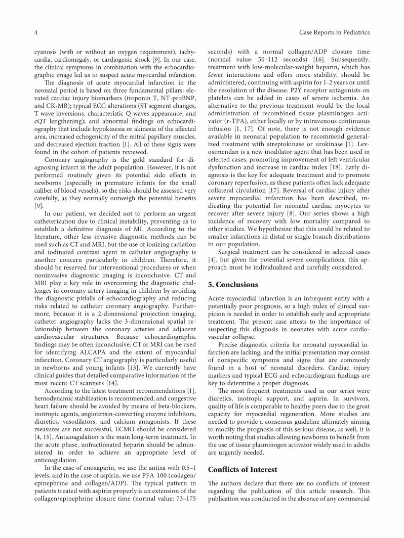
Blood analysis performed at admission demonstratesmoderately deranged liver function (AST 62U/L, ALT118U/L, and CRP 15.3mg/L) and markedly elevated cardiacenzymes (troponin T: 4,046 ng/L, proBNP >35,000 pg).D-dimer was 1.621 ng/mL. Chest X-ray showed car-diomegaly without pleural effusion. Electrocardiogram(ECG) showed a QS pattern in leads I, aVL, and V6 (Fig-ure 1), and echocardiogram confirmed normal intracardiacand coronary anatomy, moderate left ventricular dysfunc-tion (EF 45%), and a thrombus at the level of the left atrialappendage, leading to the working diagnosis of acutemyocardial infarction, possibly secondary to the atrialthrombus. Hemodynamic stabilization was performed withvolume expanders and milrinone infusion. Unfractionatedheparin was started initially and then subsequently con-verted to low-molecular-weight heparin plus aspirin for fullanticoagulation. Further investigations show no evidence ofthrombophilia, and septic screen was negative. In follow-upechocardiograms, cardiac function showed almost completerecovery, and the patient was discharged at 26 days of age oncaptopril, furosemide, spironolactone, enoxaparin, and as-pirin. Catheterization performed one month later did notshow any lesion or abnormality in the coronary arteries, witha normal EF. Medications were gradually weaned off, and hehad no further concerns.
3. Cohort Study
We performed a retrospective review of all patients di-agnosed with myocardial infarction in our center over thelast 17 years. We recorded all demographic and perinataldata including gestational age, obstetric history, Apgar score,birth weight, age at diagnosis, clinical presentation, char-acteristics of the electrocardiogram, troponin T values,ventricular function, angiographic study, treatment used,and mortality in each of the cases (Table 1).
A total of six newborn babies were included.)e averagegestational age was 36 weeks, with a birth weight of 2630 gand age at diagnosis of 4 days. Two cases included obstetrichistory of fetal distress, although all had a normal Apgarscore. )ree patients presented cardiogenic shock; theremaining presented nonspecific symptoms that wereinterpreted as probable sepsis. ECGs showed typical signs ofinfarct pattern, including characteristic appearance of Qwaves and alterations of the ST segment. All patients pre-sented with troponin elevation and cardiac dysfunction onechocardiography. An angiographic study was performed inthree patients, with thickness of the coronary artery wall inonly one case. )e etiologies recorded in these cases were asfollows: myocarditis (secondary to enterovirus) (2), con-genital heart disease (global myocardial hypertrophy, aorticand pulmonary stenosis, and mitral prolapse) (1), fetalhypoxia (1), and antecedent umbilical vein catheterization(1). In one case, no cause was identified. Treatment includeddiuretics, inotropic support, and aspirin. Mortality rate inour series was 33%. At the time of the review, all survivorsare asymptomatic, without treatment and with normalcardiac function.
4. Discussion
Myocardial infarction is an uncommon event in neonatescompared with adults, and addressing the true incidence ofthis condition is difficult due to diagnostic challenges in thispopulation. )e most important causes of myocardial in-farction in the neonatal period are congenital heart diseases,among which coronary vessel anomalies are the most fre-quent. Other reported causes include perinatal asphyxia,erythrocytosis [10], myocarditis, eosinophilic endocarditis,coagulation alterations, congenital diaphragmatic hernia,intrauterine infection, sepsis, umbilical vein catheterization,thromboembolism associated with umbilical hematoma inthe renal vein or ductus venosus, and maternal diabetesduring pregnancy (due to myocardial hypertrophy); con-genital heart abnormalities are other possible causes. )emost relevant predisposing factors are maternal arterialhypertension, fetal-maternal transfusion, fetal-fetal trans-fusion, and dystocic delivery [1, 11]. A significant number ofcases remain classified as idiopathic [1, 2, 9].
We present a small series of infants with infarct pattern,in which myocarditis and structural heart disease were themost prominent underlying diagnoses. In the case thatmotivated this revision, an LA thrombus was identifiedpresumably as a cause of the myocardium ischemia, but acoronary angiography was not performed because of thecritical condition of the newborn, so it is not possible to ruleout completely other causes that can mimic an MI. In someneonates and infants with underlying cardiac disease whoare homozygous for the 4G/4G variant of the PAI-1 pro-motor polymorphism, there is a high risk of developing earlythromboembolism during cardiac catheterization or withinsertion of central venous lines [12], but this variant was notfound in our patient.
)e presentation ofMI in neonates is nonspecific andmayinclude respiratory distress, vomiting, feeding difficulties,
2 Case Reports in Pediatrics

Figure 1: Electrocardiogram (ECG) showing a QS pattern in leads I, aVL, and V6.
Table 1: Demographic and perinatal data.
Caseandyear
Gestationalage
Birthweight(g)
Age atdiagnosis(days oflife)
Clinicalpresentation
Ventricularfunction
Angiographicstudy Etiology Treatment Mortality
7(2002) 38 2820 14 Sepsis
Dysfunctionand
hypokinesiaLateral basal
andlateroapical
Unrealized Myocarditisenterovirus
DiureticImmunoglobulins No
2(2007) 38 3190 1 Shock/
arrythmia
Dysfunctionand
posterolateralhypochinesia
Normal Fetalhypoxia
ACE inhibitorEnoxaparin
ASANo
3(2010) 34 1720 8 Shock
Dysfunctionand
posterolateralhypochinesia
UnrealizedMyocarditisenterovirus(necropsy)
ImmunoglobulinsDiureticInotropic
Yes
4(2011) 30 1700 1 Shock Anterolateral
hypochinesia NormalUmbilicalvenous
channeling
ACE inhibitorEnoxaparin
ASANo
5(2013) 40 3090 1 Heart
murmur
GlobalmyocardialhypertrophyAortic andpulmonarystenosisMitralprolapse
Coronary wallHypertrophicMyocardial
infarction in VI(necropsy)
Congenitalheart disease Diuretic Yes
6(2017) 39 3270 4
Respiratorydistress andacrocyanosis
Moderate/severe
DysfunctionVI
Lateral/posterior and
apicalhypokinesia)rombus inleft atrialappendage
UnrealizedUmbilicalvenous
channeling
DiureticInotropic
ACE inhibitorHeparinASA
No
Case Reports in Pediatrics 3
cyanosis (with or without an oxygen requirement), tachy-cardia, cardiomegaly, or cardiogenic shock [9]. In our case,the clinical symptoms in combination with the echocardio-graphic image led us to suspect acute myocardial infarction.
)e diagnosis of acute myocardial infarction in theneonatal period is based on three fundamental pillars: ele-vated cardiac injury biomarkers (troponin T, NT-proBNP,and CK-MB); typical ECG alterations (ST segment changes,T wave inversions, characteristic Q waves appearance, andcQT lengthening); and abnormal findings on echocardi-ography that include hypokinesia or akinesia of the affectedarea, increased echogenicity of the mitral papillary muscles,and decreased ejection fraction [1]. All of these signs werefound in the cohort of patients reviewed.
Coronary angiography is the gold standard for di-agnosing infarct in the adult population. However, it is notperformed routinely given its potential side effects innewborns (especially in premature infants for the smallcaliber of blood vessels), so the risks should be assessed verycarefully, as they normally outweigh the potential benefits[9].
In our patient, we decided not to perform an urgentcatheterization due to clinical instability, preventing us toestablish a definitive diagnosis of MI. According to theliterature, other less invasive diagnostic methods can beused such as CT and MRI, but the use of ionizing radiationand iodinated contrast agent in catheter angiography isanother concern particularly in children. )erefore, itshould be reserved for interventional procedures or whennoninvasive diagnostic imaging is inconclusive. CT andMRI play a key role in overcoming the diagnostic chal-lenges in coronary artery imaging in children by avoidingthe diagnostic pitfalls of echocardiography and reducingrisks related to catheter coronary angiography. Further-more, because it is a 2-dimensional projection imaging,catheter angiography lacks the 3-dimensional spatial re-lationship between the coronary arteries and adjacentcardiovascular structures. Because echocardiographicfindings may be often inconclusive, CTor MRI can be usedfor identifying ALCAPA and the extent of myocardialinfarction. Coronary CT angiography is particularly usefulin newborns and young infants [13]. We currently haveclinical guides that detailed comparative information of themost recent CT scanners [14].
According to the latest treatment recommendations [1],hemodynamic stabilization is recommended, and congestiveheart failure should be avoided by means of beta-blockers,inotropic agents, angiotensin-converting enzyme inhibitors,diuretics, vasodilators, and calcium antagonists. If thesemeasures are not successful, ECMO should be considered[4, 15]. Anticoagulation is the main long-term treatment. Inthe acute phase, unfractionated heparin should be admin-istered in order to achieve an appropriate level ofanticoagulation.
In the case of enoxaparin, we use the antixa with 0.5–1levels, and in the case of aspirin, we use PFA-100 (collagen/epinephrine and collagen/ADP). )e typical pattern inpatients treated with aspirin properly is an extension of thecollagen/epinephrine closure time (normal value: 73–175
seconds) with a normal collagen/ADP closure time(normal value: 50–112 seconds) [16]. Subsequently,treatment with low-molecular-weight heparin, which hasfewer interactions and offers more stability, should beadministered, continuing with aspirin for 1-2 years or untilthe resolution of the disease. P2Y receptor antagonists onplatelets can be added in cases of severe ischemia. Analternative to the previous treatment would be the localadministration of recombined tissue plasminogen acti-vator (r-TPA), either locally or by intravenous continuousinfusion [1, 17]. Of note, there is not enough evidenceavailable in neonatal population to recommend general-ized treatment with streptokinase or urokinase [1]. Lev-osimendan is a new inodilator agent that has been used inselected cases, promoting improvement of left ventriculardysfunction and increase in cardiac index [18]. Early di-agnosis is the key for adequate treatment and to promotecoronary reperfusion, as these patients often lack adequatecollateral circulation [17]. Reversal of cardiac injury aftersevere myocardial infarction has been described, in-dicating the potential for neonatal cardiac myocytes torecover after severe injury [8]. Our series shows a highincidence of recovery with low mortality compared toother studies. We hypothesize that this could be related tosmaller infarctions in distal or single branch distributionsin our population.
Surgical treatment can be considered in selected cases[4], but given the potential severe complications, this ap-proach must be individualized and carefully considered.
5. Conclusions
Acute myocardial infarction is an infrequent entity with apotentially poor prognosis, so a high index of clinical sus-picion is needed in order to establish early and appropriatetreatment. )e present case attests to the importance ofsuspecting this diagnosis in neonates with acute cardio-vascular collapse.
Precise diagnostic criteria for neonatal myocardial in-farction are lacking, and the initial presentation may consistof nonspecific symptoms and signs that are commonlyfound in a host of neonatal disorders. Cardiac injurymarkers and typical ECG and echocardiogram findings arekey to determine a proper diagnosis.
)e most frequent treatments used in our series werediuretics, inotropic support, and aspirin. In survivors,quality of life is comparable to healthy peers due to the greatcapacity for myocardial regeneration. More studies areneeded to provide a consensus guideline ultimately aimingto modify the prognosis of this serious disease, as well; it isworth noting that studies allowing newborns to benefit fromthe use of tissue plasminogen activator widely used in adultsare urgently needed.
Conflicts of Interest
)e authors declare that there are no conflicts of interestregarding the publication of this article research. )ispublication was conducted in the absence of any commercial
4 Case Reports in Pediatrics

or financial relationships that could be construed as po-tential conflicts of interest.
References
[1] K. Papneja, A. K. Chan, T. K. Mondal, and B. Paes, “Myo-cardial infarction in neonates: a review of an entity withsignificant morbidity and mortality,” Pediatric Cardiology,vol. 38, no. 3, pp. 427–441, 2017.
[2] N. Poonai, A. Kornecki, I. Buffo, and D. Pepelassis, “Umbilicalvenous catheterization: a case report,” Paediatrics and ChildHealth, vol. 14, no. 8, pp. 539–541, 2009.
[3] J. P. Arguello, L. Rıos, A. Jove, P. Aguirres, andC. D. Cardiologıa, “Infarto agudo de miocardio neonatal,”CONAREC, vol. 95, pp. 8–10, 2008.
[4] S. Perrier, A. Parker, C. P. Brizard et al., “Surgical manage-ment of extensive perinatal myocardial infarction,” (e An-nals of (oracic Surgery, vol. 104, no. 6, pp. e435–e437, 2017.
[5] A. Moscicka, T. Mendaluk, M. Szymankiewicz, B. Mrozinski,and I. Maroszynska, “Fatal myocardial infarction in termneonate,” Archives of Perinatal Medicine, vol. 20, no. 1,pp. 49–53, 2014.
[6] K. M. Farooqi, N. Sutton, S. Weinstein, M. Menegus,H. Spindola-Franco, and R. H. Pass, “Successful managementof neonatal myocardial infarction with ECMO,” CongenitHeart Dis, vol. 7, no. 6, pp. E97–E102, 2012.
[7] D. S. Celermajer, G. F. Sholler, R. Howman-Giles, andJ. M. Celermajer, “Myocardial infarction in childhood: clinicalanalysis of 17 cases and medium term follow up of survivors,”Heart, vol. 65, no. 6, pp. 332–336, 1991.
[8] B. J. Haubner, J. Schneider, U. U. Schweigmann et al.,“Functional recovery of a human neonatal heart after severemyocardial infarction,” Circulation Research, vol. 118, no. 2,pp. 216–221, 2016.
[9] L. De. Vetten, K. A. Bergman, N. J. Elzenga, J. P. van Melle,A. Timmer, and B. Bartelds, “Neonatal myocardial infarctionor myocarditis?,” Pediatric Cardiology, vol. 32, no. 4,pp. 492–497, 2011.
[10] E. Caruso, A. Di Pino, D. Poli, L. Manuri, and P. Guccione,“Erythrocytosis and severe asphyxia: two different causes ofneonatal myocardial infarction,” Cardiology in the Young,vol. 24, no. 1, pp. 178–181, 2014.
[11] A. Tillett, B. Hartley, and J. Simpson, “Paradoxical embolismcausing fatal myocardial infarction in a newborn infant,”Archives of Disease in Childhood—Fetal and Neonatal Edition,vol. 85, no. 2, pp. F137–F138, 2001.
[12] V. De Lucia, M. G. Andreassi, L. Sabatini, L. Ait-Ali,I. Spadoni, and S. Giusti, “Myocardial infarction and arterialthrombosis in identical newborn twins with homozygosity forthe PAI-1 4 G/5 G polymorphism,” International Journal ofCardiology, vol. 137, no. 1, pp. e1–e4, 2009.
[13] H. W. Goo, “Coronary artery imaging in children,” KoreanJournal of Radiology, vol. 16, no. 2, pp. 239–250, 2015.
[14] S. H. Hong, H. W. Goo, E. Maeda, K. S. Choo, and I.-C. Tsai,“User-friendly vendor-specific guideline for pediatric car-diothoracic computed tomography provided by the Asiansociety of cardiovascular imaging congenital heart diseasestudy group: part 1. Imaging techniques,” Korean Journal ofRadiology, vol. 20, no. 2, pp. 190–204, 2019.
[15] M.-A. Deutsch, J. Cleuziou, C. Noebauer et al., “Successfulmanagement of neonatal myocardial infarction with ECMOand intracoronary r-tPA lysis,” Congenital Heart Disease,vol. 9, no. 5, pp. E169–E174, 2014.
[16] G. Campuzano Maya, “PFA-100: una nueva prueba de fun-cionplaquetaria sustituta del tiempo de sangrıa,” Medicinaand Laboratorio, vol. 1, pp. 511–548, 2013.
[17] G. Giralt, F. Gran, P. Betrian, and Q. Ferrer, “Infarto agudo demiocardio en un neonato causado por trombosis coronaria:un gran reto diagnostico y terapeutico,” Revista Española deCardiologıa, vol. 68, no. 10, pp. 903-904, 2015.
[18] Y. C. Luther, I. Schulze-Neick, B. Stiller et al., “Saugling mittherapie-refraktarer suprasystemischer pulmonaler hyper-tonie nach myokardinfarkt,” Zeitschrift Far Kardiologie,vol. 93, no. 3, pp. 234–239, 2004.
Case Reports in Pediatrics 5

Stem Cells International
Hindawiwww.hindawi.com Volume 2018
Hindawiwww.hindawi.com Volume 2018
MEDIATORSINFLAMMATION
of
EndocrinologyInternational Journal of
Hindawiwww.hindawi.com Volume 2018
Hindawiwww.hindawi.com Volume 2018
Disease Markers
Hindawiwww.hindawi.com Volume 2018
BioMed Research International
OncologyJournal of
Hindawiwww.hindawi.com Volume 2013
Hindawiwww.hindawi.com Volume 2018
Oxidative Medicine and Cellular Longevity
Hindawiwww.hindawi.com Volume 2018
PPAR Research
Hindawi Publishing Corporation http://www.hindawi.com Volume 2013Hindawiwww.hindawi.com
The Scientific World Journal
Volume 2018
Immunology ResearchHindawiwww.hindawi.com Volume 2018
Journal of
ObesityJournal of
Hindawiwww.hindawi.com Volume 2018
Hindawiwww.hindawi.com Volume 2018
Computational and Mathematical Methods in Medicine
Hindawiwww.hindawi.com Volume 2018
Behavioural Neurology
OphthalmologyJournal of
Hindawiwww.hindawi.com Volume 2018
Diabetes ResearchJournal of
Hindawiwww.hindawi.com Volume 2018
Hindawiwww.hindawi.com Volume 2018
Research and TreatmentAIDS
Hindawiwww.hindawi.com Volume 2018
Gastroenterology Research and Practice
Hindawiwww.hindawi.com Volume 2018
Parkinson’s Disease
Evidence-Based Complementary andAlternative Medicine
Volume 2018Hindawiwww.hindawi.com
Submit your manuscripts atwww.hindawi.com
Related Documents