DOI: 10.1016/j.athoracsur.2007.10.052 2008;85:901-908 Ann Thorac Surg Schussler and Alain Carpentier Juan C. Chachques, Jorge C. Trainini, Noemi Lago, Miguel Cortes-Morichetti, Olivier (MAGNUM Trial): Clinical Feasibility Study Myocardial Assistance by Grafting a New Bioartificial Upgraded Myocardium http://ats.ctsnetjournals.org/cgi/content/full/85/3/901 located on the World Wide Web at: The online version of this article, along with updated information and services, is Print ISSN: 0003-4975; eISSN: 1552-6259. Southern Thoracic Surgical Association. Copyright © 2008 by The Society of Thoracic Surgeons. is the official journal of The Society of Thoracic Surgeons and the The Annals of Thoracic Surgery by on June 4, 2013 ats.ctsnetjournals.org Downloaded from

Welcome message from author
This document is posted to help you gain knowledge. Please leave a comment to let me know what you think about it! Share it to your friends and learn new things together.
Transcript

DOI: 10.1016/j.athoracsur.2007.10.052 2008;85:901-908 Ann Thorac Surg
Schussler and Alain Carpentier Juan C. Chachques, Jorge C. Trainini, Noemi Lago, Miguel Cortes-Morichetti, Olivier
(MAGNUM Trial): Clinical Feasibility StudyMyocardial Assistance by Grafting a New Bioartificial Upgraded Myocardium
http://ats.ctsnetjournals.org/cgi/content/full/85/3/901located on the World Wide Web at:
The online version of this article, along with updated information and services, is
Print ISSN: 0003-4975; eISSN: 1552-6259. Southern Thoracic Surgical Association. Copyright © 2008 by The Society of Thoracic Surgeons.
is the official journal of The Society of Thoracic Surgeons and theThe Annals of Thoracic Surgery
by on June 4, 2013 ats.ctsnetjournals.orgDownloaded from

MBTJMDS
oaebamoc
lcm1m
vHgvv
Rrocftemmml
iart
A
Al7
©P
CA
RD
IOV
ASC
ULA
R
yocardial Assistance by Grafting a Newioartificial Upgraded Myocardium (MAGNUMrial): Clinical Feasibility Study
uan C. Chachques, MD, PhD, Jorge C. Trainini, MD, PhD, Noemi Lago, MD,iguel Cortes-Morichetti, MD, Olivier Schussler, MD, and Alain Carpentier, MD, PhD
epartment of Cardiovascular Surgery, Pompidou Hospital, Paris, France; and Departments of Cardiology and Cardiovascular
urgery, Avellaneda Hospital, Buenos Aires, Argentine21vc(161f33
bcttia
Background. Cell transplantation for the regenerationf ischemic myocardium is limited by poor graft viabilitynd low cell retention. In ischemic cardiomyopathy, thextracellular matrix is deeply altered; therefore, it coulde important to associate a procedure aiming at regener-ting myocardial cells and restoring the extracellularatrix function. We evaluated the feasibility and safety
f intrainfarct cell therapy associated with a cell-seededollagen scaffold grafted onto infarcted ventricles.
Methods. In 20 consecutive patients presenting witheft ventricular postischemic myocardial scars and indi-ation for coronary artery bypass graft surgery, bonearrow cells were implanted during surgery. In the last
0 patients, we added a collagen matrix seeded with bonearrow cells, placed onto the scar.Results. There was no mortality and any related ad-
erse events (follow-up 10 � 3.5 months). New Yorkeart Association functional class improved in both
roups from 2.3 � 0.5 to 1.3 � 0.5 (matrix, p � 0.0002)ersus 2.4 � 0.5 to 1.5 � 0.5 (no matrix, p � 0.001). Left
entricular end-diastolic volume evolved from 142.4 �ntst
iAficiptldtTaetpl
ar Surgery, Georges Pompidou European Hospital, 20 rue Leblanc, Paris,5015, France; e-mail: [email protected].
2008 by The Society of Thoracic Surgeonsublished by Elsevier Inc
ats.ctsnetjournDownloaded from
4.5 mL to 112.9 � 27.3 mL (matrix, p � 0.02) versus38.9 � 36.1 mL to 148.7 � 41 mL (no matrix, p � 0.57), leftentricular filling deceleration time improved signifi-antly in the matrix group from 162 � 7 ms to 198 � 9 msp � 0.01) versus the no-matrix group (from 159 � 5 ms to67 � 8 ms, p � 0.07). Scar area thickness progressed from� 1.4 to 9 mm � 1.1 mm (matrix, p � 0.005) versus 5 �
.5 mm to 6 � 0.8 mm (no matrix, p � 0.09). Ejectionraction improved in both groups, from 25.3% � 7.3% to2% � 5.4% (matrix, p � 0.03) versus 27.2% � 6.9% to4.6% � 7.3% (no matrix, p � 0.031).
Conclusions. This tissue-engineered approach is feasi-le and safe and appears to improve the efficiency ofellular cardiomyoplasty. The cell-seeded collagen ma-rix increases the thickness of the infarct scar with viableissue and helps to normalize cardiac wall stress innjured regions, thus limiting ventricular remodelingnd improving diastolic function.
(Ann Thorac Surg 2008;85:901–8)
© 2008 by The Society of Thoracic Surgeonsesident stem cells have been identified in the heart[1–5] as in many other organs, some of them with
ecognized proliferative activity, as skin [6] and blood [7]r others more static such as central nervous systemomponents [8]. Cardiac stem cells have been isolatedrom human and murine hearts and have been charac-erized [9]. However, the evolution of patients makes itvident that, even in presence of native stem cells, theyocardium self-renewal is, at least, an insufficientechanism to resolve the heart damage. In addition,igration of stem cells from extracardiac source is
imited.In contrast with some nonmammalian animals [10, 11],
n humans, regeneration is reduced in favor of reparationnd efficient wound healing with scar formation. Thisesolution should be a complex and multifactor processhat includes not only muscle proliferation but also
ccepted for publication Oct 12, 2007.
ddress correspondence to Dr Chachques, Department of Cardiovascu-
eoangiogenesis and electric and mechanical stabiliza-ion. A defective resolution results in temporary compen-atory mechanisms that at some moment start a cascadehat progresses to heart failure [12].
The objective of cellular cardiomyoplasty by stem cellmplantation is to regenerate the myocardium [13–15].lthough some clinical studies showed functional bene-ts after stem cell therapy [16–18], until now isolatedellular cardiomyoplasty failed to clearly demonstratemprovements of ventricular function [19–21]. The mainroblem appears to be that cardiac effects of stem cell
ransplantation are limited by poor graft viability and byow cell retention into the treated area [22]. In ischemicisease, both the contracting cells (cardiomyocytes) and
he extracellular matrix are pathologically modified.herefore, it could be important to associate a procedureiming at regenerating both myocardial cells and thextracellular matrix [23]. Preclinical investigations usingissue-engineering technologies showed that this ap-roach may contribute to improve the efficiency of cellu-
ar therapy for organ regeneration [24–27]. Associating a
0003-4975/08/$34.00doi:10.1016/j.athoracsur.2007.10.052
by on June 4, 2013 als.org

cpt[mto
tlarptBic
P
STcsaortbfrTte
uesttoiht
zAGT“anb
PT(ip
Fb0
Fdsi
902 CHACHQUES ET AL Ann Thorac SurgMAGNUM TRIAL CLINICAL FEASIBILITY STUDY 2008;85:901–8
CA
RD
IOV
ASC
ULA
R
ell-seeded matrix with cellular cardiomyoplasty in ex-erimental ischemic hearts, we have demonstrated func-
ional benefits over isolated cell therapy or matrix alone28]. Other investigations demonstrated that collagen
atrices enhance survival of transplanted cells and con-ribute to functional improvements and to the limitationf postischemic ventricular remodeling [29].The goal of this clinical feasibility study was to evaluate
he potential of a biodegradable three-dimensional col-agen type I matrix seeded with bone marrow cells (BMC)nd grafted onto the infarcted ventricle to support andegenerate postischemic lesions. Two groups of patientsresenting with ischemic heart disease were compared:
he first group was treated with intrainfarct injection ofMC; the second group was treated with intrainfarct
njection of BMC associated with the implantation of aell-loaded matrix placed onto the infarct scar.
atients and Methods
tudy Designhe study was a nonrandomized, controlled phase Ilinical trial. Eligibility for inclusion was based on (1)ystolic left ventricular (LV) dysfunction, as reflected byn echocardiographic LV ejection fraction (LVEF) of 35%r less; (2) a history of myocardial infarction with aesidual akinetic and nonviable scar; and (3) an indica-ion for concomitant single off-pump coronary arteryypass graft surgery (OP-CABG). These patients were
ree of angina symptoms, there was no indication ofevascularization of other territories due to poor targets.he preoperative myocardial viability was assessed using
hallium/sestamibi gated myocardial single-photonmission computed tomography (SPECT). Baseline eval-
ig 1. Macroscopic view of the collagen type I, three-dimensionaliodegradable matrix used for myocardial repair (size: 7 � 5 �
b.6 cm).
ats.ctsnetjournDownloaded from
ation was assessed within one week before surgery. Wexcluded patients who had cardiogenic shock or end-tage congestive heart failure, history of leukopenia orhrombocytopenia, evidence for malignant disease orerminal disease, patients under treatment with steroidsr oncology drugs or immunologic suppression, renal
nsufficiency (serum creatinine �2.5 mg/dL) or knownepatic insufficiency, and stroke or major surgery during
he last month.The procedures were performed in the Hospital Inter-
onal “Presidente Perón” of Avellaneda (Buenos Aires,rgentina) in collaboration with the surgical team of theeorges Pompidou European Hospital (Paris, France).he Institutional Review Board of the Hospital InterzonalPresidente Perón” of Avellaneda approved this study,nd informed written consent stating the experimentalature of the study was obtained from each patientefore the procedure.
atient Selection and Group Assignmentwo groups of patients were created. The first group
BMC) included 10 successive ischemic patients withndication for CABG and stem cell therapy; these 10atients were the last of a series of 33 patients treated
ig 2. This image (scanning electron microscopy) shows the three-imensional arrangement of collagen fibers of the matrix used to beeeded with bone marrow cells and grafted onto the left ventricularnfarcted wall.
etween January 2003 and May 2005 in the Department of
by on June 4, 2013 als.org
Cgsacw
BFmccdAulpsm3i
CCaCm
lToammcufb9
SAutpao
csozsm
903Ann Thorac Surg CHACHQUES ET AL2008;85:901–8 MAGNUM TRIAL CLINICAL FEASIBILITY STUDY
CA
RD
IOV
ASC
ULA
R
ardiovascular Surgery of the Avellaneda Hospital (Ar-entina). The second group (BMC � matrix) included 10uccessive ischemic patients with indication for CABGnd stem cell therapy; in these 10 patients a cell-seededollagen matrix was added to the previous treatment andere operated on since May 2005 in the same institution.
one Marrow Cells Isolation and Preparationour hours before the cardiac surgical procedure, bonearrow was aspirated from the ilium bone and pro-
essed to obtain mononuclear bone marrow cells. Theell suspension was loaded on Ficoll-Paque density gra-ient (specific gravity � 1.077 [Amersham Biosciences,rlington Heights, Illinois]), and centrifuged for 20 min-tes at 2,000g. Bone marrow cells were isolated from the
ayer between the Ficoll-Paque reagent and blondlasma, and washed two times in phosphate-bufferedaline (PBS [Sigma, St. Louis, Missouri]). A suspension ofononuclear BMC was obtained (CD34� 8%, AC133�
%) and diluted in autologous patient serum to performntramyocardial injections and matrix seeding.
ollagen Matrix Preparationollagen matrix was prepared from a commercially avail-ble CE Mark collagen kit (Pangen 2; Urgo Laboratory,henove, France). This three-dimensional biodegradableatrix (size: 5 � 7 � 0.6 cm) was manufactured using a
ats.ctsnetjournDownloaded from
yophilized nondenatured, native type I collagen (Fig 1).he matrix pores measured 50 to 100 �m (Fig 2). In theperating room, matrices were placed into a Petri dish;fterward, the cell suspension was seeded onto eachatrix. To promote a regular distribution of BMC into theatrix pores, Petri dishes containing the collagen matri-
es were shaken continuously for 10 minutes at 120gsing an Orbital Shaker (Stuart Scientific, Stone, Staf-
ordshire, United Kingdom [Fig 3]). This procedure cane also performed using centrifugation (10 minutes at00g) of the matrix and cells.
urgical Proceduresfter sternotomy, a single OP-CABG was performedsing left interior mammary artery. At the end of surgery,
he intrainfarct implantation of the autologous BMC waserformed (250 � 28 million cells), within and principallyround the infarct, with a 25G � 40 mm retrobulbarphthalmic needle, in all patients.In the last 10 patients, a three-dimensional type I
ollagen matrix (size: 7 � 5 � 0.6 cm) seeded with theame number of BMC (250 � 28 million cells) was addedn the scarred area, covering the infarct and peri-infarctones. It was fixed onto the epicardium by 6 single PDSutures (6-0) and covered by a second noncellularizedatrix (Fig 4). The number of BMC injected into the heart
Fig 3. (A) Histologic study of theoriginal collagen type I matrix.(Hematoxylin-eosin staining;original magnification �100.)(B) Histologic study of the colla-gen matrix after cells seeding.(Hematoxylin-eosin staining;original magnification �100.)
Fig 4. Surgical procedure associ-ating the intrainfarct implanta-tion of stem cells (A) followedby the fixation of a cell-seededmatrix onto the epicardial sur-face (B).
by on June 4, 2013 als.org

we
ATsam
dmaium
cfda
mGLvsgtaovlaSehanhfcimtafebiori
SRmgs
R
Iosi3w
casfgti
pmicaapsu
da
Tt
AMI
LBNR
M
a
D
AllR
904 CHACHQUES ET AL Ann Thorac SurgMAGNUM TRIAL CLINICAL FEASIBILITY STUDY 2008;85:901–8
CA
RD
IOV
ASC
ULA
R
as selected according with our previous successful clinicalxperience with skeletal myoblast transplantation [30].
ssessment of Outcomeshe primary endpoints were feasibility and safety. Fea-ibility referred to the ability to perform bone marrowspiration, BMC injections, matrix seeding, and surgicalatrix implantation onto the epicardium.We assessed safety on the basis of the following:
evelopment of major adverse cardiac events (death, newyocardial infarction, admittance to hospital due to
ggravation of ischaemia, or heart failure); clinical statusncluding dyspnea, chest pain; and detection of ventric-lar arrhythmias by 24-hour Holter electrocardiographiconitoring study.After surgery, cardiac rhythm was monitored with
ontinuous in-hospital telemetry. All patients underwentollow-up visits, 24-hour Holter recordings, and echocar-iograms before hospital discharge at 1, 3, and 6 months,nd at the end of follow-up.The secondary endpoint was efficacy, which was pri-arily assessed by two-dimensional echocardiography.lobal LVEF (%) was calculated from measurements ofV end-diastolic volume (LVEDV) and LV end-systolicolume (LVESV; both in mL) using the biplane Simp-on’s rule, as ([LVEDV � LVESV]/LVEDV) � 100. Re-ional contractile function was assessed semiquantita-ively after division of the left ventricle into 16 segments,s recommended by the American Society of Echocardi-graphy [31]. Care was taken to optimize endocardialisualization on three parasternal short-axis views of theeft ventricle (at the base, mid-ventricular level, andpex) and apical two-, three-, and four-chamber views.egmental thickening of each segment was then visuallyvaluated and graded as dyskinetic, akinetic, severelyypokinetic, moderately hypokinetic, or normal. Thisssessment was performed within 1 week before surgery,ot during episodes of cardiac failure, and at rest, beforeospital discharge and at 3 months and at the end of
ollow-up. Echocardiographic measurement assessedhanges in global and regional systolic function as well asn diastolic function. We made conventional Doppler
easurements of early and late diastolic transmitral flow,he ratio between the two, and deceleration time. Inddition to measurements of systolic and diastolic LVunction, serial changes in LV segmental thickening werevaluated preoperatively and at the end of follow-upy echocardiography and SPECT Tc99m sestamibi imag-
ng. Image analysis was performed by two investigators,ne of whom was blinded to the timing of echocardiog-aphy, and at the sites of cell injections and matrixmplantation.
tatisticsesults were analyzed and reported as percentage orean � SD. The Student t test was used to compare the
roups. A value of p less than 0.05 was considered to be
tatistically significant. aats.ctsnetjournDownloaded from
esults
n this phase I nonrandomized trial, after considerationf inclusion and exclusion criteria, we enrolled 20 con-ecutive patients (90% male) presenting with chronicschemic heart disease. The age of the infarcts was 8.2 �.5 months (range, 3 to 25). The baseline characteristicsere comparable in both groups (Table 1).Bone marrow was aspirated from the posterior iliac
rest during a brief general anesthesia with midazolamnd etomidate; no bleeding complications at the harvestite were noted. On average, 200 to 250 mL were obtainedor the no-matrix group and 350 to 400 mL for the matrixroup. The increased bone marrow volume obtained forhe matrix group patients was used to inject 50% of BMCnto the infarct and 50% to be seeded into the matrix.
After sternotomy, a single OP-CABG was successfullyerformed in all patients, using the left interior mam-ary artery. At the end of surgery, the intrainfarct
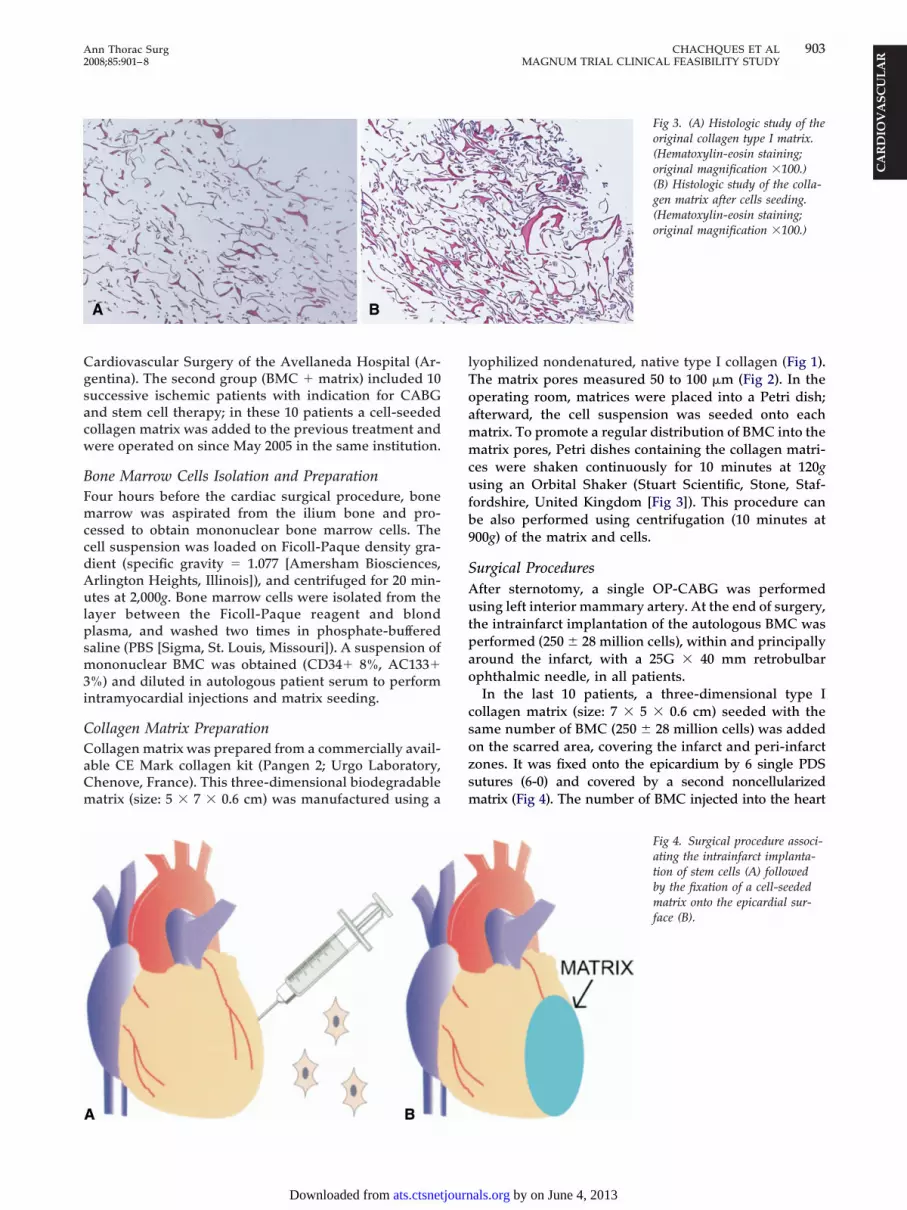
mplantation of the autologous BMC (250 � 28 millionells) was performed into well-exposed LV ischemicreas, permitting 12 � 3 injections points within andround the infarct. Afterward, the cell-seeded matrix wasositioned over the infarct and peri-infarct zones andurgically fixed (Fig 5). A second noncellular matrix wassed to cover this area.All patients had an uneventful recovery and were
ischarged from the intensive care unit 3.4 � 2.6 daysfter surgery. Patients were discharged from the hospital
able 1. Baseline Characteristics of Patients Enrolled inhe Study
Matrix Group(n � 10)
No-MatrixGroup (n � 10)
gea (years) 52.6 56.8ale:female 10:0 8:2
nfarcted related artery(LAD:LCx:RCA)
7:3:0 8:2:0
VEFa (%) 25.3 � 7.3 27.2 � 6.9ody surface area (m2) 1.83 � 0.09 1.79 � 0.08YHA class (II-III-IV) 7:3:0 6:4:0isk factorsHypertension 6 5Diabetes mellitus 4 3Hypercholesterolemia 6 7Current cigarette smoking 4 6edicationAspirin 10 10Statine 7 8Beta-blocker 7 7ACE inhibitor/AT-II
receptor blocker9 10
Mean (SD).
ata are number of patients unless otherwise indicated.
CE � angiotensin-converting enzyme; AT � angiotensin; LAD �eft anterior descending artery; LCx � left circumflex artery; LVEF �eft ventricular ejection fraction; NYHA � New York Heart Association;CA � right coronary artery.
t 11 � 4 postoperative days. At a mean follow-up of 10 �
by on June 4, 2013 als.org
3eN
tht6v
i0
wsof1
tm1t
1n01m
tci
C
Tsads
Fmp
TE
E
L
L
a
C
V
BLl
TRLTP
V
N
R
a
B
905Ann Thorac Surg CHACHQUES ET AL2008;85:901–8 MAGNUM TRIAL CLINICAL FEASIBILITY STUDY
CA
RD
IOV
ASC
ULA
R
.5 months, without mortality and any related adversevents, no malignant cardiac arrhythmias were reported.o patient was lost to follow-up.There were no differences between groups with respect
o the number of premature ventricular complexes perour and the occurrence of nonsustained ventricular
achycardias by Holter monitoring at 6 weeks, 3 months,months, and at the end of follow-up. No sustained
entricular tachycardias was observed in either group.New York Heart Association functional class improved
n both groups from 2.3 � 0.5 to 1.3 � 0.5 (matrix, p �.0002) versus 2.4 � 0.5 to 1.5 � 0.5 (no matrix, p � 0.001).
Echocardiographic studies demonstrated that LVEFas comparable at baseline in both groups and that LVEF
ignificantly increased in both groups at 10 � 3.5 monthsf follow-up. There were no statistically significant dif-erences between the treatment groups at baseline or at0 � 3.5 months of follow-up (Table 2).
Significant differences were observed at long term inhe matrix-treated group concerning postischemic re-
odeling: LVEDV evolved from 142.4 � 24.5 mL to12.9 � 27.3 mL (matrix, p � 0.02) versus 138.9 � 36.1 mLo 148.7 � 41 mL (no matrix, p � 0.57; Table 2).
Deceleration time improved in the matrix group from62 � 7 ms to 198 � 9 ms (p � 0.01), but not enough in theo-matrix group (from 159 � 5 ms to 167 � 8 ms, p �.07). Scar area thickness progress from 6 � 1.4 mm to 9 �.1 mm (matrix, p � 0.005) versus 5 � 1.5 mm to 6 � 0.8m (no matrix, p � 0.09).Blind radioisotopic analysis showed that 58% � 9% of
he cell-implanted segments versus 62% � 5% of theell-plus-matrix–treated segments improved their kinet-
ig 5. Clinical case showing the bone marrow cell–seeded collagenatrix grafted onto the heart, immediately after coronary artery by-
ass graft surgery and intrainfarct cell therapy.
cs and viability (p � 0.06; Table 3, Fig 6). V
ats.ctsnetjournDownloaded from
omment
his study attempts to demonstrate the feasibility andafety of simultaneous intramyocardial injection of BMCnd fixation of a BMC-seeded matrix onto the epicar-ium of infarcted ventricles. The cardiac connective tis-ue is mainly composed of collagen, with smaller
able 2. Myocardial Function Variables Assessed bychocardiography
Therapy Stratum
pValuea
MatrixGroup
BMCGroup
jection fractionBaseline (1 week
before CABG) (%)25.3 � 7.3 27.2 � 6.9 NS
10 � 3.5 months (%) 32 � 5.4 34.6 � 7.3 NSp valueb 0.03 0.031� ejection fraction
(% points)6.7 � 5.3 7.4 � 7.5 NS
VEDVBaseline (1 week
before CABG) (%)142.4 � 24.5 138.9 � 36.1 NS
10 � 3.5 months (%) 112.9 � 27.3 148.7 � 41 0.033p valueb 0.02 NS� LVEDV (mL) �29.5 � 12.3 9.8 � 12.1 0.01
VESVBaseline (1 week
before CABG) (%)117.8 � 24.5 113.5 � 33.3 NS
10 � 3.5 months (%) 77.7 � 23.5 99.1 � 36.3 NSp valueb 0.001 NS� LVESV (mL) �40.1 � 10.1 �14.4 � 14.9 0.003
Between treatment groups. b Between baseline (1 week beforeABG) and 10 � 3.5 months follow-up by echocardiography.
alues represent mean � SD.
MC � bone marrow cell; CABG � coronary artery bypass graft surgery;VEDV � left ventricular end-diastolic volume; LVESV � left ventricu-
ar end-systolic volume; NS � not significant.
able 3. Single-Photon Emission Computed Tomographyadioisotopic Analysis of Myocardial Viability and Regionaleft Ventricular Contractility in Bone Marrow Cell (BMC)reated Patients (n � 10) and BMC Plus Matrix Treatedatients (n � 10)
ariable Baseline 10 Months p Valuea
onviablesegments
BMC 74 � 5.1 31 � 2.4 0.001BMC � matrix 76 � 3.4 29 � 3.1 0.001
ecuperatedsegments
BMC 43 � 3.7 (58% � 9%)BMC � matrix 47 � 3.2 (62% � 5%)
Between baseline (1 week before CABG) and 10 � 3.5 months follow-up;MC group versus BMC � matrix-treated group (p � 0.06).
alues represent mean � SD.
by on June 4, 2013 als.org

athfiiecsiaraof
etcBetrtg
bit
wpama
bapsrdboHt
eewlarracsh
mfacrctaigtbtsrtsacrtttb
trdeat
FmuctuadAtcmt
906 CHACHQUES ET AL Ann Thorac SurgMAGNUM TRIAL CLINICAL FEASIBILITY STUDY 2008;85:901–8
CA
RD
IOV
ASC
ULA
R
mounts of elastin, laminin, and fibronectin. There arewo main types of collagen fibers in the normal adulteart, types I and III, produced by fibroblasts and myo-broblasts. Experimental observations have shown that
n the process of ischemic heart disease, the myocardialxtracellular matrix is deeply altered, the reserve ofollagen type I, which is responsible for the structuralupport, can decrease, down from 80% to 40% afternfarction [32]. Collagen fibers provide structural supportnd give the heart properties that include stiffness andesistance to deformation. In addition, they have shownn important role as a link between contractile elementsf adjacent myocytes, carrying some information usefulor cell function [23].
We designed this nonrandomized trial involving isch-mic patients in which the first 10 were treated with BMCherapy and the next 10 patients were treated with aombined approach of BMC implant and fixation of aMC-seeded collagen scaffold onto the epicardium. Ourxperimental studies [28] and others [24, 29] suggest thathis combined approach may offer further benefits withespect to cell therapy alone. Thus, we tested whetherhese experimental procedures could be clinically up-raded so as to make them compatible with human use.There were no perioperative complications related to
one marrow aspiration, BMC injections, or matrix seed-ng, and cellularized matrix surgical implantation ontohe epicardium.
The number of BMC obtained by crest iliac aspirationas enough for both treatments. The cell matrix seedingrocedures were easily carried out. After the OP-CABGnd BMC intramyocardial injections, the cellularizedatrix was placed in a few minutes onto the infarcted
rea without any technical problem.The data of the present study demonstrate the feasi-
ility of this combined approach in humans and providebasis for ongoing studies and future developments. Theresented data suggest that this therapeutic approach isafe; specifically, there was no evidence for an increasedate of proarrhythmic effects. In fact, there were noifference between both groups with respect of the num-er of premature ventricular complexes per hour and theccurrence of nonsustained ventricular tachycardias byolter monitoring during follow-up. No sustained ven-
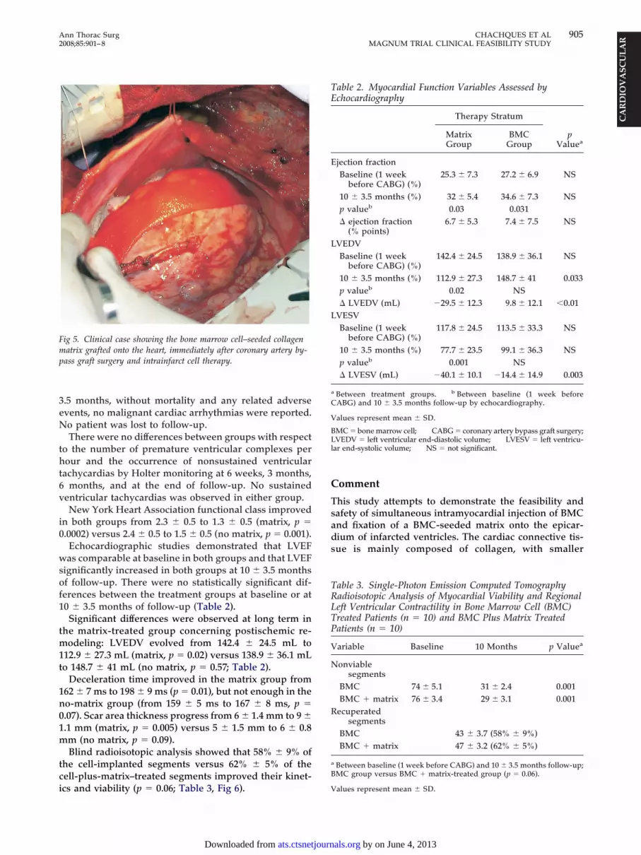
ig 6. (A) Preoperative assess-ent of myocardial perfusion
sing single-photon emissionomputed tomography Tc99m ses-amibi imaging. The left ventric-lar anterolateroapical infarctedrea is stained in blue (three-imensional representation). (B)t 6 months’ follow-up, the pa-
ient treated with stem cells asso-iated with a cell-seeded collagenatrix shows a 62% reduction of
he infarcted area (blue stain).
ricular tachycardia was observed in either group. t
ats.ctsnetjournDownloaded from
Our phase I clinical study was not designed to assessfficacy of this combined treatment. In our opinion, thenhanced recovery of LV function must be consideredith caution. In fact, the small number of patients ana-
yzed does not allow us to draw definitive conclusions,nd the improvement in symptoms and LVEF may alsoeflect efficacious CABG revascularization. Nevertheless,esults suggest that this combined approach has andditional beneficial effect on LV remodelling whenompared with cell therapy alone; the interaction effect oftem cells associated with the matrix appears to makeeart repair more efficient.The underlying mechanisms by which BMC-seededatrix promote functional recovery after myocardial in-
arction are not clearly elucidated, but data derived fromnimal experimentations let us suppose that the use of aellularized collagen matrix favors intramyocardial celletention and creates a microatmosphere that promotesell survival [24–29]. The enhanced recovery of LV func-ion may be due to a significant reduction in the extentnd magnitude of regional LV dysfunction between thenfarct and peri-infarct areas. These cell-seeded biode-radable collagen scaffolds may be able to provide struc-ural integrity within the body, and eventually it willreak down, leaving the newly formed tissue that will
ake over the mechanical load; in this way the matrixeems to help normalize cardiac wall stress in injuredegions [33–38]. Interestingly, we found the thickness ofhe infarcted wall to be significantly greater in the cell-eeded matrix group than in the cell therapy group. Suchfinding suggests that the seeded grafts may improve
ardiac function by minimizing adverse ventricularemodeling or wall thinning. A passive girdling effect ofhe cell-seeded collagen matrix generating a reduction ofhe ventricular wall stress and improving strain distribu-ion and infarct scar elasticity could explain the functionalenefits observed in the matrix group patients.The reduction of the scar area assessed by radioiso-
opic studies (Fig 6) suggests that fibrotic tissue waseplaced by living cells, resulting in improved myocar-ium elastic properties and compliance. This fact mayxplain the improvements in the diastolic filling time. Inddition, no restrictive effects were observed in thereated areas; the collagen matrix (5 � 7 cm) only par-
ially covers the pathologically dilated ventricles. It isby on June 4, 2013 als.org

i(icttmtwp
Bscftpmedcoe
LApttcssmstoimsos
trwio
aBcsviraCpii
Tfi(ACMa
R
1
1
1
1
1
1
1
1
1
1
907Ann Thorac Surg CHACHQUES ET AL2008;85:901–8 MAGNUM TRIAL CLINICAL FEASIBILITY STUDY
CA
RD
IOV
ASC
ULA
R
mportant to remark that this resorbable collagen matrixPangen) has high hemostatic capacity and is frequentlyndicated and used in our hospital and in other Europeanenters as compresses for local hemostasis during cardio-horacic and abdominal surgical operations, when con-rol of bleeding by ligaturing or other conventional
eans is ineffective or impractical. In cardiac use, long-erm follow-up never demonstrated a restrictive processith impairment of diastolic function (ie, constrictiveericarditis).Recent publications have highlighted the potential of
MC to promote paracrine effects in ischemic tissues (eg,ecretion of angiogenic factors), and suggest that para-rine signaling, rather than cell incorporation, promotesunctional recovery [39–42]. Transplanted BMC and ma-rix seeded cells may release growth factors that mayreserve extracellular matrix and promote the recruit-ent of cardiac stem cells that would provide a new
ndogenous pool of contractile cells. In addition, trans-ifferentiation of BMC-derived hemopoietic stem cells toardiomyocytes and potential cardiomyogenic propertiesf mesenchymal BMC can account for the beneficialffects of this new therapeutic procedure [43–46].
imitationsfirst limitation pertains to the semiquantitative ap-
roach used for assessing LV segmental function. Al-hough two-dimensional echocardiography has becomehe technique of choice for direct visualization of endo-ardial motion and wall thickening, this assessment maytill be confounded by contractile changes of the adjacentegments, as long as myocardial strain is not directlyeasured. The improved kinetics of the revascularized
egments could therefore have skewed the grading ofransplanted areas. We have used the LVEF as an indexf systolic function, but one of the limitations of the LVEF
s its load dependence, which confounds accurate assess-ent of LV systolic function. An alternative, strain and
train-rate images, could allow quantitative assessmentf regional myocardial wall motion and may be moreensitive to regional myocardial ischemia.
The second limitation was the association with a CABG;hus, functional improvements can not be conclusivelyelated to the cells and matrix. Another limitation is that thisas a nonrandomized trial. In spite of this choice, our
nclusion and exclusion criteria resulted in the enrollmentf a relatively homogeneous patient population.In conclusion, our data suggest that simultaneous
utologous intramyocardial injection of mononuclearMC and fixation of a cell-seeded matrix onto the epi-ardium is a feasible and safe procedure. This matrixeems to increase the thickness of the infarct scar withiable tissues and helps to normalize cardiac wall stressn injured regions. However, large randomized trials areequired to show a beneficial effect not only on systolicnd diastolic LV function, but also on clinical endpoints.ardiac tissue engineering is emerging as a new thera-eutic tool and extends even more the amazing possibil-
ties of cell therapies in cardiology, becoming a promis-
ng way for the creation of a “bioartificial myocardium.”ats.ctsnetjournDownloaded from
he authors would like to acknowledge Osvaldo H. Masoli, MD,rom Argerich Hospital (Buenos Aires, Argentina) for the radio-sotopic studies; Marc Marie, MD, from Urgo LaboratoryChenove, France) for illustration 2; and Jorge Genovese, MD,mit Patel, MD, Cristiano Spadaccio, MD, and Emmanuelhachques, BS, from the McGowan Institute for Regenerativeedicine (Pittsburgh, Pennsylvania) for the histological studies
nd illustration 3.
eferences
1. Hierlihy AM, Seale P, Lobe CG, Rudnicki MA, Megeney LA.The post-natal heart contains a myocardial stem cell popu-lation. FEBS Lett 2002;530:239–43.
2. Linke A, Muller P, Nurzynska D, et al. Stem cells in the dogheart are self-renewing, clonogenic, and multipotent andregenerate infarcted myocardium, improving cardiac func-tion. Proc Natl Acad Sci USA 2005;102:8966–71.
3. Matsuura K, Nagai T, Nishigaki N, et al. Adult cardiacsca-1-positive cells differentiate into beating cardiomyo-cytes. J Biol Chem 2004;279:11384–11391.
4. Oh H, Bradfute SB, Gallardo TD, et al. Cardiac progenitorcells from adult myocardium: homing, differentiation, andfusion after infarction. Proc Natl Acad Sci USA 2003;100:12313–8.
5. Urbanek K, Quaini F, Tasca G, et al. Intense myocyteformation from cardiac stem cells in human cardiac hyper-trophy. Proc Natl Acad Sci USA 2003;100:10440–5.
6. Pellegrini G, Dellambra E, Golisano O, et al. p63 identifieskeratinocyte stem cells. Proc Natl Acad Sci USA 2001;98:3156–61.
7. Jaffredo T, Nottingham W, Liddiard K, Bollerot K, Pouget C, deBruijn M. From hemangioblast to hematopoietic stem cell: anendothelial connection? Exp Hematol 2005;33:1029–40.
8. Sanai N, Alvarez-Buylla A, Berger MS. Neural stem cells andthe origin of gliomas. N Engl J Med 2005;353:811–22.
9. Messina E, De Angelis L, Frati G, et al. Isolation andexpansion of adult cardiac stem cells from human andmurine heart. Circ Res 2004;95:911–21.
0. Poss KD, Wilson LG, Keating MT. Heart regeneration inzebrafish. Science 2002;298:2188–90.
1. Bettencourt-Dias M, Mittnacht S, Brockes JP. Heterogeneousproliferative potential in regenerative adult newt cardiomy-ocytes. J Cell Sci 2003;116:4001–9.
2. Pfeffer MA, Braunwald E. Ventricular remodelling aftermyocardial infarction. Experimental observations and clini-cal implications. Circulation 1990;81:1161–72.
3. Laflamme MA, Murry CE. Regenerating the heart. NatBiotechnol 2005;23:845–56.
4. Rajnoch C, Chachques JC, Berrebi A, Bruneval P, BenoitMO, Carpentier A. Cellular therapy reverses myocardialdysfunction. J Thorac Cardiovasc Surg 2001;121:871–8.
5. Chachques JC, Acar C, Herreros J, et al. Cellular cardiomyo-plasty: clinical application. Ann Thorac Surg 2004;77:1121–30.
6. Stamm C, Kleine HD, Choi YH, et al. Intramyocardial deliveryof CD133� bone marrow cells and coronary artery bypassgrafting for chronic ischemic heart disease: safety and efficacystudies. J Thorac Cardiovasc Surg 2007;133:717–25.
7. Assmus B, Honold J, Schachinger V, et al. Transcoronarytransplantation of progenitor cells after myocardial infarc-tion. N Engl J Med 2006;355:1222–32.
8. Schachinger V, Erbs S, Elsasser A, et al, for the REPAIR-AMIInvestigators. Intracoronary bone marrow-derived progeni-tor cells in acute myocardial infarction. N Engl J Med2006;355:1210–21.
9. Meyer GP, Wollert KC, Lotz J, et al. Intracoronary bonemarrow cell transfer after myocardial infarction: eighteenmonths’ follow-up data from the randomized, controlledBOOST (BOne marrOw transfer to enhance ST-elevation
infarct regeneration trial). Circulation 2006;113:1287–94.by on June 4, 2013 als.org

2
2
2
2
2
2
2
2
2
2
3
3
3
3
3
3
3
3
3
3
4
4
4
4
4
4
4
908 CHACHQUES ET AL Ann Thorac SurgMAGNUM TRIAL CLINICAL FEASIBILITY STUDY 2008;85:901–8
CA
RD
IOV
ASC
ULA
R
0. Janssens S, Dubois C, Bogaert J, et al. Autologous bonemarrow-derived stem-cell transfer in patients with ST-segment elevation myocardial infarction: double-blind, ran-domised controlled trial. Lancet 2006;367:113–21.
1. Lunde K, Solheim S, Aakhus S, et al. Intracoronary injectionof mononuclear bone marrow cells in acute myocardialinfarction. N Engl J Med 2006;355:1199–209.
2. Teng CJ, Luo J, Chiu RC, Shum-Tim D. Massive mechanicalloss of microspheres with direct intramyocardial injection inthe beating heart: implications for cellular cardiomyoplasty.J Thorac Cardiovasc Surg 2006;132:628–32.
3. Brown L. Cardiac extracellular matrix: a dynamic entity.Am J Physiol Heart Circ Physiol 2005;289:H973–4.
4. Piao H, Kwon JS, Piao S, et al. Effects of cardiac patchesengineered with bone marrow-derived mononuclear cellsand PGCL scaffolds in a rat myocardial infarction model.Biomaterials 2007;28:641–9.
5. Barandon L, Couffinhal T, Dufourcq P, et al. Repair ofmyocardial infarction by epicardial deposition of bone-marrow-cell-coated muscle patch in a murine model. AnnThorac Surg 2004;78:1409–17.
6. Zimmermann WH, Melnychenko I, Wasmeier G, et al.Engineered heart tissue grafts improve systolic and diastolicfunction in infarcted rat hearts. Nat Med 2006;12:452–8.
7. Christman KL, Lee RJ. Biomaterials for the treatment ofmyocardial infarction. J Am Coll Cardiol 2006;48:907–13.
8. Cortes-Morichetti M, Frati G, Schussler O, et al. Associationbetween a cell-seeded collagen matrix and cellular cardio-myoplasty for myocardial support and regeneration. TissueEng 2007;13:2681–7.
9. Kutschka I, Chen IY, Kofidis T, et al. Collagen matricesenhance survival of transplanted cardiomyoblasts and con-tribute to functional improvement of ischemic rat hearts.Circulation 2006;114(Suppl):I167–73.
0. Chachques JC, Herreros J, Trainini J, et al. Autologoushuman serum for cell culture avoids the implantation ofcardioverter-defibrillators in cellular cardiomyoplasty. IntJ Cardiol 2004;95(Suppl 1):29–33.
1. Schiller N, Shah P, Crawford M, et al. American Society ofEchocardiography committee on standards, subcommitteeon quantitation of two-dimensional echocardiograms: rec-ommendations for quantitation of the left ventricle by two-dimensional echocardiography. J Am Soc Echocardiogr 1989;2:358–67.
2. Herpel E, Pritsch M, Koch A, Dengler TJ, Schirmacher P,Schnabel PA. Interstitial fibrosis in the heart: differences inextracellular matrix proteins and matrix metalloproteinasesin end-stage dilated, ischaemic and valvular cardiomyopa-
thy. Histopathology 2006;48:736–47.ats.ctsnetjournDownloaded from
3. Lunkenheimer PP, Redmann K, Westermann P, et al. Themyocardium and its fibrous matrix working in concert as aspatially netted mesh: a critical review of the purportedtertiary structure of the ventricular mass. Eur J CardiothoracSurg 2006;29(Suppl 1):41–9.
4. Kresh JY. Cell replacement therapy: the functional impor-tance of myocardial architecture and intercellular gap-junction distribution. J Thorac Cardiovasc Surg 2006;131:1310–3.
5. Eschenhagen T, Zimmermann WH. Engineering myocardialtissue. Circ Res 2005;97:1220–31.
6. Leor J, Landa N, Cohen S. Renovation of the injured heartwith myocardial tissue engineering. Expert Rev CardiovascTher 2006;4:239–52.
7. Kofidis T, de Bruin JL, Hoyt G, et al. Myocardial restorationwith embryonic stem cell bioartificial tissue transplantation.J Heart Lung Transplant 2005;24:737–44.
8. Giraud MN, Armbruster C, Carrel T, Tevaearai HT. Currentstate of the art in myocardial tissue engineering. Tissue Eng2007;13:1825–36.
9. Fedak PW, Szmitko PE, Weisel RD, et al. Cell transplantationpreserves matrix homeostasis: a novel paracrine mechanism.J Thorac Cardiovasc Surg 2005;130:1430–9.
0. Tang YL, Zhao Q, Qin X, et al. Paracrine action enhances theeffects of autologous mesenchymal stem cell transplantationon vascular regeneration in rat model of myocardial infarc-tion. Ann Thorac Surg 2005;80:229–37.
1. Kubal C, Sheth K, Nadal-Ginard B, Galinanes M. Bonemarrow cells have a potent anti-ischemic effect againstmyocardial cell death in humans. J Thorac Cardiovasc Surg2006;132:1112–8.
2. Thum T, Bauersachs J, Poole-Wilson PA, Volk HD, AnkerSD. The dying stem cell hypothesis: immune modulation asa novel mechanism for progenitor cell therapy in cardiacmuscle. J Am Coll Cardiol 2005;46:1799–802.
3. Ebelt H, Jungblut M, Zhang Y, et al. Cellular cardiomyoplasty:improvement of left ventricular function correlates with therelease of cardioactive cytokines. Stem Cells 2007;25:236–44.
4. Chachques JC, Duarte F, Cattadori B, et al. Angiogenicgrowth factors and/or cellular therapy for myocardial regen-eration: a comparative study. J Thorac Cardiovasc Surg2004;128:245–53.
5. Chachques JC, Salanson-Lajos C, Lajos P, Shafy A, AlshamryA, Carpentier A. Cellular cardiomyoplasty for myocardial re-generation. Asian Cardiovasc Thorac Ann 2005;13:287–96.
6. Dzau VJ, Gnecchi M, Pachori AS. Enhancing stem celltherapy through genetic modification. J Am Coll Cardiol
2005;46:1351–3.by on June 4, 2013 als.org

DOI: 10.1016/j.athoracsur.2007.10.052 2008;85:901-908 Ann Thorac Surg
Schussler and Alain Carpentier Juan C. Chachques, Jorge C. Trainini, Noemi Lago, Miguel Cortes-Morichetti, Olivier
(MAGNUM Trial): Clinical Feasibility StudyMyocardial Assistance by Grafting a New Bioartificial Upgraded Myocardium
& ServicesUpdated Information
http://ats.ctsnetjournals.org/cgi/content/full/85/3/901including high-resolution figures, can be found at:
References http://ats.ctsnetjournals.org/cgi/content/full/85/3/901#BIBL
This article cites 46 articles, 23 of which you can access for free at:
Citations http://ats.ctsnetjournals.org/cgi/content/full/85/3/901#otherarticles
This article has been cited by 11 HighWire-hosted articles:
Subspecialty Collections
http://ats.ctsnetjournals.org/cgi/collection/molecular_biology Molecular biology
following collection(s): This article, along with others on similar topics, appears in the
Permissions & Licensing
[email protected]: orhttp://www.us.elsevierhealth.com/Licensing/permissions.jsp
in its entirety should be submitted to: Requests about reproducing this article in parts (figures, tables) or
Reprints [email protected]
For information about ordering reprints, please email:
by on June 4, 2013 ats.ctsnetjournals.orgDownloaded from
Related Documents