Myeloproliferative Neoplasms: New Approaches Myeloproliferative Neoplasms: New Approaches to Diagnosis and Disease Monitoring to Diagnosis and Disease Monitoring John L Frater, MD Jeffery M Klco, MD, PhD Department of Pathology and Immunology Washington University School of Medicine St Louis, Missouri

Welcome message from author
This document is posted to help you gain knowledge. Please leave a comment to let me know what you think about it! Share it to your friends and learn new things together.
Transcript

Myeloproliferative Neoplasms: New Approaches Myeloproliferative Neoplasms: New Approaches
to Diagnosis and Disease Monitoringto Diagnosis and Disease Monitoring
John L Frater, MD
Jeffery M Klco, MD, PhD
Department of Pathology and Immunology
Washington University School of Medicine
St Louis, Missouri

Myeloproliferative Neoplasms
2008 WHO Classification
• Chronic myelogenous leukemia, BCR-ABL1positive
• Chronic neutrophilic leukemia
• Chronic eosinophilic leukemia, NOS
• Polycythemia vera• Polycythemia vera
• Primary myelofibrosis
• Essential thrombocythemia
• Mastocytosis
• Chronic myeloproliferative disease, unclassifiable


Philadelphia Chromosome
• The Philadelphia chromosome (Ph) is commonly found in hematologic malignancies
– CML: 90-95%
– Adult ALL: 20%
– Pediatric ALL:5%
– AML: 2%
• BCR-ABL functions– Aberrant tyrosine kinase activity
– Activation of Ras
– Secondary lesions that promote blast crisis
• Differentiation arrest
• Impaired genomic surveillance
• DNA repair defects
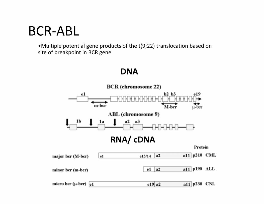
DNA
BCR-ABL•Multiple potential gene products of the t(9;22) translocation based on site of breakpoint in BCR gene
RNA/ cDNA
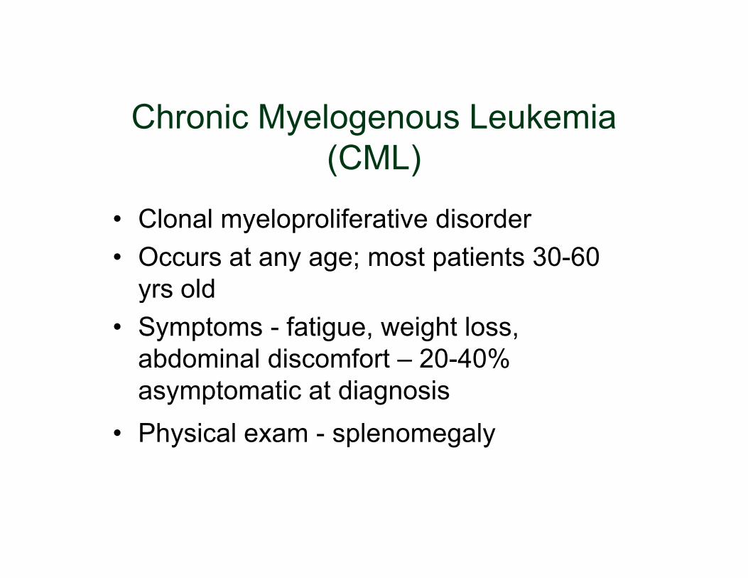
Chronic Myelogenous Leukemia
(CML)
• Clonal myeloproliferative disorder
• Occurs at any age; most patients 30-60
yrs oldyrs old
• Symptoms - fatigue, weight loss,
abdominal discomfort – 20-40%
asymptomatic at diagnosis
• Physical exam - splenomegaly

Chronic Myelogenous Leukemia (CML)
• Chronic phase – inc. myelopoeisis, basophilia, eosinophilia
– Median survival 7 years with interferon therapy
Accelerated phase• Accelerated phase
– 6-18 months
• Blast phase
– Myeloid: median survival 3-4 months
– Lymphoid: median survival 9-12 months

Chronic Myelogenous Leukemia
Accelerated Phase (WHO)
• Blasts 10-19% of WBCs in PB and/or of nucleated bone marrow cells
• Peripheral blood basophils ≥20%
• Persistent thrombocytopenia (<100x109/L) unrelated to therapy, or persistent unrelated to therapy, or persistent thrombocytosis (>1000x109/L) unresponsive to therapy
• Increasing spleen size and increasing WBC count unresponsive to therapy
• Clonal evolution

Chronic Myelogenous Leukemia
Accelerated Phase

Chronic Myelogenous Leukemia
Blast Phase (WHO)
• Blasts >20% of PB WBCs/nucleated bone
marrow cells
• Extramedullary blast proliferation
• Large foci/clusters of blasts in bone marrow • Large foci/clusters of blasts in bone marrow
biopsy

Blast Phase of CML

Blast Phase of CML
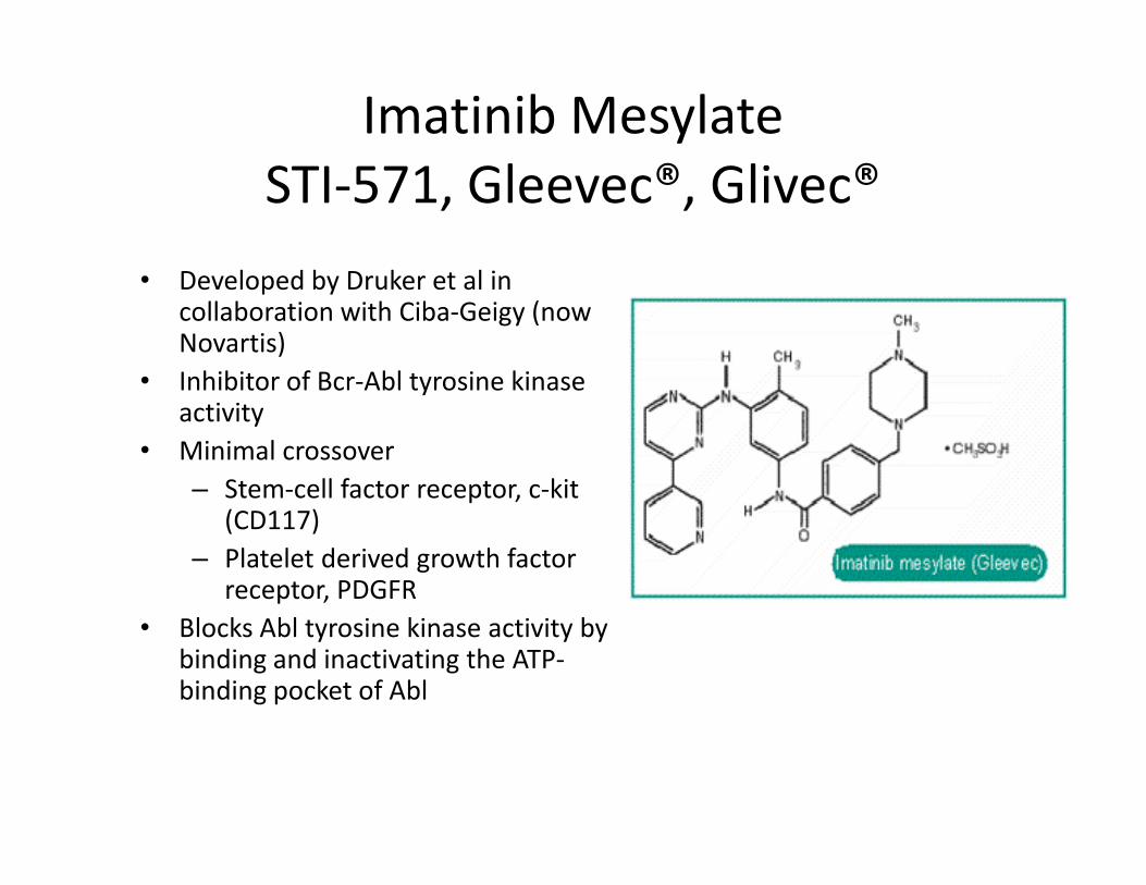
Imatinib Mesylate
STI-571, Gleevec®, Glivec®
• Developed by Druker et al in collaboration with Ciba-Geigy (now Novartis)
• Inhibitor of Bcr-Abl tyrosine kinase activity
• Minimal crossover• Minimal crossover
– Stem-cell factor receptor, c-kit (CD117)
– Platelet derived growth factor receptor, PDGFR
• Blocks Abl tyrosine kinase activity by binding and inactivating the ATP-binding pocket of Abl

Actuarial probability of disease progression according to the level of
cytogenetic and molecular response after 12 months of imatinib (P <
.001; log-rank test).
Merx et al. Leukemia. 2002;16:1579–1583.

Bone Marrow Cellularity
Month 0 Month 3 Month 6

Bone Marrow Cellularity
Month 9 Month 12 Month 15 Month 18

Frater JL et al. Am J Clin Pathol 2003;119:833-41
Patient who became t(9;22) negative

Frater JL et al. Am J Clin Pathol 2003;119:833-41
Patient who remained t(9;22) positive
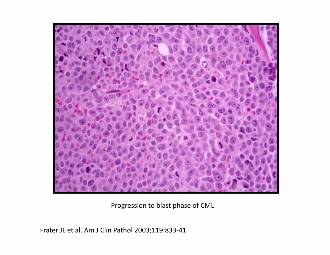
Progression to blast phase of CML
Frater JL et al. Am J Clin Pathol 2003;119:833-41
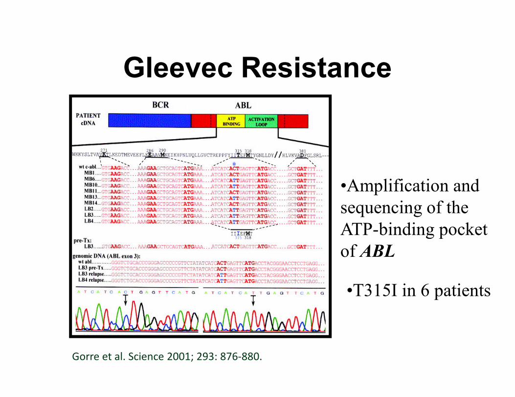
Gleevec Resistance
•Amplification and
sequencing of the sequencing of the
ATP-binding pocket
of ABL
•T315I in 6 patients
Gorre et al. Science 2001; 293: 876-880.
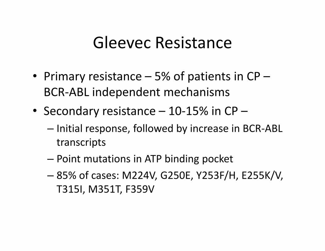
Gleevec Resistance
• Primary resistance – 5% of patients in CP –
BCR-ABL independent mechanisms
• Secondary resistance – 10-15% in CP –
– Initial response, followed by increase in BCR-ABL – Initial response, followed by increase in BCR-ABL
transcripts
– Point mutations in ATP binding pocket
– 85% of cases: M224V, G250E, Y253F/H, E255K/V,
T315I, M351T, F359V

Gleevec Resistance
BCR-ABL Wildtype BCR-ABL T315I MutantGorre et al. Science 2001; 293: 876-880.

Kaplan-Meier survival curves for patients with mutations
Branford et al. Blood. 2003;102:276–283.

Presumptive CML
Conventional Cytogenetics
t(9;22)+ (~95%) t(9;22)-
Molecular Molecular
BCR-ABL1+ BCR-ABL1-
(2.5%, likely not CML)
BCR-ABL1+
(~2.5%, CML)
Adapted from Ou et al. Am J Hematol 2007; 83: 296-302.
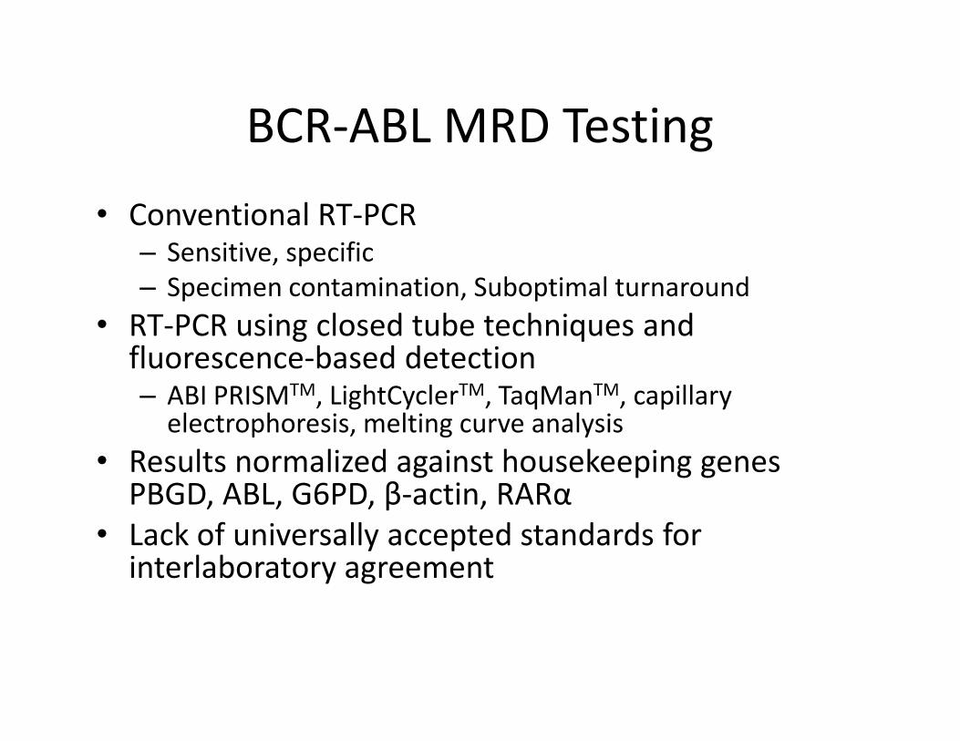
BCR-ABL MRD Testing
• Conventional RT-PCR– Sensitive, specific
– Specimen contamination, Suboptimal turnaround
• RT-PCR using closed tube techniques and fluorescence-based detection fluorescence-based detection – ABI PRISMTM, LightCyclerTM, TaqManTM, capillary
electrophoresis, melting curve analysis
• Results normalized against housekeeping genes PBGD, ABL, G6PD, β-actin, RARα
• Lack of universally accepted standards for interlaboratory agreement

BCR-ABL MRD Testing
• Quantitative RT-PCR analysis is technically feasible, reproducible with excellent intralaboratory agreement, useful in assessing response to therapy
• Current recommendations – serial assessment of BCR-ABL levels at 3 month intervals in patients treated with imatinib
• 4 transcript level patterns: continual decline, undetectable, • 4 transcript level patterns: continual decline, undetectable, stable/ plateau, rising
• MMR (major molecular response): therapeutic goal (IRIS study), ≥3 log reduction in BCR-ABL transcript compared to the standardized baseline
• IRIS study: patients with MMR + CCR (complete cytogenetic response) at 12 months have 100% rate of progression free survival

Every 2 weeks Every 3 months
Every 6 months Yearly
Every 2-3 months
CBC
Cytogenetics
FISH (PB)
CCR
CCR
CHR
Every 3 months ** Every 3 months **Molecular (PB or BM)
MMR
CCR
*
***
In cases of treatment failure, suboptimal
Response, or increasing transcript levels
*= qualitative RT-PCR
** = quantitative RT-PCR
*** = mutational testing
Adapted from Ou et al. Am J Hematol 2007; 83: 296-302 and Baccarani M et al. Blood
2006; 108: 1809-1820.

Failure to Respond
3 mo 6 mo 12 mo 18 mo anytime
No CHR <CHR,
No CR
<PaCR <CCR Loss of CHR,
Loss of CR
Suboptimal Response
<CHR <PaCR <CCR <MMR ACA in Ph+ cells,
Loss of MMR
Adapted from Ou et al. Am J Hematol 2007; 83: 296-302 and Baccarani M et al. Blood
2006; 108: 1809-1820.
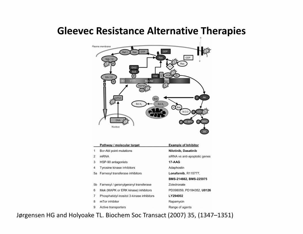
Gleevec Resistance Alternative Therapies
Jørgensen HG and Holyoake TL. Biochem Soc Transact (2007) 35, (1347–1351)

Philadelphia-Chromosome–Negative
Classic MPNs
• Polycythemia vera (PV)
• Essential thrombocythemia (ET)
• Primary myelofibrosis (PMF)
• Clonal expansion of 1 or more bone marrow • Clonal expansion of 1 or more bone marrow
lineages
W. Dameshek 1900-1969

Polycythemia Vera
• Absolute increase in erythrocyte cell mass
• Increased hematocrit
• Increased blood volume
• Increased blood viscosity• Increased blood viscosity

Polycythemia VeraClinical Features
• Skin: Rubor, pruritus
• Vascular: Thromboses
• Gastrointestinal: Peptic ulcers, hemorrhage
• Splenomegaly• Splenomegaly
• Dyspnea
• CNS: Headache, vertigo, syncope, visual
disturbance, tinnitus, stroke

Polycythemia Vera
Laboratory Features
• Increased absolute red blood cell mass
• Erythrocytosis (7-10,000,000/mm3)
• Increased hemoglobin (18-24g/dL)
• Reticulocyte count not increased
• Leukocytosis (25-30,000/mm3)• Leukocytosis (25-30,000/mm
• Thrombocytosis (500,000-1,000,000/mm3)
• Increased total blood volume
• Increased blood viscosity
• Increased leukocyte alkaline phosphatase (LAP)
• Increased serum vitamin B12 (increased transcobalamin I)

Polycythemia VeraMorphologic Features
• Hypercellular bone marrow
• Multilineage hyperplasia
• Megakaryocyte clustering
• Minimal fibrosis• Minimal fibrosis
Klco JM, et al. Am J Clin Pathol 2010;
133: 602-615

Polycythemia VeraNatural History
Evolution Manifestation Transformation
10-15% mimic “ET”
Fibrosis
Post-polycythemic myeloid
Metaplasia ~20%
Jak2 +/-
Jak2+/+
Epo
EECs+
Definite increase
In RBC mass
10-15 years
Pre-polycythemic
stagePolycythemic stage
Splenomegaly
Terminal phase
Post-PV MF with
blastic transformation
<10%
Adapted from Swerdlow et al (2008)

Polycythemia Vera
Klco JM, et al. Am J Clin Pathol 2010; 133: 602-615
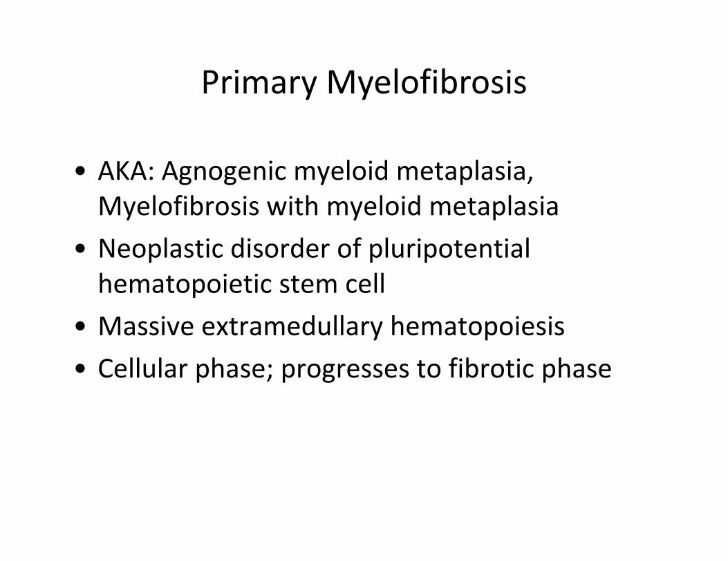
Primary Myelofibrosis
• AKA: Agnogenic myeloid metaplasia,
Myelofibrosis with myeloid metaplasia
• Neoplastic disorder of pluripotential
hematopoietic stem cellhematopoietic stem cell
• Massive extramedullary hematopoiesis
• Cellular phase; progresses to fibrotic phase
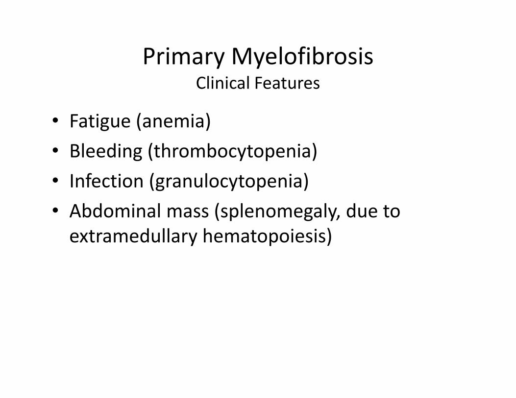
Primary Myelofibrosis Clinical Features
• Fatigue (anemia)
• Bleeding (thrombocytopenia)
• Infection (granulocytopenia)
• Abdominal mass (splenomegaly, due to • Abdominal mass (splenomegaly, due to
extramedullary hematopoiesis)
Primary Myelofibrosis
Cellular Phase: Laboratory Features
• Peripheral blood
– Leukocytosis
– Thrombocytosis
– Basophilia
– Eosinophilia– Eosinophilia
• Bone marrow
– Hypercellularity
– Granulocytic, megakaryocytic, erythroid hyperplasia
– Minimal fibrosis
– Megakaryocyte clustering

Primary Myelofibrosis
Fibrotic Phase: Laboratory Features
• Peripheral blood
– Dacyrocytes (teardrop RBCs)
– Nucleated RBCs
– Immature granulocytes– Immature granulocytes
– Anemia/ leukopenia/ thrombocytopenia
– Increased LAP
• Bone marrow
– Obliterative fibrosis
– Osteosclerosis
Primary Myelofibrosis Natural History
• Progressive bone marrow failure
• Death due to infection or hemorrhage
• Conversion to acute leukemia in <10% of
patientspatients
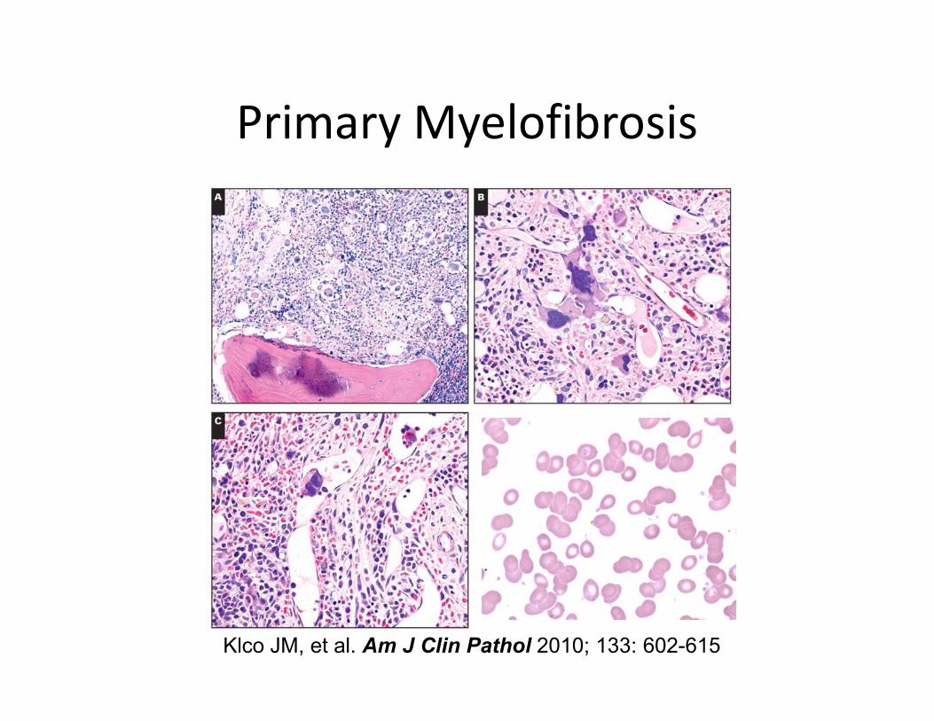
Primary Myelofibrosis
Klco JM, et al. Am J Clin Pathol 2010; 133: 602-615

Essential Thrombocythemia
• Clonal neoplasm derived from pluripotential
hematopoietic stem cell
• Marked hyperplasia of bone marrow
megakaryocytesmegakaryocytes
• Peripheral thrombocytosis

Essential ThrombocythemiaClinical Manifestations
• Thrombocytosis/ bleeding due to platelet
dysfunction
• Splenomegaly due to extramedullary
hematopoiesishematopoiesis
Essential Thrombocythemia
Laboratory Features, Peripheral Blood
• Thrombocytosis >1,000,000/mm3
• Abnormal platelet morphology
• Abnormal platelet function
• Leukocytosis 15-40,000/mm3• Leukocytosis 15-40,000/mm3
• Eosinophilia
• Basophilia

Essential Thrombocythemia
Laboratory Features, Bone Marrow
• Hypercellular bone marrow
• Marked megakaryocytic hyperplasia
• Variable hypercellularity of granulocytic and
erythroid lineageserythroid lineages
• Minimal bone marrow fibrosis

Essential ThrombocythemiaNatural History
• Episodic bleeding and/or thrombosis
• <1% progress to acute leukemia

Essential Thrombocythemia
Klco JM, et al. Am J Clin Pathol 2010; 133: 602-615
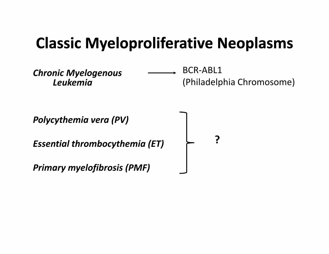
Classic Myeloproliferative NeoplasmsClassic Myeloproliferative Neoplasms
Chronic Myelogenous Leukemia
BCR-ABL1
(Philadelphia Chromosome)
Polycythemia vera (PV)
Essential thrombocythemia (ET)
Primary myelofibrosis (PMF)
?

Janus Kinase 2• Member of Janus family (Jak1, Jak2, Jak3 and
Tyk2) of non-receptor tyrosine kinases that associate with cytokine/chemokine receptors– Shared structure consisting of adjacent kinase (JH1)
and pseudokinase domains (JH2)
• Jak2 V617F: G to T somatic mutation in exon 14 (JH2) domain
• Disrupts the interaction between JH2 and JH1, resulting in constitutive activity
Courtesy Google Images
•and JH1, resulting in constitutive activity
• Likely not disease initiating in humans but studies in mice do mimic components of the human disease
FERM SH2 JH2 JH1
V6
17
F
Tyrosine kinase
activity
Regulation of
kinase activity
Phospho-tyrosine
binding
Membrane localization
Adapted from Abdel-Wahab
Current Opinion Hematology, 2011
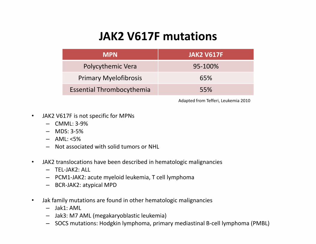
JAK2 V617F mutations
• JAK2 V617F is not specific for MPNs
– CMML: 3-9%
MPN JAK2 V617F
Polycythemic Vera 95-100%
Primary Myelofibrosis 65%
Essential Thrombocythemia 55%
Adapted from Tefferi, Leukemia 2010
– CMML: 3-9%
– MDS: 3-5%
– AML: <5%
– Not associated with solid tumors or NHL
• JAK2 translocations have been described in hematologic malignancies
– TEL-JAK2: ALL
– PCM1-JAK2: acute myeloid leukemia, T cell lymphoma
– BCR-JAK2: atypical MPD
• Jak family mutations are found in other hematologic malignancies
– Jak1: AML
– Jak3: M7 AML (megakaryoblastic leukemia)
– SOCS mutations: Hodgkin lymphoma, primary mediastinal B-cell lymphoma (PMBL)
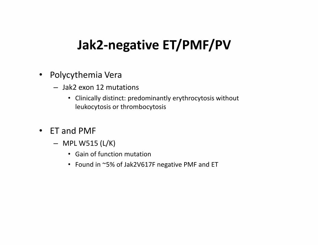
Jak2-negative ET/PMF/PV
• Polycythemia Vera
– Jak2 exon 12 mutations
• Clinically distinct: predominantly erythrocytosis without
leukocytosis or thrombocytosis
• ET and PMF
– MPL W515 (L/K)
• Gain of function mutation
• Found in ~5% of Jak2V617F negative PMF and ET

Levine et al, 2007

Mutational potpourri
• Recent studies have established a lengthy list of mutations found at low frequencies in MPNs
• These mutations are neither sensitive nor specific for MPNs and there are currently no implications for clinical testing
Gene Frequency in MPN
(PV, PMF, ET)
Other myeloid disorders
(PV, PMF, ET)
TET2 ~10% AML, BP-MPN, MDS, CMML
IDH1/IDH2 <5% AML, BP-MPN, MDS, CMML
DNMT3a 5-10% AML, BP-MPN, MDS, CMML
EZH2 ~5% AML, BP-MPN, MDS, CMML
LNK rare BP-MPN
ASXL1 ~5% AML, BP-MPN, MDS, CMML
Adapted from Tefferi, Leukemia (2011)
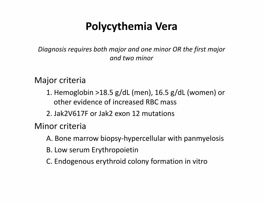
Polycythemia Vera
Diagnosis requires both major and one minor OR the first major
and two minor
Major criteria
1. Hemoglobin >18.5 g/dL (men), 16.5 g/dL (women) or
other evidence of increased RBC massother evidence of increased RBC mass
2. Jak2V617F or Jak2 exon 12 mutations
Minor criteria
A. Bone marrow biopsy-hypercellular with panmyelosis
B. Low serum Erythropoietin
C. Endogenous erythroid colony formation in vitro
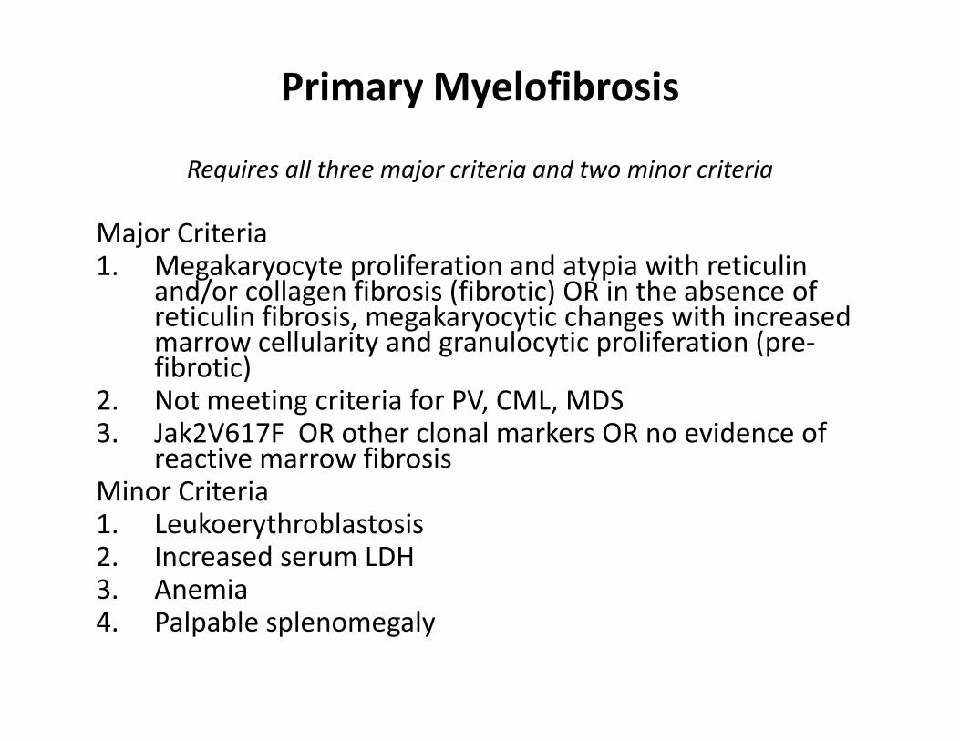
Primary Myelofibrosis
Requires all three major criteria and two minor criteria
Major Criteria1. Megakaryocyte proliferation and atypia with reticulin
and/or collagen fibrosis (fibrotic) OR in the absence of reticulin fibrosis, megakaryocytic changes with increased marrow cellularity and granulocytic proliferation (pre-fibrotic)fibrotic)
2. Not meeting criteria for PV, CML, MDS3. Jak2V617F OR other clonal markers OR no evidence of
reactive marrow fibrosisMinor Criteria1. Leukoerythroblastosis2. Increased serum LDH3. Anemia4. Palpable splenomegaly
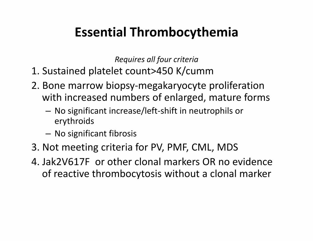
Essential Thrombocythemia
Requires all four criteria
1. Sustained platelet count>450 K/cumm
2. Bone marrow biopsy-megakaryocyte proliferation with increased numbers of enlarged, mature forms
– No significant increase/left-shift in neutrophils or erythroidserythroids
– No significant fibrosis
3. Not meeting criteria for PV, PMF, CML, MDS
4. Jak2V617F or other clonal markers OR no evidence of reactive thrombocytosis without a clonal marker
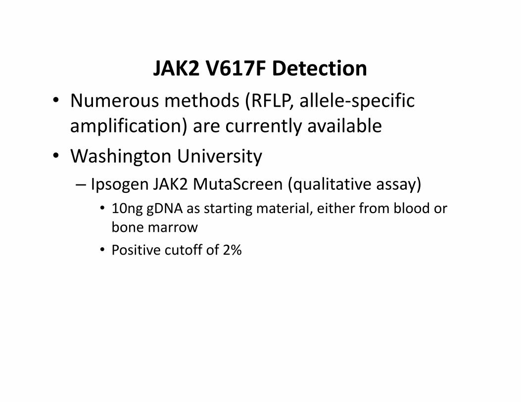
JAK2 V617F Detection
• Numerous methods (RFLP, allele-specific
amplification) are currently available
• Washington University
– Ipsogen JAK2 MutaScreen (qualitative assay)
• 10ng gDNA as starting material, either from blood or • 10ng gDNA as starting material, either from blood or
bone marrow
• Positive cutoff of 2%

JAK2 V617F Detection
• Issues to be resolved
– Is there a role for reporting allele frequency?
• Mouse models and clinical data suggest that allele burden helps shape the disease phenotype
– ET has lowest allele burden– ET has lowest allele burden
• Increasing allele burden has been associated with increased fibrosis, splenomegaly and leukocyte count
– Standardized JAK2 V617F monitoring has not been established
• Currently unclear if JAK2V617F can be used for disease monitoring similar to BCR-ABL1 in CML

WHO 2008
• Myeloproliferative Neoplasms (MPN)– Chronic myelogenous leukemia
– Polycythemia vera
– Essential thrombocythemia
– Primary myelofibrosis
– Chronic neutrophilic leukemia– Chronic neutrophilic leukemia
– Chronic eosinophilic leukemia, not otherwise categorized
– Hypereosinophilic syndrome
– Mast cell disease
– MPNs, unclassified

Mast Cell Disease
• Clinical heterogenous group of diseases due to clonal proliferation of mast cells
• Multiple WHO categories
– Cutaneous mastocytosis
– Indolent systemic mastocytosis– Indolent systemic mastocytosis
– Systemic mastocytosis with associated clonal hematological non-mast cell lineage disease (SM-AHNMD)
– Aggressive systemic mastocytosis
– Mast cell leukemia
– Mast cell sarcoma
– Extracutaneous mastocytoma

Mast Cell Disease
• Typical morphologic/immunophenotypic features– Clusters (>15 cells) of spindled mast cells
– Atypical expression of CD2 and CD25
• SM-AHNMD• SM-AHNMD– Concurrent clonal hematologic malignancy (commonly
CMML)
• Activating mutations in c-kit – D816V is most common and occurs within kinase domain
and thus is insensitive to Gleevec
– Other mutations may be present depending on additional hematologic disorders (SM-AHNMD)
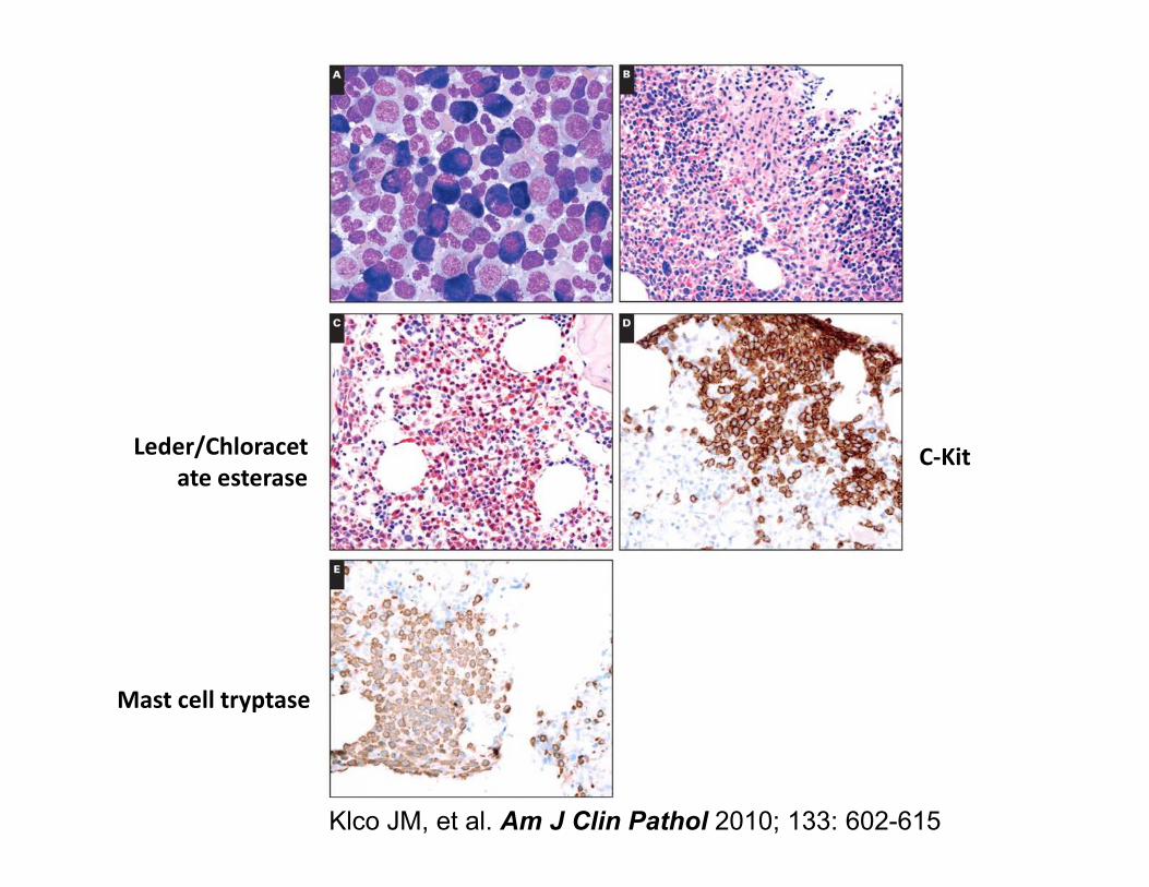
C-KitLeder/Chloracet C-Kit
Mast cell tryptase
Leder/Chloracet
ate esterase
Klco JM, et al. Am J Clin Pathol 2010; 133: 602-615

Myeloid neoplasms associated with
eosinophilia
– Chronic eosinophilic leukemia, not otherwise categorized
– Hypereosinophilic syndrome
– Myeloid and lymphoid neoplasms with eosinophilia and – Myeloid and lymphoid neoplasms with eosinophilia and abnormalities of PDGFRB
– Myeloid and lymphoid neoplasms with eosinophilia and abnormalities of PDGFRA
– Myeloid and lymphoid neoplasms with eosinophilia and abnormalities of FGFR

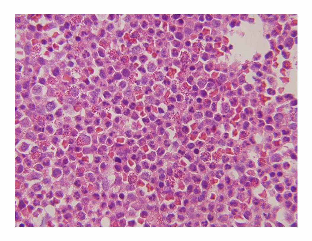
Case presentation
• 42 year old man with mild leukocytosis (15.1
K/cumm )
– Eosinophils: 12% (1.8 K/cumm; nl 0.0-0.5)
– Neutrophils: 68% (8.26 K/cumm; nl 1.8-6.6)– Neutrophils: 68% (8.26 K/cumm; nl 1.8-6.6)
– Monocytes: 4% (0.48 K/cumm; nl 0.2-1.2)
• Presented with chief complaint of fatigue
• All other indices were within normal limits
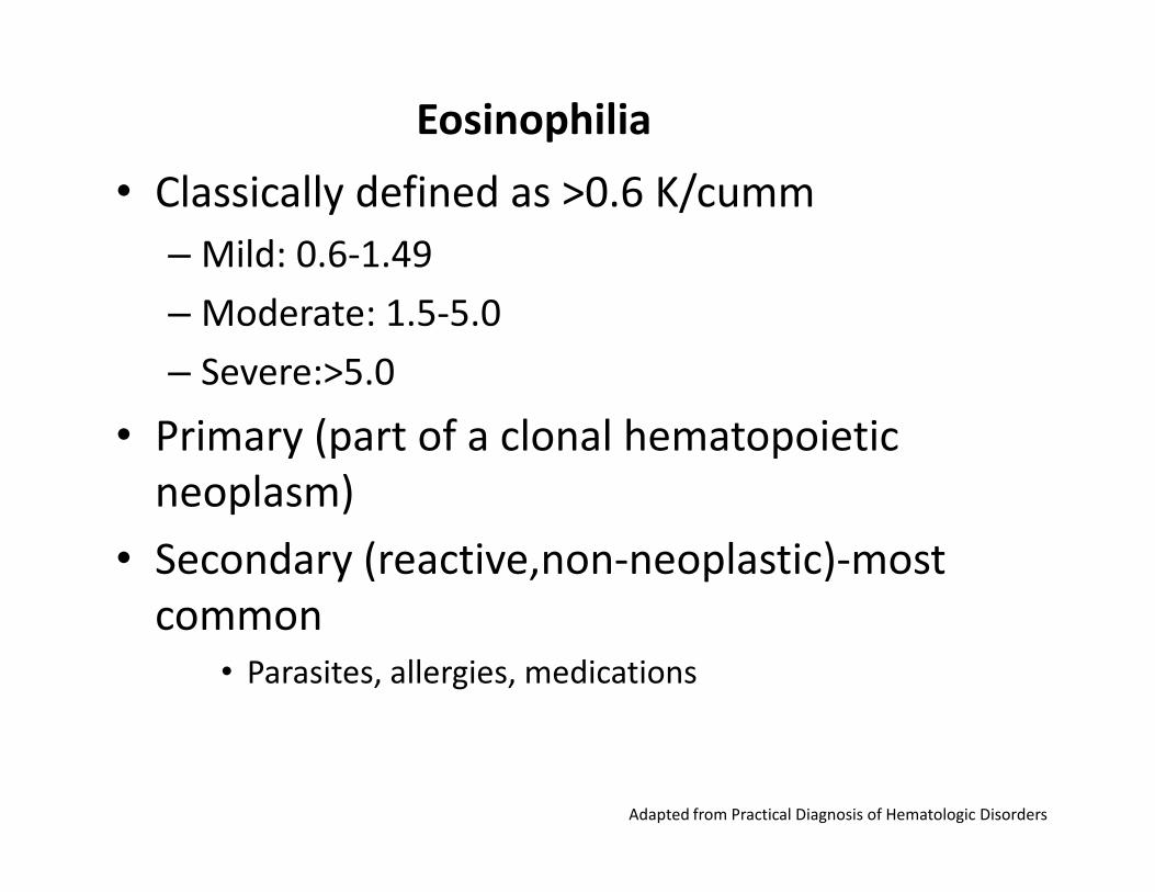
Eosinophilia
• Classically defined as >0.6 K/cumm
– Mild: 0.6-1.49
– Moderate: 1.5-5.0
– Severe:>5.0
• Primary (part of a clonal hematopoietic • Primary (part of a clonal hematopoietic
neoplasm)
• Secondary (reactive,non-neoplastic)-most
common
• Parasites, allergies, medications
Adapted from Practical Diagnosis of Hematologic Disorders

Primary Eosinophilia
• Hypereosinophilic Syndrome (non-clonal)– Persistent eosinophilia (>6 mo) of >1.5 K/cumm
– Rule out all reactive conditions
– Rule out all other hematolymphoid neoplasms associated with eosinophilia
– Presence of tissue damage due to eosinophilia– Presence of tissue damage due to eosinophilia• If absent-idiopathic hypereosinophilia
• Chronic eosinophilic leukemia– Rule out all other hematolymphoid neoplasms
associated with eosinophilia
– Cytogenetic abnormality or blasts >2% in PB or >5% in BM

Other Hematolymphoid malignancies
associated with eosinophilia
• Chronic myelogenous leukemia– Evaluate for BCR-ABL1
• Mast cell disease– Evaluate for D816V C-Kit mutation
• B lymphoblastic leukemia/lymphoma with t(5;14)(q31;q32)– IL3-IgH– IL3-IgH
• Acute myeloid leukemia with inv(16)(p13.1q22) or t(16;16)(p13.1; q22)– CBFB-MYH11
– Myelomonocytic leukemia with immature eosinophils with basophilic granules
• Myeloid and lymphoid neoplasms with associated abnormalities of PDGFRA, PDGFRB and FGFR1– Eosinophilia is characteristic but not always present
• Other disorders: T cell lymphoma, Hodgkin lymphoma, LCH


46,XY,t(5;12)(q33;p13)[20] Dr. Shashi Kulkarni
Department of Pathology and Immunology
Washington University School of Medicine

5’ PDGFRB
3’ PDGFRB
nuc ish(PDGFRBx2)(5'PDGFRB sep 3'PDGFRBx1)[181/200]
Dr. Shashi Kulkarni
Department of Pathology and Immunology
Washington University School of Medicine

Alteration Age/Gender Disease
Presentation
Histologic
Features
Translocation
partners
Molecular
confirmation
Gleevec
Sensitivity
PDGFRA M>>F (20:1) Chronic
eosinophilic
leukemia
Tissue
infiltration by
eosinophils; +/-
atypia
FIP1L1-PDGFRA;
rare variants
have been
reported
Karyotype-no
FISH/PCR
Yes
PDGFRB M>F (2:1); 4th-
5th decade
CMML with
eosinophilia
Hypercellular
BM with
granulocytic
hyperplasia;
Increased mast
cells
ETV6-PDGFRB/
t(5;12)
Over 20
possible
partners
Karyotype, FISH
or PCR
Yes
FGFR1 (aka
8p11 syndrome)
Slight male
predominance;
3rd decade
MPN, AML, T-
ALL, B-ALL,
mixed
Varies
depending on
presentation
ZNF198-FGFR1;
CEP110-FGFR1
Karyotype, FISH
or PCR
No
3rd decade mixed
phenotype AL
presentation
Adapted from WHO 2008

Persistent eosinophilia
(secondary conditions
ruled out)
Bone marrow biopsyTryptase stain to evaluate
for mast cell diseaseT cell receptor studies
Molecular studies to evaluate for Molecular studies to evaluate for
PDGFRA, PDGFRB and FGFR1
rearrangements
If above studies negative-chronic
eosinophilic leukemia vs
hypereosinophilic syndrome
Adapted from WHO 2008

• Majority of MPNs now have a defined molecular event that can be used in their
diagnosis
Myeloproliferative Neoplasm Molecular Alteration
Chronic Myelogenous Leukemia BCR-ABL1
Polycythemia Vera JAK2 V617F
Essential Thrombocythemia/Primary Myelofibrosis JAK2 V617F
MPL W515
Mast Cell Disease KIT D816V
Conclusions
Mast Cell Disease KIT D816V
Myeloid diseases associated with clonal eosinophilia PDGFRA/PDGFRB/FGFR1
translocations
•Detection of these events in MRD testing has yet to be universally accepted and
validated
•Role of detecting less common mutations (i.e. DNMT3a, TET2) in the diagnosis of
myeloproliferative neoplasms is unclear
Future Clinical Directions
Related Documents