Review Article Mycoplasma agalactiae, an Etiological Agent of Contagious Agalactia in Small Ruminants: A Review Amit Kumar, 1 Anu Rahal, 2 Sandip Chakraborty, 3 Amit Kumar Verma, 4 and Kuldeep Dhama 5 1 Department of Veterinary Microbiology, Uttar Pradesh Pandit Deen Dayal Upadhayay Pashu Chikitsa Vigyan Vishwavidhyalaya Evum Go-Anusandhan Sansthan (DUVASU), Mathura 281001, India 2 Division of Pharmacology and Toxicology, Indian Veterinary Research Institute, Izatnagar 243122, India 3 Animal Resources Development Department, Pt. Nehru Complex, Agartala 799006, India 4 Department of Veterinary Epidemiology and Preventive Medicine, Uttar Pradesh Pandit Deen Dayal Upadhayay Pashu Chikitsa Vigyan Vishwavidhyalaya Evum Go-Anusandhan Sansthan (DUVASU), Mathura 281001, India 5 Division of Pathology, Indian Veterinary Research Institute, Izatnagar 243122, India Correspondence should be addressed to Anu Rahal; [email protected] Received 23 February 2014; Accepted 19 May 2014; Published 3 July 2014 Academic Editor: Praveen Malik Copyright © 2014 Amit Kumar et al. is is an open access article distributed under the Creative Commons Attribution License, which permits unrestricted use, distribution, and reproduction in any medium, provided the original work is properly cited. Mycoplasma agalactiae is one of the causal agents of classical contagious agalactia (CA), a serious, economically important but neglected enzootic disease of small ruminants. It occurs in many parts of the world and most notably in the Mediterranean Basin. Following the infection common complications are septicaemia, mastitis, arthritis, pleurisy, pneumonia, and keratoconjunctivitis. Primary or tentative diagnosis of the organism is based upon clinical signs. Various serological tests, namely, growth precipitation, immunofluorescence, complement fixation test, haemagglutination inhibition, agglutination, immunodiffusion, enzyme immunoassays, immunoelectrophoresis, blotting techniques, and others, are available. Molecular tools seem to be much more sensitive, specific, and faster and help to differentiate various strains. e real-time PCR, multiplex PCR, quantitative PCR, PCR-RFLP, MLST, and gene probes, complementary to segments of chromosomal DNA or 16S ribosomal RNA (rRNA), have strengthened the diagnosis of M. agalactiae. Both live attenuated and adjuvant (alum precipitated or saponified) inactivated vaccines are available with greater use of inactivated ones due to lack of side effects. e present review discusses the etiology, epidemiology, pathogenesis, and clinical signs of contagious agalactia in small ruminants along with trends and advances in its diagnosis, treatment, vaccination, prevention, and control strategies that will help in countering this disease. 1. Introduction Contagious agalactia, a disease with the involvement of mul- tiple organs, produces systemic infections and is supposed to be among the most serious diseases of small ruminants, produced by mycoplasmas aſter contagious caprine pleu- ropneumonia (CCPP) [1–3]. In many parts of the world countries most notably in the Mediterranean basin, are severely affected economically due to outbreaks. It is a listed disease by World Organization for Animal Health (OIE), which is responsible for severe losses to dairy industry [4, 5]. Mycoplasma agalactiae is the classical etiological agent of this disease which primarily affects goats and sheep along with many wild species. e impressive diffusion of this disease is due to several factors including primitive herding practices, inefficiency of antimicrobial therapies, and adoption of very few prophylactic measures [6]. e disease has been reported from almost all the countries and continents of the world and is responsible for heavy economic losses to shepherds mainly due to high morbidity rather than high mortality in sheep population throughout the world [1, 4, 7–9]. In the European countries major economic losses are incurred upon by the disease due to reduced or suppressed production of milk and abortion along with high morbidity as well as mortality rates Hindawi Publishing Corporation Veterinary Medicine International Volume 2014, Article ID 286752, 13 pages http://dx.doi.org/10.1155/2014/286752

Welcome message from author
This document is posted to help you gain knowledge. Please leave a comment to let me know what you think about it! Share it to your friends and learn new things together.
Transcript

Review ArticleMycoplasma agalactiae, an Etiological Agent of ContagiousAgalactia in Small Ruminants: A Review
Amit Kumar,1 Anu Rahal,2 Sandip Chakraborty,3
Amit Kumar Verma,4 and Kuldeep Dhama5
1 Department of Veterinary Microbiology, Uttar Pradesh Pandit Deen Dayal Upadhayay Pashu Chikitsa Vigyan VishwavidhyalayaEvum Go-Anusandhan Sansthan (DUVASU), Mathura 281001, India
2Division of Pharmacology and Toxicology, Indian Veterinary Research Institute, Izatnagar 243122, India3 Animal Resources Development Department, Pt. Nehru Complex, Agartala 799006, India4Department of Veterinary Epidemiology and Preventive Medicine, Uttar Pradesh Pandit Deen Dayal Upadhayay Pashu ChikitsaVigyan Vishwavidhyalaya Evum Go-Anusandhan Sansthan (DUVASU), Mathura 281001, India
5 Division of Pathology, Indian Veterinary Research Institute, Izatnagar 243122, India
Correspondence should be addressed to Anu Rahal; [email protected]
Received 23 February 2014; Accepted 19 May 2014; Published 3 July 2014
Academic Editor: Praveen Malik
Copyright © 2014 Amit Kumar et al. This is an open access article distributed under the Creative Commons Attribution License,which permits unrestricted use, distribution, and reproduction in any medium, provided the original work is properly cited.
Mycoplasma agalactiae is one of the causal agents of classical contagious agalactia (CA), a serious, economically important butneglected enzootic disease of small ruminants. It occurs in many parts of the world and most notably in the Mediterranean Basin.Following the infection common complications are septicaemia, mastitis, arthritis, pleurisy, pneumonia, and keratoconjunctivitis.Primary or tentative diagnosis of the organism is based upon clinical signs. Various serological tests, namely, growthprecipitation, immunofluorescence, complement fixation test, haemagglutination inhibition, agglutination, immunodiffusion,enzyme immunoassays, immunoelectrophoresis, blotting techniques, and others, are available. Molecular tools seem to be muchmore sensitive, specific, and faster and help to differentiate various strains. The real-time PCR, multiplex PCR, quantitative PCR,PCR-RFLP, MLST, and gene probes, complementary to segments of chromosomal DNA or 16S ribosomal RNA (rRNA), havestrengthened the diagnosis of M. agalactiae. Both live attenuated and adjuvant (alum precipitated or saponified) inactivatedvaccines are available with greater use of inactivated ones due to lack of side effects. The present review discusses the etiology,epidemiology, pathogenesis, and clinical signs of contagious agalactia in small ruminants along with trends and advances in itsdiagnosis, treatment, vaccination, prevention, and control strategies that will help in countering this disease.
1. Introduction
Contagious agalactia, a disease with the involvement of mul-tiple organs, produces systemic infections and is supposedto be among the most serious diseases of small ruminants,produced by mycoplasmas after contagious caprine pleu-ropneumonia (CCPP) [1–3]. In many parts of the worldcountries most notably in the Mediterranean basin, areseverely affected economically due to outbreaks. It is a listeddisease by World Organization for Animal Health (OIE),which is responsible for severe losses to dairy industry [4, 5].Mycoplasma agalactiae is the classical etiological agent of this
disease which primarily affects goats and sheep along withmany wild species. The impressive diffusion of this disease isdue to several factors including primitive herding practices,inefficiency of antimicrobial therapies, and adoption of veryfew prophylactic measures [6].The disease has been reportedfrom almost all the countries and continents of the world andis responsible for heavy economic losses to shepherds mainlydue to high morbidity rather than high mortality in sheeppopulation throughout the world [1, 4, 7–9]. In the Europeancountries major economic losses are incurred upon by thedisease due to reduced or suppressed production of milk andabortion along with high morbidity as well as mortality rates
Hindawi Publishing CorporationVeterinary Medicine InternationalVolume 2014, Article ID 286752, 13 pageshttp://dx.doi.org/10.1155/2014/286752

2 Veterinary Medicine International
in adult sheep. Along with this the cost of diagnosis is a majorproblem which has been estimated to be approximately 20million Euros for a year [3, 10].
Mycoplasma agalactiae is the second one in mycoplasmaspecies, after M. mycoides subsp. mycoides type SC. It wasfirst reported, dating back to 1923, when Bridre and Donatiencultivated the microbe responsible for causing contagiousagalactia (CA) in goats for the first time [11]. In 1925, Bridreand Donatien for the first time reported CA as a diseaseof sheep and goats characterized by mastitis, arthritis, andkeratoconjunctivitis and succeeded in growing the causalorganism [12]. However, the disease was first notified in 1816in Italy. Initially in 1931, the organism was named as Anu-lomyces agalaxie [13] and after the advent of new taxonomyof mycoplasmas, Freundt named it Mycoplasma agalactiae[14]. Initially,M. agalactiae was considered to be the classicaletiological agent of contagious agalactia [4]. However, nowthis designation of M. agalactiae disease as “contagiousagalactia” appears to be misnomer as disease occurs in bothsexes. Further the involvement of other species is also wellestablished in mycoplasma induced agalactia. It is becausethe complex of disease conditions, namely, mastitis, agalactia,keratoconjunctivitis, and pneumonia (MAKePS syndrome),which was earlier assigned toM. agalactiae is supposed to bedue to the cluster includingM.mycoides subsp.mycoides largecolony type (LC), M. capricolum subsp. capricolum, and M.mycoides subsp. capri [15]. Moreover, a disease with almostsimilar clinical and pathological manifestations is also causedbyMycoplasma putrefaciens in goats [16]. StillM. agalactiae issupposed to be the major pathogen which accounts for 90%outbreaks of contagious agalactia syndrome in goats [17] andalmost 100% in sheep [18, 19]. Most importantly, control anderadication of contagious agalactia can be obtained throughbetter diagnostic tests and through a more efficient vaccine[20]. The present review discusses some salient features ofM. agalactiae and the disease (contagious agalactia) causedin small ruminants with regards to epidemiology, pathogen-esis, and clinical signs, along with focusing the trends andadvances on its diagnosis, treatment, vaccination, prevention,and control strategies that will help in countering this diseasein a better way.
2. Etiology
2.1. Morphology, Cultural and Biochemical Characteristics.Mycoplasma agalactiae is a polymorphic bacterium withthe size in the range of 124–250 nm and has a very smallgenome (1 × 10⁹Da). The isolation of M. agalactiae is bittime taking due to slow adaptation of bacterium to newenvironment. Freshly isolated strains of the bacterium areslow growing, but when adapted to laboratory conditionsthese grow easily inmajority of the commonly usedmedia formycoplasma growth [21, 22].M. agalactiae produces colonieswith dark centers producing typical fried-egg appearance andthis phenomenon is called as “film and spot”. In biochemicalcharacterization, M. agalactiae neither ferments glucose norhydrolyses urea and arginine [18, 23, 24]. Staining of themycoplasma colonies is performed with Giemsa stain from
solid agarmedia to observe the colony characteristics [21, 22].The absence of cell wall in themycoplasma leads to pink colorstaining with Gram staining [21, 25].
2.2. Growth Requirements. Initially it takes few days to aweek time to growM. agalactiae in laboratory media, but thegrowth time is reduced after adaptation [22, 26]. Moreover,the growth of strains is comparatively slow in solid media incomparison to liquid media [22]. M. agalactiae is routinelygrown at 37∘C in laboratory media enriched with sterol [22,27]. The growth on solid media in humid atmosphere issupported by 5% CO
2and the osmotic pressure of 7 to 14
atmospheres [7, 22]. M. agalactiae multiplies by budding orbinary division and grows well on special liquid and solidmedia with the addition of sterols, which is an essentialcomponent for the synthesis of plasma membrane. As theorganism is sensitive to alteration in pH, optimumpH shouldbemaintained at 7.6 with the addition of organic componentslike DNA and NADH to improve the growth [21, 22, 28].
2.3. Sensitivity and Resistance. M. agalactiae is very sensitiveto high temperature and can be easily inactivated with theexposure to 60∘C for 5min. and within a minute at 100∘C. Itcan be inactivated with the direct exposure to sunlight duringhot summer season.The survival time of the organisms variesfrom 1-2 weeks to 3-4 months at room temperature andin refrigerator at 8∘C, respectively, depending upon otherconditions like pH of media. Humid and cold conditionssupport its survival. It can survive for 8 to 9 months ofperiod at −20∘C. Exposure to ultraviolet radiation and dyesinactivates it quickly. Moreover, the organism can be easilydestroyed by commonly used disinfectants such as potassiumhydrochloride, formalin, and chloramines [16]. Similar toother mycoplasma species M. agalactiae also lacks cell walland, due to the presence of only the plasma membrane,it is resistant to penicillin and its analogues. However, itscells are sensitive to digitonin. The presence of only plasmamembrane makes it vulnerable to osmotic shock and theeffect of detergents [21, 29].
2.4. Antigenicity. In 1968, Razin [30] applied polyacrylamidegel electrophoresis (PAGE) to study the electrophoreticpatterns of mycoplasma cell proteins to resolve severaltaxonomic problems in the Mycoplasmatales. He observedthe similar patterns for several mycoplasmas, namely, M.mycoides subsp. capri, other caprinemycoplasmas,M. agalac-tiae and M. agalactiae var. bovis, and different murinemycoplasmas. However, the avian mycoplasma species,namely, M. gallisepticum, M. synoviae, M. meleagridis, M.gallinarum, and M. iners, showed easily distinguishable andspecific patterns.M. agalactiae has many cross-reactive anti-gens of heterogenous nature; hence, initially due to lackof knowledge regarding its protein heterogeneity, it wasreported to be a species with uniform antigenicity [31, 32].M.agalactiae andM. bovis are almost identical in cell and colonyform as well as in their metabolic behavior with the sharingof high number of antigens. It is difficult to differentiatethem on the basis of usual morphological, metabolical, and
Veterinary Medicine International 3
serological methods [21, 33–35]. Now the antigenic hetero-geneity of M. agalactiae has been duly established [22, 36–41]. In a recent study, SDS-PAGE revealed 24 polypeptidesin whole cell antigens (WCA) and sonicated supernatantantigen (SSA) of Indian isolates ofM. agalactiae, respectively.They are in the range of 20.89 to 181.97 kDa with sevenmajor proteins of 63.10, 60.25, 58.88, 47.86, 44.66, 33.88,and 28.84 kDa molecular weights. On immunoblotting withpolyclonal rabbit serum produced against M. agalactiae, allthe major proteins appeared immunogenic with 12 to 14immunogenic polypeptides [42]. These major immunogenicproteins are being targeted for the development of diagnosticaids for the detection aswell as differentiation ofM. agalactiaefrom other related mycoplasmas.
3. Epidemiology
The disease primarily occurs in Mediterranean countries[43, 44].M. agalactiae has been reported to be isolated fromdifferent parts of theworld in various countries, namely, India[45], Australia [46], Turkey [47], Iran [48], Mongolia [49],Nigeria [50], Senegal [51], Iraq [52], and Spain [3]. Apart fromthe above it has also been reported from regions and countriessuch as European litoral, Bulgaria, Serbia, Sudan, Russia, AsiaMinor, America, and Switzerland [4, 18, 53]. Thus by the endof the 19th century the disease had become enzootic in manyparts of the world [41].
The disease has also been noted in the countries ofWest Asia; Central as well as North and East Africa; theUnited States as well as Brazil. In both sheep and goatpopulation of Jordan, M. agalactiae is the major pathogencausing the disease contagious agalactia. In the WesternPyrenees basin of France there has been reemergence of thisparticular pathogen.There is however research gap regardingthe epidemiology of the disease in Spain which is amongone of the countries of European Union containing largepopulation of sheep [54, 55].
M. agalactiae infection represents a risk for populationdensity andmaintenance inwild populations, namely, Iberianibex (Capra pyrenaica) in Spain [56].Thepredisposing factorsfor the occurrence of the disease are sex (in females), age(young animals), andmetapopulation [57]. In the populationof wild ibex the strains ofM. agalactiae have been found to behighly related and appeared to originate from an individualparental clone spreading to another species of wild ungulate(chamois) in same geographical location. Strains found inEurope are clearly different from those found nearby. Thepathogenesis ofM. agalactiae infection is not clear in ibexes,but in Alpine there has been atypical strain emergence.This has given rise to the thought that wild fauna can actpotentially as reservoirs of mycoplasmas that are pathogenic[5].
4. Transmission of Disease
The sustainability of organism at room temperature supportsits rapid spread through contact from infected to healthy ani-mals. The main sources of infection include auricular, ocular
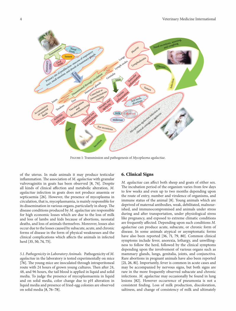
and nasal secretions, faeces, milk, urine, and excretions fromjoint lesions [58]. Sexual transmission through infected malehas been reported. Contaminated utensils andmilker’s handsare vital source of infection. Vertical transmission is observedthrough contaminated colostrum or milk [18, 59, 60]. Thevarious sources of disease transmission have been depictedin Figure 1.
In majority of cases chronic or persistence infection forseveral months in flock is observed with clinically positiveanimals during favorable environmental conditions as at thetime of hot and humid summer. Young, malnourished, preg-nant, and immunocompromised animals are comparativelymore susceptible to the infection [61]. There are reports ofexcretion of organisms in milk even after 8 years of infectionwithmild and with or without clinical signs [37, 62].Thus thepresence of asymptomatic carriers in a herd which carry theinfectious agent is ofmajor concern. Persistence of antibodiescould be observed up to 8 and 3 years of clinical disease ingoats and sheep, respectively [63, 64]. Animal species otherthan homologous hosts as cattle, camel, and many other wildsmall ruminants can also act as reservoir of the infection.These carrier states are more frequently observed in females,particularly in their genital tracts [16, 56, 65].
5. Pathogenesis
M. agalactiae is comparatively stable at room temperatureand in general is transmitted through oral, respiratory,and mammary route. The different routes of transmissionand process of disease development have been depicted inFigure 1. It has been isolated fromnasal secretions [21, 66, 67],faecal samples [68], milk [59, 69], and aborted fetus [70]. Itsuggests that the primary site of predilection is the mucosaof respiratory tract, small intestine, and alveoli of mammaryglands, respectively, depending upon the respiratory, oral,and mammary routes [21]. However, as such no diseasecondition is reported with the involvement of small intestine.Once infection is set up, fever is observed due to bacteremiaaccompanied by fever. Then following the initial multipli-cation organisms are disseminated through circulation todifferent vital organs, namely, lungs, lymph nodes, eyes,mammary glands, joints, and tendons, producing variousclinical signs [8, 26]. Involvement of connective tissues inmammary glands leads to initial inflammation which ulti-mately turns in catarrhal or parenchymatous mastitis leadingto atrophy and agalactia [21, 53]. Animals suffering frommastitis can spread disease to young ones through colostrumsor milk [71]. In general, lung lesions are observed with M.agalactiae infection, although outbreaks of pleurisy amonggoats with the isolation of mycoplasma have been reported[8, 43]. Painful swelling of joints with the accumulationof synovial fluids leads to arthritis mainly in carpal andtarsal joints. In chronic cases eventual loss of joints leadsto ankylosis. Affections in eye cause severe losses of cornea,ultimately leading to blindness through vascularisation andkeratoconjunctivitis [26, 60, 72, 73]. Affections of genitalorgans are also observed with occasional abortions or stillbirths in pregnant animals, mainly due to the inflammation
4 Veterinary Medicine International
Milk Colostrum
Milkman
Chronic
Infe
ctio
n
Semen
Semen
Transmission Nasalroute
Oralroute
Mam
mar
yro
ute
Primary site
of predilection–mucosa of respiratory
tract, small in
testine, and alveoli o
f mammary glands
Pyrexia,
bacteremia
Accumulation
of synovial
fluids
Joints
Joint
Tendons
Lungs
Lymph nodes
Eyes
Mammary glandsGenitalia
Pleurisy
Lymphadenitis
Male
Fem
ale
Nasal secretions, sneezing,
and coughing
Catarrhal or
parenchymatous
mastitis
Vascularisation
and
keratoconjunctivitis
Arthriti
s,
ankylosis
Testicu
lar
inflammati
on
Uterine
inflammatio
n
Atrophy and
agalactia
Corneal loss
and blindness
Secreti
on
Eye secretionsMyco
plasma agalactia
e infec
tion or
classic
al contagious agalactia
Still birth,
abortionssss
MMaMMMM
ry gyyyy
Disseminated through circulation
Figure 1: Transmission and pathogenesis ofMycoplasma agalactiae.
of the uterus. In male animals it may produce testicularinflammation.The association ofM. agalactiae with granularvulvovaginitis in goats has been observed [8, 74]. Despiteall kinds of clinical affection and metabolic alteration, M.agalactiae infection in goats does not produce anaemia orsepticaemia [26]. However, the presence of mycoplasma incirculation, that is, mycoplasmamia, is mainly responsible forits dissemination in various organs, particularly in sheep.Thedisease conditions produced byM. agalactiae are responsiblefor high economic losses which are due to the loss of milkand loss of lambs and kids because of abortions, neonataldeaths, and loss of animals themselves. Moreover, losses alsooccur due to the losses caused by subacute, acute, and chronicforms of disease in the form of physical weaknesses and theclinical complications which affects the animals in infectedherd [35, 50, 74, 75].
5.1. Pathogenicity in Laboratory Animals. Pathogenicity ofM.agalactiae in the laboratory is tested experimentally on mice[76]. The young mice are inoculated through intraperitonealroute with 24 hours of grown young cultures. Then after 24,48, and 96 hours, the tail blood is applied in liquid and solidmedia. To judge the presence of mycoplasmaemia in liquidand on solid media, color change due to pH alteration inliquid media and presence of fried-egg colonies are observedon solid media [8, 76–78].
6. Clinical Signs
M. agalactiae can affect both sheep and goats of either sex.The incubation period of the organism varies from few daysto few weeks and even up to two months depending uponthe route of entry, number and virulence of organisms, andimmune status of the animal [8]. Young animals which aredeprived of maternal antibodies, weak, debilitated, malnour-ished, and immunocompromised and animals under stressduring and after transportation, under physiological stresslike pregnancy, and exposed to extreme climatic conditionsare frequently affected. Depending upon such conditionsM.agalactiae can produce acute, subacute, or chronic form ofdisease. In some animals atypical or asymptomatic formshave also been reported [36, 71, 79, 80]. Common clinicalsymptoms include fever, anorexia, lethargy, and unwilling-ness to follow the herd, followed by the clinical symptomsdepending upon the involvement of various organs such asmammary glands, lungs, genitalia, joints, and conjunctiva.Rare abortions in pregnant animals have also been reported[21, 26, 81]. Importantly, fever is common in acute cases andmay be accompanied by nervous signs, but both signs arerare in the more frequently observed subacute and chronicinfections. M. agalactiae may occasionally be found in lunglesions [82]. However occurrence of pneumonia is not aconsistent finding. Loss of milk production, discoloration,saltiness, and change of consistency of milk and ultimately
Veterinary Medicine International 5
agalactia are commonly observed. Young ones receivinginfected colostrums and milk might lead to septicaemia,arthritis, or pneumonia with high mortality of the kids[8, 71]. Chronic involvement of joints and severe losses tocornea lead to lameness along with inability to walk orstand and blindness, respectively [72, 73]. The conditionslike pleurisy, arthritis, pneumonia, keratoconjunctivitis, andmastitis usually result from infection with M. mycoides toobecause this organismhas one of the widest geographicaldistributions and is found wherever contagious agalactia isreported [69]. Congenital polyarthritis has also been reportedfrom goat kid [83, 84]. The clinical conditions producedduring infection have been elicited in Figure 1.
7. Diagnosis
7.1. Conventional Diagnosis. Primary or tentative diagnosisof the organism is based upon clinical signs, namely, loss ofmilk production, mastitis, keratoconjunctivitis, and articularlesions. Discoloration ofmilk in yellowish-green color, oculardischarges, articular swellings, and lameness are suggestive ofM. agalactiae infection.The clinical diagnosis is confirmed byisolation and identification of the organism in the laboratory[85]. Samples of milk, auricular, ocular, vaginal, or nasaldischarges, articular exudates, blood, and urine are used forthe diagnosis [21, 26, 86]. For the isolation purposes frominfected tissues, samples are collected aseptically from themammary glands, regional lymph nodes, pulmonary lesions,and articular exudates during postmortem examination [26].Isolation ofM. agalactiae from liver, kidney, and spleen couldbe performed during the phase of mycoplasma. Cultivationis carried out in liquid or on solid media which supportmycoplasma growth [18, 21].M. agalactiae produces fried-eggcolonies. Characterization of isolates based on biochemicaltests is not usually recommended [22, 87] due tomorphology,growth, andmetabolic similarity to some other mycoplasmas[28, 88]. Various methods of diagnosis have been depicted inFigure 2.
7.2. Serological Diagnosis. Serological tests of importancefor detecting M. agalactiae include growth precipitation(GP), immunofluorescence (IF), complement fixation test(CFT), indirect haemagglutination (IHA), haemagglutina-tion inhibition (HI), agglutination, latex agglutination test(LAT), double immunodiffusion (DID), single radial immun-odiffusion (SRID), enzyme linked immunosorbent assay(ELISA), radio immunoassay (RIA), and immunoperoxidase(IP) [1, 9, 22, 87, 89–96]. They also include many elec-trophoretic techniques such as gel electrophoresis, immuno-electrophoresis (IEP), countercurrent immunoelectrophore-sis (CCE), and crossed immunoelectrophoresis [22, 40, 97,98]. Immunoblotting has been used to demonstrate theantigenic specificity by the use of hyperimmune sera fromrabbit which is monospecific [42]. Other than these meth-ods, the techniques to separate protein antigens, namely,polyacrylamide gel electrophoresis (PAGE), sodium dodecylsulphate polyacrylamide gel electrophoresis (SDS-PAGE),two-dimensional immunoelectrophoresis, western blotting,
dot blotting, and immunobinding assay, have also beendeveloped and attempted to diagnose caprine agalactia [34,35, 40, 99–103]. However, to overcome the difficulties andlimitations in identification of the organism, diagnosis ofM.agalactiae can be carried out by the complement fixationtest (CFT) or monoclonal antibody based ELISA techniquesagainst individual mycoplasma species or by means of geneamplification techniques [36, 79, 80, 104]. Serological testshave been efficiently used for the diagnosis of contagiousagalactia due to M. agalactiae from the field cases, butdependency of these tests on crude antigens, in general, maynot render them very specific and sensitive. Therefore, manyof these tests cannot differentiate between the mycoplasmaspecies due to the presence of common antigens [22, 34,85, 87]. For differentiation of M. agalactiae and M. mycoides(large colony) and many other related species monoclonalantibody as well as recombinant protein based ELISA hasbeen described [36, 79, 80, 104]. In small ruminants, affectedwith contagious agalactia, correlation study conducted onELISA activity with various other serological tests under thefield conditions indicated the ability of the test to detect thesubclinical infection caused by the organism and also theability to screen the goat herds for the presence of carrieranimals [9, 61]. The ELISA, CFT, and immunoblotting aresupposed to be standard serological tests as per the guidelinesof OIE.
7.3. Molecular Diagnosis. The recent advances in molecularbiology and biotechnology have strengthened the diagno-sis, characterization, and differentiation of mycoplasmasincludingM. agalactiae. Cross-reactive closely related speciescan be certainly differentiated by the use of gene probes,complementary to segments of chromosomal DNA or 16Sribosomal RNA (rRNA) [63, 105, 106] with mixed success(Figure 2). However, the use of polymerase chain reaction(PCR) technique that seems to be even more sensitive andeffective tool for the identification purposes is commonlypracticed [107]. A simple method for detection ofM. agalac-tiae from sheepmilk byDNA extraction and subsequent PCRhas proven to be faster than cultural isolation of the organismand has reduced the time required for diagnosis from daysto hours [39, 108–110]. Use of pulsed-field gel electrophoresis(PFGE) has also strengthened the M. agalactiae diagnostics[39, 111]. PCR techniques based on 16S rRNA [79, 112], uvrCgene [113], and multiplex PCR [15, 26, 100, 114, 115] are beingroutinely used for the identification of M. agalactiae andhave high diagnostic value (Figure 2). Molecular detectionbased on uvrC gene is of prime importance according to therecommendation of OIE.
By amplifying the 16S rRNA gene it is possible to identifythe M. agalactiae isolates by means of PCR and it has beenfound that 99.8 percent similarity is shared by M. agalactiaeas well asM. bovis isolates. Certain other diagnostic strategiesinclude unknown sequence amplification or amplification ofcertain particular gene apart from uvrC like mb-mp81 geneencoding the P81 membrane protein. PCR-restriction frag-ment length polymorphism (PCR-RFLP) technique formsthe basis of thismethod [107, 116]. Real-time quantitative PCR

6 Veterinary Medicine International
Primary or tentative diagnosis based upon clinical signs
Sample Collection
Loss of milk production
Samples of milk, auricular, ocular, vaginal, or nasal discharges, articular exudates, blood, and urine from live animals, while mammary glands, regional lymph nodes, pulmonary lesions and articular exudates during postmortem examination
Secondary and confirmatory diagnosis
Conventional detectionSerological detection
Molecular detectionChromosomal
DNA /16S rRNA/uvrC
gene/mb-mp81 gene Growth precipitation, CFT,
immunofluorescence, indirect haemagglutination, immunoperoxidase
immunoelectrophoresis, crossedimmunoelectrophoresis, immunoblotting,
immunobinding assay, dot blotting,
Isolation
Cultural examination
Morphological examination
Metabolic characterization
Growth inhibition
PCR, multiplex PCR,real-time PCR, Q-PCR,
and MLSTassay, LAT, HI, SRID, DID, ELISA, RIA,
Combined
(1) Antigenic profiling and molecular typing
(2) Optical mapping and genome sequencing
Confrmation
strategy
Mastitis Keratoconjunctivitis Articular lesions Still birth, abortions
and PFGE
Figure 2: Diagnosis ofMycoplasma agalactiae infection.
(Q-PCR) assay has been used for quantifying the organismabsolutely and is becoming increasingly popular for thepurpose of diagnosis in both clinical and food microbiology[117]. Higher specificity as well as sensitivity of analysis isprovided by this technique thus reducing the chances ofcross-contamination. Chemistry of molecular beacon hasbeen used by certain workers for developing a real-timePCR detection methodology. This method targets a regionof 117 base pairs (bp) of the mb-mp81 gene of M. agalactiaeencoding P81 lipoprotein gene [118]. In each of the reactionmixtures it ismandatory to add internal amplification control(IAC) for assessing the potential inhibitory effect of PCR orthermocycler malfunctioning. It is known that a chimericnontarget DNA fragment is IAC present in each of the reac-tion mixtures and target sequence can be used to coamplifyit [119]. However, in an another method, alternate templateinstead of being used in same PCR reaction mixture, it is rununder similar amplification conditions in separate PCR wells[118, 120].
Multilocus sequence typing (MLST), a robust moleculartool, has also been used for comparison of genetic sequencesofM. agalactiae [121, 122].M. agalactiae possesses a capacityfor phenotypic diversification of its surface antigens [36,79]. In this regard, analysis of the antigenic variation ofseveral M. agalactiae wild strains using different sera fromnaturally infected sheep followed by characterization of twostrongly immunogenic membrane surface proteins of 55 kDa
and 35 kDa, respectively [110] is quiet noteworthy. The geneencoding the P48 major surface lipoprotein has been char-acterized and reported to play a crucial role in the immuneresponse of infected animals. Analysis of a recombinant P48expressed in E. coli by using western blot and indirect ELISAproves to be a diagnostically relevant marker ofM. agalactiaeinfection [123].
A combined strategy including antigenic profiling,molecular typing, and optical mapping as well as sequencingof the whole genome has shown the presence of 35 codingsequence. These sequences are based on gene involved,expression of antigens, and vice versa. They are contained ina large prophage and have confirmed the characterization ofisolates in wild ungulates [5].
8. Treatment
Initial therapy for the infection included the use of arsenicals,particularly sodium and zinc salts of acetarsol. The use ofthese compounds and their continuous therapeutic usagehad adverse effects. Presently all over the world preferredtherapy is the use of antibiotics based on drug sensitivity.Commonly used antibiotics include tetracycline, macrolide,clindamycin, florfenicol, tylosin, tiamulin, tilmicosin, andfluoroquinolones [35, 124–126]. Systemic use of antibioticresponds well; however, local application in advanced stages

Veterinary Medicine International 7
to avoid damages inmammary glands, conjunctiva, and jointsshould accompany systemic treatment [127, 128]. Fluoro-quinolones, particularly enrofloxacin which is converted tociprofloxacin after metabolism [129], might have less chancesof resistance development. Moreover, the peak value ofminimum inhibitory concentration (MIC) of ciprofloxacin isreached within few minutes in sheep [129–131] so it would bemore useful in acute cases.Theuse of traditional antibiotics inacute cases is followed by the long acting preparations whichhave vital role in the subclinical and chronically affectedanimals. The confirmation and culling policy has limited theuse of antibiotics; however precious animals and suspectedanimals are always treated with parental therapy followed bylong acting oily preparations.
9. Prevention and Control
The multiple sources of infection and excretion ofMycoplasma agalactiae through various body secretionslead to rapid spread of infection. Thus the timely and quickresponse to the infection is essential for the prevention andcontrol of the spread of infection to susceptible animals[35].M. agalactiae infection could be prevented by adoptinggood managemental practices and following continuoussurveillance/monitoring for the pathogen. Many timessubclinically infected animals may also spread the infection;hence there is always a need to apply specific, sensitiveand rapid diagnostic procedure for its early detection.Till confirmation suspected animals should be isolatedand kept under observation. Immediately after diseaseconfirmation, culling of all the contact and affected animalsis recommended. Proper disposal of litter and othermaterials, namely, discharges and aborted fetus, and propersterilization of contaminated utensils are recommended.Use of disinfectants as hypochloric acid, formalin, cresols,and phenolic substances along with commonly usedquaternary ammonium compounds is effective against theorganism [21, 35]. Proper screening of the semen for artificialinsemination and bucks to be used should be conducted onregular basis. To avoid the vertical transmission ewes shouldbe vaccinated. In endemic areas vaccination with locallydeveloped vaccine is effectively applied throughout world.
10. Vaccines
Similar to many other bacterial agents, both live attenuatedand inactivated vaccines are available for caprine agalactia[2, 111, 132–140]. These vaccines are both safe and effective[68, 137, 138, 141, 142]. The vaccination against Mycoplasmaagalactiae in sheep induces both specific and nonspecific,humoral, and cellular response irrespective of type of vaccine.However, duration of persistence of antibodies depends uponmultiple factors, namely, strain used, adjuvant incorporated,dose of vaccine, routes of inoculation, physiological statusof animal, and so forth. Live attenuated vaccines are moreeffective and have been reported to provide better protectionin ewes and their lambs than the inactivated vaccines but canproduce a transient infection with shedding of mycoplasma
through milk. Importantly, the live vaccines should be partof a regional plan in which all flocks from which animalsare likely to come into contact be vaccinated at the sametime. Inactivated vaccines are much safer with no side effectsbut have shorter period of protection with doubted efficacy[71, 140, 143]. It is possible that in some instances theapparent lack of protection given by vaccines can be theresult of infection of animals with one of the other fourmycoplasmas involved in the contagious agalactia syndrome.The issue for vaccine is duration, the levels of immunityare being addressed, and a combined inactivated vaccinewith aluminium hydroxide gel and saponin and with mineraloil as adjuvant was also attempted in laboratory [139, 140]and field condition [137, 138, 144]. A saponified vaccinewas reported to be effective in initial laboratory trial inmice [145]. Three inactivated vaccines, namely, A, B, andC, with several adjuvants (oil-emulsified) prepared with M.agalactiae have been evaluated for immunogenicity as wellas efficacy purposes. For this purpose, animals have beendivided into three groups and immunized with same vaccineusing different adjuvant. After challenge with the organism,clinical protection has been induced by all the vaccineformulations. Full protection however has been induced byonly the vaccine C which contains Montanide ISA-563 aswell asMarcol-52 andMontane-80 (ratio: 30% : 63% : 7%) andhas been found to induce protection at full level in animalsthat are challenged. This has helped in preventing both theclinical signs’ onset and infection [136]. Out of available liveattenuated and adjuvant (alum precipitated or saponified)vaccines, inactivated vaccines are of greater use due to lackof side effects. However, the protection period is short incomparison to live vaccines.
11. Conclusion and Future Perspectives
Contagious agalactia is considered as a neglected disease ofsmall ruminants because of the complex disease distributionpattern, ubiquitous nature of the causal agent, and poorsheep and goat farm managemental practices, especially indeveloping and underdeveloped countries like India. Rapidspread and multiple sources of infection along with verticaland horizontal mode of transmission are matter of immenseconcern and severely affect the local economy. Dependingupon conditions like deprivation of maternal antibodies,immunocompromised state, stress due to transportation,pregnancy, or extreme climatic conditions, animals maysuffer from acute, subacute, chronic, or asymptomatic formsof disease. The isolation of M. agalactiae is a difficult taskdue to its property of lack of cell wall resembling otherorganisms of the genus Mycoplasma, and serological testscan efficiently identify M. agalactiae. However, in recentyears, the isolation ofM. capricolum subsp. capricolum (Mcc)and M. mycoides subsp. capri (formerly M. mycoides subsp.mycoides; large colonies) from sheep and goat havingmastitisand arthritis complicated the situation. The cross-reactivityof many antigens of these mycoplasmas may lead to falsereactivity. Under such conditions, the advanced moleculardetection techniques like 16S rRNA based PCR andmultiplex

8 Veterinary Medicine International
PCR certainly help in differentiation of closely related speciesof the organism which often cause confusion in the mind ofthe diagnostician. Systemic uses of tetracycline, macrolide, orquinolone group of antibiotics along with local application inadvanced stages are useful treatment options. Good manage-mental practices like isolation of sick animals along with testand slaughter policy form the basis of disease prevention. Forthe prevention and control of disease in particular to endemicareas vaccination is only effective strategy. Mono-, bi-, andtrivalent live attenuated and adjuvant inactivated vaccinesare available with local strains with limited success rate.However, further research on the molecular epidemiologyof the organism in both domestic and wild animals isnecessary to fully understand the disease distribution patternto effectively manage the populations of goat and sheep andprotect them against the infection. Similarly, there is need toexplore the advancementsmade in the field of vaccinology forthe management of the disease more efficiently in sheep andgoat population.
Conflict of Interests
The authors declare that there is no conflict of interestsregarding the publication of this paper.
Acknowledgments
The authors are thankful to the Hon’ble Vice Chancellor ofUttar Pradesh Pandit Deen Dayal Upadhyaya Pashu ChikitsaVigyan Vishwavidyalaya Evam Go Anusandhan Sansthan(DUVASU), Mathura, Uttar Pradesh, India, Director of Ani-mal Resources Development Department, Agartala, Tripura,India, and Director of Indian Veterinary Research Institute,Izatnagar, Bareilly, Uttar Pradesh, India.
References
[1] D. G. Edward, “Organisms of the pleuropneumonia groupcausing disease of goats,” Veterinary Record, vol. 65, pp. 873–875, 1953.
[2] L. Leon-Vizcaino, F. Garrido-Abellan, M. J. Cubero-Pablo,and A. Perales, “Immunoprophylaxis of caprine contagiousagalactia due to Mycoplasma agalactiae with an inactivatedvaccine,” Veterinary Record, vol. 137, no. 11, pp. 266–269, 1995.
[3] J. Ariza-Miguel, D. Rodriguez-Lazaro, and M. Hernandez, “Asurvey ofMycoplasma agalactiae in dairy sheep farms in Spain,”BMC Veterinary Research, vol. 8, article 171, 2012.
[4] K. Sarris, “Contagious agalactia,” in Mycoplasmas of Rumi-nants: Pathogenicity, Diagnostics, Epidemiology and MolecularGenetics, J. Frey and K. Sarris, Eds., pp. 12–15, EUR 16934,COST, EuropeanCommission, EuropeanCommunitiesOfficialPublications Office, Luxembourg, 1996.
[5] F. Tardy, E. Baranowski, L.-X. Nouvel et al., “Emergence of atyp-ical Mycoplasma agalactiae strains harboring a new prophageand associated with an alpine wild ungulate mortality episode,”Applied and Environmental Microbiology, vol. 78, no. 13, pp.4659–4668, 2012.
[6] M. C. Gil, M. H. de Mendoza, J. Rey, J. M. Alonso, J. B. Poveda,and J. de Hermoso Mendoza, “Aetiology of caprine contagious
agalactia syndrome in extra madura, Spain,” Veterinary Record,vol. 144, no. 1, pp. 24–25, 1999.
[7] G. S. Cottew, “Infections withMollicutes in sheep and goats,” inInfektionen durchMycoplasmates, I. Gyrlstorff, Ed., pp. 368–386,VEB Gustaf Fischer, Jena, Germany, 1985.
[8] S. Razin, D. Yogev, and Y. Naot, “Molecular biology andpathogenicity of mycoplasmas,” Microbiology and MolecularBiology Reviews, vol. 62, no. 4, pp. 1094–1156, 1998.
[9] F. Poumarat, D. le Grand, P. Gaurivaud et al., “Comparativeassessment of two commonly used commercial ELISA testsfor the serological diagnosis of contagious agalactia of smallruminants caused by Mycoplasma agalactiae,” BMC VeterinaryResearch, vol. 8, article 109, 2012.
[10] J. C. Corrales, A. Esnal, C. de la Fe et al., “Contagious agalactiain small ruminants,” Small Ruminant Research, vol. 68, no. 1-2,pp. 154–166, 2007.
[11] J. Bridre and A. Donatien, “Lemicrobe de l’agalaxie contagieuseet sa culture in vitro,”Comptes Rendus de l’Academie des Sciences,vol. 177, pp. 841–843, 1923.
[12] J. Bridre and A. Donatien, “Le microbe de l’agalaxie du moutonet de la chevre,” Annals of Institute of Pasteur, vol. 39, pp. 925–951, 1925.
[13] W.Wroblewski, “Morphologie et cycle evolutif des microbes dela peripneumonie des bovides et de l’agalaxie contagieuse deschevres et des moutons,” Annals of Institute of Pasteur, vol. 47,pp. 94–115, 1931.
[14] E. A. Freundt, “Order X. Mycoplasmatales Freundt,” in Bergey’sManual of Determinative Bacteriology, R. S. Breed, E. G. D.Murray, andN. R. Smith, Eds., pp. 914–926,Williams&Wilkins,Baltimore, 7th edition, 1957.
[15] C. A. Becker, F. Ramos, E. Sellal, S. Moine, F. Poumarat,and F. Tardy, “Development of a multiplex real-time PCR forcontagious agalactia diagnosis in small ruminants,” Journal ofMicrobiological Methods, vol. 90, no. 2, pp. 73–79, 2012.
[16] D. Bergonier, X. Berthelot, and F. Poumarat, “Contagiousagalactia of small ruminants: current knowledge concerningepidemiology, diagnosis and control,” OIE Review in ScienificTechique, vol. 16, no. 3, pp. 848–873, 1997.
[17] F. Garrido, J. L. Leon, L. Cuellar, and M. A. Diaz, ContagiousAgalactia and Other Mycoplasmal Diseases of Ruminants, eds.by M. Lambert and G. Jones, Commission of the EuropeanCommunity, Luxembourg, 1987.
[18] M. Lambert, “Contagious agalactia of sheep and goats,” OIEReview in Scienific Techique, vol. 6, no. 3, pp. 699–711, 1987.
[19] R. A. J. Nicholas, “Contagious agalactia: an update,” in Myco-plasmas of Ruminants: Pathogenicity, Diagnostics, Epidemiologyand Molecular Genetics, J. Frey and K. Sarris, Eds., pp. 60–62,EUR 16934, COST, European Commission, European Commu-nities Official Publications Office, Luxembourg, 1996.
[20] G. Greco, M. Corrente, D. Buonavoglia, A. Aliberti, andA. Fasanella, “Inactivated vaccine induces protection againstMycoplasma agalactiae infection in sheep,”NewMicrobiologica,vol. 25, no. 1, pp. 17–20, 2002.
[21] N. C. Srivastava, Studies on the mycoplasma and acholeplasmaof respiratory tract of buffaloes [Ph.D. thesis], Rohilkhand Uni-versity, Bareilly, India, 1982.
[22] A. Kumar, Characterization of protein antigens of Mycoplasmaagalactiae [M.S. thesis], Indian Veterinary Research Institute,Izatnagar, India, 2000.
[23] L. Khan,G. Loria, K.Abu-Amero, R.A. J. Nicholas,M.Halablab,and R. J. Miles, “Distinctive biochemical characteristics of

Veterinary Medicine International 9
Mycoplasma agalactiae andMycoplasma bovis,” inMycoplasmasof Ruminants: Pathogenicity, Diagnostics, Epidemiology andMolecular Genetics, J. B. Poveda, A. Fernandez, J. Frey, and K.E. Johansson, Eds., vol. 5, pp. 60–63, European Commission,Brussels, Belgium, 2001.
[24] L. A. Khan, G. R. Loria, A. S. Ramirez, R. A. J. Nicholas, R.J. Miles, and M. D. Fielder, “Biochemical characterisation ofsome non fermenting, non arginine hydrolysing mycoplasmasof ruminants,”VeterinaryMicrobiology, vol. 109, no. 1-2, pp. 129–134, 2004.
[25] C. de la Fe, P. Assuinao, T. Antunes, R. S. Rosales, and J.B. Poveda, “Microbiological survey for Mycoplasma spp. in acontagious agalactia endemic area,”The Veterinary Journal, vol.170, no. 2, pp. 257–259, 2005.
[26] O. Kizil and H. Ozdemir, “Clinical, haematological and bio-chemical studies in goats naturally infected with Mycoplasmaagalactiae,” Bulletin of the Veterinary Institute in Pulawy, vol. 50,no. 3, pp. 325–328, 2006.
[27] H. ErnØ and L. Stipkovits, “Bovine mycoplasmas: cultural andbiochemical studies,” Acta Veterinaria Scandinavica, vol. 14, no.3, pp. 436–449, 1973.
[28] J. B. Poveda and R. A. J. Nicholas, “Serological identificationof mycoplasmas by growth and metabolic inhibition tests,” inMycoplasma Protocols, R. J. Miles and R. A. J. Nicholas, Eds.,pp. 105–111, Humana Press, Totowa, NJ, USA, 1998.
[29] W. A. Clyde, “Mycoplasma species identification based upongrowth inhibition by specific antisera,” Journal of Immunology,vol. 92, pp. 958–965, 1964.
[30] S. Razin, “Mycoplasma taxonomy studiedy electrophoresis ofcell proteins,” Journal of Bacteriology, vol. 96, no. 3, pp. 687–694,1968.
[31] J. M. Al Aubaidi and A. H. Dardiri, “Biochemical characteriza-tion ofMycoplasma agalactiae subsp. agalactiae (Wroblewski),”International Journal of Systematic Bacteriology, vol. 24, no. 1,pp. 136–138, 1974.
[32] V. Manceau, J. P. Crampe, P. Boursot, and P. Taberlet, “Identifi-cation of evolutionary significant units in the Spanish wild goat,Capra pyrenaica (Mammalia, Artiodactyla),” Animal Conserva-tion, vol. 2, no. 1, pp. 33–39, 1999.
[33] Y. R. C. Gonzalez, C. R. Bascunana, G. Bolske, J. G.Mattsson, C.F.Molina, andK.-E. Johansson, “In vitro amplification of the 16SrRNAgenes fromMycoplasma bovis andMycoplasma agalactiaeby PCR,” Veterinary Microbiology, vol. 47, no. 1-2, pp. 183–190,1995.
[34] I. Gummelt, H. Hotzel, M. Runge, andH. Kirchoff, “Toxonomicrelationship between M. bovis and M. agalactiae,” in Cost826: Mycoplasmas of Ruminants: Pathogenicity, Diagnostics,Epidemiology andMolecular Genetics, J. Frey and K. Sarris, Eds.,pp. 27–29, EUR 16934, COST, EuropeanCommission, EuropeanCommunities Official Publications Office, Luxembourg, 1996.
[35] A. Kumar, A. K. Verma, N. Gangwar, and A. Rahal, “Isolation,characterization and antibiogram ofMycoplasma bovis in sheeppneumonia,” Asian Journal of Animal and Veterinary Advances,vol. 7, no. 2, pp. 149–157, 2012.
[36] D. Bergonier, F. de Simone, P. Russo, M. Solsona, M. Lambert,and F. Poumarat, “Variable expression and geographic distribu-tion of Mycoplasma agalactiae surface epitopes demonstratedwith monoclonal antibodies,” FEMS Microbiology Letters, vol.143, no. 2-3, pp. 159–165, 1996.
[37] D. Bergonier, M. Solsona, F. de Simone, P. Russo, and F.Poumarat, “Study ofMycoplasmaagalactiae antigenic variability
using monoclonal antibodies,” in Mycoplasmas of Ruminants:Pathogenicity, Diagnostics, Epidemiology and Molecular Genet-ics, J. Frey and K. Sarris, Eds., pp. 52–54, EUR 16934, COST,European Commission, European Communities Official Pub-lications Office, Luxembourg, 1996.
[38] M. Solsona, M. Lambert, and F. Poumarat, “Genomic, proteinhomogeneity and antigenic variability of Mycoplasma agalac-tiae,” Veterinary Microbiology, vol. 50, no. 1-2, pp. 45–58, 1996.
[39] S. Tola, G. Idini, D.Manunta et al., “Comparison ofMycoplasmaagalactiae isolates by pulsed field gel eIectrophoresis, SDS-PAGE and immunoblotting,” FEMS Microbiology Letters, vol.143, no. 2-3, pp. 259–265, 1996.
[40] A. Kumar and V. P. Singh, “Characterization of Mycoplasmaagalactiae sonicated supernatant protein antigens (SSA) bySephadex G-200 column chromatography and SDS-PAGE,”Indian Veterinary Journal, vol. 88, no. 5, pp. 9–10, 2011.
[41] L.-X. Nouvel, M. S. Marenda, M. D. Glew et al., “Molecular typ-ing of Mycoplasma agalactiae: tracing European-wide geneticdiversity and an endemic clonal population,” ComparativeImmunology, Microbiology and Infectious Diseases, vol. 35, no.5, pp. 487–496, 2012.
[42] A. Kumar, N. C. Srivastava, and V. P. Singh, “Antigeniccharacterization of Mycoplasma agalactiae by SDS-PAGE andImmunoblotting,” Research Journal Microbiology, vol. 9, no. 1,pp. 59–65, 2014.
[43] G. S. Cottew, “Caprine-ovine mycoplasmas,” in The Mycoplas-mas. II. Human and Animal Mycoplasmas, J. G. Tully and R. F.Whitcomb, Eds., pp. 103–132, Academic Press, San Francisco,Calif, USA, 1979.
[44] G. E. Jones, “Contagious agalactia and other mycoplasmaldiseases of small ruminants,” in Proceedings of the Agricul-ture Commission of the European Communities, Nice, France,September 1985.
[45] H. S. Bawa, “Some observations on “Wah” of goats and sheep inShind,” Indian Journal of Veterinary Sciences, vol. 14, pp. 105–110,1944.
[46] G. S. Cottew and L. C. Lloyd, “An outbreak of pleurisy andpneumonia in goats in Australia attributed to a mycoplasmaspecies,” Journal of Comparative Pathology, vol. 75, no. 4, pp.363–374, 1965.
[47] W. A. Watson, G. S. Cottew, O. Erdag, and F. Arisoy, “Thepathogenicity of mycoplasma organisms isolated from sheepand goats in Turkey,” Journal of Comparative Pathology, vol. 78,no. 3, pp. 283–291, 1968.
[48] J. Nicolet, “Animal mycoplasmoses: a general introduction,”Review Scientific Techiques Office of International Epizootics, vol.15, no. 4, pp. 1233–1240, 1996.
[49] C.Damdinsuren, “Mycoplasmosis in farm animals inMongolia:immunization of sheep and goats against contagious agalactia,”Archives Experimental VeterinaryMedicine, vol. 43, pp. 769–772,1989.
[50] M. O. Ojo, “Caprine pneumonia. II. Biochemical characteri-sation and serological identification of mycoplasmas,” TropicalAnimal Health and Production, vol. 8, no. 3, pp. 137–146, 1976.
[51] M. Doutre, P. Perreau, and A. M. Ndiaye, “Contagious reservoirof agalactia in goats caused byM. agalactiae in Senegal,” RevueD’Elevage et de Medicine Veterinary des Pays Tropical, vol. 34,no. 1, pp. 11–14, 1981.
[52] M. M. M. Al-Graibawi, A. J. N. Al-Shammari, V. K. Sharma,and O. M. Saed, “Isolation and identification of M. agalactiaefrom angora goats in Iraq,” Indian Journal of Comparative

10 Veterinary Medicine International
Microbiology, Immunology and Infectious Diseases, vol. 10, pp.156–158, 1989.
[53] G. E. Jones, “Contagious caprine pleuropneumonia,” TechnicalSeries no. 9, Office of International des Epizootics, Paris, France,1989.
[54] W.Al-Momani, R. A. J. Nicholas, andM.N.Abo-Shehada, “Riskfactors associatedwithMycoplasma agalactiae infection of smallruminants in northern Jordan,” Preventive Veterinary Medicine,vol. 83, no. 1, pp. 1–10, 2008.
[55] J. Amores, A. Gomez-Martın, J. C. Corrales, A. Sanchez, A.Contreras, and C. de la Fe, “Presence of contagious agalactiacausing mycoplasmas in Spanish goat artificial inseminationcentres,”Theriogenology, vol. 75, no. 7, pp. 1265–1270, 2011.
[56] G. Verbisck, M. Gonzalez-Candela, M. J. Cubero, L. Leon, E.Serrano, and A. Perales, “Mycoplasma agalactiae in Iberian ibex(Capra pyrenaica) in Spain,” Veterinary Record, vol. 167, no. 11,pp. 425–426, 2010.
[57] G. Verbisck-Bucker, M. Gonzalez-Candela, J. Galian, M. J.Cubero-Pablo, P. Martın-Atance, and L. Leon-Vizcaıno, “Epi-demiology of Mycoplasma agalactiae infection in free-rangingSpanish ibex (Capra pyrenaica) in Andalusia, southern Spain,”Journal of Wildlife Diseases, vol. 44, no. 2, pp. 369–380, 2008.
[58] A. Gomez-Martin, C. de la Fe, J. Amores et al., “Anatomiclocation ofMycoplasma mycoides subsp. capri andMycoplasmaagalactiae in naturally infected goat male auricular carriers,”Veterinary Microbiology, vol. 157, no. 3-4, pp. 355–362, 2012.
[59] H. Kinde, A. J. da Massa, P. S. Wakenell, and R. Petty,“Mycoplasma infection in a commercial goat dairy causedby Mycoplasma agalactiae and Mycoplasma mycoides subsp.mycoides (caprine biotype),” Journal of Veterinary DiagnosticInvestigation, vol. 6, no. 4, pp. 423–427, 1994.
[60] F. Real, S. Deniz, B. Acosta, O. Ferrer, and J. B. Poveda, “Caprinecontagious agalactia caused by Mycoplasma agalactiae in theCanary Islands,” Veterinary Record, vol. 135, no. 1, pp. 15–16,1994.
[61] S. Levisohn, I. Davidson, M. R. C. Caro Vergara, and E.Rapoport, “Use of an ELISA for differential diagnosis ofMycoplasma agalactiae and M. mycoides subspecies mycoides(LC) in naturally infected goat herds,” Research in VeterinaryScience, vol. 51, no. 1, pp. 66–71, 1991.
[62] A. J. daMassa and D. L. Brooks, “The external ear canal of goatsand other animals as a mycoplasma habitat,” Small RuminantResearch, vol. 4, no. 1, pp. 85–93, 1991.
[63] S. Tola, P. Rizzu, and G. Leori, “A species-specific DNA probefor the detection of Mycoplasma agalactiae,” Veterinary Micro-biology, vol. 41, no. 4, pp. 355–361, 1994.
[64] R. A. J. Nicholas, “Surveillance for contagious agalactia inGreat Britain,” in Mycoplasmas of Ruminants: Pathogenicity,Diagnostics, Epidemiology and Molecular Genetics, G. Leori,F. Santini, E. Scanziani, and J. Frey, Eds., vol. 2, pp. 95–97,European Commission, Brussels, Belgium, 1998.
[65] S. Cokrevski, D. Crcev, G. R. Loria, and R. A. J. Nicholas,“Outbreaks of contagious agalactia in small ruminants in theRepublic of Macedonia,” Veterinary Record, vol. 148, no. 21,article 667, 2001.
[66] A. J. da Massa, P. S. Wakenell, and D. L. Brooks, “Mycoplasmasof goats and sheep,” Journal of Veterinary Diagnostic Investiga-tion, vol. 4, no. 1, pp. 101–113, 1992.
[67] J. Amores, A. Sanchez, A. G. Martın, J. C. Corrales, A. Contr-eras, and C. de la Fe, “Viability of Mycoplasma agalactiae andMycoplasma mycoides subsp. capri in goat milk samples stored
under different conditions,” Veterinary Microbiology, vol. 145,no. 3-4, pp. 347–350, 2010.
[68] S. A.Hasso, J.M.Al-Aubaidi, andA.M.Al-Darraji, “Contagiousagalactia in goats: it's severity as related to the route of infectionand pregnancy,” Small Ruminant Research, vol. 10, no. 3, pp.263–275, 1993.
[69] A. J. da Massa, “Recovery of Mycoplasma agalactiae frommastitic goat milk,” Journal of the American Veterinary MedicalAssociation, vol. 183, no. 5, pp. 548–549, 1983.
[70] A. Kumar and N. K. Chandiramani, “Isolation and character-ization of M. agalactiae from premature born kids of goats,”Indian Journal of Comparative Microbiology, Immunology andInfectious Diseases, vol. 8, pp. 95–97, 1987.
[71] J. Nicolet, “Mycoplasma infections in cattle, sheep and goats:methods for diagnosis and prophylaxis,” in ComprehensiveReports on Technical Items Presented to the International Com-mittee or to Regional Commissions, pp. 43–54, OIE, Paris,France, 1994.
[72] L. J. Kwantes and H. A. M. Harby, “Caprine mycoplasmalarthritis in the Sultanate of Oman,” Small Ruminant Research,vol. 16, no. 3, pp. 287–289, 1995.
[73] A.Mega, I. Tsaknakis, and K. Sarris K, “Diagnosis of contagiousagalactia using ELISA: comparison with the complement fixa-tion test,” Bulletin of Hellenic Veterinary Medicine Society, vol.44, pp. 42–48, 1983.
[74] V. Zavagli, “L’agalaxie contagieuse de la brebis et de la chevre,”Bulletin of Office of International Epizootics, vol. 36, pp. 336–362,1951.
[75] K. J. MacOwan, T. F. Brand, N. McGillveray, and A. R. Hunter,“Experimental infection of castrated lambs withM. agalactiae,”The Journal of Hygiene, vol. 93, no. 3, pp. 455–463, 1984.
[76] G. R. Smith, “Experimental infection of mice with M. agalac-tiae,” Journal of Comparative Pathology, vol. 77, no. 2, pp. 199–202, 1967.
[77] S. A. Hasso, A. M. Al-Darraji, and J. M. Al-Aubaidi, “Pathologyof experimentally induced contagious agalactia in goats,” SmallRuminant Research, vol. 13, no. 1, pp. 79–84, 1994.
[78] A. Kumar, N. C. Srivastava, and V. P. Singh, “Pathogenicityof Mycoplasma agalactiae in mice,” Indian Journal of SmallRuminant, vol. 16, no. 2, pp. 269–270, 2010.
[79] D. Bergonier, M. Solsona, J. Frey, R. Miserez, J. Nicolet, and F.Poumarat, “PCR on 16S rRNA gene, restriction endonucleaseanalysis and SDS-PAGE patterns of Mycoplasma agalactiaeisolates,” in Cost 826: Mycoplasmas of Ruminants: Pathogenicity,Diagnostics, Epidemiology and Molecular Genetics, J. Frey andK. Sarris, Eds., pp. 91–93, EUR 16934, COST, European Com-mission, European Communities Official Publications Office,Luxembourg, 1996.
[80] D. Zendulkova, A. Madanat, P. Lany, K. Rosenbergova, and Z.Pospısil, “Detection of Mycoplasma agalactiae by polymerasechain reaction in Jordanian Sheep and Goat Herds,” ActaVeterinary Brno, vol. 76, no. 1, pp. 71–77, 2007.
[81] M. C. Gil, F. J. Pena, J. H. de Mendoza, and L. Gomez, “Genitallesions in an outbreak of Caprine contagious agalactia caused byMycoplasmaagalactiae andMycoplasmaputrefaciens,” Journal ofVeterinary Medicine B, vol. 50, no. 10, pp. 484–487, 2003.
[82] G. R. Loria, C. Sammartino, R. A. J. Nicholas, and R. D.Ayling, “In vitro susceptibilities of field isolates of Mycoplasmaagalactiae to oxytetracycline, tylosin enrofloxacin, spiramycinand lincomycin-spectinomycin,” Research in Veterinary Science,vol. 75, no. 1, pp. 3–7, 1999.
Veterinary Medicine International 11
[83] R. A. J. Nicholas, R. D. Ayling, and G. R. Loria, “Ovinemycoplasmal infections,” Small Ruminant Research, vol. 76, no.1-2, pp. 92–98, 2008.
[84] G. Filioussis, N. D. Giadinis, E. J. Petridou, E. Karavanis, K.Papageorgiou, and H. Karatzias, “Congenital polyarthritis ingoat kids attributed to Mycoplasma agalactiae,” The VeterinaryRecord, vol. 169, no. 14, article 364, 2011.
[85] R. A. J. Nicholas, “Improvements in the diagnosis and controlof diseases of small ruminants caused by mycoplasmas,” SmallRuminant Research, vol. 45, no. 2, pp. 145–149, 2002.
[86] A. Gomez-Martin, J. C. Corrales, J. Amores et al., “Controllingcontagious agalactia in artificial insemination centers for goatsand detection of Mycoplasma mycoides subspecies capri insemen,”Theriogenology, vol. 77, no. 6, pp. 1252–1256, 2012.
[87] A. Kumar, A. K. Verma, and A. Rahal, “Mycoplasma bovis, amulti disease producing pathogen: an overview,” Asian Journalof Animal and Veterinary Advances, vol. 6, no. 6, pp. 537–546,2011.
[88] J. B. Poveda, “Biochemical characteristics in mycoplasma iden-tification,” in Mycoplasma Protocols, R. J. Miles and R. A. J.Nicholas, Eds., pp. 69–78, Humana Press, Totowa, NJ, USA,1998.
[89] A. D. Campbell and A. W. Turner, “Studies on contagiouspleuropneumonia of cattle. II. A complement fixation reactionfor the diagnosis of contagious bovine pleuropneumonia,”Bulletin of Conference of Science of Indian Research, vol. 97, pp.11–52, 1936.
[90] R. M. Lemcke, “The serological differentiation of mycoplasmastrains (PPLO) from various sources,” The Journal of Hygiene,vol. 62, pp. 199–219, 1964.
[91] H. E. Morton, “Mycoplasma-latex agglutination reaction,” Jour-nal of Bacteriology, vol. 92, no. 4, pp. 1196–1205, 1966.
[92] A. Krogsgaard-Jensen, “Indirect hemagglutination withMycoplasma antigens: effects of pH on antigen sensitizationof tanned fresh and formalinized sheep erythrocytes,” Appliedmicrobiology, vol. 22, no. 5, pp. 756–759, 1971.
[93] H. ErnØ and K. Jurmanova, “Bovine mycoplasmas: serologicalstudies by double immunodiffusion, growth precipitation andgrowth inhibition,”Acta Veterinaria Scandinavica, vol. 14, no. 4,pp. 524–537, 1973.
[94] W. Schaeren and J. Nicolet, “Micro- ELISA for detecting con-tagious agalactia in goats,” Schweizer Archiv fur Tierheilkunde,vol. 124, no. 4, pp. 163–177, 1982.
[95] J. M. Bradbury, “Identification of mycoplasmas by immunoflu-orescence,” in Mycoplasma Protocols, R. J. Miles and R. A. J.Nicholas, Eds., pp. 119–125, Humana Press, Totowa, NJ, USA,1998.
[96] M. Giangaspero, R. A. Nicholas, M. Hlusek et al., “Seroepi-demiological survey of sheep flocks from Northern Japanfor Mycoplasma ovipneumoniae and Mycoplasma agalactiae,”Tropical Animal Health and Production, vol. 44, no. 3, pp. 395–398, 2012.
[97] O. Erdag and G. S. Cottew, “Gel electrophoresis separation ofthe proteins of mycoplasma organisms isolated from sheep andgoats,” Pendik Veteriner Kontrol ve Arastirma Enstitusu Dergisi,vol. 4, no. 1, pp. 78–84, 1971.
[98] H. J. Cho and E. V. Langford, “Rapid detection of bovinemycoplasma antigens by counterimmunoelectrophoresis,” Jour-nal of Applied Microbiology, vol. 28, no. 5, pp. 897–899, 1974.
[99] F. Poumarat, “Identification of mycoplasmas by dot immuno-binding on membrane filtration (MF Dot),” in Mycoplasma
Protocols, R. J. Miles and R. A. J. Nicholas, Eds., pp. 113–118,Humana Press, Totowa, NJ, USA, 1998.
[100] M. Kumar, V. P. Singh, N. C. Srivastava et al., “Rapid and specificdetection ofM.Mycoides cluster and differentiation ofmycoidesgroup from capricolum group by PCR,” Indian Journal ofComparative Microbiology, Immunology and Infectious Diseases,vol. 22, pp. 118–121, 2001.
[101] A.Kumar,N.C. Srivastava, andV. P. Singh, “Rapid identificationof M. agalactiae and M. bovis by immuno binding assay,”Indian Journal of Comparative Microbiology, Immunology andInfectious Diseases, vol. 23, pp. 161–163, 2002.
[102] A. Kumar, N. C. Srivastava, and V. P. Singh, “Poly acrylamidegel electrophoresis (PAGE) of Mycoplasma agalactiae andMycoplasma bovis,” Indian Journal of Small Ruminant, vol. 16,pp. 271–273, 2010.
[103] S. Tola, D. Manunta, M. Cocco et al., “Characterization ofmembrane surface proteins of Mycoplasma agalactiae duringnatural infection,” FEMSMicrobiology Letters, vol. 154, no. 2, pp.355–362, 1997.
[104] M. Lambert, M. Calamel, P. Dufour, E. Cabasse, C. Vitu, andM.Pepin, “Detection of false-positive sera in contagious agalactiawith a multiantigen ELISA and their elimination with a proteinG conjugate,” Journal of Veterinary Diagnostic Investigation, vol.10, no. 4, pp. 326–330, 1998.
[105] J. G. Mattsson, H. Gersdorf, U. B. Gobel, and K.-E. Johansson,“Detection ofMycoplasma bovis andMycoplasma agalactiae byoligonucleotide probes complementary to 16S rRNA,”Molecularand Cellular Probes, vol. 5, no. 1, pp. 27–35, 1991.
[106] L. A. Dedieu, V. Mady, and P. C. Lefevre, “Development ofa species—specific DNA probe for Mycoplasma capricolum,”Veterinary Microbiology, vol. 32, no. 2, pp. 189–197, 1992.
[107] J. B. Bashiruddin, J. Frey, M. H. Konigsson et al., “Evaluationof PCR systems for the identification and differentiation ofMycoplasma agalactiae and Mycoplasma bovis: a collaborativetrial,”The Veterinary Journal, vol. 169, no. 2, pp. 268–275, 2005.
[108] L. Dedieu, V. Mady, and P.-C. Lefevre, “Development of twoPCR assays for the identification of mycoplasmas causingcontagious agalactia,” FEMS Microbiology Letters, vol. 129, no.2-3, pp. 243–250, 1995.
[109] S. Tola, G. Idini, D. Manunta et al., “Rapid and specific detec-tion of Mycoplasma agalactiae by polymerase chain reaction,”Veterinary Microbiology, vol. 51, no. 1-2, pp. 77–84, 1996.
[110] S. Tola, A. Angioi, A. M. Rocchigiani et al., “Detection ofMycoplasma agalactiae in sheep milk samples by polymerasechain reaction,” Veterinary Microbiology, vol. 54, no. 1, pp. 17–22, 1997.
[111] S. Tola, G. Idini, A. M. Rocchigiani et al., “Comparison ofrestriction pattern polymorphism of Mycoplasma agalactiaeand Mycoplasma bovis by pulsed field gel electrophoresis,”Journal of Veterinary Medicine B, vol. 46, no. 3, pp. 199–206,1999.
[112] T. R. Abo-Elnaga, A. S. A. Gouda, Y. M. Kamel, M. A.Mahmoud, and W. A. Osman, “Application of the polymerasechain reaction for identification of Mycoplasma isolated fromaborted She Camel,” Global Veterinaria, vol. 8, no. 4, pp. 386–392, 2012.
[113] S. Subramaniam, D. Bergonier, F. Poumarat et al., “Speciesidentification of Mycoplasma bovis and Mycoplasma agalactiaebased on the uvrC genes by PCR,” Molecular and CellularProbes, vol. 12, no. 3, pp. 161–169, 1998.
[114] G. Greco, M. Corrente, V. Martella, A. Pratelli, and D. Buon-avoglia, “A multiplex-PCR for the diagnosis of contagious

12 Veterinary Medicine International
agalactia of sheep and goats,”Molecular and Cellular Probes, vol.15, no. 1, pp. 21–25, 2001.
[115] A. Foddai, G. Idini,M. Fusco et al., “Rapid differential diagnosisof Mycoplasma agalactiae and Mycoplasma bovis based ona multiplex-PCR and a PCR-RFLP,” Molecular and CellularProbes, vol. 19, no. 3, pp. 207–212, 2005.
[116] M.H. Konigsson, G. Bolske, and K.-E. Johansson, “Intraspecificvariation in the 16S rRNA gene sequences of Mycoplasmaagalactiae and Mycoplasma bovis strains,” Veterinary Microbi-ology, vol. 85, no. 3, pp. 209–220, 2002.
[117] D. Rodrıguez-Lazaro, M. Hernandez, M. Scortti, T. Esteve, J.A. Vazquez-Boland, and M. Pla, “Quantitative detection ofListeria monocytogenes and Listeria innocua by real-time PCR:assessment of hly, iap, and lin02483 targets and AmpliFluortechnology,” Applied and Environmental Microbiology, vol. 70,no. 3, pp. 1366–1377, 2004.
[118] A. Lorusso, N. Decaro, G. Greco, M. Corrente, A. Fasanella,and D. Buonavoglia, “A real-time PCR assay for detectionand quantification of Mycoplasma agalactiae DNA,” Journal ofApplied Microbiology, vol. 103, no. 4, pp. 918–923, 2007.
[119] J. Hoorfar, N. Cook, B.Malorny et al., “Diagnostic PCR:makinginternal amplification control mandatory,” Journal of AppliedMicrobiology, vol. 96, no. 2, pp. 221–222, 2004.
[120] K. Oravcova, L. Lopez-Enriquez, D. Rodriguez-Lazaro, and M.Hernandez, “Mycoplasma agalactiae p40 gene, a novel markerfor diagnosis of contagious agalactia in sheep by real-time PCR:assessment of analytical performance and in-house validationusing naturally contaminated milk samples,” Journal of ClinicalMicrobiology, vol. 47, no. 2, pp. 445–450, 2009.
[121] L. McAuliffe, F. Gosney, M. Hlusek et al., “Multilocus sequencetyping ofMycoplasma agalactiae,” Journal of Medical Microbiol-ogy, vol. 60, no. 6, pp. 803–811, 2011.
[122] L. Manso-Silvan, V. Dupuy, I. Lysnyansky, U. Ozdemir, and F.Thiaucourt, “Phylogeny and molecular typing of Mycoplasmaagalactiae and Mycoplasma bovis by multilocus sequencing,”Veterinary Microbiology, vol. 161, no. 1-2, pp. 104–112, 2012.
[123] S. Rosati, P. Robino,M. Fadda, S. Pozzi, A.Mannelli, andM. Pit-tau, “Expression and antigenic characterization of recombinantMycoplasma agalactiae P48 major surface protein,” VeterinaryMicrobiology, vol. 71, no. 3-4, pp. 201–210, 2000.
[124] L. Stipkovits, Z. Varga, G. Laber, and J. Bockmann, “A compar-ison of the effect of tiamulin hydrogen fumarate and tylosintartrate on mycoplasmas of ruminants and some animal ure-aplasmas,” Veterinary Microbiology, vol. 9, no. 2, pp. 147–153,1984.
[125] A. Fadda, E. A. Cannas, G. M. Cubeddu, S. de Palmas, andG. Petracca, “Susceptibility of Mycoplasma agalactiae to tilmi-cosin,” in Proceedings of the 3rd International Mastitis Seminar,pp. 126–127, Lachmann Printers, Tel Aviv, Israel, May 1995.
[126] M. L. de Garnica, R. S. Rosales, C. Gonzalo, J. A. Santos, and R.A. Nicholas, “Isolation, molecular characterization and antimi-crobial susceptibilities of isolates ofMycoplasma agalactiae frombulk tank milk in an endemic area of Spain,” Journal of AppliedMicrobiology, vol. 114, no. 6, pp. 1575–1581, 2013.
[127] A. Bhaumik, B. B. Verma, D. K. Thakur, S. N. Pandey, and N.C. Benerjee, “Effect of oral administration of tylosin tartratein the treatment of experimental and natural cases of caprinemycoplasmosis,” Indian Veterinary Journal, vol. 67, no. 10, pp.948–951, 1990.
[128] A. Esnal, J. J. Aduriz, M. Romeo, R. A. Juste, and A. Contreras,“Agalaxia contagiosa: control,” Ovis, vol. 30, pp. 35–62, 1994.
[129] A. Rahal, A. Kumar, A. H. Ahmad, J. K. Malik, and V. Ahuja,“Pharmacokinetics of enrofloxacin in sheep following intra-venous and subcutaneous administration,” Journal of VeterinaryPharmacology andTherapeutics, vol. 29, no. 4, pp. 321–324, 2006.
[130] A. Rahal, A. Kumar, A. H. Ahmad, and J. K. Malik, “Pharma-cokinetics of diclofenac and its interaction with enrofloxacin insheep,” Research in Veterinary Science, vol. 84, no. 3, pp. 452–456, 2008.
[131] A. Rahal, A. Kumar, A. H. Ahmad, and J. K. Malik, “Pharma-cokinetics of ciprofloxacin in sheep following intravenous andsubcutaneous administration,” Small Ruminant Research, vol.73, no. 1–3, pp. 242–245, 2007.
[132] A. Foggie, J. R. Etheridge, O. Erdag, and F. Arisoy, “Contagiousagalactia of sheep and goats studies on live and dead vaccines inlactating sheep,” Journal of Comparative Pathology, vol. 81, no. 1,pp. 165–172, 1971.
[133] A. Foggie, J. R. Etheridge, O. Erdag, and F. Arisoy, “Contagiousagalactia of sheep and goats,” Journal of Comparative Pathology,vol. 81, no. 3, pp. 393–400, 1971.
[134] O. Erdag, “Investigations on the preparation and application ofvaccine against contagiousMycoplasma agalactiae or sheep andgoats in Turkey,” in Proceeding of International Symposium onMycoplasmosis andTheileriosi, pp. 20–22, Pendik, Turkey, 1989.
[135] K. Sarris, A. Zdrakas, A. Papasteriadis, and O. Papadopoulos,“Experimental contagious agalactia vaccine,” Bulletin HellenicVeterinary Medicine Society, vol. 40, pp. 71–74, 1989.
[136] D. Buonavoglia, A. Fasanella, P. Sagazio, M. Tempesta, G.Iovane, and C. Buonavoglia, “Persistence of antibodies toMycoplasma agalactiae in vaccinated sheep,” New Microbiolog-ica, vol. 21, no. 2, pp. 209–212, 1998.
[137] C. de la Fe, P. Assuncao, P. Saavedra, A. Ramirez, and J. B.Poveda, “Field trial of a combined vaccine against caprinecontagious agalactia: Humoral immune response in lactatinggoats,”The Veterinary Journal, vol. 174, no. 3, pp. 610–615, 2007.
[138] C. de la Fe, P. Assuncao, P. Saavedra, S. Tola, C. Poveda, and J.B. Poveda, “Field trial of two dual vaccines againstMycoplasmaagalactiae and Mycoplasma mycoides subsp. mycoides (largecolony type) in goats,” Vaccine, vol. 25, no. 12, pp. 2340–2345,2007.
[139] D. Buonavoglia, G. Greco, M. Corrente et al., “Long-termimmunogenicity and protection againstMycoplasma agalactiaeinduced by an oil adjuvant vaccine in sheep,” Research inVeterinary Science, vol. 88, no. 1, pp. 16–19, 2010.
[140] D. Buonavoglia, G. Greco, V. Quaranta, M. Corrente, V.Martella, and N. Decaro, “An oil-emulsion vaccine induces full-protection against Mycoplasma agalactiae infection in sheep,”New Microbiologica, vol. 31, no. 1, pp. 117–123, 2012.
[141] M. Lambert and E. Cabasse, “Serologie de l’agalactie con-tagieuse des brebis: comparison ELISA-fixation du comple-ment,” Review Medicine Veterinary, vol. 140, pp. 107–112, 1989.
[142] J. Sunder, N. C. Srivastava, and V. P. Singh, “Preliminary trialson development of vaccine againstMycoplasmamycoides subsp.mycoides type LC infection in goats,” Journal of Applied AnimalResearch, vol. 21, no. 1, pp. 75–80, 2002.
[143] Food andAgricultureOrganisation (FAO),Animal Health Year-book, FAO/Office International des Epizooties (OIE)/WorldHealth Oraganisation (WHO), Rome, Italy, 1992.

Veterinary Medicine International 13
[144] A. Ramirez, C. de la Fe, P. Assuncao, M. Gonzalez, andJ. B. Poveda, “Preparation and evaluation of an inactivatedpolyvalent vaccine against Mycoplasma spp on infected goats,”in Mycoplasmas of Ruminants: Pathogenicity, Diagnostics, Epi-demiology and Molecular Genetics, J. B. Poveda, A. Fernandez,J. Frey, and K. E. Johansson, Eds., vol. 5, pp. 154–157, EuropeanCommission, Brussels, Belgium, 2001.
[145] A. Kumar, N. C. Srivastava, andV. P. Singh, “Immunogenicity ofMycoplasma agalactiae saponin vaccine inmice,” Indian Journalof Comparative Microbiology, Immunology and Infectious Dis-eases, vol. 30, pp. 61–62, 2009.

Submit your manuscripts athttp://www.hindawi.com
Veterinary MedicineJournal of
Hindawi Publishing Corporationhttp://www.hindawi.com Volume 2014
Veterinary Medicine International
Hindawi Publishing Corporationhttp://www.hindawi.com Volume 2014
Hindawi Publishing Corporationhttp://www.hindawi.com Volume 2014
International Journal of
Microbiology
Hindawi Publishing Corporationhttp://www.hindawi.com Volume 2014
AnimalsJournal of
EcologyInternational Journal of
Hindawi Publishing Corporationhttp://www.hindawi.com Volume 2014
PsycheHindawi Publishing Corporationhttp://www.hindawi.com Volume 2014
Evolutionary BiologyInternational Journal of
Hindawi Publishing Corporationhttp://www.hindawi.com Volume 2014
Hindawi Publishing Corporationhttp://www.hindawi.com
Applied &EnvironmentalSoil Science
Volume 2014
Biotechnology Research International
Hindawi Publishing Corporationhttp://www.hindawi.com Volume 2014
Agronomy
Hindawi Publishing Corporationhttp://www.hindawi.com Volume 2014
International Journal of
Hindawi Publishing Corporationhttp://www.hindawi.com Volume 2014
Journal of Parasitology Research
Hindawi Publishing Corporation http://www.hindawi.com
International Journal of
Volume 2014
Zoology
Hindawi Publishing Corporationhttp://www.hindawi.com
GenomicsInternational Journal of
Volume 2014
InsectsJournal of
Hindawi Publishing Corporationhttp://www.hindawi.com Volume 2014
The Scientific World JournalHindawi Publishing Corporation http://www.hindawi.com Volume 2014
Hindawi Publishing Corporationhttp://www.hindawi.com Volume 2014
VirusesJournal of
ScientificaHindawi Publishing Corporationhttp://www.hindawi.com Volume 2014
Cell BiologyInternational Journal of
Hindawi Publishing Corporationhttp://www.hindawi.com Volume 2014
Hindawi Publishing Corporationhttp://www.hindawi.com Volume 2014
Case Reports in Veterinary Medicine
Related Documents