Mycolactones: immunosuppressive and cytotoxic polyketides produced by aquatic mycobacteria Hui Hong, a Caroline Demangel, b Sacha J. Pidot, c Peter F. Leadlay a and Tim Stinear * c Received 25th February 2008 First published as an Advance Article on the web 17th April 2008 DOI: 10.1039/b803101k Covering: up to January 2008 Mycolactones are a family of highly related macrocyclic polyketides that exhibit immunosuppressive and cytotoxic properties. First discovered in 1999, they are the primary virulence factors produced by the environmental human pathogen Mycobacterium ulcerans, the causative agent of Buruli ulcer, and by some closely-related aquatic mycobacteria that cause disease in fish and frogs. Mycolactones are characterized by a common 12-membered lactone core to which is appended an unsaturated fatty acyl side-chain of variable length and oxidation state. This Highlight summarizes recent progress in understanding the structural diversity of the mycolactones, their biological activity and mode of action in mammalian cells, and the genetics, evolution, and enzymology of their biosynthesis. 1 Introduction In the 1960s, pathologists in Uganda studying Buruli ulcer, an unusual skin disease caused by Mycobacterium ulcerans, noted in histological sections that tissue necrosis and a marked lack of inflammation extended beyond zones containing bacteria, suggesting that M. ulcerans produced a diffusible toxin. 1,2 In support of this idea, culture filtrates of M. ulcerans were later found to be cytotoxic to eukaryotic cells, provoke necrosis following injection in guinea pig skin or mouse footpads, and to have immunosuppressive properties. 3–6 Finally, the putative toxin was successfully purified from a clinical isolate of M. ulcerans and revealed as a polyketide that was named myco- lactone. 7,8 It was originally identified as a mixture of cis and trans isomers designated mycolactone A and B. 7,9 Since then it has been shown that different strains of M. ulcerans and closely related mycobacterial species naturally produce at least five structurally distinct molecules, each of which likely also exists in cis and trans forms. The importance of mycolactones for the survival of M. ulcerans is unknown, although a recent study suggests mycolactones are a major constituent of an abundant extracellular matrix produced by the bacterium that may confer an enhanced capacity to colonize certain ecological niches. 10 2 Mycolactones: structural characterization and diversity Mycolactones A and B were first isolated from M. ulcerans MU1615, a Malaysian strain that makes the same mycolactones as African isolates of M. ulcerans. 7,8 Their overall structures were shown by 2D NMR experiments to be, respectively, Z-D 4 0 ,5 0 and E-D 4 0 ,5 0 isomers of a 12-membered macrocyclic polyketide in which a second highly unsaturated polyketide side-chain is appended via an ester linkage 9 (Fig. 1). The absolute configura- tion of mycolactone A and B was established soon afterwards by chemical synthesis. 11–13 Subsequent investigations of myco- lactone structure have been hampered by the small (usually microgram) quantities available from laboratory-scale cultiva- tion. However, either by scaling-up to 150 L scale, 14 or by use of sensitive mass spectrometry techniques, 15,16 analysis of culture extracts of a typical strain of M. ulcerans has revealed the pres- ence of minor amounts of additional mycolactones, differing from mycolactones A and B only in the side-chain. In particular, use of LC-MS n , combining ion trap mass spectrometry (quadruple ion trap or FT-ICR) with multi-stage collision- induced fragmentation experiments, 15 has provided detailed structural information. Such analysis of cell extracts of the African strain MUAgy99 revealed that apart from mycolactone A/B with [M + Na] + at m/z 765 as the major species, there were present small amounts of other mycolactones with [M + Na] + at m/z 763, 749, 747, 745 and 781. 15 Analysis of cell extracts of M. ulcerans strains from Malaysia and Japan has revealed very similar mycolactone production profiles (H. H., P. F. L. and T. S., unpublished data). MS n analysis showed that these myco- lactone congeners share the same core lactone structure as mycolactone A/B, with the structural variations being confined to the distal end of the side-chain. The species with [M + Na] + at m/z 749 has been identified, for example, as mycolactone C (Fig. 1) in which the normal late-stage hydroxylation of the side- chain at C-12 0 has not taken place. 15,16 The other species can be accounted for if the polyketide synthase governing the side-chain biosynthesis assembles the chain with less than perfect speci- ficity 15 (see also sections 4 and 5). Intriguingly, other clinical isolates of M. ulcerans show distinctly different patterns of mycolactone production. For example, Australian strains produce almost exclusively myco- lactone C ([M + Na] + at m/z 749) 16,44 (Fig. 1). The missing hydroxyl group at C-12 0 of the side-chain compared to a Department of Biochemistry, University of Cambridge, 80 Tennis Court Road, Cambridge, CB2 1GA, UK b Unite´ Postulante Pathoge´nomique Mycobacte´rienne Inte´gre´e, Institut Pasteur, 25 Rue du Dr Roux, Paris, 75015, France c Department of Microbiology, Monash University, Wellington Road, Clayton, 3800, Australia This journal is ª The Royal Society of Chemistry 2008 Nat. Prod. Rep., 2008, 25, 447–454 | 447 HIGHLIGHT www.rsc.org/npr | Natural Product Reports Open Access Article. Published on 17 April 2008. Downloaded on 27/05/2013 14:39:59. View Article Online / Journal Homepage / Table of Contents for this issue

Welcome message from author
This document is posted to help you gain knowledge. Please leave a comment to let me know what you think about it! Share it to your friends and learn new things together.
Transcript
HIGHLIGHT www.rsc.org/npr | Natural Product Reports
Ope
n A
cces
s A
rtic
le. P
ublis
hed
on 1
7 A
pril
2008
. Dow
nloa
ded
on 2
7/05
/201
3 14
:39:
59.
View Article Online / Journal Homepage / Table of Contents for this issue
Mycolactones: immunosuppressive and cytotoxic polyketides producedby aquatic mycobacteria
Hui Hong,a Caroline Demangel,b Sacha J. Pidot,c Peter F. Leadlaya and Tim Stinear*c
Received 25th February 2008
First published as an Advance Article on the web 17th April 2008
DOI: 10.1039/b803101k
Covering: up to January 2008
Mycolactones are a family of highly related macrocyclic polyketides that exhibit immunosuppressive
and cytotoxic properties. First discovered in 1999, they are the primary virulence factors produced by
the environmental human pathogen Mycobacterium ulcerans, the causative agent of Buruli ulcer, and
by some closely-related aquatic mycobacteria that cause disease in fish and frogs. Mycolactones are
characterized by a common 12-membered lactone core to which is appended an unsaturated fatty acyl
side-chain of variable length and oxidation state. This Highlight summarizes recent progress in
understanding the structural diversity of the mycolactones, their biological activity and mode of action
in mammalian cells, and the genetics, evolution, and enzymology of their biosynthesis.
1 Introduction
In the 1960s, pathologists in Uganda studying Buruli ulcer, an
unusual skin disease caused by Mycobacterium ulcerans, noted in
histological sections that tissue necrosis and a marked lack of
inflammation extended beyond zones containing bacteria,
suggesting that M. ulcerans produced a diffusible toxin.1,2 In
support of this idea, culture filtrates of M. ulcerans were later
found to be cytotoxic to eukaryotic cells, provoke necrosis
following injection in guinea pig skin or mouse footpads, and to
have immunosuppressive properties.3–6 Finally, the putative
toxin was successfully purified from a clinical isolate of M.
ulcerans and revealed as a polyketide that was named myco-
lactone.7,8 It was originally identified as a mixture of cis and trans
isomers designated mycolactone A and B.7,9 Since then it has
been shown that different strains of M. ulcerans and closely
related mycobacterial species naturally produce at least five
structurally distinct molecules, each of which likely also exists in
cis and trans forms. The importance of mycolactones for the
survival of M. ulcerans is unknown, although a recent study
suggests mycolactones are a major constituent of an abundant
extracellular matrix produced by the bacterium that may confer
an enhanced capacity to colonize certain ecological niches.10
2 Mycolactones: structural characterization anddiversity
Mycolactones A and B were first isolated from M. ulcerans
MU1615, a Malaysian strain that makes the same mycolactones
as African isolates of M. ulcerans.7,8 Their overall structures were
shown by 2D NMR experiments to be, respectively, Z-D40 ,50 and
aDepartment of Biochemistry, University of Cambridge, 80 Tennis CourtRoad, Cambridge, CB2 1GA, UKbUnite Postulante Pathogenomique Mycobacterienne Integree, InstitutPasteur, 25 Rue du Dr Roux, Paris, 75015, FrancecDepartment of Microbiology, Monash University, Wellington Road,Clayton, 3800, Australia
This journal is ª The Royal Society of Chemistry 2008
E-D40 ,50 isomers of a 12-membered macrocyclic polyketide in
which a second highly unsaturated polyketide side-chain is
appended via an ester linkage9 (Fig. 1). The absolute configura-
tion of mycolactone A and B was established soon afterwards by
chemical synthesis.11–13 Subsequent investigations of myco-
lactone structure have been hampered by the small (usually
microgram) quantities available from laboratory-scale cultiva-
tion. However, either by scaling-up to 150 L scale,14 or by use of
sensitive mass spectrometry techniques,15,16 analysis of culture
extracts of a typical strain of M. ulcerans has revealed the pres-
ence of minor amounts of additional mycolactones, differing
from mycolactones A and B only in the side-chain. In particular,
use of LC-MSn, combining ion trap mass spectrometry
(quadruple ion trap or FT-ICR) with multi-stage collision-
induced fragmentation experiments,15 has provided detailed
structural information. Such analysis of cell extracts of the
African strain MUAgy99 revealed that apart from mycolactone
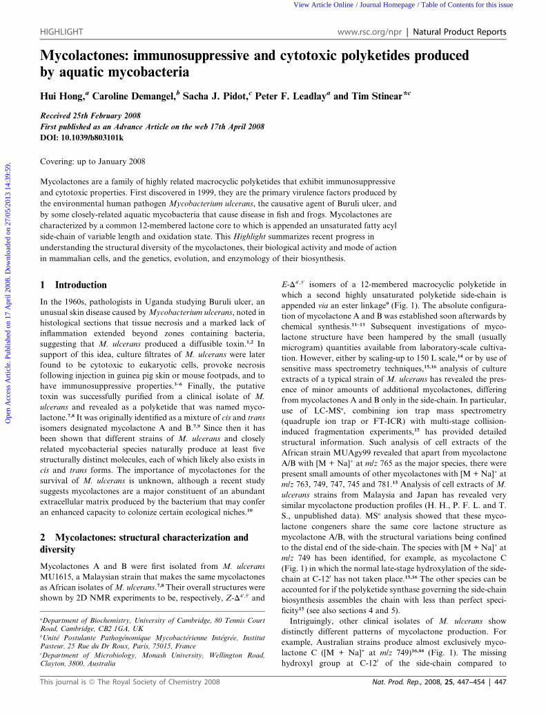
A/B with [M + Na]+ at m/z 765 as the major species, there were
present small amounts of other mycolactones with [M + Na]+ at
m/z 763, 749, 747, 745 and 781.15 Analysis of cell extracts of M.
ulcerans strains from Malaysia and Japan has revealed very
similar mycolactone production profiles (H. H., P. F. L. and T.
S., unpublished data). MSn analysis showed that these myco-
lactone congeners share the same core lactone structure as
mycolactone A/B, with the structural variations being confined
to the distal end of the side-chain. The species with [M + Na]+ at
m/z 749 has been identified, for example, as mycolactone C
(Fig. 1) in which the normal late-stage hydroxylation of the side-
chain at C-120 has not taken place.15,16 The other species can be
accounted for if the polyketide synthase governing the side-chain
biosynthesis assembles the chain with less than perfect speci-
ficity15 (see also sections 4 and 5).
Intriguingly, other clinical isolates of M. ulcerans show
distinctly different patterns of mycolactone production. For
example, Australian strains produce almost exclusively myco-
lactone C ([M + Na]+ at m/z 749)16,44 (Fig. 1). The missing
hydroxyl group at C-120 of the side-chain compared to
Nat. Prod. Rep., 2008, 25, 447–454 | 447
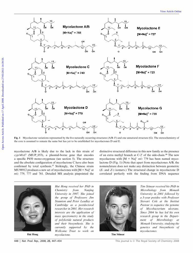
Fig. 1 Mycolactone variations represented by the five naturally occurring structures (A/B–F) and one unnatural structure (G). The stereochemistry of
the core is assumed to remain the same but has yet to be established for mycolactones D and E.
Ope
n A
cces
s A
rtic
le. P
ublis
hed
on 1
7 A
pril
2008
. Dow
nloa
ded
on 2
7/05
/201
3 14
:39:
59.
View Article Online
mycolactone A/B is likely due to the lack in this strain of
cyp140A7 (MUP_053), a plasmid-borne gene that encodes
a specific P450 mono-oxygenase (see section 5). The structure
and the absolute configuration of mycolactone C have also been
confirmed by total synthesis.17 Strikingly, the Chinese strain
MU98912 produces a new set of mycolactones with [M + Na]+ at
m/z 779, 777 and 761. Detailed MS analysis pinpointed the
Hui Hong
Hui Hong received her PhD in
Chemistry from Nanjing
University in 1997. She joined
the group of Professors Jim
Staunton and Peter Leadlay at
Cambridge as a postdoctoral
researcher in 2001. Her research
interests are the application of
mass spectrometry in the study
of polyketide natural products
and their biosynthesis. She is
currently supported by the
Wellcome Trust to work on
mycolactone.
448 | Nat. Prod. Rep., 2008, 25, 447–454
distinctive structural difference in this new family as the presence
of an extra methyl branch at C-20 of the side-chain.18 The new
mycolactone with [M + Na]+ m/z 779 has been named myco-
lactone D (Fig. 1) (Note that apart from mycolactones A/B, the
nomenclature does not make any distinction between geometric
(E- and Z-) isomers.) The structural change in mycolactone D
correlated perfectly with the finding from DNA sequence
Tim Stinear
Tim Stinear received his PhD in
Microbiology from Monash
University in 2001 followed by
a 3-year postdoc with Professor
Stewart Cole at the Institut
Pasteur to sequence the genome
of Mycobacterium ulcerans.
Since 2004 he has led his own
research group in the Depart-
ment of Microbiology at
Monash University, studying the
genetics and biosynthesis of
mycolactones.
This journal is ª The Royal Society of Chemistry 2008
Fig. 2 Summary of the known effects of mycolactones on various
mammalian cell types. Abbreviations LPS: lipopolysaccharide; TNF:
tumour necrosis factor; IL: interleukin; NF-kB: nuclear factor kappa B.
Ope
n A
cces
s A
rtic
le. P
ublis
hed
on 1
7 A
pril
2008
. Dow
nloa
ded
on 2
7/05
/201
3 14
:39:
59.
View Article Online
analysis of the corresponding genes in the Chinese strain, that the
malonyl-CoA-specific acyltransferase (AT) domain in module 7
of the side-chain polyketide synthase (PKS)19 (which governs
incorporation of this part of the chain) was substituted by an AT
domain specific for methylmalonyl-CoA.18 The absolute config-
uration of mycolactone D remains to be established.
Mycolactones have also been found in aquatic mycobacteria
related to M. ulcerans. In 2001, a frog pathogen was discovered
and named as Mycobacterium liflandii.20 Analysis of the lipid
extracts revealed another family of mycolactones, with [M +
Na]+ at m/z 737 (major) and 735 (minor).21,22 The new myco-
lactone with [M+Na]+ at m/z 737 was designated as mycolactone
E. Alternative structures were initially proposed for mycolactone
E21,22 but MS analysis in combination with deuterium exchange
and chemical transformation firmly supports the structure shown
in Fig. 1,22 in which (compared to mycolactone A/B) the number
of conjugated double bonds in the side-chain is reduced from five
to four, the terminal C-140 methyl group is replaced by an ethyl
group, and there is no hydroxylation at C-100, the position
equivalent to C-120 in mycolactone A/B. Mycolactones have also
been detected from recently-described fish pathogens, such as
Mycobacterium marinum DL045, M. marinum CC240299, M.
marinum DL240290 and Mycobacterium pseudoshottsii L15. Cell
extracts from each of these strains contain mycolactones with m/z
at 723 (major component) and m/z 721 (minor component).23,24
The mycolactone with m/z 723 has been named mycolactone F
(Fig. 1). MS23 and NMR24 analysis have both shown that in
terms of overall structure mycolactone F differs from myco-
lactone E only in its lack of the terminal C-150 methyl group in
the side-chain. Intriguingly, a recent total synthesis of myco-
lactone F by the Kishi group25 has defined the absolute config-
uration of natural mycolactone F as that shown in Fig. 1, in
which the configuration of the C0-11 and C0-13 hydroxyl groups
in the distal part of the side-chain is reversed from that of the
other mycolactones (A/B, C) whose absolute configuration is
known. The relevant ketoreductase activities of the side-chain
PKS (section 5) are predicted to have reversed stereospecificities
in such strains, compared to M. ulcerans. It would appear that
the structural variation in natural mycolactones extends also to
differences in configuration at comparable stereocentres.
3 Assessment of biological activity and explorationof the mode of action
Although the precise molecular mechanisms of mycolactone
action on eukaryotic cells remain unknown, significant progress
has been made in the characterisation of its biological activity, as
discussed below (Fig. 2).
3.1 Uptake and sub-cellular localization
A derivative of mycolactone bearing a fluorescent reporter group
in the side-chain (with 10-fold lower biological activity) has been
shown to accumulate in the cytosol of murine fibroblasts in vitro,
in a non-saturable and non-competitive manner.26 Similar results
have been obtained with human epithelial cells and lymphocytes
exposed to a 14C-labelled form of the toxin, showing that
mycolactone accumulates in a time- and dose-dependent fashion
in the cell cytoplasm, and not in either the plasma cell membrane
This journal is ª The Royal Society of Chemistry 2008
or the nucleus (C. D., unpublished data). These data are
consistent with a non-cell-specific passive diffusion of the toxin
through the plasma membrane, followed by interaction with
a cytosolic target. Identifying the molecular target(s) and
pathway of action of mycolactone would undoubtedly assist the
identification of molecules capable of blocking the activity of the
toxin in vivo, which would obviously be of therapeutic interest.
3.2 Cytotoxic effects of mycolactone
Treatment of murine L929 fibroblasts with mycolactone in vitro
triggers, as an early event, the loss of attachment to the extra-
cellular matrix in a dose-dependent manner. Induction of
apoptosis, with increases in intracellular calcium and induction
of caspases,26 arrest in the G0/G1 phase of the cell cycle, and
eventual cell death are seen only after 2–3 days of incubation
with the toxin, whereas cells become detached and round up after
only 24 h of treatment.8 In fact, mycolactone causes dramatic cell
skeletal rearrangements in L929 cells after only 4 h of exposure to
mycolactone. Such cells show foci of F-actin, and the normal
actin stress fibre structures have disappeared. This deleterious
effect on the actin skeleton may account for the decreased
phagocytic activity of mycolactone-treated macrophages.27–29
However, the toxin appears not to interact directly with the actin
skeleton, since actin staining never co-localises with a fluorescent
derivative of mycolactone in toxin-treated L929 cells26 and the in
vitro kinetics of actin polymerization and depolymerization are
both unchanged in the presence of up to 20 mM mycolactone (C.
D., unpublished data).
Although the histopathological examination of ulcers suggests
that mycolactone activity is not cell-specific, in vitro studies have
revealed differences in cellular sensitivity to mycolactone. For
example, the toxin blocked the proliferation of a murine pre-B
cell line but not a human T cell line.6 Also, the cytotoxicity of
mycolactone towards human dendritic cells was higher during
Nat. Prod. Rep., 2008, 25, 447–454 | 449
Ope
n A
cces
s A
rtic
le. P
ublis
hed
on 1
7 A
pril
2008
. Dow
nloa
ded
on 2
7/05
/201
3 14
:39:
59.
View Article Online
the immature state.26 These observations suggest that the
molecular target of mycolactone is ubiquitous, but may be
differentially expressed depending on the cell type or the state of
activation of the cell.
3.3 Immunosuppressive effects
Before mycolactone was purified, the inhibitory action of a M.
ulcerans lipid extracts on the production of TNF by human
monocytes and of TNF-induced NF-kB activation was noted.6
Suppression of TNF production without loss of cell viability was
later confirmed by exogenously added, or endogenously
produced, toxin in murine macrophages.29,30 The suppressive
effect of mycolactone on inflammatory cytokines such as TNF
was nevertheless marginal in human dendritic cells. However, in
these cells, which are considered the key initiators and regulators
of immune responses, functional maturation and production of
chemokines was abolished in the presence of nanomolar
concentrations of mycolactone.31 As these chemotactic signals
are critical for the induction of local inflammatory responses, this
effect of mycolactone may account for the lack of inflammatory
infiltrates in infected tissues.31 The observations that systemic
IFN-g responses are suppressed in Buruli ulcer patients32–38
strongly suggest that mycolactone may also affect the biology of
T cells. Further evidence for this comes from the fact that
partially purified toxin has been found to inhibit the production
of IL-2 by human T cell lines.
3.4 Structure–function studies
The structural heterogeneity of mycolactones produced by M.
ulcerans clinical isolates and other mycolactone-producing
mycobacteria raises the question of which structural features
determine the cytotoxic and immunosuppressive activities. The
absence of the fatty acyl side-chain, for example, renders the
macrolactone core 104-fold less cytotoxic than mycolactone
A/B.16 Furthermore, variations in the side-chain strongly
influence biological activity. Acetylation of hydroxyl groups, or
hydrogenation resulting in saturation of the double bonds,
reportedly abrogated the cytotoxicity of mycolactone A/B.7 A
periodate-oxidized mycolactone lacking the hydrophilic end of
the side chain showed a 10-fold reduced activity.26 When myco-
lactone variants were compared in assays of cytokine production
by human lymphocytes, mycolactones A/B proved to be the most
potent inhibitor of IL-2 production.23 Mycolactones C, E and F
showed lower inhibitory effects, while an engineered form
bearing an aldehyde group in place of the methyl substituent at
position C-80 of mycolactone F (mycolactone G) was the least
active of all.23 Together, these data suggest that the presence of
a hydroxyl group on C-120 is critically important for immuno-
suppression by mycolactone A/B.
4 Genetics and evolution of mycolactone biosynthesis
4.1 Discovery of the mycolactone biosynthesis PKS gene
cluster on a megaplasmid
Genome sequencing of Mycobacterium ulcerans strain Agy99,
a mycolactone A/B-producer, revealed a 174 kb megaplasmid,
named pMUM001. The plasmid harbours three very large genes
450 | Nat. Prod. Rep., 2008, 25, 447–454
(mlsA1 51 kb; mlsA2 7 kb; mlsB 42 kb) encoding type 1 modular
polyketide synthases that account for 60% of the total plasmid
sequence. The predicted Mls proteins conform to the conven-
tional assembly-line concept for type I polyketide synthases,39
involving the processive decarboxylative condensation of acti-
vated thioesters of malonyl- and methylmalonyl-CoA. The
observed module and domain structure corresponds to expec-
tations based on mycolactone structure whereby MlsA1 and
MlsA2 produce the 12-membered lactone core and MlsB
produces the unsaturated side-chain (Fig. 3). Transposon
mutagenesis of the mls genes has confirmed their respective roles
in mycolactone synthesis.19
4.2 Mycolactones are produced by a family of closely related
mycobacteria
Despite being assigned various species names such as Mycobac-
terium ulcerans, Mycobacterium liflandii, and Mycobacterium
pseudoshottsii, mycolactone-producing mycobacteria are geneti-
cally very closely related and have all evolved from a common
Mycobacterium marinum ancestor that acquired the pMUM
plasmid.40 M. marinum without pMUM is an environmental
bacterium that is a pathogen of fish, frogs and other ectotherms.
Interestingly, while M. ulcerans is a major human pathogen,
other species of mycolactone-producing mycobacteria have
never been isolated from humans and are usually (but not
always) associated with disease in fish and frogs.21,41,42
4.3 High DNA sequence repetition among domains of the mls
locus
One of the most surprising features of the mycolactone PKS is
the extraordinary level of sequence identity among domains of
the same function. Modular PKS domains routinely share 30–
80% aa identity. For example, intra-module amino acid (aa)
identity among the 14 KS domains of the rapamycin cluster is
66%.43 In comparison, the 16 modules of the Mls locus have an
intra-KS domain aa identity of 97% (Fig. 3).19 Identity scores are
even higher among the other Mls domains, ranging from 98.7–
100%. The repetitive nature of this locus is highlighted in Fig. 3
where the identical colours for domains or modules indicates
both identical function and amino acid sequence (97–100%). The
entire 100 kb locus, encompassing mlsA1, mlsA2 and mlsB,
comprises only 9.5 kb of unique DNA.19 One conclusion from
these data is that the region has likely evolved very recently from
a series of in-frame recombination and duplication events.
4.4 The mls locus is unstable yet stable
Not surprisingly, the extreme sequence identity between modules
leads to instability within the mls locus. Loss of mycolactone
production caused by spontaneous deletion of fragments of the
mls genes is frequently observed in subcultured M. ulcerans.44
However, strains isolated from African and Asian countries over
a 40-year period all produce the same mycolactone A/B,
suggesting that there are very strong selective forces acting on M.
ulcerans populations to preserve those bacteria that produce
mycolactones. The vital role that mycolactones appear to play
for M. ulcerans remains to be discovered.
This journal is ª The Royal Society of Chemistry 2008
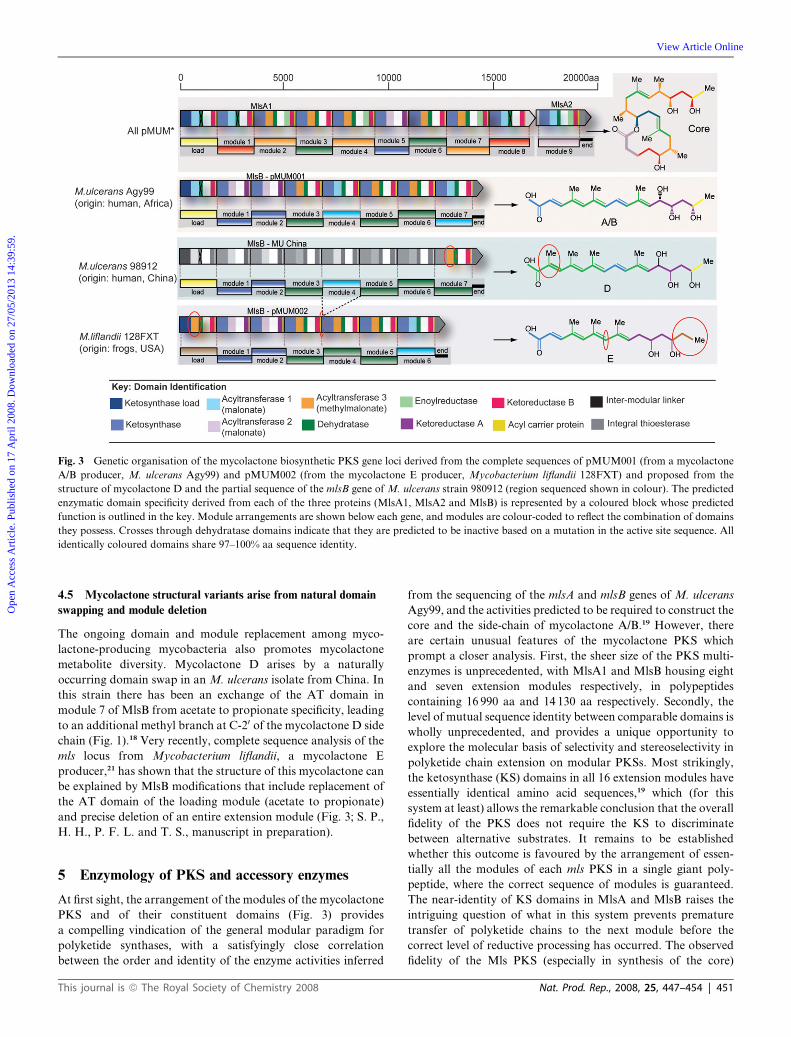
Fig. 3 Genetic organisation of the mycolactone biosynthetic PKS gene loci derived from the complete sequences of pMUM001 (from a mycolactone
A/B producer, M. ulcerans Agy99) and pMUM002 (from the mycolactone E producer, Mycobacterium liflandii 128FXT) and proposed from the
structure of mycolactone D and the partial sequence of the mlsB gene of M. ulcerans strain 980912 (region sequenced shown in colour). The predicted
enzymatic domain specificity derived from each of the three proteins (MlsA1, MlsA2 and MlsB) is represented by a coloured block whose predicted
function is outlined in the key. Module arrangements are shown below each gene, and modules are colour-coded to reflect the combination of domains
they possess. Crosses through dehydratase domains indicate that they are predicted to be inactive based on a mutation in the active site sequence. All
identically coloured domains share 97–100% aa sequence identity.
Ope
n A
cces
s A
rtic
le. P
ublis
hed
on 1
7 A
pril
2008
. Dow
nloa
ded
on 2
7/05
/201
3 14
:39:
59.
View Article Online
4.5 Mycolactone structural variants arise from natural domain
swapping and module deletion
The ongoing domain and module replacement among myco-
lactone-producing mycobacteria also promotes mycolactone
metabolite diversity. Mycolactone D arises by a naturally
occurring domain swap in an M. ulcerans isolate from China. In
this strain there has been an exchange of the AT domain in
module 7 of MlsB from acetate to propionate specificity, leading
to an additional methyl branch at C-20 of the mycolactone D side
chain (Fig. 1).18 Very recently, complete sequence analysis of the
mls locus from Mycobacterium liflandii, a mycolactone E
producer,21 has shown that the structure of this mycolactone can
be explained by MlsB modifications that include replacement of
the AT domain of the loading module (acetate to propionate)
and precise deletion of an entire extension module (Fig. 3; S. P.,
H. H., P. F. L. and T. S., manuscript in preparation).
5 Enzymology of PKS and accessory enzymes
At first sight, the arrangement of the modules of the mycolactone
PKS and of their constituent domains (Fig. 3) provides
a compelling vindication of the general modular paradigm for
polyketide synthases, with a satisfyingly close correlation
between the order and identity of the enzyme activities inferred
This journal is ª The Royal Society of Chemistry 2008
from the sequencing of the mlsA and mlsB genes of M. ulcerans
Agy99, and the activities predicted to be required to construct the
core and the side-chain of mycolactone A/B.19 However, there
are certain unusual features of the mycolactone PKS which
prompt a closer analysis. First, the sheer size of the PKS multi-
enzymes is unprecedented, with MlsA1 and MlsB housing eight
and seven extension modules respectively, in polypeptides
containing 16 990 aa and 14 130 aa respectively. Secondly, the
level of mutual sequence identity between comparable domains is
wholly unprecedented, and provides a unique opportunity to
explore the molecular basis of selectivity and stereoselectivity in
polyketide chain extension on modular PKSs. Most strikingly,
the ketosynthase (KS) domains in all 16 extension modules have
essentially identical amino acid sequences,19 which (for this
system at least) allows the remarkable conclusion that the overall
fidelity of the PKS does not require the KS to discriminate
between alternative substrates. It remains to be established
whether this outcome is favoured by the arrangement of essen-
tially all the modules of each mls PKS in a single giant poly-
peptide, where the correct sequence of modules is guaranteed.
The near-identity of KS domains in MlsA and MlsB raises the
intriguing question of what in this system prevents premature
transfer of polyketide chains to the next module before the
correct level of reductive processing has occurred. The observed
fidelity of the Mls PKS (especially in synthesis of the core)
Nat. Prod. Rep., 2008, 25, 447–454 | 451

Ope
n A
cces
s A
rtic
le. P
ublis
hed
on 1
7 A
pril
2008
. Dow
nloa
ded
on 2
7/05
/201
3 14
:39:
59.
View Article Online
suggests that KS-mediated inter-module transfer is either
uniformly slow relative to the rates of reductive steps, or that the
KS has an intrinsic preference for reduced substrates in this
transfer reaction. Finally, the mls gene cluster contains three
additional genes proposed to play essential roles in toxin
biosynthesis, but where until recently biochemical or genetic
evidence for these roles has been lacking.
5.1 Studies of discrete mycolactone PKS domains in vitro
As yet, there is no convenient system available for genetic
manipulation of mls PKS genes in M. ulcerans, and there have
been no reports of successful heterologous expression of these
genes in a faster growing, more genetically tractable host.
However, there has been progress in the expression and purifi-
cation of individual domains in recombinant E. coli, and study of
their reaction with surrogate substrates. In particular, a detailed
analysis has been published on the ketoreduction of model
diketide thioester substrates by individual ketoreductase (KR)
domains excised from the mycolactone PKS.45 The core lactone
PKS MlsA contains an A-type KR domain (KR-A) in extension
module 5, and the side-chain PKSMlsB contains two such KR-A
domains in extension modules 1 and 2. All the other extension
modules contain B-type KR domains (the designations A and B
refer to the alternative stereochemistries of reduction which are
correlated with specific amino acid motifs in the KR active
sites).46 Although the KR-B domains are 100% identical to each
other, as are the KR-A domains to each other, both types of KR
process PKS-bound polyketide intermediates of several different
lengths and incorporating different structural features. It was
found that, while the KR-A domain acted on a diketide substrate
with complete stereospecificity and stereoselectivity, the KR-B
domain showed low stereochemical fidelity. Taken together,
these results underline the small energetic differences between
alternative modes of presentation of the substrates to the KR
active site, and the likely importance of the tethering of poly-
ketide intermediates to ACP domains in ensuring the correct
stereochemical outcome.45
Another intriguing feature of the mycolactone PKS is
presented by the C-terminal domains of MlsA2 and MlsB, which
are each predicted to house a thioesterase/acyltransferase/cyclase
(type I TE) activity involved in polyketide chain termination.19
By analogy with other modular PKS, theMlsA2 type I TE would
catalyse cyclisation to form the 12-membered core macrolactone.
The MlsB type I TE might catalyse transfer of the side-chain
polyketide chain to coenzyme A, to provide the active donor for
esterification of the core lactone,19 or alternatively it might itself
catalyse the direct acyl transfer to the core lactone. The type I TE
domains are essentially identical in sequence, except for a few
additional amino acids at the C-terminus of MlsA2 type I TE.
They differ however from conventional chain-terminating TE
domains of modular PKS in the apparent absence of the expected
His and Asp/Glu residues, which form part of the highly-
conserved triad of amino acid residues (Ser-His-Asp/Glu)
characteristic of these enzymes. Also, the sequence around the
predicted Ser active site nucleophile (X-X-Ser-X-X) deviates
significantly from the consensus sequence (Gly-X-Ser-X-Gly) for
these enzymes. Homology modelling of the TE-I of MlsA2
(B. Popovic, A. Moore and P. F. L., unpublished data) has
452 | Nat. Prod. Rep., 2008, 25, 447–454
confirmed that the active site shows significant differences from
the arrangement in authentic TE active sites. Either the Mls TE-I
domains represent a new sub-class of a,b-hydrolases, or they are
inactive, and other enzymes catalyse the required steps in
mycolactone assembly.
5.2 Role of auxiliary enzymes in mycolactone biosynthesis
The mycolactone gene cluster contains the gene cyp140A7
(MUP_053) encoding a cytochrome P450 hydroxylase. The
observed distribution of this enzyme in different mycolactone-
producing strains strongly suggests that it is responsible for the
specific hydroxylation of mycolactone C at C-120 to produce
mycolactone A/B (Fig. 1), although this conversion has not yet
been reproduced in vitro with purified enzyme. No other natural
mycolactones undergo such hydroxylation in the side-chain,
a modification that seems to correlate with increased cytotoxicity
(section 4). When the cyp140A7 gene was expressed in the
mycolactone F-producing strain M. marinum DL045, a novel
mycolactone (mycolactone G) was produced (Fig. 1), whose
structure reveals that hydroxylation had apparently occurred,
not as expected on C-100, but on the neighbouring methyl branch
at C-80, presumably because of subtle alterations in the geometry
of the enzyme–substrate interaction.23
Another auxiliary enzyme encoded in the mls gene cluster is
a discrete thioesterase (type II TE), an activity which is found
associated with most modular PKS and which is thought to have
a ‘proof-reading’ function.47 The mycolactone type II TE has
been expressed in E. coli and shown to catalyse the hydrolysis of
model thioester substrates as for authentic type II TE enzymes
(A. Moore and P. F. L., unpublished data) but this does not rule
out additional roles for this enzyme. Given that the type I TE
domains of the mycolactone PKS are atypical, and possibly even
inactive, it is interesting that phylogenetic analysis of the myco-
lactone type II TE shows that it has closest similarity not to
‘proof-reading’ type II TE enzymes, but rather to chain-termi-
nating thioesterases of the MonCII family, involved in release of
intermediates in polyether biosynthesis.48 Finally, the myco-
lactone gene cluster contains a gene encoding a ketosynthase-like
(FabH-like) enzyme, MUP_045, which has been proposed to act
as the acyltransferase that couples the activated polyunsaturated
side-chain of mycolactone onto the core.19 This hypothesis
remains to be tested by biochemical studies with purified enzyme
and the appropriate substrates.
6 Outlook
Studies of mycolactones have been hampered by the slow growth
of the host mycobacteria, the relatively small amounts of
material produced and the high frequency of spontaneous loss-
of-function mutations in the mls genes. Altogether, these factors
lead to considerable difficulties in producing sufficient quantities
of material for structural studies or other research. There are no
reports yet of transfer of pMUM001 or of the mls and myco-
lactone accessory genes, to permit mycolactone production in
a more tractable heterologous host, and this remains a formi-
dable challenge.
Meanwhile, although informative for the biology of
M. ulcerans, the structural variation of natural mycolactones is
This journal is ª The Royal Society of Chemistry 2008

Ope
n A
cces
s A
rtic
le. P
ublis
hed
on 1
7 A
pril
2008
. Dow
nloa
ded
on 2
7/05
/201
3 14
:39:
59.
View Article Online
presently too limited to allow detailed structure–function studies
that might shed further light on the molecular mechanisms of
mycolactone action. Structural variants with lower cytotoxicity
or immunosuppressive activity might compete with mycolactone
for binding to its molecular target, and thereby constitute
valuable functional inhibitors of toxin production. Alternative
strategies for generation of mycolactone variants are clearly
needed, and in the absence of a system for heterologous
expression of the genes which would open the way to engineered
biosynthesis of new analogues, the most promising route to such
analogues is offered by total chemical synthesis.
The first synthetic route to mycolactone A/B was reported in
2002,11–13 making use of an earlier route to the side-chain
proposed by others.49 More recently Kishi and colleagues have
published a significantly more efficient synthetic route to this
molecule,50 as well as syntheses of mycolactones C17 and F.25
Several other groups have also recently reported their efforts to
generate general and flexible routes to analogues of the myco-
lactone core51,52 and side-chain.53,54 If successful, this work would
allow direct testing of the proposed functions of the enzymes
encoded in the mls locus, and also provide important tools
for exploration of the enigmatic mechanism of action of
mycolactone.
7 Acknowledgements
We gratefully acknowledge the financial support of theWellcome
Trust (H. H. and P. F. L.) and the National Health and Medical
Research Council of Australia (T. S.).
8 References
1 D. H. Connor and H. F. Lunn, Int. J. Lepr., 1965, 33, supplement,p. 698.
2 H. F. Lunn, D. H. Connor, N. E. Wilks, G. R. Barnley, F. Kamunvi,J. K. Clancey and J. D. Bee, E. Afr. Med. J., 1965, 42, 275.
3 J. K. Read, C. M. Heggie, W. M. Meyers and D. H. Connor, Infect.Immun., 1974, 9, 1114.
4 M. Pimsler, T. A. Sponsler and W. M. Meyers, J. Infect. Dis., 1988,157, 577.
5 K. M. George, L. P. Barker, D. M. Welty and P. L. C. Small, Infect.Immun., 1998, 66, 587.
6 A. A. Pahlevan, D. J. M. Wright, C. Andrews, K. M. George,P. L. C. Small and B. M. Foxwell, J. Immun., 1999, 163, 3928.
7 K. M. George, D. Chatterjee, G. Gunawardana, D. Welty,J. Hayman, R. Lee and P. L. C. Small, Science, 1999, 283, 854.
8 K. M. George, L. Pascopella, D. M. Welty and P. L. C. Small, Infect.Immun., 2000, 68, 877.
9 G. Gunawardana, D. Chatterjee, K. M. George, P. J. Brennan,D. Whittern and P. L. C. Small, J. Am. Chem. Soc., 1999, 121, 6092.
10 L.Marsollier, P. Brodin,M. Jackson, J. Kordulokova, P. Tafalmeyer,E. Carbonelle, J. Aubry, G. Milon, P. Legras, J. P. Saint Andre,C. Leroy, J. Cottin, M. L. J. Guillou, G. Reysset and S. T. Cole,PLoS Path., 2007, 3, e62.
11 A. B. Benowitz, S. Fidanze, P. L. C. Small and Y. Kishi, J. Am. Chem.Soc., 2001, 123, 5128.
12 S. Fidanze, S. F. Song, M. Szoslek-Pinaud, P. L. C. Small andY. Kishi, J. Am. Chem. Soc., 2001, 123, 10117.
13 F. Song, S. Fidanze, A. B. Benowitz and Y. Kishi, Org. Lett., 2002, 4,647.
14 L. D. Cadapan, R. L. Arslanian, J. R. Carney, S. M. Zavala,P. L. Small and P. Licari, FEMS Microbiol. Lett., 2001, 205, 385.
15 H. Hong, P. J. Gates, J. Staunton, T. Stinear, S. T. Cole, P. F. Leadlayand J. B. Spencer, Chem. Commun., 2003, 2822.
16 A. Mve-Obiang, R. E. Lee, F. Portaels and P. L. C. Small, Infect.Immun., 2003, 71, 774.
This journal is ª The Royal Society of Chemistry 2008
17 T. C. Judd, A. Bischoff, Y. Kishi, S. Adusumilli and P. L. Small, Org.Lett., 2004, 6, 4901.
18 H. Hong, J. B. Spencer, J. L. Porter, P. F. Leadlay and T. Stinear,ChemBioChem, 2005, 6, 643.
19 T. Stinear, A. Mve-Obiang, P. L. Small, W. Frigui, M. J. Pryor,R. Brosch, G. A. Jenkin, P. D. Johnson, J. K. Davies, R. E. Lee,S. Adasumilli, T. Garnier, S. F. Haydock, P. F. Leadlay andS. T. Cole, Proc. Natl. Acad. Sci. U. S. A., 2004, 101, 1345.
20 K. A. Trott, B. A. Stacy, B. D. Lifland, H. E. Diggs, R. M. Harland,M. K. Khokha, T. A. Grammer and J. M. Parker, Comp. Med., 2004,54, 309.
21 A. Mve-Obiang, R. E. Lee, E. S. Umstot, K. A. Trott,T. C. Grammer, J. M. Parker, B. S. Ranger, R. Grainger,E. A. Mahrous and P. L. Small, Infect. Immun., 2005, 73, 3307.
22 H. Hong, T. Stinear, P. Skelton, J. B. Spencer and P. F. Leadlay,Chem. Commun., 2005, 34, 4306.
23 H. Hong, T. Stinear, J. L. Porter, C. Demangel and P. F. Leadlay,ChemBioChem, 2007, 8, 2043.
24 B. S. Ranger, E. A. Mahrous, L. Mosi, S. Adusumilli, R. E. Lee,A. Colorni,M.Rhodes and P. L. Small, Infect. Immun., 2006, 74, 6037.
25 H. J. Kim and Y. Kishi, J. Am. Chem. Soc., 2008, 130, 1842.26 D. S. Snyder and P. L. C. Small, Microbial Pathogenesis, 2003, 34, 91.27 E. Coutanceau, J. Decalf, A. Martino, A. Babon, N. Winter,
S. T. Cole, M. L. Albert and C. Demangel, J. Exp. Med., 2007, 204,1395.
28 S. Adusumilli, A. Mve-Obiang, T. Sparer, W.Meyers, J. Hayman andP. L. C. Small, Cell. Microbiol., 2005, 7, 1295.
29 E. Coutanceau, L. Marsollier, R. Brosch, E. Perret, P. Goossens,M. Tanguy, S. T. Cole, P. L. C. Small and C. Demangel, Cell.Microbiol., 2005, 7, 1187.
30 E. Torrado, S. Adusumilli, A. G. Fraga, P. L. Small, A. G. Castro andJ. Pedrosa, Infect. Immun., 2007, 75, 3979.
31 J. Guarner, J. Bartlett, E. A. S. Whitney, P. L. Raghunathan,Y. Stienstra, K. Asamoa, S. Etuaful, E. Klutse, E. Quarshie,T. S. van der Werf, W. T. A. van der Graaf, C. H. King andD. A. Ashford, Emerging Infect. Dis., 2003, 9, 651.
32 T. M. Gooding, P. D. R. Johnson, D. E. Campbell, J. A. Hayman,E. L. Hartland, A. S. Kemp and R. M. Robins-Browne, Infect.Immun., 2001, 69, 1704–1707.
33 T. M. Gooding, P. D. R. Johnson, M. Smith, A. S. Kemp andR. M. Robins-Browne, Infect. Immun., 2002, 70, 5562.
34 T. M. Gooding, A. S. Kemp, R. M. Robins-Browne, M. Smith andP. D. R. Johnson, Clin. Infect. Dis., 2003, 36, 1076.
35 R. Phillips, C. Horsfield, S. Kuijper, S. F. Sarfo, J. Obeng-Baah,S. Etuaful, B. Nyamekye, P. Awuah, K. M. Nyarko, F. Osei-Sarpong, S. Lucas, A. H. Kolk and M. Wansbrough-Jones, Clin.Vaccine Immunol., 2006, 13, 253.
36 G. Prevot, E. Bourreau, H. Pascalis, R. Pradinaud, A. Tanghe,K. Huygen and P. Launois, Infect. Immun., 2004, 72, 958.
37 B. D. Westenbrink, Y. Stienstra, M. G. Huitema, W. A. Thompson,E. O. Klutse, E. O. Ampadu, H. M. Boezen, P. C. Limburg andT. S. van der Werf, Clin. Diagn. Lab. Immunol., 2005, 12, 125.
38 D. Yeboah-Manu, E. Peduzzi, E. Mensah-Quainoo, A. Asante-Poku,D. Ofori-Adjei, G. Pluschke and C. A. Daubenberger, J. LeukocyteBiol., 2006, 79, 1150.
39 K. J. Weissman and P. F. Leadlay, Nat. Rev. Microbiol., 2005, 3, 925.40 M. J. Yip, J. L. Porter, J. A. M. Fyfe, C. J. Lavender, F. Portaels,
M. Rhodes, H. Kator, A. Colorni, G. A. Jenkin and T. Stinear,J. Bacteriol., 2007, 189, 2021.
41 M. W. Rhodes, H. Kator, A. McNabb, C. Deshayes, J. M. Reyrat,B. A. Brown-Elliott, R. Wallace, Jr., K. A. Trott, J. M. Parker,B. Lifland, G. Osterhout, I. Kaattari, K. Reece, W. Vogelbein andC. A. Ottinger, Int. J. Syst. Evol. Microbiol., 2005, 55, 1139.
42 M. Ucko and A. Colorni, J. Clin. Microbiol., 2005, 43, 892.43 J. F. Aparicio, I. Molnar, T. Schwecke, A. Konig, S. F. Haydock,
L. E. Khaw, J. Staunton and P. F. Leadlay, Gene, 1996, 169, 9.44 T. P. Stinear, H. Hong,W. Frigui, M. J. Pryor, R. Brosch, T. Garnier,
P. F. Leadlay and S. T. Cole, J. Bacteriol., 2005, 187, 1668.45 S. Bali and K. J. Weissman, ChemBioChem, 2007, 8, 2043.46 P. Caffrey, ChemBioChem, 2003, 4, 654.47 M. L. Heathcote, J. Staunton and P. F. Leadlay, Chem. Biol., 2001, 8,
207.48 B. M. Harvey, H. Hong, M. A. Jones, Z. A. Hughes-Thomas,
R. M. Goss, M. L. Heathcote, V. M. Bolanos-Garcia, W. Kroutil,J. Staunton and P. F. Leadlay, ChemBioChem, 2006, 7, 1435.
Nat. Prod. Rep., 2008, 25, 447–454 | 453
Ope
n A
cces
s A
rtic
le. P
ublis
hed
on 1
7 A
pril
2008
. Dow
nloa
ded
on 2
7/05
/201
3 14
:39:
59.
View Article Online
49 M. K. Gurjar and J. Cherian, Heterocycles, 2001, 55, 1095.50 F. Song, S. Fidanze, A. B. Benowitz and Y. Kishi, Tetrahedron, 2007,
63, 5739.51 M. D. Alexander, S. D. Fontaine, J. J. La Clair, A. G. Dipasquale,
A. L. Rheingold and M. D. Burkart, Chem. Commun., 2006, 4602.
454 | Nat. Prod. Rep., 2008, 25, 447–454
52 F. Feyen, A. Jantsch and K. Altmann, Synlett, 2007, 415–418.53 R. P. Van Summeren, B. L. Feringa and A. J. Minnaard, Org. Biomol.
Chem., 2005, 3, 2524.54 N. Yin, G. Wang, M. Qian and E. Negishi, Angew. Chem., Int. Ed.,
2006, 45, 2916.
This journal is ª The Royal Society of Chemistry 2008
Related Documents