Mycobacterium tuberculosis nuoG Is a Virulence Gene That Inhibits Apoptosis of Infected Host Cells Kamalakannan Velmurugan 1 , Bing Chen 2 , Jessica L. Miller 1 , Sharon Azogue 1 , Serdar Gurses 1 , Tsungda Hsu 2 , Michael Glickman 3 , William R. Jacobs Jr. 2,4,5 , Steven A. Porcelli 4,5 , Volker Briken 1* 1 Department of Cell Biology and Molecular Genetics, University of Maryland, College Park, Maryland, United States of America, 2 Howard Hughes Medical Institute, Albert Einstein College of Medicine, Bronx, New York, United States of America, 3 Division of Infectious Diseases, Immunology Program, Memorial Sloan Kettering Cancer Center, New York, New York, United States of America, 4 Department of Microbiology and Immunology, Albert Einstein College of Medicine, Bronx, New York, United States of America, 5 Department of Medicine, Albert Einstein College of Medicine, Bronx, New York, United States of America The survival and persistence of Mycobacterium tuberculosis depends on its capacity to manipulate multiple host defense pathways, including the ability to actively inhibit the death by apoptosis of infected host cells. The genetic basis for this anti-apoptotic activity and its implication for mycobacterial virulence have not been demonstrated or elucidated. Using a novel gain-of-function genetic screen, we demonstrated that inhibition of infection-induced apoptosis of macrophages is controlled by multiple genetic loci in M. tuberculosis. Characterization of one of these loci in detail revealed that the anti-apoptosis activity was attributable to the type I NADH-dehydrogenase of M. tuberculosis, and was mainly due to the subunit of this multicomponent complex encoded by the nuoG gene. Expression of M. tuberculosis nuoG in nonpathogenic mycobacteria endowed them with the ability to inhibit apoptosis of infected human or mouse macrophages, and increased their virulence in a SCID mouse model. Conversely, deletion of nuoG in M. tuberculosis ablated its ability to inhibit macrophage apoptosis and significantly reduced its virulence in mice. These results identify a key component of the genetic basis for an important virulence trait of M. tuberculosis and support a direct causal relationship between virulence of pathogenic mycobacteria and their ability to inhibit macrophage apoptosis. Citation: Velmurugan K, Chen B, Miller JL, Azogue S, Gurses S, et al. (2007) Mycobacterium tuberculosis nuoG is a virulence gene that inhibits apoptosis of infected host cells. PLoS Pathog 3(7): e110. doi:10.1371/journal.ppat.0030110 Introduction Tuberculosis is an infectious disease of enormous and increasing global importance. Currently, about one third of all humans are latently infected with its etiologic agent, Mycobacterium tuberculosis (Mtb), and an estimated 2.5 million people die of tuberculosis annually [1]. After infection of a mammalian host, Mtb is able to resist innate host defenses sufficiently to increase the local bacterial burden and disseminate throughout the body. With the onset of the adaptive immune response, however, the bacterial numbers are controlled in over 90% of infected individuals. Never- theless, the host is not able to completely clear the bacterial burden, thus leading to persistence of Mtb within the lungs and other tissues of healthy individuals. These latent infections can be reactivated to generate full-blown disease, a process that is accelerated by immunocompromised states resulting from senescence, malnutrition, and co-infection with HIV, which is a major source of morbidity and mortality associated with the current HIV epidemics in many countries [2–5]. Programmed cell death (apoptosis) plays an important role in the innate immune response against pathogens and comprises an evolutionarily conserved defense strategy that extends even into the plant world [6,7]. It is therefore essential for persisting intracellular pathogens to have strong anti-apoptosis mechanisms [8–12]. While a few studies have suggested that under some conditions Mtb may induce host cell apoptosis [13–16], a substantial body of evidence points strongly to the expression of strong anti-apoptotic mecha- nisms by Mtb and other closely related virulent bacteria. Furthermore, this capacity is not found in avirulent species, suggesting a causal link between virulence and inhibition of macrophage apoptosis [17–19]. This hypothesis is supported by the recent discovery that the genetic predisposition of different inbred mouse strains to mycobacterial infections is linked to the capacity of their macrophages to undergo apoptosis or necrosis upon infection, with the former response imparting a resistant and the latter a susceptible host phenotype [20]. Further confirmation of the findings that Mtb inhibits host cell apoptosis is provided by a number of studies that have addressed its molecular mechanism. The importance of Mtb- induced upregulation of anti-apoptosis genes in infected macrophages for apoptosis inhibition was supported by functional data using either anti-sense oligonucleotides to Editor: William Bishai, Johns Hopkins School of Medicine, United States of America Received April 3, 2007; Accepted June 13, 2007; Published July 20, 2007 Copyright: Ó 2007 Velmurugan et al. This is an open-access article distributed under the terms of the Creative Commons Attribution License, which permits unrestricted use, distribution, and reproduction in any medium, provided the original author and source are credited. Abbreviations: BCG, M. bovis Bacille Calmette-Gue ´rin; CFU, colony-forming unit; Mtb, Mycobacterium tuberculosis; NDH-1, type I NADH dehydrogenase complex * To whom correspondence should be addressed. E-mail: [email protected] PLoS Pathogens | www.plospathogens.org July 2007 | Volume 3 | Issue 7 | e110 0972

Welcome message from author
This document is posted to help you gain knowledge. Please leave a comment to let me know what you think about it! Share it to your friends and learn new things together.
Transcript

Mycobacterium tuberculosis nuoGIs a Virulence Gene That Inhibits Apoptosisof Infected Host CellsKamalakannan Velmurugan
1, Bing Chen
2, Jessica L. Miller
1, Sharon Azogue
1, Serdar Gurses
1, Tsungda Hsu
2,
Michael Glickman3
, William R. Jacobs Jr.2,4,5
, Steven A. Porcelli4,5
, Volker Briken1*
1 Department of Cell Biology and Molecular Genetics, University of Maryland, College Park, Maryland, United States of America, 2 Howard Hughes Medical Institute, Albert
Einstein College of Medicine, Bronx, New York, United States of America, 3 Division of Infectious Diseases, Immunology Program, Memorial Sloan Kettering Cancer Center,
New York, New York, United States of America, 4 Department of Microbiology and Immunology, Albert Einstein College of Medicine, Bronx, New York, United States of
America, 5 Department of Medicine, Albert Einstein College of Medicine, Bronx, New York, United States of America
The survival and persistence of Mycobacterium tuberculosis depends on its capacity to manipulate multiple hostdefense pathways, including the ability to actively inhibit the death by apoptosis of infected host cells. The geneticbasis for this anti-apoptotic activity and its implication for mycobacterial virulence have not been demonstrated orelucidated. Using a novel gain-of-function genetic screen, we demonstrated that inhibition of infection-inducedapoptosis of macrophages is controlled by multiple genetic loci in M. tuberculosis. Characterization of one of these lociin detail revealed that the anti-apoptosis activity was attributable to the type I NADH-dehydrogenase of M.tuberculosis, and was mainly due to the subunit of this multicomponent complex encoded by the nuoG gene.Expression of M. tuberculosis nuoG in nonpathogenic mycobacteria endowed them with the ability to inhibit apoptosisof infected human or mouse macrophages, and increased their virulence in a SCID mouse model. Conversely, deletionof nuoG in M. tuberculosis ablated its ability to inhibit macrophage apoptosis and significantly reduced its virulence inmice. These results identify a key component of the genetic basis for an important virulence trait of M. tuberculosis andsupport a direct causal relationship between virulence of pathogenic mycobacteria and their ability to inhibitmacrophage apoptosis.
Citation: Velmurugan K, Chen B, Miller JL, Azogue S, Gurses S, et al. (2007) Mycobacterium tuberculosis nuoG is a virulence gene that inhibits apoptosis of infected host cells.PLoS Pathog 3(7): e110. doi:10.1371/journal.ppat.0030110
Introduction
Tuberculosis is an infectious disease of enormous andincreasing global importance. Currently, about one third ofall humans are latently infected with its etiologic agent,Mycobacterium tuberculosis (Mtb), and an estimated 2.5 millionpeople die of tuberculosis annually [1]. After infection of amammalian host, Mtb is able to resist innate host defensessufficiently to increase the local bacterial burden anddisseminate throughout the body. With the onset of theadaptive immune response, however, the bacterial numbersare controlled in over 90% of infected individuals. Never-theless, the host is not able to completely clear the bacterialburden, thus leading to persistence of Mtb within the lungsand other tissues of healthy individuals. These latentinfections can be reactivated to generate full-blown disease,a process that is accelerated by immunocompromised statesresulting from senescence, malnutrition, and co-infectionwith HIV, which is a major source of morbidity and mortalityassociated with the current HIV epidemics in many countries[2–5].
Programmed cell death (apoptosis) plays an important rolein the innate immune response against pathogens andcomprises an evolutionarily conserved defense strategy thatextends even into the plant world [6,7]. It is thereforeessential for persisting intracellular pathogens to have stronganti-apoptosis mechanisms [8–12]. While a few studies havesuggested that under some conditions Mtb may induce host
cell apoptosis [13–16], a substantial body of evidence pointsstrongly to the expression of strong anti-apoptotic mecha-nisms by Mtb and other closely related virulent bacteria.Furthermore, this capacity is not found in avirulent species,suggesting a causal link between virulence and inhibition ofmacrophage apoptosis [17–19]. This hypothesis is supportedby the recent discovery that the genetic predisposition ofdifferent inbred mouse strains to mycobacterial infections islinked to the capacity of their macrophages to undergoapoptosis or necrosis upon infection, with the formerresponse imparting a resistant and the latter a susceptiblehost phenotype [20].Further confirmation of the findings that Mtb inhibits host
cell apoptosis is provided by a number of studies that haveaddressed its molecular mechanism. The importance of Mtb-induced upregulation of anti-apoptosis genes in infectedmacrophages for apoptosis inhibition was supported byfunctional data using either anti-sense oligonucleotides to
Editor: William Bishai, Johns Hopkins School of Medicine, United States of America
Received April 3, 2007; Accepted June 13, 2007; Published July 20, 2007
Copyright: � 2007 Velmurugan et al. This is an open-access article distributedunder the terms of the Creative Commons Attribution License, which permitsunrestricted use, distribution, and reproduction in any medium, provided theoriginal author and source are credited.
Abbreviations: BCG, M. bovis Bacille Calmette-Guerin; CFU, colony-forming unit;Mtb, Mycobacterium tuberculosis; NDH-1, type I NADH dehydrogenase complex
* To whom correspondence should be addressed. E-mail: [email protected]
PLoS Pathogens | www.plospathogens.org July 2007 | Volume 3 | Issue 7 | e1100972

knock down mcl-1 expression [19] or A1 knock-out micelacking the anti-apoptosis gene A1 [21,22]. These resultsimplicate the intrinsic (mitochondria-mediated) apoptosispathway as a target for Mtb-mediated apoptosis inhibition,because mcl-1 and A1 are both members of the large family ofBcl-2–like proteins that localize prominently to mitochon-dria. However, this is contradicted by the finding thatoverexpression of Bcl-2 (another mitochondrial anti-apop-totic protein) could not rescue cells from undergoingapoptosis after infection with nonvirulent mycobacteria, thussuggesting that the extrinsic pathway (death receptor–mediated) is involved in the infection-induced apoptosis[23]. Consistently, virulent Mtb strains could inhibit FasL-induced apoptosis in Fas-expressing cells [18]. The samegroup reported very recently that lipoglycans of the Mtb cellwall stimulate the activation of NF-kB via TLR-2 and that thesubsequent upregulation of cellular FLIP leads to inhibitionof FasL-mediated apoptosis [24]. Furthermore, it was sug-gested that Mtb stimulated the secretion of soluble TNF-R2,which led to the reduction of bioactive TNF-a in the mediumand therefore less stimulation of the TNF-R1 [25]. Altogether,it seems that virulent Mtb is able to inhibit induction of hostcell apoptosis via multiple pathways, and probably encodesmechanisms to interfere with both intrinsic and extrinsicpathways for initiation of programmed cell death.
The inhibition of macrophage apoptosis by Mtb is believedto provide a number of advantages to the bacterium in itsstruggle to resist the host immune response. These includepreservation of a favorable host cell environment duringgrowth and persistence [26,27], evasion of apoptosis-linkedbactericidal effects [28,29], and avoidance of efficientcytotoxic T cell priming via the detour pathway of antigencross-presentation [15,30–32]. This last point is of potentialimportance to the improvement of tuberculosis vaccines,because attenuated mycobacterial strains that induce higherlevels of host cell apoptosis would be expected to stimulatemore robust cellular immunity, as suggested by a recent studyusing recombinant M. bovis Bacille Calmette-Guerin (BCG)expressing listeriolysin [33,34]. Therefore, the identification
of mycobacterial genes required for prevention of apoptosiscould lead to specific strategies for designing more efficaciousforms of BCG or other attenuated mycobacterial vaccinestrains.In order to clarify the role of mycobacteria in host cell
apoptosis and to address its importance for bacterialvirulence, we sought to identify anti-apoptosis genes via again-of-function genetic screen. Using this approach, wesuccessfully identified two independent genomic regions ofvirulent Mtb (strain H37Rv) that mediate the inhibition ofhost cell apoptosis by the facultative pathogen M. kansasii.The analysis of a defined set of bacterial mutants of M.kansasii in immunocompromised (SCID) mice demonstrated acausal relationship between inhibition of apoptosis andvirulence. These findings were confirmed via a loss-of-function strategy using the newly identified anti-apoptosisgene, nuoG, and demonstrated attenuation of Mtb nuoGmutants in immunocompromised and immunocompetentmice. Altogether our findings allowed, to our knowledge forthe first time, the demonstration of a causal relationshipbetween inhibition of host cell apoptosis and virulence ofmycobacteria.
Results/Discussion
Gain-of-Function Genetic Screen for Anti-Apoptosis Genesin MtbTo identify genes in Mtb responsible for anti-apoptotic
effects, we established a gain-of-function genetic screen usingthe nonpathogenic M. smegmatis mc2155 strain, which is a fast-growing mycobacterium that is extremely efficient for trans-formation [35]. The human cell line THP-1 was chosen for useas the host cells for this screen because published workindicates that these cells provide an accurate model for theapoptotic response of Mtb-infected primary human alveolarmacrophages [23]. Our initial studies established that M.smegmatis infection of THP-1 cells induced strong apoptosisafter 1.5 d of infection when compared to BCG-infectedmacrophages, as assessed by disruption of the cell monolayerand by staining for DNA strand breakage using the TUNELassay (Figure 1A). The failure of BCG to induce apoptosisseems to contradict the report by Kean et al. [17] in whichseven different species of mycobacteria were compared fortheir capacity to induce apoptosis in primary human alveolarmacrophages, and BCG was among the apoptosis-inducingmycobacterial species. Nevertheless, in that study, no fast-growing species were included, and all apoptosis assays wereperformed after 5–7 d of infection. In an independent study,we demonstrated that M. smegmatis and other nonpathogenicmycobacteria like M. fortuitum have a very strong capacity toinduce apoptosis with rapid kinetics, even when compared tofacultative pathogenic mycobacteria like M. kansasii and BCG(A. Bohsali and V. Briken, unpublished data). Therefore, theinduction of apoptosis byM. smegmatis had to be analyzed veryearly after infection (16–36 h; Figure 1A and 1B), and at thistime point, little or no induction of macrophage apoptosis byBCG could be observed.The gain-of-function screen was performed using a library
of 312 M. smegmatis clones containing Mtb genomic DNAfragments on an episomal cosmid. Two clones (designated J21and M24) containing cosmids with separate nonoverlappingMtb genomic DNA inserts gave significantly reduced levels of
PLoS Pathogens | www.plospathogens.org July 2007 | Volume 3 | Issue 7 | e1100973
Virulence of Mtb and Macrophage Apoptosis
Author Summary
The infection-induced suicide of host cells following invasion byintracellular pathogens is an ancient defense mechanism observedin multicellular organisms of both the animal and plant kingdoms. Itis therefore not surprising that persistent pathogens of viral,bacterial, and protozoal origin have evolved to inhibit the inductionof host cell death. M. tuberculosis, the etiological agent oftuberculosis, has latently infected about one third of the world’spopulation and can persist for decades in the lungs of infected,asymptomatic individuals. In the present study we have identifiednuoG of M. tuberculosis, which encodes a subunit of the type I NADHdehydrogenase complex, as a critical bacterial gene for inhibition ofhost cell death. A mutant of M. tuberculosis in which nuoG wasdeleted triggered a marked increase in apoptosis by infectedmacrophages, and subsequent analysis of this mutant in the mousetuberculosis model provided direct evidence for a causal linkbetween the capacity to inhibit apoptosis and bacterial virulence.The discovery of anti-apoptosis genes in M. tuberculosis couldprovide a powerful approach to the generation of better attenuatedvaccine strains, and may also identify a new group of drug targetsfor improved chemotherapy.
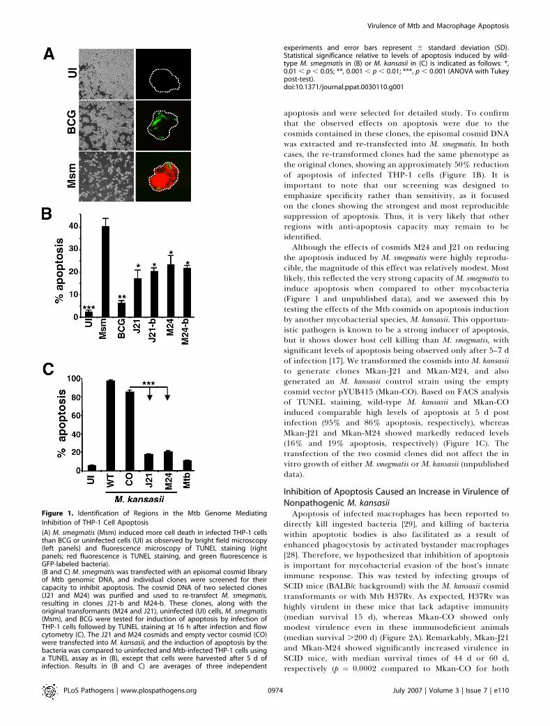
apoptosis and were selected for detailed study. To confirmthat the observed effects on apoptosis were due to thecosmids contained in these clones, the episomal cosmid DNAwas extracted and re-transfected into M. smegmatis. In bothcases, the re-transformed clones had the same phenotype asthe original clones, showing an approximately 50% reductionof apoptosis of infected THP-1 cells (Figure 1B). It isimportant to note that our screening was designed toemphasize specificity rather than sensitivity, as it focusedon the clones showing the strongest and most reproduciblesuppression of apoptosis. Thus, it is very likely that otherregions with anti-apoptosis capacity may remain to beidentified.Although the effects of cosmids M24 and J21 on reducing
the apoptosis induced by M. smegmatis were highly reprodu-cible, the magnitude of this effect was relatively modest. Mostlikely, this reflected the very strong capacity ofM. smegmatis toinduce apoptosis when compared to other mycobacteria(Figure 1 and unpublished data), and we assessed this bytesting the effects of the Mtb cosmids on apoptosis inductionby another mycobacterial species, M. kansasii. This opportun-istic pathogen is known to be a strong inducer of apoptosis,but it shows slower host cell killing than M. smegmatis, withsignificant levels of apoptosis being observed only after 5–7 dof infection [17]. We transformed the cosmids into M. kansasiito generate clones Mkan-J21 and Mkan-M24, and alsogenerated an M. kansasii control strain using the emptycosmid vector pYUB415 (Mkan-CO). Based on FACS analysisof TUNEL staining, wild-type M. kansasii and Mkan-COinduced comparable high levels of apoptosis at 5 d postinfection (95% and 86% apoptosis, respectively), whereasMkan-J21 and Mkan-M24 showed markedly reduced levels(16% and 19% apoptosis, respectively) (Figure 1C). Thetransfection of the two cosmid clones did not affect the invitro growth of either M. smegmatis or M. kansasii (unpublisheddata).
Inhibition of Apoptosis Caused an Increase in Virulence ofNonpathogenic M. kansasiiApoptosis of infected macrophages has been reported to
directly kill ingested bacteria [29], and killing of bacteriawithin apoptotic bodies is also facilitated as a result ofenhanced phagocytosis by activated bystander macrophages[28]. Therefore, we hypothesized that inhibition of apoptosisis important for mycobacterial evasion of the host’s innateimmune response. This was tested by infecting groups ofSCID mice (BALB/c background) with the M. kansasii cosmidtransformants or with Mtb H37Rv. As expected, H37Rv washighly virulent in these mice that lack adaptive immunity(median survival 15 d), whereas Mkan-CO showed onlymodest virulence even in these immunodeficient animals(median survival .200 d) (Figure 2A). Remarkably, Mkan-J21and Mkan-M24 showed significantly increased virulence inSCID mice, with median survival times of 44 d or 60 d,respectively (p ¼ 0.0002 compared to Mkan-CO for both
Figure 1. Identification of Regions in the Mtb Genome Mediating
Inhibition of THP-1 Cell Apoptosis
(A) M. smegmatis (Msm) induced more cell death in infected THP-1 cellsthan BCG or uninfected cells (UI) as observed by bright field microscopy(left panels) and fluorescence microscopy of TUNEL staining (rightpanels; red fluorescence is TUNEL staining, and green fluorescence isGFP-labeled bacteria).(B and C) M. smegmatis was transfected with an episomal cosmid libraryof Mtb genomic DNA, and individual clones were screened for theircapacity to inhibit apoptosis. The cosmid DNA of two selected clones(J21 and M24) was purified and used to re-transfect M. smegmatis,resulting in clones J21-b and M24-b. These clones, along with theoriginal transformants (M24 and J21), uninfected (UI) cells, M. smegmatis(Msm), and BCG were tested for induction of apoptosis by infection ofTHP-1 cells followed by TUNEL staining at 16 h after infection and flowcytometry (C). The J21 and M24 cosmids and empty vector cosmid (CO)were transfected into M. kansasii, and the induction of apoptosis by thebacteria was compared to uninfected and Mtb-infected THP-1 cells usinga TUNEL assay as in (B), except that cells were harvested after 5 d ofinfection. Results in (B and C) are averages of three independent
experiments and error bars represent 6 standard deviation (SD).Statistical significance relative to levels of apoptosis induced by wild-type M. smegmatis in (B) or M. kansasii in (C) is indicated as follows: *,0.01 , p , 0.05; **, 0.001 , p , 0.01; ***, p , 0.001 (ANOVA with Tukeypost-test).doi:10.1371/journal.ppat.0030110.g001
PLoS Pathogens | www.plospathogens.org July 2007 | Volume 3 | Issue 7 | e1100974
Virulence of Mtb and Macrophage Apoptosis
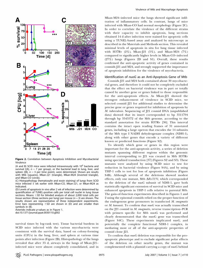
survival times by log-rank test). Tissue bacterial burdens inSCID mice infected with the various mycobacteria wereconsistent with the survival data, based on colony-formingunits (CFUs) in the lung, liver, and spleen at various timepoints after infection (Figure 2B). Histopathology of the lungsrevealed that after 35 d, airways in the lungs of Mkan-J21–infected mice were almost completely consolidated, and in
Mkan-M24–infected mice the lungs showed significant infil-tration of inflammatory cells. In contrast, lungs of miceinfected with Mkan-CO had normal morphology (Figure 2C).In order to correlate the virulence of the different strainswith their capacity to inhibit apoptosis, lung sectionsobtained 14 d after infection were stained for apoptotic cellsusing a TUNEL-based assay and analyzed by microscopy asdescribed in the Materials and Methods section. This revealedminimal levels of apoptosis in situ for lung tissue infectedwith H37Rv (2%), Mkan-J21 (5%), and Mkan-M24 (7%)compared to significantly higher levels in Mkan-CO–infected(27%) lungs (Figures 2D and S1). Overall, these resultsconfirmed the anti-apoptotic activity of genes contained incosmids J21 and M24, and strongly supported the importanceof apoptosis inhibition for the virulence of mycobacteria.
Identification of nuoG as an Anti-Apoptosis Gene of MtbCosmids J21 and M24 both contained about 30 mycobacte-
rial genes, and therefore it could not be completely excludedthat the effect on bacterial virulence was in part or totallycaused by another gene or genes linked to those responsiblefor the anti-apoptosis effects. As Mkan-J21 showed thestrongest enhancement of virulence in SCID mice, weselected cosmid J21 for additional studies to determine theprecise gene or genes required for inhibition of apoptosis byM. tuberculosis. Sequencing of J21 cosmid DNA (unpublisheddata) showed that its insert corresponded to bp 3511794through bp 3545572 of the Mtb genome, according to thestandard annotation for strain H37Rv [36]. This intervalcontains the intact open reading frames of 31 annotatedgenes, including a large operon that encodes the 14 subunitsof the Mtb type I NADH dehydrogenase complex (NDH-1),along with other genes that encode a variety of differentknown or predicted functions (Figure S2).To identify which gene or genes in this region were
important for the anti-apoptosis activity, a series of deletionmutants spanning different regions within the genomicinterval corresponding to J21 was created in Mtb H37Rvusing specialized transduction [37] (Figures S2 and S3). Thesemutants were analyzed by using SCID mice to test forreduction in bacterial virulence (Figure S4A), and by usingTHP-1 cells to test for loss of apoptosis inhibition (FigureS4B). Although several of the deletions showed modesteffects, only one mutant, Mtb DRv3151, which correspondedto the deletion of the nuoG subunit of NDH-1, gave bothstatistically significant extension of survival in SCID mice andenhanced apoptosis in THP-1 cells relative to parental Mtb.The gain-of-function experiments described in Figures 1 and2 using the episomal cosmids relied on the proper function ofthe endogenous gene promoters in transfected M. smegmatisor M. kansasii. To confirm that nuoG was actually transcribedon the J21 cosmid in M. smegmatis, reverse transcription–PCRwith primers specific for Mtb nuoG was performed andclearly demonstrated that the nuoG gene was transcribed(Figure S4C). These experiments implicated nuoG, andpotentially the complete functional NDH-1 complex, inmediating most or all of the anti-apoptotic properties ofcosmid clone J21.To confirm that nuoG deletion was responsible for the pro-
apoptotic phenotype and to exclude significant polar effectsof the deletion on other nearby genes, the mutant wascomplemented with a plasmid carrying a copy of nuoG behind
Figure 2. Correlation between Apoptosis Inhibition and Mycobacterial
Virulence
(A and B) SCID mice were infected intravenously with 106 bacteria andsurvival ([A], n ¼ 7 per group), or the bacterial load in lung, liver, andspleen ([B], n ¼ 3 per time point), were determined. Shown are resultswith Mtb (squares), Mkan-J21 (triangle), Mkan-M24 (inverted triangle),and Mkan-CO (circle).(C) Histopathology (hematoxylin and eosin staining) of lungs from SCIDmice infected 5 wk earlier with Mkan-CO, Mkan-J21, or Mkan-M24 asindicated.(D) Levels of apoptosis in vivo after 2 wk of infection were determined byquantification of TUNEL-positive cells per total cell nuclei in lung tissuesections. Means 6SD for blinded analysis of about 2,000 total cells pertissue section (three sections per lung) per mouse (n¼ 3) are shown. Theresults shown are representative of three independent experiments.Error bars representing 6SD are shown in (D) and are smaller thansymbols in (B).Asterisks indicate p-values as in Figure 1.doi:10.1371/journal.ppat.0030110.g002
PLoS Pathogens | www.plospathogens.org July 2007 | Volume 3 | Issue 7 | e1100975
Virulence of Mtb and Macrophage Apoptosis
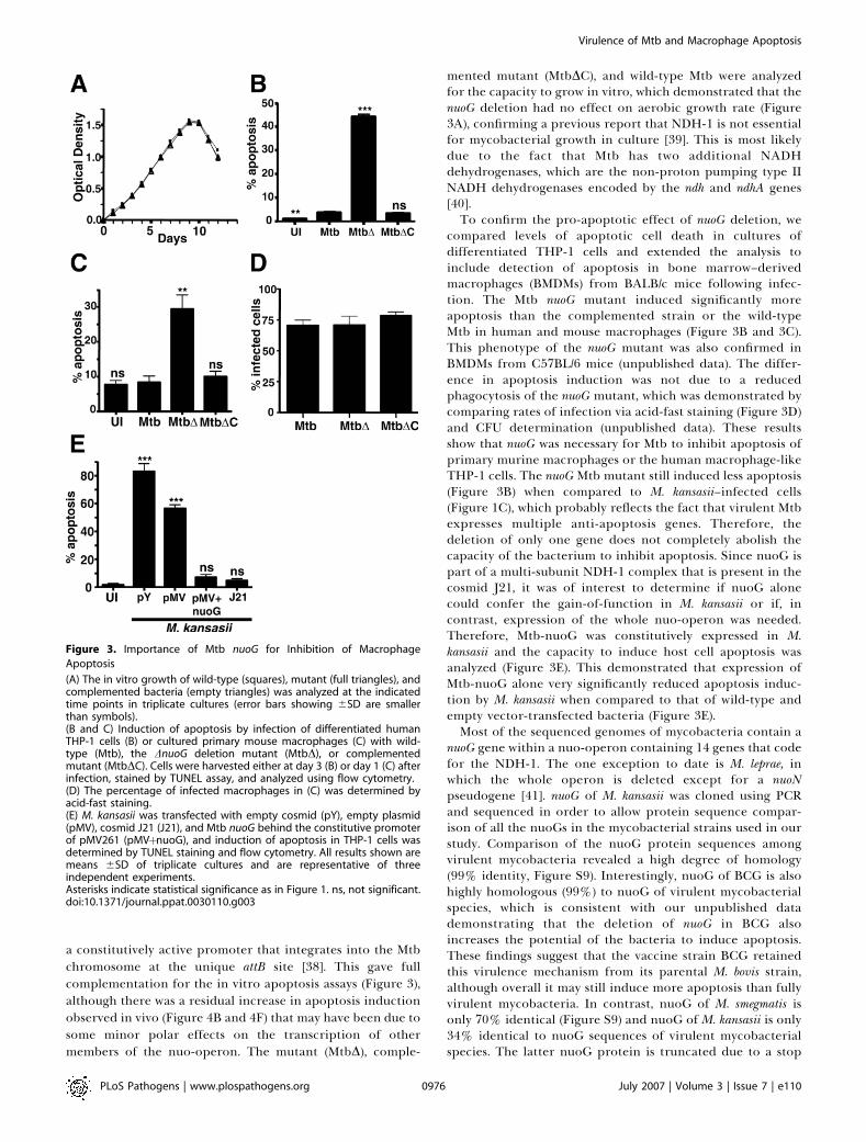
a constitutively active promoter that integrates into the Mtbchromosome at the unique attB site [38]. This gave fullcomplementation for the in vitro apoptosis assays (Figure 3),although there was a residual increase in apoptosis inductionobserved in vivo (Figure 4B and 4F) that may have been due tosome minor polar effects on the transcription of othermembers of the nuo-operon. The mutant (MtbD), comple-
mented mutant (MtbDC), and wild-type Mtb were analyzedfor the capacity to grow in vitro, which demonstrated that thenuoG deletion had no effect on aerobic growth rate (Figure3A), confirming a previous report that NDH-1 is not essentialfor mycobacterial growth in culture [39]. This is most likelydue to the fact that Mtb has two additional NADHdehydrogenases, which are the non-proton pumping type IINADH dehydrogenases encoded by the ndh and ndhA genes[40].To confirm the pro-apoptotic effect of nuoG deletion, we
compared levels of apoptotic cell death in cultures ofdifferentiated THP-1 cells and extended the analysis toinclude detection of apoptosis in bone marrow–derivedmacrophages (BMDMs) from BALB/c mice following infec-tion. The Mtb nuoG mutant induced significantly moreapoptosis than the complemented strain or the wild-typeMtb in human and mouse macrophages (Figure 3B and 3C).This phenotype of the nuoG mutant was also confirmed inBMDMs from C57BL/6 mice (unpublished data). The differ-ence in apoptosis induction was not due to a reducedphagocytosis of the nuoG mutant, which was demonstrated bycomparing rates of infection via acid-fast staining (Figure 3D)and CFU determination (unpublished data). These resultsshow that nuoG was necessary for Mtb to inhibit apoptosis ofprimary murine macrophages or the human macrophage-likeTHP-1 cells. The nuoG Mtb mutant still induced less apoptosis(Figure 3B) when compared to M. kansasii–infected cells(Figure 1C), which probably reflects the fact that virulent Mtbexpresses multiple anti-apoptosis genes. Therefore, thedeletion of only one gene does not completely abolish thecapacity of the bacterium to inhibit apoptosis. Since nuoG ispart of a multi-subunit NDH-1 complex that is present in thecosmid J21, it was of interest to determine if nuoG alonecould confer the gain-of-function in M. kansasii or if, incontrast, expression of the whole nuo-operon was needed.Therefore, Mtb-nuoG was constitutively expressed in M.kansasii and the capacity to induce host cell apoptosis wasanalyzed (Figure 3E). This demonstrated that expression ofMtb-nuoG alone very significantly reduced apoptosis induc-tion by M. kansasii when compared to that of wild-type andempty vector-transfected bacteria (Figure 3E).Most of the sequenced genomes of mycobacteria contain a
nuoG gene within a nuo-operon containing 14 genes that codefor the NDH-1. The one exception to date is M. leprae, inwhich the whole operon is deleted except for a nuoNpseudogene [41]. nuoG of M. kansasii was cloned using PCRand sequenced in order to allow protein sequence compar-ison of all the nuoGs in the mycobacterial strains used in ourstudy. Comparison of the nuoG protein sequences amongvirulent mycobacteria revealed a high degree of homology(99% identity, Figure S9). Interestingly, nuoG of BCG is alsohighly homologous (99%) to nuoG of virulent mycobacterialspecies, which is consistent with our unpublished datademonstrating that the deletion of nuoG in BCG alsoincreases the potential of the bacteria to induce apoptosis.These findings suggest that the vaccine strain BCG retainedthis virulence mechanism from its parental M. bovis strain,although overall it may still induce more apoptosis than fullyvirulent mycobacteria. In contrast, nuoG of M. smegmatis isonly 70% identical (Figure S9) and nuoG of M. kansasii is only34% identical to nuoG sequences of virulent mycobacterialspecies. The latter nuoG protein is truncated due to a stop
Figure 3. Importance of Mtb nuoG for Inhibition of Macrophage
Apoptosis
(A) The in vitro growth of wild-type (squares), mutant (full triangles), andcomplemented bacteria (empty triangles) was analyzed at the indicatedtime points in triplicate cultures (error bars showing 6SD are smallerthan symbols).(B and C) Induction of apoptosis by infection of differentiated humanTHP-1 cells (B) or cultured primary mouse macrophages (C) with wild-type (Mtb), the DnuoG deletion mutant (MtbD), or complementedmutant (MtbDC). Cells were harvested either at day 3 (B) or day 1 (C) afterinfection, stained by TUNEL assay, and analyzed using flow cytometry.(D) The percentage of infected macrophages in (C) was determined byacid-fast staining.(E) M. kansasii was transfected with empty cosmid (pY), empty plasmid(pMV), cosmid J21 (J21), and Mtb nuoG behind the constitutive promoterof pMV261 (pMVþnuoG), and induction of apoptosis in THP-1 cells wasdetermined by TUNEL staining and flow cytometry. All results shown aremeans 6SD of triplicate cultures and are representative of threeindependent experiments.Asterisks indicate statistical significance as in Figure 1. ns, not significant.doi:10.1371/journal.ppat.0030110.g003
PLoS Pathogens | www.plospathogens.org July 2007 | Volume 3 | Issue 7 | e1100976
Virulence of Mtb and Macrophage Apoptosis

codon introduced at codon 295, and thus nuoG of M. kansasiiis missing about 512 of the C-terminal amino acids.Interestingly, the M. kansasii nuoG is quite homologous upto amino acid 288 (94%). In conclusion, it seems likely that
both nuoG of M. smegmatis and M. kansasii have lost theirapoptosis-inhibiting function within the NDH-1 complex,and therefore the overexpression of Mtb nuoG is able torestore the capacity of the bacteria to inhibit host cellapoptosis. It will be of great interest to explore thishypothesis further by examining the expression of the variousnuoG proteins in the nuoG deletion mutant of Mtb andanalyzing the apoptosis induction of these complementedbacteria.How does nuoG mediate apoptosis inhibition? For nuoG to
have a direct effect on host cell apoptosis pathways, onewould assume that it needs to be secreted in order to interactwith host cell proteins or lipids. However, if the structure ofthe NDH-1 complex of Mtb is similar to that of otherbacterial NDH-1 complexes, nuoG will be located in thecytosol of the bacterium [42]. To determine whether MtbnuoG is secreted experimentally, we created a phoA-nuoGfusion protein. phoA can convert a colorless substrate into ablue product, but only if it is secreted by the bacterium [43].This assay failed to detect secretion of nuoG (Figure S7),which is consistent with the absence of a signal peptide andthe predicted cytosolic localization of this component ofNDH-1. Altogether, it thus seems unlikely that nuoG issecreted, and a direct effect of nuoG onto the host cell canalso be judged to be unlikely.The disruption of the NDH-1 system in the nuoG mutant
might have a very profound impact on the metabolism andproteome of the mycobacterium, which might result in anindirect effect on host cell apoptosis induction. Nevertheless,the absence of an in vitro growth defect in the nuoG mutant(Figure 3A) would argue against a profound effect of thedeletion on bacterial metabolism. In order to address theeffect of the nuoG mutation on the proteome of themycobacteria, the proteins of wild-type and mutant Mtbwere separated via 2-D gel electrophoresis. This revealed thatthe nuoG mutation did not induce a major change in theproteome (Figure S8). Thus, we found that the deletion ofnuoG did not have a major impact on the general bacterialmetabolism or its proteome. Instead, we propose that nuoGexerts its anti-apoptotic and virulence-promoting functionvia the enzymatic activity of the NDH-1 complex in a morespecific way.
Importance of Inhibition of Host Cell Apoptosis forVirulence of MtbRegardless of the remaining questions about the potential
mechanism of the nuoG/NDH-1–mediated apoptosis inhib-ition, our identification of an apoptosis-inducing mutant ofMtb allowed us to analyze the importance of apoptosisinhibition for bacterial virulence. First, the importance ofhost cell apoptosis inhibition in innate immune defense wasanalyzed by infecting immunodeficient SCID mice. Themedian survival times for mice infected with Mtb wild-typeor the complemented strain were not significantly different(14 d and 16.5 d, respectively). In contrast, mice infected withDnuoG Mtb survived twice as long as those infected with wild-type bacteria (median survival of 27.5 d, p , 0.0001, log-ranktest; Figure 4A), even though all mice received similar initialbacterial doses as confirmed by CFU counts at day 1 afterinfection (Figure S10). Consistently, the amount of apoptosisinduced in lung sections of these mice was significantlyincreased from less than 1% with wild-type bacteria to about
Figure 4. Identification of nuoG as a Mycobacterial Virulence Determi-
nant
(A) Survival of SCID mice after intravenous infection with 106 wild-typeMtb (squares), DnuoG mutant (filled triangles), or complemented mutantbacilli (open triangles). n¼ 7 mice per group.(B) Apoptotic cells in lung tissues of SCID mice after 14 d of infectionwere quantified using TUNEL peroxidase staining by microscopy asexplained in Figure 2.(C) Survival of immunocompetent BALB/c mice infected with bacterialstrains as in (A); n¼ 7 mice per group.(D) The bacterial burden in the lungs of infected BALB/c mice wasfollowed (n¼ 3 per time point; symbols indicate bacterial strains as in [Aand C]).(E) Lung histopathology (hematoxylin and eosin staining) at 3 and 20 wkfor BALB/c mice infected as in (C) with wild-type Mtb, DnuoG mutant(MtbD), or complemented mutant Mtb (MtbDC).(F) The percentages of apoptotic cells in lung sections were determinedby TUNEL peroxidase assay and quantified in blinded fashion as in Figure2. All results shown are representative of two independent experiments.Statistically significant differences compared to wild-type Mtb areindicated by asterisks as in the Figure 1 legend.doi:10.1371/journal.ppat.0030110.g004
PLoS Pathogens | www.plospathogens.org July 2007 | Volume 3 | Issue 7 | e1100977
Virulence of Mtb and Macrophage Apoptosis
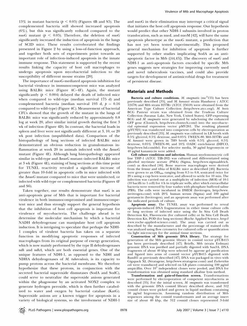
13% in mutant bacteria (p , 0.05) (Figures 4B and S5). Thecomplemented bacteria still showed increased apoptosis(6%), but this was significantly reduced compared to thenuoG mutant (p , 0.05). Therefore, the deletion of nuoGsignificantly increased the induction of apoptosis in the lungsof SCID mice. These results corroborated the findingspresented in Figure 2 by using a loss-of-function approach,and together both sets of experiments point towards animportant role of infection-induced apoptosis in the innateimmune response. This statement is supported by the recentresults linking the capacity of host cell macrophages toundergo apoptosis upon mycobacterial infection to thesusceptibility of different mouse strains [20].
The importance of nuoG-mediated apoptosis inhibition forbacterial virulence in immunocompetent mice was analyzedusing BALB/c mice (Figure 4C–4F). Again, the mutantsignificantly (p , 0.004) delayed the death of infected micewhen compared to wild-type (median survival 175 d) andcomplemented bacteria (median survival 193 d, p ¼ 0.16compared to wild-type) (Figure 4C). Measurement of bacterialCFUs showed that the growth of the mutant in the lungs ofBALB/c mice was significantly reduced by approximately 0.8log at week 20, after similar initial growth during the first 3wk of infection (Figure 4D). In contrast, the bacterial loads inspleen and liver were not significantly different at 3, 10, or 20wk post infection (unpublished data). Comparison of thehistopathology of lung sections of infected BALB/c micedemonstrated an obvious reduction in granulomatous in-flammation at week 20 in animals infected with the DnuoGmutant (Figure 4E). Although lung histopathology appearedsimilar in wild-type and DnuoG mutant–infected BALB/c miceat 3 wk (Figure 4E), staining of lung sections at this time pointfor TUNEL reactivity revealed a significant increase ofgreater than 10-fold in apoptotic cells in mice infected withthe DnuoG mutant compared to mice that were uninfected, orinfected with wild-type or complemented bacteria (Figures 4Fand S6).
Taken together, our results demonstrate that nuoG is ananti-apoptosis gene of Mtb that is important for bacterialvirulence in both immunocompromised and immunocompe-tent mice and thus strongly support the general hypothesisthat the inhibition of host cell apoptosis is important forvirulence of mycobacteria. The challenge ahead is todetermine the molecular mechanism by which a bacterialNADH dehydrogenase can manipulate host cell apoptosisinduction. It is intriguing to speculate that perhaps the NDH-1 complex of virulent bacteria has taken on a separatefunction in modifying apoptotic responses of infectedmacrophages from its original purpose of energy generation,which is now mainly performed by the type II dehydrogenasesndh and ndhA, which are both essential genes. One of theunique features of NDH-1, as opposed to the NDH andNDHA dehydrogenases of M. tuberculosis, is its capacity topump protons across the bacterial membrane. We thereforehypothesize that these protons, in conjunction with thesecreted bacterial superoxide dismutases (SodA and SodC),could serve to neutralize the superoxide anions generatedwithin the phagosome by an activated NOX2 complex togenerate hydrogen peroxide, which is then further catabol-ized to water and oxygen by bacterial catalase (KatG).Superoxide anions are a known trigger for apoptosis in avariety of biological systems, so the involvement of NDH-1
and nuoG in their elimination may interrupt a critical signalthat initiates the host cell apoptosis response. Our hypothesiswould predict that other NDH-1 subunits involved in protontranslocation, such as nuoL and nuoM [42], will have the sameapoptosis phenotype as the nuoG mutant, a prediction thathas not yet been tested experimentally. This proposedgeneral mechanism for inhibition of apoptosis is furthersupported by other studies implicating SodA as an anti-apoptotic factor in Mtb ([44,45]). The discovery of nuoG andNDH-1 as anti-apoptosis factors encoded by specific Mtbgenes suggests new strategies for improving currently usedand novel tuberculosis vaccines, and could also providetargets for development of antimicrobial drugs for treatmentof persistent disease.
Materials and Methods
Bacteria and culture conditions. M. smegmatis (mc2155) has beenpreviously described [35], and M. kansasii strain Hauduroy ( ATCC12478) and Mtb strain H37Rv (ATCC 25618) were obtained from theAmerican Type Culture Collection (http://www.atcc.org/). M. bovisBCG Pasteur strain was obtained from the Trudeau CultureCollection (Saranac Lake, New York, United States). GFP-expressingBCG and M. smegmatis were generated by subcloning the enhancedGFP gene (Clontech, http://www.clontech.com/) into the mycobacte-rial episomal expression vector pMV261. The resulting plasmid(pYU921) was transfected into competent cells by electroporation aspreviously described [35]. M. smegmatis was cultured in LB broth with0.5% glycerol, 0.5% dextrose, and 0.05% TWEEN-80. Mtb H37Rv andM. kansasii were grown in 7H9 broth with 0.5% glycerol, 0.5%dextrose, 0.05% TWEEN-80, and 10% OADC enrichment (DIFCO,http://www.bd.com/ds/). For selective media, 50 lg/ml hygromycin or40 lg/ml kanamycin were added.
Cell culture conditions and infection. Human myelomonocytic cellline THP-1 (ATCC TIB-202) was cultured and differentiated usingphorbol myristate acetate (PMA) (Sigma, http://www.sigmaaldrich.com/) as described [46]. Bone marrow macrophages were derivedfrom the femur and tibia of BALB/c mice as described [46]. Bacteriawere grown to an OD600 ranging from 0.5 to 0.8, sonicated twice for20 s using a cup horn sonicator, and allowed to settle for 10 min. Theinfection was carried out at a multiplicity of infection (MOI) of 10:1(10 bacilli to 1 cell) for 4 h in triplicate wells, after which extracellularbacteria were removed by four washes with phosphate buffered saline(PBS). The cells were incubated in DMEM (Invitrogen, http://www.invitrogen.com/) with 20% human serum (Sigma) and 100 lg/mlgentamicin (Invitrogen), and an apoptosis assay was performed afterthe indicated periods of culture.
Apoptosis assay. The TUNEL assay was performed to revealapoptosis-induced DNA fragmentation in either tissue culture cellsor lung sections of infected mice using the In Situ Cell DeathDetection Kit, Fluorescein (for cultured cells) or In Situ Cell DeathDetection Kit, POD (for lung sections) (Roche Applied Science, http://www.roche-applied-science.com/). The assay was carried out asdescribed by the manufacturer and the percentage of stained cellswas analyzed using flow cytometry for cultured cells or quantificationvia light microscopy for the animal tissue sections.
Construction of Mtb genomic DNA library. The strategy forgeneration of the Mtb genomic library in cosmid vector pYUB415has been previously described [47]. Briefly, Mtb (strain Erdman)genomic DNA was purified and partially digested with Sau3A. DNAfragments of about 40 kbp were selected by agarose gel purificationand ligated into arms of cosmid vector pYUB415 digested withBamH1 as previously described [47]. DNA was packaged in vitro withGigapack XL (Stratagene, http://www.stratagene.com/) and Escherichiacoli were transduced and selected on LB plates containing 100 lg/mlampicillin. Over 105 independent clones were pooled, and DNA fortransformation was obtained using standard alkaline lysis method.
Transformation and gain-of-function screen. Transformationswere performed by electroporation of competent mycobacteria asdescribed [35]. For the initial screen, M. smegmatis was transformedwith the genomic DNA cosmid library described above, and 312cosmid clones were picked and grown in liquid medium containing50 lg/ml hygromycin. Assuming random distribution of Mtbsequences among the cosmid transformants and an average insertsize of about 40 kbp, the 312 cosmid clones represented 3-fold
PLoS Pathogens | www.plospathogens.org July 2007 | Volume 3 | Issue 7 | e1100978
Virulence of Mtb and Macrophage Apoptosis

coverage of the entire Mtb genome. After three successive screensusing bright field microscopy or flow cytometry to assess levels of celldeath, 12 clones were selected for quantitative assessment usingTUNEL staining followed by flow cytometry. This identified threeclones of greatest interest (M24, J21, and I16), and their cosmid DNAwas purified and screened by restriction digest (not shown). Thisrevealed that the inserts of M24 and I16 were identical, but differentfrom the insert of J21. For cosmid J21, the 59 and 39 ends of the insertDNA were sequenced and aligned with the published genomic MtbDNA sequence (Figure S2), and subsequently the whole insert wassequenced to confirm that is corresponded to the sequence publisheddata.
Specialized transduction for construction of deletion mutants inMtb. Specific genes of Mtb were disrupted using specialized trans-duction as described [37]. To create the nuoG::hyg-null allele, thehygromycin resistance cassette was introduced between the first 4 bpof the nuoG 59 end and the last 163 bp of the 39 end of the openreading frame. The successful deletion of the gene was demonstratedby Southern blotting as described previously (Figure S3). Forcomplementation of the DnuoG mutation, nuoG was amplified byPCR and cloned behind the hsp60 constitutive promoter into theplasmid pMV361, which allows integration of a single copy into thegenome of Mtb [38].
Animal studies. BALB/c or SCID/Ncr (BALB/c background) mice (4-to 6-wk-old females) were infected intravenously through the lateraltail vein with 13106 bacteria. For survival studies, groups of ten micewere infected, and after 24 h, three mice per group were sacrificed todetermine the bacterial load in the organs. In order to follow thebacterial growth, an additional three mice per time point wereinfected. The organs (lung, spleen, liver) were homogenized sepa-rately in PBS/0.05% TWEEN-80, and colonies were enumerated on7H10 plates grown at 37 8C for 3–4 wk. For histopathology, tissueswere fixed in 10% buffered formalin and embedded in paraffin; 4-lmsections were stained with hematoxylin and eosin. TUNEL stainingwas performed on the paraffin-embedded tissue sections using the InSitu Cell Death Detection Kit, POD (Roche Applied Science) per themanufacturer’s protocol. Quantification was performed on codedspecimens by a blinded observer by counting the number ofapoptotic nuclei per ;200 total nuclei in eight separate areas oftwo lung sections for each of the three mice per group. All animalswere maintained in accordance with protocols approved by theAlbert Einstein College of Medicine and University of MarylandInstitutional Animal Care and Use Committees.
Supporting Information
Figure S1. Representative Lung Sections of Infected and UninfectedSCID Mice
Found at doi:10.1371/journal.ppat.0030110.sg001 (27.6 MB PPT).
Figure S2. Insert of Cosmid J21
Found at doi:10.1371/journal.ppat.0030110.sg002 (43 KB PPT).
Figure S3. Creation of nuoG Null Mutant in M. tuberculosisFound at doi:10.1371/journal.ppat.0030110.sg003 (90 KB PPT).
Figure S4. Identification of nuoG as a Mycobacterial Virulence Gene
Found at doi:10.1371/journal.ppat.0030110.sg004 (95 KB PPT).
Figure S5. TUNEL Staining of Lung Sections of Infected SCID Mice
Found at doi:10.1371/journal.ppat.0030110.sg005 (811 KB PPT).
Figure S6. TUNEL Staining of Lung Sections of Infected BALB/c Mice
Found at doi:10.1371/journal.ppat.0030110.sg006 (528 KB PPT).
Figure S7. A nuoG-phoA Fusion Protein Was Not Secreted
Found at doi:10.1371/journal.ppat.0030110.sg007 (192 KB PPT).
Figure S8. nuoG Deletion Did Not Lead to a Major Change in MtbProteome
Found at doi:10.1371/journal.ppat.0030110.sg008 (7.8 MB PPT).
Figure S9. nuoG Protein Sequence Alignment
Found at doi:10.1371/journal.ppat.0030110.sg009 (159 KB PPT).
Figure S10. Similar Bacterial Burden in SCID Mice Infected with Mtb,Mutant, and Complemented Mutant Strain after 24 h
Found at doi:10.1371/journal.ppat.0030110.sg010 (95 KB PPT).
Accession Numbers
The GenBank (http://www.ncbi.nlm.nih.gov/Genbank/index.html) ac-cession numbers for the sequences of the nuoG proteins from thefollowing mycobacteria are M.bovis AF2122/97 (CAD95267), M. bovisBCG-Pasteur (YP979258), M. kansasii Hauduroy (EF607211), M.smegmatis mc2155 (YP886418), M. tuberculosis CDC1551 (AAK47578),and M. tuberculosis H37Rv (CAB06288).
Acknowledgments
We would like to thank M. Braunstein for providing vectors pMB111and pMB124. Flow cytometry studies were supported by the FlowCytometry Core Facilities of the Cancer Center and the Center forAIDS Research at Albert Einstein College of Medicine.
Author contributions. KV, JM, SA, SG, and VB conceived anddesigned the experiments. KV, BC, JM, SA, SG, and VB performed theexperiments. KV, WRJ, SAP, and VB analyzed the data. TH, MG, andWRJ contributed reagents/materials/analysis tools. SAP and VB wrotethe paper.
Funding. This work was supported by the National Institutes ofHealth (VB, SAP, MG, WRJ), the Burroughs Wellcome Fund (SAP),The Hirschl Trust (SAP), the Ellison Medical Foundation (MG), andthe Howard Hughes Medical Institute (WRJ).
Competing interests. The authors have declared that no competinginterests exist.
References1. Dye C, Watt CJ, Bleed DM, Hosseini SM, Raviglione MC (2005) Evolution of
tuberculosis control and prospects for reducing tuberculosis incidence,prevalence, and deaths globally. JAMA 293: 2767–2775.
2. Manabe YC, Bishai WR (2000) LatentMycobacterium tuberculosis—Persistence,patience, and winning by waiting. Nat Med 6: 1327–1329.
3. Honer zu Bentrup K, Russell DG (2001) Mycobacterial persistence: Adapta-tion to a changing environment. Trends Microbiol 9: 597–605.
4. Hingley-Wilson SM, Sambandamurthy VK, Jacobs WR Jr (2003) Survivalperspectives from the world’s most successful pathogen, Mycobacteriumtuberculosis. Nat Immunol 4: 949–955.
5. Smith CV, Sharma V, Sacchettini JC (2004) TB drug discovery: Addressingissues of persistence and resistance. Tuberculosis (Edinb) 84: 45–55.
6. Abramovitch RB, Martin GB (2004) Strategies used by bacterial pathogensto suppress plant defenses. Curr Opin Plant Biol 7: 356–364.
7. Stuart LM, Ezekowitz RA (2005) Phagocytosis: Elegant complexity.Immunity 22: 539–550.
8. Boya P, Roques B, Kroemer G (2001) New EMBO members’ review: Viraland bacterial proteins regulating apoptosis at the mitochondrial level.EMBO J 20: 4325–4331.
9. Dockrell DH (2003) The multiple roles of Fas ligand in the pathogenesis ofinfectious diseases. Clin Microbiol Infect 9: 766–779.
10. Hilleman MR (2004) Strategies and mechanisms for host and pathogensurvival in acute and persistent viral infections. Proc Natl Acad Sci U S A101 (Suppl 2): 14560–14566.
11. Monack DM, Mueller A, Falkow S (2004) Persistent bacterial infections: theinterface of the pathogen and the host immune system. Nat Rev Microbiol2: 747–765.
12. Hacker G, Kirschnek S, Fischer SF (2006) Apoptosis in infectious disease:How bacteria interfere with the apoptotic apparatus. Med MicrobiolImmunol 195: 11–19.
13. Rojas M, Barrera LF, Puzo G, Garcia LF (1997) Differential induction ofapoptosis by virulent Mycobacterium tuberculosis in resistant and susceptiblemurine macrophages: Role of nitric oxide and mycobacterial products. JImmunol 159: 1352–1361.
14. Danelishvili L, McGarvey J, Li YJ, Bermudez LE (2003) Mycobacteriumtuberculosis infection causes different levels of apoptosis and necrosis inhuman macrophages and alveolar epithelial cells. Cell Microbiol 5: 649–660.
15. Schaible UE, Winau F, Sieling PA, Fischer K, Collins HL, et al. (2003)Apoptosis facilitates antigen presentation to T lymphocytes through MHC-I and CD1 in tuberculosis. Nat Med 9: 1039–1046.
16. Derrick SC, Morris SL (2007) The ESAT6 protein of Mycobacteriumtuberculosis induces apoptosis of macrophages by activating caspaseexpression. Cell Microbiol 9: 1547–1555.
17. Keane J, Remold HG, Kornfeld H (2000) Virulent Mycobacterium tuberculosisstrains evade apoptosis of infected alveolar macrophages. J Immunol 164:2016–2020.
18. Oddo M, Renno T, Attinger A, Bakker T, MacDonald HR, et al. (1998) Fasligand-induced apoptosis of infected human macrophages reduces the
PLoS Pathogens | www.plospathogens.org July 2007 | Volume 3 | Issue 7 | e1100979
Virulence of Mtb and Macrophage Apoptosis

viability of intracellular Mycobacterium tuberculosis. J Immunol 160: 5448–5454.
19. Sly LM, Hingley-Wilson SM, Reiner NE, McMaster WR (2003) Survival ofMycobacterium tuberculosis in host macrophages involves resistance toapoptosis dependent upon induction of antiapoptotic Bcl-2 family memberMcl-1. J Immunol 170: 430–437.
20. Pan H, Yan BS, Rojas M, Shebzukhov YV, Zhou H, et al. (2005) Ipr1 genemediates innate immunity to tuberculosis. Nature 434: 767–772.
21. Kausalya S, Somogyi R, Orlofsky A, Prystowsky MB (2001) Requirement ofA1-a for bacillus Calmette-Guerin-mediated protection of macrophagesagainst nitric oxide-induced apoptosis. J Immunol 166: 4721–4727.
22. Kremer L, Estaquier J, Brandt E, Ameisen JC, Locht C (1997) Mycobacteriumbovis Bacillus Calmette Guerin infection prevents apoptosis of restinghuman monocytes. Eur J Immunol 27: 2450–2456.
23. Riendeau CJ, Kornfeld H (2003) THP-1 cell apoptosis in response toMycobacterial infection. Infect Immun 71: 254–259.
24. Loeuillet C, Martinon F, Perez C, Munoz M, Thome M, et al. (2006)Mycobacterium tuberculosis subverts innate immunity to evade specificeffectors. J Immunol 177: 6245–6255.
25. Balcewicz-Sablinska MK, Keane J, Kornfeld H, Remold HG (1998)Pathogenic Mycobacterium tuberculosis evades apoptosis of host macrophagesby release of TNF-R2, resulting in inactivation of TNF-alpha. J Immunol161: 2636–2641.
26. McKinney JD, Honer zu Bentrup K, Munoz-Elias EJ, Miczak A, Chen B, et al.(2000) Persistence of Mycobacterium tuberculosis in macrophages and micerequires the glyoxylate shunt enzyme isocitrate lyase. Nature 406: 735–738.
27. Stewart GR, Robertson BD, Young DB (2003) Tuberculosis: A problem withpersistence. Nat Rev Microbiol 1: 97–105.
28. Fratazzi C, Arbeit RD, Carini C, Remold HG (1997) Programmed cell deathof Mycobacterium avium serovar 4-infected human macrophages prevents themycobacteria from spreading and induces mycobacterial growth inhibitionby freshly added, uninfected macrophages. J Immunol 158: 4320–4327.
29. Molloy A, Laochumroonvorapong P, Kaplan G (1994) Apoptosis, but notnecrosis, of infected monocytes is coupled with killing of intracellularbacillus Calmette-Guerin. J Exp Med 180: 1499–1509.
30. Winau F, Kaufmann SH, Schaible UE (2004) Apoptosis paves the detourpath for CD8 T cell activation against intracellular bacteria. Cell Microbiol6: 599–607.
31. Winau F, Hegasy G, Kaufmann SH, Schaible UE (2005) No life withoutdeath-apoptosis as prerequisite for T cell activation. Apoptosis 10: 707–715.
32. Kaufmann SH (2006) Tuberculosis: Back on the immunologists’ agenda.Immunity 24: 351–357.
33. Grode L, Seiler P, Baumann S, Hess J, Brinkmann V, et al. (2005) Increasedvaccine efficacy against tuberculosis of recombinant Mycobacterium bovisbacille Calmette-Guerin mutants that secrete listeriolysin. J Clin Invest 115:2472–2479.
34. Kaufmann SH (2006) Envisioning future strategies for vaccination againsttuberculosis. Nat Rev Immunol 6: 699–704.
35. Snapper SB, Melton RE, Mustafa S, Kieser T, Jacobs WR Jr (1990) Isolationand characterization of efficient plasmid transformation mutants ofMycobacterium smegmatis. Mol Microbiol 4: 1911–1919.
36. Cole ST, Brosch R, Parkhill J, Garnier T, Churcher C, et al. (1998)Deciphering the biology of Mycobacterium tuberculosis from the completegenome sequence. Nature 393: 537–544.
37. Bardarov S, Bardarov Jr S, Pavelka Jr MS, Sambandamurthy V, Larsen M, etal. (2002) Specialized transduction: An efficient method for generatingmarked and unmarked targeted gene disruptions in Mycobacterium tuber-culosis, M. bovis BCG and M. smegmatis. Microbiology 148: 3007–3017.
38. Stover CK, de la Cruz VF, Fuerst TR, Burlein JE, Benson LA, et al. (1991)New use of BCG for recombinant vaccines. Nature 351: 456–460.
39. Sassetti CM, Boyd DH, Rubin EJ (2003) Genes required for mycobacterialgrowth defined by high density mutagenesis. Mol Microbiol 48: 77–84.
40. Shi L, Sohaskey CD, Kana BD, Dawes S, North RJ, et al. (2005) Changes inenergy metabolism of Mycobacterium tuberculosis in mouse lung and under invitro conditions affecting aerobic respiration. Proc Natl Acad Sci U S A102: 15629–15634.
41. Weinstein EA, Yano T, Li LS, Avarbock D, Avarbock A, et al. (2005)Inhibitors of type II NADH:menaquinone oxidoreductase represent a classof antitubercular drugs. Proc Natl Acad Sci U S A 102: 4548–4553.
42. Friedrich T, Bottcher B (2004) The gross structure of the respiratorycomplex I: A Lego system. Biochim Biophys Acta 1608: 1–9.
43. Braunstein M, Brown AM, Kurtz S, Jacobs WR Jr. (2001) Two nonredundantSecA homologues function in mycobacteria. J Bacteriol 183: 6979–6990.
44. Hinchey J, Lee S, Jeon BY, Basaraba RJ, Venkataswamy MM, et al. (2007)Enhanced priming of adaptive immunity by a proapoptotic mutant ofMycobacterium tuberculosis. J Clin Invest. In press.
45. Edwards KM, Cynamon MH, Voladri RK, Hager CC, DeStefano MS, et al.(2001) Iron-cofactored superoxide dismutase inhibits host responses toMycobacterium tuberculosis. Am J Respir Crit Care Med 164: 2213–2219.
46. Dao DN, Kremer L, Guerardel Y, Molano A, Jacobs WR Jr, et al. (2004)Mycobacterium tuberculosis lipomannan induces apoptosis and interleukin-12production in macrophages. Infect Immun 72: 2067–2074.
47. Bange FC, Collins FM, Jacobs WR Jr (1999) Survival of mice infected withMycobacterium smegmatis containing large DNA fragments fromMycobacteriumtuberculosis. Tuber Lung Dis 79: 171–180.
PLoS Pathogens | www.plospathogens.org July 2007 | Volume 3 | Issue 7 | e1100980
Virulence of Mtb and Macrophage Apoptosis
Related Documents