MYCOBACTERIUM TUBERCULOSIS CYP130: CRYSTAL STRUCTURE, BIOPHYSICAL CHARACTERIZATION, AND INTERACTIONS WITH ANTIFUNGAL AZOLE DRUGS * Hugues Ouellet, Larissa M. Podust, and Paul R. Ortiz de Montellano † From the Department of Pharmaceutical Chemistry, University of California, San Francisco, California 94158-2517 Abstract CYP130 is one of the 20 Mycobacterium tuberculosis cytochrome P450 enzymes, only two of which, CYP51 and CYP121, have so far been studied as individually expressed proteins. Herein we characterize a third heterologously expressed Mycobacterium tuberculosis cytochrome P450, CYP130, by UV-visible spectroscopy, isothermal titration calorimetry, and x-ray crystallography, including determination of the crystal structures of ligand-free and econazole-bound CYP130 at a resolution of 1.46 Å and 3.0 Å, respectively. Ligand-free CYP130 crystallizes in an ‘open’ conformation as a monomer, whereas the econazole-bound form crystallizes in a ‘closed’ conformation as a dimer. Conformational changes enabling the ‘open-closed’ transition involve repositioning of the BC loop and the F and G helices that envelop the inhibitor in the binding site and reshape the protein surface. Crystal structure analysis shows that the portion of the BC-loop relocates as much as 18 Å between the open and closed conformations. Binding of econazole to CYP130 involves a conformational change and is mediated by both a set of hydrophobic interactions with amino acid residues in the active site and coordination of the heme iron. CYP130 also binds miconazole with virtually the same binding affinity as econazole and clotrimazole and ketoconazole with somewhat lower affinities, which makes it a plausible target for this class of therapeutic drugs. Overall, binding of the azole inhibitors is a sequential two-step entropy-driven endothermic process. Binding of econazole and clotrimazole exhibits positive cooperativity that may reflect a propensity of CYP130 to associate into a dimeric structure. The pathogenic bacterium Mycobacterium tuberculosis (Mtb) 1 continues to be an enormous threat to human health. It is responsible for more deaths worldwide than any other infectious agent, and is the major cause of death for HIV-infected individuals in developing countries. An aggravating factor associated with the global resurgence of tuberculosis is the proliferation of strains resistant to isoniazid and rifampicin, the two major frontline antitubercular drugs. Therefore, new drug strategies are needed to combat the rising incidence of tuberculosis, * This work was supported by NIH RO1 grants GM25515 and AI74824 (to P.O.M.), and GM078553 (to L.M.P.). The Advanced Light Source is supported by the Director, Office of Science, Office of Basic Energy Sciences, of the U.S. Department of Energy under Contract No. DE-AC02-05CH11231. † Address Correspondence to: Dr. Paul R. Ortiz de Montellano, Department of Pharmaceutical Chemistry, N572D, Tel.: 415-476-2903, [email protected]. Protein Data Bank accession numbers. Atomic coordinates and structure factors determined in this study (Protein Data Bank IDs 2UUQ and 2UVN) have been deposited in the Protein Data Bank, Macrolomecular Structure Database Group, European Bioinformatics Institute. 1 The abbreviations are: Mtb, Mycobacterium tuberculosis; TAP, tetracycline/aminoglycoside-resistance protein; P450 or CYP, cytochrome P450; ITC, isothermal titration calorimetry; TB, terrific broth; LB, Luria-Bertani; FPLC, fast protein liquid chromatography; KPi, potassium phosphate; MAD, multiwavelength anomalous dispersion. NIH Public Access Author Manuscript J Biol Chem. Author manuscript; available in PMC 2010 October 21. Published in final edited form as: J Biol Chem. 2008 February 22; 283(8): 5069–5080. doi:10.1074/jbc.M708734200. NIH-PA Author Manuscript NIH-PA Author Manuscript NIH-PA Author Manuscript

Welcome message from author
This document is posted to help you gain knowledge. Please leave a comment to let me know what you think about it! Share it to your friends and learn new things together.
Transcript
MYCOBACTERIUM TUBERCULOSIS CYP130: CRYSTALSTRUCTURE, BIOPHYSICAL CHARACTERIZATION, ANDINTERACTIONS WITH ANTIFUNGAL AZOLE DRUGS*
Hugues Ouellet, Larissa M. Podust, and Paul R. Ortiz de Montellano†From the Department of Pharmaceutical Chemistry, University of California, San Francisco,California 94158-2517
AbstractCYP130 is one of the 20 Mycobacterium tuberculosis cytochrome P450 enzymes, only two of which,CYP51 and CYP121, have so far been studied as individually expressed proteins. Herein wecharacterize a third heterologously expressed Mycobacterium tuberculosis cytochrome P450,CYP130, by UV-visible spectroscopy, isothermal titration calorimetry, and x-ray crystallography,including determination of the crystal structures of ligand-free and econazole-bound CYP130 at aresolution of 1.46 Å and 3.0 Å, respectively. Ligand-free CYP130 crystallizes in an ‘open’conformation as a monomer, whereas the econazole-bound form crystallizes in a ‘closed’conformation as a dimer. Conformational changes enabling the ‘open-closed’ transition involverepositioning of the BC loop and the F and G helices that envelop the inhibitor in the binding siteand reshape the protein surface. Crystal structure analysis shows that the portion of the BC-looprelocates as much as 18 Å between the open and closed conformations. Binding of econazole toCYP130 involves a conformational change and is mediated by both a set of hydrophobic interactionswith amino acid residues in the active site and coordination of the heme iron. CYP130 also bindsmiconazole with virtually the same binding affinity as econazole and clotrimazole and ketoconazolewith somewhat lower affinities, which makes it a plausible target for this class of therapeutic drugs.Overall, binding of the azole inhibitors is a sequential two-step entropy-driven endothermic process.Binding of econazole and clotrimazole exhibits positive cooperativity that may reflect a propensityof CYP130 to associate into a dimeric structure.
The pathogenic bacterium Mycobacterium tuberculosis (Mtb)1 continues to be an enormousthreat to human health. It is responsible for more deaths worldwide than any other infectiousagent, and is the major cause of death for HIV-infected individuals in developing countries.An aggravating factor associated with the global resurgence of tuberculosis is the proliferationof strains resistant to isoniazid and rifampicin, the two major frontline antitubercular drugs.Therefore, new drug strategies are needed to combat the rising incidence of tuberculosis,
*This work was supported by NIH RO1 grants GM25515 and AI74824 (to P.O.M.), and GM078553 (to L.M.P.). The Advanced LightSource is supported by the Director, Office of Science, Office of Basic Energy Sciences, of the U.S. Department of Energy under ContractNo. DE-AC02-05CH11231.†Address Correspondence to: Dr. Paul R. Ortiz de Montellano, Department of Pharmaceutical Chemistry, N572D, Tel.: 415-476-2903,[email protected] Data Bank accession numbers. Atomic coordinates and structure factors determined in this study (Protein Data Bank IDs 2UUQand 2UVN) have been deposited in the Protein Data Bank, Macrolomecular Structure Database Group, European Bioinformatics Institute.1The abbreviations are: Mtb, Mycobacterium tuberculosis; TAP, tetracycline/aminoglycoside-resistance protein; P450 or CYP,cytochrome P450; ITC, isothermal titration calorimetry; TB, terrific broth; LB, Luria-Bertani; FPLC, fast protein liquid chromatography;KPi, potassium phosphate; MAD, multiwavelength anomalous dispersion.
NIH Public AccessAuthor ManuscriptJ Biol Chem. Author manuscript; available in PMC 2010 October 21.
Published in final edited form as:J Biol Chem. 2008 February 22; 283(8): 5069–5080. doi:10.1074/jbc.M708734200.
NIH
-PA Author Manuscript
NIH
-PA Author Manuscript
NIH
-PA Author Manuscript

especially the multidrug-resistant forms, and to shorten the duration of tuberculosis treatment(1).
It has been demonstrated that azole drugs such as econazole and clotrimazole, which inhibitthe sterol 14α-demethylase CYP51 and were originally developed as fungal antibiotics (2),display inhibitory potential against the latent and multidrug-resistant forms of tuberculosis bothin vitro and in tuberculosis infected mice (3–7). Furthermore, econazole exhibits synergisticactivities with rifampicin and isoniazid against the multidrug-resistant Mtb strains (3). The 4.4Mb Mtb genome encodes 20 different cyp genes (8), whose biological roles are not yetunderstood. To date, physiological roles have been proposed for CYP125 and CYP142 incholesterol catabolism (9), and for CYP132 in fatty acid metabolism (10). A catalytic function,the demethylation of sterols, has been demonstrated for Mtb CYP51 (11) that, in the absenceof a sterol biosynthetic pathway in Mtb, potentially links this enzyme to cholesterol-mediatedMtb entry into macrophages and its subsequent intracellular survival (12).
The cyp130 and cyp141 genes are missing from the virulent Mycobacterium bovis strain andfrom its avirulent counterpart, Mycobacterium bovis BCG, suggesting that they are not essentialfor Mtb growth, but may be relevant for Mtb virulence and infectivity towards the human host(13). The gene Rv1256c encoding Mtb CYP130 is possibly part of a functional operon alongwith the gene Rv1258c that encodes for a tetracycline/aminoglycoside-resistance (TAP)-likeefflux pump. Both the Mycobacterium fortuitum TAP1 efflux pump and its Mtb Rv1258chomologue confer significant resistance to tetracycline and aminoglycosides, includingstreptomycin, a third major drug in antituberculosis treatment (14). Deletion of the Rv1258cgene from the M. bovis BCG chromosome increases the susceptibility of the organism to thesetwo drugs, confirming involvement of the efflux pump in the intrinsic resistance of M. bovisand Mtb to tetracycline and streptomycin (15). Furthermore, a correlation has been establishedbetween expression of the Rv1258c gene and drug resistance in a clinical Mtb isolate resistantto the two major antitubercular drugs, rifampicin and isoniazid (16). However, no evidence yetexists of a functional link between CYP130 and Rv1258c.
The large number of distinct cytochrome P450 (P450) enzymes and the susceptibility of Mtbto azole agents that target such enzymes suggest important roles for them in Mtb physiologyand, hence, their potential use as therapeutic targets. To date, only two Mtb P450 enzymes,CYP51 and CYP121, have been studied as individually expressed recombinant proteins. Bothhave been shown to tightly bind econazole, the agent of the azole class with the highest knownantimycobacterial activity, as well as other azole and triazole drugs (17). The interactions ofCYP51 and CYP121 with the azole inhibitors have been addressed by x-ray crystallographyresulting in the determination of several crystal structures, including those of their complexeswith the triazole antifungal agent fluconazole (18,19). Although econazole is so far the mostpotent antimycobacterial azole agent interacting in vitro with CYP51 and CYP121 (17), andherein with CYP130, no crystal structure of econazole bound in any P450 active site has everbeen reported.
In the current work, we report determination of the x-ray crystal structures for ligand-free andeconazole-bound Mtb CYP130. We have also examined the binding of azole drugs by UV-visible spectroscopy and isothermal titration calorimetry (ITC). Our data demonstrate that aconformational change in the protein is required for binding of econazole to CYP130 througha set of hydrophobic protein contacts and coordination to the heme iron. In addition toeconazole, CYP130 binds a number of other antifungal agents with micromolar affinity, whichmakes it a plausible target for this class of therapeutic agents. Collectively, binding azoles toCYP130 is an endothermic entropy-driven complex process, which consists of two stepsdeducible from the titration calorimetry and exhibits spectrally detectable ligand-specific
Ouellet et al. Page 2
J Biol Chem. Author manuscript; available in PMC 2010 October 21.
NIH
-PA Author Manuscript
NIH
-PA Author Manuscript
NIH
-PA Author Manuscript
binding cooperativity that can be attributed to a potential for intramolecular or intermolecularprotein-protein interactions inherent to CYP130.
EXPERIMENTAL PROCEDURESChemicals
Econazole, miconazole, clotrimazole, ketoconazole, glutaraldehyde, and other chemicals werepurchased from Sigma-Aldrich unless otherwise specified. Crystallization screening kits werepurchased from both Hampton Research and Qiagen.
Molecular Cloning of Rv1256c Encoding CYP130Genomic DNA from M. tuberculosis H37Rv was obtained through the TB Vaccine Testingand Research Materials Contract at Colorado State University. The region of the Rv1256c geneencoding the putative cytochrome P450 CYP130 was amplified by PCR using Pfu Turbo DNApolymerase (Stratagene) and upstream 5'-CTCTGCTCCATATGACATCAGTAATGTCTCACG-3' and downstream 5'-AAGCTTTCATCTAGAGGATGTCACTCGGAACG-3' primers. The letters in bold in theupstream primer indicate an engineered Nde I restriction cloning site, including the initiationcodon ATG. The underlined letters in the downstream primer indicate a Hind III restriction-cloning site. Amplification conditions were 94 °C for 5 min, 5 cycles of 94 °C for 30 s, 55 °Cfor 30 s, and 72 °C for 3 min followed by 25 cycles of 94 °C for 30 s, 65 °C for 30 s, and 72 °C for 3 min. The PCR program was ended by a polymerization step at 72 °C for 25 min. Toconfirm the DNA sequence, the PCR fragment was first cloned into a pCR2.1 TOPO vector(Invitrogen) and then the Nde I-Hind III digested fragment was subcloned into a pCWori vector,which allows the expression of the recombinant protein with an N-terminal His6-tag (20).
Expression of Native and Se-methionine Containing CYP130Recombinant CYP130, both native and as the Se-methionine containing derivative, wasexpressed under the control of the tac promoter of the pCWori vector using the Escherichiacoli DH5α cells. For the native protein, cells were grown at 37 °C with vigorous agitation (250rpm) in 2.8-L flasks containing 1 L of terrific broth (TB) medium supplemented with 200 µg/ml of ampicillin until the OD600 reached 0.5–0.8. At that time IPTG (0.5 mM), δ-aminolevulinic acid (0.5 mM), FeCl3 (250 µM) and ampicillin (200 µg/ml) were added. Thecells were incubated for an additional 36 h at 25 °C at reduced agitation (180 rpm). The cellswere harvested by centrifugation at 5,000 × g for 20 min at 4 °C and were then kept frozen at−80 °C until used.
For the Se-methionine containing CYP130 derivative, the transformed cells were grown at 37°C and 250 rpm in 2.8-L flasks containing 1 L of Luria-Bertani (LB) medium supplementedwith 200 µg/ml ampicillin until the OD600 reached 0.8–1.0. Cells were harvested bycentrifugation at 2,000 × g for 15 min at 18 °C, washed with 100 ml of SelenoMet Mediumbase (AthenaES, Baltimore, MD) according to the protocol provided by the manufacturer, andre-centrifuged. Re-centrifuged cells were resuspended, transferred into 1 L of fresh SelenoMetMedium base and incubated at 25 °C and 250 rpm for 2 h before IPTG (0.5 mM), δ-aminolevulinic acid (0.5 mM), ampicillin (200 µg/ml), and SelenoMet Nutrient Mix(AthenaES, Baltimore, MD) containing a mixture of all the amino acids except methionine,vitamins and seleno-methionine were added according to the protocol provided by themanufacturer. The cells were incubated for 36 h at 25 °C and 180 rpm and harvested bycentrifugation as described above.
Ouellet et al. Page 3
J Biol Chem. Author manuscript; available in PMC 2010 October 21.
NIH
-PA Author Manuscript
NIH
-PA Author Manuscript
NIH
-PA Author Manuscript
CYP130 PurificationBoth native and Se-methionine containing CYP130 were purified to homogeneity by fastprotein liquid chromatography (FPLC). Cells obtained from 6 L of culture were thawed on iceand resuspended in 200 ml of buffer A (50 mM Tris-HCl, pH 7.5, 0.5 M NaCl, 0.1 mM EDTA,20 mM imidazole, and 1 mM PMSF). The cell suspension was incubated on ice for 30 minafter the addition of lysozyme (0.5 mg/ml) and DNAse I (0.1 mg/ml). The cells were lysed bysonication using a Branson sonicator (3 × 4-min bursts at 50% power, with 2 min cooling onice between each burst). Cell debris was removed by centrifugation at 100,000 × g, for 1 h at4 °C. The soluble extract was loaded onto a 20 ml His/PrepFF 16/60 column (Amersham-Biosciences) equilibrated with buffer A. The column was first washed with 100 ml of bufferA and then with 100 ml of buffer B (50 mM Tris-HCl, pH 7.5, 0.1 mM EDTA, and 20 mMimidazole). The protein was eluted with 200 ml of a linear gradient (20–200 mM) of imidazolein buffer B. All the fractions containing P450 were pooled and the protein was further purifiedby flow-through chromatography on SP-sepharose Fast-Flow (Amersham Bioscience) andsubsequent binding to Q-sepharose Fast-Flow (Amersham Bioscience). The protein was elutedwith 200 ml of a linear gradient (0–250 mM) of NaCl in 50 mM Tris-HCl, pH 7.5 and 0.1 mMEDTA. The fractions were analyzed by SDS-PAGE, and those containing pure CYP130 werepooled and concentrated to at least 1 mM using an Amicon Ultra concentrating device(Millipore). The content of Se-methionine in the CYP130 Se-methionine derivative wasassessed by trypsin digestion and analysis of the tryptic fragments by MALDI-TOF MS usinga Q-STAR XL mass spectrometer (Applied Biosystems/MDS Sciex).
Optical Absorption SpectroscopyUV-visible absorption spectra of the purified CYP130 were recorded on Cary UV-visiblescanning spectophotometer (Varian) using 1-cm path length quartz cuvette at 23 °C in 50 mMpotassium phosphate (KPi) buffer, pH 7.4, containing 0.1 mM EDTA. The ferric-nitrosylspecies was obtained in anaerobic conditions by flushing pure NO gas (Matheson Tri Gas, CA)over the ferric protein solution previously flushed with argon for 20 min. Formation of theferrous carbon monoxide complex was achieved by bubbling CO gas (Airgas, CA) into theferric enzyme solution for approximately 30 s through a septum-sealed cuvette prior to theinjection of 1 mM sodium dithionite using a gas tight syringe (Hamilton, Reno, NV). Differencespectra were generated by subtracting the spectrum of the ferrous deoxy form from that of itscarbon monoxide complex. The concentration of P450 was determined from difference spectrausing the extinction coefficient 91,000 M−1cm−1 (21).
Equilibrium Binding AssayBinding of the antifungal azole agents econazole, miconazole, clotrimazole and ketoconazoleto CYP130 was monitored by UV-visible spectroscopy at 23 °C in 50 mM KPi buffer, pH 7.4,containing 0.1 mM EDTA. Stock solutions of the inhibitors at concentrations of 1 and 10 mMwere prepared in DMSO. Difference spectra were recorded following the addition of a seriesof 0.25–1.0 µl aliquots of inhibitor to the sample cuvette containing 1 ml of 2.5 µM CYP130for a maximal volume of 10 µl. The same amounts of DMSO alone were added to the referencecuvette. Increasing concentrations of KCl were added when specified in Table 3. To determinethe Kd values, titration data points were fitted to the rectangular hyperbola (Eq. 1) forketoconazole, quadratic hyperbola (Eq. 2) for miconazole, and the Hill equation (Eq. 3) forboth econazole and clotrimazole using the Kaleidagraph software (Synergy).
(Eq. 1)
Ouellet et al. Page 4
J Biol Chem. Author manuscript; available in PMC 2010 October 21.
NIH
-PA Author Manuscript
NIH
-PA Author Manuscript
NIH
-PA Author Manuscript
(Eq. 2)
(Eq. 3)
In all equations, Aobs is the absorption shift determined at any ligand concentration, Amax isthe maximal absorption shift obtained at saturation, Kd, is the apparent dissociation constantfor the inhibitor-enzyme complex, Et is the total enzyme concentration used, S is the ligandconcentration, and n is a Hill coefficient, a measure of cooperativity.
Binding stoichiometry by Job’s titrationThe stoichiometry for the binding of inhibitors to CYP130 was determined by the method ofcontinuous variation known as a Job’s titration (22) using UV-visible spectroscopy and a sub-micro quartz cuvette designed for 0.1- to 1.55-ml sample (Starna, Atascadero, CA).Experiments were carried out at 23 °C in 50 mM KPi, pH 7.4, containing 0.1 mM EDTA and0.5% DMSO. 125 µl of 15 µM CYP130 were placed into the optical cell, and the inhibitorsolution of the same concentration was added gradually, until the volume of the mixture reached1.525 ml. The sum of the concentrations of the reactants was therefore kept constant and equalto 15 µM.
Crystallization and Data CollectionPurified CYP130 diluted to a concentration of 0.2 mM was subjected to automated screeningof crystallization conditions using a nanoliter drop setter Mosquito (TTP LabTech). Bothligand-free and econazole-bound CYP130 crystallized from the different sets of crystallizationconditions, which were further optimized to generate crystals of diffraction quality. Ligand-free crystals grew from 1.6 M ammonium sulfate, 0.1 M Na citrate, pH 5.2, and 2% isopropanoland diffracted in the monoclinic space group C2 (Table 1) to a resolution of 1.46 Å. Theasymmetric unit contained one protein molecule and 40% solvent. Econazole-bound crystalsgrew from 1.4 M ammonium sulfate, 0.1 M MES, pH 6.25, 40 mM NaF, and 2 mM econazole.Crystals belonged in the space group P3(2)21 and diffracted to a resolution of 3.0 Å (Table 1).Despite a large unit cell, there were only two molecules in the asymmetric unit, both relatedby non-crystallographic two-fold symmetry. Thus, high solvent content (78%) and peculiaritiesof the molecule packing probably account for a low resolution of these crystals. Data werecollected at 100–110 K at beamline 8.3.1, Advanced Light Source, Lawrence BerkeleyNational Laboratory, USA. The images were integrated, and the intensities merged by usingHKL2000 software suite (23). Anomalous data were collected at two wavelengths using a Se-methionine derivatized crystal (Table 1).
Structure Determination and RefinementThe crystal structure of the ligand-free CYP130 was determined to 1.46 Å resolution using themultiwavelength anomalous dispersion (MAD) protocol implemented in the CNS softwaresuite (24) and a two-wavelength data set (Table 1). Electron density was traced using the ARP/wARP program (25). Model refinement was performed by alternation of automated modelbuilding with COOT (26,27) and refinement with the REFMAC5 (28) programs. The structureof the econazole-bound form was determined by molecular replacement using the programMOLREP (29) and the ligand-free CYP130 structure (PDB ID 2UUQ) as a search model (Table1). The structure was further refined by alternation of manual model building using programO (30) and refinement using CNS (24). The quality of the final structures was assessed withthe program PROCHECK (31) and the Ramachandran statistics are shown in Table 1.
Ouellet et al. Page 5
J Biol Chem. Author manuscript; available in PMC 2010 October 21.
NIH
-PA Author Manuscript
NIH
-PA Author Manuscript
NIH
-PA Author Manuscript
Isothermal Titration CalorimetryExperiments were performed using a VP-ITC calorimeter equipped with the control and dataacquisition and analysis software ORIGIN 7 (MicroCal Inc., Northampton, MA). Solutions ofthe protein and inhibitors were prepared in 50 mM KPi, pH 7.4, containing 0.1 mM EDTA and0.5% DMSO. Due to the low solubility of the azole inhibitors in aqueous solutions, theexperiments were carried out in the reverse mode. The inhibitor solution (25 µM) was placedin the calorimetric cell and titrated with the CYP130 (400 µM) in the titration syringe. Firstinjection (1 µl, omitted from the analysis) was followed by 30 injections of 4 µl with 4 minintervals. The titration syringe was continuously stirred at 305 rpm and the temperature of thecalorimetric cell was maintained at 25 °C. Injecting the protein into the buffer alone was alsocarried out as a reference titration, and the resulting heat of dilution was subtracted from theprotein-inhibitor titration.
Glutaraldehyde Cross-linking and Gel-electrophoresis AnalysisCYP130 and the reference P450 enzymes dissolved at 20 µM concentration in 50 mM MES,pH 6.5, containing KCl (when specified) at concentrations ranging from 50 to 300 mM, weremixed with freshly prepared glutaraldehyde (1%) to final concentrations of 0.05 to 0.1%. After15 min incubation the samples were loaded either directly onto 20% pre-cast polyacrylamidegel equilibrated with the native buffer or (after quenching excess of glutaraldehyde with 1/10(v/v) of 1 M Tris-glycine buffer, pH 7.5 followed by 5 min incubation at 95 °C in the presenceof SDS) onto 12.5% polyacrylamide gel equilibrated with SDS-containing buffer and run at15 °C using a FastSystem apparatus (General Electric) according to the standard protocols untilbromophenol blue dye reached the gel bottom. The protein bands were stained with CoomassieBlue.
RESULTSExpression and Purification of CYP130
CYP130 is the third Mtb P450 expressed and purified to homogeneity in a soluble recombinantform, yielding 60 mg of native protein and 30 mg of Se-methionine derivative from 1 L ofbacterial culture. In the Se-methionine derivative, 54% of the methionine was substituted bySe-methionine, as judged by mass spectrometric analysis (not shown).
Spectroscopic Characterization of CYP130UV-visible absorption spectroscopy was used for initial characterization of purified CYP130.CYP130 displayed the spectral properties typical for a ferric P450 with the heme iron in a low-spin state, exhibiting a Soret γ band at 418 nm and α and β bands at 567 and 535 nm, respectively(Fig. S1A). Bubbling of NO into the ferric CYP130 under anaerobic conditions resulted in theformation of a stable ferric nitrosyl adduct with a Soret band at 434 nm (Fig. S1A). Coordinationof the imidazole of econazole to the CYP130 ferric heme iron caused a typical Type II red shiftof the Soret band to ~424–425 nm, reflecting replacement of the heme distal water ligand bythe Nε1 atom of the azole moiety (Fig. S1A). However, the econazole-induced red shift inCYP130 was temperature dependent (Fig. S1B), progressively and reversibly shifting from421.7 nm at 15 °C to 424.2 nm at 40 °C, suggesting an equilibrium between low-spin hemeiron complexes involving either direct iron-nitrogen ligation or indirect coordination mediatedby the water molecule, as observed elsewhere for the CYP121-fluconazole interactions (19).
One-electron reduction of the iron by sodium dithionite followed by binding of CO shifted theSoret band to 447 nm, as expected for conversion of the ferric CYP130 to its ferrous-COcomplex (Fig. S1A).
Ouellet et al. Page 6
J Biol Chem. Author manuscript; available in PMC 2010 October 21.
NIH
-PA Author Manuscript
NIH
-PA Author Manuscript
NIH
-PA Author Manuscript
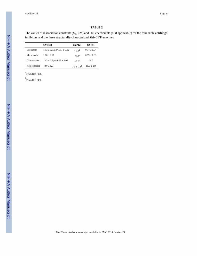
Binding of Antifungal Azole InhibitorsBinding of the azole antifungal drugs econazole, miconazole, clotrimazole, and ketoconazole(Fig. 1) to CYP130 was monitored via the type II shift of the heme Soret band caused bycoordination of the inhibitors to the heme iron atom. The Kd values for the inhibitors wereobtained from the spectral titration curves (Fig. S2) and are summarized in Table 2. Forcomparison, the Kd values for CYP121 obtained elsewhere and for CYP51 determined hereinare also listed. The sigmoid titration plots obtained for both econazole (Fig. 1A) andclotrimazole (Fig. 1B) were best fitted to the Hill equation (Eq. 3) with coefficients of 1.37 and1.93, respectively, indicating the presence of binding cooperativity. The titration curves forketoconazole (Fig. 1C) and miconazole (Fig. 1D) were fitted with the rectangular (Eq. 1) andthe quadratic (Eq. 2) hyperbolas, respectively. Collectively, the binding affinities of all theinhibitors are about an order of magnitude lower for CYP130 than for CYP121. Miconazole,clotrimazole, and ketoconazole also bind to CYP51 somewhat more tightly than to CYP130,whereas econazole has about the same binding affinities for both CYP130 and CYP51.
Cooperativity of P450-ligand BindingBinding cooperativity was observed for two (econazole and clotrimazole) of the four ligandsused in this study. Two potential sources of CYP130 binding cooperativity can be considered:(i) multiple site cooperativity, in which two or more ligands bind simultaneously to the sameprotein molecule, and (ii) multimer cooperativity, where protein-protein interactions arepromoted by the binding of a ligand to a molecule of the protein. Multiple site cooperativityhas been demonstrated by a variety of experimental techniques for a number of microsomal(CYP3A4, CYP1A2, CYP2C9) and bacterial (EryF) P450 enzymes (32,33). Simultaneousbinding of two ligands within the P450 active site has been validated by x-ray structures forEryF (34), CYP3A4 (35), and CYP158A2 (36). Although multimer binding cooperativity hasnot, to our knowledge, been reported for P450 systems, substrate-dependent P450-P450interactions have been shown to significantly influence individual functions of drug-metabolizing CYP2 and CYP3A4 enzymes (37–43), presumably by altering the rate ofassociation and/or affinity for the P450-reductase. These changes could reflect ligand-dependent dimerization or oligomerization of the P450 enzymes (44). Among the reportedP450 structures, the majority of mammalian CYP2 enzymes have been crystallized in dimeric(CYP2C9, CYP2C8, CYP2B4, CYP2R1, CYP3A4) or higher (CYP2A6, CYP2A13, CYP3A4)oligomerization states. Among the bacterial proteins, dimers with two-fold non-crystallographic symmetry have been detected for a P450 enzyme from Thermusthermophilus (PDB ID code: 1WIY) and for CYP154C1 from Streptomyces coelicolor (1GWI)(45).
Stoichiometry of CYP130-inhibitor BindingTo address the possibility that binding cooperativity may arise from the binding of multipleinhibitor molecules in the CYP130 active site, the stoichiometry for the CYP130/econazoleand CYP130/miconazole complexes was determined using the Job’s titration method (22),which is based on mixing of the reactants in such a way that their molar ratio varies, while thetotal molar concentration remains constant. The data for both econazole and miconazole werefit to a binary complex mechanism with the maximum of the bell-shaped plot located close tothe center corresponding to a molar fraction (defined as [CYP130]/([CYP130]+[inhibitor])) of~0.55, indicating virtually a 1:1 enzyme:inhibitor ratio (Fig. 2). This result suggests that thebinding cooperativity observed for econazole is unlikely due to simultaneous binding of twomolecules in the active site, which agrees with the x-ray structure data. This conclusion isconsistent with the fact that the structurally related miconazole, which binds CYP130 with thesame stoichiometry, does not exhibit binding cooperativity.
Ouellet et al. Page 7
J Biol Chem. Author manuscript; available in PMC 2010 October 21.
NIH
-PA Author Manuscript
NIH
-PA Author Manuscript
NIH
-PA Author Manuscript
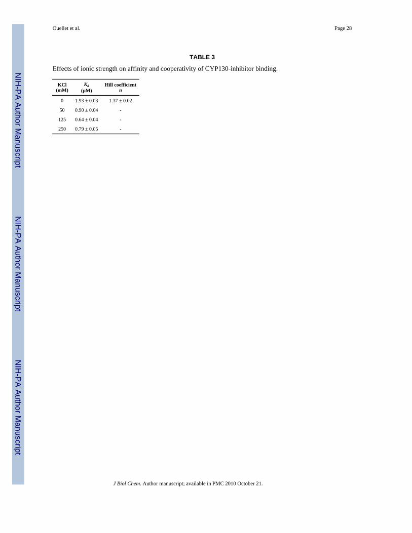
Protein-protein Interactions and Binding CooperativityTo examine a possible role for protein-protein interactions in the binding cooperativity ofeconazole and clotrimazole, a series of binding experiments was conducted in the presence ofincreasing concentrations of KCl (Table 3). The binding cooperativity of econazole wasabolished by 50 mM KCl. In the case of clotrimazole, the influence of ionic strength could notbe explored due to protein aggregation at even the lowest concentration of KCl, an effect similarto that observed for the CYP3A4/ketoconazole complex in the presence of apolar solvents andelevated ionic strength (35). These data support the inference that the binding cooperativity ofeconazole may arise from protein-protein interactions.
Overall Structure of CYP130The interaction of CYP130 with econazole was addressed by x-ray crystallography. Crystalstructures of CYP130 were determined for the ligand-free and econazole-bound forms.CYP130 has the characteristic fold common to all structurally defined cytochromes P450 butexhibits conformational and oligomerization differences between the ligand-free and -boundforms. Ligand-free CYP130 crystallized as a monomer in a relatively ‘open’ conformation(Fig. 3A), which is largely achieved by an extended conformation of the BC-loop unmaskinga route for substrate access. In the dimeric econazole-bound form, the BC-region (residues 80–91, colored pink in Fig. 3B, 4A) looses secondary structure and relocates as much as 18 Å togenerate multiple contacts with econazole, primarily with its two chlorinated phenyl moieties.At the same time, the F helix looses one helical turn while the G helix gains one helical turn,causing a drift of the connecting loop in the direction of the N-terminus along the primarysequence. Thus, the FG-loop in CYP130 is rather short (note that four amino acids are missingfrom the electron density in both the open and closed CYP130 forms (Fig. 3B, 4A)) and servesonly as a turn between two helices. Conformational mobility of the BC- and the FG-regionshas been previously observed in other structurally defined P450 enzymes and may serve toenable substrate access/product release to/from the active site.
Dimerization of CYP130 in the CrystalWe were unable to crystallize the CYP130/econazole complex under conditions that favorcrystallization of the open ligand-free form. Instead, a longer incubation under a different setof conditions was required to generate econazole-bound crystals that have a differentmorphology, unit cell dimensions, and diffract in a different space group (Table 1). The highsolvent content in these crystals (78%), indicating loose packing, partially explains the lowresolution of the diffraction data. Analysis of the econazole-bound structure and the crystalsymmetry revealed that the crystal lattice is largely stabilized by (i) formation of a CYP130dimer having two-fold rotation symmetry along the non-crystallographic axis which generatesa dimerization interface utilizing about 2000 Å2 (12.5%) of the surface of each monomer, and(ii) a two-fold crystallographic symmetry generating a dimer of dimers with 1280 Å2 of totalinterface (Fig. S3). The non-crystallographic dimerization interface involves the mostconformationally mobile P450 regions: the BC-loop, the F and G helices, and the N-terminalportion of the I-helix (Fig. 5). The interface is stabilized by partial overlap (about four helicalturns) between the I-helix N-termini and complete overlap between the G helices packed inanti-parallel orientations (Fig. 5A). Together with the F helices, they constitute two layers ofanti-parallel α-helices crossing each other at an angle of ~60°. A similar dimerization pattern,although with a smaller (600 Å2, 3.5% of the monomer surface) dimerization interface, isobserved in ligand-free CYP154C1 (45) (Fig. 5B). It is worth mentioning that the BC- and FG-regions are also involved in the dimerization of a bacterial P450 from T. thermophilus (PDBID code 1WIY).
Ouellet et al. Page 8
J Biol Chem. Author manuscript; available in PMC 2010 October 21.
NIH
-PA Author Manuscript
NIH
-PA Author Manuscript
NIH
-PA Author Manuscript

Dimerization of CYP130 in SolutionThe CYP130 dimer in the crystal is stabilized via a number of hydrophobic and H-bondinginteractions, while electrostatic interactions are involved in stabilizing the crystallographictetrameric interface. The stability of the dimer, if formed, is not sufficiently high to detect itby equilibrium techniques such as gel filtration chromatography or native gel electrophoresisat protein concentrations up to 100 µM. However, CYP130 oligomerization in solution wasdetectable by chemical cross-linking using glutaraldehyde (46). A substantial fraction ofCYP130 was found in dimeric/tetrameric forms at 20 µM concentration, whereas only marginaloligomerization was detected for two other soluble bacterial P450 enzymes, Mtb CYP51 andPikC from Streptomyces venezuelae, examined as controls (Fig. 6 A, B). No significant effectof inhibitors at up to a 500 µM concentration was observed, with the exception of a slightlyreduced content of the higher molecular weight aggregates for clotrimazole and ketoconazole(not shown).
When cross-linking was carried out with increasing KCl concentrations ranging up to 300 mM,the dimer product persisted unabated but the formation of tetramers and higher oligomers wassuppressed at the higher salt concentrations (Fig. 6 C). This is consistent with the observationthat dimer formation involves specific hydrophobic and H-bonding interactions, whereashigher oligomers are formed by relatively non-specific ionic ones. A small fraction of the dimermay be formed by such non-specific interactions, but the majority of the dimer does not involveionic contacts and persists in the presence of higher salt concentrations. Collectively, thecrystallographic and chemical cross-linking data suggest that the oligomerization of CYP130seen in the crystal can also occur in solution even in the absence of a ligand, with the closedform being susceptible to dimerization. If, as expected, CYP130 exists in an equilibriumbetween the open and closed forms that is shifted toward the closed form by econazole binding,the accumulation of the cross-linked products in the absence of azole ligand is readily explainedby irreversible removal of the closed form from the equilibrium mixture by the cross-linkingreaction.
Active Site of CYP130In the ligand-free form, the CYP130 heme iron is hexa-coordinated with a water moleculetightly bound as the distal axial ligand (bond distance 2.2 Å). This axial water is additionallystabilized via an H-bond to the carbonyl group of G243 (distance 2.7 Å) and an H-bondingnetwork formed by the cluster of water molecules bound in the proximity. To provide an H-bond to the axial water, the middle portion of the I-helix accommodating G243 closelyapproaches the porphyrin plane (distance 4.2 Å between the G243 carbonyl group and the hemeiron), imposing steric constraints on the binding of potential ligands in the active site and atthe same time preventing the axial water from premature release. None of a dozen differentcompounds of varied structure examined here was able to expel the axial water from theCYP130 active site to generate a high-spin heme iron state.
A hydrogen-bonding network of water molecules that also stabilizes the distal water axialligand is well defined in the crystal structure. This network leads from the distal water ligandalong the N-terminal portion of the I-helix to the surface of the molecule (Fig. 3A, 4B). A chainof seven well-structured water molecules is interrupted only once between the third and fourthmolecules, where the position of the missing water is taken by the hydroxyl group of I-helixresidue T239, located one helical turn away from G243 toward the N-terminus.
Econazole Binding SiteDespite the relatively low resolution (3.0 Å) of the econazole-bound crystal structure, theelectron density for econazole is unambiguously defined in each of the two monomers in theasymmetric unit (Fig. 7A). Econazole binds to CYP130 through a set of predominantly
Ouellet et al. Page 9
J Biol Chem. Author manuscript; available in PMC 2010 October 21.
NIH
-PA Author Manuscript
NIH
-PA Author Manuscript
NIH
-PA Author Manuscript
hydrophobic interactions in addition to the coordination bond (length ~ 2.75 Å) formed betweenthe heme iron and the lone pair of nitrogen electrons of the azole moiety. Econazole introducesa kink into the I-helix that displaces G243 by 2.3 Å from the hydrogen bonding position andreleases the axial water stabilized by this H-bond (Fig. 7B). The econazole binding modedeviates from the expected geometry (47), including the length of the coordination bond (ideal~2.1 Å) and the ~80° angle (ideal 90°) between the azole plane and the porphyrin macrocycle.These deviations are likely due to the steric constraints imposed by the I-helix, but are lesspronounced than those observed elsewhere for the CYP121/fluconazole complex (19). Givena weakened coordination bond and a larger volume of the active site cavity (accessible volume600 Å3, shown by mesh surface in Fig. 7C) than is required to accommodate econazole (330Å3) (Fig. 7C), alternative coordination mode(s) may arise to account for the temperaturedependent shift of the low-spin Soret band of the CYP130/econazole complex observed byspectroscopic analysis in solution (Fig. S1B). However, in contrast to the CYP121/fluconazolecomplex, no structured water molecules are observed in the vicinity (or in the active site ingeneral) at this resolution to allow us unambiguously conclude that the formation of a low-spin heme iron complex involving indirect iron-nitrogen coordination through a water molecule(19) can occur in econazole-bound CYP130.
The rest of the econazole molecule forms mainly hydrophobic contacts with the amino acidside chains of L71, T72, D85, P87, P88, M89, M91, F100, F236, T239, M240, T242, G243,G244, D246, T247, V290, Y392, and V393 situated within interaction distance (6 Å) fromeconazole (Fig. 7C). Notably, D85 is the only charged amino acid side chain in the active site,with the exception of the catalytic negative charge of D246 that is highly conserved throughoutthe CYP family. These two charged residues and the neutral N177 surround an accessible buteconazole-unoccupied extension of the otherwise hydrophobic active site cavity (Fig. 7C),suggesting that the enzyme may normally act upon a hydrophobic endogenous substrate,possibly larger than econazole that carries a positively charged functional group.
A stretch of the hydrophobic BC-loop residues (80–91) (highlighted in pink in Fig. 3B) isrelocated by up to 18 Å when econazole binds. One of the two consecutive proline residues,P87, residing in this region binds in the groove formed between the mono- and double-chlorinated econazole phenyl moieties (Fig. 7A, C). This interaction appears to be critical forpositioning of this portion of the BC-loop, which is directly involved in formation of theCYP130/econazole dimerization interface. The additional chlorine atom in miconazole isexpected to protrude toward P87, altering the local configuration of the BC-loop and, hence,the dimerization interface. Should such alterations occur, they may account for the lack ofbinding cooperativity observed with miconazole (Fig. 1C) and failure of the CYP130/miconazole complex to crystallize from >400 different crystallization conditions, includingthose which reproducibly generate ligand-free or econazole-bound crystals.
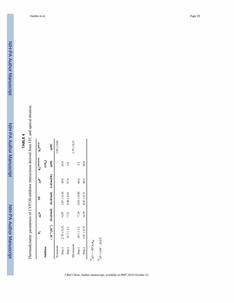
Thermodynamic Parameters of CYP130-Inhibitor Interactions by ITCBinding of econazole and miconazole to CYP130 was addressed by ITC to examine thethermodynamics of protein-inhibitor interactions independently of the accompanyingspectroscopic changes. Clotrimazole was excluded from the analysis because its low solubilityin aqueous solutions obviated data acquisition. The binding isotherms were obtained in thereverse titration mode (Fig. 8). A control titration of the protein into the buffer alone was alsoconducted and did not reveal any significant heat of dilution, confirming the monomeric stateof the protein in solution at the concentration employed (not shown). The data were best fittedto a sequential two-step binding model. The thermodynamic parameters derived from theanalysis are summarized in Table 4. Binding of both inhibitors is an endothermic and entropy-driven process, as evidenced by the large and positive ΔS values. Two sequential binding steps
Ouellet et al. Page 10
J Biol Chem. Author manuscript; available in PMC 2010 October 21.
NIH
-PA Author Manuscript
NIH
-PA Author Manuscript
NIH
-PA Author Manuscript
were deduced from the ITC data for both econazole and miconazole with one set of associationconstants close to those obtained from the optical titrations.
While the first step in econazole binding detected by ITC has a Kd (35.9 µM) much higher thanthat obtained from the optical titration (1.9 µM), the second step occurs with a Kd of 3.0 µMthat closely matches the spectroscopic dissociation constant, suggesting that full ‘Soretbinding’ is achieved during the second step. Apparently, the second step represents the energiesassociated with the conformational changes accompanying the primary ligand recognition inthe active site, presumably including protein dimerization. Collectively, the ITC andspectroscopic data indicate that these conformational changes/dimerization are beneficial tothe Soret status of econazole and concur with the assumption of multimer binding cooperativity.Unlike econazole, the first step of miconazole binding occurs with a Kd (5.3 µM) close to thatobtained from the optical titration (1.7 µM), suggesting that the majority of the Soret responseis achieved during the first step. Although the second step (Kd =28.8 µM) significantly impactsthe binding energetics (Table 4), it is virtually silent spectrally and, hence, undetectable by thespectroscopic techniques.
DISCUSSIONEconazole is an antifungal antibiotic with a potent activity against the latent and multidrug-resistant forms of tuberculosis (3–7). Mtb P450s, including CYP130, are therefore plausibletherapeutic targets for the azole class of antifungal agents. CYP130 binds a number ofantifungal drugs, including econazole, miconazole, clotrimazole, and ketoconazole with pooreraffinities than those observed for the other two characterized Mtb P450 enzymes, CYP121(17,48) and CYP51 (Table 2) (11,49). Nevertheless, the potential use of P450 enzymes astherapeutic targets in Mtb depends not only on the binding affinity toward currently availableazole drugs, but also on their biological roles.
The binding of azole inhibitors to CYP130 is an endothermic entropy-driven two-step processapparently complicated by protein-protein interactions manifested in the ligand-specificbinding cooperativity observed for econazole and clotrimazole (Fig. 1). While virtually fullSoret binding response for miconazole (the inhibitor lacking positive binding cooperativity) isachieved during the first binding step, econazole requires the second step to be completedbefore full spectral shift of the Soret band occurs (Table 4). We attribute the second bindingstep to the conformational changes associated with CYP130 dimerization. An apparent abilityof CYP130 to dimerize in solution is supported by covalent cross-linking of the protein in thepresence or absence of azole ligands (Fig. 6). The crystal structure indicates that dimerizationis likely to involve the closed form of the protein favored by econazole binding (Fig. 5A).However, in the absence of a ligand, the equilibrium distribution of accessible proteinconformers can be shifted towards the closed form as the cross-linked dimer is formed and isthus removed from the equilibrium.
It is impossible at this stage to predict whether protein-protein interactions play anyphysiological role in modulating the functional activities of CYP130 or other known bacterialP450 enzymes, e.g., via alteration of dimer affinity for an electron donor partner or othermechanisms. Both the native substrate(s) and an electron donor for CYP130, as for the majorityof bacterial P450 enzymes, remain unknown. A similarity of dimerization patterns for twounrelated bacterial P450 proteins, CYP130 and CYP154C1 (Fig. 5), suggests that theassociation between two monomers may not be random. In this regard, P450-P450 interactionshave been reported to modulate the catalytic activities of drug metabolizing mammalianmicrosomal P450 enzymes, although the occurrence and physiological significance of suchinteractions in intact cellular membranes remains to be confirmed. Nevertheless, a precedent
Ouellet et al. Page 11
J Biol Chem. Author manuscript; available in PMC 2010 October 21.
NIH
-PA Author Manuscript
NIH
-PA Author Manuscript
NIH
-PA Author Manuscript
for an inherent dimerization propensity among P450 enzymes is relevant to our understandingof P450-drug- and drug-drug interactions.
The binding of econazole was addressed in more detail by crystallographic studies. The positionof econazole in the active site of CYP130 exhibits notable deviations from the ideal geometrythat result from steric constraints imposed by the I-helix analogous to those observed for theCYP121/fluconazole complex (19). In addition, the volume of the active site cavity is largerthan is required to accommodate econazole and provides room for possible alternative ligationmode(s) to the heme iron, such as that in which a water molecule is placed between the ironand the azole nitrogen. The less than perfect protein/inhibitor fit presumably contributes to theattenuated binding affinity of the complex. For instance, the affinity of the CYP121/fluconazolecomplex (10 µM) with the strongest observed perturbations of the triazole-heme ironcoordination geometry (19) is 50- and 5-fold reduced compared to that of the CYP121/econazole (17) and CYP130/econazole complexes, respectively. Therefore, a better fit betweenthe compound and the spatial and chemical features of the P450 active site would yield strongerinhibitors. In this regard, a portion of the active site cavity surrounded by the charged and/orhydrophilic residues D85, D246, and N177 (Fig. 7C), contrasts with the almost exclusivelyhydrophobic environment of the rest of the CYP130 active site. This pocket could serve as alandmark for substrate (or inhibitor) recognition, similar to that observed in the macrolidemonooxygenase PikC, where a salt-bridge formed between the positively charged tertiaryamino group of the macrolide substrate and a negatively charged carboxylic amino acid residueis essential to achieve catalytically competent binding (50).
The high resolution (1.46 Å) of the ligand-free structure has allowed us to define a hydrogen-bonded network that includes seven water molecules and the hydroxyl group of I-helix residueT239. These water molecules are evenly distributed and spaced by H-bond distances along theN-terminal portion of the I-helix leading from the distal water ligand to the molecular surface(Fig. 3A, 4B). The involvement of the I-helix in this hydrogen bonded network stabilizes thedistal water ligand and suggests that the movement of the I-helix N-terminus that transientlyexposes the active site for substrate access may, at the same time, facilitate displacement ofthe distal water, providing yet another level of regulation of CYP130 catalysis. Therefore, theN-terminal portion of the I-helix may mediate coupling of (i) the binding of substrate possiblyassisted by protein dimerization, and (ii) the release of the axial water ligand. This couplingmay be part of a regulatory mechanism aimed at preventing unproductive oxygen binding underlimited access to nutrients such as oxygen (e.g., in granulomas, avascular environments wheredormant infectious tubercle bacilli adapt for long-term asymptomatic survival). The reluctanceof CYP130 to release the water axial ligand and thus to be reduced and bind molecular oxygenin response to a small range of potential substrates examined in this study is consistent withthis assumption.
In summary, we report expression, purification, biophysical characterization, andcrystallization of CYP130 in its ligand-free and econazole-bound forms. The crystal structureof the econazole-bound CYP130 is the first of a P450/econazole complex. Econazole bindingin the active site involves conformational selection mediated by direct coordination to the hemeiron and largely hydrophobic contacts with the active site amino acid residues. The interactionsbetween CYP130, econazole, and other potent azole antifungal drugs were characterized insome detail by UV-visible spectroscopy, ITC, and chemical cross-linking. Overall, binding ofazole inhibitors is a complex entropy-driven two-step process that appears to be assisted foreconazole and clotrimazole by protein-protein interactions resulting from a propensity of theclosed form of CYP130 to dimerize both in solution and in the crystal, providing evidence insupport of a possible role for P450-P450 interactions in biology.
Ouellet et al. Page 12
J Biol Chem. Author manuscript; available in PMC 2010 October 21.
NIH
-PA Author Manuscript
NIH
-PA Author Manuscript
NIH
-PA Author Manuscript

Supplementary MaterialRefer to Web version on PubMed Central for supplementary material.
AcknowledgmentsWe acknowledge Dr. Chris Waddling for his assistance with the software and instrumentation in the UCSF X-RayFacility, Dr. Vladimir N. Podust for mass spectrometric analysis of the Se-methionine CYP130 derivative, Dr.Youngchang Kim for fruitful discussions and valuable contributions, and Marco Moschini for excellent technicalassistance.
REFERENCES1. Zhang Y. Annu. Rev. Pharmacol. Toxicol 2005;45:529–564. [PubMed: 15822188]2. Sheehan DJ, Hitchcoch CA, Sibley CM. Clin. Microbiol. Rev 1999;12:40–79. [PubMed: 9880474]3. Ahmad Z, Sharma S, Khuller GK. FEMS Microbiology Letters 2005;251:19–22. [PubMed: 16143463]4. Ahmad Z, Sharma S, Khuller GK, Singh P, Faujdar J, Katoch VM. Int. J. Antimicrob. Agents
2006;28:543–544. [PubMed: 17101262]5. Ahmad Z, Sharma S, Khuller GK. FEMS Microbiology Letters 2006;261:181–186. [PubMed:
16907718]6. Ahmad Z, Sharma S, Khuller GK. FEMS Microbiology Letters 2006;258:200–203. [PubMed:
16640573]7. Banfi E, Scialino G, Zampieri D, Mamolo MG, Vio L, Ferrone M, Fermeglia M, Paneni MS, Pricl S.
J. Antimicrob. Chemother 2006;58:76–84. [PubMed: 16709593]8. Cole ST, Brosch R, Parkhill J, Garnier T, Churcher C, Harris D, Gordon SV, Eiglmeier K, Gas S, Barry
CE 3rd, Tekaia F, Badcock K, Basham D, Brown D, Chillingworth T, Connor R, Davies R, Devlin K,Feltwell T, Gentles S, Hamlin N, Holroyd S, Hornsby T, Jagels K, Krogh A, McLean J, Moule S,Murphy L, Oliver K, Osborne J, Quail MA, Rajandream MA, Rogers J, Rutter S, Seeger K, SkeltonJ, Squares R, Squares S, Sulston JE, Taylor K, Whitehead S, Barrell BG. Nature 1998;393:537–544.[PubMed: 9634230]
9. Van der Geize R, Yam K, Heuser T, Wilbrink MH, Hara H, Anderton MC, Sim E, Dijkhuizen L, DaviesJE, Mohn WW, Eltis LD. Proc. Natl. Acad. Sci. U S A 2007;104:1947–1952. [PubMed: 17264217]
10. Recchi C, Sclavi B, Rauzier J, Gicquel B, Reyrat JM. J. Biol. Chem 2003;278:33763–33773.[PubMed: 12826660]
11. Bellamine A, Mangla AT, Nes WD, Waterman MR. Proc. Natl. Acad. Sci. U S A 1999;96:8937–8942. [PubMed: 10430874]
12. Gatfield J, Pieters J. Science 2000;288:1647–1650. [PubMed: 10834844]13. McLean KJ, Dunford AJ, Neeli R, Driscoll MD, Munro AW. Arch. Biochem. Biophys 2007;464:228–
240. [PubMed: 17482138]14. Ainsa JA, Blokpoel MC, Otal I, Young DB, De Smet KA, Martin C. J. Bacteriol 1998;180:5836–
5843. [PubMed: 9811639]15. De Rossi E, Ainsa JA, Riccardi G. FEMS Microbiol. Rev 2006;30:36–52. [PubMed: 16438679]16. Siddiqi N, Das R, Pathak N, Banerjee S, Ahmed N, Katoch VM, Hasnain SE. Infection 2004;32:109–
111. [PubMed: 15057575]17. McLean KJ, Marshall KR, Richmond A, Hunter IS, Fowler K, Kieser T, Gurcha SS, Besra GS, Munro
AW. Microbiology 2002;148:2937–2949. [PubMed: 12368427]18. Podust LM, Poulos TL, Waterman MR. Proc. Natl. Acad. Sci. USA 2001;98:3068–3073. [PubMed:
11248033]19. Seward HE, Roujeinikova A, McLean KJ, Munro AW, Leys D. J. Biol. Chem 2006;281:39437–
39443. [PubMed: 17028183]20. Barnes HJ, Arlotto MP, Waterman MR. Proc. Natl. Acad. Sci. U S A 1991;88:5597–5601. [PubMed:
1829523]21. Omura T, Sato R. J. Biol. Chem 1964;239:2379–2385. [PubMed: 14209972]22. Job P. Ann. Chim 1928;9:113–203.
Ouellet et al. Page 13
J Biol Chem. Author manuscript; available in PMC 2010 October 21.
NIH
-PA Author Manuscript
NIH
-PA Author Manuscript
NIH
-PA Author Manuscript
23. Otwinowski Z, Minor W. Methods Enzymol 1997;276:307–326.24. Brunger AT, Adams PD, Clore GM, Delano WL, Gros P, Grosse-Kunstleve RW, Jiang J-S, Kuszewski
J, Nilges M, Pannu NS. Acta Crystallogr 1998;D54:905–921.25. Perrakis A, Morris R, Lamzin VS. Nat. Struct. Biol 1999;6:458–463. [PubMed: 10331874]26. Vagin AA, Steiner RA, Lebedev AA, Potterton L, McNicholas S, Long F, Murshudov GN. Acta
Crystallogr. D Biol. Crystallogr 2004;60:2184–2195. [PubMed: 15572771]27. Emsley P, Cowtan K. Acta Crystallogr. D Biol. Crystallogr 2004;60:2126–2132. [PubMed:
15572765]28. Murshudov GN, Vagin AA, Dodson EJ. Acta Crystallogr. D Biol. Crystallogr 1997;53:240–255.
[PubMed: 15299926]29. Vagin A, Teplyakov A. J. Appl. Crystallogr 1997;30:1022–1025.30. Jones TA, Zou JY, Cowan SW, Kjeldgaard M. Acta Crysallogr 1991;A47:110–119.31. Laskowski RA, MacArthur MW, Moss DS, Thornton JM. J. Appl. Crystallogr 1993;26:283–291.32. Xiang H, Tschirret-Guth RA, Ortiz De Montellano PR. J. Biol. Chem 2000;275:35999–36006.
[PubMed: 10956654]33. Yoon MY, Campbell AP, Atkins WM. Drug Metab. Rev 2004;36:219–230. [PubMed: 15237852]34. Cupp-Vickery J, Anderson R, Hatziris Z. Proc. Natl. Acad. Sci. U S A 2000;97:3050–3055. [PubMed:
10716705]35. Ekroos M, Sjogren T. Proc. Natl. Acad. Sci. U S A 2006;103:13682–13687. [PubMed: 16954191]36. Zhao B, Guengerich FP, Bellamine A, Lamb DC, Izumikawa M, Lei L, Podust LM, Sundaramoorthy
M, Kalaitzis JA, Reddy LM, Kelly SL, Moore BS, Stec D, Voehler M, Falck JR, Shimada T,Waterman MR. J. Biol. Chem 2005;280:11599–11607. [PubMed: 15659395]
37. Kaminsky LS, Guengerich FP. Eur. J. Biochem 1985;149:479–489. [PubMed: 3924614]38. Backes WL, Eyer CS. J. Biol. Chem 1989;264:6252–5259. [PubMed: 2495281]39. Cawley GF, Batie CJ, Backes WL. Biochemistry 1995;34:1244–1247. [PubMed: 7827074]40. Cawley GF, Zhang S, Kelley RW, Backes WL. Drug. Metab. Dispos 2001;29:1529–1534. [PubMed:
11717170]41. Backes WL, Batie CJ, Cawley GF. Biochemistry 1998;37:12852–12859. [PubMed: 9737863]42. Hazai E, Kupfer D. Drug. Metab. Dispos 2005;33:157–164. [PubMed: 15486075]43. Jamakhandi AP, Kuzmic P, Sanders DE, Miller GP. Biochemistry 2007;46:10192–10201. [PubMed:
17685587]44. Hazai E, Bikadi Z, Simonyi M, Kupfer D. J. Comput. Aided Mol. Des 2005;19:271–285. [PubMed:
16163453]45. Podust LM, Kim Y, Arase M, Neely BA, Beck BJ, Bach H, Sherman DH, Lamb DC, Kelly SL,
Waterman MR. J. Biol. Chem 2003;278:12214–12221. [PubMed: 12519772]46. Migneault I, Dartiguenave C, Bertrand MJ, Waldron KC. Biotechniques 2004;37:790–802. [PubMed:
15560135]47. Patchkovskii S, Ziegler T. Inorg. Chem 2000;39:5354–5364. [PubMed: 11154592]48. McLean KJ, Cheesman MR, Rivers SL, Richmond A, Leys D, Chapman SK, Reid GA, Price NC,
Kelly SM, Clarkson J, Smith WE, Munro AW. J. Inorg. Biochem 2002;91:527–541. [PubMed:12237220]
49. Guardiola-Diaz HM, Foster LA, Mushrush D, Vaz AD. Biochem. Pharmacol 2001;61:1463–1470.[PubMed: 11377375]
50. Sherman DH, Li S, Yermalitskaya LV, Kim Y, Smith JA, Waterman MR, Podust LM. J. Biol. Chem2006;281:26289–26297. [PubMed: 16825192]
51. Humphrey W, Dalke A, Schulten K. J. Mol. Graph 1996;14:33–38. [PubMed: 8744570]52. Evans SV. J. Mol. Graphics 1993;11:134–138.53. Pettersen EF, Goddard TD, Huang CC, Couch GS, Greenblatt DM, Meng EC, Ferrin TE. J. Comput.
Chem 2004;25:1605–1612. [PubMed: 15264254]
Ouellet et al. Page 14
J Biol Chem. Author manuscript; available in PMC 2010 October 21.
NIH
-PA Author Manuscript
NIH
-PA Author Manuscript
NIH
-PA Author Manuscript

Fig. 1. Binding of antifungal azole drugs to CYP130The concentration dependence of econazole (A), clotrimazole (B), miconazole (C) andketoconazole (D) binding deduced from the difference absorption changes obtained from thetitration of CYP130 (2.5 µM) with increasing concentrations of the inhibitor. The structure ofthe inhibitor is shown in each panel.
Ouellet et al. Page 15
J Biol Chem. Author manuscript; available in PMC 2010 October 21.
NIH
-PA Author Manuscript
NIH
-PA Author Manuscript
NIH
-PA Author Manuscript

Fig. 2. Stoichiometry of CYP130-inhibitor bindingThe bell-shaped Job plots at a total protein and inhibitor concentration of 15 µM display amaximum close to a mole fraction of 0.55, the value that corresponds to a 1:1 ratio for thebinding to CYP130 of econazole (open circles) and miconazole (closed circles).
Ouellet et al. Page 16
J Biol Chem. Author manuscript; available in PMC 2010 October 21.
NIH
-PA Author Manuscript
NIH
-PA Author Manuscript
NIH
-PA Author Manuscript
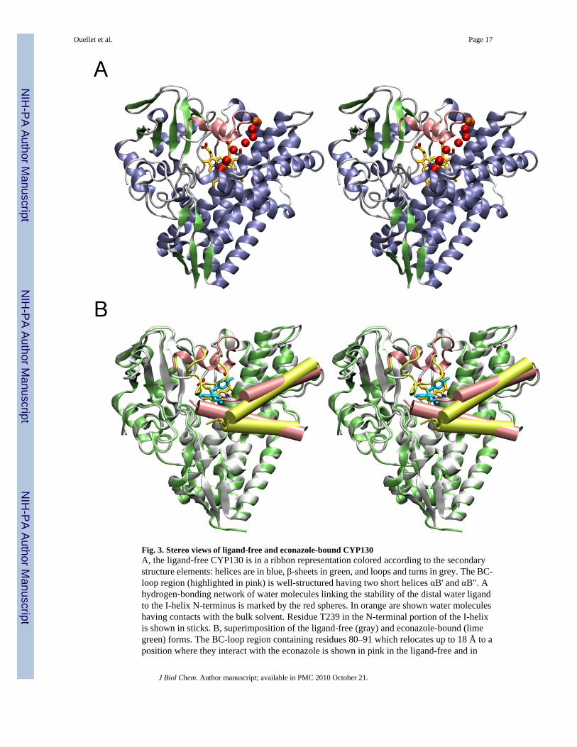
Fig. 3. Stereo views of ligand-free and econazole-bound CYP130A, the ligand-free CYP130 is in a ribbon representation colored according to the secondarystructure elements: helices are in blue, β-sheets in green, and loops and turns in grey. The BC-loop region (highlighted in pink) is well-structured having two short helices αB' and αB". Ahydrogen-bonding network of water molecules linking the stability of the distal water ligandto the I-helix N-terminus is marked by the red spheres. In orange are shown water moleculeshaving contacts with the bulk solvent. Residue T239 in the N-terminal portion of the I-helixis shown in sticks. B, superimposition of the ligand-free (gray) and econazole-bound (limegreen) forms. The BC-loop region containing residues 80–91 which relocates up to 18 Å to aposition where they interact with the econazole is shown in pink in the ligand-free and in
Ouellet et al. Page 17
J Biol Chem. Author manuscript; available in PMC 2010 October 21.
NIH
-PA Author Manuscript
NIH
-PA Author Manuscript
NIH
-PA Author Manuscript

yellow-green in the econazole-bound forms. The F and G helices are shown as pink cylindersin the ligand-free and yellow-green cylinders in econazole bound forms. The G helix is on top.Econazole is in cyan and heme is in yellow. Images were generated using VMD software(51) unless specified otherwise.
Ouellet et al. Page 18
J Biol Chem. Author manuscript; available in PMC 2010 October 21.
NIH
-PA Author Manuscript
NIH
-PA Author Manuscript
NIH
-PA Author Manuscript

Fig. 4. CYP130 active siteA, major conformational differences between the ligand-free (gray) and econazole–bound(green) states. The BC-region is in pink, heme is in yellow and econazole is in cyan. Gaps inthe protein chain between the F and G helices due to the missing electron density are markedwith the open circles. Relocation distances for selected structural elements are given inAngstroms. B, H-bonding network. The fragment of the ligand-free crystal structure showingthe water molecules (red spheres) that stabilize the distal water in CYP130. Water moleculeshaving contacts with the bulk solvent are colored in orange. Distances between oxygen atomcenters are in Angstroms. T239 is shown in sticks. The iron axial water ligand is labeled witha capital L.
Ouellet et al. Page 19
J Biol Chem. Author manuscript; available in PMC 2010 October 21.
NIH
-PA Author Manuscript
NIH
-PA Author Manuscript
NIH
-PA Author Manuscript
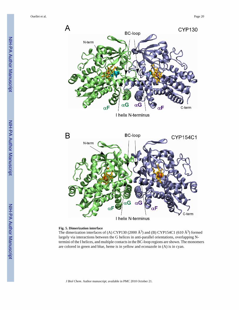
Fig. 5. Dimerization interfaceThe dimerization interfaces of (A) CYP130 (2000 Å3) and (B) CYP154C1 (610 Å3) formedlargely via interactions between the G helices in anti-parallel orientations, overlapping N-termini of the I helices, and multiple contacts in the BC-loop regions are shown. The monomersare colored in green and blue, heme is in yellow and econazole in (A) is in cyan.
Ouellet et al. Page 20
J Biol Chem. Author manuscript; available in PMC 2010 October 21.
NIH
-PA Author Manuscript
NIH
-PA Author Manuscript
NIH
-PA Author Manuscript
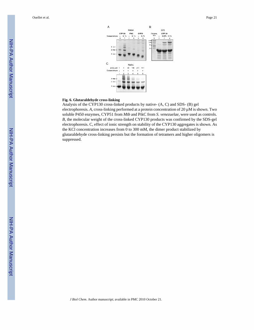
Fig. 6. Glutaraldehyde cross-linkingAnalysis of the CYP130 cross-linked products by native- (A, C) and SDS- (B) gelelectrophoresis. A, cross-linking performed at a protein concentration of 20 µM is shown. Twosoluble P450 enzymes, CYP51 from Mtb and PikC from S. venezuelae, were used as controls.B, the molecular weight of the cross-linked CYP130 products was confirmed by the SDS-gelelectrophoresis. C, effect of ionic strength on stability of the CYP130 aggregates is shown. Asthe KCl concentration increases from 0 to 300 mM, the dimer product stabilized byglutaraldehyde cross-linking persists but the formation of tetramers and higher oligomers issuppressed.
Ouellet et al. Page 21
J Biol Chem. Author manuscript; available in PMC 2010 October 21.
NIH
-PA Author Manuscript
NIH
-PA Author Manuscript
NIH
-PA Author Manuscript

Fig. 7. Econazole binding in the active siteA, stereo view of econazole (yellow-green) bound in the active site of CYP130 is shown. Clatoms are colored in green, N atoms in blue and O atoms in red. Amino acid residues within4 Å of econazole are labeled in black. Fragments of the 2Fo−Fc electron density compositeomit map contoured at 1.0 σ are in blue. To avoid excessive cluttering, heme was excludedfrom the map calculation and T242 was excluded from the view as projecting on top ofeconazole. The image was generated using the SETOR program (52). B, a kink of the I-helixintroduced by the binding of econazole is shown. The ligand-free (gray) and econazole-bound(green) CYP130 structures were superimposed with an r.m.s.d. of 0.93 Å2 for all the proteinresidues. Iron (ochre) and oxygen (red) atoms are shown as spheres. The iron axial water ligand
Ouellet et al. Page 22
J Biol Chem. Author manuscript; available in PMC 2010 October 21.
NIH
-PA Author Manuscript
NIH
-PA Author Manuscript
NIH
-PA Author Manuscript
is labeled with a capital L. The arrows show the directions of movements upon transition fromthe ligand-free to the econazole-bound state. C, alignment of the BC-loop fragment (85–91)(grey) in the groove formed between the mono- and double-chlorinated phenyl rings ofeconazole is shown. The additional chlorination site in miconazole is indicated by an arrow.The solid surface represents the van der Waals surface of econazole (volume of 330 Å3) andis colored according to the underlying atoms: oxygen in red, nitrogen in blue, chlorine in green.The mesh surface shows the accessible space in the active site (600 Å3). A fragment of the I-helix (237–247) is represented by the grey ribbon. The heme is in orange. This image wasgenerated using the program CHIMERA (53).
Ouellet et al. Page 23
J Biol Chem. Author manuscript; available in PMC 2010 October 21.
NIH
-PA Author Manuscript
NIH
-PA Author Manuscript
NIH
-PA Author Manuscript
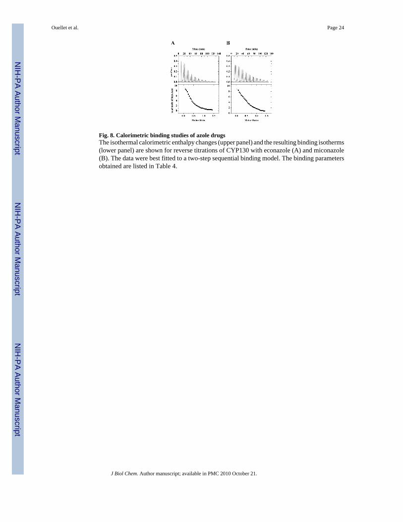
Fig. 8. Calorimetric binding studies of azole drugsThe isothermal calorimetric enthalpy changes (upper panel) and the resulting binding isotherms(lower panel) are shown for reverse titrations of CYP130 with econazole (A) and miconazole(B). The data were best fitted to a two-step sequential binding model. The binding parametersobtained are listed in Table 4.
Ouellet et al. Page 24
J Biol Chem. Author manuscript; available in PMC 2010 October 21.
NIH
-PA Author Manuscript
NIH
-PA Author Manuscript
NIH
-PA Author Manuscript

NIH
-PA Author Manuscript
NIH
-PA Author Manuscript
NIH
-PA Author Manuscript
Ouellet et al. Page 25
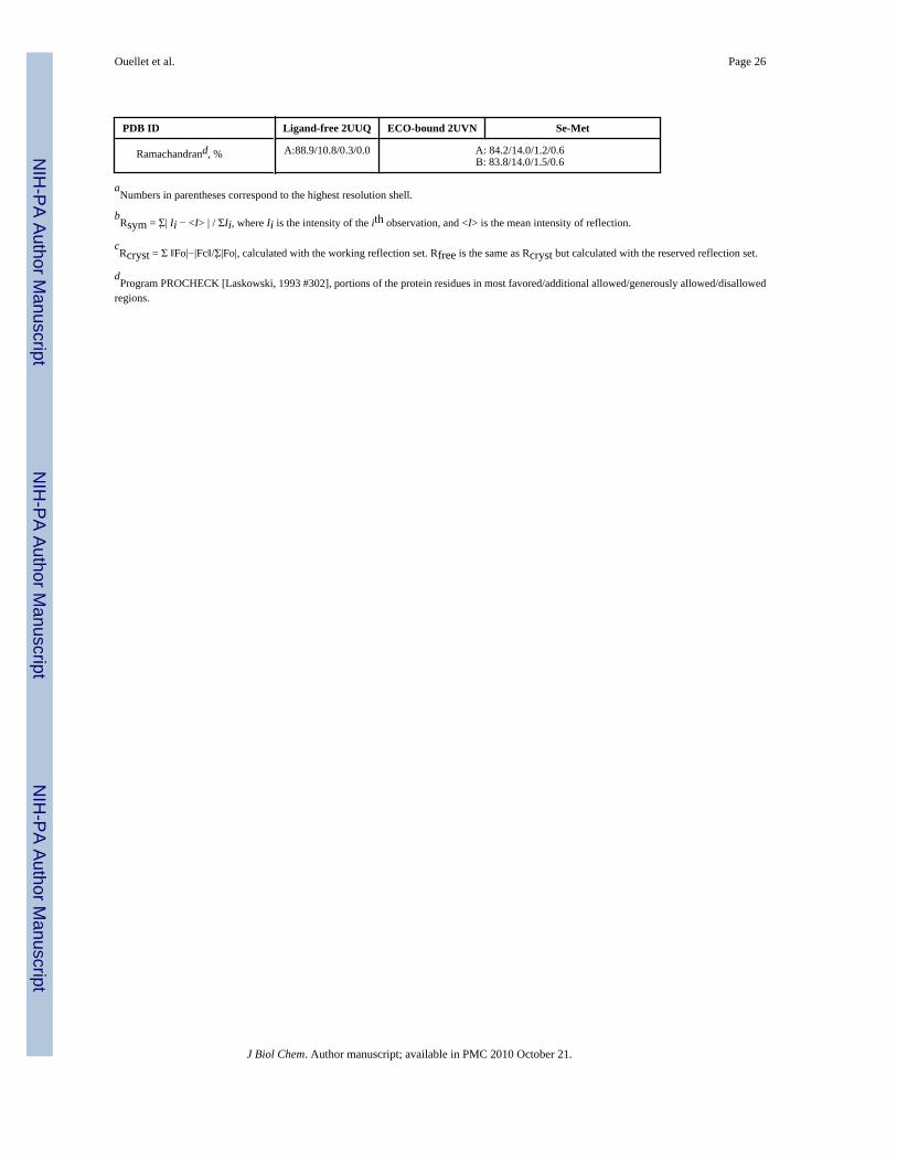
Table 1
Data collection and refinement statistics
PDB ID Ligand-free 2UUQ ECO-bound 2UVN Se-Met
Data Collection
Resolution, Å 1.52 3.0 1.75 1.75
Wavelength, Å 0.97926 1.11588 0.97970 0.96863
Space group C2 P3(2)21 C2 C2
Cell dimensionsa, b, c, Åa, b, g, °
160.0, 53.9, 43.790.0, 96.5, 90.0
131.7, 131.7, 229.490.0, 90.0, 120.0
160.3, 53.9,43.890.0, 96.6, 90.0
160.3, 53.9,43.890.0, 96.6, 90.0
Molecules in an asymmetric unit 1 2 1 1
Solvent content, % 40 78 40 40
Rsyma,b, % 4.7 (31.3) 8.1 (51.2) 4.4 (45.8) 4.4 (43.6)
I/s 21.8 (2.0) 29.9 (4.2) 26.0 (2.1) 26.2 (2.3)
Unique reflections 52466 45935 71697 70994
Completeness, % 91.6 (56.9) 100.0 (100.0) 97.5 (92.4) 97.7 (94.0)
Redundancy 4.1 (2.2) 9.8 (9.6) 3.9 (3.5) 3.9 (3.6)
Phasing
Resolution range, Å 43.5-2.2
Number of used sites 9
Phasing power 2.5
Figure of merit 0.64
After density modification 0.95
Refinement
Reflections used 44721 41548
Rcryst/Rfreec, % 18.7/23.1 20.0/23.4
No. atoms
Protein 3079 6120
Heme 43 86
Ligand N/A 48
Water/Ion 289/9 77/34
Wilson plot B-values, Å2 18.6 N/A
Mean B-factors, Å2 22.1 64.6
Protein 21.4 A: 64.4/ B: 65.4
Heme 12.5 A: 48.2/B: 60.5
Ligand N/A A: 37.7/B: 53.4
Water 31.2 43.7
r.m.s. diviations
Bond length, Å 0.010 0.008
Bond angles, ° 1.28 1.30
J Biol Chem. Author manuscript; available in PMC 2010 October 21.
NIH
-PA Author Manuscript
NIH
-PA Author Manuscript
NIH
-PA Author Manuscript
Ouellet et al. Page 26
PDB ID Ligand-free 2UUQ ECO-bound 2UVN Se-Met
Ramachandrand, % A:88.9/10.8/0.3/0.0 A: 84.2/14.0/1.2/0.6B: 83.8/14.0/1.5/0.6
aNumbers in parentheses correspond to the highest resolution shell.
bRsym = Σ̣| Ii − <I> | / ΣIi, where Ii is the intensity of the ith observation, and <I> is the mean intensity of reflection.
cRcryst = Σ ‖Fo|−|Fc‖/Σ̣|Fo|, calculated with the working reflection set. Rfree is the same as Rcryst but calculated with the reserved reflection set.
dProgram PROCHECK [Laskowski, 1993 #302], portions of the protein residues in most favored/additional allowed/generously allowed/disallowed
regions.
J Biol Chem. Author manuscript; available in PMC 2010 October 21.
NIH
-PA Author Manuscript
NIH
-PA Author Manuscript
NIH
-PA Author Manuscript
Ouellet et al. Page 27
TABLE 2
The values of dissociation constants (Kd, µM) and Hill coefficients (n, if applicable) for the four azole antifungalinhibitors and the three structurally-characterized Mtb CYP enzymes.
CYP130 CYP121 CYP51
Econazole 1.93 ± 0.03; n=1.37 ± 0.02 <0.2a 0.77 ± 0.04
Miconazole 1.70 ± 0.21 <0.2a 0.59 ± 0.03
Clotrimazole 13.3 ± 0.6; n=1.95 ± 0.05 <0.2a <1.0
Ketoconazole 48.0 ± 1.5 3.3 ± 0.3b 19.0 ± 1.9
aFrom Ref. (17).
bFrom Ref. (48).
J Biol Chem. Author manuscript; available in PMC 2010 October 21.
NIH
-PA Author Manuscript
NIH
-PA Author Manuscript
NIH
-PA Author Manuscript
Ouellet et al. Page 28
TABLE 3
Effects of ionic strength on affinity and cooperativity of CYP130-inhibitor binding.
KCl(mM)
Kd(µM)
Hill coefficientn
0 1.93 ± 0.03 1.37 ± 0.02
50 0.90 ± 0.04 -
125 0.64 ± 0.04 -
250 0.79 ± 0.05 -
J Biol Chem. Author manuscript; available in PMC 2010 October 21.
NIH
-PA Author Manuscript
NIH
-PA Author Manuscript
NIH
-PA Author Manuscript
Ouellet et al. Page 29
TAB
LE 4
Ther
mod
ynam
ic p
aram
eter
s of C
YP1
30-in
hibi
tor i
nter
actio
ns d
eriv
ed fr
om IT
C a
nd o
ptic
al ti
tratio
n.
K aΔG
aΔH
ΔSb
K dca
lorim
etry
K dop
tical
Inhi
bito
r(1
/Ka)
× 10
−4 (M
−1)
(kca
l/mol
)(k
cal/m
ol)
(cal
/mol
/K)
(µM
)(µ
M)
Econ
azol
e1.
93 ±
0.0
3
S
tep-
12.
78 ±
0.2
5−6
.07
2.87
± 0
.34
30.0
35.9
S
tep-
232
.7 ±
3.5
−7.5
19.
48 ±
0.6
157
.03.
0
Mic
onaz
ole
1.70
± 0
.21
S
tep-
118
.7 ±
1.2
−7.1
86.
05 ±
0.0
944
.45.
3
S
tep-
23.
47 ±
0.3
7−6
.19
8.47
± 0
.71
49.2
28.8
a ΔG
= −
RT ln
Ka.
b ΔS
= (Δ
H −
ΔG
)/T.
J Biol Chem. Author manuscript; available in PMC 2010 October 21.
Related Documents


