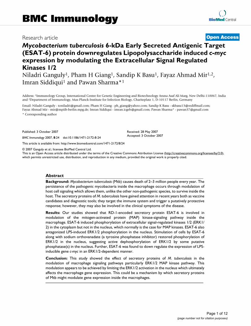
BioMed Central Page 1 of 12 (page number not for citation purposes) BMC Immunology Open Access Research article Mycobacterium tuberculosis 6-kDa Early Secreted Antigenic Target (ESAT-6) protein downregulates Lipopolysaccharide induced c-myc expression by modulating the Extracellular Signal Regulated Kinases 1/2 Niladri Ganguly 1 , Pham H Giang 1 , Sandip K Basu 1 , Fayaz Ahmad Mir 1,2 , Imran Siddiqui 1 and Pawan Sharma* 1 Address: 1 Immunology Group, International Centre for Genetic Engineering and Biotechnology Aruna Asaf Ali Marg, New Delhi-110067, India and 2 Department of Immunology, Max-Planck-Institute for Infection Biology, Chariteplatz 1, D-10117 Berlin, Germany Email: Niladri Ganguly - [email protected]; Pham H Giang - [email protected]; Sandip K Basu - [email protected]; Fayaz Ahmad Mir - [email protected]; Imran Siddiqui - [email protected]; Pawan Sharma* - [email protected] * Corresponding author Abstract Background: Mycobacterium tuberculosis (Mtb) causes death of 2–3 million people every year. The persistence of the pathogenic mycobacteria inside the macrophage occurs through modulation of host cell signaling which allows them, unlike the other non-pathogenic species, to survive inside the host. The secretory proteins of M. tuberculosis have gained attention in recent years both as vaccine candidates and diagnostic tools; they target the immune system and trigger a putatively protective response; however, they may also be involved in the clinical symptoms of the disease. Results: Our studies showed that RD-1-encoded secretory protein ESAT-6 is involved in modulation of the mitogen-activated protein (MAP) kinase-signaling pathway inside the macrophage. ESAT-6 induced phosphorylation of extracellular signal-regulated kinases 1/2 (ERK1/ 2) in the cytoplasm but not in the nucleus, which normally is the case for MAP kinases. ESAT-6 also antagonized LPS-induced ERK1/2 phosphorylation in the nucleus. Stimulation of cells by ESAT-6 along with sodium orthovanadate (a tyrosine phosphatase inhibitor) restored phosphorylation of ERK1/2 in the nucleus, suggesting active dephosphorylation of ERK1/2 by some putative phosphatase(s) in the nucleus. Further, ESAT-6 was found to down regulate the expression of LPS- inducible gene c-myc in an ERK1/2-dependent manner. Conclusion: This study showed the effect of secretory proteins of M. tuberculosis in the modulation of macrophage signaling pathways particularly ERK1/2 MAP kinase pathway. This modulation appears to be achieved by limiting the ERK1/2 activation in the nucleus which ultimately affects the macrophage gene expression. This could be a mechanism by which secretory proteins of Mtb might modulate gene expression inside the macrophages. Published: 3 October 2007 BMC Immunology 2007, 8:24 doi:10.1186/1471-2172-8-24 Received: 28 May 2007 Accepted: 3 October 2007 This article is available from: http://www.biomedcentral.com/1471-2172/8/24 © 2007 Ganguly et al.; licensee BioMed Central Ltd. This is an Open Access article distributed under the terms of the Creative Commons Attribution License (http://creativecommons.org/licenses/by/2.0 ), which permits unrestricted use, distribution, and reproduction in any medium, provided the original work is properly cited.

Welcome message from author
This document is posted to help you gain knowledge. Please leave a comment to let me know what you think about it! Share it to your friends and learn new things together.
Transcript
BioMed CentralBMC Immunology
ss
Open AcceResearch articleMycobacterium tuberculosis 6-kDa Early Secreted Antigenic Target (ESAT-6) protein downregulates Lipopolysaccharide induced c-myc expression by modulating the Extracellular Signal Regulated Kinases 1/2Niladri Ganguly1, Pham H Giang1, Sandip K Basu1, Fayaz Ahmad Mir1,2, Imran Siddiqui1 and Pawan Sharma*1Address: 1Immunology Group, International Centre for Genetic Engineering and Biotechnology Aruna Asaf Ali Marg, New Delhi-110067, India and 2Department of Immunology, Max-Planck-Institute for Infection Biology, Chariteplatz 1, D-10117 Berlin, Germany
Email: Niladri Ganguly - [email protected]; Pham H Giang - [email protected]; Sandip K Basu - [email protected]; Fayaz Ahmad Mir - [email protected]; Imran Siddiqui - [email protected]; Pawan Sharma* - [email protected]
* Corresponding author
AbstractBackground: Mycobacterium tuberculosis (Mtb) causes death of 2–3 million people every year. Thepersistence of the pathogenic mycobacteria inside the macrophage occurs through modulation ofhost cell signaling which allows them, unlike the other non-pathogenic species, to survive inside thehost. The secretory proteins of M. tuberculosis have gained attention in recent years both as vaccinecandidates and diagnostic tools; they target the immune system and trigger a putatively protectiveresponse; however, they may also be involved in the clinical symptoms of the disease.
Results: Our studies showed that RD-1-encoded secretory protein ESAT-6 is involved inmodulation of the mitogen-activated protein (MAP) kinase-signaling pathway inside themacrophage. ESAT-6 induced phosphorylation of extracellular signal-regulated kinases 1/2 (ERK1/2) in the cytoplasm but not in the nucleus, which normally is the case for MAP kinases. ESAT-6 alsoantagonized LPS-induced ERK1/2 phosphorylation in the nucleus. Stimulation of cells by ESAT-6along with sodium orthovanadate (a tyrosine phosphatase inhibitor) restored phosphorylation ofERK1/2 in the nucleus, suggesting active dephosphorylation of ERK1/2 by some putativephosphatase(s) in the nucleus. Further, ESAT-6 was found to down regulate the expression of LPS-inducible gene c-myc in an ERK1/2-dependent manner.
Conclusion: This study showed the effect of secretory proteins of M. tuberculosis in themodulation of macrophage signaling pathways particularly ERK1/2 MAP kinase pathway. Thismodulation appears to be achieved by limiting the ERK1/2 activation in the nucleus which ultimatelyaffects the macrophage gene expression. This could be a mechanism by which secretory proteinsof Mtb might modulate gene expression inside the macrophages.
Published: 3 October 2007
BMC Immunology 2007, 8:24 doi:10.1186/1471-2172-8-24
Received: 28 May 2007Accepted: 3 October 2007
This article is available from: http://www.biomedcentral.com/1471-2172/8/24
© 2007 Ganguly et al.; licensee BioMed Central Ltd. This is an Open Access article distributed under the terms of the Creative Commons Attribution License (http://creativecommons.org/licenses/by/2.0), which permits unrestricted use, distribution, and reproduction in any medium, provided the original work is properly cited.
Page 1 of 12(page number not for citation purposes)
BMC Immunology 2007, 8:24 http://www.biomedcentral.com/1471-2172/8/24
BackgroundTuberculosis, the disease caused by Mycobacterium tubercu-losis (Mtb), is the leading cause of human mortality,claiming nearly 3 million lives every year [1]. The naïve orresting macrophages are extremely prone to invasion byMtb bacilli and are unable to mount any anti-bacterialresponse associated with activated macrophages [2-7].Thus, the resting macrophage seems to provide an idealniche where intracellular tubercle bacilli may reside, rep-licate and persist [8,9]. The proteins that are secreted bymycobacteria have gained particular attention in therecent years both as vaccine candidates and virulence fac-tors [10-18]. Some of these proteins like CFP-10 andESAT-6 are encoded by the RD-1 region of Mtb genome, aregion consistently deleted in all BCG vaccine strains ofM. bovis [19-22].
Mitogen-activated protein kinases (MAPK) are evolution-arily conserved enzymes that are important in signaltransduction. They play a diverse role in cell proliferation,cell death, cytokine production and cell differentiation.Three main families of MAPKs are found in mammaliancells: c-Jun-N-terminal kinases (JNK 1, 2 and 3); the extra-cellular signal-regulated kinases 1/2 (ERK1/2); and thep38 MAPK (p38 α, β, γ and δ) [23]. They play diverse rolesin the cell, ranging from apoptosis, cell differentiation,cell proliferation, stress response, to production of proin-flammatory cytokines etc. [24-31]. Targeting the MAPkinase pathway is one of the favorable strategies adoptedby the pathogens to survive inside the macrophages [32].Mycobacteria modulate MAPK signaling to promote theirsurvival in the host cells. Studies on MAPKs have beendone using virulent and attenuated strains of mycobacte-ria. M. avium has two strains; smooth transparent (SmT)and smooth opaque (SmO) which represent a more viru-lent and a less virulent phenotype, respectively. Both SmTand SmO induced early phosphorylation of p38 uponinfection; however, only the attenuated strain elicited sus-tained activation of p38 MAPK. The virulent strains ofmycobacteria caused greater inhibition of MAP kinases,particularly ERK1/2 pathway, as compared to the aviru-lent strains [33,34]. However, the molecular mechanismsinvolved in this phenomenon have not been investigated.Here, we show for the first time that ESAT-6 protein canmodulate the ERK1/2 group of MAP kinases by limiting itsactivation in the nucleus. The MAP kinase-inducible tran-scription factor c-Myc is known to enhance cell prolifera-tion as well as apoptosis [35,36]. Here we show that bymodulating the MAP kinase ERK1/2, ESAT-6 down regu-lates the LPS-induced c-myc gene expression in the macro-phages.
ResultsESAT-6 caused activation of extracellular signal regulated kinase1/2 (ERK1/2) in cytoplasm but not in nucleusWe studied the effect of ESAT-6 on the activation status ofERK1/2 group of MAP kinases. MAP kinases are activatedby a variety of extracellular stimuli such as stress, growthfactors, and cytokines. The activation of ERK1/2 occursthrough phosphorylation; the activated or phosphor-ylated ERK1/2 (pERK1/2) translocate to the nucleus [37]where they phosphorylate and activate the downstreamcognate transcription factors such as CREB etc. [38]. Wefound that ESAT-6 (5 µg/ml) caused a time-dependentphosphorylation of ERK1/2 (Fig. 1A) in cytoplasm ofRAW264.7 cells compared to unstimulated cells. In theFigure 1 the ERK1/2 is shown as a doublet where upperband represents ERK-1 with molecular weight of 44 kDaand the lower band represents ERK-2 with molecularweight of 42 kDa. In general cytoplasmic pERK1/2 wouldhave translocated to the nucleus to activate the down-stream molecules, but in the case of ESAT-6-stimulatedcells we did not observe any pERK1/2 in the nuclearextract at any of the time points under the observationperiod (Fig. 1C). To determine whether the effect of ESAT-6 was specific for ERK1/2 or not, we checked for the phos-phorylation of another MAP kinase p38. ESAT-6 triggeredphosphorylation of p38 in both cytoplasm (Fig. 1E) andthe nucleus (Fig. 1G), therefore the effect of ESAT-6 wasspecific for ERK1/2. Total p38 levels were constant overthe experimental time period in both cytoplasm (Fig. 1F)and nucleus (Fig. 1H).
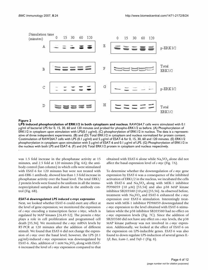
Lipopolysaccharide triggered ERK1/2 phosphorylation in both cytoplasm and the nucleusThe absence of pERK1/2 from the nucleus of ESAT-6-stim-ulated RAW264.7 cells was specific for ESAT-6 treatment.To establish this, we stimulated the cells with the bacteriallipopolysaccharide (LPS), which is a general activator ofmacrophages [39-47]. In the RAW264.7 cells stimulatedwith 0.1 µg/ml of LPS for the same time points as before,we observed time-dependent phosphorylation of ERK1/2in both cytoplasm (Fig. 2A) and nucleus (Fig. 2C).
Next we wanted to know whether LPS can overcome theESAT-6 imposed inhibition of phosphorylation of ERK1/2 in nucleus, for this RAW264.7 cells were co-stimulatedfor the same time points with LPS (0.1 µg/ml) and ESAT-6 (5 µg/ml). In the presence of ESAT-6, LPS caused onlyweak phosphorylation of ERK1/2 in nucleus (Fig. 2G)compared to the LPS alone. Thus, ESAT-6 seemed todampen the ERK1/2 signaling of the MAP kinase familyby limiting the activation of ERK1/2 in the nucleus.
Page 2 of 12(page number not for citation purposes)

BMC Immunology 2007, 8:24 http://www.biomedcentral.com/1471-2172/8/24
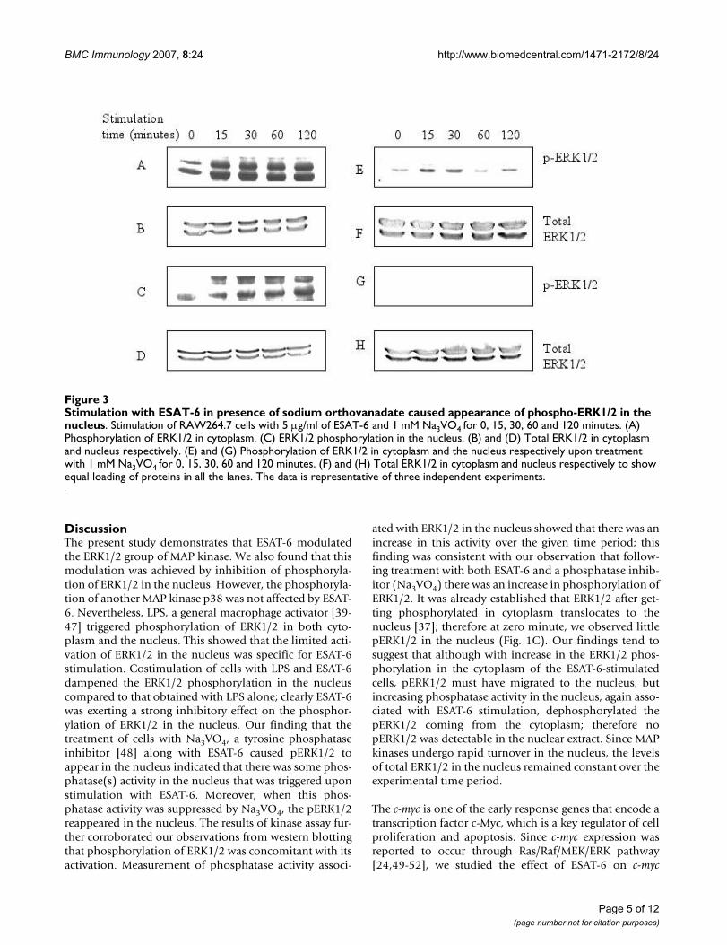
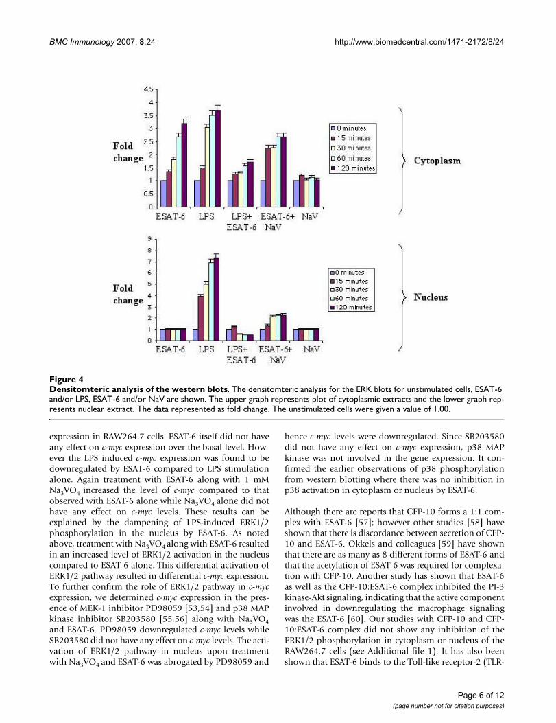
Diminished ERK1/2 activation in the nucleus by ESAT-6 was due to some tyrosine phosphataseTo evaluate whether any phosphatase was involved in thedephosphorylation of ERK1/2 in the nucleus of ESAT-6treated cells, RAW264.7 cells were stimulated with 5 µg/ml of ESAT-6 in presence of 1 mM sodium orthovanadate(Na3VO4), which is a protein tyrosine phosphatase inhib-itor [48]. We found that in the presence of Na3VO4,pERK1/2 appeared in the nucleus (Fig. 3C). The activationof ERK1/2 in cytoplasm was observed as usual (Fig. 3A).Since with ESAT-6 alone there was no pERK1/2 in thenucleus, but with Na3VO4 treatment there was phosphor-ylation of ERK1/2, so there might be some putative phos-phatase(s) dephosphorylating the pERK1/2 as ittranslocated from cytoplasm to the nucleus. We checkedwhether sodium orthovanadate alone could induce acti-vation of ERK1/2; in the cells stimulated with 1 mMNa3VO4 for the same time points, we found weakly acti-vated ERK1/2 in cytoplasm (Fig. 3E) and none in thenucleus (Fig. 3G). The graphs showing densitomtericanalysis of the above ERK blots are shown in Figure 4. Theplots for cytoplasm and nucleus are shown separately.
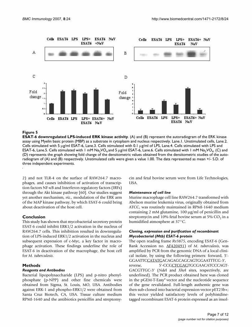
To further confirm the observations from western blot-ting, kinase assay for ERK1/2 was done. The RAW264.7cells were treated with LPS and/or ESAT-6 and ESAT-6and/or Na3VO4 for 60 minutes and the kinase activity wasassayed as described in Methods. In cytoplasm (Fig. 5A)both LPS and ESAT-6 increased ERK enzyme activity overbasal level. ESAT-6 treatment was found to have no effecton the ERK kinase activity in the nucleus over the basallevel (Fig 5B); furthermore, ESAT-6 antagonized the LPS-induced ERK activation. Concurrent treatment withNa3VO4 and ESAT-6 increased ERK activation in thenucleus by more than 4-fold compared to the ESAT-6alone (Fig. 5D). Na3VO4 alone did not have any effect onERK kinase activity over the basal level. Thus the kinaseassay confirmed the earlier western blot observations.
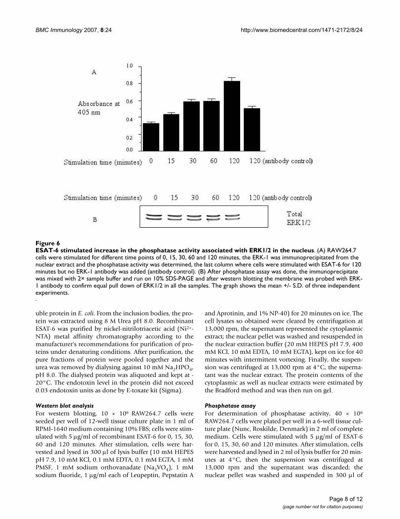
ESAT-6 stimulated phosphatase activity in the nucleusIn order to ascertain if the absence of pERK1/2 in nucleuswas really due to some phosphatase(s), we determinedphosphatase activity associated with ERK1/2 for the sametime points in the nucleus. Determination of phosphataseactivity showed that upon stimulation with ESAT-6 there
ESAT-6 induced phosphorylation of ERK1/2 in cytoplasm but not in nucleusFigure 1ESAT-6 induced phosphorylation of ERK1/2 in cytoplasm but not in nucleus. 10 × 106 RAW264.7 cells were stimu-lated with 5 µg/ml of recombinant ESAT-6 for 0, 15, 30, 60 and 120 minutes; cytoplasmic and nuclear extracts were run on gel and probed with anti-phospho-ERK1/2 antibody. (A) phosphorylation of ERK1/2 in cytoplasm. (C) phosphorylation of ERK1/2 in nucleus. (B) and (D) Total ERK1/2 in the cytoplasmic and nuclear extracts respectively at different time points to confirm equal loading of samples in all the lanes. (E) and (G) represents phosphorylated p38 in cytoplasm and the nucleus respectively. (F) and (H) shows the total p38 protein in cytoplasm and nucleus respectively. Data is a representative from three experi-ments.
Page 3 of 12(page number not for citation purposes)
BMC Immunology 2007, 8:24 http://www.biomedcentral.com/1471-2172/8/24
was 1.5 fold increase in the phosphatase activity at 15minutes, and 2.5 fold at 120 minutes (Fig. 6A); the anti-body control (last column) in which cells were stimulatedwith ESAT-6 for 120 minutes but were not treated withanti-ERK-1 antibody, showed less than 1.5 fold increase inphosphatase activity over the basal level. The total ERK1/2 protein levels were found to be uniform in all the immu-noprecipitated samples and absent in the antibody con-trol (Fig. 6B).
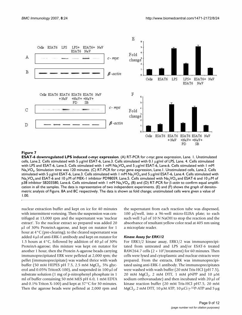
ESAT-6 downregulated LPS induced c-myc expressionNext, we looked whether ESAT-6 could exert any effect atthe level of gene expression; we monitored the expressionof c-myc encoding a transcription factor c-Myc, which isregulated by MAP kinases [24,49-52]. The protein c-Mycplays a role in cell proliferation and programmed celldeath [35,36]. We monitored the c-myc mRNA levels byRT-PCR at 120 minutes after the addition of differentstimuli. We found that ESAT-6 did not change the expres-sion of c-myc over the basal level; however, the LPS (0.1µg/ml)-induced c-myc expression was downregulated byESAT-6. Also, addition of 1 mM Na3VO4 along with ESAT-6 increased the level of c-myc expression compared to that
obtained with ESAT-6 alone while Na3VO4 alone did notaffect the basal expression level of c-myc (Fig. 7A).
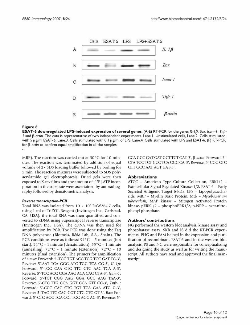
To determine whether the downregulation of c-myc geneexpression by ESAT-6 was a consequence of the inhibitedactivation of ERK1/2 in the nucleus, we incubated the cellswith ESAT-6 and Na3VO4 along with MEK-1 inhibitorPD98059 (10 µM) [53,54] and also p38 MAP kinaseinhibitor SB203580 (10 µM) [55,56]. As observed before,treatment with Na3VO4 and ESAT-6 enhanced the c-mycexpression over ESAT-6 stimulation. Interestingly treat-ment with MEK-1 inhibitor PD98059 downregulated thec-myc expression to the level obtained with ESAT-6 stimu-lation while the p38 inhibitor SB203580 had no effect onc-myc expression levels (Fig. 7C). Since the addition ofSB203580 did not have any effect on c-myc levels, the p38MAP kinase pathway was not involved in c-myc expres-sion. Additionally, we looked at the effect of ESAT-6 onthe expression on LPS-inducible genes. ESAT-6 was alsofound to down regulate LPS-induction of several genes IL-1β, Bax, Icam-1, and Tnfr-1 (Fig. 8).
LPS induced phosphorylation of ERK1/2 in both cytoplasm and nucleusFigure 2LPS induced phosphorylation of ERK1/2 in both cytoplasm and nucleus. RAW264.7 cells were stimulated with 0.1 µg/ml of bacterial LPS for 0, 15, 30, 60 and 120 minutes and probed for phospho-ERK1/2 as before. (A) Phosphorylation of ERK1/2 in cytoplasm upon stimulation with LPS(0.1 µg/ml). (C) phosphorylation of ERK1/2 in nucleus. The data is a represent-ative of three independent experiments. (B) and (D) Total ERK1/2 in cytoplasm and nucleus normalized for protein content. Costimulation of RAW264.7 cells with LPS (0.1 µg/ml) and 5 µg/ml of ESAT-6 for 0, 15, 30, 60 and 120 minutes. (E) ERK1/2 phosphorylation in cytoplasm upon stimulation with 5 µg/ml of ESAT-6 and 0.1 µg/ml of LPS. (G) Phosphorylation of ERK1/2 in the nucleus with both LPS and ESAT-6. (F) and (H) Total ERK1/2 protein in cytoplasm and nucleus respectively.
Page 4 of 12(page number not for citation purposes)
BMC Immunology 2007, 8:24 http://www.biomedcentral.com/1471-2172/8/24
DiscussionThe present study demonstrates that ESAT-6 modulatedthe ERK1/2 group of MAP kinase. We also found that thismodulation was achieved by inhibition of phosphoryla-tion of ERK1/2 in the nucleus. However, the phosphoryla-tion of another MAP kinase p38 was not affected by ESAT-6. Nevertheless, LPS, a general macrophage activator [39-47] triggered phosphorylation of ERK1/2 in both cyto-plasm and the nucleus. This showed that the limited acti-vation of ERK1/2 in the nucleus was specific for ESAT-6stimulation. Costimulation of cells with LPS and ESAT-6dampened the ERK1/2 phosphorylation in the nucleuscompared to that obtained with LPS alone; clearly ESAT-6was exerting a strong inhibitory effect on the phosphor-ylation of ERK1/2 in the nucleus. Our finding that thetreatment of cells with Na3VO4, a tyrosine phosphataseinhibitor [48] along with ESAT-6 caused pERK1/2 toappear in the nucleus indicated that there was some phos-phatase(s) activity in the nucleus that was triggered uponstimulation with ESAT-6. Moreover, when this phos-phatase activity was suppressed by Na3VO4, the pERK1/2reappeared in the nucleus. The results of kinase assay fur-ther corroborated our observations from western blottingthat phosphorylation of ERK1/2 was concomitant with itsactivation. Measurement of phosphatase activity associ-
ated with ERK1/2 in the nucleus showed that there was anincrease in this activity over the given time period; thisfinding was consistent with our observation that follow-ing treatment with both ESAT-6 and a phosphatase inhib-itor (Na3VO4) there was an increase in phosphorylation ofERK1/2. It was already established that ERK1/2 after get-ting phosphorylated in cytoplasm translocates to thenucleus [37]; therefore at zero minute, we observed littlepERK1/2 in the nucleus (Fig. 1C). Our findings tend tosuggest that although with increase in the ERK1/2 phos-phorylation in the cytoplasm of the ESAT-6-stimulatedcells, pERK1/2 must have migrated to the nucleus, butincreasing phosphatase activity in the nucleus, again asso-ciated with ESAT-6 stimulation, dephosphorylated thepERK1/2 coming from the cytoplasm; therefore nopERK1/2 was detectable in the nuclear extract. Since MAPkinases undergo rapid turnover in the nucleus, the levelsof total ERK1/2 in the nucleus remained constant over theexperimental time period.
The c-myc is one of the early response genes that encode atranscription factor c-Myc, which is a key regulator of cellproliferation and apoptosis. Since c-myc expression wasreported to occur through Ras/Raf/MEK/ERK pathway[24,49-52], we studied the effect of ESAT-6 on c-myc
Stimulation with ESAT-6 in presence of sodium orthovanadate caused appearance of phospho-ERK1/2 in the nucleusFigure 3Stimulation with ESAT-6 in presence of sodium orthovanadate caused appearance of phospho-ERK1/2 in the nucleus. Stimulation of RAW264.7 cells with 5 µg/ml of ESAT-6 and 1 mM Na3VO4 for 0, 15, 30, 60 and 120 minutes. (A) Phosphorylation of ERK1/2 in cytoplasm. (C) ERK1/2 phosphorylation in the nucleus. (B) and (D) Total ERK1/2 in cytoplasm and nucleus respectively. (E) and (G) Phosphorylation of ERK1/2 in cytoplasm and the nucleus respectively upon treatment with 1 mM Na3VO4 for 0, 15, 30, 60 and 120 minutes. (F) and (H) Total ERK1/2 in cytoplasm and nucleus respectively to show equal loading of proteins in all the lanes. The data is representative of three independent experiments.
Page 5 of 12(page number not for citation purposes)
BMC Immunology 2007, 8:24 http://www.biomedcentral.com/1471-2172/8/24
expression in RAW264.7 cells. ESAT-6 itself did not haveany effect on c-myc expression over the basal level. How-ever the LPS induced c-myc expression was found to bedownregulated by ESAT-6 compared to LPS stimulationalone. Again treatment with ESAT-6 along with 1 mMNa3VO4 increased the level of c-myc compared to thatobserved with ESAT-6 alone while Na3VO4 alone did nothave any effect on c-myc levels. These results can beexplained by the dampening of LPS-induced ERK1/2phosphorylation in the nucleus by ESAT-6. As notedabove, treatment with Na3VO4 along with ESAT-6 resultedin an increased level of ERK1/2 activation in the nucleuscompared to ESAT-6 alone. This differential activation ofERK1/2 pathway resulted in differential c-myc expression.To further confirm the role of ERK1/2 pathway in c-mycexpression, we determined c-myc expression in the pres-ence of MEK-1 inhibitor PD98059 [53,54] and p38 MAPkinase inhibitor SB203580 [55,56] along with Na3VO4and ESAT-6. PD98059 downregulated c-myc levels whileSB203580 did not have any effect on c-myc levels. The acti-vation of ERK1/2 pathway in nucleus upon treatmentwith Na3VO4 and ESAT-6 was abrogated by PD98059 and
hence c-myc levels were downregulated. Since SB203580did not have any effect on c-myc expression, p38 MAPkinase was not involved in the gene expression. It con-firmed the earlier observations of p38 phosphorylationfrom western blotting where there was no inhibition inp38 activation in cytoplasm or nucleus by ESAT-6.
Although there are reports that CFP-10 forms a 1:1 com-plex with ESAT-6 [57]; however other studies [58] haveshown that there is discordance between secretion of CFP-10 and ESAT-6. Okkels and colleagues [59] have shownthat there are as many as 8 different forms of ESAT-6 andthat the acetylation of ESAT-6 was required for complexa-tion with CFP-10. Another study has shown that ESAT-6as well as the CFP-10:ESAT-6 complex inhibited the PI-3kinase-Akt signaling, indicating that the active componentinvolved in downregulating the macrophage signalingwas the ESAT-6 [60]. Our studies with CFP-10 and CFP-10:ESAT-6 complex did not show any inhibition of theERK1/2 phosphorylation in cytoplasm or nucleus of theRAW264.7 cells (see Additional file 1). It has also beenshown that ESAT-6 binds to the Toll-like receptor-2 (TLR-
Densitomteric analysis of the western blotsFigure 4Densitomteric analysis of the western blots. The densitomteric analysis for the ERK blots for unstimulated cells, ESAT-6 and/or LPS, ESAT-6 and/or NaV are shown. The upper graph represents plot of cytoplasmic extracts and the lower graph rep-resents nuclear extract. The data represented as fold change. The unstimulated cells were given a value of 1.00.
Page 6 of 12(page number not for citation purposes)
BMC Immunology 2007, 8:24 http://www.biomedcentral.com/1471-2172/8/24
2) and not TLR-4 on the surface of RAW264.7 macro-phages, and causes inhibition of activation of transcrip-tion factors NF-κB and Interferon regulatory factors (IRFs)through the Akt kinase pathway [60]. Our studies suggestyet another mechanism, viz., modulation of the ERK armof the MAP kinase pathway, by which ESAT-6 could bringabout deactivation of the host cell.
ConclusionThis study has shown that mycobacterial secretory proteinESAT-6 could inhibit ERK1/2 activation in the nucleus ofRAW264.7 cells. This inhibition resulted in downregula-tion of LPS-induced ERK1/2 activation in the nucleus andsubsequent expression of c-Myc, a key factor in macro-phage activation. These findings underline the role ofESAT-6 in deactivation of the macrophage, the host cellfor M. tuberculosis.
MethodsReagents and AntibodiesBacterial lipopolysaccharide (LPS) and p-nitro phenyl-phosphate (p-NPP) and other fine chemicals wereobtained from Sigma, St. Louis, MO, USA. Antibodiesagainst ERK-1 and phospho-ERK1/2 were obtained fromSanta Cruz Biotech, CA, USA. Tissue culture mediumRPMI-1640 and the antibiotics penicillin and streptomy-
cin and fetal bovine serum were from Life Technologies,USA.
Maintenance of cell lineMurine macrophage cell line RAW264.7 transformed withAbelson murine leukemia virus, originally obtained fromATCC, was routinely maintained in RPMI-1640 mediumcontaining 2 mM glutamine, 100 µg/ml of penicillin andstreptomycin and 10% fetal bovine serum at 5% CO2 in ahumidified atmosphere at 37°C.
Cloning, expression and purification of recombinant Mycobacterial (Mtb) ESAT-6 proteinThe open reading frame Rv3875, encoding ESAT-6 (Gen-Bank Accession no. AF420491) of M. tuberculosis, wasamplified by PCR from the genomic DNA of a local clini-cal isolate, by using the following primers: forward, 5'-GGAATTCCATATGACAGAGCAGCAGTGGAATTTCG-3',reverse, 5'-CCGCTCGAGTGCGAACATCCCAGT-GACGTTGC-3' (NdeI and XhoI sites, respectively, areunderlined). The PCR product obtained here was clonedin the pGEM-T-Easy® vector and the nucleotide sequenceof the gene revalidated. Full-length authentic gene wasthen sub-cloned into bacterial expression vector pET23b+;this vector yielded satisfactory levels of polyhistdine-tagged recombinant ESAT-6 protein expressed as an insol-
ESAT-6 downregulated LPS-induced ERK kinase activityFigure 5ESAT-6 downregulated LPS-induced ERK kinase activity. (A) and (B) represent the autoradiogram of the ERK kinase assay using Myelin basic protein (MBP) as a substrate in cytoplasm and nucleus respectively. Lane.1. Unstimulated cells, Lane.2. Cells stimulated with 5 µg/ml ESAT-6, Lane.3. Cells stimulated with 0.1 µg/ml of LPS, Lane.4. Cells stimulated with LPS and ESAT-6, Lane.5. Cells stimulated with 1 mM Na3VO4 and 5 µg/ml ESAT-6, Lane.6. Cells stimulated with 1 mM Na3VO4. (C) and (D) represents the graph showing fold change of the densitometric values obtained from the densitometric studies of the auto-radiogram of (A) and (B) respectively. Unstimulated cells were given a value 1.00. The data represented as mean +/- S.D. of three independent experiments.
Page 7 of 12(page number not for citation purposes)
BMC Immunology 2007, 8:24 http://www.biomedcentral.com/1471-2172/8/24
uble protein in E. coli. From the inclusion bodies, the pro-tein was extracted using 8 M Urea pH 8.0. RecombinantESAT-6 was purified by nickel-nitrilotriacetic acid (Ni2+-NTA) metal affinity chromatography according to themanufacturer's recommendations for purification of pro-teins under denaturing conditions. After purification, thepure fractions of protein were pooled together and theurea was removed by dialysing against 10 mM Na2HPO4,pH 8.0. The dialysed protein was aliquoted and kept at -20°C. The endotoxin level in the protein did not exceed0.03 endotoxin units as done by E-toxate kit (Sigma).
Western blot analysisFor western blotting, 10 × 106 RAW264.7 cells wereseeded per well of 12-well tissue culture plate in 1 ml ofRPMI-1640 medium containing 10% FBS; cells were stim-ulated with 5 µg/ml of recombinant ESAT-6 for 0, 15, 30,60 and 120 minutes. After stimulation, cells were har-vested and lysed in 300 µl of lysis buffer (10 mM HEPESpH 7.9, 10 mM KCl, 0.1 mM EDTA, 0.1 mM EGTA, 1 mMPMSF, 1 mM sodium orthovanadate (Na3VO4), 1 mMsodium fluoride, 1 µg/ml each of Leupeptin, Pepstatin A
and Aprotinin, and 1% NP-40) for 20 minutes on ice. Thecell lysates so obtained were cleared by centrifugation at13,000 rpm, the supernatant represented the cytoplasmicextract; the nuclear pellet was washed and resuspended inthe nuclear extraction buffer (20 mM HEPES pH 7.9, 400mM KCl, 10 mM EDTA, 10 mM EGTA), kept on ice for 40minutes with intermittent vortexing. Finally, the suspen-sion was centrifuged at 13,000 rpm at 4°C, the superna-tant was the nuclear extract. The protein contents of thecytoplasmic as well as nuclear extracts were estimated bythe Bradford method and was then run on gel.
Phosphatase assayFor determination of phosphatase activity, 40 × 106
RAW264.7 cells were plated per well in a 6-well tissue cul-ture plate (Nunc, Roskilde, Denmark) in 2 ml of completemedium. Cells were stimulated with 5 µg/ml of ESAT-6for 0, 15, 30, 60 and 120 minutes. After stimulation, cellswere harvested and lysed in 2 ml of lysis buffer for 20 min-utes at 4°C, then the suspension was centrifuged at13,000 rpm and the supernatant was discarded; thenuclear pellet was washed and suspended in 300 µl of
ESAT-6 stimulated increase in the phosphatase activity associated with ERK1/2 in the nucleusFigure 6ESAT-6 stimulated increase in the phosphatase activity associated with ERK1/2 in the nucleus. (A) RAW264.7 cells were stimulated for different time points of 0, 15, 30, 60 and 120 minutes, the ERK-1 was immunoprecipitated from the nuclear extract and the phosphatase activity was determined, the last column where cells were stimulated with ESAT-6 for 120 minutes but no ERK-1 antibody was added (antibody control). (B) After phosphatase assay was done, the immunoprecipitate was mixed with 2× sample buffer and run on 10% SDS-PAGE and after western blotting the membrane was probed with ERK-1 antibody to confirm equal pull down of ERK1/2 in all the samples. The graph shows the mean +/- S.D. of three independent experiments.
Page 8 of 12(page number not for citation purposes)
BMC Immunology 2007, 8:24 http://www.biomedcentral.com/1471-2172/8/24
nuclear extraction buffer and kept on ice for 40 minuteswith intermittent vortexing. Then the suspension was cen-trifuged at 13,000 rpm and the supernatant was 'nuclearextract'. To the nuclear extract so prepared was added 20µl of 30% ProteinA-agarose, and kept on nutator for 1hour at 4°C (pre-clearing); to the cleared supernatant wasadded 4 µl of anti-ERK-1 antibody and kept on nutator for1.5 hours at 4°C, followed by addition of 40 µl of 30%ProteinA-agarose; this mixture was kept on nutator foranother 1 hour, then the Protein A-agarose beads carryingimmunoprecipitated ERK were pelleted at 2,000 rpm; thepellet (immunoprecipitate) was washed thrice with washbuffer (50 mM HEPES pH 7.5, 2.5 mM MgCl2, 5% glyc-erol and 0.05% TritonX-100), and suspended in 100 µl ofsubstrate solution (1 mg of p-nitrophenyl phosphate in 1ml of buffer containing 50 mM MES pH 6.0, 1 mM EDTAand 0.1% Triton X-100) and kept at 37°C for 30 minutes.Then the agarose beads were pelleted at 2,000 rpm and
the supernatant from each reaction tube was dispensed,100 µl/well, into a 96-well micro-ELISA plate; to eachsuch well 5 µl of 10 N NaOH to stop the reaction and theabsorbance of resultant yellow color read at 405 nm usinga microplate reader.
Kinase Assay for ERK1/2For ERK1/2 kinase assay, ERK1/2 was immunoprecipi-tated from untreated and LPS and/or ESAT-6 treatedRAW264.7 cells (2 × 10⁷/treatment) for 60 minutes. Thencells were lysed and cytoplasmic and nuclear extracts wereprepared. From the extracts, ERK was immunoprecipi-tated using anti-ERK-1 antibody. The immunoprecipitateswere washed with wash buffer (20 mM Tris-HCl (pH 7.5),20 mM MgCl2, 2 mM DTT, 1 mM pNPP and 10 µMsodium orthovanadate) and then incubated with 20 µl ofkinase reaction buffer (20 mM Tris-HCl pH7.5, 20 mMMgCl2, 2 mM DTT, 10 µM ATP, 10 µCi γ-32P-ATP and 5 µg
ESAT-6 downregulated LPS induced c-myc expressionFigure 7ESAT-6 downregulated LPS induced c-myc expression. (A) RT-PCR for c-myc gene expression, Lane. 1. Unstimulated cells, Lane.2. Cells stimulated with 5 µg/ml ESAT-6, Lane.3. Cells stimulated with 0.1 µg/ml of LPS, Lane. 4. Cells stimulated with LPS and ESAT-6, Lane.5. Cells stimulated with 1 mM Na3VO4 and 5 µg/ml ESAT-6, Lane.6. Cells stimulated with 1 mM Na3VO4. Stimulation time was 120 minutes. (C) RT-PCR for c-myc gene expression, Lane.1. Unstimulated cells, Lane.2. Cells stimulated with 5 µg/ml ESAT-6, Lane.3. Cells stimulated with 1 mM Na3VO4 and 5 µg/ml ESAT-6, Lane.4. Cells stimulated with Na3VO4 and ESAT-6 and 10 µM of MEK-1 inhibitor PD98059, Lane.5. Cells simulated with Na3VO4 and ESAT-6 and 10 µM of p38 inhibitor SB203580, Lane.6. Cells stimulated with 1 mM Na3VO4, (B) and (D) RT-PCR for β-actin to confirm equal amplifi-cation in all the samples. The data is representative of two independent experiments. (E) and (F) shows the graph of densito-metric analysis of Figure. 8A and 8C respectively. The data is shown as fold change; unstimulated cells were given a value of 1.00.
Page 9 of 12(page number not for citation purposes)
BMC Immunology 2007, 8:24 http://www.biomedcentral.com/1471-2172/8/24
MBP). The reaction was carried out at 30°C for 10 min-utes. The reaction was terminated by addition of equalvolume of 2× SDS loading buffer followed by boiling for5 min. The reaction mixtures were subjected to SDS poly-acrylamide gel electrophoresis. Dried gels were thenexposed to X-ray films and the amount of [32P]-ATP incor-poration in the substrate were ascertained by autoradiog-raphy followed by densitometric analysis.
Reverse transcription-PCRTotal RNA was isolated from 10 × 106 RAW264.7 cells,using 1 ml of TriZOL Reagent (Invitrogen Inc., Carlsbad,CA, USA); the total RNA was then quantified and con-verted to cDNA using Superscript II reverse transcriptase(Invitrogen Inc., USA). The cDNA was then used foramplification by PCR. The PCR was done using the TaqDNA polynerase (Biotools, B&M Lab, S.A., Spain). ThePCR conditions were as follows: 94°C – 5 minutes (hotstart), 94°C – 1 minute (denaturation), 55°C – 1 minute(annealing), 72°C – 1 minute (extension), 72°C – 10minutes (final extension). The primers for amplificationof c-myc: Forward: 5'-TCC TGT ACC TCG TCC GAT TC-3',Reverse: 5'-AAT TCA GGG ATC TGG TCA CG-3', IL-1β:Forward: 5'-TGG CAA CTG TTC CTG AAC TCA A-3',Reverse: 5'-TCC ACG GGA AAG ACA CAG GTA-3', Icam-1:Forward: 5'-TCT CGG AAG GGA GCC AAG TAA-3',Reverse: 5'-CTC TTG CCA GGT CCA GTT CC-3', Tnfr-1:Forward: 5'-CCC CAC CTC TGT TCA GAA ATG G-3',Reverse: 5'-TAC TTC CAG CGT GTC CTC GT-3', Bax: For-ward: 5'-CTG AGC TGA CCT TGG AGC AG-3', Reverse: 5'-
CCA GCC CAT GAT GGT TCT GAT-3', β-actin: Forward: 5'-CTA TGC TCT CCC TCA CGC CA-3', Reverse: 5'-CCG CTCGTT GCC AAT AGT GAT-3'.
AbbreviationsATCC – American Type Culture Collection, ERK1/2 –Extracellular Signal Regulated Kinases1/2, ESAT-6 – EarlySecreted Antigenic Target 6-kDa, LPS – Lipopolysaccha-ride, MBP – Myelin Basic Protein, Mtb – Mycobacteriumtuberculosis, MAP kinase – Mitogen Activated Proteinkinase, pERK1/2 – phosphoERK1/2, p-NPP – para-nitro-phenyl phosphate.
Authors' contributionsNG performed the western blot analysis, kinase assay andphosphatase assay. SKB and IS did the RT-PCR experi-ments. PHG and FAM helped in the expression and puri-fication of recombinant ESAT-6 and in the western blotanalysis. PS and NG were responsible for conceptualizingand designing the study as well as for writing the manu-script. All authors have read and approved the final man-uscript.
ESAT-6 downregulated LPS-induced expression of several genesFigure 8ESAT-6 downregulated LPS-induced expression of several genes. (A-E) RT-PCR for the genes IL-1β, Bax, Icam-1, Tnfr-1 and β-actin. The data is representative of two independent experiments. Lane.1. Unstimulated cells, Lane.2. Cells stimulated with 5 µg/ml ESAT-6, Lane.3. Cells stimulated with 0.1 µg/ml of LPS, Lane.4. Cells stimulated with LPS and ESAT-6. (F) RT-PCR for β-actin to confirm equal amplification in all the samples.
Page 10 of 12(page number not for citation purposes)
BMC Immunology 2007, 8:24 http://www.biomedcentral.com/1471-2172/8/24
Additional material
AcknowledgementsNG and SKB were recipients of JRF/SRF fellowships from CSIR; PHG is recipient of an International Pre-Doctoral Fellowship from ICGEB. The Sharma lab receives generous financial support from the Department of Biotechnology (Govt. of India), New Delhi.
References1. Raviglione MC: The TB epidemic from 1992 to 2002. Tuberculo-
sis (Edinb) 2003, 83:4-14.2. Rosenberger CM, Finlay BB: Phagocyte sabotage: disruption of
macrophage signalling by bacterial pathogens. Nat Rev Mol CellBiol 2003, 4:385-396.
3. Koul A, Herget T, Klebl B, Ullrich A: Interplay between mycobac-teria and host signalling pathways. Nat Rev Microbiol 2004,2:189-202.
4. Malik Z, Iyer SS, Kusner DJ: Mycobacterium tuberculosis phago-somes exhibit altered calmodulin-dependent signal trans-duction: contribution to inhibition of phagosome-lysosomefusion and intracellular survival in human macrophages. JImmunol 2001, 166:3392-3401.
5. Malik ZA, Thompson CR, Hashimi S, Porter B, Iyer SS, Kusner DJ:Cutting edge: Mycobacterium tuberculosis blocks Ca2+ signal-ing and phagosome maturation in human macrophages viaspecific inhibition of sphingosine kinase. J Immunol 2003,170:2811-2815.
6. Ting LM, Kim AC, Cattamanchi A, Ernst JD: Mycobacterium tuber-culosis inhibits IFN-gamma transcriptional responses with-out inhibiting activation of STAT1. J Immunol 1999,163:3898-3906.
7. Noss EH, Harding CV, Boom WH: Mycobacterium tuberculosisinhibits MHC class II antigen processing in murine bone mar-row macrophages. Cell Immunol 2000, 201:63-74.
8. Balcewicz-Sablinska MK, Keane J, Kornfeld H, Remold HG: Patho-genic Mycobacterium tuberculosis evades apoptosis of hostmacrophages by release of TNF-R2, resulting in inactivationof TNF-alpha. J Immunol 1998, 161:2636-2641.
9. Keane J, Remold HG, Kornfeld H: Virulent Mycobacterium tuber-culosis strains evade apoptosis of infected alveolar macro-phages. J Immunol 2000, 164:2016-2020.
10. Harboe M, Oettinger T, Wiker HG, Rosenkrands I, Andersen P: Evi-dence for occurrence of the ESAT-6 protein in Mycobacte-rium tuberculosis and virulent Mycobacterium bovis and for itsabsence in Mycobacterium bovis BCG. Infect Immun 1996,64:16-22.
11. Berthet FX, Rasmussen PB, Rosenkrands I, Andersen P, Gicquel B: AMycobacterium tuberculosis operon encoding ESAT-6 and anovel low-molecular-mass culture filtrate protein (CFP-10).Microbiology 1998, 144:3195-3203.
12. Skjot RL, Oettinger T, Rosenkrands I, Ravn P, Brock I, Jacobsen S,Andersen P: Comparative evaluation of low-molecular-massproteins from Mycobacterium tuberculosis identifies mem-bers of the ESAT-6 family as immunodominant T-cell anti-gens. Infect Immun 2000, 68:214-220.
13. Colangeli R, Spencer JS, Bifani P, Williams A, Lyashchenko K, KeenMA, Hill PJ, Belisle J, Gennaro ML: MTSA-10, the product of theRv3874 gene of Mycobacterium tuberculosis, elicits tuberculo-
sis-specific, delayed-type hypersensitivity in guinea pigs.Infect Immun 2000, 68:990-993.
14. Trajkovic V, Singh G, Singh B, Singh S, Sharma P: Effect of Mycobac-terium tuberculosis-specific 10-kilodalton antigen on macro-phage release of tumor necrosis factor alpha and nitricoxide. Infect Immun 2002, 70:6558-6566.
15. Pym AS, Brodin P, Brosch R, Huerre M, Cole ST: Loss of RD1 con-tributed to the attenuation of the live tuberculosis vaccinesMycobacterium bovis BCG and Mycobacterium microti. MolMicrobiol 2002, 46:709-717.
16. Pym AS, Brodin P, Majlessi L, Brosch R, Demangel C, Williams A, Grif-fiths KE, Marchal G, Leclerc C, Cole ST: Recombinant BCGexporting ESAT-6 confers enhanced protection againsttuberculosis. Nat Med 2003, 9:533-539.
17. Brodin P, de Jonge MI, Majlessi L, Leclerc C, Nilges M, Cole ST, Bro-sch R: Functional analysis of early secreted antigenic target-6, the dominant T-cell antigen of Mycobacterium tuberculosis,reveals key residues involved in secretion, complex forma-tion, virulence, and immunogenicity. J Biol Chem 2005,280:33953-33959.
18. Brodin P, Majlessi L, Marsollier L, de Jonge MI, Bottai D, Demangel C,Hinds J, Neyrolles O, Butcher PD, Leclerc C, Cole ST, Brosch R: Dis-section of ESAT-6 system 1 of Mycobacterium tuberculosisand impact on immunogenicity and virulence. Infect Immun2006, 74:88-98.
19. Mahairas GG, Sabo PJ, Hickey MJ, Singh DC, Stover CK: Molecularanalysis of genetic differences between Mycobacterium bovisBCG and virulent M. bovis. J Bacteriol 1996, 178:1274-1282.
20. Lewis KN, Liao R, Guinn KM, Hickey MJ, Smith S, Behr MA, ShermanDR: Deletion of RD1 from Mycobacterium tuberculosis mimicsbacille Calmette-Guerin attenuation. J Infect Dis 2003,187:117-123.
21. Guinn KM, Hickey MJ, Mathur SK, Zakel KL, Grotzke JE, LewinsohnDM, Smith S, Sherman DR: Individual RD1-region genes arerequired for export of ESAT-6/CFP-10 and for virulence ofMycobacterium tuberculosis. Mol Microbiol 2004, 51:359-370.
22. Fortune SM, Jaeger A, Sarracino DA, Chase MR, Sassetti CM, Sher-man DR, Bloom BR, Rubin EJ: Mutually dependent secretion ofproteins required for mycobacterial virulence. Proc Natl AcadSci USA 2005, 102:10676-10681.
23. Johnson GL, Lapadat R: Mitogen-activated protein kinase path-ways mediated by ERK, JNK, and p38 protein kinases. Science2002, 298:1911-1912.
24. van der Bruggen T, Nijenhuis S, van Raaij E, Verhoef J, van Asbeck BS:Lipopolysaccharide-induced tumor necrosis factor alphaproduction by human monocytes involves the raf-1/MEK1-MEK2/ERK1-ERK2 pathway. Infect Immun 1999, 67:3824-3829.
25. Reiling N, Blumenthal A, Flad HD, Ernst M, Ehlers S: Mycobacteria-induced TNF-alpha and IL-10 formation by human macro-phages is differentially regulated at the level of mitogen-acti-vated protein kinase activity. J Immunol 2001, 167:3339-3345.
26. Carter AB, Monick MM, Hunninghake GW: Both Erk and p38kinases are necessary for cytokine gene transcription. Am JRespir Cell Mol Biol 1999, 20:751-758.
27. Kyriakis JM, Avruch J: Sounding the alarm: protein kinase cas-cades activated by stress and inflammation. J Biol Chem 1996,271:24313-24316.
28. Bhattacharyya A, Pathak S, Basak C, Law S, Kundu M, Basu J: Execu-tion of macrophage apoptosis by Mycobacterium aviumthrough apoptosis signal-regulating kinase 1/p38 mitogen-activated protein kinase signaling and caspase 8 activation. JBiol Chem 2003, 278:26517-26525.
29. Feng GJ, Goodridge HS, Harnett MM, Wei XQ, Nikolaev AV, HigsonAP, Liew FY: Extracellular signal-related kinase (ERK) and p38mitogen-activated protein (MAP) kinases differentially regu-late the lipopolysaccharide-mediated induction of induciblenitric oxide synthase and IL-12 in macrophages: Leishmaniaphosphoglycans subvert macrophage IL-12 production by tar-geting ERK MAP kinase. J Immunol 1999, 163:6403-6412.
30. Roux PP, Blenis J: ERK and p38 MAPK-activated proteinkinases: a family of protein kinases with diverse biologicalfunctions. Microbiol Mol Biol Rev 2004, 68:320-344.
31. Rescan C, Coutant A, Talarmin H, Theret N, Glaise D, Guguen-Guil-louzo C, Baffet G: Mechanism in the sequential control of cellmorphology and S phase entry by epidermal growth factor
Additional file 1Western blot analysis of ERK1/2 phosphorylation upon stimulation by CFP-10 and CFP10:ESAT6 complex. The data shows the phosphorylation of ERK1/2 in cytoplasmic and nuclear extracts upon stimulation with CFP-10 and CFP10:ESAT6 complex for 0, 15, 30, 60 and 120 minutes.Click here for file[http://www.biomedcentral.com/content/supplementary/1471-2172-8-24-S1.pdf]
Page 11 of 12(page number not for citation purposes)
http://www.ncbi.nlm.nih.gov/entrez/query.fcgi?cmd=Retrieve&db=PubMed&dopt=Abstract&list_uids=9725266
http://www.ncbi.nlm.nih.gov/entrez/query.fcgi?cmd=Retrieve&db=PubMed&dopt=Abstract&list_uids=9725266
http://www.ncbi.nlm.nih.gov/entrez/query.fcgi?cmd=Retrieve&db=PubMed&dopt=Abstract&list_uids=9725266
http://www.ncbi.nlm.nih.gov/entrez/query.fcgi?cmd=Retrieve&db=PubMed&dopt=Abstract&list_uids=8557334
http://www.ncbi.nlm.nih.gov/entrez/query.fcgi?cmd=Retrieve&db=PubMed&dopt=Abstract&list_uids=8557334
http://www.ncbi.nlm.nih.gov/entrez/query.fcgi?cmd=Retrieve&db=PubMed&dopt=Abstract&list_uids=9846755
http://www.ncbi.nlm.nih.gov/entrez/query.fcgi?cmd=Retrieve&db=PubMed&dopt=Abstract&list_uids=9846755
http://www.ncbi.nlm.nih.gov/entrez/query.fcgi?cmd=Retrieve&db=PubMed&dopt=Abstract&list_uids=8631702
http://www.ncbi.nlm.nih.gov/entrez/query.fcgi?cmd=Retrieve&db=PubMed&dopt=Abstract&list_uids=8798679
BMC Immunology 2007, 8:24 http://www.biomedcentral.com/1471-2172/8/24
Publish with BioMed Central and every scientist can read your work free of charge
"BioMed Central will be the most significant development for disseminating the results of biomedical research in our lifetime."
Sir Paul Nurse, Cancer Research UK
Your research papers will be:
available free of charge to the entire biomedical community
peer reviewed and published immediately upon acceptance
cited in PubMed and archived on PubMed Central
yours — you keep the copyright
Submit your manuscript here:http://www.biomedcentral.com/info/publishing_adv.asp
BioMedcentral
involves distinct MEK/ERK activations. Mol Biol Cell 2001,12:725-738.
32. Ruckdeschel K, Machold J, Roggenkamp A, Schubert S, Pierre J,Zumbihl R, Liautard JP, Heesemann J, Rouot B: Yersinia enterocol-itica promotes deactivation of macrophage mitogen-acti-vated protein kinases extracellular signal-regulated kinase-1/2, p38, and c-Jun NH2-terminal kinase. Correlation with itsinhibitory effect on tumor necrosis factor-alpha production.J Biol Chem 1997, 272:15920-15927.
33. Blumenthal A, Ehlers S, Ernst M, Flad HD, Reiling N: Control ofmycobacterial replication in human macrophages: roles ofextracellular signal-regulated kinases 1 and 2 and p38mitogen-activated protein kinase pathways. Infect Immun2002, 70:4961-4967.
34. Roach SK, Schorey JS: Differential regulation of the mitogen-activated protein kinases by pathogenic and nonpathogenicmycobacteria. Infect Immun 2002, 70:3040-3052.
35. Lemaitre JM, Buckle RS, Mechali M: c-Myc in the control of cellproliferation and embryonic development. Adv Cancer Res1996, 70:95-144.
36. Herbst A, Hemann MT, Tworkowski KA, Salghetti SE, Lowe SW, Tan-sey WP: A conserved element in Myc that negatively regu-lates its proapoptotic activity. EMBO Rep 2005, 6:177-183.
37. Brunet A, Roux D, Lenormand P, Dowd S, Keyse S, Pouyssegur J:Nuclear translocation of p42/p44 mitogen-activated proteinkinase is required for growth factor-induced gene expressionand cell cycle entry. Embo J 1999, 18:664-674.
38. Deak M, Clifton AD, Lucocq LM, Alessi DR: Mitogen- and stress-activated protein kinase-1 (MSK1) is directly activated byMAPK and SAPK2/p38, and may mediate activation ofCREB. Embo J 1998, 17:4426-4441.
39. Arndt PG, Suzuki N, Avdi NJ, Malcolm KC, Worthen GS: Lipopoly-saccharide-induced c-Jun NH2-terminal kinase activation inhuman neutrophils: role of phosphatidylinositol 3-Kinase andSyk-mediated pathways. J Biol Chem 2004, 279:10883-10891.
40. Durando MM, Meier KE, Cook JA: Endotoxin activation ofmitogen-activated protein kinase in THP-1 cells; diminishedactivation following endotoxin desensitization. J Leukoc Biol1998, 64:259-264.
41. Geppert TD, Whitehurst CE, Thompson P, Beutler B: Lipopolysac-charide signals activation of tumor necrosis factor biosyn-thesis through the ras/raf-1/MEK/MAPK pathway. Mol Med1994, 1:93-103.
42. Hopkins HA, Monick MM, Hunninghake GW: Lipopolysaccharideupregulates surface expression of CD14 on human alveolarmacrophages. Am J Physiol 1995, 269:L849-854.
43. Meng F, Lowell CA: Lipopolysaccharide (LPS)-induced macro-phage activation and signal transduction in the absence ofSrc-family kinases Hck, Fgr, and Lyn. J Exp Med 1997,185:1661-1670.
44. Sweet MJ, Hume DA: Endotoxin signal transduction in macro-phages. J Leukoc Biol 1996, 60:8-26.
45. Weinstein SL, Gold MR, DeFranco AL: Bacterial lipopolysaccha-ride stimulates protein tyrosine phosphorylation in macro-phages. Proc Natl Acad Sci USA 1991, 88:4148-4152.
46. Yang RB, Mark MR, Gurney AL, Godowski PJ: Signaling eventsinduced by lipopolysaccharide-activated toll-like receptor 2.J Immunol 1999, 163:639-643.
47. Shi L, Kishore R, McMullen MR, Nagy LE: Lipopolysaccharidestimulation of ERK1/2 increases TNF-alpha production viaEgr-1. Am J Physiol Cell Physiol 2002, 282:C1205-1211.
48. Sugano M, Tsuchida K, Makino N: A protein tyrosine phos-phatase inhibitor accelerates angiogenesis in a rat model ofhindlimb ischemia. J Cardiovasc Pharmacol 2004, 44:460-465.
49. Cheng M, Wang D, Roussel MF: Expression of c-Myc in responseto colony-stimulating factor-1 requires mitogen-activatedprotein kinase kinase-1. J Biol Chem 1999, 274:6553-6558.
50. Kanazawa S, Soucek L, Evan , Okamoto GT, Peterlin BM: c-Mycrecruits P-TEFb for transcription, cellular proliferation andapoptosis. Oncogene 2003, 22:5707-5711.
51. Murphy LO, MacKeigan JP, Blenis J: A network of immediateearly gene products propagates subtle differences inmitogen-activated protein kinase signal amplitude and dura-tion. Mol Cell Biol 2004, 24:144-153.
52. Marampon F, Ciccarelli C, Zani BM: Down-regulation of c-Mycfollowing MEK/ERK inhibition halts the expression of malig-
nant phenotype in rhabdomyosarcoma and in non muscle-derived human tumors. Mol Cancer 2006, 5:31.
53. Lester RD, Jo M, Campana WM, Gonias SL: Erythropoietin pro-motes MCF-7 breast cancer cell migration by an ERK/mitogen-activated protein kinase-dependent pathway and isprimarily responsible for the increase in migration observedin hypoxia. J Biol Chem 2005, 280:39273-39277.
54. Lu R, Wang X, Chen ZF, Sun DF, Tian XQ, Fang JY: Inhibition of theExtracellular Signal-regulated Kinase/Mitogen-activatedProtein Kinase Pathway Decreases DNA Methylation inColon Cancer Cells. J Biol Chem 2007, 282:12249-12259.
55. Adler HS, Kubsch S, Graulich E, Ludwig S, Knop J, Steinbrink K: Acti-vation of MAP kinase p38 is critical for the cell cycle control-led suppressor function of regulatory T cells. Blood 2007,109:4351-4359.
56. Zhou Y, Wang Q, Mark Evers B, Chung DH: Oxidative stress-induced intestinal epithelial cell apoptosis is mediated by p38MAPK. Biochem Biophys Res Commun 2006, 350:860-865.
57. Renshaw PS, Panagiotidou P, Whelan A, Gordon SV, Hewinson RG,Williamson RA, Carr MD: Conclusive evidence that the majorT-cell antigens of the Mycobactrium tuberculosis complexESAT-6 and CFP-10 form a tight 1:1 complex and character-ization of the structural properties of ESAT-6, CFP-10 andthe ESAT-6*CFP-10 complex. Implications for pathogenesisand virulence. J Biol Chem 2002, 277:21598-21603.
58. Gao LY, Guo S, McLaughlin B, Morisaki H, Engel JN, Brown EJ: Amycobacterial virulence gene cluster extending RD1 regionis required for cytolysis, bacterial spreading and ESAT-6secretion. Mol Microbiol 2004, 53:1677-1693.
59. Okkels LM, Muller EC, Schmid M, Rosenkrands I, Kaufmann SH,Andersen P, Jungblut PR: CFP-10 discriminates between non-acetylated and acetylated ESAT-6 of Mycobacterium tuber-culosis by differential interaction. Proteomics 2004, 4:2954-2960.
60. Pathak SK, Basu S, Basu KK, Banerjee A, Pathak S, Bhattacharyya A,Kaisho T, Kundu M, Basu J: Direct extracellular interactionbetween the early secreted antigen ESAT-6 of Mycobacte-rium tuberculosis and TLR2 inhibits TLR signaling in macro-phages. Nat Immunol 2007, 8:610-618.
Page 12 of 12(page number not for citation purposes)
http://www.ncbi.nlm.nih.gov/entrez/query.fcgi?cmd=Retrieve&db=PubMed&dopt=Abstract&list_uids=9188492
http://www.ncbi.nlm.nih.gov/entrez/query.fcgi?cmd=Retrieve&db=PubMed&dopt=Abstract&list_uids=9188492
http://www.ncbi.nlm.nih.gov/entrez/query.fcgi?cmd=Retrieve&db=PubMed&dopt=Abstract&list_uids=8902055
http://www.ncbi.nlm.nih.gov/entrez/query.fcgi?cmd=Retrieve&db=PubMed&dopt=Abstract&list_uids=8902055
http://www.ncbi.nlm.nih.gov/entrez/query.fcgi?cmd=Retrieve&db=PubMed&dopt=Abstract&list_uids=9927426
http://www.ncbi.nlm.nih.gov/entrez/query.fcgi?cmd=Retrieve&db=PubMed&dopt=Abstract&list_uids=9927426
http://www.ncbi.nlm.nih.gov/entrez/query.fcgi?cmd=Retrieve&db=PubMed&dopt=Abstract&list_uids=9927426
http://www.ncbi.nlm.nih.gov/entrez/query.fcgi?cmd=Retrieve&db=PubMed&dopt=Abstract&list_uids=9687510
http://www.ncbi.nlm.nih.gov/entrez/query.fcgi?cmd=Retrieve&db=PubMed&dopt=Abstract&list_uids=9687510
http://www.ncbi.nlm.nih.gov/entrez/query.fcgi?cmd=Retrieve&db=PubMed&dopt=Abstract&list_uids=9687510
http://www.ncbi.nlm.nih.gov/entrez/query.fcgi?cmd=Retrieve&db=PubMed&dopt=Abstract&list_uids=9715266
http://www.ncbi.nlm.nih.gov/entrez/query.fcgi?cmd=Retrieve&db=PubMed&dopt=Abstract&list_uids=9715266
http://www.ncbi.nlm.nih.gov/entrez/query.fcgi?cmd=Retrieve&db=PubMed&dopt=Abstract&list_uids=9715266
http://www.ncbi.nlm.nih.gov/entrez/query.fcgi?cmd=Retrieve&db=PubMed&dopt=Abstract&list_uids=8790605
http://www.ncbi.nlm.nih.gov/entrez/query.fcgi?cmd=Retrieve&db=PubMed&dopt=Abstract&list_uids=8790605
http://www.ncbi.nlm.nih.gov/entrez/query.fcgi?cmd=Retrieve&db=PubMed&dopt=Abstract&list_uids=8790605
http://www.ncbi.nlm.nih.gov/entrez/query.fcgi?cmd=Retrieve&db=PubMed&dopt=Abstract&list_uids=8572247
http://www.ncbi.nlm.nih.gov/entrez/query.fcgi?cmd=Retrieve&db=PubMed&dopt=Abstract&list_uids=8572247
http://www.ncbi.nlm.nih.gov/entrez/query.fcgi?cmd=Retrieve&db=PubMed&dopt=Abstract&list_uids=8572247
http://www.ncbi.nlm.nih.gov/entrez/query.fcgi?cmd=Retrieve&db=PubMed&dopt=Abstract&list_uids=9151903
http://www.ncbi.nlm.nih.gov/entrez/query.fcgi?cmd=Retrieve&db=PubMed&dopt=Abstract&list_uids=9151903
http://www.ncbi.nlm.nih.gov/entrez/query.fcgi?cmd=Retrieve&db=PubMed&dopt=Abstract&list_uids=9151903
http://www.ncbi.nlm.nih.gov/entrez/query.fcgi?cmd=Retrieve&db=PubMed&dopt=Abstract&list_uids=8699127
http://www.ncbi.nlm.nih.gov/entrez/query.fcgi?cmd=Retrieve&db=PubMed&dopt=Abstract&list_uids=8699127
http://www.ncbi.nlm.nih.gov/entrez/query.fcgi?cmd=Retrieve&db=PubMed&dopt=Abstract&list_uids=1709735
http://www.ncbi.nlm.nih.gov/entrez/query.fcgi?cmd=Retrieve&db=PubMed&dopt=Abstract&list_uids=1709735
Related Documents