Downloaded from https://journals.lww.com/continuum by mAXWo3ZnzwrcFjDdvMDuzVysskaX4mZb8eYMgWVSPGPJOZ9l+mqFwgfuplwVY+jMyQlPQmIFeWtrhxj7jpeO+505hdQh14PDzV4LwkY42MCrzQCKIlw0d1O4YvrWMUvvHuYO4RRbviuuWR5DqyTbTk/icsrdbT0HfRYk7+ZAGvALtKGnuDXDohHaxFFu/7KNo26hIfzU/+BCy16w7w1bDw== on 04/25/2018 Myasthenia Gravis and Lambert-Eaton Myasthenic Syndrome Michael W. Nicolle, MD ABSTRACT Purpose of Review: This article discusses the pathogenesis, diagnosis, and management of autoimmune myasthenia gravis (MG) and Lambert-Eaton myasthenic syndrome (LEMS). Recent Findings: Recognition of new antigenic targets and improved diagnostic methods promise to improve the diagnosis of MG, although the clinical phenotypes associated with newer antibodies have not yet been defined. Future therapies might specifically target the aberrant immune response. The apparent increase in the prevalence of MG is not fully explained. Results of a long-awaited trial of thymectomy support the practice of performing a thymectomy under specific conditions. Summary: The current treatment options are so effective in most patients with MG or LEMS that in patients with refractory disease the diagnosis should be reconsidered. The management of MG is individualized, and familiarity with mechanisms, adverse effects, and strategies to manage these commonly used treatments improves outcome. Patient education is important. LEMS, frequently associated with an underlying small cell lung cancer, is uncommon, and the mainstay of treatment is symptomatic in most patients. Continuum (Minneap Minn) 2016;22(6):1978–2005. INTRODUCTION Immune myasthenia gravis (MG) is the best delineated of human autoimmune diseases. Fatigable weakness is the hall- mark of both MG and Lambert-Eaton myasthenic syndrome (LEMS). The ex- panding repertoire of antibody assays has improved the diagnosis of MG and may allow treatments to be tailored to specific antibodies. Management options for MG include symptomatic as well as immunosuppressive and immunomodu- latory treatments and, in select circum- stances, thymectomy. LEMS is uncommon and often occurs as a paraneoplastic disorder with an underlying small cell lung cancer. NEUROMUSCULAR JUNCTION AND NEUROMUSCULAR TRANSMISSION Understanding normal neuromuscular transmission is important to appreciate the rationale for diagnostic tests and treatments in MG and LEMS. 1 The neuromuscular junction includes the pre- synaptic nerve terminal, basal laminaY containing synaptic cleft, and postsyn- aptic muscle fiber endplate. Nerve depolarization allows calcium influx through nerve terminal voltage- gated calcium channels (VGCCs). This results in release of acetylcholine into the synaptic cleft. Acetylcholine binds to its receptor (acetylcholine receptor [AChR]), opening AChR channels and producing an influx of cations, mostly sodium, at the endplate. Depolarization of the muscle membrane produces an endplate potential, which, if of suffi- cient amplitude, results in an all or none muscle fiber action potential and even- tually muscle movement. An excess of released acetylcholine and AChRs at the muscle endplate provides a safety fac- tor of neuromuscular transmission. Address correspondence to Dr Michael W. Nicolle, London Health Sciences Centre, University Hospital, 339 Windermere Rd, London, ON N6A 5A5, Canada, [email protected]. Relationship Disclosure: Dr Nicolle has given expert medical testimony in court cases for the Canadian Medical Protective Association. Unlabeled Use of Products/Investigational Use Disclosure: Dr Nicolle discusses the unlabeled/investigational use of azathioprine, mycophenolate, and rituximab for the treatment of myasthenia gravis and Lambert-Eaton myasthenic syndrome and the use of 3,4-diaminopyridine for the treatment of Lambert-Eaton myasthenic syndrome. * 2016 American Academy of Neurology. 1978 www.ContinuumJournal.com December 2016 Review Article Copyright © American Academy of Neurology. Unauthorized reproduction of this article is prohibited.

Welcome message from author
This document is posted to help you gain knowledge. Please leave a comment to let me know what you think about it! Share it to your friends and learn new things together.
Transcript

Dow
nloadedfrom
https://journals.lww.com
/continuumby
mAXW
o3ZnzwrcFjD
dvMDuzVysskaX4m
Zb8eYMgW
VSPGPJO
Z9l+mqFw
gfuplwVY+jM
yQlPQ
mIFeW
trhxj7jpeO+505hdQ
h14PDzV4Lw
kY42MCrzQ
CKIlw
0d1O4YvrW
MUvvH
uYO4R
RbviuuW
R5D
qyTbTk/icsrdbT0HfRYk7+ZAG
vALtKGnuD
XDohH
axFFu/7KNo26hIfzU
/+BCy16w
7w1bD
w==
on04/25/2018
Downloadedfromhttps://journals.lww.com/continuumbymAXWo3ZnzwrcFjDdvMDuzVysskaX4mZb8eYMgWVSPGPJOZ9l+mqFwgfuplwVY+jMyQlPQmIFeWtrhxj7jpeO+505hdQh14PDzV4LwkY42MCrzQCKIlw0d1O4YvrWMUvvHuYO4RRbviuuWR5DqyTbTk/icsrdbT0HfRYk7+ZAGvALtKGnuDXDohHaxFFu/7KNo26hIfzU/+BCy16w7w1bDw==on04/25/2018
Myasthenia Gravisand Lambert-EatonMyasthenic Syndrome
Michael W. Nicolle, MD
ABSTRACTPurpose of Review: This article discusses the pathogenesis, diagnosis, and management ofautoimmune myasthenia gravis (MG) and Lambert-Eaton myasthenic syndrome (LEMS).Recent Findings: Recognition of new antigenic targets and improved diagnostic methodspromise to improve the diagnosis of MG, although the clinical phenotypes associated withnewer antibodies have not yet been defined. Future therapies might specifically targetthe aberrant immune response. The apparent increase in the prevalence of MG is notfully explained. Results of a long-awaited trial of thymectomy support the practice ofperforming a thymectomy under specific conditions.Summary: The current treatment options are so effective in most patients with MG orLEMS that in patients with refractory disease the diagnosis should be reconsidered. Themanagement of MG is individualized, and familiarity with mechanisms, adverse effects,and strategies to manage these commonly used treatments improves outcome. Patienteducation is important. LEMS, frequently associated with an underlying small cell lungcancer, is uncommon, and the mainstay of treatment is symptomatic in most patients.
Continuum (Minneap Minn) 2016;22(6):1978–2005.
INTRODUCTIONImmune myasthenia gravis (MG) is thebest delineated of human autoimmunediseases. Fatigable weakness is the hall-mark of both MG and Lambert-Eatonmyasthenic syndrome (LEMS). The ex-panding repertoire of antibody assayshas improved the diagnosis of MG andmay allow treatments to be tailored tospecific antibodies. Management optionsfor MG include symptomatic as well asimmunosuppressive and immunomodu-latory treatments and, in select circum-stances, thymectomy. LEMS is uncommonand often occurs as a paraneoplasticdisorder with an underlying small celllung cancer.
NEUROMUSCULAR JUNCTION ANDNEUROMUSCULAR TRANSMISSIONUnderstanding normal neuromusculartransmission is important to appreciatethe rationale for diagnostic tests and
treatments in MG and LEMS.1 Theneuromuscular junction includes the pre-synaptic nerve terminal, basal laminaYcontaining synaptic cleft, and postsyn-aptic muscle fiber endplate.
Nerve depolarization allows calciuminflux through nerve terminal voltage-gated calcium channels (VGCCs). Thisresults in release of acetylcholine intothe synaptic cleft. Acetylcholine bindsto its receptor (acetylcholine receptor[AChR]), opening AChR channels andproducing an influx of cations, mostlysodium, at the endplate. Depolarizationof the muscle membrane produces anendplate potential, which, if of suffi-cient amplitude, results in an all or nonemuscle fiber action potential and even-tually muscle movement. An excess ofreleased acetylcholine and AChRs at themuscle endplate provides a safety fac-tor of neuromuscular transmission.
Address correspondence toDr Michael W. Nicolle, LondonHealth Sciences Centre,University Hospital,339 Windermere Rd,London, ON N6A 5A5, Canada,[email protected].
Relationship Disclosure:
Dr Nicolle has givenexpert medical testimonyin court cases for theCanadian MedicalProtective Association.
Unlabeled Use ofProducts/InvestigationalUse Disclosure:Dr Nicolle discusses theunlabeled/investigationaluse of azathioprine,mycophenolate, andrituximab for the treatmentof myasthenia gravis andLambert-Eaton myasthenicsyndrome and the use of3,4-diaminopyridine for thetreatment of Lambert-Eatonmyasthenic syndrome.
* 2016 American Academyof Neurology.
1978 www.ContinuumJournal.com December 2016
Review Article
Copyright © American Academy of Neurology. Unauthorized reproduction of this article is prohibited.

Acetylcholine binds to its receptor tran-siently and either diffuses out of theneuromuscular junction or is hydro-lyzed by acetylcholinesterase, anchoredin the basal lamina. This terminates theeffects of nerve depolarization.
AChR localization at the neuromus-cular junction and its function are in-fluenced by several other proteins at themuscle endplate. Agrin, secreted by thenerve terminal, interacts with muscle-specific tyrosine kinase (MuSK) via itscoreceptor, low-density lipoproteinreceptor-related protein 4 (LRP4). Theresultant MuSK-LRP4 complex results inthe activation and clustering of AChRs atthe neuromuscular junction. MuSK alsoanchors acetylcholinesterase at the syn-aptic basal lamina. In MG, antibodiesagainst the AChR or other neuromuscu-lar junction targets reduce the number,function, or clustering of AChRs at theneuromuscular junction. In LEMS, anti-bodies against the VGCC inhibit calciuminflux into the nerve terminal and re-duce acetylcholine release into the syn-aptic cleft after nerve depolarization.
Three other phenomena during nor-mal neuromuscular transmission areimportant to understand; these phenom-ena are seen electrophysiologically andclinically in MG and LEMS. First, duringlow-frequency (2 Hz to 5 Hz) stimulation,acetylcholine release gradually declinesas presynaptic stores are depleted, witha nadir at the fourth or fifth stimulationin a train of stimuli. In healthy individ-uals, this never reaches significance be-cause of the safety factor. However, whenthe amount of acetylcholine available forrelease is diminished (as in LEMS) or thenumber of available AChRs is reduced(as in MG), muscle fiber depolarizationmay fail. If this fails at a single fiber, this isdetected on single fiber EMG as blockingor is detected as jitter if the endplatepotential amplitude is sufficient to pro-duce a delayed muscle fiber depolariza-tion. When many fibers fail, this results
in a decrement with low-frequency re-petitive nerve stimulation studies andweakness clinically. Second, duringhigh-frequency (20 Hz to 50 Hz) stimu-lation or after brief (10 seconds) maximalvoluntary contraction, intracellular cal-cium is increased in the presynaptic nerveterminal, increasing acetylcholine release.Maximal voluntary contraction mimicshigh-frequency stimulation, as the nervefires at approximately 20 Hz during amaximal contraction. The increased in-tracellular calcium may overcome defec-tive neuromuscular transmission; inMG, this results in postexercise repairon repetitive nerve stimulation studies,and in LEMS, this results in facilitation orincrement after high-frequency stimu-lation or maximal voluntary contraction.This also explains the return of a deeptendon reflex after maximal voluntarycontraction in LEMS. Third, longer pe-riods of exercise result in less wellunderstood pre- and postsynaptic ef-fects, which also worsen neuromusculartransmission and cause postexerciseexhaustion as is seen in repetitive nervestimulation studies or fatigable weak-ness, which is seen clinically.
MYASTHENIA GRAVISMG is a disorder where neuromusculartransmission is disrupted as a result of anautoimmune attack on postsynaptic anti-genic targets, producing weakness of ske-letal muscles. The clinical hallmark of theweakness is its variability and fatigability.
PathophysiologyIn MG, antibodies against proteins atthe neuromuscular junction interferewith neuromuscular transmission. Anti-bodies against the most common anti-genic target in MG, the AChR, are foundin 85% of patients with generalized MGand 50% of patients with ocular MG anddisrupt neuromuscular transmissionthrough several mechanisms. They canreversibly block acetylcholine binding,
KEY POINTS
h The function ofacetylcholine at theneuromuscular junction isinfluenced by several otherkey proteins, which arenow known to be targetsof the autoimmuneresponse in some patientswith myasthenia gravis.
h Antibodies against themost common antigenictarget in myastheniagravis, the acetylcholinereceptor, are found in85% of patients withgeneralized myastheniagravis and 50% ofpatients with ocularmyasthenia gravis anddisrupt neuromusculartransmission throughseveral mechanisms.
1979Continuum (Minneap Minn) 2016;22(6):1978–2005 www.ContinuumJournal.com
Copyright © American Academy of Neurology. Unauthorized reproduction of this article is prohibited.

cross-link adjacent cell surface AChRsresulting in their internalization into themuscle cell, or, for some immunoglo-bulin subclasses, cause complementfixation and destruction of the neuro-muscular junction.2,3 A mixture of anti-bodies with each of these mechanismsoccurs in an individual patient, althoughcomplement-fixing antibodies are likelythe most common.
In more than one-half of the remain-ing patients who have AChR-negativegeneralized MG, antibodies target otherproteins at the neuromuscular junction.The first described and still most prev-alent are antibodies against MuSK. Anti-MuSK antibodies are mostly of thenonYcomplement-fixing IgG4 subclassand disrupt neuromuscular transmissionby interfering with LRP4/MuSK interac-tion, reducing AChR clustering at themuscle endplate.4 More recently, anti-bodies against LRP4, agrin, and cortactinhave been described.5 The significance ofthese antibodies is less well delineated.
Pathologic thymic involvement, eitherthymic hyperplasia or a thymoma, isfound in the majority of patients withAChRMG.6 Thymic hyperplasia occurs in50% to 80% of postpubertal juvenilecases and early-onset adult cases of AChRMG.6 Thymic hyperplasia is uncommonin MuSK MG and less common in sero-negative and late-onset MG, where thethymus is often normal, atrophic, or athymoma is found.6,7 The hyperplasticthymus is a significant source of anti-AChRantibodies.6,8 A thymoma is found in10% to 20% of patients with MG, and MGoccurs in 30% to 50% of thymoma cases.
Although the etiology for MG remainsunknown, some subsets of AChRMG areassociated with specific human leuko-cyte antigen (HLA) determinants, andsubsets of MuSK MG are associated withother HLA determinants. A recentgenome-wide association survey in whitepatients with AChR MG showed associa-tion with specific loci that might influence
the pathogenesis of MG.9 Although nota hereditary disorder in the mendeliansense, about 3% to 5% of patients withMG will have a family member with im-mune MG, similar to the data from morethan 800 patients in the author’s MGclinic.10 The prevalence of other autoim-mune disorders, especially thyroid, isincreased in patients with MG and theirfamily members, occurring in 13% to 30%of patients with MG compared with 5% to8% in the general population.11,12 Thissuggests a genetic influence to autoim-mune diseases, of which MG is one ofthe least frequent.
EpidemiologyStudies show significant heterogeneityin the incidence and prevalence of MG,in part because of geographic and ethnicvariation. However, recent evidence sug-gests that MG is increasingly common,especially in the elderly.13 The incidenceof MG ranges between 9 to 30 out of1 million, and the prevalence rangesfrom 100 to 140 out of 1 million. How-ever, recent studies have shown a pre-valence of more than 200 in 1 million.7,14
Women are more likely to have MG thanmen in the first 5 decades of life whereasmen are more likely to be diagnosedwith MG after the age of 50.9 Ocular MGis more common in patients with prepu-bertal juvenile MG, especially in people ofAsian descent and in men with late-onsetMG. MuSK MG is more frequent inyounger women and possibly in thenonwhite population.
ClassificationMG can be classified in several ways,each of which is useful when consider-ing diagnostic tests, therapeutic options,and prognosis.15,16
Age at onset. Congenital myasthenicsyndromes are genetic disorders with amutation in a presynaptic, synaptic, or post-synaptic protein involved in neuromus-cular transmission. Congenital myasthenic
KEY POINTS
h In more than one-half ofthe remaining patientswho have acetylcholinereceptorYnegativegeneralized myastheniagravis, antibodies targetother proteins at theneuromuscular junction.The first describedand still most prevalentare antibodies againstmuscle-specifictyrosine kinase.
h The frequent pathologicinvolvement of thethymus, especially withthymic hyperplasia, insome forms of myastheniagravis supports the rationalefor its removal in selectcircumstances, as shownby a recent internationalclinical trial.
h The different ways ofclassifying myastheniagravis are useful whenconsidering the sensitivityof diagnostic tests as wellas management options.
h Congenital myasthenicsyndromes are geneticdisorders with a mutationin a presynaptic, synaptic,or postsynaptic proteininvolved in neuromusculartransmission.
1980 www.ContinuumJournal.com December 2016
Myasthenia Gravis and LEMS
Copyright © American Academy of Neurology. Unauthorized reproduction of this article is prohibited.

syndromes are not discussed in detail inthis article but are considered in thedifferential diagnosis of immune MG.
Neonatal MG occurs after the transpla-cental transmission of AChR or MuSKantibodies and had been said to occur inabout 10% to 15% of babies of motherswith MG. However, it may be less fre-quent now, and in more than 50 mothersthat the author has managed throughpregnancy, not a single case of neonatalMG has occurred. The antibody-producing cells are not transmitted, soneonatal MG is a self-limited disorder.
Juvenile myasthenia makes up 10% to15% of most series of patients with MGand is arbitrarily defined as an age ofonset of less than 18, excluding congen-ital myasthenic syndromes and neonatalMG. Cases of prepubertal onset are morelikely to be seronegative, have a benignclinical course, a higher prevalence ofocular and mild generalized disease, andaremore common in Asians. Postpubertaljuvenile MG has similar rates of seropos-itivity and thymic hyperplasia comparedto early-onset adult MG.
Early-onset MG is variably defined asage at onset after 18 years but before 40to 60 years of age, with 50 being themost common age cutoff. A cutoff age of45 years was suggested by a clusteranalysis of patients with MG that con-sidered age at onset and thymic hy-perplasia.15 Women outnumber men inearly-onset MG. This is an importantclassification as the likelihood of thymichyperplasia and of response to thymec-tomy in nonthymomatous MG is greaterin early-onset MG. Patients with MuSKMGare also more likely to have an early onset.
Late-onset myasthenia gravis. Late-onset MG includes patients with onsetafter 50 years of age who are more oftenmen and have ocular MG. In late-onsetMG, a thymoma is more common thanthymic hyperplasia. Evidence exists foran increasing prevalence of MG in theelderly, especially those older than
age 65, although it is often not re-cognized and is most commonly mis-diagnosed as a stroke.13,14,16
Thymic pathology. The likelihoodand type of thymic pathology is associ-ated with age at onset, clinical manifes-tations, and serologic status. Thymichyperplasia is present in 50% to 80% ofpatients with AChR-positive early-onsetMG, is less common in late-onsetMG, andis rare in MuSK MG.6Y8 The benefits ofthymectomy in MG are presumed torelate to the removal of a hyperplasticthymus. A thymoma, found in 10% to20% of all cases of MG, is more com-mon with onset after 40 years of age,where it occurs in 25% to 35% of cases.17
Thymomatous MG is usually more severeand less likely to be ocular.18 A thymomais usually discovered at the time of MGdiagnosis although can present later. Thevast majority of thymomatous MG caseshave positive AChR antibodies, so AChRnegativity essentially rules out a thy-moma.19,20 A report of a possible medi-astinal abnormality on a CT chest in apatient negative for AChR will almostalways turn out to be something otherthan a thymoma. Patients with AChR-positive thymoma without clinical mani-festations of MG have been described.However, a careful history and examina-tion will almost always reveal featuressuggesting MG, or the patient will even-tually develop MG.20 A thymoma isalmost never found in MuSK MG.
Serologic status. Serologic status isarguably the most important classifica-tion. Given their high specificity, if eitherAChR or MUSK antibodies are positive, adiagnosis of MG is certain. The specificityof antibodies against agrin, LRP4, or cor-tactin is less well defined. As describedpreviously, when AChR antibodies are neg-ative, an underlying thymoma is very rare,and thymic hyperplasia is less frequent. InMuSK MG the thymus is usually normal,the disease may be more severe, and pa-tients are less responsive to pyridostigmine
KEY POINTS
h Neonatal myastheniagravis is a self-limiteddisorder that occurs afterthe transplacentaltransmission ofacetylcholine receptor ormuscle-specific tyrosinekinase antibodies.
h Juvenile myastheniamakes up 10% to 15%of most series of patientswith myasthenia gravisand is arbitrarily definedas an age of onset ofless than 18, excludingcongenital myasthenicsyndromes and neonatalmyasthenia gravis.
h Thymic hyperplasia ispresent in 50% to 80%of patients with acetyl-choline receptorYpositiveearly-onset myastheniagravis, is less common inlate-onset myastheniagravis, and is rare inmuscle-specific tyrosinekinase myasthenia gravis.
h A thymoma, found in10% to 20% of all casesof myasthenia gravis, ismore common with onsetafter 40 years of age,where it occurs in 25% to35% of cases.
h Given their high specificity,if either acetylcholinereceptor or muscle-specifictyrosine kinase antibodiesare positive, a diagnosisof myasthenia gravisis certain.
1981Continuum (Minneap Minn) 2016;22(6):1978–2005 www.ContinuumJournal.com
Copyright © American Academy of Neurology. Unauthorized reproduction of this article is prohibited.

or IV immunoglobulin (IVIg).21 In the 5%to 10% of patients with immuneMGwhoare negative for all known antibodies, thediagnosis of MG must always be ques-tioned, especially when patients do notrespond to treatment.
Clinical manifestations and severity.About 50% to 60% of patients present ini-tially with isolated ocular involvement,most of whom will generalize. Fifteenpercent to 25% of patients have onlyocular involvement throughout theircourse (ocular MG).22,23 Ocular MG ismore likely seronegative for AChR anti-bodies and rarely positive for MuSK.The role for thymectomy in ocular MG isless certain, and sensitivities of mostdiagnostic tests are lower than in gener-alized MG. Generalized MG includespatients with weakness outside of theocular muscles, many of whom will alsohave ocular manifestations. The Myas-thenia Gravis Foundation of America,Inc classification is used mostly forresearch studies but is useful whenconsidering the management optionsfor MG.3
Taking the history. When taking thehistory, it is useful to have the patientdescribe what they mean by weakness. Ahistory more suggestive of pain, lack ofenergy, exhaustion, diffuse nonspecificfatigue, or somnolence may not be con-sistent with MG. The prevalence of ob-structive sleep apnea is higher in patientswithMG; therefore, somnolence secondaryto a sleep disorder may coexist with MG.24
The distinguishing clinical feature inMG is fatigable weakness. Fluctuation insymptoms is characteristic, although notuniversal, and can occur over short orlonger periods of time. Worsening atthe end of the day is common, althoughsome patients experience worse symp-toms first thing in the morning. Thepatient who has no ptosis first thing inthe morning and whose eyes arecompletely closed at night almost cer-tainly has MG. Fluctuation over longer
periods is sometimes spontaneouslydescribed. However, patients commonlymay not remember previous symptomsunless asked specifically. They may havebeen told that previous symptoms werebecause of a transient ischemic attack orBell’s palsy, so a careful history of bothcurrent and previous symptoms is impor-tant. The patient with unilateral ptosiswho had a previous episode of self-limited ptosis on the opposite sidealmost certainly has MG.
Although characteristic for MG, fati-gability should be distinguished fromfatigue. The way that a history of fatigueis elicited is important. An unpromptedhistory of significant fluctuation given bythe patient is the most specific. Lessdirected questions (eg, ‘‘Are there timeswhen your weakness is better or worse?’’)are more useful than asking directlywhether the weakness is worse at theend of the day. No matter what the causefor weakness, direct questions will oftenelicit a history suggesting fatigable weak-ness and will result in MG being inap-propriately considered in the differential.
Ocular symptoms. Most patients(50% to 85%) with MG present withocular symptoms with or without gener-alized weakness. About 50% to 60% ofpatients with MG present with isolatedocular involvement, although many ofthese (50% to 60%) develop generalizedweakness often within the first 3 yearsafter onset. Ocular involvement in-cludes ptosis, diplopia, or a combina-tion of these.
Ptosis can be unilateral and, if bilateral,is usually asymmetric. Persistently sym-metric ptosis is more suggestive of amyopathic etiology, especially chronicprogressive external ophthalmoplegia.MG is one of few disorders that can causecomplete unilateral (or rarely bilateral)ptosis or a history of ptosis alternatingsides over time.
Many patients describe diplopia. Milderinvolvement may produce blurred vision
KEY POINTS
h When antibodies arenegative in myastheniagravis, especially whenpatients do not respondto treatment as expected,the diagnosis shouldbe reconsidered.
h The way in which ahistory of fatigue is elicitedis important; directquestioning aboutworsening weakness atthe end of the day mayfalsely suggest a diagnosisof myasthenia gravis.
h Most patients (50% to85%) with myastheniagravis present with ocularsymptoms with or withoutgeneralized weakness.About 50% to 60% ofpatients with myastheniagravis present with isolatedocular involvement,although many of these(50% to 60%) developgeneralized weaknessoften within the first3 years after onset.
1982 www.ContinuumJournal.com December 2016
Myasthenia Gravis and LEMS
Copyright © American Academy of Neurology. Unauthorized reproduction of this article is prohibited.

or a halo around objects instead. Resolu-tion of these symptoms when one eye iscovered suggests subtle diplopia, althoughmany patients have not attempted this.Persistent diplopia with one eye coveredsuggests an ophthalmic or functionalcause. Monocular vision is not affectedin MG, and abnormalities in monocularvision require an ophthalmologic assess-ment. Photophobia, with worsening ofeither ptosis or diplopia in bright lights,is not uncommon although the reasonsfor this are unclear. Some patients are sotroubled by this that they wear darksunglasses. Patients with LEMS rarely, ifever, present with ocular symptoms.25
Generalized weakness. The weak-ness experienced by patients with MGcan involve any striated muscle but char-acteristically affects some muscles morethan others. The reasons for this are notclear given that antibodies can accessany neuromuscular junction. Bulbarweakness can affect almost any of thecraniobulbar striatedmuscles. Facial weak-ness is often not recognized subjectively.Some patients describe difficulties keep-ing water out of their eyes in the showeror while swimming. Dysphagia whenconsuming solids or liquids is commonand can occur because of facial (labial),tongue, masseter, or pharyngeal weak-ness. The temperature dependency ofneuromuscular transmission means that,rarely, patients will describe more diffi-culty swallowing hot liquids than cold.Having patients point to where food getsstuck is useful. Localization below thesternal notch implies esophageal involve-ment and is not typical for MG. Nasalregurgitation when swallowing is some-times described in MG. Clearing thethroat or coughing after eating, even inthe absence of subjective dysphagia, sug-gests possible aspiration, which is oftensilent. Fatigable weakness of chewing isalso suggestive of MG, with jaw closuremuch more affected than jaw opening.When severe, patients may manually
move their jaws to chew. Fatigable dysar-thria is also common. Complete aphoniais extremely rare in MG. Laryngeal in-volvement with stridor is rare but can belife-threatening. Neck weakness can af-fect flexors, extensors, or both. Althoughflexor weakness is more common, MGshould be considered in the differentialof patients presenting with a head drop.MG is painless, although sometimespatients can develop secondary myofas-cial pain including neck and shoulderpain with a head drop without recog-nizing that weakness is the cause forthis pain.
Respiratory involvement in MG usu-ally occurs associated with significantbulbar weakness. Exertional dyspnea isnonspecific and can occur in elderly pa-tients with comorbidities or in any patientafter deconditioning or weight gain. Moreuseful for diagnosis is orthopnea, whichsuggests diaphragm weakness, althoughthis too is not specific for MG. Somepatients describe almost instantaneousdyspnea on bending over, presumablysecondary to the abdominal contentspushing against a failing diaphragm.
Extremity weakness in MG almostalways occurs along with ocular and bul-bar manifestations, although isolatedlimb-girdle weakness occurs in about5% of patients with MG. Extremity weak-ness affects proximal armsmore than legsand is usually symmetric. Patients maydescribe increasing difficulties with theprolonged use of their arms over theirheads. However, fatigable weakness ofthe arms, of which the patient may beunaware, may be found on examination.Less common manifestations includedistal or asymmetric weakness, whichhappens in about 5% of patients withMG. This almost always occurs later inthe course of the disease in patientswith more characteristic ocular, bulbar,and proximal extremity weakness. Distalweakness can produce a finger drop,which can sometimes be surprisingly
KEY POINT
h Distal weakness, which issometimes surprisinglyasymmetric, occurs inabout 5% of patients withmyasthenia gravis.
1983Continuum (Minneap Minn) 2016;22(6):1978–2005 www.ContinuumJournal.com
Copyright © American Academy of Neurology. Unauthorized reproduction of this article is prohibited.

asymmetric and even focal within a handand, very rarely, a footdrop.26
Muscle-specific tyrosine kinase.Whenconsidered as a group, clinical differencesexist between patients with AChR andMuSK MG. However, significant overlapoccurs with the clinical manifestations ofAChR MG so that predicting MuSK anti-body positivity in an individual patient isdifficult. Patients with MuSK antibodiesare more likely to be female, have early-onset MG, and have more severe disease,especially affecting bulbar and respiratorymuscles. Rarely is MuSK MG purely ocu-lar. Patients with MuSK MG may be morelikely to be oligosymptomatic or mono-symptomatic. Isolated dysphagia, a headdrop, or dyspnea without other bulbarweakness is uncommon in AChR MGbut occasionally occurs in MuSK MG(Case 11-1).
DiagnosisThe diagnosis of MG is made based onclinical suspicion, the history and neuro-logic examination, and is then supportedby electrophysiologic and serologic stud-ies. Few diagnostic tests are infallible andsome, such as single fiber EMG, are notspecific for MG. Undue reliance on diag-nostic tests when the clinical picture doesnot fit may lead to a false diagnosis ofMG.27 Although diagnostic tests in pa-tients with mild, especially ocular, MGmay be normal, if no objective supportexists for the diagnosis on at least onetest in a patient with significant weakness,other diagnoses should be considered.
Clinical. The biggest delay in diagno-sis often results from failure to considerMG in the differential. MG is uncommon,and most non-neurologists are not famil-iar with its clinical features. Patients whoreport significant weakness the previousday but have no objective weakness thenext morning are often labeled as func-tional. Most elderly patients with MGare first diagnosed as having a stroke ortransient ischemic attack. Once MG is
suspected, the examiner should look forthe characteristic and fatigable weakness.
To examine a patient for suspectedfatigable ptosis, the examiner should holdthe test object up for at least 60 secondsand watch for obvious worsening. Re-peated blinking, whichmimics a brief max-imal voluntary contraction, may transientlyimprove neuromuscular transmissionand mask fatigable ptosis. As the differ-ential for unilateral ptosis is differentthan for bilateral, looking for enhancedptosis in the patient with what appearsto be unilateral ptosis can be useful.Manual elevation of the obviously ptoticeyelid may bring on fatigable ptosis onthe opposite side as the patient is lessreliant on the compensatory use of thefrontalis muscle, which may have ob-scured ptosis on the less affected side.The specificity and sensitivity of theCogan eyelid twitch sign (twitching ofthe upper eyelid seen when the eyes aremoved from downgaze back to theprimary position) is widely variable; theauthor does not find this sign useful indiagnosing MG.
The first step when assessing diplo-pia is to establish that it is binocular. Ifmonocular (two objects persist whenone eye is covered), an ophthalmic orfunctional origin is likely. MG can pro-duce any pattern of pupil-sparing extra-ocular muscle involvement, includingsixth or third cranial nerve palsy or inter-nuclear ophthalmoplegia mimics. Down-gaze is less affected in MG. Althoughobjective limitations in extraocular mus-cle movement may be present, usuallythe weakness is subtle and not appreci-ated by the examiner. It is important forthe examiner to ask the patient to reportdouble vision when assessing the extra-ocular muscles. Complete symmetric oph-thalmoplegia, especially if early in thecourse, is very uncommon in MG andmore suggestive of chronic progressiveexternal ophthalmoplegia. Althoughptosis can take 60 seconds to fatigue,
KEY POINTS
h The diagnosis ofmyasthenia gravis ismade based on clinicalsuspicion, the history andneurologic examination,and is then supported byelectrophysiologic andserologic studies.
h Myasthenia gravis canproduce any pattern ofpupil-sparing extraocularmuscle involvement,including sixth or thirdcranial nerve palsyor internuclearophthalmoplegia mimics.
1984 www.ContinuumJournal.com December 2016
Myasthenia Gravis and LEMS
Copyright © American Academy of Neurology. Unauthorized reproduction of this article is prohibited.
diplopia usually appears within 15 to30 seconds of sustained gaze.
In any patient who reports dysphagia orother significant bulbar weakness, bed-side testing of swallowing is best left to aspeech and language pathologist withexperience in assessing patients with MG.
A simple bedside assessment of re-spiratory function involves observing
whether the patient can speak in fullsentences. A single breath count, in whichthe patient counts out loud at approxi-mately 2 Hz from full inspiration until heor she needs to take a breath, correlatesroughly with the forced vital capacity inpulmonary function studies. Counting to20 or more suggests that significant re-spiratory involvement is unlikely. During
KEY POINTS
h Although ptosis can take60 seconds to fatigue,diplopia usually appearswithin 15 to 30 secondsof sustained gaze.
h A single breath count, inwhich the patient countsout loud at approximately2 Hz from full inspirationuntil he or she needs totake a breath, correlatesroughly with the forcedvital capacity inpulmonary functionstudies.
Case 11-1A 47-year-old woman presented for evaluation of a possible myopathy. Whilefishing 3 years earlier, she had developed a painless head drop after leaningover a railing for several hours. This symptom had improved but several dayslater, after a sudden forced flexion of her neck, her head drop returned andpersisted. Initially she was thought to be functional and was referred to achiropractor, which did not help her condition. Shortly after, she developedsevere orthopnea and had to sleep in a chair, and she also developed acutedyspnea on bending over. She required bilevel positive airway pressure (BiPAP)for her dyspnea and developed severe pulmonary hypertension. Her symptomsbecame worse at the end of the day. On questioning she reported dysphagia.
Studies done prior to her visit included concentric needle EMG that showedincreases in spontaneous activity with 1+ positive sharp waves in paraspinaland trapezius muscles. Repetitive nerve stimulation of ulnar, axillary, and facialnerves was reported as normal. Single fiber EMG in extensor digitorumcommunis was also reported as normal.
On examination at age 47, she had no diplopia or ptosis. She had mild facialand severe neck flexor and extensor weakness and moderate fatigable deltoidand triceps weakness with equivocal hip flexor weakness. Repeat nerveconduction studies and EMG showed 1+ fibrillation potentials and numerous‘‘myopathic’’ motor unit potentials in the cervical paraspinals, trapezius, and deltoidmuscles. Repetitive nerve stimulation of the median nerve was normal, but a40% decrement was seen in spinal accessory studies. Single fiber EMG inorbicularis oculi showed abnormal jitter in 14 of 21 fiber pairs with blocking infour of the 14. Acetylcholine receptor antibodies were negative, and the CT chestshowed no thymic abnormalities.
She was treated with pyridostigmine, prednisone, and azathioprine butworsened and required IV immunoglobulin (IVIg). She improved, although sherequired several more treatments over the next 2 years, improving after eachtreatment. When testing for muscle-specific tyrosine kinase (MuSK) antibodiesbecame available 2 years after presentation, the patientwas found to be positive.She slowly improved, and at follow-up 5 years later, she was off pyridostigmineand prednisone and remained on azathioprine alone. Her dyspnea improved, andshe was able to sleep supine, although she remained on BiPAP for severeobstructive sleep apnea.
Comment. This case demonstrates the characteristic features of MuSKmyasthenia gravis with female predominance, prominent bulbar and respiratoryweakness, and atypical findings on electrophysiologic studies with abnormalitiesmuch more frequent in bulbar and proximal muscles. Frequently, patients withMuSK myasthenia gravis either do not respond or may even worsen aftertreatment with pyridostigmine.
1985Continuum (Minneap Minn) 2016;22(6):1978–2005 www.ContinuumJournal.com
Copyright © American Academy of Neurology. Unauthorized reproduction of this article is prohibited.
the single breath count the examinershould listen for fatigable dysarthria.The dysarthria in MG is bulbar and canusually be readily distinguished fromdysarthria in other disorders that mightbe mistaken for MG.
When assessing fatigable limb weak-ness in patients with possible MG, theauthor prefers the hands-on method, inwhich the patient contracts repeatedlyfor at least five contractions against resis-tance. The examiner then looks for grad-ual worsening in power with the last fewcontractions. With profound weaknessat baseline, demonstrating additionalfatigue is difficult. Others prefer toassess power of a single contractionfollowed by the patient exercising (eg,clapping hands over the head 20 times)and then reassessing power. The hands-ontechnique allows a better assessment ofthe pattern of fatigue to differentiate fromnonorganic fatigue, which is often suddenwith tremulous recruitment. Muscles eas-ily assessed for fatigue include deltoids,triceps, and hip flexors. It is moredifficult to convincingly demonstrate fa-tigue in distal muscles, which are easilyovercome in normal individuals. Limbweakness in MG is almost always proximaland symmetric and affects armsmore thanlegs. In the arms, involvement of deltoidsand triceps is typical for MG, whereasmyopathies more commonly cause weak-ness of deltoids and biceps.
The quantitative myasthenia gravis testand similar scales incorporate measuresof ocular, bulbar, respiratory, and ex-tremity strength and fatigue.28 Althoughused mostly for research trials, thequantitative myasthenia gravis test scorecan be used in clinical practice to followpatients during treatment.
The ice pack and edrophonium testshave similar sensitivities and specific-ities.29 Increasing difficulties in obtain-ing edrophonium means that this test israrely done. If performed, theremust be aclear end point (usually ptosis) that can
be objectively assessed. Ideally, theedrophonium test should be done dou-ble blinded, with syringes of saline oredrophonium drawn up by a third per-son, and the response to each assessed byboth patient and examiner blinded towhat is being administered. As there isoften a subjective element to the inter-pretation of this test, by blinding theedrophonium test, the risk of examinerand patient bias is reduced. In the icepack test, crushed ice in a plastic bag isapplied over a ptotic eyelid for 2 to5 minutes and then removed.30 PreYand postYice pack test eyelid positionsare then compared, ideally by a blindedexaminer looking at photographs.
Electrophysiologic studies. The testsused to diagnose MG include repetitivenerve stimulation and single fiber EMG.1
Routine nerve conduction studies andneedle EMG are usually normal in pa-tients with MG but should almost alwaysbe done first as myopathic or neurogenicconditions may confuse the interpreta-tion of repetitive nerve stimulation andsingle fiber EMG. Reduced motor ampli-tudes on motor nerve conduction studyis suggestive of LEMS. Both LEMS andamyotrophic lateral sclerosis may alsoproduce a decrement on repetitive nervestimulation and an abnormal single fiberEMG. Denervation on needle EMG sug-gests motor neuron disease. However,abnormalities on needle EMG, includingfibrillation potentials, positive sharp waves,and ‘‘myopathic’’ motor unit potentials,can be seen in some patients with MG, es-pecially in patients with MuSK MG.31
When a diagnosis of MG is serologi-cally proven, electrophysiologic testingmay not be necessary. However, repet-itive nerve stimulation is a useful exten-sion of the clinical examination whendetermining whether the patient’s symp-toms are secondary to MG. However,many muscles, including extraocular andmost bulbar and leg muscles, are notaccessible for repetitive nerve stimulation
KEY POINTS
h The tests used to diagnosemyasthenia gravis includerepetitive nervestimulation and singlefiber EMG.
h Routine nerve conductionstudies and needle EMGare usually normal inpatients with myastheniagravis but should almostalways be done first asmyopathic or neurogenicconditions may confusethe interpretation ofrepetitive nerve stimulationand single fiber EMG.
h Reduced motoramplitudes on motornerve conduction studyis suggestive ofLambert-Eatonmyasthenic syndrome.
h Abnormalities on needleEMG, including fibrillationpotentials, positivesharp waves, and‘‘myopathic’’ motor unitpotentials, can be seen insome patients withmyasthenia gravis,especially in patients withmuscle-specific tyrosinekinase myasthenia gravis.
1986 www.ContinuumJournal.com December 2016
Myasthenia Gravis and LEMS
Copyright © American Academy of Neurology. Unauthorized reproduction of this article is prohibited.

studies. Normal repetitive nerve stimula-tion results in a weak muscle suggeststhat the weakness is not related to MG.Patients with MG who have what appearsto be severe weakness should have atleast one objective abnormality on diag-nostic testing, particularly electrophysi-ologic testing.
Repetitive nerve stimulation can beperformed in several different nerve-muscle pairs. Sensitivities are higherin proximal muscles. If accessible, theweakest muscles should be studied. Withoculobulbar and mild proximal armweakness, normal studies in a distalhand muscle do not exclude MG. Usefulproximal nerve-muscle pairs are facial tonasalis or orbicularis oculi, spinal acces-sory to trapezius, axillary to deltoid, andsometimes radial to extensor forearmmuscles. In the leg, studies of the fibular(peroneal) nerve to tibialis anterior arepossible, but for technical reasons it isnot possible to study proximal leg mus-cles. In ocular MG, the sensitivity ofrepetitive nerve stimulation is low(approximately 20%) but in generalizedMG, the sensitivity is higher (approxi-mately 80%), providing that studies ofsymptomatic or proximal muscles aredone. In MuSK MG the yield of bothrepetitive nerve stimulation and single
fiber EMG is higher in proximal orbulbar muscles.
Single fiber EMG provides no addi-tional diagnostic information when thediagnosis has been established serolog-ically or if a significant decrement is seenon repetitive nerve stimulation, both ofwhich have a higher specificity for MG. Assingle fiber EMG is most useful when allother tests are negative, it is used mostlyto diagnose ocular MG. Studies of orbi-cularis oculi or frontalis are more sensi-tive than distal muscles. Although highlysensitive, an abnormal single fiber EMGis not specific for MG, and results needto be correlated with the clinical features(Figure 11-1). The performance and in-terpretation of single fiber EMG requiresan experienced electromyographer. Oneof the reasons for a false diagnosis of MGis undue reliance on abnormal singlefiber EMG studies without consideringspecificity. Single fiber EMG can be ab-normal in a wide range of neurogenicand myopathic conditions including con-genital myasthenic syndromes, LEMS,progressive external ophthalmoplegia,and amyotrophic lateral sclerosis.32Y34
Abnormalities on single fiber EMG canpersist for at least a year after botuli-num toxin injection, especially with re-peated injections, and can be found in
KEY POINT
h Although highly sensitive,and sometimes the onlypositive diagnostic test inocular myasthenia gravis,abnormalities on singlefiber EMG are nonspecificand can be seen in manyother myopathic orneurogenic conditions.Single fiber EMG resultsshould always becorrelated with theclinical presentation.
FIGURE 11-1 Single fiber EMG showing A, jitter at more than 200 �sec; B, jitter atmore than 700 �sec and blocking in more than 60% of muscle fiberaction potentials.
1987Continuum (Minneap Minn) 2016;22(6):1978–2005 www.ContinuumJournal.com
Copyright © American Academy of Neurology. Unauthorized reproduction of this article is prohibited.

muscles remote from the injection siteso that an abnormal single fiber EMGmust always be considered with cautionafter botulinum toxin.35
Serologic studies. Although the sen-sitivity of antibody testing is low in somesituations (eg, ocular MG), the specific-ity for the diagnosis of MG is very high(more than 99% for AChR antibodies).29,36
In the right clinical context, positive anti-bodies confirm a diagnosis of MG. Posi-tive AChR antibodies also correlate withthymic pathology (either thymic hyper-plasia or a thymoma), whereas MuSKantibodies, for the most part, do not.15
Thus, knowledge of the serostatus guidesinvestigations and treatment options.
AChR antibodies are present in approx-imately 50% of patients with ocular MGand 85% of patients with generalizedMG.29 A small percentage of initiallyAChR-negative patients may become se-ropositive, usually in the first year.37 Morerecent improvements in the assay usingclustered AChRs in cell-based assays re-veal positive AChR antibodies in 50% ofpreviously seronegative patients. Thisassay is more demanding technically andis not yet widely available.7,38 The AChRantibody titer does not correlate well withclinical severity in an individual patient,and it is not useful to follow antibodylevels serially.39 Negative AChR antibodiesmake a thymoma extremely unlikely.15,40
MuSK antibodies are found in approx-imately 40% (range of 0% to 70%) of the15% AChR-negative generalized MGgroup but are rarely positive in patientswith ocular MG.21,37,41 The range of MuSKsensitivities may reflect geographic andethnic variation as MuSK is more com-mon in the nonwhite population.14,21,42,43
More recently, LRP4 antibodies havebeen found in a small number (approxi-mately 18%) of patients who are negativefor AChR and MuSK,42 although otherstudies suggest a lower frequency.44 Theirrole in MG is less certain as they may oc-cur in patients who are positive for AChR
or MuSK as well as in other disorders,including amyotrophic lateral sclerosis.42
It is uncertain whether a specific clinicalphenotype is associated, although earlyevidence suggests that LRP4 is foundin ocular as well as in mild to moderategeneralized MG.42
Evenmore recently, antibodies againstagrin or cortactin have been reported inMG.Their pathogenic relevance and associatedclinical phenotype are uncertain21,45,46
Although often used as a diagnosticcriterion in published studies of MG,especially ocular MG, a frequent diag-nostic pitfall is overreliance on ‘‘response’’to treatment. This false-positive responseis especially common with pyridostigmineand IVIg (Case 11-2) for several reasons,including a placebo effect, especially inpatients with ‘‘pseudo-MG.’’ Many patientswith nonspecific fatigue pursue an Inter-net diagnosis leading to a self-diagnosisof MG. Improvement with treatment istaken to confirm the diagnosis of MG,even when all other diagnostic tests arenegative. Some patients come to theneurologist well versed in the symptomsof MG, and asking about specific detailsof each symptom is essential to helppatients differentiate what is occurringfrom what they have read about. Subjec-tive response to treatment should only beused to diagnose MG if it is unequivocallyand objectively verified. IVIg may pro-duce a nonspecific sense of well-being,independent of its specific benefits inMG. Finally, some disorders in the differ-ential for immune MG may also respondto symptomatic treatments includingcongenital myasthenic syndromes, mito-chondrial myopathies, and LEMS. If indoubt, with the patient’s consent, ablinded trial of active therapy versusplacebo can sometimes establish whetherthe benefit is biological (Case 11-2).
Ancillary investigations. A CT chestis indicated in patients with AChR MG,although many neurologists will arrangeone in all MG patients. Its main indication
KEY POINTS
h The acetylcholine receptorantibody titer does notcorrelate well with clinicalseverity in an individualpatient, and it is not usefulto follow antibodylevels serially.
h Novel antigenic targetshave been recentlydiscovered in patientswith myasthenia gravis.
h Positive serologic tests inmyasthenia gravis bothconfirm the diagnosis aswell as help guidemanagement options.
h Response to treatment,especially to pyridostigmineand IV immunoglobulin,may be useful as adiagnostic test but onlywhen supported byclinical features and,preferably, other objectivediagnostic tests.
1988 www.ContinuumJournal.com December 2016
Myasthenia Gravis and LEMS
Copyright © American Academy of Neurology. Unauthorized reproduction of this article is prohibited.

is to look for a thymoma, although it canbe difficult to distinguish focal hyperplasiafrom a small thymoma.47 Thymic hyper-
plasia is a pathologic diagnosis made atthe time of thymectomy. A normal thy-mus in a young, healthy control can look
Case 11-2A 45-year-old woman was referred for a neurologic consultation afterreceiving a diagnosis of myasthenia gravis (MG) made by her internist 1 yearearlier and because of ongoing symptoms despite treatment. Her symptomsincluded a hoarse voice with aphonia after prolonged talking, dysphagia forsolids and liquids with choking, weakness of jaw muscles when chewing, facialweakness, exertional dyspneawithout orthopnea, and a head drop. No diplopia orptosis had occurred. In addition, the patient had described generalizedexhaustion, fatigue, and daytime somnolence. Because of her extremityweakness, she would often fall to the ground and could not work. She had readextensively about MG. She had thought that pyridostigmine had made a ‘‘hugedifference’’ in her symptoms. Despite continued use of pyridostigmine, she hadworsened and had become unable to walk. Prednisone and azathioprine hadbeen started by her previous physician, and she was also being treated with IVimmunoglobulin (IVIg),which shehad continuedonamonthlybasis thereafter. Boththe patient andher previous physician had perceived significant benefit after IVIg,although it had caused headaches requiring narcotics. Because of worseningweakness, her frequency of IVIg treatments had been increased to every2 weeks prior to referral for the second opinion.
On examination, she had blepharospasm but no objective weakness. Allinvestigations were normal including repetitive nerve stimulation (several times),single fiber EMG, CT chest, acetylcholine receptor (AChR) (several times)antibodies, and muscle-specific tyrosine kinase (MuSK) antibodies. A speech andlanguage pathology consultation did not show any objective evidence ofdysarthria or dysphagia consistent with MG.
When suggested by the neurologist, she agreed to an n-of-1 (ie, doubleblinded trial of treatment in a single patient) placebo versus IVIg trial in which shewould receive IVIg or placebo every 3weeks for a total of three treatments of each,with each treatment received in randomized order and with a standardizedobjective clinical and electrophysiologic assessment at each visit. Both she and theneurologist were blinded to the treatment received. After the fourth scheduledtreatment, she asked for the trial to be stopped as she was convinced that IVIgproduced significant improvement (as well as a headache requiring narcotics) oneach of the three occasions that she felt she had received it. No significant changesin the clinical examination were noted by the neurologist, and no changes in thequantitative MG scoring or any abnormalities on repetitive nerve stimulationwere noted with any of the four treatments. When the neurologist wasunblinded to her treatment, she had received placebo each of the three timesshe felt that she had improved and had received IVIg on the occasion sheperceived no benefit. She chose not to continue follow-upwith the neurologist asshe disagreed with his opinion that she did not have MG. She sought a secondopinion with another neurologist. Despite being told again by that neurologistthat her condition was not MG, 3 years later she continued to be treated withpyridostigmine, prednisone, azathioprine, and IVIg every 2 to 4 weeks, prescribedby her first physician for a ‘‘myasthenialike syndrome.’’
Comment. This case demonstrates the perils of accepting a patient’s responseto treatment as definitive evidence that the diagnosis is MG. Ultimately, responseis often subjective and, if it is the only diagnostic clue, may lead to a false-positivediagnosis of MG.
1989Continuum (Minneap Minn) 2016;22(6):1978–2005 www.ContinuumJournal.com
Copyright © American Academy of Neurology. Unauthorized reproduction of this article is prohibited.
enlarged, and a normal or small thymuson CT may still be hyperplastic.47 Thymichyperplasia can occur in other autoim-mune conditions including systemic lupuserythematosus and autoimmune thyroiddisease. If considering thyroid eye disease,looking for enlarged extraocular muscleson a CT or MRI of the orbits is useful.
The prevalence of other autoimmunediseases is increased in MG. Without sug-gestive clinical features, the yield ofscreening for most autoimmune diseasesis low. However, the routine determina-tion of vitamin B12 and thyroid levels isuseful as vitamin B12 deficiency or thy-roid disease may produce nonspecificsymptoms that can complicate the man-agement of MG if not recognized.
Differential DiagnosisWith a characteristic history of fluctuat-ing weakness and an appropriate patternof weakness, a clinical diagnosis of MGoften seems secure. However, manypatients report worsening of their weak-ness at the end of the day even when theultimate diagnosis is not MG, and someconditions can mimic the symptoms andsigns of MG.
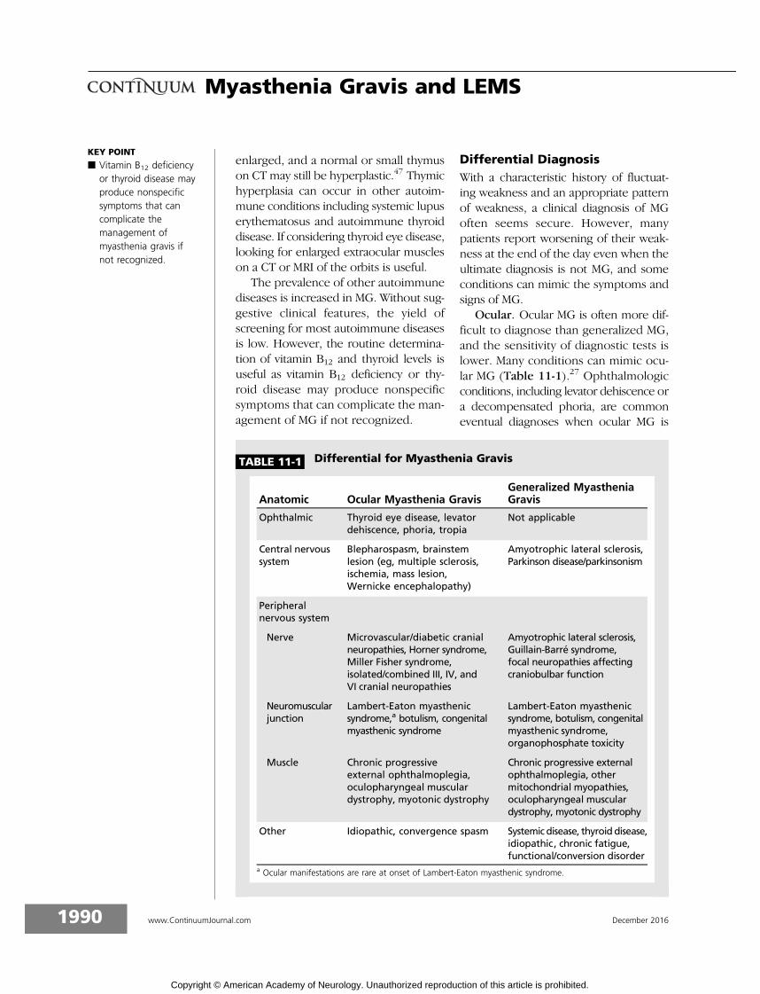
Ocular. Ocular MG is often more dif-ficult to diagnose than generalized MG,and the sensitivity of diagnostic tests islower. Many conditions can mimic ocu-lar MG (Table 11-1).27 Ophthalmologicconditions, including levator dehiscence ora decompensated phoria, are commoneventual diagnoses when ocular MG is
KEY POINT
h Vitamin B12 deficiencyor thyroid disease mayproduce nonspecificsymptoms that cancomplicate themanagement ofmyasthenia gravis ifnot recognized.
TABLE 11-1 Differential for Myasthenia Gravis
Anatomic Ocular Myasthenia GravisGeneralized MyastheniaGravis
Ophthalmic Thyroid eye disease, levatordehiscence, phoria, tropia
Not applicable
Central nervoussystem
Blepharospasm, brainstemlesion (eg, multiple sclerosis,ischemia, mass lesion,Wernicke encephalopathy)
Amyotrophic lateral sclerosis,Parkinson disease/parkinsonism
Peripheralnervous system
Nerve Microvascular/diabetic cranialneuropathies, Horner syndrome,Miller Fisher syndrome,isolated/combined III, IV, andVI cranial neuropathies
Amyotrophic lateral sclerosis,Guillain-Barre syndrome,focal neuropathies affectingcraniobulbar function
Neuromuscularjunction
Lambert-Eaton myasthenicsyndrome,a botulism, congenitalmyasthenic syndrome
Lambert-Eaton myasthenicsyndrome, botulism, congenitalmyasthenic syndrome,organophosphate toxicity
Muscle Chronic progressiveexternal ophthalmoplegia,oculopharyngeal musculardystrophy, myotonic dystrophy
Chronic progressive externalophthalmoplegia, othermitochondrial myopathies,oculopharyngeal musculardystrophy, myotonic dystrophy
Other Idiopathic, convergence spasm Systemic disease, thyroid disease,idiopathic, chronic fatigue,functional/conversion disorder
a Ocular manifestations are rare at onset of Lambert-Eaton myasthenic syndrome.
1990 www.ContinuumJournal.com December 2016
Myasthenia Gravis and LEMS
Copyright © American Academy of Neurology. Unauthorized reproduction of this article is prohibited.

being considered. Microvascular neurop-athies, including diabetic or hypertensivemicrovascular cranial neuropathies, af-fecting the III, IV, or VI cranial nervesshould be considered, especially withperiorbital pain or headache at the onsetof diplopia. Symmetric restriction inextraocular muscles and progressive sym-metric ptosis, evenwith a history ofminorfluctuations, suggests chronic progressiveexternal ophthalmoplegia. Thyroid eyedisease may mimic or coexist with ocularMG. In about 25% of patients who have acombination of ptosis and diplopia, nodiagnosis is determined.48
Generalized. In a typical case of gen-eralized MG, the differential is limited(Table 11-1), and the sensitivity of diag-nostic tests is higher.27 A congenitalmyasthenic syndrome should be consid-ered in patients who are seronegativeand who have onset at birth, early in life,and even in adulthood. Some congenitalmyasthenic syndromes can present inadolescence or even adulthood causedby mutations in the genes RAPSN, DOK7,or COLQ. When ocular symptoms areabsent and hyporeflexia or areflexia andautonomic dysfunction occur, considerLEMS. Other conditions to be consid-ered when the deep tendon reflexes arereduced include Guillain-Barre syn-drome and its Miller Fisher variant andrare cases of multifocal neuropathy withconduction block with cranial nerve mus-cle involvement. In oculopharyngeal mus-cular dystrophy, ptosis and generalizedweakness can occur, but extraocularmuscle involvement is usually absent.Mitochondrial myopathies can be difficultto distinguish clinically from MG, andsingle fiber EMG can be abnormal inmany myopathic disorders includingoculopharyngeal muscular dystrophyand mitochondrial myopathies.34 Centralnervous systemdisorders such as Parkinsondisease, progressive supranuclear palsy,or brainstem lesions are usually not diffi-cult to distinguish fromMG after a careful
history and examination. Functional weak-ness is also commonly in the differential.
ManagementThe crucial first steps in managing MGare to ensure that the diagnosis is cor-rect and that the symptoms being treatedare those of MG. Causes of refractory MGinclude an incorrect diagnosis or symp-toms that are not due to MG. Whenpatients do not improve as expected, itis useful to reconsider the diagnosis,especially if the patient is seronegative.If a patient is seropositive, it is importantto establish that the symptoms are con-sistent with MG. A patient with MG whoimproves with treatment but then reportsincreasing weakness may be describingfatigue, exhaustion, or daytime somno-lence, which may be symptoms of ob-structive sleep apnea.24 In addition to aclinical assessment, repeat electrophysio-logic studies (ie, repetitive nerve stimula-tion) may help determine whether theweakness is secondary to MG. Other rea-sons for worsening symptoms includeadverse effects from medications (pred-nisone in particular), mood disorders,and social circumstances.
Education of patients and primary carephysicians is crucial. Expectations of thetreating physician or patient about whentreatments should start being effectiveare sometimes unreasonable.49 Under-dosing (or occasionally overdosing) orabandoning a treatment too early canboth result in treatment ‘‘failure.’’ Adiscussion about each medication, pos-sible adverse effects and strategies tomanage them, expected time course ofbenefits (hours to days for pyridostig-mine, weeks to many months for predni-sone, and manymonths to a year or morefor azathioprine) is crucial. For somemed-ications, monitoring for adverse effects isessential, and a discussion of the impor-tance of this increases compliance. Forphysicians managing many patients withMG, having this educational information
KEY POINTS
h A congenital myasthenicsyndrome should beconsidered in patientswho are seronegative andwho have onset at birth,early in life, and even inadulthood.
h Causes of refractorymyasthenia gravis includean incorrect diagnosis orsymptoms that are notdue to myasthenia gravis.When patients do notimprove as expected, it isuseful to reconsider thediagnosis, especially if thepatient is seronegative.
1991Continuum (Minneap Minn) 2016;22(6):1978–2005 www.ContinuumJournal.com
Copyright © American Academy of Neurology. Unauthorized reproduction of this article is prohibited.

in print form in addition to a verbaldiscussion saves time and reinforces themessage. Sending a list of the medica-tions that should be avoided in patientswith MG to the primary care physicianis useful, although this is not infre-quently ignored. Finally, a discussionof which symptoms might be secondarytoMG (weakness) and which almost neveroccur (eg, pain, memory loss, sensorysymptoms, systemic disorders) maximizesefficiency of the neurologist’s time.
Management of ocular myastheniagravis. Although not life-threatening, ocu-lar MG can be disabling. The first decisionis whether symptoms require treatment.When symptoms are mild and infrequent,it may be best to defer treatment untilthey become troublesome. A trial ofpyridostigmine may be warranted, butthe medication is often ineffective orminimally effective, especially for diplo-pia. The risk to benefit ratio of prednisonemay not beworth accepting. Retrospectiveevidence suggests that prednisone mayreduce the risk of generalization.50 How-ever, it may be better to avoid predni-sone when not required, knowing that itis just as likely to be effective if and whenMG becomes generalized.
MG is treated symptomatically withpyridostigmine, which inhibits acetyl-cholinesterase at the neuromuscularjunction and increases available acetylcho-line but does not treat the underlyingimmunopathogenesis. Pyridostigmine isthe usual first step in treating ocular MG.It has few serious side effects and, if ef-fective, works quickly. Starting at 30 mgevery 4 hours during the waking day, withthe first tablet taken within an hour ofarising, is a reasonable strategy. Unlessnocturnal symptoms occur, a bedtimetablet is wasted. If required, the dosecan be increased to 60 mg every 4 hoursin 3 to 7 days. The regular 60 mg pyri-dostigmine pills have a more consistentbioavailability, and the author rarelyuses the sustained-release formulation.
Rarely, patients need lower initial dosesor a slower rate of escalation. Havingalready discussed the next options withthe patient at the time of medicationinitiation, the author has the patient callto provide an update on his or her re-sponse 2 weeks after starting the medica-tion. Even when a complete responseoccurs, occasionally symptoms will breakthrough over the next several months. If apartial response occurs, further increasesof 30 mg to 60 mg at each dose can bemade at intervals of 1 to 2 weeks up toa maximum of 480 mg/d, depending ontolerance. Higher doses are unlikely toproduce additional benefit. Diarrhea,one of the more common side effects,is often self-limited, but loperamide helpsin most cases. If no response occurs, theauthor moves on to immunosuppression,usually adding to pyridostigmine, althoughif it is clear that no response has oc-curred, the medication could be stopped.
Prednisone is very useful in patientswith ocular MG, although patience is re-quired. The evidence in adults that pred-nisone given on alternate days is lesslikely to cause adverse effects is prac-tically nonexistent, but starting low doses(eg, 25mg every other day) of prednisonewill help most patients in about 3 to 4months.51 Occasionally, patients fluctu-ate and are worse on the off day, andglycemic control using the alternate-dayapproach is difficult in diabetics, sosometimes starting or reverting to anequivalent daily dose is required. If at3 to 4 months the symptoms are notimproving (they may not be completelygone), doubling the dose to either 25mg/dor 50 mg on alternate days helps most ofthe remaining patients. Physicians shouldbe familiar with common side effects ofprednisone and discuss these with thepatient.3 Anticipating worsening in hy-pertension and glycemic control andenlisting the help of the primary carephysician in managing these is helpful.Individuals older than age 50 taking more
KEY POINTS
h A complete lack ofresponse to treatment isunusual in patients withmyasthenia gravis andshould prompt physiciansto reconsider the diagnosisof myasthenia gravis inpatients who areseronegative or reconsiderwhether myastheniagravis is the cause of thesymptoms in patients whoare seropositive.
h Many treatments ofmyasthenia gravis taketime to reach maximalefficacy, and anothercause for nonresponse totreatment is unreasonableexpectations about howlong it should take beforeimprovement begins.
1992 www.ContinuumJournal.com December 2016
Myasthenia Gravis and LEMS
Copyright © American Academy of Neurology. Unauthorized reproduction of this article is prohibited.

than 7.5 mg/d for more than 3 monthsshould be on osteoporosis prophylaxis atthe outset.52
With patience, prednisone doseshigher than 25 mg/d to 30 mg/d arerarely required in patients with ocularMG. About 2 to 3 months after thesymptoms have resolved, it may beappropriate to start tapering the dose.With doses of more than 20 mg/d, taperby 10 mg a month, and with doses of lessthan 20 mg/d, taper by 5 mg a month(10 mg on the alternate day). It is oftenuseful to taper by 2.5 mg or even 1 mgreductions every 1 to 2 months withdoses of less than 5 mg/d to 10 mg/d.Symptoms often recur at doses of lessthan 5 mg/d to 10 mg/d, and many pa-tients require long-term low-dose pred-nisone. Tapering too quickly or whilethe patient is still symptomatic will almostalways result in a relapse, often severalmonths after a reduction.
The databases used by most pharma-cies in North America sometimes flag thecombination of prednisone and pyridos-tigmine as a potentially harmful interac-tion. The vast majority of patients withMG will be on this combination at somepoint. The literature supporting thiswarning is decades old and likely relatesto the early worsening that can be seenwith the use of high-dose prednisone andnot to an interaction between the two.To avoid future telephone calls and con-fusion on the part of the patient, theauthor often sends a form letter with theprescription politely suggesting thatthe pharmacist ignore this warning.
Azathioprine can be used in ocularMG, either alone (if the patient is willingto wait) or along with prednisone in pa-tients where long-term/high-dose predni-sone is best avoided. Azathioprine canalso be added later if no response toprednisone occurs by 6 to 9 months orif the patient worsens when tapering.Other immunosuppressants can also beused in patients with ocular MG. The
author avoids combining more than two(prednisone and one other) immuno-suppressants because of the increasedrisk of infections.
Management of generalized myas-thenia gravis. The treatment of MG ishighly individualized, and only generalguidelines are discussed. The optionschosen depend on serostatus (AChR ver-sus other), age, comorbidities, and dis-ease severity. For most choices, no trialevidence exists to establish effective-ness.53 However, the collective experi-ence supports the most commonly usedmedications and treatments. The choiceoften comes down to physician experi-ence and familiarity as well as patienttolerance. For all treatment options, adiscussion of expected benefits, includ-ing the time frame that is involved, aswell as common adverse effects andmonitoring required, is useful.
Symptomatic. As is the case for ocularMG, in mild to moderate generalizedMG, pyridostigmine may be the onlytreatment at first. The same strategy andadvice about pyridostigmine previouslydiscussed for ocular MG also applies tothe treatment of generalized MG. A cho-linergic crisis, in which weakness isworsened by increased doses of pyri-dostigmine, rarely occurs. Almost always,a careful history will reveal worsening inMG symptoms prior to increases in thedose of pyridostigmine. In mild to mod-erate generalized MG, the concomitantuse of prednisone and sometimes anotherimmunosuppressant is often required.In more severe MG, immunosuppres-sion and sometimes immunomodula-tion should be started at the same timeas pyridostigmine.
Immunosuppression. For mild gen-eralized MG, when pyridostigmine aloneis not helpful, low-dose alternate-dayprednisone is useful. For moderate gen-eralized MG, higher doses may be re-quired. For severeMG, especially if bulbaror respiratory weakness occurs, higher
KEY POINT
h With patience, mostpatients with myastheniagravis will respond tolower doses of prednisone,although this may takeseveral months.
1993Continuum (Minneap Minn) 2016;22(6):1978–2005 www.ContinuumJournal.com
Copyright © American Academy of Neurology. Unauthorized reproduction of this article is prohibited.

doses are effective more rapidly. How-ever, if high doses (the author considersdoses higher than 30 mg/d as the cutofffor concerns about this phenomenon)are used initially, about 40% of patientswith MG may worsen initially before theystart to improve, and 10% overall worsensignificantly.54 This usually starts 4 to 5days after beginning prednisone andlasts about 4 to 7 days before improve-ment then occurs. Strategies to avoidthis initial worsening of symptoms in-clude starting at low doses (eg, 10 mg/d)with increases every 3 to 5 days in 10 mgsteps until the desired dose is reached.55
The use of IVIg or plasma exchange whenprednisone is started may also preventinitial worsening. Prednisone doses ofmore than 1 mg/kg/d are rarely required,and the author usually uses a maximumdose of 0.5 mg/kg/d to 0.75 mg/kg/d.Recent trial evidence suggests that, withpatience, most patients respond to rela-tively low doses of prednisone.56 Predni-sone takes months (usually 3 to 6 monthsand sometimes longer) before maximumbenefit occurs. Higher doses might accel-erate this slightly but definitely increasethe risk of adverse effects. With bulbar orrespiratory weakness, when a more rapidresponse is required, immunomodulatorytreatments should be used (see the fol-lowing section on immunomodulation).
In mild generalized MG, if predni-sone is best avoided, azathioprine canbe used alone provided the patient canwait the 12 to 18 months (or more) that itmay take before optimal benefit is seen.57
Starting prednisone and azathioprine atthe same time takes advantage of theearlier benefits from prednisone aswell as the eventual steroid-sparingeffects of azathioprine. Tapering regi-mens are similar to ocular MG (largerreductions at doses of more than 30mg/dand smaller below this). Starting aza-thioprine at 25 mg/d with increasesevery 2 weeks to 50 mg/d, 100 mg/d,and 150 mg/d lessens some adverse
effects. In the author’s experience, about1% to 2% of patients, higher in others’experience, will experience a flulike re-action within the first 2 weeks that almostalways requires discontinuation. Monitorhepatic transaminases (alanine amino-transferase [ALT], aspartate amino-transferase [AST], and most importantly,-glutamyltransferase [GGT]) and a com-plete blood count and differential weeklyfor the first 8 weeks and monthly there-after. Hepatotoxicity, usually mild andreversible, occurs in 15% and mye-losuppression in 10% at a median ofabout 6 weeks after starting.58 Both willresolve after a dose reduction, althoughif these adverse effects are rapid orsignificant, discontinuation may be re-quired. Neutropenia is the main concern,whereas lymphopenia and macrocytosisare common and benign findings that areseen when monitoring blood work withlong-term azathioprine use. After 6 to12 months on azathioprine, if no im-provement occurs at 150 mg/d, in mostpatients not at maximal doses based onweight (ie, the patient weighs more than50 kg [110 lb]), the dose can be increasedin 50 mg to 100 mg steps every 3 to 6months to a maximum of 2.5 mg/kg/d to3.0 mg/kg/d based on actual body weight.Long-term azathioprine use may increasethe risk of malignancies, particularly der-matologic, although the absolute risk ofthis is low.59 Strategies to reduce this areprudent.60 If a risk of or previoushistory of skin cancers already exists,using mycophenolate might pose alower risk.61
In patients who do not respond to ortolerate azathioprine, other immuno-suppressants can be used. Mycophenolate(either mofetil or sodium) is a commonnext step. Although two trials failed toshow an additional benefit comparedto prednisone alone over 36 weeks,this may reflect the efficacy of predni-sone in relatively mild MG within thefirst 9 months rather than a failure of
KEY POINT
h Caution is advised whenstarting patients withmyasthenia gravis whohave any bulbar orrespiratory weakness onhigh doses of prednisoneas patients mayworsen initially.
1994 www.ContinuumJournal.com December 2016
Myasthenia Gravis and LEMS
Copyright © American Academy of Neurology. Unauthorized reproduction of this article is prohibited.
mycophenolate.56 Mycophenolate iswidely used in MG and likely takes atleast 6 months and perhaps longer to pro-duce significant benefit. Mycophenolateis generally well tolerated and may bemore suitable in patients with skincancer. Other immunosuppressants usedfrequently in MG include cyclosporine,tacrolimus, methotrexate, and cyclophos-phamide. Some retrospective and limitedtrial evidence supports the efficacy ofeach of these. Most of these agents havegreater toxicity than azathioprine andmycophenolate and require more in-tense monitoring.
Patients with MuSK antibodiesmay be less likely to respond to (andmay even worsen) and have moreadverse effects from pyridostigmine.42,62
MuSK MG may also be less likely torespond to IVIg and perhaps immuno-suppressants in general, but azathio-prine in particular.42,63
Immunomodulation. In a myasthen-ic crisis, respiratory weakness requiresintubation. Historically, a myasthenic crisisoccurred in 15% to 30% of patients withMG but is less common now with effectivemanagement options. A myasthenic crisisusually occurs within the first few yearsafter disease onset and is sometimesthe presenting incident. Along withsevere bulbar weakness producing as-piration, a myasthenic crisis was thecause of mortality rates of 50% or moreprior to effective treatment. With thecurrent options and improved intensivecare, mortality from a myasthenic crisisis less than 5%. Severe MG with dyspneamanaged with noninvasive ventilationor severe dysphagia should be managedsimilarly to a crisis. Common precipi-tants for a myasthenic crisis includeunderlying infection (often pneumo-nia), aspiration, surgery including thy-mectomy, hormonal changes during orafter pregnancy, tapering or dis-continuation of MG medications, andhigh doses of prednisone or other
medications, which can interfere withneuromuscular transmission.64 In al-most one-third of cases, a cause isnot identified.
For patients who have an acute exac-erbation of MG, pyridostigmine is ofteninsufficient, and its adverse effects ofincreased oral and tracheobronchialsecretions may worsen the situation.Immunosuppressants may take months;therefore, in addition to treating the un-derlying precipitant, immunomodulatorytreatments are useful for a crisis or severeexacerbation and include IVIg and plasmaexchange.65,66 Both treatments are simi-larly effective, although plasma exchangemay be slightly faster and more effectivein a myasthenic crisis and more useful inpatients with MuSK MG.42,67 Either IVIgor plasma exchange is also used pre-operatively, before thymectomy or otherurgent surgery, in a patient with MG whois significantly symptomatic. Neither is re-quired routinely, and their use should berestricted to those patients with signifi-cant preoperative respiratory or bulbarweakness. IVIg or plasma exchange haveno effect on the long-term course of MGand, with few exceptions, are not indi-cated for the chronic management ofMG.68 Rarely, a patient will require on-going treatment with IVIg or plasmaexchange for refractory MG.
Immunomodulatory treatments target-ing B lymphocytes have shown significantpromise in patients with MuSK or AChRMG. Rituximab, which targets CD20 on theB lymphocyte cell surface, works in manypatients with MG with severe diseaseand who are refractory to the usualtreatments. It has been suggested thatMuSK MG is more likely to respond torituximab.21,42 However, a meta-analysisshowed efficacy of rituximab in more than80% of patients with MG regardless ofwhich antibody was positive.69 The onsetof action is probably slower than IVIg orplasma exchange but the duration ofimprovement is often 6 to 12 months or
KEY POINT
h In a myasthenic crisis,pyridostigmine is oftennot sufficient, andimmunosuppressiveoptions, althougheffective, can take monthsto produce benefit.Immunomodulation, witheither IV immunoglobulinor plasma exchange,produces earlierimprovement in mostpatients and bridges thegap while waiting forimmunosuppressionto work.
1995Continuum (Minneap Minn) 2016;22(6):1978–2005 www.ContinuumJournal.com
Copyright © American Academy of Neurology. Unauthorized reproduction of this article is prohibited.

more.70 Rituximab may offer significantbenefits in severe or refractory MG,especially in those being treated withlong-term IVIg or plasma exchange.
IgG3 and, to a lesser extent, IgG1and IgG2 AChR antibodies, which fixcomplement, can destroy the muscleendplate. Therapies targeting comple-ment including eculizumab have beenassessed in MG and its experimentalmodel and appear promising.70
Thymectomy. Indications for a thy-mectomy in patients with MG includeremoval of a thymoma and to increasethe chance of a sustained drug-free remis-sion, but these two indications are notsynonymous. Removing a thymoma prob-ably does not improve the course of MGbut is almost always indicated to reducethe chance of local growth, invasion,and metastases.71 If removed at an earlystage, the long-term survival is good.Once local invasion or metastases occur,adjuvant therapy with radiation or che-motherapy is usually indicated.
Given its role in the production ofAChR antibodies, removal of a hyper-plastic thymus has long been considereda management option for generalizedMG. The evidence supporting this ismostly retrospective, and evidence-basedreviews concluded that thymectomymay be a management option in somepatients with MG.72,73 A recently con-cluded randomized trial supports therole for thymectomy under specific con-ditions in MG.74 Although the specificsof each indication are still somewhatcontroversial, the most accepted indica-tions for a trans-sternal thymectomyinclude patients with generalized early-onset MG who are AChR antibody pos-itive and within 5 years of disease onset.The role for thymectomy outside theseindications is less certain. Improvementmay take 1 to 2 years after thymectomy.Patients with MG should be medicallywell controlled and ideally on lower dosesof prednisone prior to thymectomy.
Other surgical approaches are usedin some centers. Most consider a cervicalthymectomy suboptimal because of therisk that thymic tissue will be left behind.The role forminimally invasive approaches(robotic or video-assisted thoracoscopy)remains to be proven, although theseapproaches are increasingly used inmany centers. Despite occasional reportsof efficacy of thymectomy in MuSK MG,given the lack of thymic pathology inMuSK MG this remains a controversialindication.21 Thymectomy in seronega-tive MG, where thymic hyperplasia isless prominent, is also less proven and achance always exists that a patient who isseronegative does not have MG.
Pregnancy. For the most part MG ismanaged during pregnancy as usual. Plan-ning any changes in management in ad-vance, including before conception whenpossible, is important. When MG is wellcontrolled, the risks to mother and childare minimal. Vaginal deliveries are en-couraged, and indications for a cesareandelivery are no different than in a motherwithout MG.75 Although neonatal MG israre in the author’s experience, motherswith MG should be managed as a high-risk pregnancy in a center capable ofcaring for both neonate and mother.Most medications used in MG, includingpyridostigmine, prednisone, and azathio-prine, appear to be safe in pregnancy.21,76,77
Methotrexate and mycophenolate mustbe avoided as they are teratogenic. IVIgand plasma exchange are also relativelysafe in pregnancy. The severity of theweakness in MG may improve or worsen,the latter especially in the puerperium,during pregnancy.
Other medications and myastheniagravis. There are many lists of drugs toavoid in MG as they might worsen theweakness in a patient with MG. Theevidence supporting inclusion of someof these is often weak. Many patientswith MG, especially the elderly, will beon one or more of these (beta-blockers
KEY POINTS
h Although the specifics ofeach indication are stillsomewhat controversial,the most acceptedindications for atrans-sternal thymectomyinclude patients withgeneralized early-onsetmyasthenia gravis who areacetylcholine receptorantibody positive andwithin 5 years ofdisease onset.
h Many commonly usedmedications to treatmyasthenia gravis,including pyridostigmine,prednisone, andazathioprine, are safe foruse during pregnancy.
1996 www.ContinuumJournal.com December 2016
Myasthenia Gravis and LEMS
Copyright © American Academy of Neurology. Unauthorized reproduction of this article is prohibited.

and calcium channel blockers in parti-cular) with no worsening in their MG.78
Some antibiotics (aminoglycosides,macrolides, and fluoroquinolones, forinstance) are probably best avoided ifpossible but even then, many patientswith MG have been on one or more ofthese without ill effect. Most shouldbe considered a relative contraindica-tion, and patients and physicians shouldweigh the benefits of that specific medi-cation versus the risk that the MG maybe worsened while on the medication. Ifworsening does occur, it is sometimesdifficult to exclude worsening becauseof an underlying infection, which is acommon reason for exacerbation in MG.Patients with MG undergoing a generalanesthetic may be more sensitive tonondepolarizing neuromuscular blockingagents and less sensitive to depolarizingneuromuscular blocking agents. Spinalor local anesthetics are generally saferoptions. Any patient with MG who hassignificant weakness, especially if bulbaror respiratory, and who is started on oneof the potentially offending medicationsshould be monitored carefully.
TrendsRecent advances in identifying new anti-genic targets and improved assays forexisting antibodies promise to furtherreduce the cases of generalized MG thatare difficult to diagnose. Therapies thatare directed to specific components ofthe aberrant immune response, includ-ing complement and CTLA4, may proveinteresting. Trials that demonstrate ef-ficacy of current off-label options willexpand the list of available treatmentsfor many patients with MG.
LAMBERT-EATONMYASTHENIC SYNDROMEAlthough much less common than MG, itis important for neurologists to recognizethe clinical features of LEMS and befamiliar with its management.
PathophysiologyThe antigenic target in LEMS is the P/Qtype voltage-gated calcium channel(VGCC) on the presynaptic nerve termi-nal.79 VGCC antibodies reduce calciuminflux into the nerve terminal and, there-fore, reduce the amount of acetylcholinereleased into the synaptic cleft. At theneuromuscular junction of skeletal mus-cle, this can result in neuromusculartransmission failure. Involvement of thesame VGCC at autonomic synapses pro-duces autonomic dysfunction. VGCCantibodies can be found in other para-neoplastic disorders including subacutecerebellar degeneration.
LEMS is divided into paraneoplasticand primary autoimmune groups. Inparaneoplastic LEMS, an underlyingsmall cell lung cancer is almost alwayspresent. In children with LEMS, lym-phoproliferative disorders may be asso-ciated instead.
EpidemiologyLEMS is rare, with an estimated incidenceof 0.5 out of 1 million and a prevalenceof 2.3 out of 1 million. Relative to MG,the prevalence of LEMS is reducedcompared to its incidence.80 This reflectsthe poor survival in paraneoplasticLEMS. Approximately 40% to 50% ofpatients with LEMS have a primaryautoimmune disorder, and in 50% to60%, LEMS occurs as a paraneoplasticdisorder, almost always with an under-lying small cell lung cancer.81 Studiessuggest that LEMS occurs in 2% to 3%of small cell lung cancer cases and islikely underrecognized.81,82
Clinical FeaturesThe clinical triad characteristic for LEMSis best remembered by the three A’s:apraxia, areflexia, and autonomic involve-ment.83,84 Although not true apraxia,this is a useful way of remembering thatleg weakness producing difficultieswith gait is the most prominent clinical
KEY POINT
h Although somemedications mightproduce worsening inpatients with myastheniagravis, few medicationsare absolutelycontraindicated, andthe potential benefitsof a medication shouldbe weighed againstthe evidence that itmight produceworsening weakness.
1997Continuum (Minneap Minn) 2016;22(6):1978–2005 www.ContinuumJournal.com
Copyright © American Academy of Neurology. Unauthorized reproduction of this article is prohibited.

feature and that, frequently, patientswith LEMS have more functional diffi-culties than predicted by the strengthof individual muscles.
The clinical features of LEMS usuallyprecede the diagnosis of underlying smallcell lung cancer, which is more often at alimited stage or occult when comparedto small cell lung cancer without LEMS.Weakness almost always begins in theproximal legs and causes difficultieswalking.83,85 Arm weakness is also com-mon. The onset is often subacute, andfluctuation is less prominent than in MG.However, ocular and bulbar weaknessmay be absent or, if present, occur as alate manifestation. The presence of ocu-lar and perhaps bulbar involvement atonset is strongly against a diagnosis ofLEMS and suggests MG.25 Thus, LEMSstarts in the legs and ascends while MGoften starts with craniobulbar involve-ment and descends. Respiratory involve-ment is uncommon.
Deep tendon reflexes are almost al-ways reduced or absent, especially inthe legs. Involvement of sympatheticand perhaps more frequently parasympa-thetic systems occurs eventually in 80%to 90% of patients with LEMS and canproduce almost any autonomic manifes-tation. A dry mouth, constipation, anderectile dysfunction in men are par-ticularly common but loss of sweating,orthostatic hypotension, and pupillaryabnormalities are also seen.84
Sensory loss or cerebellar features arenot features of LEMS but might suggestan overlapping paraneoplastic disorderassociated with an underlying small celllung cancer.
DiagnosisOnce suspected, the diagnosis ofLEMS can often be made based oncharacteristic electrophysiologic abnor-malities, which distinguish it from MG.Serologic confirmation of the diagnosisis important.
Clinical. The biggest delay to a diag-nosis of LEMS is not suspecting thecondition. Abnormalities on the neuro-logic examination will usually be mostprominent in the extremities, especiallythe legs. Reduced deep tendon reflexesin a patient where MG is being consid-ered suggests LEMS instead. Prominentocular findings at onset is against a diag-nosis of LEMS.25 Characteristic in LEMSis the paradox between significant func-tional impairment with walking but onlymild weakness on examination.
The author has not found clinicalfacilitation in strength, in which thesecond contraction of a muscle grouphas increased power relative to the first,to be a reliable sign when elicited by anunbiased examiner. However, in up to40% of patients with LEMS, a previouslyabsent or significantly reduced deeptendon reflex will return to normal after10 seconds of maximal voluntary con-traction.84 Autonomic involvement isbest established by a careful history.Many patients will not volunteer symp-toms suggesting autonomic involvementunless asked.
Weakness in a patient with small celllung cancer is often blamed on cachexia,malnutrition, or chemotherapy, and para-neoplastic LEMS is often not suspected.LEMS should be considered when apatient with an underlying small cell lungcancer develops prominent leg weaknessand the deep tendon reflexes are reducedor autonomic involvement occurs.
The prediction of an underlyingsmall cell lung cancer in patients withLEMS may be increased when the pa-tient has a higher Dutch-English LEMSTumor Association Prediction (DELTA-P)score. This uses clinical features such asbulbar weakness (dysarthria, dysphagia,chewing weakness, and neck weakness),autonomic involvement (erectile dys-function), weight loss, tobacco use, ageof older than 50 years, and a KarnofskyPerformance Status Scale score of less
KEY POINT
h Patients withLambert-Eatonmyasthenic syndromeoften present withdifficulty walking andwith leg weakness, withareflexia and autonomicinvolvement comprisingthe other two key features.
1998 www.ContinuumJournal.com December 2016
Myasthenia Gravis and LEMS
Copyright © American Academy of Neurology. Unauthorized reproduction of this article is prohibited.

than 70, with 1 point allocated for each.A total score of 3 or more predicted agreater than 90% chance of an underly-ing small cell lung cancer.86
Electrophysiologic studies. As forMG,routine nerve conduction studies andneedle EMG should be done first. Thetriad of electrophysiologic abnormali-ties in LEMS consists of the following:
& Diffusely reducedmotor amplitudeson motor nerve conduction studies,
often less than 50% of thelaboratory’s lower limits of normal.
& Decrement with low-frequencystimulation; as opposed toMG, wherethe decrement is usually maximal atthe fourth or fifth stimulation in thetrain, in LEMS themaximal decrementmay occur later in the train.87
& Increment with high-frequencystimulation or facilitation after10 seconds of maximal voluntarycontraction (Figure 11-2);
KEY POINT
h Although a decrementon repetitive nervestimulation can be seen ineither myasthenia gravisor Lambert-Eatonmyasthenic syndrome, areduction in baselinemotor amplitudes andincrement after eithermaximal voluntarycontraction orhigh-frequencystimulation arecharacteristic ofLambert-Eatonmyasthenic syndrome.
FIGURE 11-2 PreY and postYmaximal voluntary contractionulnar motor nerve conduction study in apatient with Lambert-Eaton myasthenic
syndrome. A 32-year-old woman presented with a 1-yearhistory of fatigable weakness of her legs. She also described adry mouth. She was a lifetime nonsmoker. On examination, inaddition tomildweakness of hip flexors, shewas areflexic. PreYand postYmaximal voluntary contraction motor nerve conduc-tion studies of the ulnar nerve with stimulation at the wrist andrecording over abductor digiti minimi are shown, demonstrat-ing an over 500% increment inmotor amplitude after maximalvoluntary contraction.
MVC = maximal voluntary contraction.
1999Continuum (Minneap Minn) 2016;22(6):1978–2005 www.ContinuumJournal.com
Copyright © American Academy of Neurology. Unauthorized reproduction of this article is prohibited.

postYmaximal voluntary contractionstudies are as sensitive and bettertolerated than high-frequencystimulation.88 Increments of morethan 100% are very suggestive forLEMS but not specific for LEMSand occur in some cases ofbotulism and MG.
The abnormalities may be subtleearly in the disease course. Althoughleg weakness is most predominant inLEMS, electrodiagnostic studies of theulnar or median nerves have the highestsensitivities.89 Single fiber EMG abnor-malities usually do not distinguish LEMSfrom MG.
Serologic studies. VGCC antibodiesare more than 90% sensitive in primaryautoimmune LEMS and approach 100% inparaneoplastic LEMS.84 VGCC antibodiesare not specific and are found in smallcell lung cancer without LEMS as well asother paraneoplastic disorders.83,90
Ancillary investigations. Given itsassociation with LEMS, patients shouldbe investigated for an underlying smallcell lung cancer, especially in elderlysmokers with weight loss. If an initial CTchest is negative, a bronchoscopy orpositron emission tomography (PET)scan may be indicated in high-riskpatients. When initial investigations arenegative in a high-risk patient, repeatingthe CT chest every 3 to 6 months for atleast the first 2 years is suggested, afterwhich the chance of a small cell lungcancer being found is less.91
Differential DiagnosisMG is the disorder most commonly in thedifferential for LEMS. A myopathy, espe-cially dermatomyositis, might present likeLEMS in the setting of an underlying smallcell lung cancer. Guillain-Barre syndrome,chronic inflammatory demyelinating poly-radiculoneuropathy (CIDP), and othermotor predominant subacute neuropa-thies occasionally mimic LEMS. In thecontext of small cell lung cancer, the
clinical features of LEMS are oftenattributed to the effects of cachexia andmalnutrition. Electrolyte and metabolicabnormalities affecting calcium or mag-nesium, as well as hypothyroidism arealso in the differential.
ManagementThe most effective symptomatic treat-ment in LEMS is 3,4-diaminopyridine(3,4-DAP).92 Through blocking voltage-gated potassium channels, 3,4-DAPprolongs nerve terminal depolarizationand increases acetylcholine release.79,92,93
In small trials, 3,4-DAP has been shownto produce clinical and electrophysio-logic benefit in patients with LEMS.92,94
Improvement after each dose is usuallyseen within 30 minutes and is maximalat 90 minutes.92 Starting doses areusually 5 mg to 10 mg three to fourtimes a day, with gradual increases upto 80 mg/d, divided into four to sixdoses. Many patients respond to 40 mg/dto 60 mg/d. Common adverse effectsinclude perioral and acral paresthesia,maximal about an hour after each dose,nausea, abdominal pain, tachycardia, andpalpitations.92,95 Insomnia is minimizedby avoiding the last dose at bedtime.Doses of more than 100 mg/d mayincrease the risk of seizures, althoughthis is likely a rare adverse effect.92,95
Seizures are more likely with other pre-dispositions, so caution is advised withbrain metastases or the use of othermedications that might lower the thresh-old for seizures.
In theory, pyridostigmine should besynergistic with 3,4-DAP but many pa-tients with LEMS have no benefit frompyridostigmine either on its own or incombination with 3,4-DAP.96,97
Given its immunopathogenesis, im-munosuppression may also be useful totreat LEMS.97 The choice of immunosup-pressant drugs in LEMS is similar toMG. However, avoiding immunosup-pression in paraneoplastic LEMS may
KEY POINT
h Whereas pyridostigminemay not producesignificant benefit,3,4-diaminopyridine is avery effectivesymptomatic treatmentof Lambert-Eatonmyasthenic syndrome.
2000 www.ContinuumJournal.com December 2016
Myasthenia Gravis and LEMS
Copyright © American Academy of Neurology. Unauthorized reproduction of this article is prohibited.
be advisable because of concerns aboutreduced immunosurveillance and tumorprogression. Immunosuppressants canbe useful in primary autoimmune LEMSif 3,4-DAP alone is insufficient. IVIg,plasma exchange, and rituximab mightalso be useful, although the evidencefor their efficacy in treating LEMS isweaker.94,97 Treatment of the underlyingsmall cell lung cancer may improveparaneoplastic LEMS, although it is diffi-cult to distinguish an effect of this fromimmunosuppression.97,98
PrognosisPrimary autoimmune LEMS has an ex-cellent prognosis, and most patients re-spond well to treatment, although lifelongtreatment is often required. The prog-nosis in paraneoplastic LEMS is poorand is determined by the underlyingsmall cell lung cancer.97 The presence ofLEMS may improve the prognosis com-pared with small cell lung cancer withoutLEMS.97,99,100
Trends3,4-DAP is likely to remain the mainstayof symptomatic treatment in patientswith LEMS. Current trials hope to obtainregulatory approval, making it easier toobtain, although concerns exist aboutwhether the cost of approved productswill be out of reach of many patients.Experimental approaches to pharmaco-logic improvement of calcium influxand presynaptic acetylcholine releaseare also being investigated.79
CONCLUSIONThe diagnosis and, to a certain extent,specifics of management of MG are aidedsignificantly by results of serologic assays.Most patients with MG respond to treat-ment, and a lack of response may meanthat the diagnosis is incorrect or that thetarget symptoms are not directly relatedto the disease. Symptomatic treatment
with pyridostigmine helps many patientswith MG, although it is less effective forpatients with ocular MG. Many optionsexist for immunosuppression, the choiceof which often depends on practitionerfamiliarity. The goal should be to balancebenefits with potential adverse effects;with patience, most patients will respondto lower doses than are commonly used.An underlying small cell lung cancer willbe found in more than one-half ofpatients with LEMS. 3,4-DAP is the mosteffective symptomatic treatment in LEMS,and immunosuppression is best reservedfor nonparaneoplastic LEMS that doesnot respond to 3,4-DAP.
REFERENCES1. Keesey JC. AAEE Minimonograph #33:
electrodiagnostic approach to defects ofneuromuscular transmission. Muscle Nerve1989;12(8):613Y626. doi:10.1002/mus.880120802.
2. Drachman DB, Adams RN, Stanley EF, et al.Mechanisms of acetylcholine receptor lossin myasthenia gravis. J Neurol NeurosurgPsychiatry 1980;43(7):601Y610. doi:10.1136/jnnp.43.7.601.
3. Nicolle MW. Myasthenia gravis. Neurologist2002;8(1):2Y21.
4. Koneczny I, Cossins J, Vincent A. The role ofmuscle-specific tyrosine kinase (MuSK) andmystery of MuSK myasthenia gravis. J Anat2014;224(1):29Y35. doi:10.1111/joa.12034.
5. Berrih-Aknin S. Cortactin: a new target inautoimmune myositis and Myasthenia Gravis.Autoimmun Rev 2014;13(10):1001Y1002.doi:10.1016/j.autrev.2014.08.037.
6. Berrih-Aknin S, Le Panse R. Myasthenia gravis:a comprehensive review of immunedysregulation and etiological mechanisms.J Autoimmun 2014;52:90Y100. doi:10.1016/j.jaut.2013.12.011.
7. Binks S, Vincent A, Palace J. Myasthenia gravis:a clinical-immunological update. J Neurol 2016;263(4):826j834. doi:10.1007/s00415-015-7963-5.
8. Hohlfeld R,WekerleH. The thymus in myastheniagravis. Neurol Clin 1994;12(2):331Y342.
9. Renton AE, Pliner HA, Provenzano C, et al.A genome-wide association study of myastheniagravis. JAMA Neurol 2015;72(4):396Y404.doi:10.1001/jamaneurol.2014.4103.
10. Salvado M, Canela M, Ponseti JM, et al. Studyof the prevalence of familial autoimmunemyasthenia gravis in a Spanish cohort. J NeurolSci 2016;360:110Y114. doi:10.1016/j.jns.2015.11.049.
2001Continuum (Minneap Minn) 2016;22(6):1978–2005 www.ContinuumJournal.com
Copyright © American Academy of Neurology. Unauthorized reproduction of this article is prohibited.
11. Nacu A, Andersen JB, Lisnic V, et al.Complicating autoimmune diseases inmyasthenia gravis: a review. Autoimmunity2015;48(6):362Y368. doi:10.3109/08916934.2015.1030614.
12. Gilhus NE, Nacu A, Andersen JB, et al.Myasthenia gravis and risks for comorbidity.Eur J Neurol 2015;22(1):17Y23. doi:10.1111/ene.12599.
13. Pakzad Z, Aziz T, Oger J. Increasing incidenceof myasthenia gravis among elderly inBritish Columbia, Canada. Neurology 2011;76(17):1526Y1528. doi:10.1212/WNL.0b013e318217e735.
14. Carr AS, Cardwell CR, McCarron PO, et al.A systematic review of population basedepidemiological studies in MyastheniaGravis. BMC Neurol 2010;10:46. doi:10.1186/1471-2377-10-46.
15. Gilhus NE, Verschuuren JJ. Myasthenia gravis:subgroup classification and therapeuticstrategies. Lancet Neurol 2015;14(10):1023Y1036. doi:10.1016/S1474-4422(15)00145-3.
16. Akaishi T, Yamaguchi T, Suzuki Y, et al.Insights into the classification of myastheniagravis. PLoS One 2014;9(9):e106757.doi:10.1371/journal.pone.0106757.
17. Vincent A, Clover L, Buckley C, et al. Evidenceof underdiagnosis of myasthenia gravis inolder people. J Neurol Neurosurg Psychiatry2003;74(8):1105Y1108. doi:10.1136/jnnp.74.8.1105.
18. Mao ZF, Mo XA, Qin C, et al. Incidenceof thymoma in myasthenia gravis: asystematic review. J Clin Neurol 2012;8(3):161Y169. doi:10.3988/jcn.2012.8.3.161.
19. Evoli A, Batocchi AP, Provenzano C, et al.Thymectomy in the treatment of myastheniagravis: report of 247 patients. J Neurol 1988;235(5):272Y276. doi:10.1007/BF00314173.
20. Somnier FE. Clinical implementation ofanti-acetylcholine receptor antibodies. JNeurol Neurosurg Psychiatry 1993;56(5):496Y504. doi:10.1136/jnnp.56.5.496.
21. Evoli A, Minicuci GM, Vitaliani R, et al.Paraneoplastic diseases associated withthymoma. J Neurol 2007;254(6):756Y762.doi:10.1007/s00415-006-0429-z
22. Nagia L, Lemos J, Abusamra K, et al.Prognosis of ocular myasthenia gravis:retrospectivemulticenter analysis. Ophthalmology2015;122(7):1517Y1521. doi:10.1016/j.ophtha.2015.03.010.
23. Grob D, Arsura EL, Brunner NG, et al. Thecourse of myasthenia gravis and therapiesaffecting outcome. Ann N Y Acad Sci1987;505:472Y499. doi:10.1111/j.1749-6632.1987.tb51317.x.
24. Nicolle MW, Rask S, Koopman WJ, et al.Sleep apnea in patients with myastheniagravis. Neurology 2006;67(1):140Y142.doi:10.1212/01.wnl.0000223515.15691.26.
25. Wirtz PW, Sotodeh M, Nijnuis M, et al.Difference in distribution of muscleweakness between myasthenia gravisand the Lambert-Eaton myasthenicsyndrome. J Neurol Neurosurg Psychiatry2002;73(6):766Y768. doi:10.1136/jnnp.73.6.766.
26. Nicolle MW. Wrist and finger drop inmyasthenia gravis. J Clin Neuromuscul Dis2006;8(2):65Y69. doi:10.1097/01.cnd.0000245218.70927.97.
27. Engstrom JW. Myasthenia gravis:diagnostic mimics. Semin Neurol 2004;24(2):141Y147. doi:10.1055/s-2004-830903.
28. Barohn RJ, McIntire D, Herbelin L, et al.Reliability testing of the quantitativemyasthenia gravis score. Ann NY Acad Sci1998;841:769Y772. doi:10.1111/j.1749-6632.1998.tb11015.x.
29. Benatar M. A systematic review ofdiagnostic studies in myasthenia gravis.Neuromuscul Disord 2006;16(7):459Y467.doi:10.1016/j.nmd.2006.05.006
30. Larner AJ. The place of the ice pack test inthe diagnosis of myasthenia gravis. Int J ClinPract 2004;58(9):887Y888. doi:10.1111/j.1742-1241.2004.00053.x.
31. Sanders DB, El-Salem K, Massey JM, et al.Clinical aspects of MuSK antibody positiveseronegative MG. Neurology2003;60(12):1978Y1980. doi:10.1212/01.WNL.0000065882.63904.53.
32. Stalberg EV, Trontelj J, Sanders DB. Singlefiber electromyography. Studies in healthyand diseased muscle. 3rd ed. Fiskebackskil,Sweden: Edshagen Publishing House, 2010.
33. Sanders DB, Stålberg EV. AAEM minimonograph#25: single-fiber electromyography. MuscleNerve 1996;19(9):1069Y1083. doi:10.1002/(SICI)1097-4598(199609)19:9G1069::AID-MUS193.0.CO;2-Y.
34. Ukachoke C, Ashby P, Basinski A, et al.Usefulness of single fiber EMG fordistinguishing neuromuscular from othercauses of ocular muscle weakness. Can JNeurol Sci 1994;21(2):125Y128.
35. Sanders DB, Massey EW, Buckley EG.Botulinum toxin for blepharospasm:single-fiber EMG studies. Neurology 1986;36(4):545Y547. doi:10.1212/WNL.36.4.545.
36. Vincent A, Newsom-Davis J. Acetylcholinereceptor antibody as a diagnostic test formyasthenia gravis: results in 153 validated casesand 2967 diagnostic assays. J Neurol NeurosurgPsychiatry 1985;48(12):1246Y1252. doi:10.1136/jnnp.48.12.1246.
2002 www.ContinuumJournal.com December 2016
Myasthenia Gravis and LEMS
Copyright © American Academy of Neurology. Unauthorized reproduction of this article is prohibited.

37. Chan KH, Lachance DH, Harper CM, et al.Frequency of seronegativity in adult-acquiredgeneralized myasthenia gravis. Muscle Nerve2007;36(5):651Y658. doi:10.1002/mus.20854.
38. Rodrıguez Cruz PM, Al-Hajjar M, Huda S,et al. Clinical features and diagnosticusefulness of antibodies to clusteredacetylcholine receptors in the diagnosis ofseronegative myasthenia gravis. JAMA Neurol2015;72(6):642Y649. doi:10.1001/jamaneurol.2015.0203.
39. Sanders DB, Burns TM, Cutter GR, et al.Does change in acetylcholine receptorantibody level correlate with clinical changein myasthenia gravis? Muscle Nerve2014;49(4):483Y486. doi:10.1002/mus.23944.
40. Choi Decroos E, Hobson-Webb LD, Juel VC,et al. Do acetylcholine receptor and striatedmuscle antibodies predict the presence ofthymoma in patients with myasthenia gravis?Muscle Nerve 2014;49(1):30Y34. doi:10.1002/mus.23882.
41. Luchanok U, Kaminski H. Ocular myasthenia:diagnostic and treatment recommendations andthe evidence base. Curr Opin Neurol2008;21(1):8Y15. doi:10.1097/WCO.0b013e3282f4098e.
42. Evoli A, Iorio R. Characteristics ofmyasthenia gravis with antibodies tomuscle-specific kinase and low-densitylipoprotein-related receptor protein 4.Clin Exp Neuroimmunol 2015;6(1):40Y48.doi:10.1111/cen3.12173.
43. Stickler DE, Massey JM, Sanders DB.MuSK-antibody positive myasthenia gravis:clinical and electrodiagnostic patterns.Clin Neurophysiol 2005;116(9):2065Y2068.doi:10.1016/j.clinph.2005.06.003.
44. Rodriguez Cruz PM, Huda S, Lopez-Ruiz P,et al. Use of cell-based assays in myastheniagravis and other antibody-mediateddiseases. Exp Neurol 2015;270:66Y71.doi:10.1016/j.expneurol.2015.01.011.
45. Gallardo E, Martınez-Hernandez E,Titulaer MJ, et al. Cortactin autoantibodiesin myasthenia gravis. Autoimmun Rev2014;13(10):1003Y1007. doi:10.1016/j.autrev.2014.08.039.
46. Zhang B, Shen C, Bealmear B, et al.Autoantibodies to agrin in myasthenia gravispatients. PLoS One 2014;9(3):e91816.doi:10.1371/journal.pone.0091816.
47. Priola AM, Priola SM. Imaging of thymus inmyasthenia gravis: from thymic hyperplasia tothymic tumor. Clin Radiol 2014;69(5):e230Ye245. doi:10.1016/j.crad.2014.01.005.
48. Batocchi AP, Evoli A, Majolini L, et al. Ocularpalsies in the absence of other neurologicalor ocular symptoms: analysis of 105 cases.J Neurol 1997;244(10):639Y645. doi:10.1007/s004150050160
49. Hilton-Jones D. When the patient fails torespond to treatment: myastheniagravis. Pract Neurol 2007;7(6):405Y411.doi:10.1136/jnnp.2007.134130.
50. Kupersmith MJ. Does early immunotherapyreduce the conversion of ocularmyasthenia gravis to generalizedmyasthenia gravis? J Neuroophthalmol2003;23(4):249Y250.
51. Benatar M, Mcdermott MP, Sanders DB,et al. Efficacy of prednisone for thetreatment of ocular myasthenia (EPITOME): arandomized, controlled trial. Muscle Nerve2016;53(3):363Y369. doi:10.1002/mus.24769.
52. Papaioannou A, Morin S, Cheung AM, et al.2010 clinical practice guidelines for thediagnosis and management of osteoporosis inCanada: summary. CMAJ 2010;182(17):1864Y1873. doi:10.1503/cmaj.100771.
53. Hart IK, Sathasivam S, Sharshar T.Immunosuppressive agents for myastheniagravis. Cochrane Database Syst Rev2007;(4):CD005224.
54. Arsura E, Brunner NG, Namba T, et al.High-dose intravenous methylprednisolonein myasthenia gravis. Arch Neurol1985;42(12):1149Y1153. doi:10.1001/archneur.1985.04060110031011.
55. Seybold ME, Drachman DB. Graduallyincreasing doses of prednisone inmyasthenia gravis. Reducing the hazardsof treatment. N Engl J Med 1974;290(2):81Y84.doi:10.1056/NEJM197401102900204.
56. Sanders DB, Siddiqi ZA. Lessons fromtwo trials of mycophenolate mofetil inmyasthenia gravis. Ann N Y Acad Sci2008;1132:249Y253. doi:10.1196/annals.1405.031.
57. Palace J, Newsom-Davis J, Lecky B.A randomized double-blind trial ofprednisolone alone or with azathioprine inmyasthenia gravis. Myasthenia Gravis StudyGroup. Neurology 1998;50(6):1778Y1783.doi:10.1212/WNL.50.6.1778.
58. Jack KL, Koopman WJ, Hulley D, et al. Areview of azathioprine-associatedhepatotoxicity and myelosuppression inmyasthenia gravis. J Clin Neuromusc Dis2016;18(1):12Y20. doi:10.1097/CND.0000000000000133.
59. Pedersen EG, Pottegård A, Hallas J, et al.Risk of non-melanoma skin cancer inmyasthenia patients treated withazathioprine. Eur J Neurol 2014;21(3):454Y458. doi:10.1111/ene.12329.
60. McGurgan IJ, McGuigan C. Nonmelanomaskin cancer risk awareness inazathioprine-treated myasthenia gravispatients. Brain Behav 2015;5(10):e00396.doi:10.1002/brb3.396.
2003Continuum (Minneap Minn) 2016;22(6):1978–2005 www.ContinuumJournal.com
Copyright © American Academy of Neurology. Unauthorized reproduction of this article is prohibited.
61. Chockalingam R, Downing C, Tyring SK.Cutaneous squamous cell carcinomas inorgan transplant recipients. J Clin Med2015;4(6):1229Y1239. doi:10.3390/jcm4061229.
62. Guptill JT, Sanders DB, Evoli A. Anti-MuSKantibody myasthenia gravis: clinicalfindings and response to treatment intwo large cohorts. Muscle Nerve 2011;44(1):36Y40. doi:10.1002/mus.22006.
63. Evoli A, Bianchi MR, Riso R, et al. Responseto therapy in myasthenia gravis withanti-MuSK antibodies. Ann N Y Acad Sci2008;1132:76Y83. doi:10.1196/annals.1405.012.
64. Thomas CE, Mayer SA, Gungor Y, et al.Myasthenic crisis: clinical features, mortality,complications, and risk factors for prolongedintubation. Neurology 1997;48(5):1253Y1260. doi:10.1212/WNL.48.5.1253.
65. Gajdos P, Chevret S, Toyka KV. Intravenousimmunoglobulin for myasthenia gravis.Cochrane Database Syst Rev 2012;12:CD002277. doi:10.1002/14651858.CD002277.pub4.
66. Bril V, Barnett-Tapia C, Barth D, et al. IVIGand PLEX in the treatment of myastheniagravis. Ann N Y Acad Sci 2012;1275:1Y6.doi:10.1111/j.1749-6632.2012.06767.x.
67. Dhawan PS, Goodman BP, Harper CM,et al. IVIG versus PLEX in the treatmentof worsening myasthenia gravis: whatis the evidence?: a critically appraisedtopic. Neurologist 2015;19(5):145Y148.doi:10.1097/NRL.0000000000000026.
68. Dalakas MC. IVIg in the chronic managementof myasthenia gravis: is it enough for yourmoney? J Neurol Sci 2014;338(1Y2):1Y2.doi:10.1016/j.jns.2013.12.034.
69. Iorio R, Damato V, Alboini PE, et al.Efficacy and safety of rituximab formyasthenia gravis: a systematic review andmeta-analysis. J Neurol 2015;262(5):1115Y1119.doi:10.1007/s00415-014-7532-3.
70. Dalakas MC. Future perspectives intarget-specific immunotherapies ofmyasthenia gravis. Ther Adv NeurolDisord 2015;8(6):316Y327. doi:10.1177/1756285615605700.
71. Cosi V, Romani A, Lombardi M, et al.Prognosis of myasthenia gravis: aretrospective study of 380 patients.J Neurol 1997;244(9):548Y555.doi:10.1007/s004150050142.
72. Gronseth GS, Barohn RJ. Practiceparameter: thymectomy for autoimmunemyasthenia gravis (an evidence-basedreview): report of the quality standardssubcommittee of the American Academyof Neurology. Neurology 2000;55(1):7Y15.doi:10.1212/WNL.55.1.7.
73. Skeie GO, Apostolski S, Evoli A, et al.Guidelines for treatment of autoimmuneneuromuscular transmission disorders. Eur JNeurol 2010;17(7):893Y902. doi:10.1111/j.1468-1331.2010.03019.x.
74. Wolfe GI, Kaminski HJ, Aban IB, et al.Randomized trial of thymectomy inmyasthenia gravis. N Engl J Med2016;375(6):511Y522. doi:10.1056/NEJMoa1602489.
75. Ferrero S, Pretta S, Nicoletti A, et al.Myasthenia gravis: management issues duringpregnancy. Eur J Obstet Gynecol Reprod Biol2005;121(2):129Y138. doi:10.1016/j.ejogrb.2005.01.002.
76. Norwood F, Dhanjal M, Hill M, et al.Myasthenia in pregnancy: best practiceguidelines from a UK multispecialtyworking group. J Neurol NeurosurgPsychiatry 2014;85(5):538Y543. doi:10.1136/jnnp-2013-305572.
77. Sussman J, Farrugia ME, Maddison P, et al.Myasthenia gravis: Association of BritishNeurologist’s management guidelines. PractNeurol 2015;15(3):199Y206. doi:10.1136/practneurol-2015-001126.
78. Jonkers I, Swerup C, Pirskanen R,et al. Acute effects of intravenousinjection of beta-adrenoreceptor- andcalcium channel at antagonists andagonists in myasthenia gravis. MuscleNerve 1996;19(8):959Y965. doi:10.1002/(SICI)1097-4598(199608)19:8G959::AID-MUS493.0.CO;2-7.
79. Tarr TB, Wipf P, Meriney SD. Synapticpathophysiology and treatment ofLambert-Eaton myasthenic syndrome.Mol Neurobiol 2015;52(1):456Y463.doi:10.1007/s12035-014-8887-2.
80. Wirtz PW, Nijnuis MG, Sotodeh M, et al.The epidemiology of myasthenia gravis,Lambert-eaton myasthenic syndrome andtheir associated tumours in the northernpart of the province of South Holland. JNeurol 2003;250(6):698Y701. doi:10.1007/s00415-003-1063-7.
81. O’Neill JH, Murray NM, Newsom-Davis J.The Lambert-Eaton myasthenic syndrome.A review of 50 cases. Brain 1988;111(Pt 3):577Y596. doi:10.1093/brain/111.3.577.
82. Payne M, Bradbury P, Lang B, et al.Prospective study into the incidence ofLambert Eaton myasthenic syndrome insmall cell lung cancer. J Thorac Oncol2010;5(1):34Y38. doi:10.1097/JTO.0b013e3181c3f4f1.
83. Gilhus NE. Lambert-Eaton myasthenicsyndrome; pathogenesis, diagnosis, andtherapy. Autoimmune Dis 2011;2011:973808.doi:10.4061/2011/973808.
2004 www.ContinuumJournal.com December 2016
Myasthenia Gravis and LEMS
Copyright © American Academy of Neurology. Unauthorized reproduction of this article is prohibited.
84. Titulaer MJ, Lang B, Verschuuren JJ.Lambert-Eaton myasthenic syndrome: fromclinical characteristics to therapeutic strategies.Lancet Neurol 2011;10(12):1098Y1107.doi:10.1016/S1474-4422(11)70245-9.
85. Titulaer MJ, Wirtz PW, Kuks JB, et al.The Lambert-Eaton myasthenic syndrome1988Y2008: a clinical picture in 97 patients.J Neuroimmunol 2008;201Y202:153Y158.doi:10.1016/j.jneuroim.2008.05.025.
86. Titulaer MJ, Maddison P, Sont JK, et al. ClinicalDutch-English Lambert-Eaton Myasthenicsyndrome (LEMS) tumor association predictionscore accurately predicts small-cell lungcancer in the LEMS. J Clin Oncol 2011;29(7):902Y908. doi:10.1200/JCO.2010.32.0440.
87. Baslo MB, Deymeer F, Serdaroglu P, et al.Decrement pattern in Lambert-Eatonmyasthenic syndrome is different frommyasthenia gravis. Neuromuscul Disord2006;16(7):454Y458. doi:10.1016/j.nmd.2006.05.009.
88. Tim RW, Sanders DB. Repetitive nervestimulation studies in the Lambert-Eatonmyasthenic syndrome. Muscle Nerve1994;17(9):995Y1001. doi:10.1002/mus.880170906.
89. Tim RW, Massey JM, Sanders DB.Lambert-Eaton myasthenic syndrome (LEMS).Clinical and electrodiagnostic features andresponse to therapy in 59 patients.Ann N Y Acad Sci 1998;841:823Y826.doi:10.1111/j.1749-6632.1998.tb11024.x.
90. Elrington GM, Murray NM, Spiro SG,et al. Neurological paraneoplastic syndromesin patients with small cell lung cancer.A prospective survey of 150 patients. J NeurolNeurosurg Psychiatry 1991;54(9):764Y767.doi:10.1136/jnnp.54.9.764.
91. Titulaer MJ, Soffietti R, Dalmau J, et al.Screening for tumours in paraneoplasticsyndromes: report of an EFNS task force.Eur J Neurol 2011;18(1):19Ye3. doi:10.1111/j.1468-1331.2010.03220.x.
92. Lindquist S, Stangel M. 3,4-Diaminopyridine(amifampridine) for the treatment of
Lambert-Eaton myasthenic syndrome. ExpertOpin Orphan Drugs 2014;2(3):293Y300.doi:10.1517/21678707.2014.887464.
93. Lindquist S, Stangel M. Update on treatmentoptions for Lambert-Eaton myasthenicsyndrome: focus on use of amifampridine.Neuropsychiatr Dis Treat 2011;7:341Y349.doi:10.2147/NDT.S10464.
94. Keogh M, Sedehizadeh S, Maddison P.Treatment for Lambert-Eaton myasthenicsyndrome. Cochrane Database Syst Rev2011;(2):CD003279. doi:10.1002/14651858.CD003279.pub3.
95. Flet L, Polard E, Guillard O, et al.3,4-Diaminopyridine safety in clinicalpractice: an observational, retrospectivecohort study. J Neurol 2010;257(6):937Y946.doi:10.1007/s00415-009-5442-6.
96. Wirtz PW, Verschuuren JJ, van Dijk JG,et al. Efficacy of 3,4-diaminopyridineand pyridostigmine in the treatment ofLambert-Eaton myasthenic syndrome:a randomized, double-blind,placebo-controlled, crossover study. ClinPharmacol Ther 2009;86(1):44Y48.doi:10.1038/clpt.2009.35.
97. Verschuuren JJ, Wirtz PW, Titulaer MJ, et al.Available treatment options for themanagement of Lambert-Eaton myasthenicsyndrome. Expert Opin Pharmacother2006;7(10):1323Y1336. doi:10.1517/14656566.7.10.1323
98. Chalk CH, Murray NM, Newsom-Davis J et al.Response of the Lambert-Eaton myasthenicsyndrome to treatment of associatedsmall-cell lung carcinoma. Neurology 1990;40(10):1552Y1556. doi:10.1212/WNL.40.10.1552.
99. Maddison P, Lang B. Paraneoplastic neurologicalautoimmunity and survival in small-cell lungcancer. J Neuroimmunol 2008;201Y202:159Y162.doi:10.1016/j.jneuroim.2008.05.024.
100. Wirtz PW, Lang B, Graus F, et al. P/Q-typecalcium channel antibodies, Lambert-Eatonmyasthenic syndrome and survival in small celllung cancer. J Neuroimmunol2005;164(1Y2):161Y165. doi:10.1016/j.jneuroim.2005.04.001.
2005Continuum (Minneap Minn) 2016;22(6):1978–2005 www.ContinuumJournal.com
Copyright © American Academy of Neurology. Unauthorized reproduction of this article is prohibited.
Related Documents


