Mutations in Potassium Channel Kir2.6 Cause Susceptibility to Thyrotoxic Hypokalemic Periodic Paralysis Devon P. Ryan, 1,2,14 Magnus R. Dias da Silva, 2,14,15 Tuck Wah Soong, 4,8 Bertrand Fontaine, 5 Matt R. Donaldson, 2,16 Annie W.C. Kung, 6 Wallaya Jongjaroenprasert, 7 Mui Cheng Liang, 8 Daphne H.C. Khoo, 10 Jin Seng Cheah, 9 Su Chin Ho, 10 Harold S. Bernstein, 11 Rui M.B. Maciel, 12 Robert H. Brown, Jr., 13 and Louis J. Pta ´ cek 1,2,3, * 1 Neuroscience Graduate Program 2 Department of Neurology 3 Howard Hughes Medical Institute University of California, San Francisco, San Francisco, CA, 94158, USA 4 Ion Channel and Transporter Laboratory, National Neuroscience Institute, Singapore 308433, Republic of Singapore 5 INSERM, Universite ´ Pierre et Marie Curie-UPMC, UMRS 546, and Assistance Publique-Ho ˆ pitaux de Paris, Centre de Re ´ fe ´ rence des Canalopathies Musculaires, Groupe Hospitalier Pitie ´ -Salpe ˆ trie ` re, 75013 Paris, France 6 Department of Medicine, University of Hong Kong, Queen Mary Hospital, Hong Kong SAR, China 7 Department of Medicine, Ramathibodi Hospital, Mahidol University, Bangkok 10400, Thailand 8 Department of Physiology 9 Department of Medicine Yong Loo Lin School of Medicine, National University of Singapore, Singapore 308433, Republic of Singapore 10 Department of Clinical Research, Singapore General Hospital, Singapore 169608, Republic of Singapore 11 Cardiovascular Research Institute, University of California, San Francisco, San Francisco, CA 94143-0130, USA 12 Department of Medicine, Division of Endocrinology, Universidade Federal de Sa ˜ o Paulo, Sa ˜ o Paulo 04039-032, Brazil 13 Massachusetts General Hospital, 16th Street, Navy Yard, Charlestown, MA 02129, USA 14 These authors contributed equally to this work 15 Present address: Department of Biochemistry, Universidade Federal de Sa ˜ o Paulo, Sa ˜ o Paulo 04044-020, Brazil 16 Present address: Department of Dermatology, Texas Tech University, Lubbock, TX 79409, USA *Correspondence: [email protected] DOI 10.1016/j.cell.2009.12.024 SUMMARY Thyrotoxic hypokalemic periodic paralysis (TPP) is characterized by acute attacks of weakness, hypo- kalemia, and thyrotoxicosis of various etiologies. These transient attacks resemble those of patients with familial hypokalemic periodic paralysis (hypoKPP) and resolve with treatment of the underlying hyperthyroidism. Because of the pheno- typic similarity of these conditions, we hypoth- esized that TPP might also be a channelopathy. While sequencing candidate genes, we identified a previously unreported gene (not present in human sequence databases) that encodes an inwardly rectifying potassium (Kir) channel, Kir2.6. This channel, nearly identical to Kir2.2, is expressed in skeletal muscle and is transcriptionally regulated by thyroid hormone. Expression of Kir2.6 in mammalian cells revealed normal Kir currents in whole-cell and single-channel recordings. Kir2.6 mutations were present in up to 33% of the unre- lated TPP patients in our collection. Some of these mutations clearly alter a variety of Kir2.6 properties, all altering muscle membrane excitability leading to paralysis. INTRODUCTION The periodic paralyses and nondystrophic myotonias comprise a group of muscle disorders characterized by abnormal muscle relaxation (myotonia) and/or paroxysmal muscle weakness. These disorders are subdivided on the basis of the presence of myotonia with weakness, response to potassium, temperature sensitivity, association with cardiac arrhythmia and facial dys- morphology, and induction by thyrotoxicosis. Many of these entities are now known to result from ion channel mutations, termed channelopathies (Ryan and Pta ´ cek, 2007). TPP is a sporadic muscle disorder characterized by episodic attacks of weakness and hypokalemia in some thyrotoxic indi- viduals. Patients are normal between attacks, which resolve with treatment of thyrotoxicosis. Episodic weakness in TPP is clinically similar to the known channelopathies familial hypoKPP and Andersen-Tawil syndrome (ATS) (Bulman et al., 1999; Plaster et al., 2001; Pta ´ cek et al., 1994) and is the most common form of periodic paralysis, being most prevalent in young Asian and Latin American men (Kung, 2006; Silva et al., 2004). In these populations, up to 10% of thyrotoxic male individuals develop 88 Cell 140, 88–98, January 8, 2010 ª2010 Elsevier Inc.

Welcome message from author
This document is posted to help you gain knowledge. Please leave a comment to let me know what you think about it! Share it to your friends and learn new things together.
Transcript

Mutations in Potassium Channel Kir2.6Cause Susceptibility to ThyrotoxicHypokalemic Periodic ParalysisDevon P. Ryan,1,2,14 Magnus R. Dias da Silva,2,14,15 Tuck Wah Soong,4,8 Bertrand Fontaine,5 Matt R. Donaldson,2,16
Annie W.C. Kung,6 Wallaya Jongjaroenprasert,7 Mui Cheng Liang,8 Daphne H.C. Khoo,10 Jin Seng Cheah,9 Su Chin Ho,10
Harold S. Bernstein,11 Rui M.B. Maciel,12 Robert H. Brown, Jr.,13 and Louis J. Pta�cek1,2,3,*1Neuroscience Graduate Program2Department of Neurology3Howard Hughes Medical Institute
University of California, San Francisco, San Francisco, CA, 94158, USA4Ion Channel and Transporter Laboratory, National Neuroscience Institute, Singapore 308433, Republic of Singapore5INSERM, Universite Pierre et Marie Curie-UPMC, UMRS 546, and Assistance Publique-Hopitaux de Paris, Centre de Reference des
Canalopathies Musculaires, Groupe Hospitalier Pitie-Salpetriere, 75013 Paris, France6Department of Medicine, University of Hong Kong, Queen Mary Hospital, Hong Kong SAR, China7Department of Medicine, Ramathibodi Hospital, Mahidol University, Bangkok 10400, Thailand8Department of Physiology9Department of Medicine
Yong Loo Lin School of Medicine, National University of Singapore, Singapore 308433, Republic of Singapore10Department of Clinical Research, Singapore General Hospital, Singapore 169608, Republic of Singapore11Cardiovascular Research Institute, University of California, San Francisco, San Francisco, CA 94143-0130, USA12Department of Medicine, Division of Endocrinology, Universidade Federal de Sao Paulo, Sao Paulo 04039-032, Brazil13Massachusetts General Hospital, 16th Street, Navy Yard, Charlestown, MA 02129, USA14These authors contributed equally to this work15Present address: Department of Biochemistry, Universidade Federal de Sao Paulo, Sao Paulo 04044-020, Brazil16Present address: Department of Dermatology, Texas Tech University, Lubbock, TX 79409, USA
*Correspondence: [email protected] 10.1016/j.cell.2009.12.024
SUMMARY
Thyrotoxic hypokalemic periodic paralysis (TPP) ischaracterized by acute attacks of weakness, hypo-kalemia, and thyrotoxicosis of various etiologies.These transient attacks resemble those of patientswith familial hypokalemic periodic paralysis(hypoKPP) and resolve with treatment of theunderlying hyperthyroidism. Because of the pheno-typic similarity of these conditions, we hypoth-esized that TPP might also be a channelopathy.While sequencing candidate genes, we identifieda previously unreported gene (not present in humansequence databases) that encodes an inwardlyrectifying potassium (Kir) channel, Kir2.6. Thischannel, nearly identical to Kir2.2, is expressed inskeletal muscle and is transcriptionally regulatedby thyroid hormone. Expression of Kir2.6 inmammalian cells revealed normal Kir currents inwhole-cell and single-channel recordings. Kir2.6mutations were present in up to 33% of the unre-lated TPP patients in our collection. Some of thesemutations clearly alter a variety of Kir2.6 properties,
88 Cell 140, 88–98, January 8, 2010 ª2010 Elsevier Inc.
all altering muscle membrane excitability leading toparalysis.
INTRODUCTION
The periodic paralyses and nondystrophic myotonias comprise
a group of muscle disorders characterized by abnormal muscle
relaxation (myotonia) and/or paroxysmal muscle weakness.
These disorders are subdivided on the basis of the presence of
myotonia with weakness, response to potassium, temperature
sensitivity, association with cardiac arrhythmia and facial dys-
morphology, and induction by thyrotoxicosis. Many of these
entities are now known to result from ion channel mutations,
termed channelopathies (Ryan and Ptacek, 2007).
TPP is a sporadic muscle disorder characterized by episodic
attacks of weakness and hypokalemia in some thyrotoxic indi-
viduals. Patients are normal between attacks, which resolve
with treatment of thyrotoxicosis. Episodic weakness in TPP is
clinically similar to the known channelopathies familial hypoKPP
and Andersen-Tawil syndrome (ATS) (Bulman et al., 1999;
Plaster et al., 2001; Ptacek et al., 1994) and is the most common
form of periodic paralysis, being most prevalent in young Asian
and Latin American men (Kung, 2006; Silva et al., 2004). In these
populations, up to 10% of thyrotoxic male individuals develop
episodic weakness. In Caucasians, this number is less than
0.1% (Kelley et al., 1989). Although thyrotoxicosis is more
common in women, most individuals with TPP are men.
Aside from similarity to the familial periodic paralyses, several
observations indicate that TPP may be a genetically conditioned
disorder unmasked by thyrotoxicosis. Although weakness is
a common symptom of thyrotoxicosis, episodic flaccid paralysis
arises in only some thyrotoxic patients. TPP recurs in patients
with recurring hyperthyroidism, such as relapses of Graves
Disease (Kung, 2002), and, as with other channelopathies,
a similar phenotype is seen across species (Nemzek et al., 1994).
While screening skeletal muscle ion channel candidate genes
in TPP patients, we serendipitously identified a previously unre-
ported gene—here named KCNJ18—encoding an inwardly
rectifying potassium channel, Kir2.6. This subfamily, which
includes Kir2.1 through Kir2.4 and Kir2.2v/Kir2.5, is one of seven
subfamilies (Kir1.x to Kir7.x). Kir subunits have two membrane-
spanning domains, are selective for potassium, stabilize the
resting membrane potential near the potassium equilibrium
potential, and tetramerize to form functional, inwardly rectifying
channels (Krapivinsky et al., 1998; Nichols and Lopatin, 1997).
The structural and functional homology of these channels
suggests they arose through gene duplication with subsequent
diversification. This process plays a crucial role in evolution,
allowing redundant gene variants to acquire novel characteris-
tics and functions, with a likely role in expanding protein diver-
sity. Kir channels have assumed specific functions in excitable
tissues and mutations in these channels can cause human
disease. Kir channelopathies include the episodic muscle and
developmental features of ATS (Kir2.1), the renal tubular secre-
tion defects seen with Barter syndrome type III (Kir1.1), and
defective insulin secretion in persistent hyperinsulinemic hyper-
glycemia of infancy (Kir6.2) (Abraham et al., 1999; Plaster et al.,
2001).
Kir2.6 shares 99% identity with Kir2.2 and was considered an
excellent candidate for TPP because it is primarily expressed in
skeletal muscle and probably contributes to cell membrane
excitability. Furthermore, KCNJ18 has a thyroid hormone
response cis element (TRE) within its promoter, which might
drive increased expression of Kir2.6 to protect against mem-
brane potential instability.
Genetic analysis revealed mutations in Kir2.6 in multiple TPP
patients. Electrophysiological analysis indicates that some of
these mutations have large effects on Kir2.6 currents either
inherently or via thyroid hormone-inducible mechanisms. As
such, mutations may cause predisposition for the episodic
weakness seen only during thyrotoxicosis.
RESULTS
Screening Candidate Genes and GenomicCharacterization of KCNJ18
Since ion channel mutations cause familial periodic paralysis, we
screened ion channels known to be expressed in skeletal muscle
with putative thyroid response elements (TREs) of the DR-4 motif
in their promoter regions, for mutations (Table S1 available
online). While screening KCNJ12, we noted a number of poly-
morphisms that violated Hardy-Weinberg equilibrium, suggest-
ing the existence of another gene. By changing PCR methods
and primers, we specifically amplified a highly homologous
sequence that we have submitted under NCBI accession
number FJ434338 as KCNJ18.
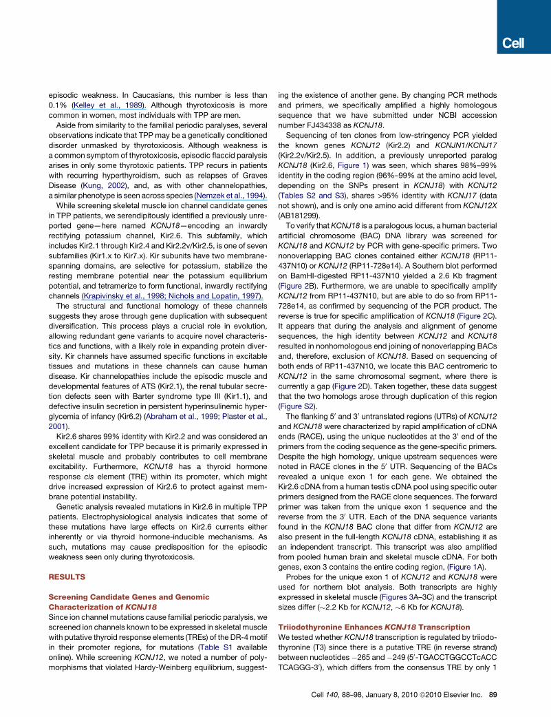
Sequencing of ten clones from low-stringency PCR yielded
the known genes KCNJ12 (Kir2.2) and KCNJN1/KCNJ17
(Kir2.2v/Kir2.5). In addition, a previously unreported paralog
KCNJ18 (Kir2.6, Figure 1) was seen, which shares 98%–99%
identity in the coding region (96%–99% at the amino acid level,
depending on the SNPs present in KCNJ18) with KCNJ12
(Tables S2 and S3), shares >95% identity with KCNJ17 (data
not shown), and is only one amino acid different from KCNJ12X
(AB181299).
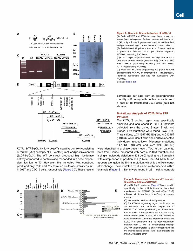
To verify that KCNJ18 is a paralogous locus, a human bacterial
artificial chromosome (BAC) DNA library was screened for
KCNJ18 and KCNJ12 by PCR with gene-specific primers. Two
nonoverlapping BAC clones contained either KCNJ18 (RP11-
437N10) or KCNJ12 (RP11-728e14). A Southern blot performed
on BamHI-digested RP11-437N10 yielded a 2.6 Kb fragment
(Figure 2B). Furthermore, we are unable to specifically amplify
KCNJ12 from RP11-437N10, but are able to do so from RP11-
728e14, as confirmed by sequencing of the PCR product. The
reverse is true for specific amplification of KCNJ18 (Figure 2C).
It appears that during the analysis and alignment of genome
sequences, the high identity between KCNJ12 and KCNJ18
resulted in nonhomologous end joining of nonoverlapping BACs
and, therefore, exclusion of KCNJ18. Based on sequencing of
both ends of RP11-437N10, we locate this BAC centromeric to
KCNJ12 in the same chromosomal segment, where there is
currently a gap (Figure 2D). Taken together, these data suggest
that the two homologs arose through duplication of this region
(Figure S2).
The flanking 50 and 30 untranslated regions (UTRs) of KCNJ12
and KCNJ18 were characterized by rapid amplification of cDNA
ends (RACE), using the unique nucleotides at the 30 end of the
primers from the coding sequence as the gene-specific primers.
Despite the high homology, unique upstream sequences were
noted in RACE clones in the 50 UTR. Sequencing of the BACs
revealed a unique exon 1 for each gene. We obtained the
Kir2.6 cDNA from a human testis cDNA pool using specific outer
primers designed from the RACE clone sequences. The forward
primer was taken from the unique exon 1 sequence and the
reverse from the 30 UTR. Each of the DNA sequence variants
found in the KCNJ18 BAC clone that differ from KCNJ12 are
also present in the full-length KCNJ18 cDNA, establishing it as
an independent transcript. This transcript was also amplified
from pooled human brain and skeletal muscle cDNA. For both
genes, exon 3 contains the entire coding region, (Figure 1A).
Probes for the unique exon 1 of KCNJ12 and KCNJ18 were
used for northern blot analysis. Both transcripts are highly
expressed in skeletal muscle (Figures 3A–3C) and the transcript
sizes differ (�2.2 Kb for KCNJ12, �6 Kb for KCNJ18).
Triiodothyronine Enhances KCNJ18 TranscriptionWe tested whether KCNJ18 transcription is regulated by triiodo-
thyronine (T3) since there is a putative TRE (in reverse strand)
between nucleotides�265 and�249 (50-TGACCTGGCCTcACC
TCAGGG-30), which differs from the consensus TRE by only 1
Cell 140, 88–98, January 8, 2010 ª2010 Elsevier Inc. 89

GGG
T
TGC TA
I D H S H F H K T Y E V P S T P R C 357
L W G H R F E P V L F E E K N Q F K 339
ATC ACC ATC TTG CAT GA I T I L H E I D E A S P L L G I S R 285
L I K P R V T E E G E Y I P L D Q V 249
C
G A I M A K M A R P K K R A H T L L 195
T T I G Y G L R C V T E E C L V A V 159
A V A H G D L E P A E G H G R T P C 123
S L A F L A S W L L F G V I F W V I 105
G K V H T Q H R C R N R F V K K N G 51
Q C N I A F A N M D E K S Q R Y L A 69
GAC ATG TTC ACC ACC TGT GTG GAC ATC CGC TGG CG
E E D G L H L V T M S G A K G F G N 33
M T A A S Q A N P Y S I V S L 15
agcaggccacagggaggcttgggcacagtcagctttccgtcctgttgggaagcctgtttctgctttcagtgc 72
ctacgtggtgtcaggaacgccggtcacagtgaacgcctgcaggacccacactgctgcatcgtctctgtggga 144
cagatactgaagccagggcttggcttaccctcgtgacagtctcccgag^gatgttctagtgactggatccttt 216
ccagctggtgcctgggaaatggcagccactactaattcagccttgaagacagtaccgtgcctactcagcacc 288
attacgtagaagacagaactcaagagg^agccgccctgcctggagctagcctgggggtgagccagggtccccc 360
aacccccggg ATG ACC GCG GCC AGC CAG GCC AAC CCC TAC AGC ATC GTG TCA TTG 415
GAG GAG GAC GGG CTG CAC CTG GTC ACC ATG TCA GGC GCC AAA GGC TTC GGC AAC 469
GGC AAG GTG CAC ACG CAG CAC AGG TGC CGC AAC CGC TTC GTC AAG AAG AAT GGC 523
CAG TGC AAC ATT GCG TTC GCC AAC ATG GAC GAG AAG TCA CAG CGC TAC CTG GCT 577
D M F T T C V D I R W R Y M L L I F 87C TAC ATG CTG CTC ATC TTC 631
TCA CTG GCC TTC CTT GCC TCC TGG CTG CTG TTC GGC GTC ATC TTC TGG GTC ATC 685
GCG GTG GCA CAT GGT GAC CTG GAG CCG GCT GAG GGC CAC GGC CGC ACA CCC TGT 739
V M Q V H G F M A A F L F S I E T Q 141 GTG ATG CAG GTG CAC GGC TTC ATG GCG GCC TTC CTC TTC TCC ATC GAG ACG CAG 793
ACC ACC ATC GGC TAC GGG CTG CGC TGT GTG ACG GAG GAG TGC CTG GTG GCC GTC 847
F M V V A Q S I V G C I I D S F M I 177 TTC ATG GTG GTG GCC CAG TCC ATC GTG GGC TGC ATC ATC GAC TCC TTC ATG ATT 901
GGT GCC ATC ATG GCC AAG ATG GCA AGG CCC AAG AAG CGG GCA CAC ACG CTG CTG 955
F S H N A V V A L R D G K L C L M W 213 TTC AGC CAC AAC GCC GTG GTG GCC CTG CGT GAC GGC AAG CTC TGC CTC ATG TGG 1009
R V G N L R K S H I V E A H V R A Q 231 CGT GTG GG AAC CTG CGC AAG AGC CAC ATT GTG GAG GCC CAT GTG CGC GCG CAG 1063
CTC ATC AAG CCG CGG GTC ACC GAG GAG GGC GAG TAC ATC CCG CTG GAC CAG GTC 1117
D I D V G F D K G L D R I F L V S P 267 GAC ATC GAT GTG GGC TTC GAC AAG GGC CTG GAC CGC ATC TTT CTG GTG TCG CCC 1171
A ATT GAC GAG GCC AGC CCA CTC TTG GGC ATC AGC CGG 1225
Q D L E T D D F E I V V I L E G M V 303 CAG GAC CTG GAG ACG GAC GAC TTT GAG ATC GTG GTC ATC CTG GAA GGC ATG GTG 1279
E A T A M T T Q A R S S Y L A N E I 321 GAG GCC ACA GCC ATG ACC ACC CAG GCC CGC AGC TCC TAC CTG GCC AAT GAG ATC 1333
CTG TGG GGT CAC CGC TTT GAG CCC GTG CTC TTT GAG GAG AAG AAC CAG TTC AAG 1387
ATT GAC CAC TCG CAC TTC CAC AAG ACC TAT GAG GTG CCC TCT ACG CCC CGC TGC 1441
S A K D L V E N K F L L P S A N S F 375 AGT GCG AAG GAT CTG GTA GAG AAC AAG TTC CTG CTG CCC AGT GCC AAC TCC TTC 1495
C Y E N E L A F L S R D E E D E A D 393T GAG AAC GAG CTG GCC TTC CTG AGC CGT GAC GAG GAG GAT GAG GCG GAC 1549
G D Q D G R S R D G L S P Q A R H D 411 GGA GAC CAG GAC GGC CGA AGC CGG GA GGC CTC AGC CCC CAG GCC AGG CAT GAC 1603
F D R L Q A G G G V L E Q R P Y R R 429 TTT GAC AGA CTC CAG GCT GGC GGC GGG GTC CTG GAG CAG CGG CCC TAC AGA CGG 1657
G S E I STOP 433 TCA GAG ATC tgagccaaccttggccgacatgcagcatccacccctggccggggagaggccccgc 1724
ggtcgctcaggggccctgggtttgagcagaacgggcccagtgccctgggttgcagactcagtagcgttttag 1796
tcgttttatgtttctttgcaaaggcctcagaaggttggccggagagggggcagccagagcggcagcccccgg 1868
cctcagaggctatcacaggctcagggcaaagaagtggcctcctggggggccaggccacgggggccagggctt 1940
ctgcctgaagatggagctgcagcctgcggggaagcagcccagctcgatggtgggcccagcctctgctgtcca 2012
aggctggctagctgcggtgctccttgctggtttttaacttggggagaaacaccgggtttcagctttctcgac 2084
cttagcttgggtgagactgtttacaaaaaaaaaaaaataccatgcaattggagaaaaaaattttaattcata 2156
gggggcaaaaagaacaattagaattccatgggtctgccagg 2197
Exon 1Intron 1
Exon 2Intron 2
Exon 3
43% 96% 96% 97% 82% 99% 97%
A
B
0%
N
C
CI144fs
R205H
T354M
K366R
R399XQ407X
Figure 1. Structure and Sequence of KCNJ18 and Kir2.6
(A) KCNJ18 shares a high degree of identity with KCNJ12 in both exons (boxes) and introns (lines). The coding region of both KCNJ18 and KCNJ12 is contained
within exon 3 (black region). The first intron of KCNJ12 is longer than that of KCNJ18, causing 0% identity in this nonoverlapping region (dotted line).
(B) KCNJ18 sequence-exon boundaries are denoted with a caret (^). Coding sequence is capitalized with the corresponding amino acid above. Underlined nucle-
otides denote differences between KCNJ18 and KCNJ12, with nonsynonymous differences having a gray background.
(C) Diagram of Kir2.6 with the relative locations of TPP associated mutations.
See also Figure S1.
base pair (c). This is a direct motif repeat with a 4 nt link (DR4). A
luciferase assay in mammalian cells overexpressing thyroid
hormone (TR) and retinoid X receptor (RXR) was used to
90 Cell 140, 88–98, January 8, 2010 ª2010 Elsevier Inc.
determine whether this TRE motif is functional. Mouse skeletal
muscle myoblasts (C2C12) or human embryonic kidney cells
(293T) were transfected with reporter constructs containing the
A
CM
D
MAP2K3 KCNJ12 KCNJ18
RP11-728E14
RP11-45G12
CTD-3147E12
RP11-822E23
CTD2591M19
RP11-937K3
CTD2546N20
RP11-1104K18
RP11-1002M13
RP11-14N6
RP11-437N10
RH
80924
WI5966
RH
69047D
17S624
RH
65630
SHG
C-85872
SHG
C-82547
D17S2040
D17S687
RH
64889
Chr17 p12 11.2 11.2 q12 22 23.2 25.3
Gap
125bp
6kb
4kb
2.02kb
2.3kb
564bp
9kb
23kb
Hin
dIII
Bam
HI
Eco
RI
Marker
A1-Used for PCR exon1 boundaries
A2-Used as probe for Southern blot
5’ KCNJ18 3’A1 A2
B
hG 728E14 728E14 437N10437N10
KCNJ12 KCNJ18
Figure 2. Genomic Characterization of KCNJ18
(A) Both KCNJ12 and KCNJ18 have three recognized
exons (hatched regions). Probes constructed from exon
1 (A1, unique for each gene) were used for northern blot
and genome walking to determine exon 1 boundaries.
(B) Radiolabeled A2 primers from exon 3 were used as
a probe for Southern blot upon BamH1-digested
KCNJ18 containing BAC DNA,
(C) KCNJ12 specific primers were able to yield PCR prod-
ucts from control human genomic (hG) DNA and BAC
RP11-728E14 (containing KCNJ12) but not RP11-
437N10 (containing KCNJ18).
(D) From this BAC end sequencing, we place KCNJ18
centromeric to KCNJ12 on chromosome 17 in a previously
identified sequencing gap and not overlapping with
KCNJ12.
See also Figure S2.
KCNJ18/TRE-pGL3 wild-type (WT), negative controls consisting
of mutant (Mut) or empty pGL3 vector (Emp), and positive control
(2xDR4-pGL3). The WT construct produced high luciferase
activity compared to controls and responded in a dose-depen-
dent fashion to T3. However, the truncated Mut construct
produced only 25% and 7% as much luciferase activity as WT
in 293T and C2C12 cells, respectively (Figure 3D). These results
9.57.5
4.5
2.4
1.35
0.24
2.4
Hea
rtB
rain
Plac
enta
Lun
gL
iver
Skel
etal
Mus
cle
Kid
ney
Panc
reas
Hea
rtB
rain
Plac
enta
Lun
gL
iver
Skel
etal
Mus
cle
Kid
ney
Panc
reas
1.35
A B
C
Mutated TRE-200nM30nM0nM
KCNJ18 TRE enhancer-200nM30nM0nM
2xDR4 Positive control-200nM30nM0nM
Empty Negative Control-200nM30nM0nM
D
Proliferating C2CDifferentiating CHEK293T
0 250 500 750 1000 1250 1500 1750 2000 2
Relative Luciferase Activity
Cell 14
corroborate our data from an electrophoretic
mobility shift assay with nuclear extracts from
a pool of TR-transfected 293T cells (data not
shown).
Mutational Analysis of KCNJ18 in TPPPatientsThe KCNJ18 coding region was specifically
amplified and sequenced in 30 TPP patients
collected from the United States, Brazil, and
France. Five mutations were found. Two C-to-
T transitions, c.C1195T (R399X) and c.C1219T
(Q407X), were identified in one and five affected
individuals, respectively. Missense mutations
c.C1061T (T354M) and c.A1097G (K366R)
were identified in a single patient each. Two further patients,
both from France, presented with both a T140M mutation and
a single nucleotide deletion (c.428 delC) leading to a frameshift
with a stop codon at position 151 (I144fs). The T140M mutation
appears alongside the I144fs mutation, which is the likely caus-
ative change. These mutated residues are well conserved in Kir2
channels (Figure S1). None were found in 281 healthy controls
122C12
250
Figure 3. Expression Pattern and Transcrip-
tional Regulation of KCNJ18
(A and B) The A1 probe set (Figure 2A) was used to
specifically probe multiple tissue northern blot
membranes for KCNJ18 (A) and KCNJ12 (B)
mRNAs, which are found specifically in skeletal
muscle.
(C) b-actin was used as a loading control.
(D) The KCNJ18 regulatory region can function as
an enhancer for luciferase expression in
HEK293T and either proliferating or differentiated
C2C12 cells. A DR4-positive control, an empty
vector control, and a mutated KCNJ18 TRE control
were also tested. Luciferase expression by the WT
KCNJ18 is enhanced in a T3 dose-dependent
manner from 0 nM T3 (hypothyroid) through
200 nM (hyperthyroid) T3 after compensating for
the internal renilla control. Error bars indicate the
mean ± standard error.
0, 88–98, January 8, 2010 ª2010 Elsevier Inc. 91

-4
-3
-2
-1
0
1
Cu
rren
t (n
A)
200150100500
Time (ms)
A
C
B
D
-100
-200
-300
-400
-500
-600
Cu
rre
nt D
en
sity
(p
A/p
F)
-60 -40 -20 20 40 60
Driving Force (mV)
EGFP-Kir 2.6
Kir 2.6 WT-EGFP
EGFP-Kir2.6 I144fs
EGFP-Kir2.6 R205H
EGFP-Kir2.6 K366R
EGFP-Kir2.6 T354M
EGFP-Kir2.6 R399X
EGFP-Kir2.6 Q407X
Untransfected
-1.0
-0.8
-0.6
-0.4
-0.2
No
rm
alized
C
urren
t
-60 -40 -20 20 40 60
Driving Force (mV)
EGFP-Kir 2.6
Kir 2.6 WT-EGFP
EGFP-Kir 2.6 R205H
EGFP-Kir 2.6 T354M
EGFP-Kir 2.6 K366R
EGFP-Kir 2.6 R399X
EGFP-Kir 2.6 Q407X
0 -100 -200 -300 -400 -500 -600
pA/pF
EGFP-Kir 2.6 WT
Kir 2.6 WT-EGFP
EGFP-Kir 2.6 I144fs
EGFP-Kir 2.6 R205H
EGFP-Kir 2.6 T354M
EGFP-Kir 2.6 K366R
EGFP-Kir 2.6 R399X
EGFP-Kir 2.6 Q407X
Untransfected
***
***
*
Figure 4. KCNJ18 Encodes an Inwardly
Rectifying Potassium Channel Whose
Conductance Properties Are Altered by
Some TPP Mutations
(A) When expressed in 293T cells, Kir2.6 produces
stereotypical Kir currents. Voltage steps were per-
formed from the resting membrane potential (0 mV
driving force) to between�60 and +60 mV in incre-
ments of 10 mV.
(B) Normalization of these values to maximal
current allows for comparison of rectification
between WT and mutant channels. TPP mutations
do not lead to altered rectification.
(C) Current density can instead be measured by
normalizing currents to cellular capacitance.
(D) Both the I144fs and T354M mutations lead to
decreased current density, most easily seen at
�60 mV. p values were calculated with a t test
versus EGFP-Kir2.6 WT. There are five to 12 cells
per data point. Error bars indicate the mean ± stan-
dard error.
See also Figure S3.
(137 Caucasians, 48 Mexicans, 45 African Americans, 43
Chinese, and eight Japanese-Brazilians). Since TPP is highly
prevalent in Asian thyrotoxic patients, we sequenced KCNJ18
in TPP patients from Hong Kong, Thailand, and Singapore.
One of 83 TPP subjects from Hong Kong harbored a mutation
(R205H), none of 31 Thai subjects harbored mutations, and
seven of 27 Singaporean subjects harbored mutations (all
R399X). R399X was found in one of 76 Singaporean control
samples, as expected from the prevalence of TPP among thyro-
toxic individuals. No mutations were found in 98 Hong Kong thy-
rotoxic controls not manifesting TPP. Results are summarized in
Table S4, and the mutation positions within Kir2.6 are indicated
in Figure 1C.
Electrophysiology of WT and Mutant Kir2.6Kir2.6 channels were fused to EGFP (N-terminal for the mutant
and both C-terminal and N-terminal for the WT) and expressed
in 293T cells. Whole-cell recordings of WT-expressing cells indi-
cate that Kir2.6 produces typical inwardly rectifying currents
(Figure 4A). This inward rectification is vital for the proper func-
tion of skeletal muscle as it allows potassium current around
the resting membrane potential but not during action potentials.
Normalization of currents to that produced at �60 mV allows
comparison of rectification, which is unaffected by any of the
TPP mutations (Figure 4B). Some of the mutations alter current
density (Figures 4C and 4D). The I144fs mutation results in
a complete loss of current while the T354M mutation causes
a small decrease in current density. Cotransfection of the
I144fs mutation with WT channels (fused to the C terminus of
EYFP) indicates that I144fs does not exert a dominant negative
effect (Figure S3).
Kir2.2 has a single-channel conductance of 30–34 pS and
a high open probability (Po) (Preisig-Muller et al., 2002; Takaha-
shi et al., 1994). Kir2.6 has identical single-channel characteris-
tics, with a conductance of approximately 34 pS and a Po of
�80% (Figures 5A–5C).
92 Cell 140, 88–98, January 8, 2010 ª2010 Elsevier Inc.
PKC activation, which is increased during thyrotoxicosis,
results in decreased Kir2.2 whole-cell currents, and threonine
354 is involved in this process (Karle et al., 2002). As the equiv-
alent Kir2.6 residue is mutated in one TPP patient, we tested
whether phosphorylation of this residue alters single-channel
characteristics and whether the T354M mutation abrogates
these alterations. The single-channel conductance and Po of
the T354M mutant are identical to WT Kir2.6 channels (Figures
5B and 5C). Activation of PKC with phorbol 12-myristate
13-acetate (PMA) does not seem to alter the single-channel
conductance of either the mutant T354M (35.7 ± 0.85 pS with
PMA versus 33.8 ± 0.89 pS without) or WT channels
(32.8 ± 3.0 pS with PMA versus 34.2 ± 0.9 pS without,
Figure 5B). However, while PKC activation does not alter the
Po of mutant channels, it nearly abolishes that of WT channels
(WT, 78.9% ± 0.9%; WT + PMA, 0.00069% ± 0.00033%;
T354M, 82.1% ± 0.8%; T354M + PMA, 84.2% ± 0.8% average
across voltages, Figure 5C). Similarly, mimicking pseudoconstit-
utive phosphorylation of this residue (T354E) results in unaltered
single-channel conductance (30.5 ± 0.17 pS) and a decreased
Po (0.00093% ± 0.00048%).
Kir channel interactions with phosphatidylinositol-(4,5) bi-
sphosphate (PIP2) occur via positively charged residues in the
C termini and are required for the opening of mammalian Kir
channels. Mutation of such residues in Kir2.1 leads to ATS
(Donaldson et al., 2003). PIP2 turnover is increased during thyro-
toxicosis, and we hypothesized that the R205H and K366R
mutations may alter interactions with PIP2 (Kavok et al., 2001).
We artificially decreased the availability of membrane PIP2
with polylysine and measured the time for channel openings
to decrease, as measured by the T50 time constant. Both
the R205H and K366R mutations cause an �1.5-fold increase
in T50, (WT, T50 38.98 ± 0.016 s, base 0.85 ± 5.08e-4,
A 7.82 ± 1.68e-3; R205H, T50 55.34 ± 0.016 s, base 0 ±
3.78e�4, A 6.0 ± 7.88e�4; K366R, T50 58.47 ± 0.024 s, base
1.02 ± 8.13e-4, A 8.92 ± 1.56e-3; Figures 6A–6D, p < 0.001

O
C
1.0
0.8
0.6
0.4
0.2
0.0
Op
en
C
han
nel P
ro
bab
ility
-120 -100 -80 -60 -40 -20
Driving Force (mV)
Kir 2.6 WT
Kir 2.6 T354M
Kir 2.6 T354E
Kir 2.6 T354M + PMA
Kir 2.6 WT + PMA
*** *** *** ******
A
B
C
-1
-2
-3
-4
Sin
gle C
han
nel C
on
du
ctan
ce (p
A)
-120 -100 -80 -60 -40 -20
Driving Force (mV)
EGFP-Kir 2.6
EGFP-Kir 2.6 T354M
EGFP-Kir 2.6 T354E
EGFP-Kir 2.6 T354M + PMA
EGFP-Kir 2.6 + PMA
Figure 5. TPP Mutations in Kir2.6 Alter Single-Channel Response to
PKC
(A) Kir2.6 produces stereotypical single-channel currents. Scale bars repre-
sent 200 ms by 2 pA. Openings (O) are down, and closings (C) are up.
(B) Wild-type and mutant single-channel conductance is unaltered by PKC
activation or mimicking constitutive phosphorylation at T354 (T354E).
(C) Open probability, however, is decreased in WT but not T354M mutant
channels by the activation of PKC or T354E. p values were calculated a t test
versus EGFP-Kir2.6 WT. There are five to nine patches per data point, each
with one to three (typically two or three) channels.
according to Figure S4). This increase in T50 indicates that
mutant channels may produce more current than WT channels
during thyrotoxicosis when PIP2 turnover is increased.
DISCUSSION
Genomic Context of KCNJ18
We have identified KCNJ18 as a paralog of KCNJ12 sharing up
to 99% identity in coding sequence and having largely similar
UTR and intron sequences. Because of this remarkable simi-
larity, amplification of KCNJ12 for sequencing inadvertently
amplified KCNJ18; sequence differences were previously inter-
preted as heterozygosity for polymorphic alleles for a single
gene (KCNJ12), though we now present evidence for two unique
genes. Duplicated regions are enriched in or around sequence
gaps, such as that containing KCNJ18, and are known to be diffi-
cult to recognize (Bailey et al., 2001; Eichler et al., 2004). It has
been estimated that over 5% of the human genome is composed
of duplicons, large regions (1 to over 100 Kb) of duplicated
sequence sharing high (>90%) identity (Bailey et al., 2001).
Such regions are thought to result from inter- or intrachromoso-
mal segmental duplication, with the latter involving DNA flanking
pericentromeric regions.
KCNJ18 is not the first paralog of KCNJ12 to be reported.
KCNJ17 (Kir2.5) has 95% identity to KCNJ12, but the GYG
selectivity filter is changed to SYG, rendering it nonfunctional
(Namba et al., 1996). We verified that KCNJ17 is a third paralog
using gene-specific primers to amplify it from control and patient
DNA along with KCNJ12 and KCNJ18.
The exact positions of KCNJ18 and KCNJ17 remain unclear.
Although a BAC containing KCNJ17 has not been found, both
RP11-437N10 (containing KCNJ18) and RP11-728e14 (contain-
ing KCNJ12) have been mapped to 17p11.1-2. KCNJ12 and
KCNJ17 were originally localized to 17p11.1 by FISH (Hugnot
et al., 1997; Namba et al., 1997). According to the BAC
sequences, we localize KCNJ12 and KCNJ18 to this pericentro-
meric region. Although pericentromeric duplications are thought
to contain heterochromatic DNA and have fewer expressed
genes, we have presented several lines of evidence suggesting
that KCNJ18 is transcribed and functional and that the BAC con-
taining it is distinct.
Regulation of Kir2.6 ExpressionProtein regulation by thyroid hormones is complex, occurring
both by transcriptional and posttranslational events (Bassett
et al., 2003). Transcriptional regulation is particularly important
in skeletal muscle, where both decreased (hypothyroidism) and
increased (thyrotoxicosis) T3 has a profound effect on muscle
performance. Some channels, such as Kv1.5 and Kv4.2, are
known to be transcriptionally regulated by T3, while T3 regulation
of Kir transcription has received little attention (Le Bouter et al.,
2003). To determine the feasibility of T3 altered transcription,
we searched for putative thyroid response elements (TREs) in
the regulatory region of these genes using the publicly available
TESS-TRANSFAC v6.0 software (http://www.cbil.upenn.edu/
tess/). Based on 2 Kb of sequence 50 upstream of KCNJ18, we
identified four putative TREs with Lq scores of 0.9 to 1 (the
best is 1), with a mismatch allowance of 10%. Of these, only
a single predicted TRE of the DR4 variety had a high score.
A similar search of the regulatory region of KCNJ12 identified
no putative TREs.
To add credence to these predictions, we found that TRb-T3
complexes bind to these TRE-KCNJ18 sequences. Luciferase
assay results show that the regulatory region of KCNJ18 confers
dose-dependent T3 transcriptional modulation (Figure 3) in
HEK293 and differentiated C2C12 cells.
Possible Role of Kir2.6Multiple-tissue northern blotting revealed a skeletal muscle-
specific pattern of both Kir2.2 and Kir2.6 expression. To prevent
Cell 140, 88–98, January 8, 2010 ª2010 Elsevier Inc. 93

A B
C D
12
10
8
6
4
2
0
Num
ber o
f ope
n ch
anne
ls
300250200150100500Time (s)
Polylysine 10
8
6
4
2
0
Num
ber o
f ope
n ch
anne
ls
300250200150100500Time (s)
Polylysine
20
15
10
5
0
Num
ber o
f ope
n ch
anne
ls
300250200150100500Time (s)
Polylysine
6050403020100T50 (s)
Kir 2.6 Wild-Type
Kir 2.6 R205H
Kir 2.6 K366R
***
***
Figure 6. Kir2.6-PIP2 Interactions Are
Altered by TPP Mutations
(A–C) Inside-out patches were perfused with
isotonic solution for 40 s prior to perfusion
with polylysine. Wild-type (A), R205H (B), and
K366R (C) openings decreased after polylysine
perfusion.
(D) Curve fits of idealized channel openings
(smooth black curves in A–C), were used to derive
the T50 value. Both the R205H and the K366R
mutant channels have an altered interaction with
PIP2 and take significantly longer to reach half-
maximal opening than WT channels. There are
five to ten cells and nine to 20 total channels per
channel type. p < 0.001 in accordance to a statis-
tical model.
See also Figure S4.
nonspecific detection of similar transcripts, we performed these
blots by probing against the unique exon 1. Earlier expression
studies in human (heart only) and mouse (multiple tissues)
suggest that Kir2.2 is expressed abundantly in many tissues,
including heart, skeletal muscle, and neurons (Takahashi et al.,
1994; Wible et al., 1995). However, the probes used in these
experiments were designed against the coding region of Kir2.2
(exon 3), which shares high identity with the other members of
the Kir2 family. Thus, it is likely that probes used in previous
reports were nonspecific for Kir2.2. These blots using human
atrium and multiple mouse tissues revealed bands of multiple
sizes. As shown in Figure 2, our probes detected single tran-
scripts of 6 and 2.2 Kb. This difference in transcript size between
KCNJ12 and KCNJ18, as well as nonspecific detection of other
Kir channels, may explain the previously observed pattern that
has been interpreted as variable UTR extension. Sequences
identified through RACE are shorter than bands of 6 and 2.2Kb
detected on northern blot. Thus, there may be additional, unrec-
ognized noncoding exons.
Kir2.6 shares 96%–99% amino acid identity with Kir2.2
(depending on the SNPs present) and high homology of introns
(>95%). Kir2.2 is a member of the strong inward rectifier
subfamily (Preisig-Muller et al., 2002; Takahashi et al., 1994). It
is thought that Kir2s maintain the resting membrane potential in
excitable cells (Takahashi et al., 1994; Wible et al., 1995). Kir2.1
and Kir2.2 share over 70% identity at the amino acid level, overlap
in expression, and may coassemble, leading to the repolarization
of Ik1 current in mouse heart (Preisig-Muller et al., 2002; Zaritsky
et al., 2000; Zaritsky et al., 2001; Zobel et al., 2003). While the role
of Kir2 channels in skeletal muscle has received much less atten-
tion, it appears largely similar to that in cardiac myocytes.
Increased or decreased ion channel transcription during thyro-
toxicosis may cause drastic changes in resting membrane poten-
tial and potassium accumulation and can lead to weakness in
otherwise normal patients. We propose that Kir2.6 levels are
increased during thyrotoxicosis to aid in proper membrane
potential maintenance, warding off more severe weakness.
94 Cell 140, 88–98, January 8, 2010 ª2010 Elsevier Inc.
Mutations in Kir2.6 Cause Susceptibility to TPPWe have found a total of six mutations in Kir2.6 associated with
TPP. These mutations are largely localized in the channel’s intra-
cellular C terminus with a single frame-shift truncation mutation
in the pore region. These mutations account for up to 33% of
patients in the United States/Brazil/France group, indicating
that other TPP genes exist. Kir2.6 mutations were identified in
�25% of Singaporean patients (seven of 27) but in only one of
83 patients from Hong Kong and 0 of 31 Thai patients. Thus,
there are unique genetic contributions to TPP in different ethnic
populations. Genetic complexity is not surprising given the
complex affect of thyroid hormones on both transcription and
posttranslational modification. These differences in mutation
frequency between Asian populations are likely due to genetic
heterogeneity between and (as for the Chinese population) within
Asian populations.
Inward rectification is one of the most important properties of
Kir channels, allowing for asymmetrically large current at hyper-
polarized potentials. Rectification is caused by a voltage-depen-
dent conduction-pore blockage by intracellular polyamines and
Mg2+ (Lu, 2004). A potential gradient across the cell membrane
removes this blockage during hyperpolarization but allow these
polyvalent cations to occlude the ion-conducting pore during
depolarizations. Rectification is important for setting the resting
potential and aiding in repolarization of cells while not shunting
current during depolarizations. We showed that none of the
TPP mutations alter Kir2.6 rectification (Figure 4B).
The I144fs mutation results in a stop codon in the pore region
and completely nonfunctional channels (Figures 4C and 4D).
Truncated subunits lack the entire C terminus and M2 mem-
brane-spanning helix. The M2 helix lines a portion of the ion-
conducting pore and is involved in intersubunit coupling. The
C terminus is involved in both gating and intersubunit assembly
(Soom et al., 2001; Tinker et al., 1996). Coexpression studies
showed that the I144fs mutation does not coassemble with WT
subunits (Figure S3), presumably leading to the TPP phenotype
through haploinsufficiency (Figure 7).

Increased transcription Non-genomic affects
T3
PIP2 & PKC
IKir IKir
Difficulty reachingthreshold
Inactivationof NaV & CaV
IKir
Inactivationof NaV & CaV
Hyper
mor
ph
Hypom
orph
Hyperpolarization
Depolarization
Antimorph
Hypomor
phAmorph
Depolarization
Altered MuscleExcitability
Weakness/Paralysis
Stressor
I144fs
R205H, T35
4M, K
366R
Figure 7. Model of TPP Pathophysiology
Amorphic and antimorphic alleles would cause decreased K+ currents leading
to depolarization and gradual transitioning of voltage-gated channels to their
inactivated states. Hypermorphic alleles, causing hyperpolarization, con-
versely, would cause difficulty reaching threshold. These opposite shifts in
membrane excitability, when coupled with a stressor event, are both predicted
to lead to the weakness and paralysis observed in TPP. How the genomic and
nongenomic effects of thyrotoxicosis on other ion channels interact with these
proposed Kir current alterations remains to be elucidated.
The T354M mutation also leads to a small decrease in overall
whole-cell current (Figure 4C) that is attributable to alterations of
neither conductance nor open channel probability (Figures 5C
and 5D). Whether this small change is physiologically relevant
is unclear. We found that Kir2.6 single-channel conductance is
unaltered by PKC activation while Po is decreased dramatically
in WT but not T354M channels (Figures 5B and 5C). The T354E
mutation has properties identical to WT channels in the presence
of activated PKC, indicating that phosphorylation of T354 causes
current decrease by changing open probability. These data
demonstrate that PKC activation leads to closure of WT chan-
nels but not T354M mutant channels. Such changes in PKC
activity are expected during thyrotoxicosis and may result in
weakness in the presence of the T354M hypermorphic mutation
(Figure 7).
PKC is activated during thyrotoxicosis because of increased
PIP2 turnover (Kavok et al., 2001; Lin et al., 1999). Kir channels
interact directly with PIP2 in the membrane during normal gating,
suggesting that currents are decreased during thyrotoxicosis by
a decreased ability to open. This protein-lipid interaction occurs
at a number of positively charged residues in the C terminus. In
ATS, a number of these residues are mutated and result in
decreased PIP2 affinity (Lopes et al., 2002). Two TPP mutations,
R205H and K366R, occur in positively charged residues in the
C terminus and may alter PIP2 affinity. Inside-out patches con-
taining mutant or WT channels were perfused with polylysine
to screen membrane charges and disrupt protein-lipid interac-
tions (Lopes et al., 2002). We found that the time required for
half-maximal current degradation for both of the mutants is
significantly longer than that of WT channels (Figure 6D), sug-
gesting that these are hypermorphic mutations (Figure 7).
The disease causing affects of two TPP-associated mutations
(R399X and Q407X) have not yet been fully elucidated. Both of
these mutations are located at the very C terminus of the channel
and result in the absence of a putative PDZ interacting domain
important for proper subcellular localization of Kir2.2 in skeletal
muscle (Leonoudakis et al., 2004a, 2004b). We found that these
mutant channels are functional in 293T cells and localize to the
membrane in both these and C2C12 cells. However, mutant
channels may either have improper trafficking or localization in
native skeletal muscle. Such changes likely result in decreased
or misplaced Kir2.6 currents, possibly in a dominant negative
fashion. Ongoing experiments are aimed at addressing these
questions.
We present a previously unreported paralog of Kir2.2 that
encodes a strong inwardly rectifying potassium channel,
Kir2.6, sharing up to 99% amino acid identity. As mutations in
other ion channels are known to cause familial periodic paralysis,
we screened this and other ion channels for mutations. We
present genetic and electrophysiological evidence that muta-
tions in Kir2.6 underlie TPP in a significant portion of patients.
These mutations probably lead to paralysis only during thyrotox-
icosis because Kir2.6 levels are increased by these conditions,
nongenomic effects of T3 alter posttranslational modifications,
and muscle membrane potential is already metabolically
stressed by changes in many other ionic currents. Further func-
tional characterization will determine the affects of the remainder
of TPP associated mutations and whether mutant channels can
coassemble with other WT Kir subunits with physiologically rele-
vant effects. Homologs of ion channel genes identified as the
cause of familial periodic paralysis were later shown to cause
cardiac arrhythmias including long-QT syndrome. It is therefore
interesting to speculate that the atrial fibrillation frequently
seen in thyrotoxic patients may be due, in part, to both genomic
and nongenomic affects of thyroid hormones on inward rectifiers
and other channels expressed in the heart.
EXPERIMENTAL PROCEDURES
Identification of Patients with TPP
We collected DNA and clinical data on unrelated index patients. TPP was diag-
nosed in patients with episodic flaccid paralysis, hypokalemia during at least
one of the attacks, and altered thyroid function tests. Written consent was
obtained from all subjects in compliance with the Institutional Review Boards
and Ethics committees at the University of California at San Francisco, the
Federal University of Sao Paulo, the University of Hong Kong, Groupe Hospi-
talier-Pitie-Salpetriere, Ramathibodi Hospital, Singapore General Hospital,
National University Hospital (Singapore), and the National Neuroscience Insti-
tute (Singapore). Ethnicity of both patients and controls was determined by
self-report.
We confirmed the diagnosis of thyrotoxicosis through the findings of
suppressed TSH and elevated free T4 and/or total T3, measured by an
Cell 140, 88–98, January 8, 2010 ª2010 Elsevier Inc. 95

immunofluorimetric method (Delphia-Wallac, Finland); TSH-receptor anti-
bodies (TRAb) by a radioassay (BRAHMS, Germany); and anti-thyroid peroxi-
dase and anti-thyroglobulin antibodies by radioimmunoassays (BRAHMS,
Germany). Potassium was measured via ion-specific electrode (normal is
between 3.5 and 5.0 mmol/l). Clinical results from select patients are summa-
rized in Table S5.
Low-Stringency PCR
Genomic DNA was extracted from leucocytes with PUREGENE DNA isolation
blood kits (GENTRA Systems, Minneapolis, MN). PCR primers were designed
against the GenBank accession NM_021012 sequence with the forward
primer containing two nucleotide mismatches to favor amplification of diverse
products. In brief, a standard outer PCR reaction (primers: TGGTGTC
GTCTCTGTTCC/CTGGGCCCGTTCTGCTC) was performed employment of
100–200 ng genomic DNA in 50 ml containing 90% Platinum PCR SuperMix
(Invitrogen, Carlsbad, CA). Cycling conditions were as follows: 5 min at 94�C
followed by 38 cycles of 20 s at 94�C, a low-stringency annealing step of
30 s at 56�C, and an extension step of 2 min at 72�C.
We performed nested PCR of Kir2.2 with 20 ng outer PCR product in 50 ml
containing 10 mM Tris-HCl (pH 9 at 25�C), 50 mM KCl, 0.1% Triton X-100,
200 mM each dNTP, 1.5 mM MgCl2, 1 U Taq DNA polymerase, and 25 pmol
forward and reverse primers (CGAGGAGGGCGAGTACATC/CAAGATGGT
GATGGGCG) under the following conditions: five cycles at 66�C for 30 s and
72�C for 1 min, plus 30 cycles at 64�C for 30 s and 72�C for 1 min, each cycle
preceded by a 94�C step. The resulting products were subcloned into pCR2.1
(Invitrogen, Carlsbad, CA) and sequenced.
BAC Southern Blot
Five micrograms of purified BAC DNA was digested with EcoRI and BamHI
(New England Biolabs, Beverly, MA) for 16 hr and then fractionated on a 1%
agarose gel before blotting to a Nylon membrane (Roche, Indianapolis, IN)
by capillary transfer. KCNJ12 PCR product was released from pCR2.1 by
EcoRI digestion, gel purified, and random prime performed at 37�C with the
Digoxigenin-labeled system (DIG Luminescent Detection Kit, Roche). Hybrid-
ization was performed at 68�C overnight in hybridization buffer (63 SSC,
53 Denhardt’s reagent, 0.5% SDS, and 100 mg/mL salmon sperm DNA). The
membrane was washed 2 3 20 min in 23 SSC/0.1% SDS at room temperature
followed by 2 3 20 min washes in SSC/0.1% SDS at 65�C. The membrane was
then exposed for 40 min with Lumi-Film Chemiluminescent Detection film
(Roche Applied Science) with an intensifying screen. The gel band that
matched with the blot band was cut, purified, and subcloned into pBluescript
SK(+). This clone contained a 2.6 Kb BAC DNA fragment containing KCNJ18.
For the supplemental figures, 20 mg BAC DNA was digested with either
BamHI or HindIII and fractionated on a 0.8% agarose gel prior to transfer to
an Amersham Hybond-N+ membrane (GE Healthcare) and probed as above.
Overnight hybridization was performed at 40�C with biotinylated oligonucleo-
tide per the manufacturer’s instructions (North2South Chemiluminescent
Hybridization and Detection Kit, Pierce). Exon 1 probes are CTGTTGGGAA
GCCTGTTTC and GTCACGAGGGTAAGCCAAGC. Exon 3 probes are
CAACCCCTACAGCATCGTGTC and TCCACACAGGTGGTGAACAT.
Characterization of the 50 and 30 UTRs of KCNJ18
Based on the differences between KCNJ12 (NCBI database) and KCNJ18 (2.6
Kb clone), we performed 50 and 30 RACE to characterize both transcripts and
their exon-intron genomic structure. The first round of RACE-PCR used 10 mM
of the provided adaptor primer 1 and 10 mM of the gene specific primer
(50-RACE: CTGCATCACACAGGGTGTGCGGCCGT; 30-RACE: CTGCTGCCC
AGTGCCAACTCCTTCTGCTAT) in a 25 ml reaction containing 0.1 ng/ml of
human testis or brain Marathon-Ready cDNA, followed by an inner PCR reac-
tion with the nested adaptor primer 2 and the nested gene specific primer
(50-RACE: CTGCATCACACAGGGTGTGCGGCCGT; 30-RACE: GTCCTGGAG
CAGCGGCCCTACAGACGGGG). RACE was performed per Advantage-GC
2 PCR kit manufacturer standard protocol (BD Biosciences). PCR products
were then subcloned into pCR2.1-TOPO vector for sequencing.
According to the cloned consensus RACE products, we designed new
primers flanking KCNJ18 (see Mutational Analysis, below). The PCR was per-
formed in a 25 ml reaction with 5 ml of substrate (0.1 ng/ml of human testis Mara-
96 Cell 140, 88–98, January 8, 2010 ª2010 Elsevier Inc.
thon-Ready cDNA) (Clontech, Palo Alto, CA) under the following touchdown
PCR conditions: 5 min at 94�C, followed by 38 cycles of 20 s at 94�C, touch-
down annealing temperature step of 30 s at 68�C /�0.5�C per cycle, and an
extension step of 2 min at 72�C. This was repeated using skeletal muscle
cDNA from FirstChoice RACE-Ready cDNA (Ambion, Austin, TX) to validate
the full-length KCNJ18 cDNA. Products were subcloned into pCR2.1-TOPO
and sequenced.
Kir2.6 Multiple-Tissue Northern Blot Expression
Exon 1 of KCNJ12 (primers: GAGATCAGATAACAGCCGGCGGG/CTTCTC
TGCAAAGCGGATCG) and KCNJ18 (primers: CTCTGTGGGACAGATACT
GAAGCC/GACACGATGCTGTAGGGGTTG) were used as probes after
random prime labeling with 32P dCTP at 37�C for 4 hr with the Rediprime II
Random Prime Labeling kit (Amersham Biosciences, Piscataway, NJ).
Ready-to-use Multiple Tissue Northern membrane (Clontech, Palo Alto, CA)
was then hybridized at 68�C in ExpressHyb Hybridization Solution (BD Biosci-
ences, Palo Alto, CA) at a concentration of 1–2 3 106 cpm/ml (2–10 ng/ml).
After overnight hybridization, the membrane was washed 2 3 20 min in
23 SSC/0.05% SDS at room temperature, followed by another two washes
for 20 min each in 0.13 SSC/0.1% SDS at 55�C for KCNJ18 and at room
temperature for KCNJ12. The timing of washes was optimized according to
radioactivity counting and the blot was exposed for 60 hr with Kodak
x-OMAT ARfilm (Eastman Kodak, Rochester, NY)film andan intensifyingscreen.
Promoter Constructs
Inspection of the possible KCNJ18 promoter sequence revealed a region
containing a DR4 motif (50-TGACCTggccTCACCTcaggg-30), located 265 bp
upstream of exon 1. We used the above TRE sequence as an oligonucleotide
with an inner EcoRI restriction site combined with NheI linker in the forward
strand (50-CTAGCggaattccTGACCTGGCCTcACCTCAGGG-30) and BglII
linker in the reverse (50-TCTAGCCCTGAGGTgAGGCCAGGTCAggaattcc).
The NheI/BglII-linked TRE sequence was ligated into pGL3-Basic (Promega,
Madison, WI) to produce the WT construct. Negative controls were made
either by disruption of the TRE sequence (50-CTAGCggaattccTGgaCTGDCT
cgaCTCAGGG-30) (mutant construct) or by release of the NheI/BglII fragment
and recircularization of the vector (‘‘Empty’’ pGL3). A positive control was con-
structed with two TRE binding motifs (2xDR-4pGL3).
T3 Treatment and Luciferase Assay
Wild-type, mutant, and empty vectors were used for transient transfection of
C2C12 or 293T cells. Transfections were carried out in duplicate using Lipo-
fectamine 2000 (Invitrogen) according to the manufacturer’s instructions.
C2C12 cells (1.25 3 10 cells/well for ‘‘proliferating’’ and 3 3 10 cells/well for
‘‘differentiated’’ stage, which is believed to be more similar to native muscle,
in 24-well plates) were seeded each day prior to transfection. The cells were
transfected with 0.5 mg pGL3 construct, 0.1 mg internal control plasmid
pRL-TK (Promega) that constitutively expresses renilla luciferase, and 0.5 mg
expression plasmid for human thyroid receptor (TRb). C2C12 cells were first
cultured in Dulbecco’s modified Eagle’s medium (DMEM) with 10% fetal
bovine serum (FBS) and 10% horse serum (HS), followed by 10% (proliferating)
or 2% (differentiated) HS solely, containing penicillin and streptomycin
(100 mg/ml). T3-depleted serum was obtained by AG1-X8 resin (BioRad) treat-
ment for 16 hr at room temperature. The cells were harvested 48 (proliferating)
or 72 (differentiated) hours posttransfection. Serum-free medium (DMEM) sup-
plemented with 2 mg/ml of T3-free bovine serum albumin (Sigma) was used for
transfection.
In both cell types, triiodothyronine (T3, Sigma) was added 6 hr after transfec-
tion to the labeled concentration. 293T cells were harvested 24 hr after trans-
fection. Cellular expression of the luciferases was assayed using a dual lucif-
erase assay system (Promega). Light intensity was measured with a TD-20/
20 DLR luminometer (Turner BioSystems). The assay was replicated five times.
Relative light unit measures were obtained in duplicate in each assay.
Mutational Analysis
The entire coding region (1.47 Kb) of KCNJ18 was specifically amplified and
sequenced in TPP patients and healthy controls. We performed PCR of
KCNJ18 (primers: ATGCTGTCCTCTCTGTTCC/GGGCCTCTCCCCGGCCA)

using 20–100 ng genomic DNA in a 25 ml reaction using the Advantage 2 poly-
merase mix (Clontech) with the addition of 5 ml GC-melt from the Advantage-
GC 2 polymerase mix. Cycling conditions were as follows: 95�C for 1 min
followed by 35 cycles of 94�C for 30 s and 67�C for 3 min, plus a final 3 min
extension at 67�C. All sequences were collected and analyzed with
Sequencher (Gene Code Corporation, Ann Arbor, MI) software. Sequencing
was performed twice in both directions for each sample. Uniqueness of
KCNJ18 sequencing was confirmed by the lack of polymorphisms at amino
acids 15 and 430, at which KCNJ18 and KCNJ12 differ (Table S2).
Electrophysiology
293T cells were maintained as described above and transfected with 2 mg DNA
using Polyfect (QIAGEN, Valencia CA). Currents were recorded 24–72 hr after
transfection.
All recordings were conducted at room temperature with an Axopatch 200B
Amplifier (Axon Instruments, Union City, CA) and pClamp6 for data acquisition.
Data were analyzed with Igor Pro (Wavemetrics, Oswego, OR) and QUB
(http://www.qub.buffalo.edu/). Pippettes (Kimax) of 1.5–3.5 MU were coated
with Sylgard 184 (Dow Corning Corporation, Midland, MI) and heat polished
prior to use. Series resistance and capacitance compensation of 95% was
used for all whole-cell recordings. For whole-cell recordings, each trial
consisted of maintaining cells at resting membrane potential for 50 ms,
a 100 ms test pulse between �60 mV to +60 mV in 10 mV increments, and
then returning cells to resting membrane potential. Five trials were run per
cell and used to create a per-cell average. Data was sampled at 5 kHz with
a 2 kHz low-pass filter. Intracellular solution was as follows: 110 mM K-Aspar-
tate, 20 mM KCl, 1 mM MgCl2, 10 mM EGTA, 5 mM Na2-ATP, 5 mM glucose,
and 10 mM HEPES (pH 7.4). Cells were bathed in a solution of 117 mM NaCl,
30 mM KCl, 2 mM CaCl2, 1 mM MgCl2, 10 mM HEPES, 5 mM glucose, and
2 mM NaHCO3 (pH 7.3) or the equivalent solution with TEA-Cl substituted
for KCl to measure leak.
Single-channel on-cell and multichannel inside-out excised patch currents
were recorded at 50 kHz continuously for 1–7 min at the test voltage with
a 1 kHz low-pass filter. For single-channel recordings, pipette and bath solu-
tion was comprised of 150 mM KCl, 2 mM MgCl2, 1 mM EGTA, and 10 mM
HEPES (pH 7.4). This solution was also used as the bath solution for excised
patches while the pipette solution was identical to bath solution used for
whole-cell recordings. Excised patches were held at �80 mV during record-
ings. Where indicated, 100 nM PMA or 300 mg/ml polylysine (Sigma) was
added. Cells were bathed in PMA at least 45 min prior to recording. After ideal-
ization, data for excised inside-out patches was summed and fit with an expo-
nential function: base + A*exp(�x/T50). Similar results can be obtained by
instead fitting with a Hill or Boltzmann-style sigmoid function. Single-channel
current levels were calculated by curve fitting the sum of a number of Gaussian
curves to the recorded data. Open probability was then calculated as the rela-
tive areas of these Gaussian curves. For the T354E and WT + PMA recordings,
single-channel conductance was calculated at �60 mV only, as other poten-
tials lacked sufficient openings for reliable measurement. Data are reported
as mean ± standard error (n = number of cells, patches, etc.) unless otherwise
noted. For all figures, * denotes p < 0.05, ** p < 0.01, and *** p < 0.001.
ACCESSION NUMBERS
The sequence of KCNJ18 reported in this paper has been deposited in the
NCBI database under accession number FJ434338.
SUPPLEMENTAL INFORMATION
Supplemental Information includes four figures and five tables and can be
found with this article online at doi:10.1016/j.cell.2009.12.024.
ACKNOWLEDGMENTS
We thank Lily Jan, Friederike Haas, and Carol Vandenburg for helpful discus-
sions and advice and all the patients for their participation. We also thank
Kathleen Giacomini for additional DNA controls. This work was supported
by the Muscular Dystrophy Association, National Institutes of Health grant
U54 RR19481, CAPES Foundation grant 2284/01-4 (MRDS), and FAPESP
(Sao Paulo State Research Foundation) grants 2000/03442-4 (MRDS) and
1999/03688-4 (RMBM). B.F. is supported by INSERM, AFM, and ANR Mal-
adies Rares and acknowledges patient referral and fruitful discussions of
members of the clinical and research French network Resocanaux. R. Brown
received generous support from the C.B. Day Foundation and the NINDS
L.J.P. is an Investigator of the Howard Hughes Medical Institute.
Received: March 30, 2009
Revised: July 29, 2009
Accepted: December 4, 2009
Published: January 7, 2010
REFERENCES
Abraham, M.R., Jahangir, A., Alekseev, A.E., and Terzic, A. (1999). Channelo-
pathies of inwardly rectifying potassium channels. FASEB J. 13, 1901–1910.
Bailey, J.A., Yavor, A.M., Massa, H.F., Trask, B.J., and Eichler, E.E. (2001).
Segmental duplications: organization and impact within the current human
genome project assembly. Genome Res. 11, 1005–1017.
Bassett, J.H., Harvey, C.B., and Williams, G.R. (2003). Mechanisms of thyroid
hormone receptor-specific nuclear and extra nuclear actions. Mol. Cell. Endo-
crinol. 213, 1–11.
Bulman, D.E., Scoggan, K.A., van Oene, M.D., Nicolle, M.W., Hahn, A.F.,
Tollar, L.L., and Ebers, G.C. (1999). A novel sodium channel mutation in a family
with hypokalemic periodic paralysis. Neurology 53, 1932–1936.
Donaldson, M.R., Jensen, J.L., Tristani-Firouzi, M., Tawil, R., Bendahhou, S.,
Suarez, W.A., Cobo, A.M., Poza, J.J., Behr, E., Wagstaff, J., et al. (2003).
PIP2 binding residues of Kir2.1 are common targets of mutations causing
Andersen syndrome. Neurology 60, 1811–1816.
Eichler, E.E., Clark, R.A., and She, X. (2004). An assessment of the sequence
gaps: unfinished business in a finished human genome. Nat. Rev. Genet. 5,
345–354.
Hugnot, J.P., Pedeutour, F., Le Calvez, C., Grosgeorge, J., Passage, E.,
Fontes, M., and Lazdunski, M. (1997). The human inward rectifying K+ channel
Kir 2.2 (KCNJ12) gene: gene structure, assignment to chromosome 17p11.1,
and identification of a simple tandem repeat polymorphism. Genomics 39,
113–116.
Karle, C.A., Zitron, E., Zhang, W., Wendt-Nordahl, G., Kathofer, S., Thomas,
D., Gut, B., Scholz, E., Vahl, C.F., Katus, H.A., and Kiehn, J. (2002). Human
cardiac inwardly-rectifying K+ channel Kir(2.1b) is inhibited by direct protein
kinase C-dependent regulation in human isolated cardiomyocytes and in an
expression system. Circulation 106, 1493–1499.
Kavok, N.S., Krasilnikova, O.A., and Babenko, N.A. (2001). Thyroxine signal
transduction in liver cells involves phospholipase C and phospholipase D acti-
vation. Genomic independent action of thyroid hormone. BMC Cell Biol. 2, 5.
Kelley, D.E., Gharib, H., Kennedy, F.P., Duda, R.J., Jr., and McManis, P.G.
(1989). Thyrotoxic periodic paralysis. Report of 10 cases and review of electro-
myographic findings. Arch. Intern. Med. 149, 2597–2600.
Krapivinsky, G., Medina, I., Eng, L., Krapivinsky, L., Yang, Y., and Clapham,
D.E. (1998). A novel inward rectifier K+ channel with unique pore properties.
Neuron 20, 995–1005.
Kung, A.W. (2002). Thyrotoxic Periodic Paralysis. In Oxford Textbook of Endo-
crinology and Diabetes, J.A.H. Wass, S.M. Shalet, and E.A.M. Gale, eds.
(Oxford: Oxford University Press), pp. 427–429.
Kung, A.W. (2006). Clinical review: thyrotoxic periodic paralysis: a diagnostic
challenge. J. Clin. Endocrinol. Metab. 91, 2490–2495.
Le Bouter, S., Demolombe, S., Chambellan, A., Bellocq, C., Aimond, F., Tou-
maniantz, G., Lande, G., Siavoshian, S., Baro, I., Pond, A.L., et al. (2003).
Microarray analysis reveals complex remodeling of cardiac ion channel
expression with altered thyroid status: relation to cellular and integrated elec-
trophysiology. Circ. Res. 92, 234–242.
Cell 140, 88–98, January 8, 2010 ª2010 Elsevier Inc. 97

Leonoudakis, D., Conti, L.R., Anderson, S., Radeke, C.M., McGuire, L.M.,
Adams, M.E., Froehner, S.C., Yates, J.R., 3rd, and Vandenberg, C.A.
(2004a). Protein trafficking and anchoring complexes revealed by proteomic
analysis of inward rectifier potassium channel (Kir2.x)-associated proteins.
J. Biol. Chem. 279, 22331–22346.
Leonoudakis, D., Conti, L.R., Radeke, C.M., McGuire, L.M., and Vandenberg,
C.A. (2004b). A multiprotein trafficking complex composed of SAP97, CASK,
Veli, and Mint1 is associated with inward rectifier Kir2 potassium channels.
J. Biol. Chem. 279, 19051–19063.
Lin, H.Y., Davis, F.B., Gordinier, J.K., Martino, L.J., and Davis, P.J. (1999).
Thyroid hormone induces activation of mitogen-activated protein kinase in
cultured cells. Am. J. Physiol. 276, C1014–C1024.
Lopes, C.M., Zhang, H., Rohacs, T., Jin, T., Yang, J., and Logothetis, D.E.
(2002). Alterations in conserved Kir channel-PIP2 interactions underlie chan-
nelopathies. Neuron 34, 933–944.
Lu, Z. (2004). Mechanism of rectification in inward-rectifier K+ channels. Annu.
Rev. Physiol. 66, 103–129.
Namba, N., Inagaki, N., Gonoi, T., Seino, Y., and Seino, S. (1996). Kir2.2v:
a possible negative regulator of the inwardly rectifying K+ channel Kir2.2.
FEBS Lett. 386, 211–214.
Namba, N., Mori, R., Tanaka, H., Kondo, I., Narahara, K., and Seino, Y. (1997).
The inwardly rectifying potassium channel subunit Kir2.2v (KCNJN1) maps to
17p11.2—>p11.1. Cytogenet. Cell Genet. 79, 85–87.
Nemzek, J.A., Kruger, J.M., Walshaw, R., and Hauptman, J.G. (1994). Acute
onset of hypokalemia and muscular weakness in four hyperthyroid cats.
J. Am. Vet. Med. Assoc. 205, 65–68.
Nichols, C.G., and Lopatin, A.N. (1997). Inward rectifier potassium channels.
Annu. Rev. Physiol. 59, 171–191.
Plaster, N.M., Tawil, R., Tristani-Firouzi, M., Canun, S., Bendahhou, S., Tsu-
noda, A., Donaldson, M.R., Iannaccone, S.T., Brunt, E., Barohn, R., et al.
(2001). Mutations in Kir2.1 cause the developmental and episodic electrical
phenotypes of Andersen’s syndrome. Cell 105, 511–519.
Preisig-Muller, R., Schlichthorl, G., Goerge, T., Heinen, S., Bruggemann, A.,
Rajan, S., Derst, C., Veh, R.W., and Daut, J. (2002). Heteromerization of
Kir2.x potassium channels contributes to the phenotype of Andersen’s
syndrome. Proc. Natl. Acad. Sci. USA 99, 7774–7779.
98 Cell 140, 88–98, January 8, 2010 ª2010 Elsevier Inc.
Ptacek, L.J., Tawil, R., Griggs, R.C., Engel, A.G., Layzer, R.B., Kwiecinski, H.,
McManis, P.G., Santiago, L., Moore, M., Fouad, G., et al. (1994). Dihydropyr-
idine receptor mutations cause hypokalemic periodic paralysis. Cell 77,
863–868.
Ryan, D.P., and Ptacek, L.J. (2007). Ion channel disorders. In The Molecular
and Genetic Basis of Neurologic and Psychiatric Disease, R.N. Rosenberg,
S. DiMauro, H.L. Paulson, L.J. Ptacek, and E.J. Nestler, eds. (Philadelphia: Lip-
pincott Williams & Wilkins), pp. 550–568.
Silva, M.R., Chiamolera, M.I., Kasamatsu, T.S., Cerutti, J.M., and Maciel, R.M.
(2004). Thyrotoxic hypokalemic periodic paralysis, an endocrine emergency:
clinical and genetic features in 25 patients. Arq. Bras. Endocrinol. Metabol.
48, 196–215.
Soom, M., Schonherr, R., Kubo, Y., Kirsch, C., Klinger, R., and Heinemann,
S.H. (2001). Multiple PIP2 binding sites in Kir2.1 inwardly rectifying potassium
channels. FEBS Lett. 490, 49–53.
Takahashi, N., Morishige, K., Jahangir, A., Yamada, M., Findlay, I., Koyama,
H., and Kurachi, Y. (1994). Molecular cloning and functional expression of
cDNA encoding a second class of inward rectifier potassium channels in the
mouse brain. J. Biol. Chem. 269, 23274–23279.
Tinker, A., Jan, Y.N., and Jan, L.Y. (1996). Regions responsible for the
assembly of inwardly rectifying potassium channels. Cell 87, 857–868.
Wible, B.A., De Biasi, M., Majumder, K., Taglialatela, M., and Brown, A.M.
(1995). Cloning and functional expression of an inwardly rectifying K+ channel
from human atrium. Circ. Res. 76, 343–350.
Zaritsky, J.J., Eckman, D.M., Wellman, G.C., Nelson, M.T., and Schwarz, T.L.
(2000). Targeted disruption of Kir2.1 and Kir2.2 genes reveals the essential role
of the inwardly rectifying K(+) current in K(+)-mediated vasodilation. Circ. Res.
87, 160–166.
Zaritsky, J.J., Redell, J.B., Tempel, B.L., and Schwarz, T.L. (2001). The conse-
quences of disrupting cardiac inwardly rectifying K(+) current (I(K1)) as
revealed by the targeted deletion of the murine Kir2.1 and Kir2.2 genes.
J. Physiol. 533, 697–710.
Zobel, C., Cho, H.C., Nguyen, T.T., Pekhletski, R., Diaz, R.J., Wilson, G.J., and
Backx, P.H. (2003). Molecular dissection of the inward rectifier potassium
current (IK1) in rabbit cardiomyocytes: evidence for heteromeric co-assembly
of Kir2.1 and Kir2.2. J. Physiol. 550, 365–372.

Supplemental Information
T A N G C
MutantKir2.6 HumanKir2.2 HumanKir2.1 HumanKir2.3 HumanKir2.4 HumanKir2.2 CaviaKir2.2 MusKir2.2 Bos
R205HVALRDGKVALRDGKIAMRDGKISVRDGKVALRDHRVALRDGKVALRDGKVALRDGK
T354MVPSTPRCVPSTPRCVPNTPLCVAGTPCCVPGTPVCVPSTPRCVPSTPRCVPSTPRC
C C C N A G G C C
G G C N G A A G C
C G N A C
K366RVENKFLLVENKFLLAEKKYILQESKITV---KSSFVENKFLLVENKFLLVENKFLL
G C N A G
R399X
Q407X
C A G A C C A C C A T C G G C
C A G A C C A C A T C G G C
WT
I144fs
Figure S1. Kir 2.6 Mutations Found in Our TPP Cohort and Their Conservation across Species—Related to Figure 1
A total of six mutations were found in our cohort of TPP patients. Chromatograms from mutations with R205H, T354M, K366R, R399X, Q407X, and I144fs (versus
wild-type) are shown. The missense mutations (R205H, T354M, and K366R) are at residues that are well conserved both among human Kir2 family members as
well as across other species (position of the mutation is underlined).
Cell 140, 88–98, January 8, 2010 ª2010 Elsevier Inc. S1

111_Genomic AGANGGATCTCCAGGAAGTGTCTCCCTGACNGACTGGGAAGTCATCTTTATTGAAGACATTTGGCCAGAGNGAGAGGCAT112_Genomic TCACAGCACCAATTAGCTGCCACCAAAGGAATGGGAGAGAGGCAGGCAAGAGCCACATAGTCAGGGAAGGCTTCCTGGAGRP11-437N10 AGACGGATCTCCAGGAAGTGTCTCCCTGACGGACTGGGAAGTCATCTTTATTGAAGACATTTGGCCAGAGTGAGAGGCATRP11-728E14 TCACAGCACCAATTAGCTGCCACCAAAGGAATGGGAGAGAGGCAGGCAAGAGCCACATAGTCAGGGAAGGCTTCCTGGAGCons *: * * * * * ** ** * * * * * * * * * *
111_Genomic CCAGGCCCCTGAGAAACAGGGGAGGCAGAGCAAGAGGGAGGACAGAGCAGAGGCCAGAGCCCANGCAGGATACAGCATCG112_Genomic GAGGTGGTCCAAGCCAGACTTGGAGTGACTGTGGTGTCTCCTAGAGACACCAGGGCCAACGTGCGTGGTCAAATCCTAGGRP11-437N10 CCAGGCCCCTGAGAAACAGGGGAGGCAGAGCAAGAGGGAGGACAGAGCAGAGGCCAGAGCCCACGCAGGATACAGCATCGRP11-728E14 GAGGTGGTCCAAGCCAGACTTGGAGTGACTGTGGTGTCTCCTAGAGACACCAGGGCCAACGTGCGTGGTCAAATCCTAGGCons * * ** * * * * * * ** * * * : * * * *
111_Genomic TGCCACTGCCACNGGCATAAGGGGCGGTGAATTTCTATCCCAGCTTTGCCATGGACTGGCTGCNTTTCCCTAGGCACGGC112_Genomic TCTCTATTTCCACAGCCCTGGAAGTCCTGAATTTCTATCCCAGCTTTGCCATGGACTGGCTGCGTGTCCCTAGGCACAGCRP11-437N10 TGCCACTGCCACAGGCATAAGGGGCGGTGAATTTCTATCCCAGCTTTGCCATGGACTGGCTGCATTTCCCTAGGCACGGCRP11-728E14 TCTCTATTTCCACAGCCCTGGAAGTCCTGAATTTCTATCCCAGCTTTGCCATGGACTGGCTGCGTGTCCCTAGGCACAGCCons * * * ** * * ************************************:* *********** **
111_Genomic ACTCATCCTAACTGATCCCCAGTTTCTTGCCTCTATCAAAAGAAACTAATGTGAATGGAAATGGCATGTGTCACAGTGAT112_Genomic ACTCATCCTATCCGAGCCCCAGTTTCTTGCCTCTATCAAAAGAAACTGGTGTGAATGGAAGTGGCATGTGTCACAGTGACRP11-437N10 ACTCATCCTAACTGATCCCCAGTTTCTTGCCTCTATCAAAAGAAACTAATGTGAATGGAAATGGCATGTGTCACAGTGATRP11-728E14 ACTCATCCTATCCGAGCCCCAGTTTCTTGCCTCTATCAAAAGAAACTGGTGTGAATGGAAGTGGCATGTGTCACAGTGACCons ********** * ** ******************************* *********** ******************
111_Genomic GAGTGTTCCTGATGCACAGCAGTTGCGCACTTCCAGTTTACTGGGGTTGGAAAGCGTGTGTGGCAGTAAGAGGTCCGTGC112_Genomic GAGTGTTCCTGACGCACAGCAGTTGCGCAGTCCCAGTTTACTGGGGTTGGAAAGCGTGTGTGGCAGTAAGAGGTCCGTGCRP11-437N10 GAGTGTTCCTGATGCACAGCAGTTGCGCACTTCCAGTTTACTGGGGTTGGAAAGCGTGTGTGGCAGTAAGAGGTCCGTGCRP11-728E14 GAGTGTTCCTGACGCACAGCAGTTGCGCAGTCCCAGTTTACTGGGGTTGGAAAGCGTGTGTGGCAGTAAGAGGTCCGTGCCons ************ **************** * ************************************************
111_Genomic AAGGNGGTATGGGGTCAGAACAGCCTCTGTGGGGCCCCGCTCGCNACCNTCTAGCTATGGAGAGCCTGAGTCTCCTCACT112_Genomic AAAGCGGTATGGGGTCAGAACAGCCTCTGTGGGGCCCCGCTCGCTACCGTCTAGCTGTGGGGTGCCTGAGTCTCCTCACTRP11-437N10 AAGGCGGTATGGGGTCAGAACAGCCTCTGTGGGGCCCCGCTCGCTACCGTCTAGCTATGGAGAGCCTGAGTCTCCTCACTRP11-728E14 AAAGCGGTATGGGGTCAGAACAGCCTCTGTGGGGCCCCGCTCGCTACCGTCTAGCTGTGGGGTGCCTGAGTCTCCTCACTCons ** *:***************************************:***:******* *** * *****************
A
B
C
111F + + + 112F + + +
hG 437N10 728E14
1000bp
RP11-728E14
RP11-437N10
Duplicated RegionUnique Region
DuplicatedUnique
9Kb7Kb
4Kb5Kb
1 2 3 4HindIII BamHI
9Kb7Kb5Kb4Kb3Kb
2Kb
1 2 3 4HindIII BamHI
D
Figure S2. KCNJ18 and KCNJ12 Are Unique—Related to Figure 2Alignment of the BACs containing KCNJ12 (RP11-728E14) and KCNJ18 (RP11-437N10) suggests that KCNJ18 is largely a duplication of KCNJ12 (A, diagram as
in Figure 1 where dashed lines indicate duplication boundaries and arrows primer position). The duplication/unique region boundary can be specifically amplified
from each BAC using a specific forward primer (111F: CCCAATCAAGCAGAAACACA or 112F: CCTGCTAGATCCCAGCTCAG) and a non-specific reverse primer
(111R; GGAGAAACCGGAGAAACACA), indicating that the BACs are unique (B). Both primer pairs are also able to produce different PCR products from human
genomic DNA (hG). Alignment of the sequence of both BAC and human genomic PCR products indicates that the amplicons from an individual primer pair match
between BAC and human genomic DNA, but not between primer pairs except in the duplicated region (C). A portion of the sequence is shown for both genomic
DNA (111_Genomic or 112_Genomic for amplicon from 111F or 112F primer, respectively) and BAC DNA is shown. N indicates the presence of a polymorphism.
Cons shows the conservation between all four sequences with a ‘‘*’’ indicating 100% conservation and a ‘‘’’: indicating at least partial conservation, due to a poly-
morphism. The boundary of the unique and duplicated region is indicated. PCR from genomic DNA was performed with the same method used to screen for TPP
mutations. BAC PCR was performed using Phusion DNA polymerase (Finnzymes, MA) according to the manufacturers instructions. Digested BAC DNA was
probed with biotinylated synthetic probe targeted against either the unique exon 1 (CTGTTGGGAAGCCTGTTTC/ GTCACGAGGGTAAGCCAAGC, D top) or
the conserved exon 3 (CAACCCCTACAGCATCGTGTC/TCCACACAGGTGGTGAACAT, D bottom). Both BACs were digested with BamHI or HindIII for 16 hr prior
to fractionation. BAC RP11-728E14 was (digested as indicated) was run on lanes 1 and 3 while BAC Rp11-437N10 was run on lanes 2 and 4. Blots were per-
formed simultaneously from the same digested DNA and gel. Expected sizes, according to reference BAC sequence, were: 0bp, D top 1; 7174bp, D top 2;
0bp, D top 3; 8408bp, D top 4; 6589bp, D bottom 1; 6572bp, D bottom 2; 2583bp, D bottom 3; 2583bp, D bottom 4.
S2 Cell 140, 88–98, January 8, 2010 ª2010 Elsevier Inc.

-100
-200
-300
-400
-500
-600
Cur
rent
Den
sity
(pA
/pF
)
-60 -40 -20 20 40 60
Driving Force (mV)
EYFP-Kir2.6 WT EYFP-Kir2.6 WT + EGFP-Kir2.6 I144fs
Figure S3. The I144fs Mutation Does Not Act in a Dominant Negative Manner—Related to Figure 4
Wild-type (black squares) channels were expressed in HEK293 cells at half their normal levels with or without an equivalent amount of I144fs mutant (red circles
for WT + I144fs). Voltage steps indicate that the wild-type current is unaffected by coexpression of the I144fs (wild-type =�471.074 ± 123.65pA/pF and cotrans-
fected = �402.898 ± 62.69pA/pF at �60mV).
Cell 140, 88–98, January 8, 2010 ª2010 Elsevier Inc. S3

D
BA
C
10
8
6
4
2
0
250200150100500Seconds
Num
ber o
f Ope
n C
hann
els
16
14
12
10
8
6
4
2
0
250200150100500Seconds
Num
ber o
f Ope
n C
hann
els
8
6
4
2
0
250200150100500Seconds
Num
ber o
f Ope
n C
hann
els
300
250
200
150
100
50
01k
Hz
Cou
nts
70605040T50 (seconds)
60
50
40
30
20
10
0
50kHz C
ounts
Wild-TypeR205HK366R
Figure S4. Statistical Modeling of Ion Channel PIP2 Interactions Indicates Robustness of T50 Estimations—Related to Figure 6
Individual idealized ion channel open and closed states were simulated in accordance to fit parameters from Figure 6. Open state dwell times were randomly
selected from an exponential distribution with mean 220ms. Closed state dwell times were randomly selected from a similar distribution whose mean was altered
over time such that the mean open probability would follow the curve fit from Figure 6. Initial open probability for each channel type was also taken from Figure 6
(0.65 for wild-type, 0.55 for R205H mutant, or 0.49 for K366R mutant). For each run, 11 (wild-type, A), 9 (R205H mutant, B) or 19 (K366R mutant, C), idealized
openings were summed and fit with an exponential function as in Figure 6. Simulations were sampled at either 1kHz or 50kHz and the values derived from their
fit stored and displayed as a histogram (D, light bars are 1kHz simulations and dark bars are 50kHz simulations). 1000 (1kHz sampling) or 100 (50kHz sampling)
runs of each simulation were performed for each channel. The resulting T50 values have means 39.82 ± 0.07 s or 39.65 ± 0.20 s (Wild-type sampling 1kHz or
50kHz, respectively), 55.96 ± 0.13 s or 55.58 ± 0.34 s (R205H mutant 1kHz or 50kHz sampling, respectively), and 59.67 ± 0.11 s or 58.63 ± 0.40 s (K366R mutant
1kHz or 50kHz sampling, respectively). Simulations were run in Igor Pro (Wavemetrics). Similar results are seen if dwell times are chosen from Gaussian or gamma
distributions (not shown).
S4 Cell 140, 88–98, January 8, 2010 ª2010 Elsevier Inc.
Related Documents


