Mutations in Mll2, an H3K4 Methyltransferase, Result in Insulin Resistance and Impaired Glucose Tolerance in Mice Michelle Goldsworthy 1.* , Nathan L. Absalom 1. , David Schro ¨ ter 2 , Helen C. Matthews 1,3 , Debora Bogani 1 , Lee Moir 1 , Anna Long 1 , Christopher Church 1 , Alison Hugill 1 , Quentin M. Anstee 1,4 , Rob Goldin 5 , Mark Thursz 3 , Florian Hollfelder 2 , Roger D. Cox 1 1 Medical Research Council (MRC) Harwell, Diabetes Group, Harwell Science and Innovation Campus, Oxfordshire, United Kingdom, 2 Department of Biochemistry, University of Cambridge, Cambridge, United Kingdom, 3 Department of Academic Medicine, St Mary’s Hospital Campus, Imperial College London, United Kingdom, 4 Institute of Cellular Medicine, Newcastle University, The Medical School, Framlington Place, Newcastle-upon-Tyne, United Kingdom, 5 Department of Histopathology, Imperial College London, St Mary’s Hospital Campus, London, United Kingdom Abstract We employed a random mutagenesis approach to identify novel monogenic determinants of type 2 diabetes. Here we show that haplo-insufficiency of the histone methyltransferase myeloid-lineage leukemia (Mll2/Wbp7) gene causes type 2 diabetes in the mouse. We have shown that mice heterozygous for two separate mutations in the SET domain of Mll2 or heterozygous Mll2 knockout mice were hyperglycaemic, hyperinsulinaemic and developed non-alcoholic fatty liver disease. Consistent with previous Mll2 knockout studies, mice homozygous for either ENU mutation (or compound heterozygotes) died during embryonic development at 9.5–14.5 days post coitum. Heterozygous deletion of Mll2 induced in the adult mouse results in a normal phenotype suggesting that changes in chromatin methylation during development result in the adult phenotype. Mll2 has been shown to regulate a small subset of genes, a number of which Neurod1, Enpp1, Slc27a2, and Plcxd1 are downregulated in adult mutant mice. Our results demonstrate that histone H3K4 methyltransferase Mll2 is a component of the genetic regulation necessary for glucose homeostasis, resulting in a specific disease pattern linking chromatin modification with causes and progression of type 2 diabetes, providing a basis for its further understanding at the molecular level. Citation: Goldsworthy M, Absalom NL, Schro ¨ ter D, Matthews HC, Bogani D, et al. (2013) Mutations in Mll2, an H3K4 Methyltransferase, Result in Insulin Resistance and Impaired Glucose Tolerance in Mice. PLoS ONE 8(6): e61870. doi:10.1371/journal.pone.0061870 Editor: Claudia Miele, Consiglio Nazionale delle Ricerche, Italy Received July 20, 2012; Accepted March 18, 2013; Published June 24, 2013 Copyright: ß 2013 Goldsworthy et al. This is an open-access article distributed under the terms of the Creative Commons Attribution License, which permits unrestricted use, distribution, and reproduction in any medium, provided the original author and source are credited. Funding: QMA is the recipient of a Clinical Senior Lectureship Award from the Higher Education Funding Council for England (HEFCE). NLA was supported by a Wellcome Trust Research Training Fellowship (OXION) and by a Diabetes UK grant BDA:RD07/0003447. The funders had no role in study design, data collection and analysis, decision to publish, or preparation of the manuscript. Competing Interests: The authors have declared that no competing interests exist. * E-mail: [email protected] . These authors contributed equally to this work. Introduction Type 2 diabetes is a major and increasing health problem worldwide. It is estimated that the global average prevalence of Diabetes worldwide is 10% (WHO World Health Statistics 2012 report). Type 2 diabetes is generally a later onset form of diabetes and is characterized by defects in insulin action and secretion. In addition to environmental factors, such as obesity, leading to increased diabetes risk it has been clearly demonstrated that there is a complex genetic component. In recent years there has been great success using genome wide association studies to identify, in humans, candidate loci containing genes conferring risk for type 2 diabetes or sub-diabetic traits, although in the context of these studies these are small effect alleles [1–14] (reviewed [15,16]). To maintain or initiate gene expression, the local chromatin structure must be in an active state to allow access to transcription factor complexes. Histone molecules have a range of acetylation or methylation modifications that are associated with active or inactive chromatin (reviewed in [17,18]). Furthermore, transcrip- tional activity is often associated with trimethylation at the fourth lysine residue of histone H3 (H3K4) at active promoter regions [19,20]. Intrauterine growth retardation (IUGR) induced in rats causes the Pdx1 locus in pancreatic b-cells to undergo changes in histone methylation and acetylation that results in progressive transcriptional silencing and development of type 2 diabetes [21]. Similarly, adult Glut4 gene transcription is reduced in skeletal muscle in an IUGR rat model due to changes in chromatin methylation and DNA methylation [22]. Finally, Hnf4a, a gene linked to diabetes, is progressively epigentically silenced in rat beta cells due to poor maternal diet and aging [23]. In vitro studies of human monocytes under normal or high glucose indicated changes in expression of candidate genes linked to glucose dependent changes in histone methylation [24]. These studies provide limited evidence that chromatin remodeling is involved in glucose homeostasis Phenotype-driven N-ethyl-N-nitrosourea (ENU) mutagenesis screens have been shown to be an effective tool for the identification of novel murine models of human disease [25,26] PLOS ONE | www.plosone.org 1 June 2013 | Volume 8 | Issue 6 | e61870

Welcome message from author
This document is posted to help you gain knowledge. Please leave a comment to let me know what you think about it! Share it to your friends and learn new things together.
Transcript
Mutations in Mll2, an H3K4 Methyltransferase, Result inInsulin Resistance and Impaired Glucose Tolerance inMiceMichelle Goldsworthy1.*, Nathan L. Absalom1., David Schroter2, Helen C. Matthews1,3, Debora Bogani1,
Lee Moir1, Anna Long1, Christopher Church1, Alison Hugill1, Quentin M. Anstee1,4, Rob Goldin5,
Mark Thursz3, Florian Hollfelder2, Roger D. Cox1
1 Medical Research Council (MRC) Harwell, Diabetes Group, Harwell Science and Innovation Campus, Oxfordshire, United Kingdom, 2 Department of Biochemistry,
University of Cambridge, Cambridge, United Kingdom, 3 Department of Academic Medicine, St Mary’s Hospital Campus, Imperial College London, United Kingdom,
4 Institute of Cellular Medicine, Newcastle University, The Medical School, Framlington Place, Newcastle-upon-Tyne, United Kingdom, 5 Department of Histopathology,
Imperial College London, St Mary’s Hospital Campus, London, United Kingdom
Abstract
We employed a random mutagenesis approach to identify novel monogenic determinants of type 2 diabetes. Here we showthat haplo-insufficiency of the histone methyltransferase myeloid-lineage leukemia (Mll2/Wbp7) gene causes type 2 diabetesin the mouse. We have shown that mice heterozygous for two separate mutations in the SET domain of Mll2 orheterozygous Mll2 knockout mice were hyperglycaemic, hyperinsulinaemic and developed non-alcoholic fatty liver disease.Consistent with previous Mll2 knockout studies, mice homozygous for either ENU mutation (or compound heterozygotes)died during embryonic development at 9.5–14.5 days post coitum. Heterozygous deletion of Mll2 induced in the adultmouse results in a normal phenotype suggesting that changes in chromatin methylation during development result in theadult phenotype. Mll2 has been shown to regulate a small subset of genes, a number of which Neurod1, Enpp1, Slc27a2, andPlcxd1 are downregulated in adult mutant mice. Our results demonstrate that histone H3K4 methyltransferase Mll2 is acomponent of the genetic regulation necessary for glucose homeostasis, resulting in a specific disease pattern linkingchromatin modification with causes and progression of type 2 diabetes, providing a basis for its further understanding atthe molecular level.
Citation: Goldsworthy M, Absalom NL, Schroter D, Matthews HC, Bogani D, et al. (2013) Mutations in Mll2, an H3K4 Methyltransferase, Result in Insulin Resistanceand Impaired Glucose Tolerance in Mice. PLoS ONE 8(6): e61870. doi:10.1371/journal.pone.0061870
Editor: Claudia Miele, Consiglio Nazionale delle Ricerche, Italy
Received July 20, 2012; Accepted March 18, 2013; Published June 24, 2013
Copyright: � 2013 Goldsworthy et al. This is an open-access article distributed under the terms of the Creative Commons Attribution License, which permitsunrestricted use, distribution, and reproduction in any medium, provided the original author and source are credited.
Funding: QMA is the recipient of a Clinical Senior Lectureship Award from the Higher Education Funding Council for England (HEFCE). NLA was supported by aWellcome Trust Research Training Fellowship (OXION) and by a Diabetes UK grant BDA:RD07/0003447. The funders had no role in study design, data collectionand analysis, decision to publish, or preparation of the manuscript.
Competing Interests: The authors have declared that no competing interests exist.
* E-mail: [email protected]
. These authors contributed equally to this work.
Introduction
Type 2 diabetes is a major and increasing health problem
worldwide. It is estimated that the global average prevalence of
Diabetes worldwide is 10% (WHO World Health Statistics 2012
report). Type 2 diabetes is generally a later onset form of diabetes
and is characterized by defects in insulin action and secretion. In
addition to environmental factors, such as obesity, leading to
increased diabetes risk it has been clearly demonstrated that there
is a complex genetic component. In recent years there has been
great success using genome wide association studies to identify, in
humans, candidate loci containing genes conferring risk for type 2
diabetes or sub-diabetic traits, although in the context of these
studies these are small effect alleles [1–14] (reviewed [15,16]).
To maintain or initiate gene expression, the local chromatin
structure must be in an active state to allow access to transcription
factor complexes. Histone molecules have a range of acetylation or
methylation modifications that are associated with active or
inactive chromatin (reviewed in [17,18]). Furthermore, transcrip-
tional activity is often associated with trimethylation at the fourth
lysine residue of histone H3 (H3K4) at active promoter regions
[19,20]. Intrauterine growth retardation (IUGR) induced in rats
causes the Pdx1 locus in pancreatic b-cells to undergo changes in
histone methylation and acetylation that results in progressive
transcriptional silencing and development of type 2 diabetes [21].
Similarly, adult Glut4 gene transcription is reduced in skeletal
muscle in an IUGR rat model due to changes in chromatin
methylation and DNA methylation [22]. Finally, Hnf4a, a gene
linked to diabetes, is progressively epigentically silenced in rat beta
cells due to poor maternal diet and aging [23]. In vitro studies of
human monocytes under normal or high glucose indicated
changes in expression of candidate genes linked to glucose
dependent changes in histone methylation [24]. These studies
provide limited evidence that chromatin remodeling is involved in
glucose homeostasis
Phenotype-driven N-ethyl-N-nitrosourea (ENU) mutagenesis
screens have been shown to be an effective tool for the
identification of novel murine models of human disease [25,26]
PLOS ONE | www.plosone.org 1 June 2013 | Volume 8 | Issue 6 | e61870
including new mouse models of type 2 diabetes [27,28]. Using this
approach, we identified a novel mouse model of type 2 diabetes
that also exhibits features of non-alcoholic fatty liver disease
(NAFLD). By mapping and sequencing we have identified a
mutation in a histone 3 lysine 4 (H3K4) methyltransferase,
myeloid-lineage leukemia 2 (Mll2/Wbp7). We also identified
additional alleles in the mouse and confirmed the functional link
between this gene and type 2 diabetes phenotypes [29]. Finally, we
describe evidence that Mll2 contributes to glucose homeostasis
through altered gene regulation established during development.
Materials and Methods
Animal HusbandryMice were kept in accordance with UK Home Office Welfare
guidelines and project license restrictions and in addition the study
was approved by the local Ethical Review Panel committee.
Mll2FC and Mll2KO+/2 mice were a kind gift from Professor
Frances Stewart (Dresden University of technology, Germany).
B6.Cg-Tg(UBC-cre/ERT2)1Ejb/J a Tamoxifen-inducible cre was
obtained from the Jackson Laboratory (Bar harbor, ME). All
mouse lines were maintained by on a C3H/HeH background by
Backcrossing. Mice were sacrificed by either cervical dislocation or
exsanguination under general anesthesia.
Tamoxifen Knockout Mll2FCTamoxifen was resuspended in Corn Oil +2% Ethanol at a
concentration of 30 mg/ml. Mice were dosed at 8 weeks of age via
oral gavage with 200 mg/kg Tamoxifen or vehicle control once a
day for 5 days.
Intraperitoneal Glucose Tolerance test and biochemistryMice were tested using the EMPReSS IPGTT (http://empress.
har.mrc.ac.uk). In insulin Tolerance tests (ITT), mice were fasted
for 4 hours and a T0 blood sample taken and an Intraperitoneal
injection of 2iU/Kg of insulin administered. Subsequent blood
samples were taken at 15, 30, 45 and 60 minutes and blood
glucose determined using a GM9 glucose analyser (Plasma,
Analox, UK) or glucotrend strips (Whole blood). Plasma insulin
was measured using a Mercodia ultra-sensitive mouse ELISA kit
according to the manufacturer’s instructions. The plasma
concentrations of glucose, triglycerides, total cholesterol, HDL
cholesterol and LDL cholesterol were measured on an AU400
(Olympus UK).
Isolated IsletsMice were killed by cervical dislocation, the pancreas removed,
and islets isolated by liberase digestion and handpicking [30]. Cells
were maintained in this medium at 37uC in a humidified
atmosphere at 5% CO2 in air, and used 24 hours after the
isolation. Insulin secretion was measured during 1-hour static
incubations in Krebs-Ringer Buffer as described previously [30].
DNA archive screenThe Harwell DNA archive was screened utilising High-
resolution DNA melting analysis performed on the Idaho
Technology LightScanner as described previously [30].
Genotyping and sequencingGenomic DNA was extracted from either mouse tail or ear
tissue using a Qiagen DNeasy tissue kit (Qiagen, UK) according to
the manufacturer’s instructions. The DNA of the founder F1
mouse was sequenced within coding regions of 26 of the 27 genes
within the mapped interval, not including the Gapdhs gene that was
also sequenced apart from 150 bp of repeat sequence (Table S1).
Genotyping of the two Mll2 ENU mutations was performed by
pyrosequencing. The Mll2 KO mice were genotyped using a
generic Neo PCR assay.
Histology and Islet immunohistochemistryThe pancreas and liver from each mouse was fixed in neutral
buffered formaldehyde and mounted in wax longitudinally. Serial
sections were cut and stained with Hematoxylin and Eosin (H&E).
A rabbit ABC staining system (Santa Cruz Biotechnology) was
used according to manufacturer’s instructions. The following
primary antibodies were used: rabbit anti-human glucagon (1:40;
AbD Serotec), rabbit anti-somatostatin (2 mg/ml; Chemicon
International). Sections were counterstained with Gill’s formula-
tion no. 2 hematoxylin. H-E–stained sections from each mouse
were photographed completely, and islet area calculated using
Adobe Photoshop to measure islet area and total pancreas section
area in each image. Liver histology was assessed by an expert
histopathologist (RG) for steatosis and steatohepatitis using the
NIDDK NASH Clinical Research Network histological classifica-
tion [31].
Embryo DissectionMice heterozygous for both ENU mutations were crossed to
mice heterozygous for the same mutation or the Mll2 KO allele.
Embryo dissections were carried out between 8.5 and 14.5 days
post coitum (dpc) and the embryonic phenotype was visually
analyzed.
Quantitative RT-PCRRNA was extracted from snap frozen tissues with RNeasy Mini
Prep (Qiagen) kits according to the manufactures instructions.
cDNA generated by Superscript II enzyme (Invitrogen, UK) was
analysed by quantitative RT-PCR using the TaqMan system
based on real-time detection of accumulated fluorescence (ABI
Prism 7700, Perkin-Elmer Inc., USA). Gene expression was
normalised relative to the expression of glyceraldehyde-3-phos-
phate dehydrogenase (Gapdh). Taqman Probes with FAM tags
were purchased from Applied Biosystems (ABI, USA). Samples
were tested in triplicate and results expressed relative to Gapdh.
Site-directed mutagenesis and Protein expressionThe SET domain from MLL1 comprising the last 178 amino
acids (with 86% sequence identity to the MLL2 SET domain
sequence, Figure S1) was expressed as a glutathione S-transferase
(GST) fusion protein from the expression vector pGEX-2T (GE
Healthcare) in Escherichia coli Rosetta (DE3) (Novagen). (Several
Mll2 constructs failed to express under identical conditions.)
Expression was induced with 0.2 mM isopropyl- D-thiogalacto-
pyranoside for 24 hours at 20uC. The enzyme was purified over a
glutathione-Sepharose 4B column (GE Healthcare). After dialysis
against a buffer containing 50 mM Tris/HCl, pH 8.5, 100 mM
NaCl, 1 mM dithiothreitol, and 10% glycerol the enzyme was
stored at 280uC. The enzyme concentration was determined
using the FluroProfileH Protein Quantification kit (Sigma-Aldrich).
The expression and purification procedure of the M3884K mutant
of MLL1 was identical to the wild-type enzyme.
In vitro methylation assaysThe biotinylated peptide substrates comprising the first 21 N-
terminal amino acids of human histone H3 were immobilized on
streptavidin-coated 96 well microtiter plates (Sigma) by incubating
Mll2 and Impaired Glucose Tolerance
PLOS ONE | www.plosone.org 2 June 2013 | Volume 8 | Issue 6 | e61870
50 mL of 5 mg/mL peptide in PBS per well for 1 h at room
temperature. After washing three times with water, each well was
incubated with 200 mL of 3% bovine serum albumin in PBS for
2 h at room temperature. Following a washing step with PBS (3
times) each well was incubated with 50 ml of methylation reaction
mixture (1 mM methyltransferase, 1 mM S-adenosyl-L-methionine
(Fluka; purity $80%, stored in 10 mM H2SO4 at 220uC), 50 mM
Tris-Cl, pH 8.5, 100 mM NaCl, 2 mM dithiothreitol) or the
control reaction (1 mM S-adenosyl-L-methionine, 50 mM Tris-Cl,
pH 8.5, 100 mM NaCl, 2 mM dithiothreitol). The plates were
incubated for the indicated time intervals at 30uC and subse-
quently washed three times with PBS. To detect the respective
modifications each well was incubated with 50 ml of anti-
H3K4Me1, anti-H3K4Me2, or anti-H3K4Me3 antibody (diluted
1:250 in 3% bovine serum albumin, PBS) for 45 min at room
temperature. The wells were then washed twice with PBST (PBS-
0.5% Tween-20)/500 mM NaCl followed by washing twice with
PBST and 3 times with PBS. The wells were then incubated with
50 mL of the secondary anti-rabbit IgG horseradish peroxidase
conjugate (Sigma-Aldrich) (diluted 1:5000 in 3% bovine serum
albumin, PBS) for 45 min at room temperature. After a washing
step with PBST (three times) and with water (three times) each well
was incubated with 100 ml of TMB Microwell Horseradish
Peroxidase Substrate (KPL) for 10 min. 50 ml of 1M H3PO4 were
added and the absorbance measured at 450 nm on a Spectra-
MaxPlus reader (Molecular Devices). For each peptide substrate,
the values obtained without enzyme were defined as background
and subtracted from those obtained for the enzymatic methylation.
The synthetic peptides used for the methylation assays correspond
to amino acids 1–21 of human histone H3 with lysine4
unmodified, monomethylated or dimethylated followed by a GG
linker and biotinylated lysine (Upstate).
Statistical AnalysisThe calculations and statistical analyses (2-sample t-test were
conducted using Excel and Prism). Unless otherwise specified data
is expressed as mean 6 SD. Differences with a p,0.05 were
defined as significant.
Results
Identification of a mouse hyperglycaemia modelA random phenotype driven N-ethyl-N-nitrosourea (ENU)
mutagenesis screen, where BALB/c male mice were treated with
ENU and then crossed to female C3H/HeH mice was performed
as previously described [25]. The Mll2 line was identified as an
individual G1 (BALB/c x C3H/HeH) male mouse (called
GENA263) with elevated free-fed blood glucose [32]. Inheritance
of the phenotype was confirmed by generating offspring by
backcrossing to C3H/HeH mice and measuring glucose tolerance
at 12 weeks of age in an intraperitoneal glucose tolerance test
(IPGTT, data not shown).
Identification of mutations in Mll2To identify the underlying mutation, mutant mice were
backcrossed to C3H/HeH for two generations, phenotyped by
an IPGTT and placed into affected or unaffected groups where
affected mice had glucose levels 2 standard deviations (SD) or
more above the population mean (data not shown). A genome-
wide scan was performed on DNA from these crosses and a region
of BALB/c DNA identified on chromosome 7 between D7Mit267
and D7Mit25 that was associated with the impaired glucose
tolerance phenotype. Successive backcrossing to C3H/HeH mice
and genotyping of DNA recombination events narrowed this
candidate region to a 350 kilo-base-pair (kbp) region of
chromosome 7. All the coding regions of 26 of the 27 genes
within the region, not including spermatogenic Gapdh, (Table S1)
were sequenced in the founder F1 DNA to identify heterozygous
base pairs. An ENU induced thymine to adenine (T7883A,
transcript Wbp7-001, ENSMUST00000108154, NCBIM37)
transversion was identified in exon 36 of the gene encoding
myeloid-lineage leukemia 2 (Mll2/Wbp7) (NM_029274,
NP_083550). The Mll2T7883A mutation causes a methionine to
lysine amino acid change at residue 2628 (M2628K) within the
highly conserved SET (Su(var)3-9, enhancer(zeste)) methyltrans-
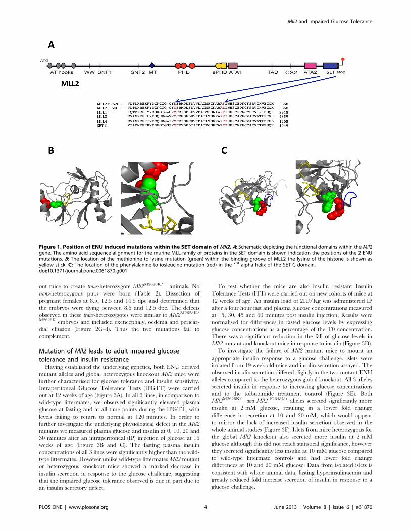
ferase domain (Figure 1A) [33]. As the crystal structure of the
homologous MLL1 SET domain has been elucidated it is possible
to map the Mll2M2628K mutation onto this structure where it
corresponds to residue MLL1M3884 and occupies a position in the
lysine binding groove (Figure 1B) [34].
To probe whether the observed phenotypes can be ascribed to
an impairment of the methyltransferase function of the SET
domain, a M3884K mutant MLL1 (the SET domains are highly
conserved between the two genes, Figure S1) equivalent to
M2628K in MLL2 (that in our hands could not be expressed) was
tested in in vitro assays (Figure S2). The SET domain of wild-type
and mutant MLL1 was thus expressed in E.coli, purified by affinity
chromatography and histone mono-, di- and trimethylation was
quantified by ELISA (Figure S3) [35]. The MLL1M3884K protein
had significantly reduced methyltransferase activity compared to
the MLL1 wild type, demonstrating this residue was indeed
required for methyltransferase activity (Figure S2).
In order to confirm that mutation of Mll2 underlies the
phenotypes we sought to identify additional mutant alleles of Mll2.
Firstly, we screened for mutations within the Mll2 SET
methyltransferase domain using the Harwell ENU DNA/Sperm
archive [29,36]. An additional thymine to adenine (T7942A)
transversion was identified and results in a phenylalanine to
isoleucine amino acid change at residue 2648 (NP_083550,
F2648I). The Mll2F2648I mutation maps to MLL1F3904 and is
located in an a5 helix of the SET-C domain in a region of the
protein important for interaction with the co-factor AdoHcy
(Figure 1C) [34]. Secondly, a global knockout of the Mll2 allele
was kindly provided by Professor Frances Stewart (Dresden
University of Technology, Germany) for additional analysis [33].
Homozygous Mll2 mutants are embryonic lethalHomozygous Mll2 knockout mice die of widespread apoptosis
prior to 11.5 dpc and we therefore tested the functionality of our 2
ENU mutations in homozygous individuals [33]. No homozygous
pups were identified from these matings (Table 1) showing that
homozygosity of both the ENU alleles is non-viable. To determine
the time and cause of lethality, we dissected pregnant females at
8.5, 12.5 and 14.5 dpc and genotyped the embryos for the
Mll2M2628K/M2628K allele. The percentage of homozygous indi-
viduals was lower than expected at both 12.5 and 14.5dpc
(Table 1). This suggested that the majority of Mll2M2628K/M2628K
embryos were dying between 8.5 and 11.5 dpc. The most
common defects identified in the embryos were evident as early
as 10.5 dpc and up to 12.5 dpc and included pericardial effusion,
abnormal heart looping, exencephaly and various head abnor-
malities and anterior truncation defects (Figure 2A–F). The
observed heart and/or circulation defects are the most likely
cause of non-viabilty in Mll2M2628K/M2628K embryos.
As the homozygous lethal phenotype of Mll2M2628K/M2628K is
not identical to the published phenotype of Mll2 knockout mice we
investigated the effect of combining the two mutations. We set up
crosses between Mll2M2628K/+ and heterozygous Mll2+/2 knock-
Mll2 and Impaired Glucose Tolerance
PLOS ONE | www.plosone.org 3 June 2013 | Volume 8 | Issue 6 | e61870
out mice to create trans-heterozygote Mll2M2628K/2 animals. No
trans-heterozygous pups were born (Table 2). Dissection of
pregnant females at 8.5, 12.5 and 14.5 dpc and determined that
the embryos were dying between 8.5 and 12.5 dpc. The defects
observed in these trans-heterozygotes were similar to Mll2M2628K/
M2628K embryos and included exencephaly, oedema and pericar-
dial effusion (Figure 2G–I). Thus the two mutations fail to
complement.
Mutation of Mll2 leads to adult impaired glucosetolerance and insulin resistance
Having established the underlying genetics, both ENU derived
mutant alleles and global heterozygous knockout Mll2 mice were
further characterised for glucose tolerance and insulin sensitivity.
Intraperitoneal Glucose Tolerance Tests (IPGTT) were carried
out at 12 weeks of age (Figure 3A). In all 3 lines, in comparison to
wild-type littermates, we observed significantly elevated plasma
glucose at fasting and at all time points during the IPGTT, with
levels failing to return to normal at 120 minutes. In order to
further investigate the underlying physiological defect in the Mll2
mutants we measured plasma glucose and insulin at 0, 10, 20 and
30 minutes after an intraperitoneal (IP) injection of glucose at 16
weeks of age (Figure 3B and C). The fasting plasma insulin
concentrations of all 3 lines were significantly higher than the wild-
type littermates. However unlike wild-type littermates Mll2 mutant
or heterozygous knockout mice showed a marked decrease in
insulin secretion in response to the glucose challenge, suggesting
that the impaired glucose tolerance observed is due in part due to
an insulin secretory defect.
To test whether the mice are also insulin resistant Insulin
Tolerance Tests (ITT) were carried out on new cohorts of mice at
12 weeks of age. An insulin load of 2IU/Kg was administered IP
after a four hour fast and plasma glucose concentrations measured
at 15, 30, 45 and 60 minutes post insulin injection. Results were
normalised for differences in fasted glucose levels by expressing
glucose concentrations as a percentage of the T0 concentration.
There was a significant reduction in the fall of glucose levels in
Mll2 mutant and knockout mice in response to insulin (Figure 3D).
To investigate the failure of Mll2 mutant mice to mount an
appropriate insulin response to a glucose challenge, islets were
isolated from 19 week old mice and insulin secretion assayed. The
observed insulin secretion differed slightly in the two mutant ENU
alleles compared to the heterozygous global knockout. All 3 alleles
secreted insulin in response to increasing glucose concentrations
and to the tolbutamide treatment control (Figure 3E). Both
Mll2M2628K/+ and Mll2 F2648I/+ alleles secreted significantly more
insulin at 2 mM glucose, resulting in a lower fold change
difference in secretion at 10 and 20 mM, which would appear
to mirror the lack of increased insulin secretion observed in the
whole animal studies (Figure 3F). Islets from mice heterozygous for
the global Mll2 knockout also secreted more insulin at 2 mM
glucose although this did not reach statistical significance, however
they secreted significantly less insulin at 10 mM glucose compared
to wild-type littermate controls and had lower fold change
differences at 10 and 20 mM glucose. Data from isolated islets is
consistent with whole animal data; fasting hyperinsulinaemia and
greatly reduced fold increase secretion of insulin in response to a
glucose challenge.
Figure 1. Position of ENU induced mutations within the SET domain of Mll2. A: Schematic depicting the functional domains within the Mll2gene. The amino acid sequence alignment for the murine MLL-family of proteins in the SET domain is shown indication the positions of the 2 ENUmutations. B: The location of the methionine to lysine mutation (green) within the binding groove of MLL2 the lysine of the histone is shown asyellow stick. C: The location of the phenylalanine to iosleucine mutation (red) in the 1st alpha helix of the SET-C domain.doi:10.1371/journal.pone.0061870.g001
Mll2 and Impaired Glucose Tolerance
PLOS ONE | www.plosone.org 4 June 2013 | Volume 8 | Issue 6 | e61870
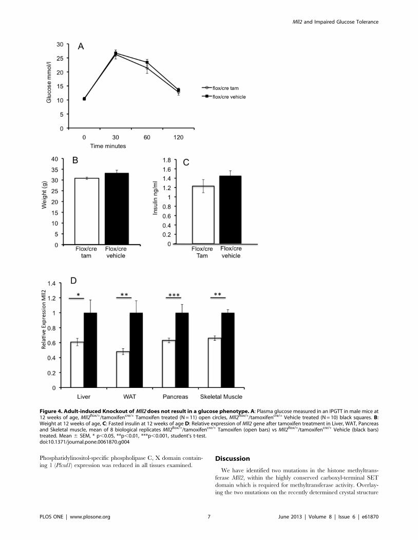
Tamoxifen induced Knockdown of Mll2 in adult micedoes not result in a glucose phenotype
In order to test whether the observed adult glucose phenotype
was the result of early developmental effects or changes in the
adult animal we carried out a heterozygous adult-induced
knockout. A floxed Mll2 allele was crossed with a tamoxifen-
inducible ubiquitin-Cre and mice of all genotype classes treated
with tamoxifen or vehicle at 8 weeks of age, in order to generate a
knockout allele. Mice were phenotyped at 12 weeks using an
IPGTT test. No significant difference was seen in either glucose
tolerance, weight or fasted insulin in heterozygous mice (Figure 4A,
B and C and Figure S4). Levels of Mll2 knockdown in tamoxifen
treated mice was assayed by quantitative RTPCR (Figure 4D) and
41% reduction in Mll2 levels was observed.
Biochemical and histological analysis of Mll2 mutants;evidence of fatty liver disease
As these mice were insulin resistant we further investigated them
for other metabolic disturbances. At 19 weeks of age mice were
fasted for four hours then sacrificed and metabolic tissues were
collected, additionally epididymal white fat pads and livers were
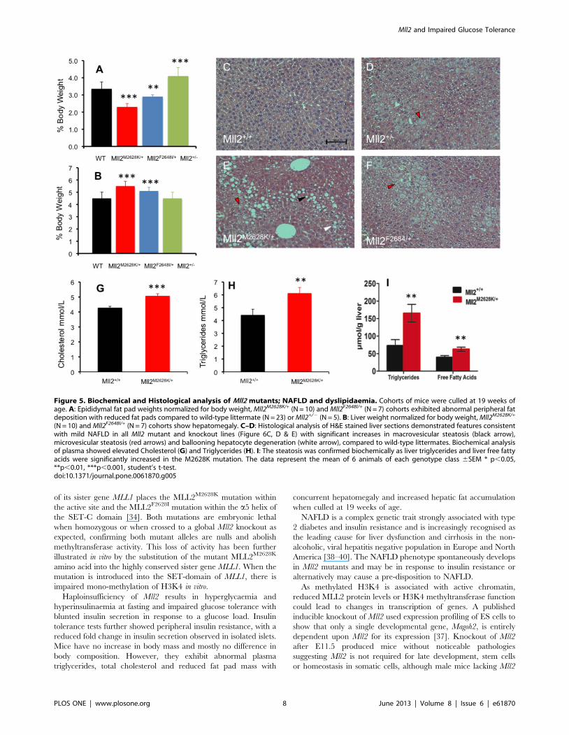
weighed. The two mutant ENU alleles of Mll2 exhibited
dyslipidaemia and epididymal fat pads were significantly lighter
at 19 weeks of age in the mutants versus wild-type littermates
(Figure 5A). They also showed significant hepatomegaly compared
to wild-type littermates (Figure 5B). There was no significant
increase in body mass observed in the 2 ENU mutants or Mll2+/2
knockout compared to wild-type littermates. Body composition of
the Mll2M2628K/+ mutant allele was additionally measured by
DEXA analysis at 8, 12 and 18 weeks of age (Figure S5) with a
brief increase in percentage fat mass observed at 12 weeks of age in
the mutant (31.0464.25% vs 26.8564.87%). Histological analysis
of liver sections demonstrated features consistent with NAFLD
(Figure 5C–F). The severity was formally assessed using the semi-
quantitative NIDDK histological score [31] and revealed signif-
icant increases in steatosis and ballooning hepatocyte degeneration
compared to wild-type littermates. Biochemical analysis of plasma
samples showed significantly increased cholesterol and triglyceride
(Figure 5G and H). Biochemical analysis of liver tissue showed
increased triglyceride and free fatty acid content in Mll2M2628K/+
compared to wild-type littermates (Figure 5I).
Histological analysis of Pancreatic IsletsPancreatic sections from Mll2M2628K/+ animals and wild-type
littermates were examined for differences in both islet mass and
architecture, 3 sections for 8 mice of each genotype class were
examined. There was small significant increase (p,0.01) in islet
area in heterozygous Mll2M2628K/+ mice (1.23760.077%) com-
pared to wild-type litter mates (1.00260.358%) in their percentage
of islet areas (the percentage of a histological section identified as
islet Table S2). Histological staining showed no qualitative
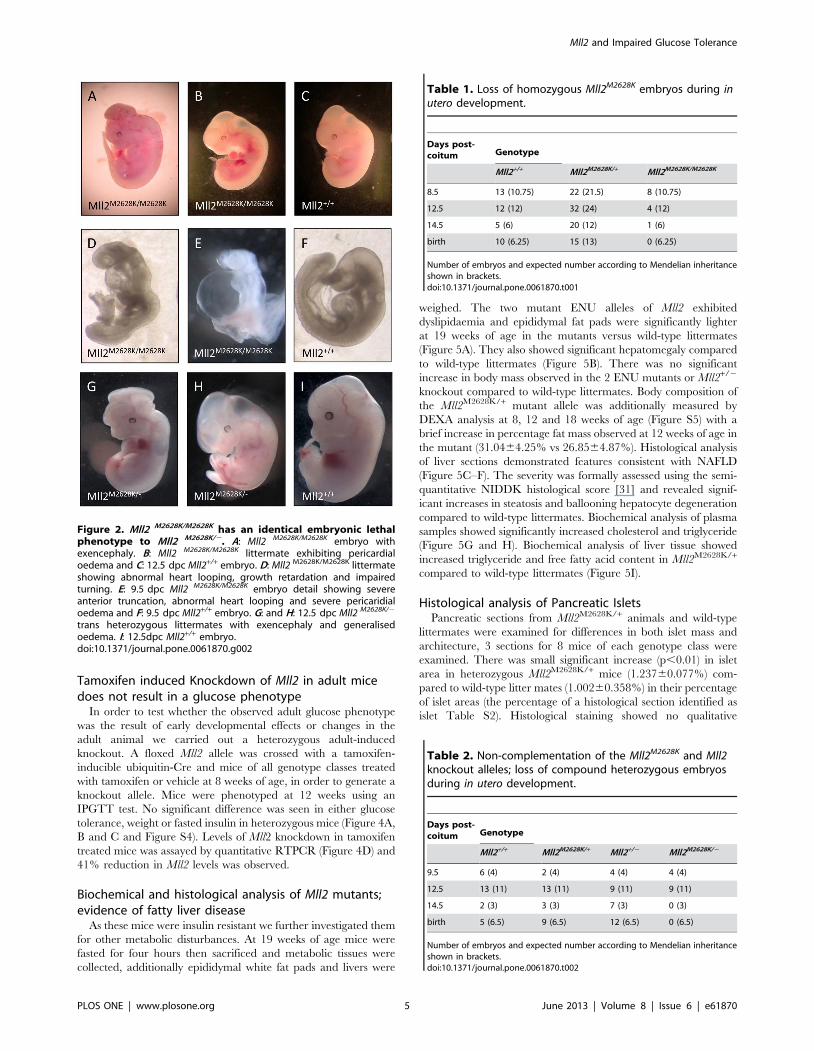
Figure 2. Mll2 M2628K/M2628K has an identical embryonic lethalphenotype to Mll2 M2628K/2. A: Mll2 M2628K/M2628K embryo withexencephaly. B: Mll2 M2628K/M2628K littermate exhibiting pericardialoedema and C: 12.5 dpc Mll2+/+ embryo. D: Mll2 M2628K/M2628K littermateshowing abnormal heart looping, growth retardation and impairedturning. E: 9.5 dpc Mll2 M2628K/M2628K embryo detail showing severeanterior truncation, abnormal heart looping and severe pericaridialoedema and F: 9.5 dpc Mll2+/+ embryo. G: and H: 12.5 dpc Mll2 M2628K/2
trans heterozygous littermates with exencephaly and generalisedoedema. I: 12.5dpc Mll2+/+ embryo.doi:10.1371/journal.pone.0061870.g002
Table 1. Loss of homozygous Mll2M2628K embryos during inutero development.
Days post-coitum Genotype
Mll2+/+ Mll2M2628K/+ Mll2M2628K/M2628K
8.5 13 (10.75) 22 (21.5) 8 (10.75)
12.5 12 (12) 32 (24) 4 (12)
14.5 5 (6) 20 (12) 1 (6)
birth 10 (6.25) 15 (13) 0 (6.25)
Number of embryos and expected number according to Mendelian inheritanceshown in brackets.doi:10.1371/journal.pone.0061870.t001
Table 2. Non-complementation of the Mll2M2628K and Mll2knockout alleles; loss of compound heterozygous embryosduring in utero development.
Days post-coitum Genotype
Mll2+/+ Mll2M2628K/+ Mll2+/2 Mll2M2628K/2
9.5 6 (4) 2 (4) 4 (4) 4 (4)
12.5 13 (11) 13 (11) 9 (11) 9 (11)
14.5 2 (3) 3 (3) 7 (3) 0 (3)
birth 5 (6.5) 9 (6.5) 12 (6.5) 0 (6.5)
Number of embryos and expected number according to Mendelian inheritanceshown in brackets.doi:10.1371/journal.pone.0061870.t002
Mll2 and Impaired Glucose Tolerance
PLOS ONE | www.plosone.org 5 June 2013 | Volume 8 | Issue 6 | e61870
difference in islet architecture in terms of arrangement or relative
numbers of a- or d-cells (data not shown)
Altered gene expression in Mll2 mutantsThe H3K4 methyltransferase Mll2 is thought to function briefly
during development to alter cellular gene expression programmes
that are then maintained by other redundant mechanisms [37].
This is consistent with our results as the adult knockout suggests
that the glucose phenotype arises from changes in gene expression
set during earlier development. We therefore decided to examine
the expression of selected genes with links to glucose homeostasis
and identified as altered in expression in published ES cell gene
expression experiments using Mll2 mutants (see additional file 1 in
[37]). These included Slc27a, Enpp3 (and additionally Enpp1 as it
has been implicated in diabetes and is adjacent to its paralog Enpp3
in the genome), Plcxd1, Neurod1 and several genes downstream of
Neurod1. The expression profile of these downregulated genes was
investigated in adult Mll2M2628K/+ heterozygous mutants in the
metabolically important tissues liver, white adipose tissue, isolated
islets and skeletal muscle (Figure 6A–D).
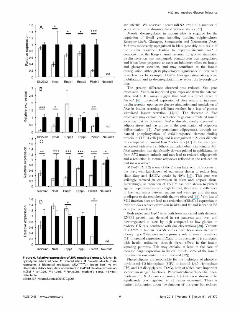
Neurod1 is an important transcriptional regulator both in the
developing pancreas and the mature beta cell. Its expression was
significantly downregulated in mutant islets, therefore the relative
expression of a number of genes regulated by Neurod1 in the beta
cell were examined. Ins1 but not Ins2 insulin gene expression was
slightly upregulated, and both Glucagon and Nnat downregulated
(Figure S6). Nnat expression was significantly reduced in liver and
white adipose tissue (Figure 6A and B). Slc27a2 expression was
significantly reduced in islets and adipose tissue. There were no
significant difference in liver or muscle (although a trend to
reduction in the latter) (Figure 6 A–D). Enpp1 and Enpp3 were
significantly reduced in liver and islets, however expression was
not altered in white adipose. However, Enpp1 showed an increase
in skeletal muscle and Enpp3 showed no difference (Figure 6D).
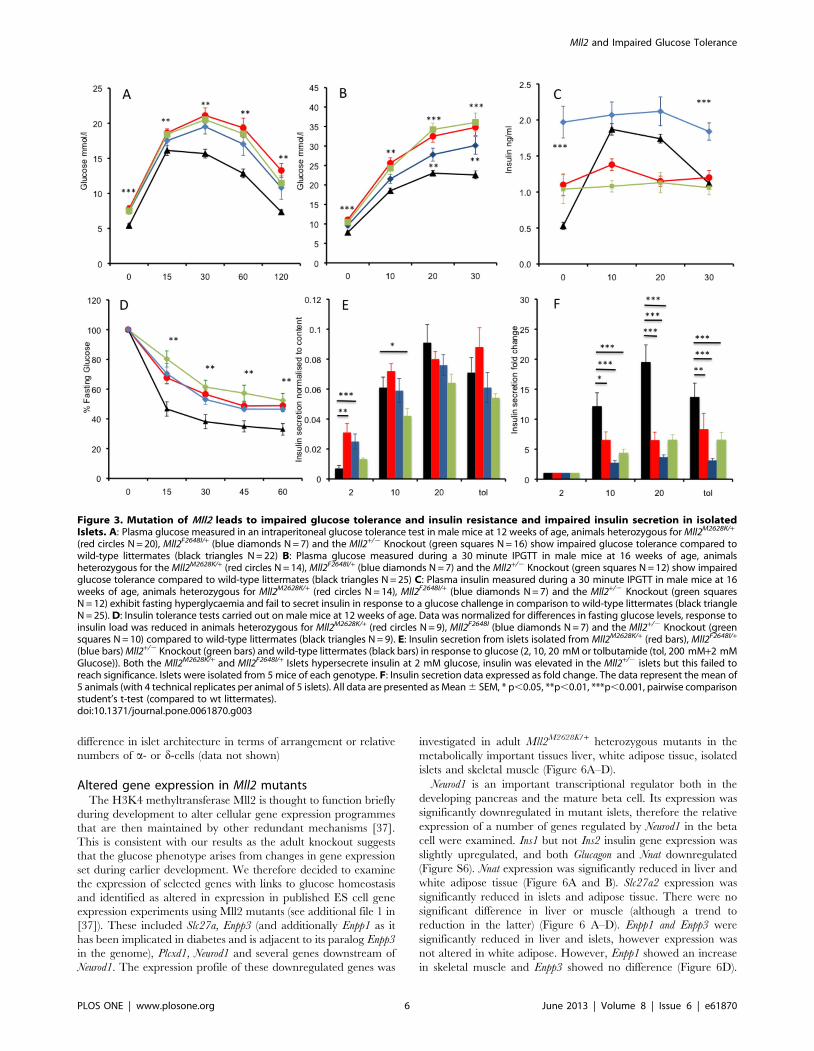
Figure 3. Mutation of Mll2 leads to impaired glucose tolerance and insulin resistance and impaired insulin secretion in isolatedIslets. A: Plasma glucose measured in an intraperitoneal glucose tolerance test in male mice at 12 weeks of age, animals heterozygous for Mll2M2628K/+
(red circles N = 20), Mll2F2648I/+ (blue diamonds N = 7) and the Mll2+/2 Knockout (green squares N = 16) show impaired glucose tolerance compared towild-type littermates (black triangles N = 22) B: Plasma glucose measured during a 30 minute IPGTT in male mice at 16 weeks of age, animalsheterozygous for the Mll2M2628K/+ (red circles N = 14), Mll2F2648I/+ (blue diamonds N = 7) and the Mll2+/2 Knockout (green squares N = 12) show impairedglucose tolerance compared to wild-type littermates (black triangles N = 25) C: Plasma insulin measured during a 30 minute IPGTT in male mice at 16weeks of age, animals heterozygous for Mll2M2628K/+ (red circles N = 14), Mll2F2648I/+ (blue diamonds N = 7) and the Mll2+/2 Knockout (green squaresN = 12) exhibit fasting hyperglycaemia and fail to secret insulin in response to a glucose challenge in comparison to wild-type littermates (black triangleN = 25). D: Insulin tolerance tests carried out on male mice at 12 weeks of age. Data was normalized for differences in fasting glucose levels, response toinsulin load was reduced in animals heterozygous for Mll2M2628K/+ (red circles N = 9), Mll2F2648I (blue diamonds N = 7) and the Mll2+/2 Knockout (greensquares N = 10) compared to wild-type littermates (black triangles N = 9). E: Insulin secretion from islets isolated from Mll2M2628K/+ (red bars), Mll2F2648I/+
(blue bars) Mll2+/2 Knockout (green bars) and wild-type littermates (black bars) in response to glucose (2, 10, 20 mM or tolbutamide (tol, 200 mM+2 mMGlucose)). Both the Mll2M2628K/+ and Mll2F2648I/+ Islets hypersecrete insulin at 2 mM glucose, insulin was elevated in the Mll2+/2 islets but this failed toreach significance. Islets were isolated from 5 mice of each genotype. F: Insulin secretion data expressed as fold change. The data represent the mean of5 animals (with 4 technical replicates per animal of 5 islets). All data are presented as Mean 6 SEM, * p,0.05, **p,0.01, ***p,0.001, pairwise comparisonstudent’s t-test (compared to wt littermates).doi:10.1371/journal.pone.0061870.g003
Mll2 and Impaired Glucose Tolerance
PLOS ONE | www.plosone.org 6 June 2013 | Volume 8 | Issue 6 | e61870
Phosphatidylinositol-specific phospholipase C, X domain contain-
ing 1 (Plcxd1) expression was reduced in all tissues examined.Discussion
We have identified two mutations in the histone methyltrans-
ferase Mll2, within the highly conserved carboxyl-terminal SET
domain which is required for methyltransferase activity. Overlay-
ing the two mutations on the recently determined crystal structure
Figure 4. Adult-induced Knockout of Mll2 does not result in a glucose phenotype. A: Plasma glucose measured in an IPGTT in male mice at12 weeks of age, Mll2flox/+/tamoxifencre/+ Tamoxifen treated (N = 11) open circles, Mll2flox/+/tamoxifencre/+ Vehicle treated (N = 10) black squares. B:Weight at 12 weeks of age, C: Fasted insulin at 12 weeks of age D: Relative expression of Mll2 gene after tamoxifen treatment in Liver, WAT, Pancreasand Skeletal muscle, mean of 8 biological replicates Mll2flox/+/tamoxifencre/+ Tamoxifen (open bars) vs Mll2flox/+/tamoxifencre/+ Vehicle (black bars)treated. Mean 6 SEM, * p,0.05, **p,0.01, ***p,0.001, student’s t-test.doi:10.1371/journal.pone.0061870.g004
Mll2 and Impaired Glucose Tolerance
PLOS ONE | www.plosone.org 7 June 2013 | Volume 8 | Issue 6 | e61870
of its sister gene MLL1 places the MLL2M2628K mutation within
the active site and the MLL2F2628I mutation within the a5 helix of
the SET-C domain [34]. Both mutations are embryonic lethal
when homozygous or when crossed to a global Mll2 knockout as
expected, confirming both mutant alleles are nulls and abolish
methyltransferase activity. This loss of activity has been further
illustrated in vitro by the substitution of the mutant MLL2M2628K
amino acid into the highly conserved sister gene MLL1. When the
mutation is introduced into the SET-domain of MLL1, there is
impaired mono-methylation of H3K4 in vitro.
Haploinsufficiency of Mll2 results in hyperglycaemia and
hyperinsulinaemia at fasting and impaired glucose tolerance with
blunted insulin secretion in response to a glucose load. Insulin
tolerance tests further showed peripheral insulin resistance, with a
reduced fold change in insulin secretion observed in isolated islets.
Mice have no increase in body mass and mostly no difference in
body composition. However, they exhibit abnormal plasma
triglycerides, total cholesterol and reduced fat pad mass with
concurrent hepatomegaly and increased hepatic fat accumulation
when culled at 19 weeks of age.
NAFLD is a complex genetic trait strongly associated with type
2 diabetes and insulin resistance and is increasingly recognised as
the leading cause for liver dysfunction and cirrhosis in the non-
alcoholic, viral hepatitis negative population in Europe and North
America [38–40]. The NAFLD phenotype spontaneously develops
in Mll2 mutants and may be in response to insulin resistance or
alternatively may cause a pre-disposition to NAFLD.
As methylated H3K4 is associated with active chromatin,
reduced MLL2 protein levels or H3K4 methyltransferase function
could lead to changes in transcription of genes. A published
inducible knockout of Mll2 used expression profiling of ES cells to
show that only a single developmental gene, Magoh2, is entirely
dependent upon Mll2 for its expression [37]. Knockout of Mll2
after E11.5 produced mice without noticeable pathologies
suggesting Mll2 is not required for late development, stem cells
or homeostasis in somatic cells, although male mice lacking Mll2
Figure 5. Biochemical and Histological analysis of Mll2 mutants; NAFLD and dyslipidaemia. Cohorts of mice were culled at 19 weeks ofage. A: Epididymal fat pad weights normalized for body weight, Mll2M2628K/+ (N = 10) and Mll2F2648I/+ (N = 7) cohorts exhibited abnormal peripheral fatdeposition with reduced fat pads compared to wild-type littermate (N = 23) or Mll2+/2 (N = 5). B: Liver weight normalized for body weight, Mll2M2628K/+
(N = 10) and Mll2F2648I/+ (N = 7) cohorts show hepatomegaly. C–D: Histological analysis of H&E stained liver sections demonstrated features consistentwith mild NAFLD in all Mll2 mutant and knockout lines (Figure 6C, D & E) with significant increases in macrovesicular steatosis (black arrow),microvesicular steatosis (red arrows) and ballooning hepatocyte degeneration (white arrow), compared to wild-type littermates. Biochemical analysisof plasma showed elevated Cholesterol (G) and Triglycerides (H). I: The steatosis was confirmed biochemically as liver triglycerides and liver free fattyacids were significantly increased in the M2628K mutation. The data represent the mean of 6 animals of each genotype class 6SEM * p,0.05,**p,0.01, ***p,0.001, student’s t-test.doi:10.1371/journal.pone.0061870.g005
Mll2 and Impaired Glucose Tolerance
PLOS ONE | www.plosone.org 8 June 2013 | Volume 8 | Issue 6 | e61870
are infertile. We observed altered mRNA levels of a number of
genes shown to be downregulated in these studies [37].
Neurod1, downregulated in mutant islets, is required for the
regulation of b-cell genes including Insulin, Sulphonylurea
Receptor (Sur1), Glucagon, Somatasatin and Neuronatin (Nnat).
Ins1 was moderately upregulated in islets, probably as a result of
the insulin resistance leading to hyperinsulinaemia. Sur1 a
component of the KATP channel essential for glucose stimulated
insulin secretion was unchanged. Somatostatin was upregulated
and it has been proposed to exert an inhibitory effect on insulin
and glucagon secretion, and may contribute to the insulin
dysregulation, although its physiological significance in these roles
is unclear (see for example [41,42]). Glucagon stimulates glucose
mobilization and its downregulation may reflect the hyperglycae-
mia.
The greatest difference observed was reduced Nnat gene
expression. Nnat is an imprinted gene expressed from the paternal
allele and CHIP assays suggest that Nnat is a direct target of
Neurod1 [43]. Increased expression of Nnat results in increased
insulin secretion upon acute glucose stimulation and knockdown of
Nnat in insulin secreting cell lines resulted in a loss of glucose
stimulated insulin secretion [43,44]. The decrease in Nnat
expression may explain the reduction in glucose stimulated insulin
secretion that we observed. Nnat is also abundantly expressed in
adipose tissue and has a role in the potentiation of adipocyte
differentiation [45]. Nnat potentiates adipogenesis through en-
hanced phosphorylation of cAMP-response element–binding
protein in 3T3-L1 cells [46], and is upregulated in Zucker diabetic
rats compared to control lean Zucker rats [47]. It has also been
associated with severe childhood and adult obesity in humans [48].
Nnat expression was significantly downregulated in epididymal fat
from Mll2 mutant animals and may lead to reduced adipogenesis
and a reduction in mature adipocyes reflected in the reduced fat
pad mass observed.
Slc27a2 (FATP2) is one of the 2 main fatty acid transporters in
the liver, with knockdown of expression shown to reduce long
chain fatty acid (LCFA) uptake by 40% [49]. This gene was
strikingly reduced in expression in islets and adipose tissue.
Interestingly, as reduction of FATP2 has been shown to protect
against hepatosteatosis on a high fat diet, there was no difference
in liver expression between mutant and wild-type and this may
predispose to the steatohepatitis that we observed [49]. Why loss of
Mll2 function does not lead to a reduction of Slc27a2 expression in
liver but does reduce expression in islets and fat and indeed in ES
cells [37] is unclear.
Both Enpp3 and Enpp1 have both been associated with diabetes.
ENPP3 protein was detected in rat pancreas and liver and
downregulated in islets by high compared to low glucose in
diabetic GK rats, consistent with our observations [50]. Variants
of ENPP1 in human GWAS studies have been associated with
obesity, type 2 diabetes and a primary role in insulin resistance
[51]. Increased expression of Enpp1 or its overactivity is correlated
with insulin resistance, through direct effects in the insulin
signaling pathway. This may explain, at least in the case of
increase Enpp1 expression in skeletal muscle, some of the insulin
resistance in our mutant mice (reviewed [52]).
Phospholipases are responsible for the hydrolysis of phospha-
tidylinositol 4-5-biphosphate (PIP2) to inositol 1,4,5-triphosphate
(IP3) and 1,2-diacylglycerol (DAG), both of which have important
second messenger functions. Phosphatidylinositol-specific phos-
pholipase C, X domain containing 1 (Plcxd1) was shown to be
significantly downregulated in all tissues examined. There is
limited information about the function of this gene but reduced
Figure 6. Relative expression of Mll2 regulated genes. A: Liver, B:Epididymal White adipose, C: Isolated islets, D: Skeletal Muscle. Datarepresents 8 biological replicates, Mll2M2628K/+ (open bars) vs wtlittermates, (black bars) data normalized to GAPDH. Relative expression6SEM * p,0.05, **p,0.01, ***p,0.001, student’s t-test. nd = notdetectable.doi:10.1371/journal.pone.0061870.g006
Mll2 and Impaired Glucose Tolerance
PLOS ONE | www.plosone.org 9 June 2013 | Volume 8 | Issue 6 | e61870
expression may reflect dysregulation of insulin signalling pathways
either through effects on IP3 or DAG.
Type 2 Diabetes is a complex disease involving many different
tissue types, euglycemic hyperinsulinemic CLAMP studies may
further dissect the tissues important for the insulin resistance
identified in this model. CHIP studies in relevant tissues may
identify other yet unidentified genes whose regulation by MLL2 at
the chromatin level may contribute to disease. It would be
informative to carry out these studies before and after onset of
overt disease to differentiate between causal and effect differences
in gene expression. Whilst the mutation identified is in a single
gene Mll2 the phenotype observed is likely to be the result of
multiple small changes in the expression of a number of genes in
many diverse tissues. Tissue specific KO of MLL2 followed by
CHIP analysis and comprehensive metabolic phenotyping should
yield insight into the relative contribution of each tissue and gene
set to onset of disease.
Conclusions
In summary, we have identified two ENU induced point
mutations M2628K and F2628I in MLL2 that give rise to a novel
murine model of insulin resistance, impaired glucose tolerance and
primary stages of NAFLD. We have provided evidence that these
are functional mutations that affect the H3K4 methyltransferase
activity of MLL2 that then leads to changes during embryonic
development, likely in chromatin and DNA methylation (see [37]),
that determine the expression of genes linked to diabetes
phenotypes in the adult. These data reveal that gene expression
controlled through histone methylation is a significant mechanism
involved in glucose homeostasis.
Supporting Information
Figure S1 Sequence alignment of the highly conservedSET domain of MLL1 and MLL2.(TIF)
Figure S2 In vitro methyltransferase assays indicate areduced activity of the MLL1 (M3884K) mutant incomparison to wild-type MLL1. The methylation of histone
H3 substrates comprising the first 21 N-terminal amino acids was
quantified enzyme linked immunoabsorbent assays (ELISAs) with
antibodies against H3K4me1, me2 and me3. A: Unmodified H3
peptide (H3K4me0) was incubated with wild-type and mutant
recombinant expressed SET-domain of MLL1 respectively and
H3K4me1 product detected (see Figure S3 for time courses of the
subsequent products H3K4me2 and H3K4me3). B: Monomethy-
lated H3 peptide (H3K4me1) was incubated with MLL and
samples were analyzed for dimethylation (forming H3K4me2 -
Figure S3 for time courses of the subsequent product H3K4me3).
C: Dimethylated H3 peptide (H3K4me2) was incubated with
MLL and H3K4me3 product detected. The activity of the
M3884K was reduced compared to wild-type in all cases. The
M3884K position of MLL1 is equivalent to the M2628K position
of Mll2 based on sequence alignment. Error bars show the SD
from the mean value of three experiments.
(TIF)
Figure S3 In vitro methyltransferase assays indicate areduced activity of the MLL1 (M3884K) mutant incomparison to wild-type MLL1. The methylation of histone
H3 substrates comprising the first 21 N-terminal amino acids was
quantified enzyme linked immunoabsorbent assays with antibodies
against H3K4me1, me2 and me3. A–C Unmodified H3 peptide
was incubated with wildtype and mutant recombinant expressed
SET-domain of MLL1 respectively. Samples were analyzed for (A)
monomethylation, (B) dimethylation and (C) trimethylation at
lysine 4 after 2, 8, 20 and 48 hours. D, E H3 peptide with a
monomethyl modified lysine 4 was incubated with the enzyme and
the mutant. The samples were analyzed for (D) dimethylation and
(E) trimethylation. (F) Lysine 4 dimethyl H3 peptide was incubated
with the wild-type enzyme and mutant and samples analyzed for a
trimethyl mark at lysine 4. Error bars show the SD from the mean
value.
(TIF)
Figure S4 Adult Knockout of Mll2. All genotype classes and
treatment groups. N = 7–11 for each group. A: Plasma glucose
measured in an intraperitoneal glucose tolerance test in male mice
at 12 weeks of age, B: Weight at 12 weeks of age. C: Fasted plasma
insulin at 12 weeks of age. Data represented as Mean 6SEM.
(TIF)
Figure S5 Dexa analysis of Mll2M2628K/+ compared towildtype litter mates. Dexa analysis at 8, 12 and 18weeks
Mll2M2628K/+ (open bars N = 12) compared to wildtype littermates
(Black bars N = 17). No significant difference was observed in total
body weight or lean mass at any of the 3 time points. A transient
significant increase in body fat in Mll2M2628K/+ was observed at 12
weeks of age (p = 0.03). Data represented as Mean 6SEM.
(TIF)
Figure S6 Relative expression of Neurod1 regulatedgenes in Isolated Islets. Data represents 8 biological replicates,
Mll2M2628K/+ (open bars) vs wt littermates (black bars), data
normalized to GAPDH. Relative expression 6SEM * p,0.05,
**p,0.01, ***p,0.001, student’s t-test.
(TIF)
Table S1 Candidate list on chromosome 7.
(TIF)
Table S2 Percentage Islet areas.(TIF)
Acknowledgments
We thank S. Gendreizig for preliminary expression experiments. We thank
Professor Francis A. Stewart (University of Technology, Dresden) for the
Mll2FC and Mll2KO mice. We also wish to thank the Mary Lyon Centre
staff for managing the mouse colonies.
Author Contributions
Conceived and designed the experiments: MG NLA HCM DB FH RDC.
Performed the experiments: MG NLA DS HCM DB LM AL CC AH
QMA RG. Analyzed the data: MG NLA HCM DB QMA RG RDC.
Wrote the paper: MG NA DB RDC. Revised manuscript: MG NLA HC
DB LM CC QMA RG MT FH RDC.
References
1. Saxena R, Voight BF, Lyssenko V, Burtt NP, de Bakker PI, et al. (2007)
Genome-wide association analysis identifies loci for type 2 diabetes and
triglyceride levels. Science 316: 1331–1336.
2. Scott LJ, Mohlke KL, Bonnycastle LL, Willer CJ, Li Y, et al. (2007) A genome-
wide association study of type 2 diabetes in Finns detects multiple susceptibility
variants. Science 316: 1341–1345.
3. Sladek R, Rocheleau G, Rung J, Dina C, Shen L, et al. (2007) A genome-wide
association study identifies novel risk loci for type 2 diabetes. Nature 445: 881–
885.
4. Steinthorsdottir V, Thorleifsson G, Reynisdottir I, Benediktsson R, Jonsdottir T,
et al. (2007) A variant in CDKAL1 influences insulin response and risk of type 2
diabetes. Nat Genet 39: 770–775.
Mll2 and Impaired Glucose Tolerance
PLOS ONE | www.plosone.org 10 June 2013 | Volume 8 | Issue 6 | e61870
5. Zeggini E, Weedon MN, Lindgren CM, Frayling TM, Elliott KS, et al. (2007)
Replication of genome-wide association signals in UK samples reveals risk locifor type 2 diabetes. Science 316: 1336–1341.
6. Zeggini E, Scott LJ, Saxena R, Voight BF, Marchini JL, et al. (2008) Meta-
analysis of genome-wide association data and large-scale replication identifiesadditional susceptibility loci for type 2 diabetes. Nat Genet 40: 638–645.
7. Lango H, Palmer CN, Morris AD, Zeggini E, Hattersley AT, et al. (2008)Assessing the combined impact of 18 common genetic variants of modest effect
sizes on type 2 diabetes risk. Diabetes 57: 3129–3135.
8. Dupuis J, Langenberg C, Prokopenko I, Saxena R, Soranzo N, et al. (2010) Newgenetic loci implicated in fasting glucose homeostasis and their impact on type 2
diabetes risk. Nat Genet 42: 105–116.9. Voight BF, Scott LJ, Steinthorsdottir V, Morris AP, Dina C, et al. (2010) Twelve
type 2 diabetes susceptibility loci identified through large-scale associationanalysis. Nat Genet 42: 579–589.
10. Qi L, Cornelis MC, Kraft P, Stanya KJ, Linda Kao WH, et al. (2010) Genetic
variants at 2q24 are associated with susceptibility to type 2 diabetes. Hum MolGenet 19: 2706–2715.
11. Yamauchi T, Hara K, Maeda S, Yasuda K, Takahashi A, et al. (2010) Agenome-wide association study in the Japanese population identifies susceptibil-
ity loci for type 2 diabetes at UBE2E2 and C2CD4A-C2CD4B. Nat Genet 42:
864–868.12. Altshuler D, Hirschhorn JN, Klannemark M, Lindgren CM, Vohl MC, et al.
(2000) The common PPARgamma Pro12Ala polymorphism is associated withdecreased risk of type 2 diabetes. Nat Genet 26: 76–80.
13. Young EH, Wareham NJ, Farooqi S, Hinney A, Hebebrand J, et al. (2007) TheV103I polymorphism of the MC4R gene and obesity: population based studies
and meta-analysis of 29 563 individuals. Int J Obes (Lond) 31: 1437–1441.
14. Tsai FJ, Yang CF, Chen CC, Chuang LM, Lu CH, et al. (2010) A genome-wideassociation study identifies susceptibility variants for type 2 diabetes in Han
Chinese. PLoS Genet 6: e1000847.15. Wheeler E, Barroso I (2011) Genome-wide association studies and type 2
diabetes. Briefings in functional genomics 10: 52–60.
16. McCarthy MI (2010) Genomics, type 2 diabetes, and obesity. The New Englandjournal of medicine 363: 2339–2350.
17. Li B, Carey M, Workman JL (2007) The role of chromatin during transcription.Cell 128: 707–719.
18. Shilatifard A (2006) Chromatin modifications by methylation and ubiquitina-tion: implications in the regulation of gene expression. Annu Rev Biochem 75:
243–269.
19. Heintzman ND, Stuart RK, Hon G, Fu Y, Ching CW, et al. (2007) Distinct andpredictive chromatin signatures of transcriptional promoters and enhancers in
the human genome. Nat Genet 39: 311–318.20. Sims RJ 3rd, Reinberg D (2006) Histone H3 Lys 4 methylation: caught in a
bind? Genes Dev 20: 2779–2786.
21. Park JH, Stoffers DA, Nicholls RD, Simmons RA (2008) Development of type 2diabetes following intrauterine growth retardation in rats is associated with
progressive epigenetic silencing of Pdx1. J Clin Invest 118: 2316–2324.22. Raychaudhuri N, Raychaudhuri S, Thamotharan M, Devaskar SU (2008)
Histone code modifications repress glucose transporter 4 expression in theintrauterine growth-restricted offspring. J Biol Chem 283: 13611–13626.
23. Sandovici I, Smith NH, Nitert MD, Ackers-Johnson M, Uribe-Lewis S, et al.
(2011) Maternal diet and aging alter the epigenetic control of a promoter-enhancer interaction at the Hnf4a gene in rat pancreatic islets. Proc Natl Acad
Sci U S A 108: 5449–5454.24. Miao F, Wu X, Zhang L, Yuan YC, Riggs AD, et al. (2007) Genome-wide
analysis of histone lysine methylation variations caused by diabetic conditions in
human monocytes. J Biol Chem 282: 13854–13863.25. Nolan PM, Peters J, Strivens M, Rogers D, Hagan J, et al. (2000) A systematic,
genome-wide, phenotype-driven mutagenesis programme for gene functionstudies in the mouse. Nat Genet 25: 440–443.
26. Hrabe de Angelis MH, Flaswinkel H, Fuchs H, Rathkolb B, Soewarto D, et al.
(2000) Genome-wide, large-scale production of mutant mice by ENUmutagenesis. Nat Genet 25: 444–447.
27. Toye AA, Moir L, Hugill A, Bentley L, Quarterman J, et al. (2004) A new mousemodel of type 2 diabetes, produced by N-ethyl-nitrosourea mutagenesis, is the
result of a missense mutation in the glucokinase gene. Diabetes 53: 1577–1583.28. Goldsworthy M, Hugill A, Freeman H, Horner E, Shimomura K, et al. (2008)
Role of the transcription factor sox4 in insulin secretion and impaired glucose
tolerance. Diabetes 57: 2234–2244.29. Quwailid MM, Hugill A, Dear N, Vizor L, Wells S, et al. (2004) A gene-driven
ENU-based approach to generating an allelic series in any gene. MammGenome 15: 585–591.
30. Shimomura K, Galvanovskis J, Goldsworthy M, Hugill A, Kaizak S, et al. (2009)
Insulin secretion from beta-cells is affected by deletion of nicotinamide
nucleotide transhydrogenase. Methods in enzymology 457: 451–480.
31. Kleiner DE, Brunt EM, Van Natta M, Behling C, Contos MJ, et al. (2005)
Design and validation of a histological scoring system for nonalcoholic fatty liver
disease. Hepatology 41: 1313–1321.
32. Hough TA, Nolan PM, Tsipouri V, Toye AA, Gray IC, et al. (2002) Novel
phenotypes identified by plasma biochemical screening in the mouse. Mamm
Genome 13: 595–602.
33. Glaser S, Schaft J, Lubitz S, Vintersten K, van der Hoeven F, et al. (2006)
Multiple epigenetic maintenance factors implicated by the loss of Mll2 in mouse
development. Development 133: 1423–1432.
34. Southall SM, Wong PS, Odho Z, Roe SM, Wilson JR (2009) Structural basis for
the requirement of additional factors for MLL1 SET domain activity and
recognition of epigenetic marks. Mol Cell 33: 181–191.
35. Nightingale KP, Gendreizig S, White DA, Bradbury C, Hollfelder F, et al.
(2007) Cross-talk between histone modifications in response to histone
deacetylase inhibitors: MLL4 links histone H3 acetylation and histone H3K4
methylation. The Journal of biological chemistry 282: 4408–4416.
36. Coghill EL, Hugill A, Parkinson N, Davison C, Glenister P, et al. (2002) A gene-
driven approach to the identification of ENU mutants in the mouse. Nat Genet
30: 255–256.
37. Glaser S, Lubitz S, Loveland KL, Ohbo K, Robb L, et al. (2009) The histone 3
lysine 4 methyltransferase, Mll2, is only required briefly in development and
spermatogenesis. Epigenetics Chromatin 2: 5.
38. Angulo P (2002) Nonalcoholic fatty liver disease. N Engl J Med 346: 1221–1231.
39. Anstee QM, Daly AK, Day CP (2011) Genetic modifiers of non-alcoholic fatty
liver disease progression. Biochimica et biophysica acta 1812: 1557–1566.
40. Anstee QM, Goldin RD (2006) Mouse models in non-alcoholic fatty liver disease
and steatohepatitis research. Int J Exp Pathol 87: 1–16.
41. Hauge-Evans AC, King AJ, Carmignac D, Richardson CC, Robinson IC, et al.
(2009) Somatostatin secreted by islet delta-cells fulfills multiple roles as a
paracrine regulator of islet function. Diabetes 58: 403–411.
42. Walker JN, Ramracheya R, Zhang Q, Johnson PR, Braun M, et al. (2011)
Regulation of glucagon secretion by glucose: paracrine, intrinsic or both?
Diabetes, obesity & metabolism 13 Suppl 1: 95–105.
43. Chu K, Tsai MJ (2005) Neuronatin, a downstream target of BETA2/NeuroD1
in the pancreas, is involved in glucose-mediated insulin secretion. Diabetes 54:
1064–1073.
44. Joe MK, Lee HJ, Suh YH, Han KL, Lim JH, et al. (2008) Crucial roles of
neuronatin in insulin secretion and high glucose-induced apoptosis in pancreatic
beta-cells. Cellular signalling 20: 907–915.
45. Suh YH, Kim WH, Moon C, Hong YH, Eun SY, et al. (2005) Ectopic
expression of Neuronatin potentiates adipogenesis through enhanced phosphor-
ylation of cAMP-response element-binding protein in 3T3-L1 cells. Biochemical
and Biophysical Research Communications 337: 481–489.
46. Suh YH, Kim WH, Moon C, Hong YH, Eun SY, et al. (2005) Ectopic
expression of Neuronatin potentiates adipogenesis through enhanced phosphor-
ylation of cAMP-response element-binding protein in 3T3-L1 cells. Biochem
Biophys Res Commun 337: 481–489.
47. Suh YH, Kim Y, Bang JH, Choi KS, Lee JW, et al. (2005) Analysis of gene
expression profiles in insulin-sensitive tissues from pre-diabetic and diabetic
Zucker diabetic fatty rats. J Mol Endocrinol 34: 299–315.
48. Vrang N, Meyre D, Froguel P, Jelsing J, Tang-Christensen M, et al. (2010) The
imprinted gene neuronatin is regulated by metabolic status and associated with
obesity. Obesity (Silver Spring) 18: 1289–1296.
49. Falcon A, Doege H, Fluitt A, Tsang B, Watson N, et al. (2010) FATP2 is a
hepatic fatty acid transporter and peroxisomal very long-chain acyl-CoA
synthetase. American Journal of Physiology-Endocrinology and Metabolism
299: E384–E393.
50. Ghanaat-Pour H, Huang Z, Lehtihet M, Sjoholm A (2007) Global expression
profiling of glucose-regulated genes in pancreatic islets of spontaneously diabetic
Goto-Kakizaki rats. J Mol Endocrinol 39: 135–150.
51. Meyre D, Bouatia-Naji N, Tounian A, Samson C, Lecoeur C, et al. (2005)
Variants of ENPP1 are associated with childhood and adult obesity and increase
the risk of glucose intolerance and type 2 diabetes. Nature Genetics 37: 863–867.
52. Goldfine ID, Maddux BA, Youngren JF, Reaven G, Accili D, et al. (2008) The
role of membrane glycoprotein plasma cell antigen 1/ectonucleotide pyrophos-
phatase phosphodiesterase 1 in the pathogenesis of insulin resistance and related
abnormalities. Endocrine reviews 29: 62–75.
Mll2 and Impaired Glucose Tolerance
PLOS ONE | www.plosone.org 11 June 2013 | Volume 8 | Issue 6 | e61870
Related Documents