Mutations in IRF6 cause Van der Woude and popliteal pterygium syndromes Shinji Kondo 1,* , Brian C. Schutte 1,2,* , Rebecca J. Richardson 3,† , Bryan C. Bjork 4,† , Alexandra S. Knight 3 , Yoriko Watanabe 1 , Emma Howard 3 , Renata L.L. Ferreira de Lima 5 , Sandra Daack-Hirsch 1 , Achim Sander 6,‡ , Donna M. McDonald-McGinn 7 , Elaine H. Zackai 7 , Edward J. Lammer 8 , Arthur S. Aylsworth 9 , Holly H. Ardinger 10 , Andrew C. Lidral 11 , Barbara R. Pober 12 , Lina Moreno 13 , Mauricio Arcos-Burgos 14 , Consuelo Valencia 14 , Claude Houdayer 15 , Michel Bahuau 15,16 , Danilo Moretti-Ferreira 5 , Antonio Richieri-Costa 17 , Michael J. Dixon 3 , and Jeffrey C. Murray 1,2,18 1 Department of Pediatrics, The University of Iowa, Iowa City, Iowa 52242, USA 2 Interdisciplinary PhD Program in Genetics, The University of Iowa, Iowa City, Iowa 52242, USA 3 School of Biological Sciences and Department of Dental Medicine and Surgery, University of Manchester, Oxford Road, Manchester, UK 4 Harvard University, Brigham and Women's Hospital, Boston, Massachusetts, USA 5 Servico de Aconselhamento, Genetico da Universidade Estadual Paulista, Botucatu S.P., Brazil 6 Clinic of Oral-Maxillofacial Surgery, University of Hamburg, Hamburg, Germany 7 Division of Human Genetics and Molecular Biology, The Children's Hospital of Philadelphia, Philadelphia, Pennsylvania, USA 8 Medical Genetics, Children's Hospital, Oakland, California, USA 9 Department of Pediatrics and Genetics, University of North Carolina, Chapel Hill, North Carolina, USA 10 Department of Pediatrics, University of Kansas, Children's Medical Center, Kansas City, Kansas, USA 11 Department of Orthodontics, The University of Iowa, Iowa City, Iowa, USA 12 Department of Genetics, Yale University School of Medicine, New Haven, Connecticut, USA 13 PhD Program in Oral Sciences, The University of Iowa, Iowa City, Iowa, USA 14 Universidad de Antioquia, Medellin, Colombia 15 Service de Biochimie et Biologie Moléculaire, Hôpital d'Enfants Armand-Trousseau, Paris, France 16 Service de Chirurgie Maxillofaciale et Plastique, Stomatologie, Hôpital d'Enfants Armand- Trousseau, Paris, France © 2002 Nature Publishing Group Correspondence should be addressed to J.C.M. ([email protected]). * These two authors contributed equally to this work. † These two authors contributed equally to this work. ‡ Deceased. Competing interests statement: The authors declare that they have no competing financial interests. NIH Public Access Author Manuscript Nat Genet. Author manuscript; available in PMC 2011 September 8. Published in final edited form as: Nat Genet. 2002 October ; 32(2): 285–289. doi:10.1038/ng985. NIH-PA Author Manuscript NIH-PA Author Manuscript NIH-PA Author Manuscript

Welcome message from author
This document is posted to help you gain knowledge. Please leave a comment to let me know what you think about it! Share it to your friends and learn new things together.
Transcript
Mutations in IRF6 cause Van der Woude and popliteal pterygiumsyndromes
Shinji Kondo1,*, Brian C. Schutte1,2,*, Rebecca J. Richardson3,†, Bryan C. Bjork4,†,Alexandra S. Knight3, Yoriko Watanabe1, Emma Howard3, Renata L.L. Ferreira de Lima5,Sandra Daack-Hirsch1, Achim Sander6,‡, Donna M. McDonald-McGinn7, Elaine H. Zackai7,Edward J. Lammer8, Arthur S. Aylsworth9, Holly H. Ardinger10, Andrew C. Lidral11, BarbaraR. Pober12, Lina Moreno13, Mauricio Arcos-Burgos14, Consuelo Valencia14, ClaudeHoudayer15, Michel Bahuau15,16, Danilo Moretti-Ferreira5, Antonio Richieri-Costa17,Michael J. Dixon3, and Jeffrey C. Murray1,2,18
1Department of Pediatrics, The University of Iowa, Iowa City, Iowa 52242, USA2Interdisciplinary PhD Program in Genetics, The University of Iowa, Iowa City, Iowa 52242, USA3School of Biological Sciences and Department of Dental Medicine and Surgery, University ofManchester, Oxford Road, Manchester, UK4Harvard University, Brigham and Women's Hospital, Boston, Massachusetts, USA5Servico de Aconselhamento, Genetico da Universidade Estadual Paulista, Botucatu S.P., Brazil6Clinic of Oral-Maxillofacial Surgery, University of Hamburg, Hamburg, Germany7Division of Human Genetics and Molecular Biology, The Children's Hospital of Philadelphia,Philadelphia, Pennsylvania, USA8Medical Genetics, Children's Hospital, Oakland, California, USA9Department of Pediatrics and Genetics, University of North Carolina, Chapel Hill, North Carolina,USA10Department of Pediatrics, University of Kansas, Children's Medical Center, Kansas City,Kansas, USA11Department of Orthodontics, The University of Iowa, Iowa City, Iowa, USA12Department of Genetics, Yale University School of Medicine, New Haven, Connecticut, USA13PhD Program in Oral Sciences, The University of Iowa, Iowa City, Iowa, USA14Universidad de Antioquia, Medellin, Colombia15Service de Biochimie et Biologie Moléculaire, Hôpital d'Enfants Armand-Trousseau, Paris,France16Service de Chirurgie Maxillofaciale et Plastique, Stomatologie, Hôpital d'Enfants Armand-Trousseau, Paris, France
© 2002 Nature Publishing GroupCorrespondence should be addressed to J.C.M. ([email protected]).*These two authors contributed equally to this work.†These two authors contributed equally to this work.‡Deceased.Competing interests statement: The authors declare that they have no competing financial interests.
NIH Public AccessAuthor ManuscriptNat Genet. Author manuscript; available in PMC 2011 September 8.
Published in final edited form as:Nat Genet. 2002 October ; 32(2): 285–289. doi:10.1038/ng985.
NIH
-PA Author Manuscript
NIH
-PA Author Manuscript
NIH
-PA Author Manuscript
17Departmento de Genetica Clinica, Hospital de Pesquisa e Reabilitacao de Lesoes Labio-Palatais, Universidade de São Paulo, Bauru, Brazil18Department of Biology, The University of Iowa, Iowa City, Iowa 52242, USA
AbstractInterferon regulatory factor 6 (IRF6) belongs to a family of nine transcription factors that share ahighly conserved helix–turn–helix DNA-binding domain and a less conserved protein-bindingdomain. Most IRFs regulate the expression of interferon-α and -β after viral infection1, but thefunction of IRF6 is unknown. The gene encoding IRF6 is located in the critical region for the Vander Woude syndrome (VWS; OMIM 119300) locus at chromosome 1q32–q41 (refs 2,3). Thedisorder is an autosomal dominant form of cleft lip and palate with lip pits4, and is the mostcommon syndromic form of cleft lip or palate. Popliteal pterygium syndrome (PPS; OMIM119500) is a disorder with a similar orofacial phenotype that also includes skin and genitalanomalies5. Phenotypic overlap6 and linkage data7 suggest that these two disorders are allelic. Wefound a nonsense mutation in IRF6 in the affected twin of a pair of monozygotic twins who werediscordant for VWS. Subsequently, we identified mutations in IRF6 in 45 additional unrelatedfamilies affected with VWS and distinct mutations in 13 families affected with PPS. Expressionanalyses showed high levels of Irf6 mRNA along the medial edge of the fusing palate, tooth buds,hair follicles, genitalia and skin. Our observations demonstrate that haploinsufficiency of IRF6disrupts orofacial development and are consistent with dominant-negative mutations disturbingdevelopment of the skin and genitalia.
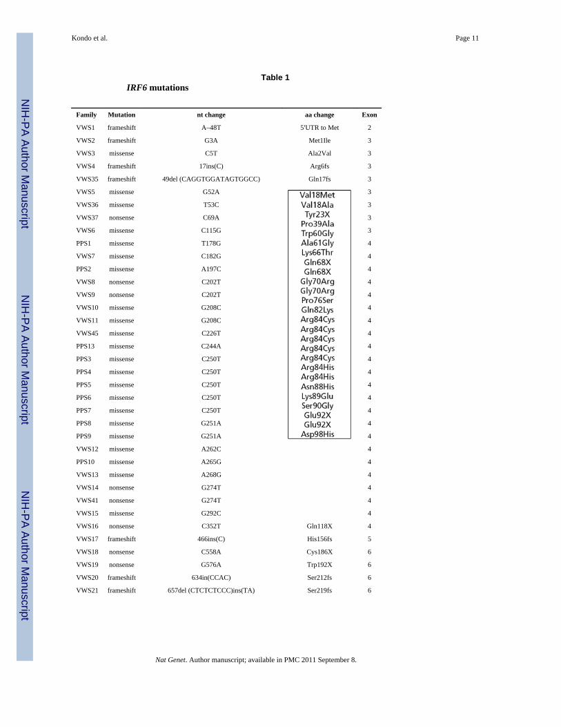
To identify the locus associated with VWS, we carried out direct sequence analysis of genesand presumptive transcripts in the 350-kilobase (kb) critical region3. This approach isconfounded by single-nucleotide polymorphisms (SNPs), normal DNA sequence variationthat occurs about once every 1,900 base pairs8 (bp). To distinguish between putativedisease-causing mutations and SNPs, we studied a pair of monozygotic twins discordant forthe VWS phenotype and whose parents were unaffected. Monozygotic status was confirmedby showing complete concordance of genotype at 20 microsatellite loci. We proposed thatthe only sequence difference between the twins would result from a somatic mutation foundonly in the affected twin. We identified a nonsense mutation in exon 4 of IRF6 in theaffected twin, which was absent in both parents and the unaffected twin (Fig. 1a). Wesubsequently identified mutations in 45 additional unrelated families affected with VWS andin 13 families affected with PPS (Fig. 1b; Table 1), demonstrating unequivocally that thesetwo syndromes are allelic6,7. These mutations were not observed in a minimum of 180control chromosomes.
Clefts of the lip with or without cleft palate and isolated cleft palate are developmentally andgenetically distinct9, yet VWS is a single-gene disorder that encompasses both cleftingphenotypes. To verify this, we analyzed pedigrees (n = 22) that had a single mutation inIRF6 and affected individuals with both phenotypes. Genotype analysis of family VWS25demonstrated that affected individuals, regardless of their phenotype, shared the 18-bpdeletion found in the proband (Fig. 1a). We observed similar results in the other families andconclude that a single mutation in IRF6 can cause both types of cleft.
To determine the effect of mutations on IRF6 gene activity, we compared the type andposition of the mutation with the phenotype. Previous identification of deletionsencompassing the VWS locus (including IRF6 in its entirety) had suggested that thephenotype is caused by haploinsufficiency10–12. In this study, we found protein-truncation(nonsense and frameshift) mutations in 22 families (Fig. 1b). Protein-truncation mutationswere significantly more common in VWS than in PPS (P = 0.004) and were consistent with
Kondo et al. Page 2
Nat Genet. Author manuscript; available in PMC 2011 September 8.
NIH
-PA Author Manuscript
NIH
-PA Author Manuscript
NIH
-PA Author Manuscript

haploinsufficiency in the VWS pedigrees. The lone exception to this relationship was anonsense mutation introducing a stop codon in place of a glutamine codon at position 393,found in pedigree PPS11, which may be a dominant-negative mutation (see below).
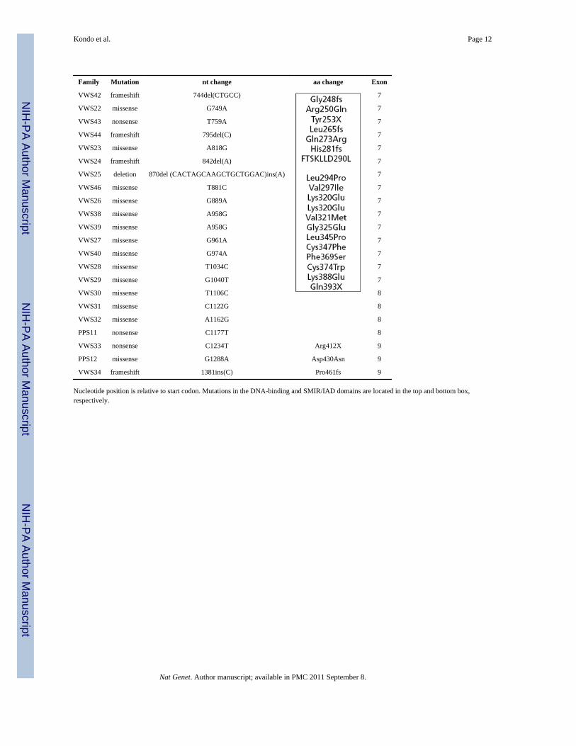
The position of the missense mutations provides insight into the structure and function of theIRF6 gene product. When we aligned the family of IRF proteins, we observed that IRF6 hastwo conserved domains (Fig. 1b), a winged-helix DNA-binding domain (amino acids 13–113) and a protein-binding domain (amino acids 226–394) termed SMIR (Smad-interferonregulatory factor–binding domain)13. Studies of IRF3 and IRF7 have shown that the SMIRdomain is required to form homo- and heterodimers14,15. The dimers then translocate to thenucleus, associate with other transcription factors and ultimately bind to their DNAtargets14. Of the missense mutations, 35 of 37 localized to regions encoding these twodomains. This distribution is non-random (P < 0.001), and we conclude that the domains arecritical for IRF6 function.
Whereas the missense mutations that cause VWS were almost evenly divided between thetwo domains, most missense mutations that cause PPS were found in the DNA-bindingdomain (11 of 13, Fig. 1b). This distribution is significant (P = 0.03) and suggests thatmissense mutations in the DNA-binding domain associated with VWS and PPS affect IRF6function differently. When we compared their positions with the crystal structure of theIRF1 DNA-binding domain16, we found that every amino-acid residue that was mutant inindividuals with PPS directly contacts the DNA, whereas only one of seven of the residuesmutant in the individuals with VWS contacts the DNA. Most notably, we observed missensemutations involving the same residue, Arg84, in seven unrelated PPS families (Fig. 1a,b).The Arg84 residue is comparable to the Arg82 residue of IRF1. It is one of four residues thatmake critical contacts with the core sequence, GAAA, and is essential for DNA binding16.The observed change of this residue to a cysteine or histidine caused a complete loss of thatessential contact (Fig. 2). One possible explanation for this apparent genotype–phenotyperelationship is that missense mutations that cause VWS are due to a complete loss offunction of the mutated IRF6 protein, affecting both DNA and protein binding, whereasmissense mutations causing PPS affect only IRF6's ability to bind DNA. The ability of themutated IRF6 to bind to other proteins is unaffected, and it therefore forms inactivetranscription complexes; thus, this is a dominant-negative mutation. Similarly, deletion ofthe DNA-binding domain of IRF3 or IRF7 exerts a dominant-negative effect on the virus-induced expression of the type I interferon genes and the RANTES gene15,17.
To correlate the expression of IRF6 with the phenotypes of VWS and PPS, we carried outRT–PCR, northern-blot analysis and whole-mount in situ hybridization. We found that Irf6was broadly expressed in embryonic and adult mouse tissues (Fig. 3a,b), a pattern also seenin human fetal and adult tissues (data not shown). Greater expression of Irf6 seemed tooccur in secondary palates dissected from day 14.5–15 mouse embryos and in adult skin.Whole-mount in situ hybridization demonstrated that Irf6 transcripts were highly expressedin the medial edges of the paired palatal shelves immediately before, and during, their fusion(Fig. 3d). Similarly high Irf6 expression was seen in the hair follicles and palatal rugae (Fig.3d), tooth germs and thyroglossal duct (Fig. 3f) and external genitalia (Fig. 3h), and in skinthroughout the body (data not shown). These observations are in accord with the VWS/PPSphenotype: notably, 20% of individuals with VWS exhibit agenesis of the second premolarteeth and 40% of individuals with PPS display genital anomalies.
Although we demonstrated that VWS and PPS are caused by mutations in a single gene, thephenotype for any given mutation varied in at least three ways even within the same family.Of the families with known mutations, we observed 32 families with multiple combinationsof orofacial anomalies, 22 families with mixed clefting phenotypes (individuals with cleft lip
Kondo et al. Page 3
Nat Genet. Author manuscript; available in PMC 2011 September 8.
NIH
-PA Author Manuscript
NIH
-PA Author Manuscript
NIH
-PA Author Manuscript
and individuals with cleft palate only in the same family) and four families affected withPPS that included individuals who exhibit orofacial (VWS) features exclusively. Themarked phenotypic variation in our cohort strongly implicates the action of stochasticfactors or modifier genes on IRF6 function. In this context, we identified the sequencevariant Val274Ile (Fig. 1b). This variant occurs at an absolutely conserved residue within theSMIR domain, is common in unaffected populations (3% in European-descended and 22%in Asian populations) and is an attractive candidate for a modifier of VWS, PPS, and otherorofusial clefting disorders.
The mixed clefting phenotype is common in families affected with VWS, but very rare infamilies with non-syndromic orofacial clefts, and is not seen in most other syndromic formsof orofacial clefts. It is, however, also seen in clefting disorders caused by mutations in thegenes MSX1 (ref. 18) and TP63 (ref. 19,20), suggesting that these may be involved in acommon genetic pathway. In support of a common pathway, we found two IRF binding sitesin the promoter of MSX1 and one in the intron, all of which are conserved between humanmouse.
We are taking an integrated approach to dissecting the complex pathways that underliedevelopment of the lip and palate, including genetic analysis to identify the mutations thatcause orofacial clefts. The discordant monozygotic twins proved useful in this effort, andprovided proof of principle21 that discordant monozygotic pairs can be used to search formodifiers or mutations, especially in regard to complex traits where mapping may beimprecise and mutation analysis may be confounded by SNPs. We also used a large numberof samples from unrelated individuals to confirm that mutations in IRF6 are pathogenic forboth VWS and PPS and to prove that IRF6 is essential for development of the lip and palateand is involved in development of the skin and external genitalia. The SMIR domain hasbeen proposed to mediate an interaction between IRFs and Smads13, a family oftranscription factors known to transduce TGF-β signals22. In addition, the expression of Irf6along the medial edge of the palate seems to overlap with Tgfb3 (ref. 23), and Tgfb3, alongwith other members of this super-family such as Tgfb2 and Inhba, is required for palatalfusion24–27. Together with our data, these observations support a role for IRF6 in thetransforming growth factor-β (TGF-β) signaling pathway, a developmental pathway offundamental significance. The identification of IRF6 as a key determinant in orofacialdevelopment will help us to further delineate and integrate the molecular pathwaysunderlying morphogenesis of the lip and palate.
MethodsFamilies
Families affected with VWS (n = 107) and PPS (n = 15) were identified and examined byone or more geneticists or clinicians as previously described12. Nearly all families are ofnorthern European descent. Sample collection and inclusion criteria for VWS and PPS weredescribed previously3. We obtained written informed consent from all subjects and approvalfor all protocols from the Institutional Review Boards at the University of Iowa and at theUniversity of Manchester.
Mutation analysisWe amplified exons 1–8 and part of exons 9 and 10 by standard PCR. The primer sequencesare available on request. The amplified products were purified (Qiagen) and directlysequenced with an ABI Prism 3700. The sequence was analyzed using the computerprogram PolyPhred.
Kondo et al. Page 4
Nat Genet. Author manuscript; available in PMC 2011 September 8.
NIH
-PA Author Manuscript
NIH
-PA Author Manuscript
NIH
-PA Author Manuscript

Protein modelingThe IRF6 protein structure was predicted from its amino-acid sequence using Expasy, andaligned with the known crystalline structure of the DNA-binding domain of IRF1 using theUNIX-based computer software package Quanta (Accelrys). To model the mutations foundat position Arg84 in the IRF6 DNA-binding domain, the residue was manually altered to acysteine or a histidine. The package predicts all possible orientations of the altered sidechain and displays the position with the highest probability.
RT–PCRWe extracted total RNA using a standard guanidinium isothiocyanate, acid–phenol protocol.RT–PCR analyses were performed and analyzed as detailed previously28 using a forwardprimer designed in exon 4 and a reverse primer designed in exon 6 of Irf6. These primersgenerate a single product of 212 bp from cDNA.
Northern-blot analysisA multiple-tissue northern blot (Seegene) was hybridized with a probe generated by PCRusing primers derived from the distal end of the 3′ untranslated region of Irf6 and labeled asrecommended by the manufacturer with the StripE-Z system (Ambion). We hybridized theblot in Express Hyb (Clontech), washed it as recommended and exposed it to X-ray film for72 h at −80 °C.
Whole-mount in situ hybridizationSense and anti-sense riboprobes were 1,600 bp in length, derived from the 3′ untranslatedregion of Irf6 and generated with Sp6 and T7 promoters, respectively. We fixed embryosdissected from time-mated MF1 mice in 4% paraformaldehyde overnight, processed themand subjected them to hybridization with sense or anti-sense probes as describedpreviously29.
Statistical analysisStatistical significance of mutation location was calculated with the Fisher's exact test usingthe assumption of equal probability for a mutation at each residue.
URLPolyPhred, http://droog.mbt.washington.edu/PolyPhred.html; Expasy, www.expasy.ch.
AcknowledgmentsWe thank our many clinical colleagues and their patients for contributing samples for this study (N. Akarsu, M.Aldred, Z. Ali-Khan, W.P. Allen, L. Bartoshesky, B. Bernhard, E. Bijlsma, E. Breslau-Siderius, C. Brewer, L.Brueton, B. Burton, J. Canady, A. Chakravarti, K. Chen, J. Clayton-Smith, M. Cunningham, A. David, B.B.A. deVries, F.R. Desposito, K. Devriendt, R. Falk, J.-P. Fryns, R.J.M. Gardner, M. Golahi, J. Graham, M. Greenstein, M.Hannibal, E. Hauselman, R. Hennekam, G. Hoganson, L. Holmes, J. Hoogeboom, E. Hoyme, S. Kirkpatrick, J.Klein, T.C. Matise, L. Meisner, Z. Miedzybrodzka, J. Mulliken, A. Newlin, R. Pauli, W. Reardon, S. Roberts, H.Saal, A. Schinzel, J. Siegel-Bartelt, D. Sternen, V. Sybert, D. Tiziani, M.-P. Vazquez, L. Williamson-Kruse, F.Wilt, C. Yardin and K. Yoshiura). We appreciate the advice of K. Buetow, J. Dixon and C. Baldock; technicalassistance from S. Hoper, M. Malik, J. Allaman, C. Hamm, N. Rorick, C. Nishimura, B. Ludwig, M. Fang, P.Hemerson, A. Westphalen and S. Lilly; administrative support from K. Krahn, D. Benton and L. Muilenburg; andsharing of unpublished results by P. Jezewski, A. Grossman and T.W. Mak. This work was supported by grantsfrom the US National Institutes of Health and by grants to M.J.D. from Wellcome Trust, Action Research,Biotechnology and Biological Sciences Research Council, The European Union and the Fundação Lucentis(R.L.L.F.L. & D.M.F.).
Kondo et al. Page 5
Nat Genet. Author manuscript; available in PMC 2011 September 8.
NIH
-PA Author Manuscript
NIH
-PA Author Manuscript
NIH
-PA Author Manuscript
References1. Taniguchi T, Ogasawara K, Takaoka A, Tanaka N. IRF family of transcription factors as regulators
of host defense. Annu Rev Immunol. 2001; 19:623–655. [PubMed: 11244049]2. Murray JC, et al. Linkage of an autosomal dominant clefting syndrome (Van der Woude) to loci on
chromosome 1q. Am J Hum Genet. 1990; 46:486–491. [PubMed: 2309700]3. Schutte BC, et al. A preliminary gene map for the Van der Woude syndrome critical region derived
from 900 kb of genomic sequence at 1q32-q41. Genome Res. 2000; 10:81–94. [PubMed: 10645953]4. Van der Woude A. Fistula labii inferioris congenita and its association with cleft lip and palate. Am
J Hum Genet. 1954; 6:244–256. [PubMed: 13158329]5. Gorlin RJ, Sedano HO, Cervenka J. Popliteal pterygium syndrome. A syndrome comprising cleft
lip-palate, popliteal and intercrural pterygia, digital and genital anomalies. Pediatrics. 1968; 41:503–509. [PubMed: 4384166]
6. Bixler D, Poland C, Nance WE. Phenotypic variation in the popliteal pterygium syndrome. ClinGenet. 1973; 4:220–228. [PubMed: 4203060]
7. Lees MM, Winter RM, Malcolm S, Saal HM, Chitty L. Popliteal pterygium syndrome: a clinicalstudy of three families and report of linkage to the Van der Woude syndrome locus on 1q32. J MedGenet. 1999; 36:888–892. [PubMed: 10593995]
8. Sachidanandam R, et al. A map of human genome sequence variation containing 1.42 million singlenucleotide polymorphisms. Nature. 2001; 409:928–933. [PubMed: 11237013]
9. Fraser FC. Thoughts on the etiology of clefts of the palate and lip. Acta Genetica. 1955; 5:358–369.10. Bocian M, Walker AP. Lip pits and deletion 1q32–q41. Am J Med Genet. 1987; 26:437–443.
[PubMed: 3812594]11. Sander A, Schmelzle R, Murray J. Evidence for a microdeletion in 1q32–41 involving the gene
responsible for Van der Woude syndrome. Hum Mol Genet. 1994; 3:575–578. [PubMed:8069301]
12. Schutte BC, et al. Microdeletions at chromosome bands 1q32-q41 as a cause of Van der Woudesyndrome. Am J Med Genet. 1999; 84:145–150. [PubMed: 10323740]
13. Eroshkin A, Mushegian A. Conserved transactivation domain shared by interferon regulatoryfactors and Smad morphogens. J Mol Med. 1999; 77:403–405. [PubMed: 10426188]
14. Mamane Y, et al. Interferon regulatory factors: the next generation. Gene. 1999; 237:1–14.[PubMed: 10524230]
15. Au WC, Yeow WS, Pitha PM. Analysis of functional domains of interferon regulatory factor 7 andits association with IRF-3. Virology. 2001; 280:273–282. [PubMed: 11162841]
16. Escalante CR, Yie J, Thanos D, Aggarwal AK. Structure of IRF-1 with bound DNA revealsdeterminants of interferon regulation. Nature. 1998; 391:103–106. [PubMed: 9422515]
17. Lin R, Heylbroeck C, Genin P, Pitha PM, Hiscott J. Essential role of interferon regulatory factor 3in direct activation of RANTES chemokine transcription. Mol Cell Biol. 1999; 19:959–966.[PubMed: 9891032]
18. van den Boogaard MJ, Dorland M, Beemer FA, van Amstel HK. MSX1 mutation is associatedwith orofacial clefting and tooth agenesis in humans. Nature Genet. 2000; 24:342–343. [PubMed:10742093]
19. Celli J, et al. Heterozygous germline mutations in the p53 homolog p63 are the cause of EECsyndrome. Cell. 1999; 99:143–153. [PubMed: 10535733]
20. McGrath JA, et al. Hay-Wells syndrome is caused by heterozygous missense mutations in theSAM domain of p63. Hum Mol Genet. 2001; 10:221–229. [PubMed: 11159940]
21. Machin GA. Some causes of genotypic and phenotypic discordance in monozygotic twin pairs. AmJ Med Genet. 1996; 61:216–228. [PubMed: 8741866]
22. Brivanlou AH, Darnell JE Jr. Signal transduction and the control of gene expression. Science.2002; 295:813–818. [PubMed: 11823631]
23. Fitzpatrick DR, Denhez F, Kondaiah P, Akhurst RJ. Differential expression of TGF β isoforms inmurine palatogenesis. Development. 1990; 109:585–595. [PubMed: 2401212]
Kondo et al. Page 6
Nat Genet. Author manuscript; available in PMC 2011 September 8.
NIH
-PA Author Manuscript
NIH
-PA Author Manuscript
NIH
-PA Author Manuscript
24. Proetzel G, et al. Transforming growth factor-β3 is required for secondary palate fusion. NatureGenet. 1995; 11:409–414. [PubMed: 7493021]
25. Sanford LP, et al. TGFβ2 knockout mice have multiple developmental defects that are non-overlapping with other TGFβ knockout phenotypes. Development. 1997; 124:2659–2670.[PubMed: 9217007]
26. Matzuk MM, et al. Functional analysis of activins during mammalian development. Nature. 1995;374:354–356. [PubMed: 7885473]
27. Kaartinen V, et al. Abnormal lung development and cleft palate in mice lacking TGF-β 3 indicatesdefects of epithelial–mesenchymal interaction. Nature Genet. 1995; 11:415–421. [PubMed:7493022]
28. Dixon J, Hovanes K, Shiang R, Dixon MJ. Sequence analysis, identification of evolutionaryconserved motifs and expression analysis of murine tcof1 provide further evidence for a potentialfunction for the gene and its human homologue, TCOF1. Hum Mol Genet. 1997; 6:727–737.[PubMed: 9158147]
29. Nieto MA, Patel K, Wilkinson DG. In situ hybridization analysis of chick embryos in whole mountand tissue sections. Methods Cell Biol. 1996; 51:219–235. [PubMed: 8722478]
Kondo et al. Page 7
Nat Genet. Author manuscript; available in PMC 2011 September 8.
NIH
-PA Author Manuscript
NIH
-PA Author Manuscript
NIH
-PA Author Manuscript
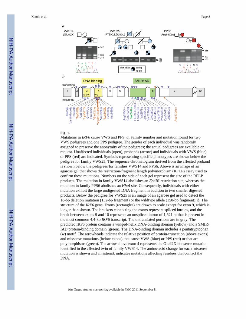
Fig. 1.Mutations in IRF6 cause VWS and PPS. a, Family number and mutation found for twoVWS pedigrees and one PPS pedigree. The gender of each individual was randomlyassigned to preserve the anonymity of the pedigrees; the actual pedigrees are available onrequest. Unaffected individuals (open), probands (arrow) and individuals with VWS (blue)or PPS (red) are indicated. Symbols representing specific phenotypes are shown below thepedigree for family VWS25. The sequence chromatogram derived from the affected probandis shown below the pedigrees for families VWS14 and PPS6. Above is an image of anagarose gel that shows the restriction-fragment length polymorphism (RFLP) assay used toconfirm these mutations. Numbers on the side of each gel represent the size of the RFLPproducts. The mutation in family VWS14 abolishes an EcoRI restriction site, whereas themutation in family PPS6 abolishes an HhaI site. Consequently, individuals with eithermutation exhibit the large undigested DNA fragment in addition to two smaller digestedproducts. Below the pedigree for VWS25 is an image of an agarose gel used to detect the18-bp deletion mutation (132-bp fragment) or the wildtype allele (150-bp fragment). b, Thestructure of the IRF6 gene. Exons (rectangles) are drawn to scale except for exon 9, which islonger than shown. The brackets connecting the exons represent spliced introns, and thebreak between exons 9 and 10 represents an unspliced intron of 1,621 nt that is present inthe most common 4.4-kb IRF6 transcript. The untranslated portions are in gray. Thepredicted IRF6 protein contains a winged-helix DNA-binding domain (yellow) and a SMIR/IAD protein-binding domain (green). The DNA-binding domain includes a pentatryptophan(w) motif. The arrowheads indicate the relative position of protein-truncation (above exons)and missense mutations (below exons) that cause VWS (blue) or PPS (red) or that arepolymorphisms (green). The arrow above exon 4 represents the Glu92X nonsense mutationidentified in the affected twin of family VWS14. The amino-acid change for each missensemutation is shown and an asterisk indicates mutations affecting residues that contact theDNA.
Kondo et al. Page 8
Nat Genet. Author manuscript; available in PMC 2011 September 8.
NIH
-PA Author Manuscript
NIH
-PA Author Manuscript
NIH
-PA Author Manuscript
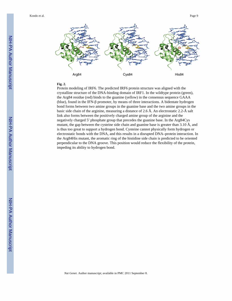
Fig. 2.Protein modeling of IRF6. The predicted IRF6 protein structure was aligned with thecrystalline structure of the DNA-binding domain of IRF1. In the wildtype protein (green),the Arg84 residue (red) binds to the guanine (yellow) in the consensus sequence GAAA(blue), found in the IFN-β promoter, by means of three interactions. A bidentate hydrogenbond forms between two amine groups in the guanine base and the two amine groups in thebasic side chain of the arginine, measuring a distance of 2.6 Å. An electrostatic 2.2-Å saltlink also forms between the positively charged amine group of the arginine and thenegatively charged 5′ phosphate group that precedes the guanine base. In the Arg84Cysmutant, the gap between the cysteine side chain and guanine base is greater than 3.10 Å, andis thus too great to support a hydrogen bond. Cysteine cannot physically form hydrogen orelectrostatic bonds with the DNA, and this results in a disrupted DNA–protein interaction. Inthe Arg84His mutant, the aromatic ring of the histidine side chain is predicted to be orientedperpendicular to the DNA groove. This position would reduce the flexibility of the protein,impeding its ability to hydrogen bond.
Kondo et al. Page 9
Nat Genet. Author manuscript; available in PMC 2011 September 8.
NIH
-PA Author Manuscript
NIH
-PA Author Manuscript
NIH
-PA Author Manuscript
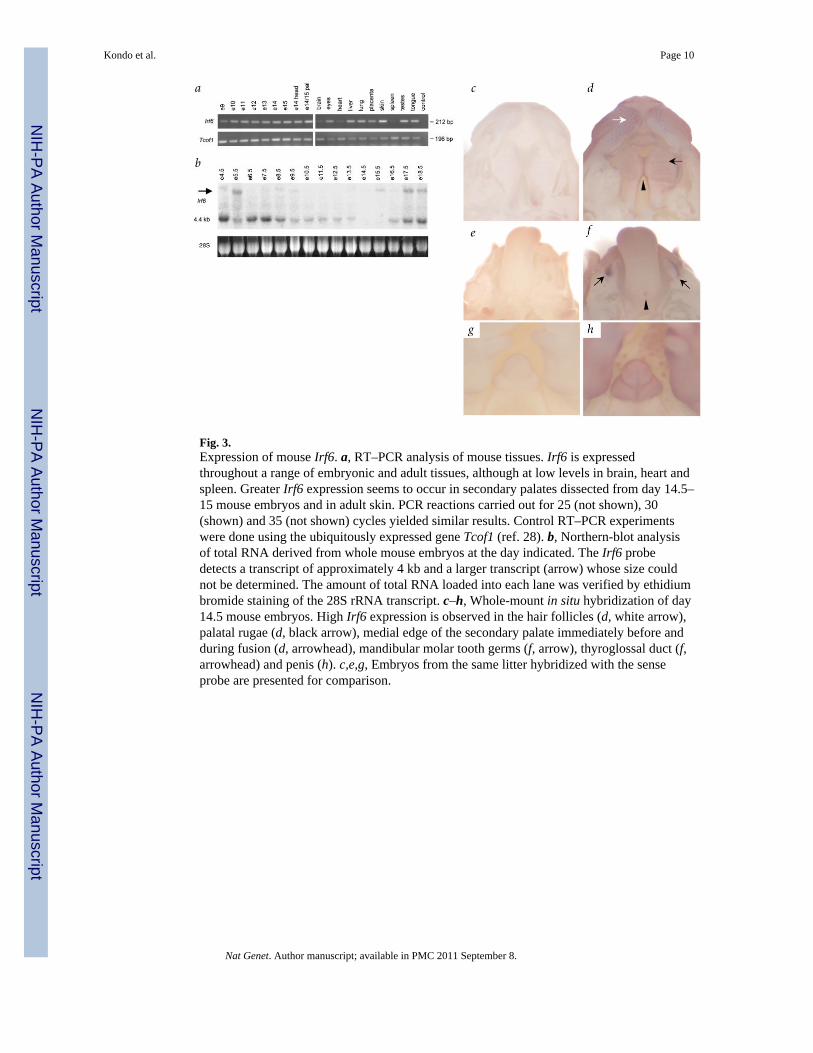
Fig. 3.Expression of mouse Irf6. a, RT–PCR analysis of mouse tissues. Irf6 is expressedthroughout a range of embryonic and adult tissues, although at low levels in brain, heart andspleen. Greater Irf6 expression seems to occur in secondary palates dissected from day 14.5–15 mouse embryos and in adult skin. PCR reactions carried out for 25 (not shown), 30(shown) and 35 (not shown) cycles yielded similar results. Control RT–PCR experimentswere done using the ubiquitously expressed gene Tcof1 (ref. 28). b, Northern-blot analysisof total RNA derived from whole mouse embryos at the day indicated. The Irf6 probedetects a transcript of approximately 4 kb and a larger transcript (arrow) whose size couldnot be determined. The amount of total RNA loaded into each lane was verified by ethidiumbromide staining of the 28S rRNA transcript. c–h, Whole-mount in situ hybridization of day14.5 mouse embryos. High Irf6 expression is observed in the hair follicles (d, white arrow),palatal rugae (d, black arrow), medial edge of the secondary palate immediately before andduring fusion (d, arrowhead), mandibular molar tooth germs (f, arrow), thyroglossal duct (f,arrowhead) and penis (h). c,e,g, Embryos from the same litter hybridized with the senseprobe are presented for comparison.
Kondo et al. Page 10
Nat Genet. Author manuscript; available in PMC 2011 September 8.
NIH
-PA Author Manuscript
NIH
-PA Author Manuscript
NIH
-PA Author Manuscript
NIH
-PA Author Manuscript
NIH
-PA Author Manuscript
NIH
-PA Author Manuscript
Kondo et al. Page 11
Table 1IRF6 mutations
Family Mutation nt change aa change Exon
VWS1 frameshift A–48T 5′UTR to Met 2
VWS2 frameshift G3A Met1Ile 3
VWS3 missense C5T Ala2Val 3
VWS4 frameshift 17ins(C) Arg6fs 3
VWS35 frameshift 49del (CAGGTGGATAGTGGCC) Gln17fs 3
VWS5 missense G52A 3
VWS36 missense T53C 3
VWS37 nonsense C69A 3
VWS6 missense C115G 3
PPS1 missense T178G 4
VWS7 missense C182G 4
PPS2 missense A197C 4
VWS8 nonsense C202T 4
VWS9 nonsense C202T 4
VWS10 missense G208C 4
VWS11 missense G208C 4
VWS45 missense C226T 4
PPS13 missense C244A 4
PPS3 missense C250T 4
PPS4 missense C250T 4
PPS5 missense C250T 4
PPS6 missense C250T 4
PPS7 missense C250T 4
PPS8 missense G251A 4
PPS9 missense G251A 4
VWS12 missense A262C 4
PPS10 missense A265G 4
VWS13 missense A268G 4
VWS14 nonsense G274T 4
VWS41 nonsense G274T 4
VWS15 missense G292C 4
VWS16 nonsense C352T Gln118X 4
VWS17 frameshift 466ins(C) His156fs 5
VWS18 nonsense C558A Cys186X 6
VWS19 nonsense G576A Trp192X 6
VWS20 frameshift 634in(CCAC) Ser212fs 6
VWS21 frameshift 657del (CTCTCTCCC)ins(TA) Ser219fs 6
Nat Genet. Author manuscript; available in PMC 2011 September 8.
NIH
-PA Author Manuscript
NIH
-PA Author Manuscript
NIH
-PA Author Manuscript
Kondo et al. Page 12
Family Mutation nt change aa change Exon
VWS42 frameshift 744del(CTGCC) 7
VWS22 missense G749A 7
VWS43 nonsense T759A 7
VWS44 frameshift 795del(C) 7
VWS23 missense A818G 7
VWS24 frameshift 842del(A) 7
VWS25 deletion 870del (CACTAGCAAGCTGCTGGAC)ins(A) 7
VWS46 missense T881C 7
VWS26 missense G889A 7
VWS38 missense A958G 7
VWS39 missense A958G 7
VWS27 missense G961A 7
VWS40 missense G974A 7
VWS28 missense T1034C 7
VWS29 missense G1040T 7
VWS30 missense T1106C 8
VWS31 missense C1122G 8
VWS32 missense A1162G 8
PPS11 nonsense C1177T 8
VWS33 nonsense C1234T Arg412X 9
PPS12 missense G1288A Asp430Asn 9
VWS34 frameshift 1381ins(C) Pro461fs 9
Nucleotide position is relative to start codon. Mutations in the DNA-binding and SMIR/IAD domains are located in the top and bottom box,respectively.
Nat Genet. Author manuscript; available in PMC 2011 September 8.
Related Documents