ARTICLE Mutations in ATP6V1E1 or ATP6V1A Cause Autosomal-Recessive Cutis Laxa Tim Van Damme, 1,25 Thatjana Gardeitchik, 2,3,25 Miski Mohamed, 2,25 Sergio Guerrero-Castillo, 4,5,26 Peter Freisinger, 6,26 Brecht Guillemyn, 1,26 Ariana Kariminejad, 7,26 Daisy Dalloyaux, 2,5 Sanne van Kraaij, 2,5 Dirk J. Lefeber, 5,8 Delfien Syx, 1 Wouter Steyaert, 1 Riet De Rycke, 9,10 Alexander Hoischen, 3 Erik-Jan Kamsteeg, 3 Sunnie Y. Wong, 11 Monique van Scherpenzeel, 5,8 Payman Jamali, 12 Ulrich Brandt, 4,5 Leo Nijtmans, 4,5 G. Christoph Korenke, 13 Brian H.Y. Chung, 14 Christopher C.Y. Mak, 14 Ingrid Hausser, 15 Uwe Kornak, 16,17 Bjo ¨rn Fischer-Zirnsak, 16,17 Tim M. Strom, 18 Thomas Meitinger, 18 Yasemin Alanay, 19 Gulen E. Utine, 20 Peter K.C. Leung, 14 Siavash Ghaderi-Sohi, 7 Paul Coucke, 1 Sofie Symoens, 1 Anne De Paepe, 1 Christian Thiel, 21 Tobias B. Haack, 18,22,23 Fransiska Malfait, 1,27 Eva Morava, 11,24,27 Bert Callewaert, 1,27, * and Ron A. Wevers 5,27, * Defects of the V-type proton (H þ ) ATPase (V-ATPase) impair acidification and intracellular trafficking of membrane-enclosed compart- ments, including secretory granules, endosomes, and lysosomes. Whole-exome sequencing in five families affected by mild to severe cutis laxa, dysmorphic facial features, and cardiopulmonary involvement identified biallelic missense mutations in ATP6V1E1 and ATP6V1A, which encode the E1 and A subunits, respectively, of the V 1 domain of the heteromultimeric V-ATPase complex. Structural modeling indicated that all substitutions affect critical residues and inter- or intrasubunit interactions. Furthermore, complexome profiling, a method combining blue-native gel electrophoresis and liquid chromatography tandem mass spectrometry, showed that they disturb either the assembly or the stability of the V-ATPase complex. Protein glycosylation was variably affected. Abnormal vesicular trafficking was evidenced by delayed retrograde transport after brefeldin A treatment and abnormal swelling and fragmentation of the Golgi apparatus. In addition to showing reduced and fragmented elastic fibers, the histopathological hallmark of cutis laxa, transmission electron microscopy of the dermis also showed pronounced changes in the structure and organization of the collagen fibers. Our find- ings expand the clinical and molecular spectrum of metabolic cutis laxa syndromes and further link defective extracellular matrix assem- bly to faulty protein processing and cellular trafficking caused by genetic defects in the V-ATPase complex. Introduction The V-type proton (H þ ) ATPase (V-ATPase) is an ATP-dependent H þ pump that establishes and main- tains the acidic environment of intracellular organelles, including secretory granules, endosomes, and lysosomes, as well as extracellular compartments. This protein com- plex is composed of a catalytic cytosolic V 1 domain and a H þ -pumping, membrane-embedded V 0 domain, both of which comprise different subunits. The V-ATPase complex is essential to physiological processes such as membrane trafficking, protein degradation, pH homeostasis, and pH-dependent (e.g., Wnt and Notch) or -independent (e.g., mTORC1 and APMK) regulation of intracellular signaling, but it also mediates pathological processes including virus and toxin entry, drug resistance, and can- cer cell survival, migration, and invasion. 1,2 Gene mutations affecting V-ATPase have been identified in several autosomal-recessive (AR) Mendelian disorders. 3 In the V 1 domain, mutations affecting the B1 (ATP6V1B1 [MIM: 192132]) and B2 (ATP6V1B2 [MIM: 606939]) sub- units cause AR distal renal tubular acidosis with early-onset 1 Center for Medical Genetics, Ghent University and Ghent University Hospital, Ghent 9000, Belgium; 2 Department of Pediatrics, Radboud University Med- ical Center, Nijmegen 6500 HB, the Netherlands; 3 Department of Human Genetics, Radboud University Medical Center, Nijmegen 6500 HB, the Netherlands; 4 Radboud Center for Mitochondrial Medicine, Department of Pediatrics, Radboud University Medical Center, Nijmegen 6500 HB, the Netherlands; 5 Translational Metabolic Laboratory, Department of Laboratory Medicine, Radboud University Medical Center, Nijmegen 6500 HB, the Netherlands; 6 Childrens’ Hospital, Klinikum am Steinenberg, Reutlingen 72764, Germany; 7 Kariminejad-Najmabadi Pathology & Genetics Center, Tehran 14656, Iran; 8 Department of Neurology, Radboud University Medical Center, Nijmegen 6500 HB, the Netherlands; 9 Department of Biomedical Mo- lecular Biology, Ghent University, Ghent 9000, Belgium; 10 Inflammation Research Center, VIB, Ghent 9000, Belgium; 11 Hayward Genetics Center, Tulane University Medical School, New Orleans, LA 70112, USA; 12 Shahrood Genetic Counseling Center, Semnan 36156, Iran; 13 Department of Neuropediatrics, Children’s Hospital Klinikum Oldenburg, Oldenburg 26133, Germany; 14 Department of Paediatrics & Adolescent Medicine, Li Ka Shing Faculty of Med- icine, University of Hong Kong, Hong Kong, China; 15 Institute of Pathology, Universita ¨tsklinikum Heidelberg, Heidelberg 69120, Germany; 16 Institute of Medical Genetics and Human Genetics, Charite ´ – Universitaetsmedizin Berlin, Berlin 13353, Germany; 17 Max Planck Institute for Molecular Genetics, Berlin 14195, Germany; 18 Institute of Human Genetics, Helmholtz Zentrum Mu ¨nchen, Neuherberg 85764, Germany; 19 Pediatric Genetics Unit, Depart- ment of Pediatrics, Acibadem University School of Medicine, Istanbul 34752, Turkey; 20 Pediatric Genetics Unit, Department of Pediatrics, Ihsan Do gramacı Children’s Hospital, Hacettepe School of Medicine, Ankara 06100, Turkey; 21 Center for Child and Adolescent Medicine, Klinik Kinderheilkunde I, Univer- sita ¨tsklinikum Heidelberg, Heidelberg 69120, Germany; 22 Institute of Human Genetics, Technische Universita ¨t Mu ¨ nchen, Munich 81675, Germany; 23 Institute of Medical Genetics and Applied Genomics, University of Tu ¨bingen, Tu ¨ bingen 72076, Germany; 24 Department of Pediatrics, University Hospital Leuven, Leuven 3000, Belgium 25 These authors contributed equally to this work 26 These authors contributed equally to this work 27 These authors contributed equally to this work *Correspondence: [email protected] (B.C.), [email protected] (R.A.W.) http://dx.doi.org/10.1016/j.ajhg.2016.12.010. The American Journal of Human Genetics 100, 1–12, February 2, 2017 1 Ó 2016 American Society of Human Genetics. Please cite this article in press as: Van Damme et al., Mutations in ATP6V1E1 or ATP6V1A Cause Autosomal-Recessive Cutis Laxa, The Amer- ican Journal of Human Genetics (2017), http://dx.doi.org/10.1016/j.ajhg.2016.12.010

Welcome message from author
This document is posted to help you gain knowledge. Please leave a comment to let me know what you think about it! Share it to your friends and learn new things together.
Transcript
Please cite this article in press as: Van Damme et al., Mutations in ATP6V1E1 or ATP6V1A Cause Autosomal-Recessive Cutis Laxa, The Amer-ican Journal of Human Genetics (2017), http://dx.doi.org/10.1016/j.ajhg.2016.12.010
ARTICLE
Mutations in ATP6V1E1 or ATP6V1ACause Autosomal-Recessive Cutis Laxa
Tim Van Damme,1,25 Thatjana Gardeitchik,2,3,25 Miski Mohamed,2,25 Sergio Guerrero-Castillo,4,5,26
Peter Freisinger,6,26 Brecht Guillemyn,1,26 Ariana Kariminejad,7,26 Daisy Dalloyaux,2,5
Sanne van Kraaij,2,5 Dirk J. Lefeber,5,8 Delfien Syx,1 Wouter Steyaert,1 Riet De Rycke,9,10
Alexander Hoischen,3 Erik-Jan Kamsteeg,3 Sunnie Y. Wong,11 Monique van Scherpenzeel,5,8
Payman Jamali,12 Ulrich Brandt,4,5 Leo Nijtmans,4,5 G. Christoph Korenke,13 Brian H.Y. Chung,14
Christopher C.Y. Mak,14 Ingrid Hausser,15 Uwe Kornak,16,17 Bjorn Fischer-Zirnsak,16,17 Tim M. Strom,18
Thomas Meitinger,18 Yasemin Alanay,19 Gulen E. Utine,20 Peter K.C. Leung,14 Siavash Ghaderi-Sohi,7
Paul Coucke,1 Sofie Symoens,1 Anne De Paepe,1 Christian Thiel,21 Tobias B. Haack,18,22,23
Fransiska Malfait,1,27 Eva Morava,11,24,27 Bert Callewaert,1,27,* and Ron A. Wevers5,27,*
Defects of the V-type proton (Hþ) ATPase (V-ATPase) impair acidification and intracellular trafficking of membrane-enclosed compart-
ments, including secretory granules, endosomes, and lysosomes. Whole-exome sequencing in five families affected by mild to severe
cutis laxa, dysmorphic facial features, and cardiopulmonary involvement identified biallelic missense mutations in ATP6V1E1 and
ATP6V1A, which encode the E1 and A subunits, respectively, of the V1 domain of the heteromultimeric V-ATPase complex. Structural
modeling indicated that all substitutions affect critical residues and inter- or intrasubunit interactions. Furthermore, complexome
profiling, a method combining blue-native gel electrophoresis and liquid chromatography tandem mass spectrometry, showed that
they disturb either the assembly or the stability of the V-ATPase complex. Protein glycosylation was variably affected. Abnormal vesicular
trafficking was evidenced by delayed retrograde transport after brefeldin A treatment and abnormal swelling and fragmentation of the
Golgi apparatus. In addition to showing reduced and fragmented elastic fibers, the histopathological hallmark of cutis laxa, transmission
electron microscopy of the dermis also showed pronounced changes in the structure and organization of the collagen fibers. Our find-
ings expand the clinical andmolecular spectrum ofmetabolic cutis laxa syndromes and further link defective extracellular matrix assem-
bly to faulty protein processing and cellular trafficking caused by genetic defects in the V-ATPase complex.
Introduction
The V-type proton (Hþ) ATPase (V-ATPase) is an
ATP-dependent Hþ pump that establishes and main-
tains the acidic environment of intracellular organelles,
including secretory granules, endosomes, and lysosomes,
as well as extracellular compartments. This protein com-
plex is composed of a catalytic cytosolic V1 domain and a
Hþ-pumping, membrane-embedded V0 domain, both of
which comprise different subunits. The V-ATPase complex
is essential to physiological processes such as membrane
1Center for Medical Genetics, Ghent University and Ghent University Hospital
ical Center, Nijmegen 6500 HB, the Netherlands; 3Department of Human
Netherlands; 4Radboud Center for Mitochondrial Medicine, Department
the Netherlands; 5Translational Metabolic Laboratory, Department of Labor
the Netherlands; 6Childrens’ Hospital, Klinikum am Steinenberg, Reutlingen
Tehran 14656, Iran; 8Department of Neurology, Radboud University Medical C
lecular Biology, Ghent University, Ghent 9000, Belgium; 10Inflammation Resea
University Medical School, New Orleans, LA 70112, USA; 12Shahrood Genetic
Children’s Hospital Klinikum Oldenburg, Oldenburg 26133, Germany; 14Dep
icine, University of Hong Kong, Hong Kong, China; 15Institute of Pathology,
of Medical Genetics and Human Genetics, Charite – Universitaetsmedizin Ber
Berlin 14195, Germany; 18Institute of Human Genetics, Helmholtz Zentrum M
ment of Pediatrics, Acibadem University School of Medicine, Istanbul 34752, T
Children’s Hospital, Hacettepe School of Medicine, Ankara 06100, Turkey; 21C
sitatsklinikum Heidelberg, Heidelberg 69120, Germany; 22Institute of Huma23Institute of Medical Genetics and Applied Genomics, University of Tubingen,
Leuven, Leuven 3000, Belgium25These authors contributed equally to this work26These authors contributed equally to this work27These authors contributed equally to this work
*Correspondence: [email protected] (B.C.), [email protected]
http://dx.doi.org/10.1016/j.ajhg.2016.12.010.
The A
� 2016 American Society of Human Genetics.
trafficking, protein degradation, pH homeostasis, and
pH-dependent (e.g., Wnt and Notch) or -independent
(e.g., mTORC1 and APMK) regulation of intracellular
signaling, but it also mediates pathological processes
including virus and toxin entry, drug resistance, and can-
cer cell survival, migration, and invasion.1,2
Gene mutations affecting V-ATPase have been identified
in several autosomal-recessive (AR) Mendelian disorders.3
In the V1 domain, mutations affecting the B1 (ATP6V1B1
[MIM: 192132]) and B2 (ATP6V1B2 [MIM: 606939]) sub-
units cause AR distal renal tubular acidosis with early-onset
, Ghent 9000, Belgium; 2Department of Pediatrics, Radboud University Med-
Genetics, Radboud University Medical Center, Nijmegen 6500 HB, the
of Pediatrics, Radboud University Medical Center, Nijmegen 6500 HB,
atory Medicine, Radboud University Medical Center, Nijmegen 6500 HB,
72764, Germany; 7Kariminejad-Najmabadi Pathology & Genetics Center,
enter, Nijmegen 6500 HB, the Netherlands; 9Department of Biomedical Mo-
rch Center, VIB, Ghent 9000, Belgium; 11Hayward Genetics Center, Tulane
Counseling Center, Semnan 36156, Iran; 13Department of Neuropediatrics,
artment of Paediatrics & Adolescent Medicine, Li Ka Shing Faculty of Med-
Universitatsklinikum Heidelberg, Heidelberg 69120, Germany; 16Institute
lin, Berlin 13353, Germany; 17Max Planck Institute for Molecular Genetics,
unchen, Neuherberg 85764, Germany; 19Pediatric Genetics Unit, Depart-
urkey; 20Pediatric Genetics Unit, Department of Pediatrics, Ihsan Do�gramacıenter for Child and Adolescent Medicine, Klinik Kinderheilkunde I, Univer-
n Genetics, Technische Universitat Munchen, Munich 81675, Germany;
Tubingen 72076, Germany; 24Department of Pediatrics, University Hospital
l (R.A.W.)
merican Journal of Human Genetics 100, 1–12, February 2, 2017 1

Please cite this article in press as: Van Damme et al., Mutations in ATP6V1E1 or ATP6V1A Cause Autosomal-Recessive Cutis Laxa, The Amer-ican Journal of Human Genetics (2017), http://dx.doi.org/10.1016/j.ajhg.2016.12.010
hearing loss (MIM: 267300) and Zimmermann-Laband syn-
drome (MIM: 616455), respectively.4,5 In the V0 domain,
mutations affecting the a4 subunit (ATP6V0A4 [MIM:
605239]) cause AR distal renal tubular acidosis with late-
onset hearing loss (MIM: 602722), whereas mutations in
TCIRG1 (MIM: 604592), encoding the osteoclast specific
long isoform of the a3 subunit, cause AR osteopetrosis
(MIM: 259700).6,7 Finally, mutations in ATP6V0A2 (MIM:
611716), encoding the a2 subunit of the V0 domain, cause
AR cutis laxa type 2A (ARCL2A [MIM: 219200]).8 ARCL2A
isa congenitaldisorderofglycosylation (CDG)characterized
by a typical facial gestalt, skin abnormalities ranging from
mildwrinkling to severe cutis laxa, andvariableneurological
and skeletal involvement. Genetic depletion of ATP6V0A2
disruptsnormal vesicular traffickingand results inabnormal
glycosylation of serum proteins, intracellular accumulation
of tropoelastin, reduced deposition of mature elastin in the
extracellularmatrix (ECM), accumulation of abnormal lyso-
somes and multivesicular bodies, and increased auto-
phagy.8,9 Cutis laxa syndromes are a heterogeneous group
of acquiredor inheriteddisorderswith redundant orwrinkly
skin and elastic-fiber fragmentation as their clinical and his-
topathologicalhallmarks.Heritable cutis laxa syndromesare
caused not only by defects in ECM molecules (e.g., elastin)
but also by defects in diverse metabolic pathways, such as
intracellular trafficking, proline synthesis, and mitochon-
drial function.10,11 Nevertheless, how these metabolic de-
fects interfere with ECM assembly remains elusive.
Here, we report biallelic mutations in ATP6V1E1 (MIM:
108746) and ATP6V1A (MIM: 607027), respectively encod-
ing the E1 and A subunits of the V1 domain of V-ATPase, as
a cause of distinct metabolic and multisystemic cutis laxa
entities. We show that these mutations affect V-ATPase
structure and assembly, protein glycosylation, Golgi traf-
ficking, and lysosomal function and lead to defects in
ECM homeostasis and architecture.
Subjects and Methods
Clinical AssessmentAll individuals were referred to one of the collaborating academic
expert centers for diagnostic workup of cutis laxa. Blood samples
were collected from seven affected individuals from five families
and their unaffected parents (Figure 1). Skin biopsieswere obtained
fromPI:1, PIII:1, PIV:1, andone individualwith compound-hetero-
zygous c.1214C>T (p.Pro405Leu) and c.1529G>T (p.Arg510Ile)
ATP6V0A2mutations (PV0A2; reported as patient 1 inHucthagow-
der et al.9). PCR-based next-generation sequencing of known
cutis-laxa-related genes did not identify causal mutations in the
probands of families I and III. The study was conducted in accor-
dance with the Declaration of Helsinki, and informed consent
was obtained from all affected individuals and/or parents.
Whole-Exome SequencingWhole-exome sequencing (WES) was performed on genomic DNA
extracted from blood leukocytes of the probands of each family on
four different platforms. Details on exome capture, sequencing,
2 The American Journal of Human Genetics 100, 1–12, February 2, 20
and variant filtering in all families are available on request. Vari-
ants were annotated with PolyPhen-2, SIFT, MutationTaster,
Align-GVGD, MAPP (Multivariate Analysis of Protein Polymor-
phism), and variant allele frequencies from the Exome Aggrega-
tion Consortium (ExAC) Browser and our in-house exome
cohorts.12–18 Sequence validation and segregation analysis of
the candidate variants were performed with Sanger sequencing
(reference sequences GenBank: NM_001696.3 [ATP6V1E1] and
NM_001690.3 [ATP6V1A]). Variant nomenclature adheres to the
Human Genome Variation Society (HGVS) guidelines, and all var-
iants were checked with Mutalyzer software (version 2.0.18).19
Expression AnalysisFor quantitative reverse-transcription PCR (qRT-PCR), total RNA
was extracted in triplicate from cultured fibroblasts with the
RNeasy Kit (QIAGEN), and cDNA was synthesized with the iScript
cDNA Synthesis Kit (Bio-Rad Laboratories). For each cDNA sample,
assays were prepared with the addition of RealTime ready DNA
Probes Master mix and ResoLight Dye (Roche) and were subse-
quently run in duplicate on a Roche LightCycler 480 System.
Data were analyzed with qbaseþ (version 3.0, Biogazelle) and
HPRT1, RPL13A, and YWHAZ as reference genes.
ImmunoblottingFor immunoblotting of the E1 and A subunits, total protein was
extracted with Laemmli Sample Buffer (Bio-Rad Laboratories)
and subjected to SDS-PAGE. Proteins were transferred to a nitrocel-
lulose membrane with the iBlot 2 Dry Blotting System (Thermo
Fisher Scientific). Membranes were blocked in 2% ECL Blocking
Agent (GE Healthcare), incubated with Abcam primary anti-
bodies against ATP6V1E1 (1:200; ab176789), ATP6V1A (1:2,000;
ab199326), or b-tubulin (1:1,000; ab6046), and subsequently incu-
bated with horseradish-peroxidase-conjugated secondary anti-
body (1:1,500; 7074S, Cell Signaling Technologies). Membranes
were scanned with a ChemiDoc-It 500 Imaging System (UVP),
and signal intensity was compared to the amount of b-tubulin.
Structural ModelingHomology models of the E1 and A subunits of the human
V-ATPasewere generatedwith I-Tasser software (Iterative Threading
ASSEmbly Refinement) with the respective subunits of the Saccharo-
myces cerevisiaeV-ATPase (PDB: 3J9T) as a template.20–23 The homol-
ogy models were subsequently inserted in and aligned to the avail-
able three-dimensional structure of the S. cerevisiae V-ATPase with
the MatchMaker function of the UCSF Chimera software package
(version 1.10.2, build 40686).24 The Dunbrack rotamers function
was used to substitute specific residues within the protein structure
tomodel theeffectof the specificproteinvariants.25TheFindHBond
function was used to identify potential hydrogen bonds.
Complexome ProfilingFor preparation of a cellular fraction enriched with V-ATPase
subunits, frozen fibroblast pellets were resuspended in ice-cold
125 mM sucrose and 10 mM Tris-HCl (pH 7.4) and incubated
for 5 min on ice. The cells were homogenized with ten strokes at
1,800 rpm with a glass and Teflon homogenizer at 0�C. The ho-
mogenate was mixed with 1.5 M sucrose at a ratio of 8:1 and sub-
sequently centrifuged at 1,000 3 g at 4�C for 10 min for the
removal of nuclei, cell debris, and intact cells. The supernatant
was again centrifuged at 6,000 3 g at 2�C for 10 min, after which
the supernatant was pelleted by ultra-speed centrifugation at
17
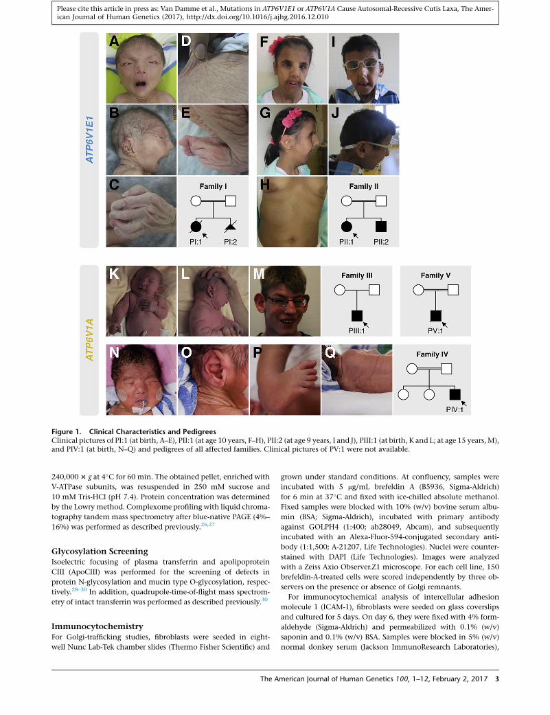
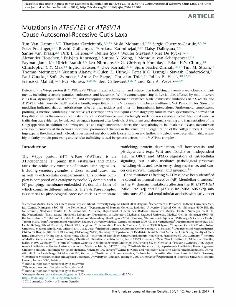
Figure 1. Clinical Characteristics and PedigreesClinical pictures of PI:1 (at birth, A–E), PII:1 (at age 10 years, F–H), PII:2 (at age 9 years, I and J), PIII:1 (at birth, K and L; at age 15 years,M),and PIV:1 (at birth, N–Q) and pedigrees of all affected families. Clinical pictures of PV:1 were not available.
Please cite this article in press as: Van Damme et al., Mutations in ATP6V1E1 or ATP6V1A Cause Autosomal-Recessive Cutis Laxa, The Amer-ican Journal of Human Genetics (2017), http://dx.doi.org/10.1016/j.ajhg.2016.12.010
240,000 3 g at 4�C for 60 min. The obtained pellet, enriched with
V-ATPase subunits, was resuspended in 250 mM sucrose and
10 mM Tris-HCl (pH 7.4). Protein concentration was determined
by the Lowrymethod. Complexome profiling with liquid chroma-
tography tandem mass spectrometry after blue-native PAGE (4%–
16%) was performed as described previously.26,27
Glycosylation ScreeningIsoelectric focusing of plasma transferrin and apolipoprotein
CIII (ApoCIII) was performed for the screening of defects in
protein N-glycosylation and mucin type O-glycosylation, respec-
tively.28–30 In addition, quadrupole-time-of-flight mass spectrom-
etry of intact transferrin was performed as described previously.30
ImmunocytochemistryFor Golgi-trafficking studies, fibroblasts were seeded in eight-
well Nunc Lab-Tek chamber slides (Thermo Fisher Scientific) and
The A
grown under standard conditions. At confluency, samples were
incubated with 5 mg/mL brefeldin A (B5936, Sigma-Aldrich)
for 6 min at 37�C and fixed with ice-chilled absolute methanol.
Fixed samples were blocked with 10% (w/v) bovine serum albu-
min (BSA; Sigma-Aldrich), incubated with primary antibody
against GOLPH4 (1:400; ab28049, Abcam), and subsequently
incubated with an Alexa-Fluor-594-conjugated secondary anti-
body (1:1,500; A-21207, Life Technologies). Nuclei were counter-
stained with DAPI (Life Technologies). Images were analyzed
with a Zeiss Axio Observer.Z1 microscope. For each cell line, 150
brefeldin-A-treated cells were scored independently by three ob-
servers on the presence or absence of Golgi remnants.
For immunocytochemical analysis of intercellular adhesion
molecule 1 (ICAM-1), fibroblasts were seeded on glass coverslips
and cultured for 5 days. On day 6, they were fixed with 4% form-
aldehyde (Sigma-Aldrich) and permeabilized with 0.1% (w/v)
saponin and 0.1% (w/v) BSA. Samples were blocked in 5% (w/v)
normal donkey serum (Jackson ImmunoResearch Laboratories),
merican Journal of Human Genetics 100, 1–12, February 2, 2017 3

Please cite this article in press as: Van Damme et al., Mutations in ATP6V1E1 or ATP6V1A Cause Autosomal-Recessive Cutis Laxa, The Amer-ican Journal of Human Genetics (2017), http://dx.doi.org/10.1016/j.ajhg.2016.12.010
incubated with a primary antibody against ICAM-1 (1:750; MA5-
13021, Thermo Fisher Scientific), and subsequently incubated
with a biotinylated donkey anti-mouse secondary antibody
(1:800; 715-065-151, Jackson ImmunoResearch Laboratories)
and streptavidin-conjugated Cy3 (1 mg/mL; 016-160-084, Jackson
ImmunoResearch Laboratories). The samples were mounted with
VECTASHIELDMounting Medium containing DAPI (Vector Labo-
ratories). Images were obtained with an Olympus BX51 System
Microscope. Particle analysis and manual counting were per-
formed with Fiji and the Cell Counter plugin (version 2.0.0-rc-
49/1.51a).31–33
Transmission Electron MicroscopySamples were fixed in 4% paraformaldehyde and 2.5% glutaralde-
hyde in 0.1 M sodium cacodylate buffer (pH 7.2). After being
washed in buffer solution, samples were postfixed in 1% OsO4
with K3Fe(CN)6 in 0.1 M sodium cacodylate buffer (pH 7.2)
and subsequently dehydrated through a graded ethanol series,
including a bulk staining with 2% uranyl acetate at the 50%
ethanol step followed by embedding in Spurr’s resin. Ultrathin
sections of a gold interference color were cut with an EM UC6
ultramicrotome (Leica Microsystems) and then consecutively
stained with uranyl acetate and lead stain in a Leica EM AC20. Sec-
tions collected on formvar-coated copper slot grids were viewed
with a JEOL JEM 1400plus transmission electronmicroscope oper-
ating at 60 kV.
Results
Clinical Phenotype
Table 1, Figure 1, and Figure S1 summarize and illustrate
the salient clinical findings in all seven affected individ-
uals. Detailed case reports are available in the Supple-
mental Note.
At birth, anthropometric parameters were mostly within
normal range. Perinatal complications were reported in
several families. PI:1 and PIV:1 died after several months
of intensive care and treatment. In family I, a second preg-
nancy (PI:2) was terminated at 21 weeks of gestation
because of cardiac abnormalities and increased nuchal
thickness. The siblings from family II were admitted
shortly after birth with respiratory distress secondary to
bilateral pneumothoraxes, and in PIV:1, the neonatal
course had been complicated by seizures, pneumonia,
sepsis, and a half-hour-long resuscitation after a dislocated
endotracheal tube.
All affected individuals presented with generalized skin
wrinkling and sparse subcutaneous fat. In addition,
PIII:1, PIV:1, and PV:1 had large skin folds and abnormal
fat distribution, especially on the buttocks. With the
exception of PIII:1, who had milder facial features, all
affected individuals had a similar progeroid facial gestalt
with a ‘‘mask-like’’ triangular face, a short forehead, hyper-
telorism, entropion, low-set ears with misfolded helices, a
beaked nose with a broad nasal base and narrow nostrils,
and a short and pointed chin.
At birth, several affected individuals had congenital
heart defects including septal defects (PII:1, PIV:1, and
4 The American Journal of Human Genetics 100, 1–12, February 2, 20
PV:1), cardiac valve defects (PII:1), and right hypoplastic
heart syndrome (PI:2). Other cardiovascular complications
included dilatation of the ascending aorta (PI:1 and PIV:1),
hypertrophic cardiomyopathy (PI:1 and PIII:1), dilatation
of the right ventricle and reduced diastolic compliance
(PII:1), and cardiac failure (PIV:1).
Three families had a history of neurologic complica-
tions. PIII:1 had a severe speech delay, and he suffered
a generalized tonic-clonic seizure at the age of 14 years.
MRI showed enlarged ventricles with white-matter
involvement and periventricular parieto-occipital gliosis.
PIV:1 developed epileptic seizures shortly after birth,
which evolved to generalized and complex partial sei-
zures at the age of 2 months. MRI showed diffuse thick-
ening of the cerebral cortex, suggestive of polymi-
crogyria, and a thin corpus callosum. Finally, in PV:1,
MRI showed an anatomical variant of the cavum septum
pellucidum.
Most affected individuals suffered from severe and often
disabling hypotonia. Additional but less frequent features
included hip dysplasia (PI:1, PII:1, and PIV:1), multiple
congenital contractures (PI:1 and PIV:1), kyphoscoliosis
(PII:1 and PII:2), marfanoid habitus (PII:1, PII:2, and
PIII:1), inguinal herniation (PII:2 and PIV:1), and cryp-
torchidy (PII:2 and PIV:1).
Biallelic Mutations in ATP6V1E1 and ATP6V1A Cause
Cutis Laxa
WES revealed biallelic missense variants in ATP6V1E1
(GenBank: NM_001696.3) and ATP6V1A (GenBank:
NM_001690.3), encoding the E1 and A subunits, respec-
tively, of the V-ATPase complex (Figures 2A and 2D
and Table S2). ATP6V1E1 and ATP6V1A were considered
strong candidate genes because mutations in ATP6V0A2,
encoding another subunit of the same complex, are
known to cause ARCL2A.8 PI:1 (family I) and PII:1
(family II) harbored homozygous variants in ATP6V1E1:
a c.383T>C (p.Leu128Pro) and c.634C>T (p.Arg212Trp),
respectively. The other three families had homozygous
ATP6V1A variants. In PIII:1 (family III), WES identified a
c.1012C>T (p.Arg338Cys) variant, whereas PIV:1 (family
IV) and PV:1 (family V) had missense variant c.215G>A
(p.Gly72Asp). All variants segregated as expected for path-
ogenic AR mutations, were designated as disease causing
by the majority of in silico predictions algorithms, were
confirmed to affect highly conserved amino acids, and
had not been reported in the ExAC Browser or our in-
house exome cohorts (Figure 2D and Table S2).18 The
p.Arg212Trp substitution in the E1 subunit had been re-
ported as a potential pathogenic mutation in a single
family affected by cutis laxa.34 Although both genes are
ubiquitously expressed, RT-PCR detected higher expres-
sion in skin and cultured fibroblasts, which is in agreement
with the severe cutis laxa phenotype (Figure S2). Notably,
expression of ATP6V1E2, which encodes the second
isoform of the E subunit, was relatively low and restricted
to the testes.
17

Table 1. Overview of Genotypes, Clinical Characteristics, and Experimental Findings
PI:1 PI:2 PII:1 PII:2 PIII:1 PIV:1 PV:1 ARCL2A
Sex female female female male male male male NA
Ethnicity Iranian Iranian Kuwaiti Kuwaiti German Pakistani Turkish NA
Age at last evaluation 5 months fetus 10 years 9 years 15 years 3 months ? NA
Genotype
Gene ATP6V1E1 ATP6V1E1 ATP6V1E1 ATP6V1E1 ATP6V1A ATP6V1A ATP6V1A ATP6V0A2
cDNA change c.383T>C c.383T>C c.634C>T c.634C>T c.1012C>T c.215G>A c.215G>A NA
Zygosity homozygous homozygous homozygous homozygous homozygous homozygous homozygous biallelic
Protein change p.Leu128Pro p.Leu128Pro p.Arg212Trp p.Arg212Trp p.Arg338Cys p.Gly72Asp p.Gly72Asp NA
Clinical Phenotype
Generalized cutis laxa þ � þ þ þ þ þ þ/�
Large skin folds � � � � þ þ/� þ �
Improving cutis laxawith age
ND � ND ND þ ND ND �
Facial dysmorphologya þ þ/� þ þ þ/� þ þ þ
Entropion þ ND þ þ � � þ �
Hip dysplasia þ ND þ � � þ � þ
Contractures þ � � � � þ � �
Kyphoscoliosis � � þ þ � � � þ
Marfanoid habitus � � þ þ þ � � �
Hypotonia þ ND þ þ þ þ þ þ
Cardiac abnormalities þ þ þ � þ þ þ �
Aortic dilation þ � � � � þ � �
Pneumothorax ND � þ þ ND ND ND �
Seizures ND ND ND ND þ þ ND þ
MRI abnormalities ND ND � ND þ þ þ/� þ
Urogenital abnormalitiesb � � � þ ND þ ND �
Cellular Phenotype
TIEF abnormalities ND ND type II type II þ/� þ/� ND type II
MS abnormalities ND ND þ þ þ þ ND þ
ICAM-1 reduction þ ND ND ND þ ND ND NR
Golgi-trafficking defects þ ND ND ND þ ND ND þ
Giant autolysosomes þ ND ND ND � ND ND þ/�
ECM defects þ ND ND ND þ þ ND þ
Abbreviations are as follows: þ, present; �, absent; þ/�, mildly or variably present; ?, unknown; NR, not reported; NA, not applicable; ND, not determined; TIEF,transferrin isoelectric focusing; MS, mass spectrometry; and ECM, extracellular matrix.aFacial dysmorphology is characterized by a ‘‘mask-like’’ triangular face, a short forehead, hypertelorism, entropion, low-set ears with misfolded helices, a beakednose with a broad nasal base and narrow nostrils, and a short and pointed chin.bInguinal herniation and cryptorchidy.
Please cite this article in press as: Van Damme et al., Mutations in ATP6V1E1 or ATP6V1A Cause Autosomal-Recessive Cutis Laxa, The Amer-ican Journal of Human Genetics (2017), http://dx.doi.org/10.1016/j.ajhg.2016.12.010
ATP6V1E1 and ATP6V1A Mutations Affect Critical
Residues in the V-ATPase Complex
We mapped the identified mutations on homology
models of the E1 and A subunits (Figure 2D). In
the E1 subunit, the Leu-to-Pro substitution at position
128 disrupts a hydrophobic interaction at the interface
between the C-terminal globular domain of the
The A
E1 subunit and the opposing B subunit. The Arg-to-
Trp substitution at position 212 is predicted to break a
salt-bridge interaction with Asp88, which is located in
the N-terminal region of the same subunit. In the A
subunit, the substitution of an Arg residue with a Cys
residue at position 338 seems to break salt-bridge inter-
actions with Glu335 and Asp339, which are located
merican Journal of Human Genetics 100, 1–12, February 2, 2017 5
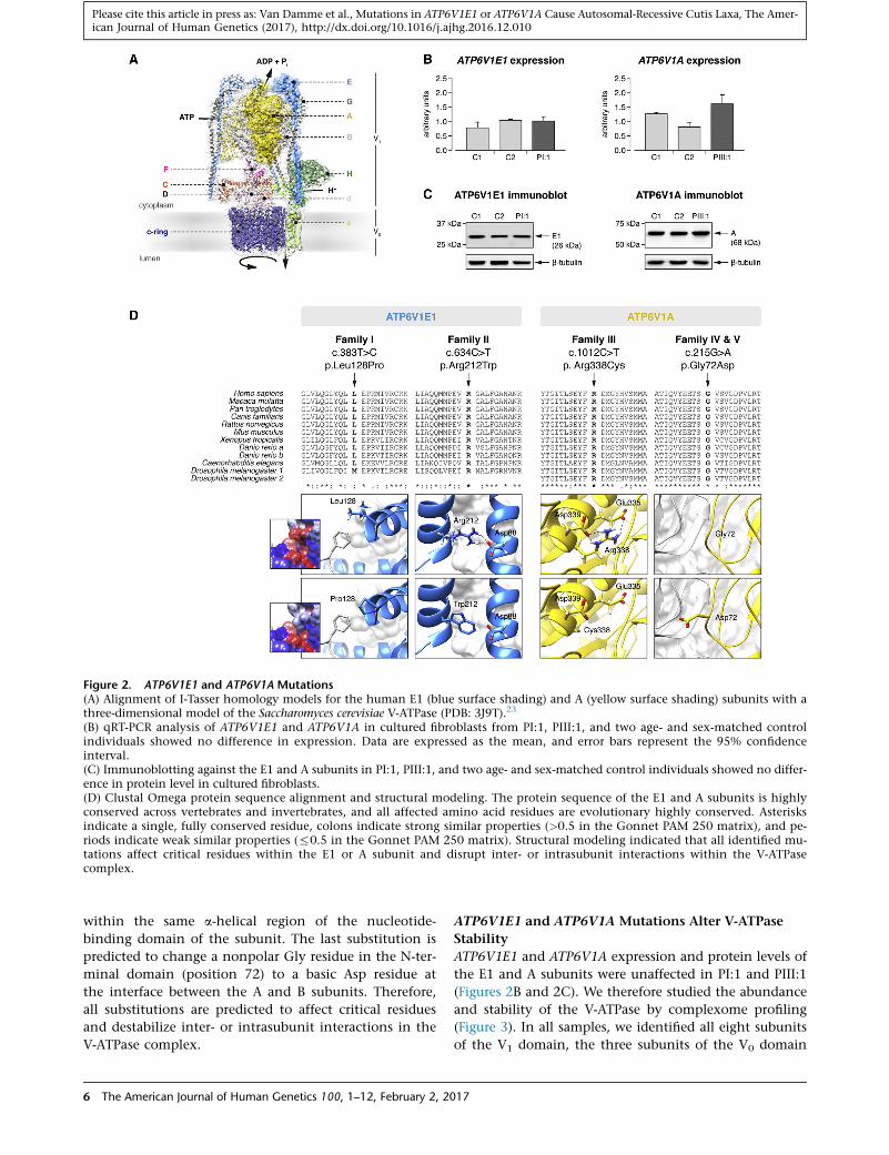
Figure 2. ATP6V1E1 and ATP6V1A Mutations(A) Alignment of I-Tasser homology models for the human E1 (blue surface shading) and A (yellow surface shading) subunits with athree-dimensional model of the Saccharomyces cerevisiae V-ATPase (PDB: 3J9T).23
(B) qRT-PCR analysis of ATP6V1E1 and ATP6V1A in cultured fibroblasts from PI:1, PIII:1, and two age- and sex-matched controlindividuals showed no difference in expression. Data are expressed as the mean, and error bars represent the 95% confidenceinterval.(C) Immunoblotting against the E1 and A subunits in PI:1, PIII:1, and two age- and sex-matched control individuals showed no differ-ence in protein level in cultured fibroblasts.(D) Clustal Omega protein sequence alignment and structural modeling. The protein sequence of the E1 and A subunits is highlyconserved across vertebrates and invertebrates, and all affected amino acid residues are evolutionary highly conserved. Asterisksindicate a single, fully conserved residue, colons indicate strong similar properties (>0.5 in the Gonnet PAM 250 matrix), and pe-riods indicate weak similar properties (%0.5 in the Gonnet PAM 250 matrix). Structural modeling indicated that all identified mu-tations affect critical residues within the E1 or A subunit and disrupt inter- or intrasubunit interactions within the V-ATPasecomplex.
Please cite this article in press as: Van Damme et al., Mutations in ATP6V1E1 or ATP6V1A Cause Autosomal-Recessive Cutis Laxa, The Amer-ican Journal of Human Genetics (2017), http://dx.doi.org/10.1016/j.ajhg.2016.12.010
within the same a-helical region of the nucleotide-
binding domain of the subunit. The last substitution is
predicted to change a nonpolar Gly residue in the N-ter-
minal domain (position 72) to a basic Asp residue at
the interface between the A and B subunits. Therefore,
all substitutions are predicted to affect critical residues
and destabilize inter- or intrasubunit interactions in the
V-ATPase complex.
6 The American Journal of Human Genetics 100, 1–12, February 2, 20
ATP6V1E1 and ATP6V1A Mutations Alter V-ATPase
Stability
ATP6V1E1 and ATP6V1A expression and protein levels of
the E1 and A subunits were unaffected in PI:1 and PIII:1
(Figures 2B and 2C). We therefore studied the abundance
and stability of the V-ATPase by complexome profiling
(Figure 3). In all samples, we identified all eight subunits
of the V1 domain, the three subunits of the V0 domain
17
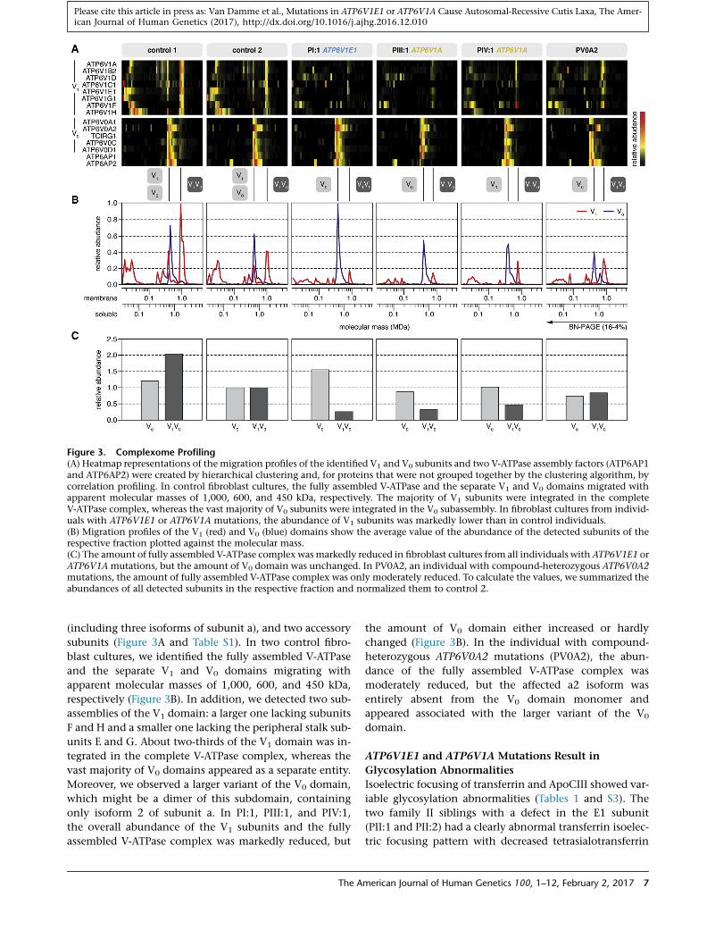
Figure 3. Complexome Profiling(A) Heatmap representations of the migration profiles of the identified V1 and V0 subunits and two V-ATPase assembly factors (ATP6AP1and ATP6AP2) were created by hierarchical clustering and, for proteins that were not grouped together by the clustering algorithm, bycorrelation profiling. In control fibroblast cultures, the fully assembled V-ATPase and the separate V1 and V0 domains migrated withapparent molecular masses of 1,000, 600, and 450 kDa, respectively. The majority of V1 subunits were integrated in the completeV-ATPase complex, whereas the vast majority of V0 subunits were integrated in the V0 subassembly. In fibroblast cultures from individ-uals with ATP6V1E1 or ATP6V1A mutations, the abundance of V1 subunits was markedly lower than in control individuals.(B) Migration profiles of the V1 (red) and V0 (blue) domains show the average value of the abundance of the detected subunits of therespective fraction plotted against the molecular mass.(C) The amount of fully assembled V-ATPase complex wasmarkedly reduced in fibroblast cultures from all individuals with ATP6V1E1 orATP6V1Amutations, but the amount of V0 domain was unchanged. In PV0A2, an individual with compound-heterozygous ATP6V0A2mutations, the amount of fully assembled V-ATPase complex was only moderately reduced. To calculate the values, we summarized theabundances of all detected subunits in the respective fraction and normalized them to control 2.
Please cite this article in press as: Van Damme et al., Mutations in ATP6V1E1 or ATP6V1A Cause Autosomal-Recessive Cutis Laxa, The Amer-ican Journal of Human Genetics (2017), http://dx.doi.org/10.1016/j.ajhg.2016.12.010
(including three isoforms of subunit a), and two accessory
subunits (Figure 3A and Table S1). In two control fibro-
blast cultures, we identified the fully assembled V-ATPase
and the separate V1 and V0 domains migrating with
apparent molecular masses of 1,000, 600, and 450 kDa,
respectively (Figure 3B). In addition, we detected two sub-
assemblies of the V1 domain: a larger one lacking subunits
F and H and a smaller one lacking the peripheral stalk sub-
units E and G. About two-thirds of the V1 domain was in-
tegrated in the complete V-ATPase complex, whereas the
vast majority of V0 domains appeared as a separate entity.
Moreover, we observed a larger variant of the V0 domain,
which might be a dimer of this subdomain, containing
only isoform 2 of subunit a. In PI:1, PIII:1, and PIV:1,
the overall abundance of the V1 subunits and the fully
assembled V-ATPase complex was markedly reduced, but
The A
the amount of V0 domain either increased or hardly
changed (Figure 3B). In the individual with compound-
heterozygous ATP6V0A2 mutations (PV0A2), the abun-
dance of the fully assembled V-ATPase complex was
moderately reduced, but the affected a2 isoform was
entirely absent from the V0 domain monomer and
appeared associated with the larger variant of the V0
domain.
ATP6V1E1 and ATP6V1A Mutations Result in
Glycosylation Abnormalities
Isoelectric focusing of transferrin and ApoCIII showed var-
iable glycosylation abnormalities (Tables 1 and S3). The
two family II siblings with a defect in the E1 subunit
(PII:1 and PII:2) had a clearly abnormal transferrin isoelec-
tric focusing pattern with decreased tetrasialotransferrin
merican Journal of Human Genetics 100, 1–12, February 2, 2017 7

Figure 4. Glycosylation and Vesicular Trafficking Studies(A) Immunocytochemistry of the highly glycosylated ICAM-1showed a severe reduction in the percentage of ICAM-1-positivefibroblasts. Scale bars represent 25 mm.(B) Retrograde translocation of Golgi membranes to the endo-plasmic reticulum was severely delayed in brefeldin-A-treated fi-broblasts from PI:1 and PIII:1. Similar to PV0A2 (with ARCL2A),PI:1 and PIII:1 showed a 2- to 3-fold higher percentage of cellsretaining Golgi remnants than control individuals. Additionally,TEM showed dilated and fragmented Golgi cisternae in PI:1,PIII:1, and PV0A2. Scale bars represent 250 nm.(C) TEM imaging of the dermis of PI:1 showed the presence oflarge, heterogeneous vacuolar structures within or near fibroblastsand in the endothelial lumen. Scale bars represent 1 mmor 200 nm(inset).
Please cite this article in press as: Van Damme et al., Mutations in ATP6V1E1 or ATP6V1A Cause Autosomal-Recessive Cutis Laxa, The Amer-ican Journal of Human Genetics (2017), http://dx.doi.org/10.1016/j.ajhg.2016.12.010
and increased di- and trisialiotransferrin (Table S3). This
pattern is indicative of a type II CDG. ApoCIII isoelectric
focusing for evaluation of mucin-type O-glycosylation
was normal in PII:1 and showed a minimal increase in
the ApoCIII-0 fraction in PII:2 (Table S3). Similar but
8 The American Journal of Human Genetics 100, 1–12, February 2, 20
milder N-glycosylation abnormalities were observed in
the individuals with a defect in the A subunit (Tables 1
and S3). For PIII:1, samples were taken at three different
ages. All samples showed a slightly increased trisialotrans-
ferrin fraction but normal tetrasialotransferrin levels.
ApoCIII isofocusing was normal at the age of 7 years, but
fully glycosylated ApoCIII was slightly decreased at the
age of 13 years. In PIV:1, a marginal increase in the tri-
sialotransferrin was observed, and ApoCIII analysis was
normal.
In all affected individuals, mass spectrometry of intact
transferrin revealed a consistent increase in the abundance
of the transferrin glycoform, corresponding to the loss of
one sialic acid (mass 79,264 amu; Figure S3). In addition,
a minor lack of galactose was observed in family II (PII:1
and PII:2), which is in agreement with known disorders
of Golgi trafficking (Figure S3).
In CDGs, there is a markedly reduced amount of the
cell-surface glycoprotein ICAM-1.35 There was a significant
decrease in the number of ICAM-1-positive cells on
immunofluorescence microscopy in both PI:1 and PIII:1
(Figure 4A).
ATP6V1E1 and ATP6V1A Mutations Disrupt Vesicular
Trafficking
Fluorescence microscopy of the Golgi marker GOLPH4
did not show abnormal Golgi morphology. Transmission
electron microscopy (TEM) of fibroblast cultures, how-
ever, showed abnormal swelling and a fragmented appear-
ance of the Golgi apparatus in PI:1 and PIII:1 (Figure 4B).
In line with previous studies on ARCL2A fibroblasts,
we observed delayed retrograde vesicular transport
between the Golgi and endoplasmic reticulum in cultured
fibroblasts from PI:1 and PIII:1: after brefeldin A treat-
ment, the number of cells retaining Golgi remnants was
two to three times higher than in control samples
(Figure 4B).8,36
Additionally, in PI:1, but not in PIII:1 or PIV:1, we
observed large heterogeneous vacuolar structures within
or in close proximity to fibroblasts or in the lumen of blood
vessels on TEM of skin biopsies (Figure 4C). These resem-
bled large autolysosomes or lysosomal storage bodies,
which are seen in lysosomal-storage disease.
ATP6V1E1 and ATP6V1A Mutations Variably Affect
ECM Assembly
Both pharmacological V-ATPase inhibition with bafilomy-
cin A1 and genetic depletion of the a2 subunit reduce
tropoelastin secretion, but their structural impact on
ECM architecture has never been studied thoroughly.5,30
The elastic fibers appeared normal in structure and amount
in PIII:1 but were irregular, fragmented, and reduced in
PIV:1 and—with the exception of some very small frag-
ments—absent in PI:1 (Figure 5). In all samples, collagen
fibrils were loosely packed and had more variable diame-
ters than the tightly assembled and regularly organized
collagen fibrils in control samples (Figure 5). Overall,
17
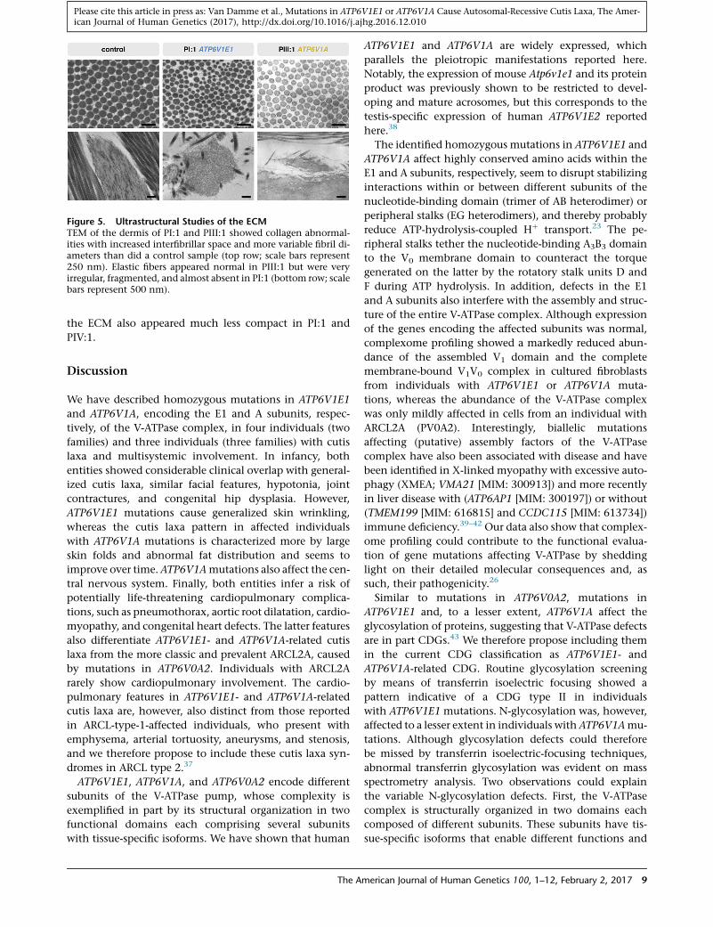
Figure 5. Ultrastructural Studies of the ECMTEM of the dermis of PI:1 and PIII:1 showed collagen abnormal-ities with increased interfibrillar space and more variable fibril di-ameters than did a control sample (top row; scale bars represent250 nm). Elastic fibers appeared normal in PIII:1 but were veryirregular, fragmented, and almost absent in PI:1 (bottom row; scalebars represent 500 nm).
Please cite this article in press as: Van Damme et al., Mutations in ATP6V1E1 or ATP6V1A Cause Autosomal-Recessive Cutis Laxa, The Amer-ican Journal of Human Genetics (2017), http://dx.doi.org/10.1016/j.ajhg.2016.12.010
the ECM also appeared much less compact in PI:1 and
PIV:1.
Discussion
We have described homozygous mutations in ATP6V1E1
and ATP6V1A, encoding the E1 and A subunits, respec-
tively, of the V-ATPase complex, in four individuals (two
families) and three individuals (three families) with cutis
laxa and multisystemic involvement. In infancy, both
entities showed considerable clinical overlap with general-
ized cutis laxa, similar facial features, hypotonia, joint
contractures, and congenital hip dysplasia. However,
ATP6V1E1 mutations cause generalized skin wrinkling,
whereas the cutis laxa pattern in affected individuals
with ATP6V1A mutations is characterized more by large
skin folds and abnormal fat distribution and seems to
improve over time.ATP6V1Amutations also affect the cen-
tral nervous system. Finally, both entities infer a risk of
potentially life-threatening cardiopulmonary complica-
tions, such as pneumothorax, aortic root dilatation, cardio-
myopathy, and congenital heart defects. The latter features
also differentiate ATP6V1E1- and ATP6V1A-related cutis
laxa from the more classic and prevalent ARCL2A, caused
by mutations in ATP6V0A2. Individuals with ARCL2A
rarely show cardiopulmonary involvement. The cardio-
pulmonary features in ATP6V1E1- and ATP6V1A-related
cutis laxa are, however, also distinct from those reported
in ARCL-type-1-affected individuals, who present with
emphysema, arterial tortuosity, aneurysms, and stenosis,
and we therefore propose to include these cutis laxa syn-
dromes in ARCL type 2.37
ATP6V1E1, ATP6V1A, and ATP6V0A2 encode different
subunits of the V-ATPase pump, whose complexity is
exemplified in part by its structural organization in two
functional domains each comprising several subunits
with tissue-specific isoforms. We have shown that human
The A
ATP6V1E1 and ATP6V1A are widely expressed, which
parallels the pleiotropic manifestations reported here.
Notably, the expression of mouse Atp6v1e1 and its protein
product was previously shown to be restricted to devel-
oping and mature acrosomes, but this corresponds to the
testis-specific expression of human ATP6V1E2 reported
here.38
The identified homozygous mutations in ATP6V1E1 and
ATP6V1A affect highly conserved amino acids within the
E1 and A subunits, respectively, seem to disrupt stabilizing
interactions within or between different subunits of the
nucleotide-binding domain (trimer of AB heterodimer) or
peripheral stalks (EG heterodimers), and thereby probably
reduce ATP-hydrolysis-coupled Hþ transport.23 The pe-
ripheral stalks tether the nucleotide-binding A3B3 domain
to the V0 membrane domain to counteract the torque
generated on the latter by the rotatory stalk units D and
F during ATP hydrolysis. In addition, defects in the E1
and A subunits also interfere with the assembly and struc-
ture of the entire V-ATPase complex. Although expression
of the genes encoding the affected subunits was normal,
complexome profiling showed a markedly reduced abun-
dance of the assembled V1 domain and the complete
membrane-bound V1V0 complex in cultured fibroblasts
from individuals with ATP6V1E1 or ATP6V1A muta-
tions, whereas the abundance of the V-ATPase complex
was only mildly affected in cells from an individual with
ARCL2A (PV0A2). Interestingly, biallelic mutations
affecting (putative) assembly factors of the V-ATPase
complex have also been associated with disease and have
been identified in X-linked myopathy with excessive auto-
phagy (XMEA; VMA21 [MIM: 300913]) and more recently
in liver disease with (ATP6AP1 [MIM: 300197]) or without
(TMEM199 [MIM: 616815] and CCDC115 [MIM: 613734])
immune deficiency.39–42 Our data also show that complex-
ome profiling could contribute to the functional evalua-
tion of gene mutations affecting V-ATPase by shedding
light on their detailed molecular consequences and, as
such, their pathogenicity.26
Similar to mutations in ATP6V0A2, mutations in
ATP6V1E1 and, to a lesser extent, ATP6V1A affect the
glycosylation of proteins, suggesting that V-ATPase defects
are in part CDGs.43 We therefore propose including them
in the current CDG classification as ATP6V1E1- and
ATP6V1A-related CDG. Routine glycosylation screening
by means of transferrin isoelectric focusing showed a
pattern indicative of a CDG type II in individuals
with ATP6V1E1 mutations. N-glycosylation was, however,
affected to a lesser extent in individuals withATP6V1Amu-
tations. Although glycosylation defects could therefore
be missed by transferrin isoelectric-focusing techniques,
abnormal transferrin glycosylation was evident on mass
spectrometry analysis. Two observations could explain
the variable N-glycosylation defects. First, the V-ATPase
complex is structurally organized in two domains each
composed of different subunits. These subunits have tis-
sue-specific isoforms that enable different functions and
merican Journal of Human Genetics 100, 1–12, February 2, 2017 9

Please cite this article in press as: Van Damme et al., Mutations in ATP6V1E1 or ATP6V1A Cause Autosomal-Recessive Cutis Laxa, The Amer-ican Journal of Human Genetics (2017), http://dx.doi.org/10.1016/j.ajhg.2016.12.010
processes.1–3 Our complexome profiling data illustrate that
different mutations affect the assembly of the holocom-
plex to different extents.
Interestingly, pneumothorax, marfanoid habitus, aortic
root dilatation, and ventricular dysfunction are all
frequent manifestations of Marfan syndrome (MFS [MIM:
154700]), a disorder caused by heterozygous mutations
in FBN1 (MIM: 134797), encoding fibrillin-1. Fibrillin-1
is a highly glycosylated protein and co-assembles with
several other glycoproteins and elastin to form elastic fi-
bers. As such, disturbed glycosylation of proteins such as
fibrillin-1 might explain the ultrastructural changes and
clinical overlap with MFS. We observed variable ultrastruc-
tural abnormalities in the elastic and collagen fibers of
dermal skin samples from the affected individuals.
Although neither elastin nor collagen is heavily glycosy-
lated, both depend on other glycosylated proteins for their
secretion and assembly into the ECM.
Impaired retrograde Golgi trafficking was first reported
in ARCL2A and has now been confirmed in these novel
ARCL entities.8,36 V-ATPases, however, acidify many other
organelles, which makes it likely that disturbed pH regula-
tion results in impaired enzymatic and sorting processes
in many of these.8 Notably, electron microscopy showed
a significant amount of vacuolar structures, resembling
autolysosomes, both intracellularly and in the vicinity of
fibroblasts in infant PI:1. The presumed autolysosomes
were not present in the other, older individuals, whose
dermal phenotype had improved over time, and their
presence might thus be a consequence of high protein
synthesis and turnover in early life. These ultrastructural
observations are supported by several studies. First, a
recent study in Drosophila melanogaster showed that ge-
netic V-ATPase defects interfere with lysosomal degrada-
tion and recycling and eventually lead to the formation
of giant autolysosomes.44 Second, lysosomal dysfunction
is a driver in the pathogenesis of XMEA, which is caused
by recessive mutations affecting the V-ATPase assembly
factor VMA21.41 Third, gingival hypertrophy is a promi-
nent feature in ATP6V1B2-related Zimmerman-Laband
syndrome and might relate to storage disease. Finally,
the concomitant intracellular aggregation of tropoelastin
and accumulation of abnormal lysosomes in ARCL2A
might suggest secretion of tropoelastin via secretory lyso-
somes rather than secretory vesicles.45 These findings
implicate lysosomal dysfunction in the pathogenesis of
these disorders and provide new insights in the cellular
processes involved in proper assembly and homeostasis
of the ECM. Further research is needed, however, to get
more insight in the exact role of lysosomes in these
disorders.
In conclusion, we have identified homozygous muta-
tions in ATP6V1E1 and ATP6V1A, respectively encoding
the E1 and A subunits of the V-ATPase complex. Affected
individuals presented with a recognizable and multisyste-
mic cutis laxa phenotype overlapping ARCL2A. We have
shown that these defects interfere with the assembly and
10 The American Journal of Human Genetics 100, 1–12, February 2, 2
functioning of the V-ATPase complex, protein glycosyla-
tion, intracellular trafficking, and lysosomal function.
Supplemental Data
Supplemental Data include a Supplemental Note, two figures, and
three tables and can be found with this article online at http://dx.
doi.org/10.1016/j.ajhg.2016.12.010.
Acknowledgments
The authors wish to thank Martin Lammens (Department of Pa-
thology, Antwerp University Hospital and University of Antwerp)
for his advice on the ultrastructural studies, Zsolt Urban (Depart-
ment of Human Genetics, Graduate School of Public Health,
University of Pittsburgh) for checking the DNA of a number of
individuals with cutis laxa, and Angel Ashikov (Department of
Neurology, Radboud University Medical Center) for his advice
on subcellular fractionation. B.C. and F.M. are senior clinical in-
vestigators and D.S. is a postdoctoral researcher of the Fund for Sci-
entific Research – Flanders. This study was financially supported
by Ghent University (Methusalem grant 08/01M01108 to
A.D.P.), the Dutch Metakids foundation (M.M.), the Netherlands
Organisation for Scientific Research (NWO project 017.008.052
to M.M.), ZonMw Medium Investment Grant 40-00506-98-9001
(D.J.L.), Vidi grant 91713359 (D.J.L.), the Dutch Stofwisselkracht
foundation (T.G.), the German Bundesministerium fur Bildung
und Forschung (BMBF, Juniorverbund in der Systemmedizin ‘‘mi-
tOmics,’’ FKZ 01ZX1405C to T.B.H.), the S.K. Yee Medical Research
Fund, the Society for the Relief of Disabled Children Research
Fund in Hong Kong, and the EURO-CDG-2 E-Rare grant by the Eu-
ropean Commission. The authors would like to thank the Exome
Aggregation Consortium and the groups that provided exome
variant data for comparison. A full list of contributing groups
can be found at http://exac.broadinstitute.org/about.
Received: September 21, 2016
Accepted: December 8, 2016
Published: January 5, 2017
Web Resources
Align GVGD, http://agvgd.hci.utah.edu
GenBank, https://www.ncbi.nlm.nih.gov/genbank/
HGVS Nomenclature, http://www.hgvs.org/mutnomen/
OMIM, http://www.omim.org
MAPP, http://www.ngrl.org.uk/Manchester/page/mapp-multivariate-
analysis-protein-polymorphism
Mutalyzer, https://mutalyzer.nl/
MutationTaster, http://www.mutationtaster.org
PolyPhen-2, http://genetics.bwh.harvard.edu/pph2/
RCSB Protein Data Bank (PDB), http://www.rcsb.org/pdb/home/
home.do
RefSeq, https://www.ncbi.nlm.nih.gov/refseq/
SIFT, http://sift.jcvi.org
References
1. Forgac, M. (2007). Vacuolar ATPases: rotary proton pumps in
physiology and pathophysiology. Nat. Rev. Mol. Cell Biol. 8,
917–929.
017

Please cite this article in press as: Van Damme et al., Mutations in ATP6V1E1 or ATP6V1A Cause Autosomal-Recessive Cutis Laxa, The Amer-ican Journal of Human Genetics (2017), http://dx.doi.org/10.1016/j.ajhg.2016.12.010
2. Cotter, K., Stransky, L., McGuire, C., and Forgac, M. (2015).
Recent Insights into the Structure, Regulation, and Function
of the V-ATPases. Trends Biochem. Sci. 40, 611–622.
3. Guillard, M., Dimopoulou, A., Fischer, B., Morava, E., Lefeber,
D.J., Kornak, U., andWevers, R.A. (2009). Vacuolar Hþ-ATPase
meets glycosylation in patients with cutis laxa. Biochim. Bio-
phys. Acta 1792, 903–914.
4. Karet, F.E., Finberg, K.E., Nelson, R.D., Nayir, A., Mocan, H.,
Sanjad, S.A., Rodriguez-Soriano, J., Santos, F., Cremers,
C.W.R.J., Di Pietro, A., et al. (1999). Mutations in the gene en-
coding B1 subunit of Hþ-ATPase cause renal tubular acidosis
with sensorineural deafness. Nat. Genet. 21, 84–90.
5. Kortum, F., Caputo, V., Bauer, C.K., Stella, L., Ciolfi, A., Alawi,
M., Bocchinfuso, G., Flex, E., Paolacci, S., Dentici, M.L., et al.
(2015). Mutations in KCNH1 and ATP6V1B2 cause Zimmer-
mann-Laband syndrome. Nat. Genet. 47, 661–667.
6. Smith, A.N., Skaug, J., Choate, K.A., Nayir, A., Bakkaloglu, A.,
Ozen, S., Hulton, S.A., Sanjad, S.A., Al-Sabban, E.A., Lifton,
R.P., et al. (2000). Mutations in ATP6N1B, encoding a new kid-
ney vacuolar proton pump 116-kD subunit, cause recessive
distal renal tubular acidosis with preserved hearing. Nat.
Genet. 26, 71–75.
7. Frattini, A., Orchard, P.J., Sobacchi, C., Giliani, S., Abinun, M.,
Mattsson, J.P., Keeling, D.J., Andersson, A.K., Wallbrandt, P.,
Zecca, L., et al. (2000). Defects in TCIRG1 subunit of the vacu-
olar proton pump are responsible for a subset of human auto-
somal recessive osteopetrosis. Nat. Genet. 25, 343–346.
8. Kornak, U., Reynders, E., Dimopoulou, A., van Reeuwijk, J.,
Fischer, B., Rajab, A., Budde, B., Nurnberg, P., Foulquier, F., Le-
feber, D., et al.; ARCL Debre-type Study Group (2008).
Impaired glycosylation and cutis laxa caused by mutations
in the vesicular Hþ-ATPase subunit ATP6V0A2. Nat. Genet.
40, 32–34.
9. Hucthagowder, V., Morava, E., Kornak, U., Lefeber, D.J.,
Fischer, B., Dimopoulou, A., Aldinger, A., Choi, J., Davis,
E.C., Abuelo, D.N., et al. (2009). Loss-of-function mutations
in ATP6V0A2 impair vesicular trafficking, tropoelastin secre-
tion and cell survival. Hum. Mol. Genet. 18, 2149–2165.
10. Mohamed, M., Kouwenberg, D., Gardeitchik, T., Kornak, U.,
Wevers, R.A., and Morava, E. (2011). Metabolic cutis laxa syn-
dromes. J. Inherit. Metab. Dis. 34, 907–916.
11. Vanakker, O., Callewaert, B., Malfait, F., and Coucke, P. (2015).
The Genetics of Soft Connective Tissue Disorders. Annu. Rev.
Genomics Hum. Genet. 16, 229–255.
12. Adzhubei, I.A., Schmidt, S., Peshkin, L., Ramensky, V.E., Gera-
simova, A., Bork, P., Kondrashov, A.S., and Sunyaev, S.R.
(2010). A method and server for predicting damaging
missense mutations. Nat. Methods 7, 248–249.
13. Kumar, P., Henikoff, S., and Ng, P.C. (2009). Predicting the ef-
fects of coding non-synonymous variants on protein function
using the SIFT algorithm. Nat. Protoc. 4, 1073–1081.
14. Schwarz, J.M., Cooper, D.N., Schuelke, M., and Seelow, D.
(2014). MutationTaster2: mutation prediction for the deep-
sequencing age. Nat. Methods 11, 361–362.
15. Tavtigian, S.V., Deffenbaugh, A.M., Yin, L., Judkins, T., Scholl,
T., Samollow, P.B., de Silva, D., Zharkikh, A., and Thomas, A.
(2006). Comprehensive statistical study of 452 BRCA1
missense substitutions with classification of eight recurrent
substitutions as neutral. J. Med. Genet. 43, 295–305.
16. Mathe, E., Olivier, M., Kato, S., Ishioka, C., Hainaut, P., and
Tavtigian, S.V. (2006). Computational approaches for predict-
ing the biological effect of p53 missense mutations: a compar-
The Am
ison of three sequence analysis based methods. Nucleic Acids
Res. 34, 1317–1325.
17. Stone, E.A., and Sidow, A. (2005). Physicochemical constraint
violation by missense substitutions mediates impairment
of protein function and disease severity. Genome Res. 15,
978–986.
18. Lek, M., Karczewski, K.J., Minikel, E.V., Samocha, K.E., Banks,
E., Fennell, T., O’Donnell-Luria, A.H., Ware, J.S., Hill, A.J.,
Cummings, B.B., et al.; Exome Aggregation Consortium
(2016). Analysis of protein-coding genetic variation in
60,706 humans. Nature 536, 285–291.
19. Wildeman, M., van Ophuizen, E., den Dunnen, J.T., and
Taschner, P.E. (2008). Improving sequence variant de-
scriptions in mutation databases and literature using the
Mutalyzer sequence variation nomenclature checker. Hum.
Mutat. 29, 6–13.
20. Zhang, Y. (2008). I-TASSER server for protein 3D structure
prediction. BMC Bioinformatics 9, 40.
21. Roy, A., Kucukural, A., and Zhang, Y. (2010). I-TASSER: a uni-
fied platform for automated protein structure and function
prediction. Nat. Protoc. 5, 725–738.
22. Yang, J., Yan, R., Roy, A., Xu, D., Poisson, J., and Zhang, Y.
(2015). The I-TASSER Suite: protein structure and function
prediction. Nat. Methods 12, 7–8.
23. Zhao, J., Benlekbir, S., and Rubinstein, J.L. (2015). Electron
cryomicroscopy observation of rotational states in a eukary-
otic V-ATPase. Nature 521, 241–245.
24. Pettersen, E.F., Goddard, T.D., Huang, C.C., Couch, G.S.,
Greenblatt, D.M., Meng, E.C., and Ferrin, T.E. (2004). UCSF
Chimera–a visualization system for exploratory research and
analysis. J. Comput. Chem. 25, 1605–1612.
25. Dunbrack, R.L., Jr. (2002). Rotamer libraries in the 21st cen-
tury. Curr. Opin. Struct. Biol. 12, 431–440.
26. Heide, H., Bleier, L., Steger, M., Ackermann, J., Drose, S.,
Schwamb, B., Zornig, M., Reichert, A.S., Koch, I., Wittig, I.,
and Brandt, U. (2012). Complexome profiling identifies
TMEM126B as a component of the mitochondrial complex I
assembly complex. Cell Metab. 16, 538–549.
27. Huynen, M.A., Muhlmeister, M., Gotthardt, K., Guerrero-Cas-
tillo, S., and Brandt, U. (2016). Evolution and structural orga-
nization of the mitochondrial contact site (MICOS) complex
and the mitochondrial intermembrane space bridging (MIB)
complex. Biochim. Biophys. Acta 1863, 91–101.
28. Bai, X., Zhou, D., Brown, J.R., Crawford, B.E., Hennet, T., and
Esko, J.D. (2001). Biosynthesis of the linkage region of glycos-
aminoglycans: cloning and activity of galactosyltransferase II,
the sixth member of the beta 1,3-galactosyltransferase family
(beta 3GalT6). J. Biol. Chem. 276, 48189–48195.
29. Nakajima, M., Mizumoto, S., Miyake, N., Kogawa, R., Iida, A.,
Ito, H., Kitoh, H., Hirayama, A., Mitsubuchi, H., Miyazaki, O.,
et al. (2013). Mutations in B3GALT6, which encodes a glycos-
aminoglycan linker region enzyme, cause a spectrum of skel-
etal and connective tissue disorders. Am. J. Hum. Genet. 92,
927–934.
30. Malfait, F., Kariminejad, A., Van Damme, T., Gauche, C., Syx,
D., Merhi-Soussi, F., Gulberti, S., Symoens, S., Vanhauwaert,
S., Willaert, A., et al. (2013). Defective initiation of glycos-
aminoglycan synthesis due to B3GALT6 mutations causes
a pleiotropic Ehlers-Danlos-syndrome-like connective tissue
disorder. Am. J. Hum. Genet. 92, 935–945.
31. Schindelin, J., Arganda-Carreras, I., Frise, E., Kaynig, V., Long-
air, M., Pietzsch, T., Preibisch, S., Rueden, C., Saalfeld, S.,
erican Journal of Human Genetics 100, 1–12, February 2, 2017 11

Please cite this article in press as: Van Damme et al., Mutations in ATP6V1E1 or ATP6V1A Cause Autosomal-Recessive Cutis Laxa, The Amer-ican Journal of Human Genetics (2017), http://dx.doi.org/10.1016/j.ajhg.2016.12.010
Schmid, B., et al. (2012). Fiji: an open-source platform for bio-
logical-image analysis. Nat. Methods 9, 676–682.
32. Schindelin, J., Rueden, C.T., Hiner, M.C., and Eliceiri, K.W.
(2015). The ImageJ ecosystem: An open platform for biomed-
ical image analysis. Mol. Reprod. Dev. 82, 518–529.
33. Schneider, C.A., Rasband, W.S., and Eliceiri, K.W. (2012). NIH
Image to ImageJ: 25 years of image analysis. Nat. Methods 9,
671–675.
34. Alazami, A.M., Al-Qattan, S.M., Faqeih, E., Alhashem, A., Al-
shammari, M., Alzahrani, F., Al-Dosari, M.S., Patel, N., Alsa-
gheir, A., Binabbas, B., et al. (2016). Expanding the clinical
and genetic heterogeneity of hereditary disorders of connec-
tive tissue. Hum. Genet. 135, 525–540.
35. He, P., Ng, B.G., Losfeld, M.-E., Zhu, W., and Freeze, H.H.
(2012). Identification of intercellular cell adhesion molecule
1 (ICAM-1) as a hypoglycosylationmarker in congenital disor-
ders of glycosylation cells. J. Biol. Chem. 287, 18210–18217.
36. Fischer, B., Dimopoulou, A., Egerer, J., Gardeitchik, T., Kidd,
A., Jost, D., Kayserili, H., Alanay, Y., Tantcheva-Poor, I., Man-
gold, E., et al. (2012). Further characterization of ATP6V0A2-
related autosomal recessive cutis laxa. Hum. Genet. 131,
1761–1773.
37. Morava, E., Guillard, M., Lefeber, D.J., and Wevers, R.A.
(2009). Autosomal recessive cutis laxa syndrome revisited.
Eur. J. Hum. Genet. 17, 1099–1110.
38. Sun-Wada, G.-H., Imai-Senga, Y., Yamamoto, A., Murata, Y.,
Hirata, T., Wada, Y., and Futai, M. (2002). A proton pump
ATPase with testis-specific E1-subunit isoform required for
acrosome acidification. J. Biol. Chem. 277, 18098–18105.
39. Jansen, J.C., Cirak, S., van Scherpenzeel, M., Timal, S., Reu-
nert, J., Rust, S., Perez, B., Vicogne, D., Krawitz, P., Wada, Y.,
12 The American Journal of Human Genetics 100, 1–12, February 2, 2
et al. (2016). CCDC115 Deficiency Causes a Disorder of Golgi
Homeostasis with Abnormal Protein Glycosylation. Am. J.
Hum. Genet. 98, 310–321.
40. Jansen, J.C., Timal, S., van Scherpenzeel, M., Michelakakis, H.,
Vicogne, D., Ashikov, A., Moraitou,M., Hoischen, A., Huijben,
K., Steenbergen, G., et al. (2016). TMEM199 Deficiency Is a
Disorder of Golgi Homeostasis Characterized by Elevated Ami-
notransferases, Alkaline Phosphatase, and Cholesterol and
Abnormal Glycosylation. Am. J. Hum. Genet. 98, 322–330.
41. Ramachandran, N., Munteanu, I., Wang, P., Ruggieri, A., Ril-
stone, J.J., Israelian, N., Naranian, T., Paroutis, P., Guo, R.,
Ren, Z.-P., et al. (2013). VMA21 deficiency prevents vacuolar
ATPase assembly and causes autophagic vacuolar myopathy.
Acta Neuropathol. 125, 439–457.
42. Jansen, E.J.R., Timal, S., Ryan, M., Ashikov, A., van Scherpen-
zeel, M., Graham, L.A., Mandel, H., Hoischen, A., Iancu, T.C.,
Raymond, K., et al. (2016). ATP6AP1 deficiency causes an im-
munodeficiency with hepatopathy, cognitive impairment and
abnormal protein glycosylation. Nat. Commun. 7, 11600.
43. van Scherpenzeel, M., Steenbergen, G., Morava, E., Wevers,
R.A., and Lefeber, D.J. (2015). High-resolution mass spectrom-
etry glycoprofiling of intact transferrin for diagnosis and
subtype identification in the congenital disorders of glycosyl-
ation. Transl. Res. 166, 639–649.e1.
44. Mauvezin, C., Nagy, P., Juhasz, G., andNeufeld, T.P. (2015). Au-
tophagosome-lysosome fusion is independent of V-ATPase-
mediated acidification. Nat. Commun. 6, 7007.
45. Hucthagowder, V., Sausgruber, N., Kim, K.H., Angle, B., Mar-
morstein, L.Y., and Urban, Z. (2006). Fibulin-4: a novel gene
for an autosomal recessive cutis laxa syndrome. Am. J. Hum.
Genet. 78, 1075–1080.
017
Related Documents