INTRODUCTION The forms of plant organs are not only beautiful, they define plant performance. The ability of leaves to capture light optimally and of stems to give support, the ability of flowers to attract pollinators and of fruits to be dispersed, and the ability of roots to permeate the soil, all depend on the shape of the organ. A plant organ acquires a specific shape through control of growth. Plants control not only the overall rate of expansion, but also the rates of expansion in different directions. When growth rates in different directions are not equal, growth is said to be ‘anisotropic’. Anisotropic growth varies both in the direction of maximal growth rate, as well as in the ratio of maximal to minimal growth rate (i.e., the degree of anisotropy). Growth anisotropy must be controlled to generate a plant organ with a specific form (Green, 1980). The anisotropy of expansion is dictated by anisotropy in stress within a cell wall and in the wall’s mechanical properties. The most prominent anisotropic component of the cell wall is cellulose. This polymer is synthesized at the plasma membrane as long chains of 1→4 β-linked glucose residues that associate laterally into microfibrils (Brown, 1999). Microfibrils are usually well oriented, which reinforces the cell wall anisotropically and guides the subsequent assembly of polymers around the cellulose framework (Cosgrove, 1997). Long-standing evidence shows that the ordered arrangement of microfibrils specifies the direction of maximal expansion (Green, 1980; Taiz, 1984). For example, in a rapidly elongating cell, the cellulose microfibrils are oriented on average perpendicular to the long axis of the cell and thus facilitate longitudinal expansion and hinder radial expansion. In turn, the orientation of microfibrils is commonly thought to be controlled in most cell types by the orientation of cortical microtubules (Baskin, 2001). However, cellulose microfibrils themselves do not restrict expansion in the direction parallel to their long axes. This can 4821 Development 129, 4821-4830 (2002) Printed in Great Britain © The Company of Biologists Limited 2002 DEV0447 The anisotropic growth of plant cells depends on cell walls having anisotropic mechanical properties, which are hypothesized to arise from aligned cellulose microfibrils. To test this hypothesis and to identify genes involved in controlling plant shape, we isolated mutants in Arabidopsis thaliana in which the degree of anisotropic expansion of the root is reduced. We report here the characterization of mutants at two new loci, RADIALLY SWOLLEN 4 (RSW4) and RSW7. The radial swelling phenotype is temperature sensitive, being moderate (rsw7) or negligible (rsw4) at the permissive temperature, 19°C, and pronounced at the restrictive temperature, 30°C. After transfer to 30°C, the primary root’s elongation rate decreases and diameter increases, with all tissues swelling radially. Swelling is accompanied by ectopic cell production but swelling is not reduced when the extra cell production is eliminated chemically. A double mutant was generated, whose roots swell constitutively and more than either parent. Based on analytical determination of acid-insoluble glucose, the amount of cellulose was normal in rsw4 and slightly elevated in rsw7. The orientation of cortical microtubules was examined with immunofluorescence in whole mounts and in semi-thin plastic sections, and the orientation of microfibrils was examined with field-emission scanning electron microscopy and quantitative polarized-light microscopy. In the swollen regions of both mutants, cortical microtubules and cellulose microfibrils are neither depleted nor disoriented. Thus, oriented microtubules and microfibrils themselves are insufficient to limit radial expansion; to build a wall with high mechanical anisotropy, additional factors are required, supplied in part by RSW4 and RSW7. Key words: Arabidopsis thaliana, Cell wall, Growth anisotropy, Microfibrils, Microtubules, Polarized-light microscopy SUMMARY Mutant alleles of Arabidopsis RADIALLY SWOLLEN 4 and 7 reduce growth anisotropy without altering the transverse orientation of cortical microtubules or cellulose microfibrils Allison M. D. Wiedemeier 1 , Jan E. Judy-March 1 , Charles H. Hocart 2 , Geoffrey O. Wasteneys 2 , Richard E. Williamson 2 and Tobias I. Baskin 1, * 1 Division of Biological Sciences, University of Missouri, Columbia, Missouri 65211-7400, USA 2 Plant Cell Biology Group, Research School of Biological Sciences, Australian NationalUniversity, Canberra ACT 2601, Australia *Author for correspondence (e-mail: [email protected]) Accepted 22 July 2002

Welcome message from author
This document is posted to help you gain knowledge. Please leave a comment to let me know what you think about it! Share it to your friends and learn new things together.
Transcript
INTRODUCTION
The forms of plant organs are not only beautiful, they defineplant performance. The ability of leaves to capture lightoptimally and of stems to give support, the ability of flowersto attract pollinators and of fruits to be dispersed, and theability of roots to permeate the soil, all depend on the shape ofthe organ. A plant organ acquires a specific shape throughcontrol of growth. Plants control not only the overall rate ofexpansion, but also the rates of expansion in differentdirections. When growth rates in different directions are notequal, growth is said to be ‘anisotropic’. Anisotropic growthvaries both in the direction of maximal growth rate, as well asin the ratio of maximal to minimal growth rate (i.e., the degreeof anisotropy). Growth anisotropy must be controlled togenerate a plant organ with a specific form (Green, 1980).
The anisotropy of expansion is dictated by anisotropy instress within a cell wall and in the wall’s mechanical properties.
The most prominent anisotropic component of the cell wall iscellulose. This polymer is synthesized at the plasma membraneas long chains of 1→4 β-linked glucose residues that associatelaterally into microfibrils (Brown, 1999). Microfibrils areusually well oriented, which reinforces the cell wallanisotropically and guides the subsequent assembly ofpolymers around the cellulose framework (Cosgrove, 1997).Long-standing evidence shows that the ordered arrangement ofmicrofibrils specifies the direction of maximal expansion(Green, 1980; Taiz, 1984). For example, in a rapidly elongatingcell, the cellulose microfibrils are oriented on averageperpendicular to the long axis of the cell and thus facilitatelongitudinal expansion and hinder radial expansion. In turn,the orientation of microfibrils is commonly thought to becontrolled in most cell types by the orientation of corticalmicrotubules (Baskin, 2001).
However, cellulose microfibrils themselves do not restrictexpansion in the direction parallel to their long axes. This can
4821Development 129, 4821-4830 (2002)Printed in Great Britain © The Company of Biologists Limited 2002DEV0447
The anisotropic growth of plant cells depends on cell wallshaving anisotropic mechanical properties, which arehypothesized to arise from aligned cellulose microfibrils. Totest this hypothesis and to identify genes involved incontrolling plant shape, we isolated mutants in Arabidopsisthaliana in which the degree of anisotropic expansion of theroot is reduced. We report here the characterization ofmutants at two new loci, RADIALLY SWOLLEN 4 (RSW4)and RSW7. The radial swelling phenotype is temperaturesensitive, being moderate (rsw7) or negligible (rsw4) at thepermissive temperature, 19°C, and pronounced at therestrictive temperature, 30°C. After transfer to 30°C, theprimary root’s elongation rate decreases and diameterincreases, with all tissues swelling radially. Swelling isaccompanied by ectopic cell production but swelling is notreduced when the extra cell production is eliminatedchemically. A double mutant was generated, whose rootsswell constitutively and more than either parent. Based on
analytical determination of acid-insoluble glucose, theamount of cellulose was normal in rsw4 and slightlyelevated in rsw7. The orientation of cortical microtubuleswas examined with immunofluorescence in whole mountsand in semi-thin plastic sections, and the orientation ofmicrofibrils was examined with field-emission scanningelectron microscopy and quantitative polarized-lightmicroscopy. In the swollen regions of both mutants,cortical microtubules and cellulose microfibrils are neitherdepleted nor disoriented. Thus, oriented microtubules andmicrofibrils themselves are insufficient to limit radialexpansion; to build a wall with high mechanical anisotropy,additional factors are required, supplied in part by RSW4and RSW7.
Key words: Arabidopsis thaliana, Cell wall, Growth anisotropy,Microfibrils, Microtubules, Polarized-light microscopy
SUMMARY
Mutant alleles of Arabidopsis RADIALLY SWOLLEN 4 and 7 reduce growth
anisotropy without altering the transverse orientation of cortical microtubules
or cellulose microfibrils
Allison M. D. Wiedemeier 1, Jan E. Judy-March 1, Charles H. Hocart 2, Geoffrey O. Wasteneys 2,Richard E. Williamson 2 and Tobias I. Baskin 1,*1Division of Biological Sciences, University of Missouri, Columbia, Missouri 65211-7400, USA 2Plant Cell Biology Group, Research School of Biological Sciences, Australian National University, Canberra ACT 2601, Australia*Author for correspondence (e-mail: [email protected])
Accepted 22 July 2002
4822
be appreciated by considering a cylindrical cell with transversemicrofibril alignment and realizing that when the cell expandsradially, the microfibrils themselves are not stretched, becausethey do not encircle the cell in closed loops. Instead,neighboring microfibrils slide past one another. Resistance tothis shear limits radial expansion, a resistance presumablymediated by the matrix enmeshing the microfibrils. Efforts tounderstand how the cell wall matrix is remodeled to permitgrowth have focused exclusively on elongation (Cosgrove,1997), and consequently we have very little idea whetherelongation and radial expansion are governed by the sameinteractions between matrix and microfibrils.
As an approach to understand the control of anisotropicexpansion, we are using developmental genetics, studyingmutants in which this control is reduced or lost. We previouslyreported the isolation of mutants at three different loci, RSW1,RSW2, RSW3, that have swollen roots (called ‘Rsw’ for radiallyswollen) (Baskin et al., 1992), and others have isolated mutantswith similar root swelling phenotypes (Benfey et al., 1993;Holding et al., 1994; Hauser et al., 1995; Scheres et al., 1995).Consistent with the central role of microtubules andmicrofibrils in determining growth anisotropy, these screenshave identified loci required for the synthesis of cellulose orfor the assembly of normal arrays of microtubules. RSW1(Arioli et al., 1998) and PROCUSTE(Fagard et al., 2000)encode catalytic subunits of cellulose synthase, RSW2(allelicto KORRIGAN) has been shown to encode an endo-1,4β-glucanase that apparently functions in the synthesis ofcellulose (Nicol et al., 1998; Lane et al., 2001; Sato et al.,2001), and RSW3encodes glucosidase II (J. Burn, personalcommunication), an ER enzyme in the N-glycosylationpathway whose operation is needed for cellulose synthesis.Other mutants that produce swollen roots have severelydefective cortical microtubules: tonneau1and ton2/fass(Traaset al., 1995; McClinton and Sung, 1997), botero/fragilefiber2(Bichet et al., 2001; Burk et al., 2001) and microtubuleorganization1(Whittington et al., 2001).
We report here the characterization of mutants at two newloci, RSW4and RSW7. Despite the extensive root swelling ofmutants at these loci, the orientations of cortical microtubulesand of cellulose microfibrils closely resembled those of thewild type. These loci thus define novel activities controlling theanisotropic expansion of plant cells.
MATERIALS AND METHODS
Plant material and growth conditionsSeeds of Arabidopsis thaliana L. (Heynh) ecotype Columbia weremutagenized with ethane-methylsulfonate as described by Baskin etal. (Baskin et al., 1992), and lines used here were selfed for fivegenerations and backcrossed at least once to wild type. Linesbackcrossed 7 times have similar phenotypes. Seeds were sterilizedin dilute bleach and germinated on nutrient-solidified agarsupplemented with 3% sucrose in Petri plates, sealed with air-permeable bandage tape, and seedlings were grown vertically undercontinuous yellow light (80 µmol/m2s) and constant temperature(19°C, permissive temperature) for up to 10 days in a growth chamber,as described by Baskin and Wilson (Baskin and Wilson, 1997). Tocause expression of the swelling phenotypes, the plates weretransferred to 30°C (non-permissive temperature) under continuousyellow light (50 µmol/m2s) in a custom made growth chamber
modified from a commercial ‘frost-free’ freezer, with light from 20W cool-white and warm-white fluorescent tubes filtered throughyellow acrylic (Plexiglas J2208, Cope Plastic, St. Louis, MO).
Elongation rate was measured by photographing seedlings on plateswith a camera inside the chamber as described by Baskin et al. (Baskinet al., 1992), whereas root diameter was measured at the end of thetreatment at the position of apparent maximal swelling or, if none, atthe site of root hair initiation by means of a compound microscopeand video digitizer. For experiments involving ethylene exposure,seeds were germinated on plates containing 100 µM AgNO3. After 7days wild-type seedlings were transferred to plates containing thesame compound ±1 µM 1-aminocyclopropane-1-carboxylic acid(ACC; Sigma, St Louis, MO) and all treatments were transferred to30°C for 2 days. For inhibitor experiments, seedlings were grown at19°C for 6 days and then transplanted onto plates containing either0.1% (v/v) DMSO (controls), 1 µM oryzalin (Chem Service, WestChester, PA), 1 µM 2, 6-dichlorobenzonitrile (DCB; Sigma), or 10µg/ml aphidicolin (Sigma) and transferred to 30°C for 2 days. Stocksof oryzalin, ACC, DCB and aphidicolin were made in DMSO andstored at –20°C. An aqueous silver nitrate stock was made fresh foreach experiment.
Genetic mapping Genomic DNA was extracted from individual mutant F2 plants froma cross to Landsberg erecta, cut with EcoRI, blotted, and probed withpooled probes (Fabri and Schäffner, 1994). Recombinationpercentages were obtained for 28-34 genomes, and converted tocentiMorgans with the Kosambi function (Koornneef and Stam,1992).
Measurement of tissue area and cell numberSeedlings were fixed and embedded in butyl-methyl methacrylate,as described by Baskin and Wilson (Baskin and Wilson, 1997). Tomeasure tissue area, cross sections were collected every 10th µmspanning the region of maximal swelling (~200-500 µm from thetip), and four sections selected for measurement having the largestoverall diameters. To count cells, cross sections were collected at 50µm intervals within a region where swelling was extensive. Fourroots were sectioned per treatment and five sections per root werescored. Sections were stained with periodic-acid-Schiff’s (PAS), ina simplified procedure where sections were incubated in 1% periodicacid for 10 minutes, rinsed briefly in water, stained in Schiff’sreagent (Sigma) for 30-60 minutes, and then rinsed extensively withwater. We determined that roots did not shrink during processing byfinding that the average diameter for wild-type roots freshlysectioned was 109±1.2 µm versus 111±5.3 µm for fixed, embeddedand sectioned roots (mean ± s.e.m., n=4). Carlemalm et al.(Carlemalm et al., 1982) likewise reported no shrinkage for a similarmixture of methacrylates.
Localization of microtubules For localizing microtubules in whole mounts of intact roots, seedlingswere fixed, extracted in EGTA followed by pectinase, incubated inantibodies, and observed with confocal fluorescence microscopy, asdescribed for A. thaliana by Liang et al. (Liang et al., 1996). Forlocalizing cortical microtubules in sections, material embedded inmethacrylate was processed for immunocytochemistry as describedpreviously (Baskin and Wilson, 1997).
Analysis of monosaccharides Seedlings were grown for 2 days at 21°C and then for 5 days at 30°C.As previously described (Lane et al., 2001), cell walls were collectedfrom whole seedlings and incubated for 1 hour in 2 M trifluoroaceticacid at 120°C, and cellulose was determined as acid-insoluble glucanby gas chromatography and mass spectrometry; additionally,monosaccharides from non-cellulosic polysaccharides solubulized byacid were quantified.
A. M. D. Wiedemeier and others
4823Growth anisotropy, microtubules and microfibrils
Field-emission scanning electron microscopy (FESEM) To image the innermost layer of the cell wall, intact seedlings wereplasmolysed in 1 M sucrose for 45 minutes, fixed in 4% paraformalde-hyde in 10 mM Pipes pH 7.0, and 0.5 M sucrose for 2 hours at roomtemperature, rinsed briefly in that buffer minus the fixative. Root tipswere excised and sandwiched between Formvar films on wire loopsas described by Baskin and Wilson (Baskin and Wilson, 1997). Roottips were infiltrated into polyethylene glycol (PEG) following aprotocol modified from that of Fowke et al. (Fowke et al., 1999).Samples were first brought to 40% PEG 400 by stepwise exchange ofthe 0.5 M sucrose, and then to 100% PEG 400. Samples were thenplaced at 55°C and progressively infiltrated first with PEG 1000, thenwith a 3:1 (v/v) mixture of PEG 1450:PEG 1000 over the following2 days, and then embedded. After excising the wire loop from theblock, nominally 15 µm thick sections were cut at 16°C and collectedon coverslips previously sputter-coated with approximately 20 nm ofgold or gold-palladium followed by brief incubation in 0.1% (v/v)polyethylene-imine. The metal reduced charging and the imineadhered the sections. The coverslips with sections were warmed on aslide warmer for 30 minutes at 40°C to flatten the sections and theywere stored in a desiccated state. Coverslips were immersed indistilled water to remove the embedment, incubated in 30 µg/mlproteinase K for 1 hour then in a solution of 250 mM NaCl and 0.05%(v/v) Triton X-100 for 3 hours or overnight to remove adherentcytoplasts, rinsed three times briefly in distilled water, dehydrated to100% ethanol, critical point dried, and sputter coated with a thin filmof platinum (nominally 2 nm).
As an alternative to PEG embedment, some samples were preparedusing the protocol developed by Sugimoto et al. (Sugimoto et al.,2000) in which roots were cut open with a cryo-ultramicrotome,cytoplasm was removed with bleach, and samples thendehydrated, critical point dried, and sputter-coated withplatinum. Samples prepared by either method were examinedwith a FESEM (Hitachi 4500 or Hitachi 4700S). Forquantification of microfibril angles, cells were selected at lowmagnification where microfibrils were invisible and then animage captured at a magnification of 60,000 times. A region ofinterest (256×256 pixels) was selected and the 2D Fouriertransform calculated (Image Processing Toolkit, version 3,Reindeer Games, Chapel Hill NC). The transforms wereelliptical and the angle was measured between the major axis ofthe ellipse and the transverse (defined as 0°), an angle thatestimates the net orientation of microfibrils.
Polarized-light microscopy Methacrylate sections (2 µm thick) were mounted in 75% glycerol,0.1% Triton X-100 and examined through a polarizing microscope(Jenapol, Carl Zeiss Jena). Birefringent retardation was assessedin longitudinal cell walls that were parallel to the section plane,and was quantified with microphotometry. The light intensitypassing through a square aperture, placed first over a cell wall and
then over the background, was measured at ±45° settings of acompensator (Brace-Köhler type, Γmax=18 nm) and retardation wascalculated from the intensity measurements, as described by Baskin etal. (Baskin et al., 1999). The area of the selected cell wall was alwayslarger than the area sampled by the aperture (3.75 µm2) and the aperturewas placed near the center of the selected cell wall, thus minimizingaberrations due to the presence of edges. The aperture was also placedto avoid any visible heterogeneity in the cell wall, such as that causedby pit fields.
RESULTS
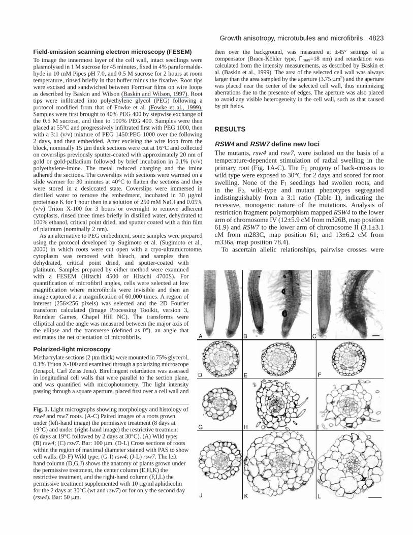
RSW4 and RSW7 define new lociThe mutants, rsw4 and rsw7, were isolated on the basis of atemperature-dependent stimulation of radial swelling in theprimary root (Fig. 1A-C). The F1 progeny of back-crosses towild type were exposed to 30°C for 2 days and scored for rootswelling. None of the F1 seedlings had swollen roots, andin the F2, wild-type and mutant phenotypes segregatedindistinguishably from a 3:1 ratio (Table 1), indicating therecessive, monogenic nature of the mutations. Analysis ofrestriction fragment polymorphism mapped RSW4to the lowerarm of chromosome IV (12±5.9 cM from m326B, map position61.9) and RSW7to the lower arm of chromosome II (3.1±3.1cM from m283C, map position 61; and 13±6.2 cM fromm336a, map position 78.4).
To ascertain allelic relationships, pairwise crosses were
Fig. 1. Light micrographs showing morphology and histology ofrsw4and rsw7 roots. (A-C) Paired images of a roots grownunder (left-hand image) the permissive treatment (8 days at19°C) and under (right-hand image) the restrictive treatment(6 days at 19°C followed by 2 days at 30°C). (A) Wild type;(B) rsw4; (C) rsw7. Bar: 100 µm. (D-L) Cross sections of rootswithin the region of maximal diameter stained with PAS to showcell walls: (D-F) Wild type; (G-I) rsw4; (J-L) rsw7. The lefthand column (D,G,J) shows the anatomy of plants grown underthe permissive treatment, the center column (E,H,K) therestrictive treatment, and the right-hand column (F,I,L) thepermissive treatment supplemented with 10 µg/ml aphidicolinfor the 2 days at 30°C (wt and rsw7) or for only the second day(rsw4). Bar: 50 µm.
4824
made between the mutants and other known radial swellingmutants: rsw1, rsw2, rsw3 (Baskin et al., 1992), rsw5,rsw6 and rsw8 (Wiedemeier, 1998). All of these crossesproduced phenotypically wild-type F1 offspring, indicatingcomplementation. RSW4maps near CUDGEL, and RSW7maps near POMPOM2and MOR1, loci previously identifiedas being involved in root morphology (Hauser et al., 1995;Whittington et al., 2001). As the only known mutant cudalleleis fully dominant and gametophytic lethal, complementationcould not be tested; however, crosses between rsw7and pom2,as well as between rsw7and mor1, produced progeny with onlya wild-type phenotype. On this basis, RSW4 and RSW7aredefined as new loci.
From the cross between rsw4 and rsw7, we isolated aputative double mutant. When this line was back crossed toeach parent, all progeny were mutant, confirming the doublemutant genotype. The radial swelling phenotype of the doublemutant was expressed constitutively, and was more extremethan either parent (data not shown). Thus, RSW4 and RSW7represent two different and additive activities needed to controlthe anisotropic expansion of the root.
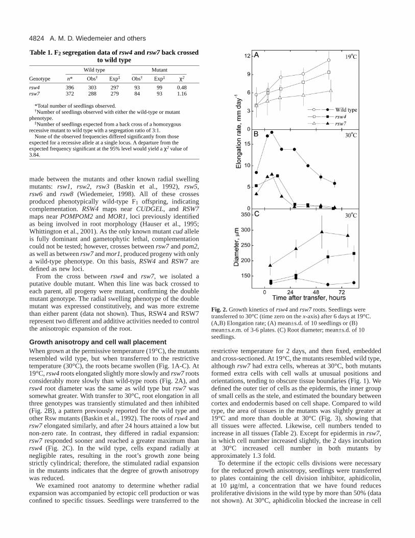
Growth anisotropy and cell wall placement When grown at the permissive temperature (19°C), the mutantsresembled wild type, but when transferred to the restrictivetemperature (30°C), the roots became swollen (Fig. 1A-C). At19°C, rsw4roots elongated slightly more slowly and rsw7rootsconsiderably more slowly than wild-type roots (Fig. 2A), andrsw4 root diameter was the same as wild type but rsw7 wassomewhat greater. With transfer to 30°C, root elongation in allthree genotypes was transiently stimulated and then inhibited(Fig. 2B), a pattern previously reported for the wild type andother Rsw mutants (Baskin et al., 1992). The roots of rsw4andrsw7elongated similarly, and after 24 hours attained a low butnon-zero rate. In contrast, they differed in radial expansion:rsw7 responded sooner and reached a greater maximum thanrsw4 (Fig. 2C). In the wild type, cells expand radially atnegligible rates, resulting in the root’s growth zone beingstrictly cylindrical; therefore, the stimulated radial expansionin the mutants indicates that the degree of growth anisotropywas reduced.
We examined root anatomy to determine whether radialexpansion was accompanied by ectopic cell production or wasconfined to specific tissues. Seedlings were transferred to the
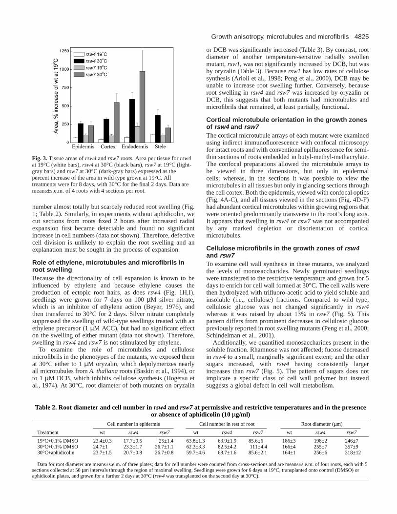
restrictive temperature for 2 days, and then fixed, embeddedand cross-sectioned. At 19°C, the mutants resembled wild type,although rsw7 had extra cells, whereas at 30°C, both mutantsformed extra cells with cell walls at unusual positions andorientations, tending to obscure tissue boundaries (Fig. 1). Wedefined the outer tier of cells as the epidermis, the inner groupof small cells as the stele, and estimated the boundary betweencortex and endodermis based on cell shape. Compared to wildtype, the area of tissues in the mutants was slightly greater at19°C and more than double at 30°C (Fig. 3), showing thatall tissues were affected. Likewise, cell numbers tended toincrease in all tissues (Table 2). Except for epidermis in rsw7,in which cell number increased slightly, the 2 days incubationat 30°C increased cell number in both mutants byapproximately 1.3 fold.
To determine if the ectopic cells divisions were necessaryfor the reduced growth anisotropy, seedlings were transferredto plates containing the cell division inhibitor, aphidicolin,at 10 µg/ml, a concentration that we have found reducesproliferative divisions in the wild type by more than 50% (datanot shown). At 30°C, aphidicolin blocked the increase in cell
A. M. D. Wiedemeier and others
Fig. 2. Growth kinetics of rsw4and rsw7roots. Seedlings weretransferred to 30°C (time zero on the x-axis) after 6 days at 19°C.(A,B) Elongation rate; (A) mean±s.d. of 10 seedlings or (B)mean±s.e.m. of 3-6 plates. (C) Root diameter; mean±s.d. of 10seedlings.
Table 1. F2 segregation data of rsw4and rsw7back crossedto wild type
Wild type Mutant
Genotype n* Obs† Exp‡ Obs† Exp‡ χ2
rsw4 396 303 297 93 99 0.48rsw7 372 288 279 84 93 1.16
*Total number of seedlings observed.†Number of seedlings observed with either the wild-type or mutant
phenotype. ‡Number of seedlings expected from a back cross of a homozygous
recessive mutant to wild type with a segregation ratio of 3:1.None of the observed frequencies differed significantly from those
expected for a recessive allele at a single locus. A departure from theexpected frequency significant at the 95% level would yield a χ2 value of3.84.
4825Growth anisotropy, microtubules and microfibrils
number almost totally but scarcely reduced root swelling (Fig.1; Table 2). Similarly, in experiments without aphidicolin, wecut sections from roots fixed 2 hours after increased radialexpansion first became detectable and found no significantincrease in cell numbers (data not shown). Therefore, defectivecell division is unlikely to explain the root swelling and anexplanation must be sought in the process of expansion.
Role of ethylene, microtubules and microfibrils inroot swellingBecause the directionality of cell expansion is known to beinfluenced by ethylene and because ethylene causes theproduction of ectopic root hairs, as does rsw4 (Fig. 1H,I),seedlings were grown for 7 days on 100 µM silver nitrate,which is an inhibitor of ethylene action (Beyer, 1976), andthen transferred to 30°C for 2 days. Silver nitrate completelysuppressed the swelling of wild-type seedlings treated with anethylene precursor (1 µM ACC), but had no significant effecton the swelling of either mutant (data not shown). Therefore,swelling in rsw4and rsw7 is not stimulated by ethylene.
To examine the role of microtubules and cellulosemicrofibrils in the phenotypes of the mutants, we exposed themat 30°C either to 1 µM oryzalin, which depolymerizes nearlyall microtubules from A. thalianaroots (Baskin et al., 1994), orto 1 µM DCB, which inhibits cellulose synthesis (Hogetsu etal., 1974). At 30°C, root diameter of both mutants on oryzalin
or DCB was significantly increased (Table 3). By contrast, rootdiameter of another temperature-sensitive radially swollenmutant, rsw1, was not significantly increased by DCB, but wasby oryzalin (Table 3). Because rsw1has low rates of cellulosesynthesis (Arioli et al., 1998; Peng et al., 2000), DCB may beunable to increase root swelling further. Conversely, becauseroot swelling in rsw4 and rsw7 was increased by oryzalin orDCB, this suggests that both mutants had microtubules andmicrofibrils that remained, at least partially, functional.
Cortical microtubule orientation in the growth zonesof rsw4 and rsw7The cortical microtubule arrays of each mutant were examinedusing indirect immunofluorescence with confocal microscopyfor intact roots and with conventional epifluorescence for semi-thin sections of roots embedded in butyl-methyl-methacrylate.The confocal preparations allowed the microtubule arrays tobe viewed in three dimensions, but only in epidermalcells; whereas, in the sections it was possible to view themicrotubules in all tissues but only in glancing sections throughthe cell cortex. Both the epidermis, viewed with confocal optics(Fig. 4A-C), and all tissues viewed in the sections (Fig. 4D-F)had abundant cortical microtubules within growing regions thatwere oriented predominantly transverse to the root’s long axis.It appears that swelling in rsw4or rsw7was not accompaniedby any marked depletion or disorientation of corticalmicrotubules.
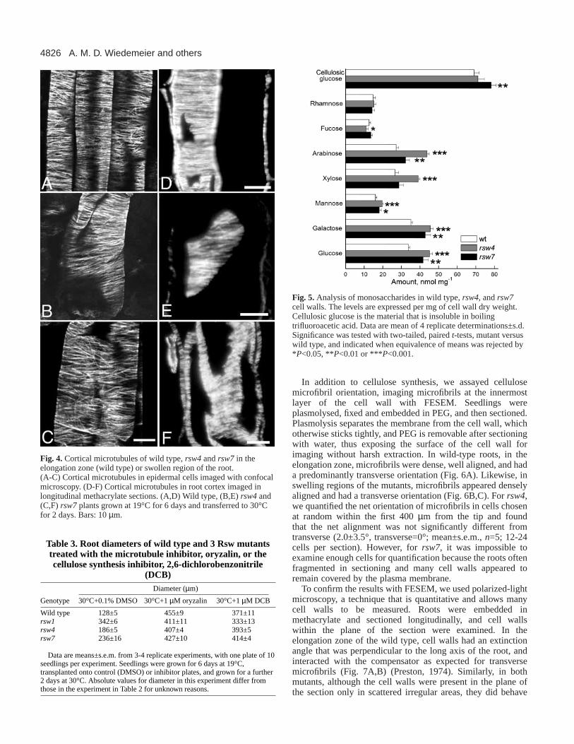
Cellulose microfibrils in the growth zones of rsw4and rsw7To examine cell wall synthesis in these mutants, we analyzedthe levels of monosaccharides. Newly germinated seedlingswere transferred to the restrictive temperature and grown for 5days to enrich for cell wall formed at 30°C. The cell walls werethen hydrolyzed with trifluoro-acetic acid to yield soluble andinsoluble (i.e., cellulose) fractions. Compared to wild type,cellulosic glucose was not changed significantly in rsw4whereas it was raised by about 13% in rsw7 (Fig. 5). Thispattern differs from prominent decreases in cellulosic glucosepreviously reported in root swelling mutants (Peng et al., 2000;Schindelman et al., 2001).
Additionally, we quantified monosaccharides present in thesoluble fraction. Rhamnose was not affected; fucose decreasedin rsw4 to a small, marginally significant extent; and the othersugars increased, with rsw4 having consistently largerincreases than rsw7 (Fig. 5). The pattern of sugars does notimplicate a specific class of cell wall polymer but insteadsuggests a global defect in cell wall metabolism.
Fig. 3. Tissue areas of rsw4 and rsw7 roots. Area per tissue for rsw4at 19°C (white bars), rsw4 at 30°C (black bars), rsw7at 19°C (light-gray bars) and rsw7at 30°C (dark-gray bars) expressed as thepercent increase of the area in wild type grown at 19°C. Alltreatments were for 8 days, with 30°C for the final 2 days. Data aremeans±s.e.m. of 4 roots with 4 sections per root.
Table 2. Root diameter and cell number in rsw4and rsw7at permissive and restrictive temperatures and in the presenceor absence of aphidicolin (10 µg/ml)
Cell number in epidermis Cell number in rest of root Root diameter (µm)
Treatment wt rsw4 rsw7 wt rsw4 rsw7 wt rsw4 rsw7
19°C+0.1% DMSO 23.4±0.3 17.7±0.5 25±1.4 63.8±1.3 63.9±1.9 85.6±6 186±3 198±2 246±730°C+0.1% DMSO 24.7±1 23.3±1.7 26.7±1.1 62.3±3.3 82.5±4.2 111±4.4 166±4 255±7 357±930°C+aphidicolin 23.7±1.5 20.7±0.8 26.7±0.8 59.7±4.6 68.7±1.6 85.6±2.1 164±1 256±6 318±12
Data for root diameter are means±s.e.m. of three plates; data for cell number were counted from cross-sections and are means±s.e.m. of four roots, each with 5sections collected at 50 µm intervals through the region of maximal swelling. Seedlings were grown for 6 days at 19°C, transplanted onto control (DMSO) oraphidicolin plates, and grown for a further 2 days at 30°C (rsw4was transplanted on the second day at 30°C).
4826
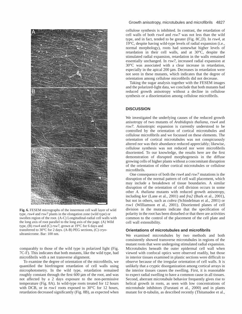
In addition to cellulose synthesis, we assayed cellulosemicrofibril orientation, imaging microfibrils at the innermostlayer of the cell wall with FESEM. Seedlings wereplasmolysed, fixed and embedded in PEG, and then sectioned.Plasmolysis separates the membrane from the cell wall, whichotherwise sticks tightly, and PEG is removable after sectioningwith water, thus exposing the surface of the cell wall forimaging without harsh extraction. In wild-type roots, in theelongation zone, microfibrils were dense, well aligned, and hada predominantly transverse orientation (Fig. 6A). Likewise, inswelling regions of the mutants, microfibrils appeared denselyaligned and had a transverse orientation (Fig. 6B,C). For rsw4,we quantified the net orientation of microfibrils in cells chosenat random within the first 400 µm from the tip and foundthat the net alignment was not significantly different fromtransverse (2.0±3.5°, transverse=0°; mean±s.e.m., n=5; 12-24cells per section). However, forrsw7, it was impossible toexamine enough cells for quantification because the roots oftenfragmented in sectioning and many cell walls appeared toremain covered by the plasma membrane.
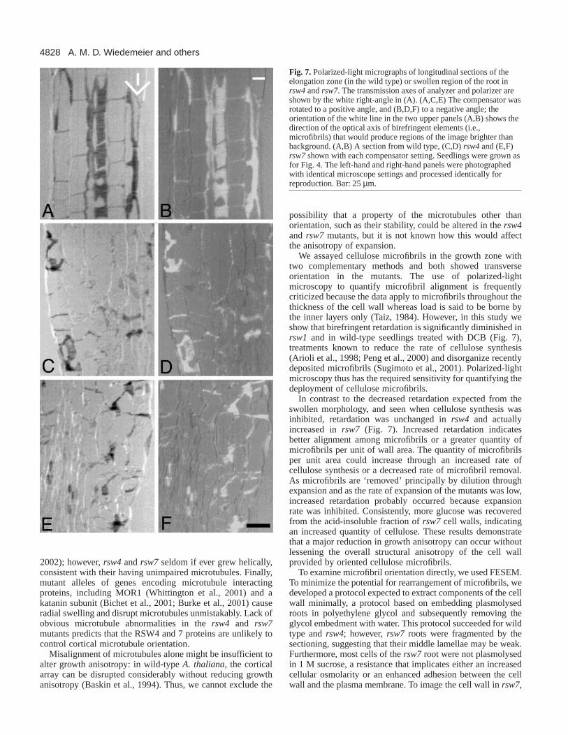
To confirm the results with FESEM, we used polarized-lightmicroscopy, a technique that is quantitative and allows manycell walls to be measured. Roots were embedded inmethacrylate and sectioned longitudinally, and cell wallswithin the plane of the section were examined. In theelongation zone of the wild type, cell walls had an extinctionangle that was perpendicular to the long axis of the root, andinteracted with the compensator as expected for transversemicrofibrils (Fig. 7A,B) (Preston, 1974). Similarly, in bothmutants, although the cell walls were present in the plane ofthe section only in scattered irregular areas, they did behave
A. M. D. Wiedemeier and others
Fig. 5. Analysis of monosaccharides in wild type, rsw4, and rsw7cell walls. The levels are expressed per mg of cell wall dry weight.Cellulosic glucose is the material that is insoluble in boilingtrifluoroacetic acid. Data are mean of 4 replicate determinations±s.d.Significance was tested with two-tailed, paired t-tests, mutant versuswild type, and indicated when equivalence of means was rejected by*P<0.05, **P<0.01 or ***P<0.001.
Fig. 4. Cortical microtubules of wild type,rsw4 and rsw7 in theelongation zone (wild type) or swollen region of the root.(A-C) Cortical microtubules in epidermal cells imaged with confocalmicroscopy. (D-F) Cortical microtubules in root cortex imaged inlongitudinal methacrylate sections. (A,D) Wild type, (B,E) rsw4and(C,F) rsw7plants grown at 19°C for 6 days and transferred to 30°Cfor 2 days. Bars: 10 µm.
Table 3. Root diameters of wild type and 3 Rsw mutantstreated with the microtubule inhibitor, oryzalin, or thecellulose synthesis inhibitor, 2,6-dichlorobenzonitrile
(DCB)Diameter (µm)
Genotype 30°C+0.1% DMSO 30°C+1 µM oryzalin 30°C+1 µM DCB
Wild type 128±5 455±9 371±11rsw1 342±6 411±11 333±13rsw4 186±5 407±4 393±5rsw7 236±16 427±10 414±4
Data are means±s.e.m. from 3-4 replicate experiments, with one plate of 10seedlings per experiment. Seedlings were grown for 6 days at 19°C,transplanted onto control (DMSO) or inhibitor plates, and grown for a further2 days at 30°C. Absolute values for diameter in this experiment differ fromthose in the experiment in Table 2 for unknown reasons.
4827Growth anisotropy, microtubules and microfibrils
comparably to those of the wild type in polarized light (Fig.7C-F). This indicates that both mutants, like the wild type, hadmicrofibrils with a net transverse alignment.
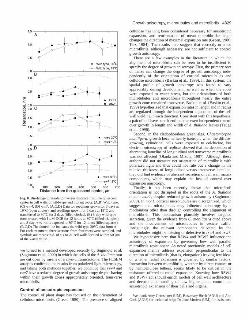
To examine the degree of orientation of the microfibrils, wequantified the birefringent retardation of cell walls usingmicrophotometry. In the wild type, retardation remainedroughly constant through the first 600 µm of the root, and wasnot affected by a 2 days exposure to the non-permissivetemperature (Fig. 8A). In wild-type roots treated for 12 hourswith DCB, or in rsw1 roots exposed to 30°C for 12 hours,retardation decreased significantly (Fig. 8B), as expected when
cellulose synthesis is inhibited. In contrast, the retardation ofcell walls of both rsw4 and rsw7 was not less than the wildtype, and in fact, tended to be greater (Fig. 8C,D). In rsw4, at19°C, despite having wild-type levels of radial expansion (i.e.,normal morphology), roots had somewhat higher levels ofretardation in their cell walls, and at 30°C, despite thestimulated radial expansion, retardation in the walls remainedessentially unchanged. In rsw7, increased radial expansion at30°C was associated with a clear increase in retardation,especially in the apical 200 µm. Decreases in retardation werenot seen in these mutants, which indicates that the degree oforientation among cellulose microfibrils did not decrease.
Taking the sugar analysis together with the FESEM imagesand the polarized-light data, we conclude that both mutants hadreduced growth anisotropy without a decline in cellulosesynthesis or a disorientation among cellulose microfibrils.
DISCUSSION
We investigated the underlying causes of the reduced growthanisotropy of two mutants of Arabidopsis thaliana, rsw4 andrsw7. Anisotropic expansion is currently understood to becontrolled by the orientation of cortical microtubules andcellulose microfibrils and we focussed on these elements. Theorientation of cortical microtubules was not conspicuouslyaltered nor was their abundance reduced appreciably; likewise,cellulose synthesis was not reduced nor were microfibrilsdisoriented. To our knowledge, the results here are the firstdemonstration of disrupted morphogenesis in the diffusegrowing cells of higher plants without a concomitant disruptionof the orientation of either cortical microtubules or cellulosemicrofibrils.
One consequence of both the rsw4and rsw7mutations is thedisruption of the normal pattern of cell wall placement, whichmay include a breakdown of tissue boundaries. A similardisruption of the orientation of cell division occurs in someother A. thaliana mutants with reduced growth anisotropy,including kor (Lane et al., 2001) and fra2 (Burk et al., 2001),but not in others, such as cobra (Schindelman et al., 2001) orrsw1 (Williamson et al., 2001). Disoriented planes of celldivision in the mutants indicate that the specification ofpolarity in the root has been disturbed or that there are activitiescommon to the control of the placement of the cell plate andcell wall extensibility.
Orientations of microtubules and microfibrilsWe examined microtubules by two methods and bothconsistently showed transverse microtubules in regions of themutant roots that were undergoing stimulated radial expansion.Microtubules beneath the outer epidermal cell wall whenviewed with confocal optics were observed readily, but thosein interior tissues examined in plastic sections were difficult toobserve because of the irregular orientation of cell walls. It isunlikely that a cryptic disorganization among cortical arrays inthe interior tissues causes the swelling. First, it is reasonableto expect radial swelling to have a common cause in all tissues.Second, aberrant microtubule behavior frequently gives rise tohelical growth in roots, as seen with low concentrations ofmicrotubule inhibitors (Furutani et al., 2000) and in plantsmutant for α-tubulin, as described recently (Thitamadee et al.,
Fig. 6. FESEM micrographs of the innermost cell wall layer of wildtype, rsw4and rsw7plants in the elongation zone (wild type) orswollen region of the root. (A-C) Longitudinal-radial cell walls withthe long axis of root parallel to the long axis of the page. (A) Wildtype, (B) rsw4and (C) rsw7, grown at 19°C for 6 days andtransferred to 30°C for 2 days. (A-B) PEG sections, (C) cryo-ultramicrome. Bar: 100 nm.
4828
2002); however, rsw4and rsw7seldom if ever grew helically,consistent with their having unimpaired microtubules. Finally,mutant alleles of genes encoding microtubule interactingproteins, including MOR1 (Whittington et al., 2001) and akatanin subunit (Bichet et al., 2001; Burke et al., 2001) causeradial swelling and disrupt microtubules unmistakably. Lack ofobvious microtubule abnormalities in the rsw4 and rsw7mutants predicts that the RSW4 and 7 proteins are unlikely tocontrol cortical microtubule orientation.
Misalignment of microtubules alone might be insufficient toalter growth anisotropy: in wild-type A. thaliana, the corticalarray can be disrupted considerably without reducing growthanisotropy (Baskin et al., 1994). Thus, we cannot exclude the
possibility that a property of the microtubules other thanorientation, such as their stability, could be altered in the rsw4and rsw7 mutants, but it is not known how this would affectthe anisotropy of expansion.
We assayed cellulose microfibrils in the growth zone withtwo complementary methods and both showed transverseorientation in the mutants. The use of polarized-lightmicroscopy to quantify microfibril alignment is frequentlycriticized because the data apply to microfibrils throughout thethickness of the cell wall whereas load is said to be borne bythe inner layers only (Taiz, 1984). However, in this study weshow that birefringent retardation is significantly diminished inrsw1 and in wild-type seedlings treated with DCB (Fig. 7),treatments known to reduce the rate of cellulose synthesis(Arioli et al., 1998; Peng et al., 2000) and disorganize recentlydeposited microfibrils (Sugimoto et al., 2001). Polarized-lightmicroscopy thus has the required sensitivity for quantifying thedeployment of cellulose microfibrils.
In contrast to the decreased retardation expected from theswollen morphology, and seen when cellulose synthesis wasinhibited, retardation was unchanged in rsw4 and actuallyincreased in rsw7 (Fig. 7). Increased retardation indicatesbetter alignment among microfibrils or a greater quantity ofmicrofibrils per unit of wall area. The quantity of microfibrilsper unit area could increase through an increased rate ofcellulose synthesis or a decreased rate of microfibril removal.As microfibrils are ‘removed’ principally by dilution throughexpansion and as the rate of expansion of the mutants was low,increased retardation probably occurred because expansionrate was inhibited. Consistently, more glucose was recoveredfrom the acid-insoluble fraction of rsw7 cell walls, indicatingan increased quantity of cellulose. These results demonstratethat a major reduction in growth anisotropy can occur withoutlessening the overall structural anisotropy of the cell wallprovided by oriented cellulose microfibrils.
To examine microfibril orientation directly, we used FESEM.To minimize the potential for rearrangement of microfibrils, wedeveloped a protocol expected to extract components of the cellwall minimally, a protocol based on embedding plasmolysedroots in polyethylene glycol and subsequently removing theglycol embedment with water. This protocol succeeded for wildtype and rsw4; however, rsw7 roots were fragmented by thesectioning, suggesting that their middle lamellae may be weak.Furthermore, most cells of the rsw7 root were not plasmolysedin 1 M sucrose, a resistance that implicates either an increasedcellular osmolarity or an enhanced adhesion between the cellwall and the plasma membrane. To image the cell wall in rsw7,
A. M. D. Wiedemeier and others
Fig. 7. Polarized-light micrographs of longitudinal sections of theelongation zone (in the wild type) or swollen region of the root inrsw4and rsw7. The transmission axes of analyzer and polarizer areshown by the white right-angle in (A). (A,C,E) The compensator wasrotated to a positive angle, and (B,D,F) to a negative angle; theorientation of the white line in the two upper panels (A,B) shows thedirection of the optical axis of birefringent elements (i.e.,microfibrils) that would produce regions of the image brighter thanbackground. (A,B) A section from wild type, (C,D) rsw4and (E,F)rsw7shown with each compensator setting. Seedlings were grown asfor Fig. 4. The left-hand and right-hand panels were photographedwith identical microscope settings and processed identically forreproduction. Bar: 25 µm.
4829Growth anisotropy, microtubules and microfibrils
we turned to a method developed recently by Sugimoto et al.(Sugimoto et al., 2000) in which the cells of the A. thalianarootare cut open by means of a cryo-ultramicrotome. The FESEManalysis confirmed the results from polarized-light microscopy,and taking both methods together, we conclude that rsw4 andrsw7have a reduced degree of growth anisotropy despite havingwithin their growth zones appropriately oriented, transversemicrofibrils.
Control of anisotropic expansion The control of plant shape has focused on the orientation ofcellulose microfibrils (Green, 1980). The presence of aligned
cellulose has long been considered necessary for anisotropicexpansion, and reorientation of mean microfibrillar anglechanges the direction of maximal expansion rate (Green, 1980;Taiz, 1984). The results here suggest that correctly orientedmicrofibrils, although necessary, are not sufficient to controlgrowth anisotropy.
There are a few examples in the literature in which thealignment of microfibrils can be seen to be insufficient tospecify the degree of growth anisotropy. First, the primary rootof maize can change the degree of growth anisotropy inde-pendently of the orientation of cortical microtubules andcellulose microfibrils (Baskin et al., 1999). In this system, thespatial profile of growth anisotropy was found to varyappreciably during development, as well as when the rootswere exposed to water stress, but the orientations of bothmicrotubules and microfibrils throughout nearly the entiregrowth zone remained transverse. Baskin et al. (Baskin et al.,1999) hypothesized that expansion rates in length and in radiusare regulated through the independent adjustment of the cellwall yielding in each direction. Consistent with this hypothesis,a pair of loci have been identified that exert independent controlover growth in length and width of A. thalianaleaves (Tsugeet al., 1996).
Second, in the cladophoralean green alga, Chaetomorphamoniligera, growth became nearly isotropic when the diffuse-growing, cylindrical cells were exposed to colchicine, butelectron microscopy of replicas showed that the deposition ofalternating lamellae of longitudinal and transverse microfibrilswas not affected (Okuda and Mizuta, 1987). Although theseauthors did not measure net orientation of microfibrils withpolarized light and thus could not rule out a change in therelative thickness of longitudinal versus transverse lamellae,they did find evidence of aberrant secretion of cell wall matrixcomponents, which may explain the loss of control overexpansion anisotropy.
Finally, it has been recently shown that microfibrilorientation is not disrupted in the roots of the A. thalianamutant mor1, despite reduced growth anisotropy (Sugimoto,2000). In mor1, cortical microtubules are disorganized, whichsuggests that microtubules may influence anisotropy by amechanism other than through controlling the alignment ofmicrofibrils. This mechanism plausibly involves targetedsecretion, given the evidence from C. moniligeracited aboveand the involvement of microtubules in vesicle traffic.Intriguingly, the relevant components delivered by themicrotubules might be missing or defective in rsw4and rsw7.
We hypothesize here that RSW4 and RSW7 influence theanisotropy of expansion by governing how well parallelmicrofibrils resist shear. As noted previously, models of cellexpansion mainly address expansion perpendicular to thedirection of microfibrils (that is, elongation) leaving few ideasof whether radial expansion is governed by similar factors.Interaction between microfibrils, whether by direct contact orby hemicellulose tethers, seems likely to be critical in theresistance offered to radial expansion. Knowing how RSW4and RSW7 act should enrich models of cell wall architectureand deepen understanding of how higher plants control theanisotropic expansion of their cells and organs.
We thank Amy Gernstetter (UM), Rosemary Birch (ANU) and AnnCork (ANU) for technical help; Dr Jane Murfett (UM) for assistance
Fig. 8. Birefringent retardation versus distance from the quiescentcenter in cell walls of wild-type and mutant roots. (A,B) Wild type;(C) rsw4; (D) rsw7. (A,C,D) Data for seedlings grown for 8 days at19°C (open circles), and seedlings grown for 6 days at 19°C andtransferred to 30°C for 2 days (filled circles). (B) 8-day wild-typeroots treated with 1 µM DCB for 12 hours at 30°C (filled triangles),and 8-day rsw1roots exposed to 30°C for 12 hours (filled squares).(B,C,D) The dotted line indicates the wild-type 30°C data from A.For each treatment, three sections from four roots were sampled, andsymbols are means±s.d. of six to 21 cell walls located within 50 µmof the x-axis value.
4830
with RFLP mapping, the Electron Microscopy Unit (ANU) and theCore Facility for Electron Microscopy (UM) for help with electronmicroscopy, and Keiko Sugimoto (ANU) for help with cryo-ultramicrotomy and insightful discussions about FESEM. A. M. D.W. was supported by a Patricia Roberts Harris Fellowship. Thisproject was funded in part by a grant to T. I. B. from the USDepartment of Energy (award No. 94ER20146), which does notconstitute endorsement by that Department of views expressed herein.
REFERENCES
Arioli, T., Peng, L., Betzner, A. S., Burn, J., Wittke, W., Herth, W.,Camilleri, C., Höfte, H., Plazinski, J., Birch, R. et al. (1998). Molecularanalysis of cellulose biosynthesis in Arabidopsis. Science279, 717-720.
Baskin, T. I. (2001). On the alignment of cellulose microfibrils by corticalmicrotubules: a review and a model. Protoplasma215, 150-171.
Baskin, T. I. and Wilson, J. E. (1997). Inhibitors of protein kinases andphosphatases alter root morphology and disorganize cortical microtubules.Plant Physiol. 113, 493-502.
Baskin, T. I., Betzner, A. S., Hoggart, R., Cork, A. and Williamson, R. E.(1992). Root morphology mutants in Arabidopsis thaliana. Aust. J. PlantPhysiol. 19, 427-437.
Baskin, T. I., Meekes, H. H. T. M., Liang, B. M. and Sharp, R. E. (1999).Regulation of growth anisotropy in well watered and water-stressed maizeroots. II. Role of cortical microtubules and cellulose microfibrils. PlantPhysiol. 119, 681-692.
Baskin, T. I., Wilson, J. E., Cork, A. and Williamson, R. E. (1994).Morphology and microtubule organization in arabidopsis roots exposed tooryzalin or taxol. Plant Cell Physiol. 35, 935-942.
Benfey, P. N., Linstead, P. J., Roberts, K., Schiefelbein, J. W., Hauser, M.-T. and Aeschbacher, R. A. (1993). Root development in Arabidopsis: fourmutants with dramatically altered root morphogenesis. Development 119,1-14.
Beyer, E. M. Jr (1976). A potent inhibitor of ethylene action in plants. PlantPhysiol. 58, 268-271.
Bichet, A., Desnos, T., Turner, S., Grandjean, O. and Höfte, H. (2001).BOTERO1is required for normal orientation of cortical microtubules andanisotropic cell expansion in Arabidopsis. Plant J. 25, 137-148.
Brown, R. M., Jr (1999) Cellulose structure and biosynthesis. Pure Appl.Chem. 71, 767-775.
Burk, D. H., Liu, B., Zhong, R. Q., Morrison, W. H. and Ye, Z. H. (2001).A katanin-like protein regulates normal cell wall biosynthesis and cellelongation. Plant Cell13, 807-827.
Carlemalm, E., Garavito, R. M. and Villiger, W. (1982). Resin developmentfor electron microscopy and an analysis of embedding at low temperature.J. Microsc. 126, 123-143.
Cosgrove, D. J. (1997). Relaxation in a high-stress environment: The molecularbases of extensible cell walls and cell enlargement. Plant Cell9, 1031-1041.
Fabri, C. O. and Schäffner, A. R. (1994). An Arabidopsis thaliana RFLPmapping set to localize mutations to chromosomal regions. Plant J. 5, 149-156.
Fagard, M., Desnos, T., Desprez, T., Goubet, F., Refregier, G., Mouille, G.,McCann, M., Rayon, C., Vernhettes, S. and Höfte, H. (2000).PROCUSTE1encodes a cellulose synthase required for normal cellelongation specifically in roots and dark-grown hypocotyls of arabidopsis.Plant Cell12, 2409-2423.
Fowke, L., Dibbayawan, T., Schwartz, O., Harper, J. and Overall, R.(1999). Combined immunofluorescence and field emission scanningelectron microscope study of plasma membrane-associated organelles inhighly vacuolated suspensor cells of white spruce somatic embryos. CellBiol. Int. 23, 389-397.
Furutani, I., Watanabe, Y., Prieto, R., Masukawa, M., Suzuki, K., Naoi,K., Thitamadee, S., Shikanai, T. and Hashimoto, T. (2000). The SPIRALgenes are required for directional central of cell elongation in Arabidopsisthaliana. Development127, 4443-4453.
Green, P. B. (1980). Organogenesis – a biophysical view. Annu. Rev. PlantPhysiol. 31, 51-82.
Hauser, M. T., Morikami, A. and Benfey, P. N. (1995). Conditional rootexpansion mutants of Arabidopsis. Development121, 1237-1252.
Hogetsu, T., Shibaoka, H. and Shimokoriyama, M. (1974). Involvement ofcellulose synthesis in actions of gibberellin and kenetin on cell expansion.
2,6-Dichlorobenzonitrile as a new cellulose-synthesis inhibitor. Plant CellPhysiol. 15, 389-393.
Holding, D. R., McKenzie, R. J. and Coomber, S. A. (1994). Genetic andstructural analysis of five arabidopsis mutants with abnormal root morphologygenerated by the seed transformation method. Ann. Bot. 74, 193-204.
Koornneef, M. and Stam, P. (1992). Genetic analysis. In: Methods inArabidopsis Research(ed. C. Koncz, N. H. Chua and J. Schell), pp. 83-99.Singapore: World Scientific.
Lane, D. R., Wiedemeier, A., Peng, L. C., Höfte, H., Vernhettes, S.,Desprez, T., Hocart, C. H., Birch, R. J., Baskin, T. I., Burn, J. E. et al.(2001). Temperature-sensitive alleles of RSW2link the KORRIGAN endo-1,4-β-glucanase to cellulose synthesis and cytokinesis in Arabidopsis. PlantPhysiol. 126, 278-288.
Liang, B. M., Dennings, A. M., Sharp, R. E. and Baskin, T. I. (1996).Consistent handedness of microtubule helical arrays in maize andArabidopisprimary roots. Protoplasma190, 8-15.
McClinton, R. S. and Sung, Z. R. (1997). Organization of corticalmicrotubules at the plasma membrane in arabidopsis. Planta201, 252-260.
Nicol, F., His, I., Jauneau, A., Vernhettes, S., Canut, H. and Höfte, H.(1998). A plasma membrane-bound putative endo-1,4-β-D-glucanase isrequired for normal wall assembly and cell elongation in Arabidopsis.EMBO J. 17, 5563-5576.
Okuda, K. and Mizuta, S. (1987). Modification in cell shape unrelated tocellulose microfibril orientation in growing thallus cells of Chaetomorphamoniligera. Plant Cell Physiol. 28, 461-473.
Peng, L. C., Hocart, C. H., Redmond, J. W. and Williamson, R. E. (2000).Fractionation of carbohydrates in Arabidopsisroot cell walls shows thatthree radial swelling loci are specifically involved in cellulose production.Planta211, 406-414.
Preston, R. D. (1974). The Physical Biology of Plant Cell Walls. pp. 68-105.London: Chapman & Hall.
Sato, S., Kato, T., Kakegawa, K., Ishii, T., Liu, Y. G., Awano, T., Takabe,K., Nishiyama, Y., Kuga, S., Nakamura, Y. et al. (2001) Role of theputative membrane-bound endo-1,4-β-glucanase KORRIGAN in cellelongation and cellulose synthesis in Arabidopsis thaliana. Plant CellPhysiol. 42, 251-263.
Scheres, B., di Laurenzio, L., Willemsen, V., Hauser, M. T., Janmaat, K.,Weisbeek, P. and Benfey, P. N. (1995). Mutations affecting the radialorganisation of the Arabidopsisroot display specific defects throughout theembryonic axis. Development121, 53-62.
Schindelman, G., Morikami, A., Jung, J., Baskin, T. I., Carpita, N. C.,Derbyshire, P., McCann, M. C. and Benfey, P. N. (2001) COBRA encodesa putative GPI-anchored protein, which is polarly localized and necessaryfor oriented cell expansion in Arabidopsis. Genes Dev. 15, 1115-1127.
Sugimoto, K. (2000). Cortical Microtubules, Cellulose Microfibrils andGrowth Anisotropy in the Roots of Arabidopsis thaliana. Ph. D. thesis,Australian National University, Canberra.
Sugimoto, K., Williamson, R. E. and Wasteneys, G. O. (2000). New techniquesenable comparative analysis of microtubule orientation, wall texture, andgrowth rate in intact roots of arabidopsis. Plant Physiol. 124, 1493-1506.
Sugimoto, K., Williamson, R. E. and Wasteneys, G. O. (2001). Wallarchitecture in the cellulose-deficient rsw1mutant of Arabidopsis thaliana:microfibrils but not microtubules lose their transverse alignment beforemicrofibrils become unrecognizable in the mitotic and elongation zones ofroots. Protoplasma215, 172-183.
Taiz, L. (1984). Plant cell expansion: regulation of cell wall mechanicalproperties. Ann. Rev. Plant Physiol. 35, 585–657.
Thitamadee, S., Tuchihara, K. and Hashimoto, T. (2002) Microtubule basisfor left-handed helical growth in arabidopsis. Nature417, 193-196.
Traas, J., Bellini, C., Nacry, P., Kronenberger, J., Bouchez, D. andCaboche, M. (1995). Normal differentiation patterns in plants lackingmicrotubular preprophase bands. Nature 375, 676-677.
Tsuge, T., Tsukaya, H. and Uchimiya, H. (1996). Two independent andpolarized processes of cell elongation regulate leaf blade expansion inArabidopsis thaliana(L.) Heynh. Development122, 1589-1600.
Whittington, A. T., Vugrek., O., Wei, K. J., Hasenbein, N. G., Sugimoto,K., Rashbrooke, M. C. and Wasteneys, G. O. (2001). MOR1 is essentialfor organizing cortical microtubules in plants. Nature411, 610-613.
Williamson, R. E., Burn, J. E., Birch, R., Baskin, T. I., Arioli, T., Betzner,A. S. and Cork, A. (2001). Morphogenesis in rsw1, a cellulose-deficientmutant of Arabidopsis thalania. Protoplasma 215, 116-127.
Wiedemeier, A. M. D. (1998). Analysis of Arabidopsis thalianaGrowthAnisotropy Mutants: Genetic, Physiological, and CytologicalCharacterization. Ph. D. Thesis, University of Missouri, Columbia.
A. M. D. Wiedemeier and others
Related Documents


