musculoskeletal system anatomy Muscles of leg Done by :-ALI HIARY

Welcome message from author
This document is posted to help you gain knowledge. Please leave a comment to let me know what you think about it! Share it to your friends and learn new things together.
Transcript

musculoskeletal system anatomy
Muscles of leg Done by :-ALI HIARY
Muscles of Leg
Lecture Objectives
• List the muscles of the leg. • Describe the attachments of the leg muscles and their nerve supply. • Describe the popliteal fossa. • List the content of the popliteal fossa. • Understand the clinical importance of the popliteal fossa.
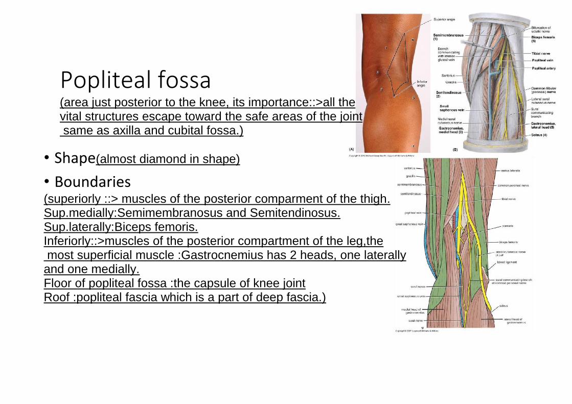
Popliteal fossa
(area just posterior to the knee, its importance::>all the vital structures escape toward the safe areas of the joint same as axilla and cubital fossa.)
• Shape(almost diamond in shape) • Boundaries (superiorly ::> muscles of the posterior comparment of the thigh. Sup.medially:Semimembranosus and Semitendinosus. Sup.laterally:Biceps femoris. Inferiorly::>muscles of the posterior compartment of the leg,the most superficial muscle :Gastrocnemius has 2 heads, one laterally and one medially. Floor of popliteal fossa :the capsule of knee joint Roof :popliteal fascia which is a part of deep fascia.)

• Content(The neurovascular bundle) • Popliteal vessels(popliteal artery +vein that come from femoral artery+vein) • Small saphenous vein(superficial to popliteal fascia and must pierce it to enter
into fossa and drains ino popliteal vein) • Common peroneal & tibial nerves(these are the terminal branches of sciatic nerve
which ends at the upper angle of popliteal fossa) *Relation:-the deepest one is artery, more superficial is vein, most superficial is tibial nerve::> cross in the middle of popliteal fossa while common peroneal runs along with tendon of biceps femoris.
• Lymph nodes(popliteal group from 2 to 4 lymph nodes) • Popliteal fascia
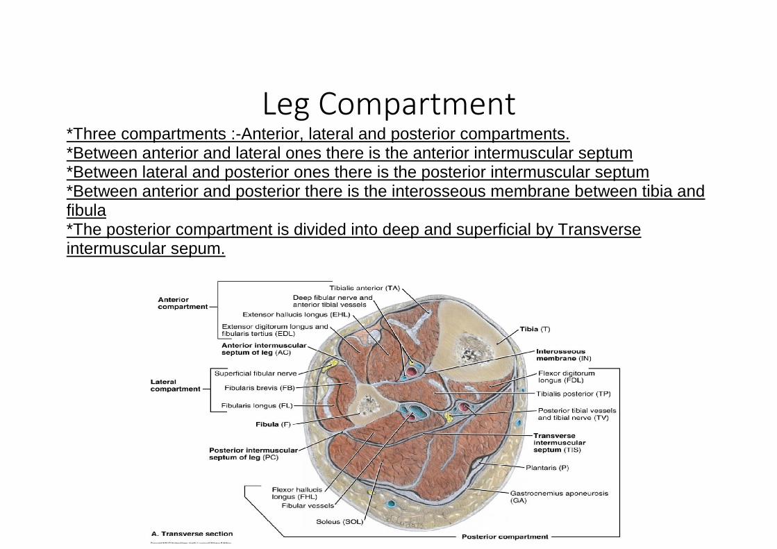
Leg Compartment *Three compartments :-Anterior, lateral and posterior compartments. *Between anterior and lateral ones there is the anterior intermuscular septum *Between lateral and posterior ones there is the posterior intermuscular septum *Between anterior and posterior there is the interosseous membrane between tibia and fibula *The posterior compartment is divided into deep and superficial by Transverse intermuscular sepum.
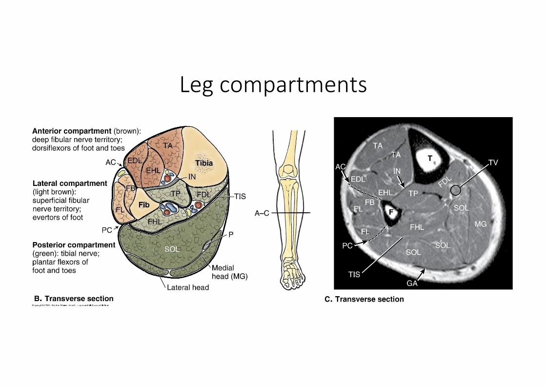
Leg compartments

Muscles of Anterior Compartment of Leg
*Aneriormedial surface of the tibia is considered as superficial structure so theres no structure,All muscles of the anterior compartment of leg are located laterally to the palpable anterior border of tibia.
Dorsiflexors(same as extensor) of the ankle( because they
cross the ankle joint anteriorly)
Deep fibular nerve(common personal nerve wraps on the neck
of fibula and enters the anteriolateral compartment by piercing the fibularis longus and divides into 2 branches :- superficial and deep) … so if there is a cut in the deep fibular nerves all the anterior muscles will be paralyzed . *Tendons of theses muscles are hold in place by retinaculi( thickening of deep fascia anteriorlly).
Superior extensor retinaculum
Inferior extensor retinaculum(Y shape)
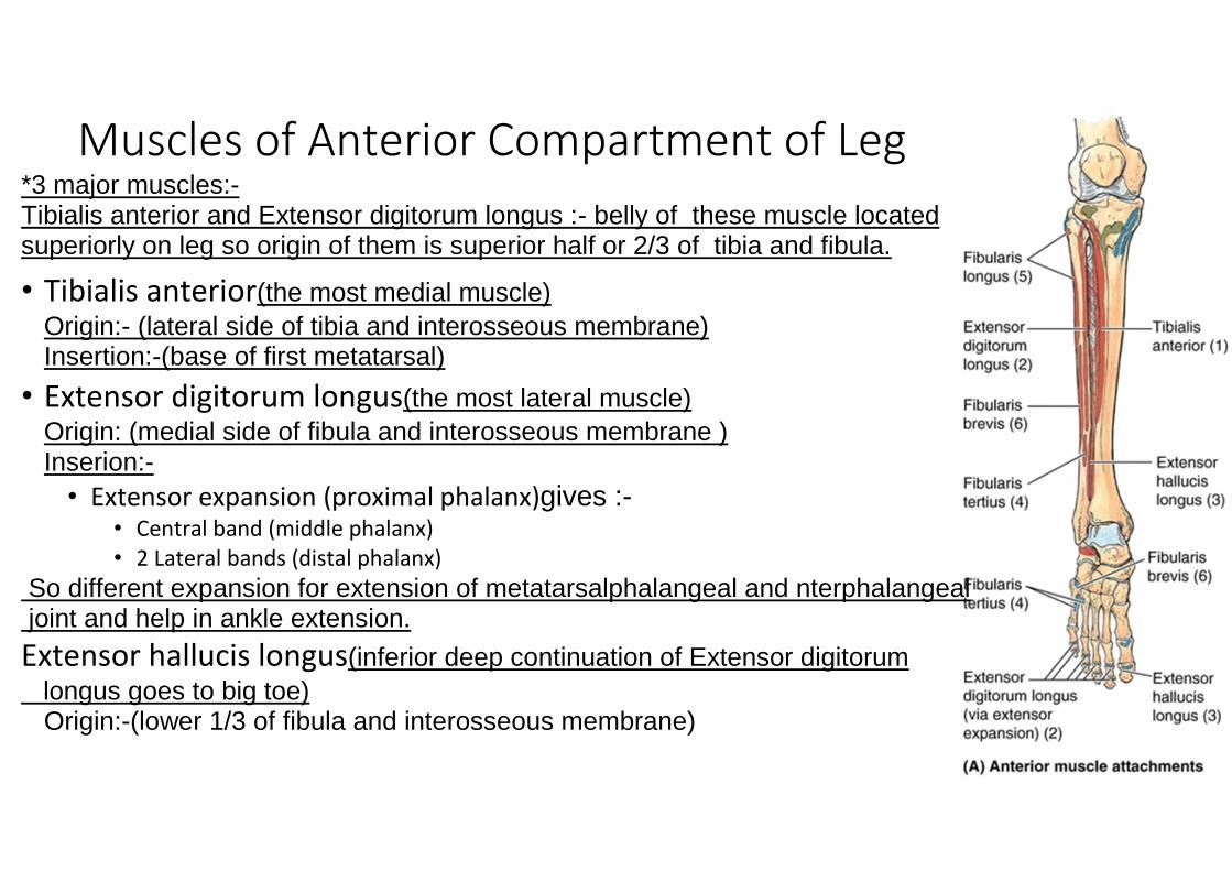
Muscles of Anterior Compartment of Leg *3 major muscles:- Tibialis anterior and Extensor digitorum longus :- belly of these muscle located superiorly on leg so origin of them is superior half or 2/3 of tibia and fibula.
• Tibialis anterior(the most medial muscle) Origin:- (lateral side of tibia and interosseous membrane) Insertion:-(base of first metatarsal)
• Extensor digitorum longus(the most lateral muscle) Origin: (medial side of fibula and interosseous membrane ) Inserion:-
• Extensor expansion (proximal phalanx)gives :- • Central band (middle phalanx) • 2 Lateral bands (distal phalanx)
So different expansion for extension of metatarsalphalangeal and nterphalangeal joint and help in ankle extension.
Extensor hallucis longus(inferior deep continuation of Extensor digitorum
longus goes to big toe) Origin:-(lower 1/3 of fibula and interosseous membrane)

Insertion:- (dital phalanx of big toe ) • Fibularis tertius(small muscle same as extensor hallucis longus and probably absent in
some people) Attachmet on base of 5
th metatarsal bone or cuboid bone.
P.S:- * Extensor from fibula and tibialis from tibia,when we get a cross section of ankle joint within retinacul we will see all the tendons are far away from each others and all superficial . *When one muscle has tendons for more than one phalanx toe ,the single movement of one toe is difficult so they all move together same as Extensor digitorum longus but the big toe has its muscle so move separately by extensor hallucis longus.

Muscles of Lateral Compartment of Leg
Eversion of foot (because of their location on lateral )
Superficial fibular nerve(innervation)(Runs between fibularis longus and brevis)
Superior peroneal
retinaculum
Inferior peroneal retinaculum
(Both retinaculi help in fixation of tendons
of fibularis longus and brevis )

Muscles of Lateral Compartment of Leg
• Peroneus (fibularis) longus m.(longer and superficial) Origin :-superior part of fibula Insertion :- more deep toward medial side on base first metatarsal of sole
So longer tract in insertion gives it more importance. • Peroneus (fibularis) brevis m.(deep)
Origin:- lower part of fibula Insertion:- base of 5
th metatarsal
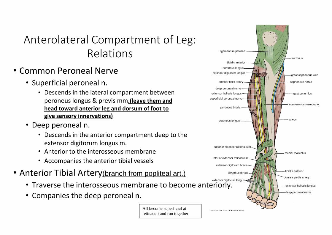
Anterolateral Compartment of Leg:
Relations
• Common Peroneal Nerve • Superficial peroneal n.
• Descends in the lateral compartment between peroneus longus & previs mm.(leave them and head toward anterior leg and dorsum of foot to give sensory innervations)
• Deep peroneal n. • Descends in the anterior compartment deep to the
extensor digitorum longus m. • Anterior to the interosseous membrane • Accompanies the anterior tibial vessels
• Anterior Tibial Artery(branch from popliteal art.) • Traverse the interosseous membrane to become anteriorly. • Companies the deep peroneal n.
All become superficial at
retinaculi and run together


Muscles of the Posterior Compartment of Leg
Planter flexion(Because they cross the ankle joint posteriorly)
Tibial nerve(all the posterior compartment +dorum of foot by terminal braches)
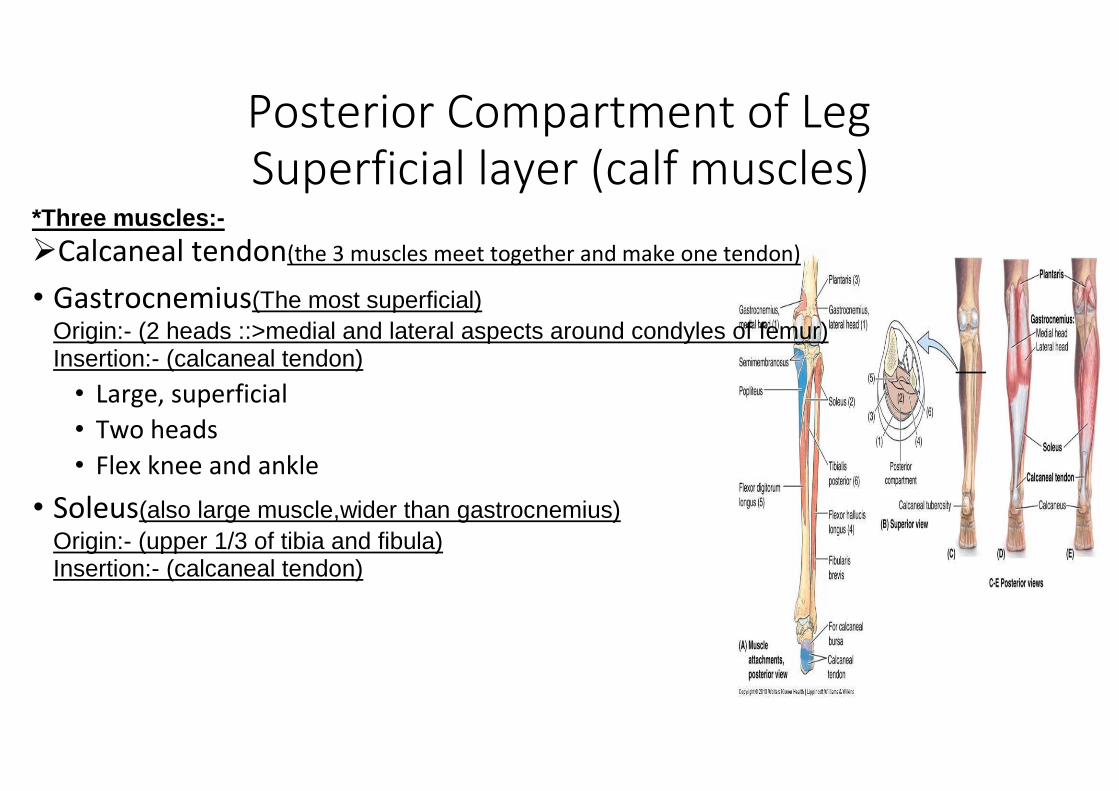
• Superficial layer (calf muscles)
Transverse intermuscular septum • Deep layer
Posterior Compartment of Leg
Superficial layer (calf muscles)
*Three muscles:-
Calcaneal tendon(the 3 muscles meet together and make one tendon)
• Gastrocnemius(The most superficial) Origin:- (2 heads ::>medial and lateral aspects around condyles of femur) Insertion:- (calcaneal tendon)
• Large, superficial • Two heads • Flex knee and ankle
• Soleus(also large muscle,wider than gastrocnemius) Origin:- (upper 1/3 of tibia and fibula) Insertion:- (calcaneal tendon)
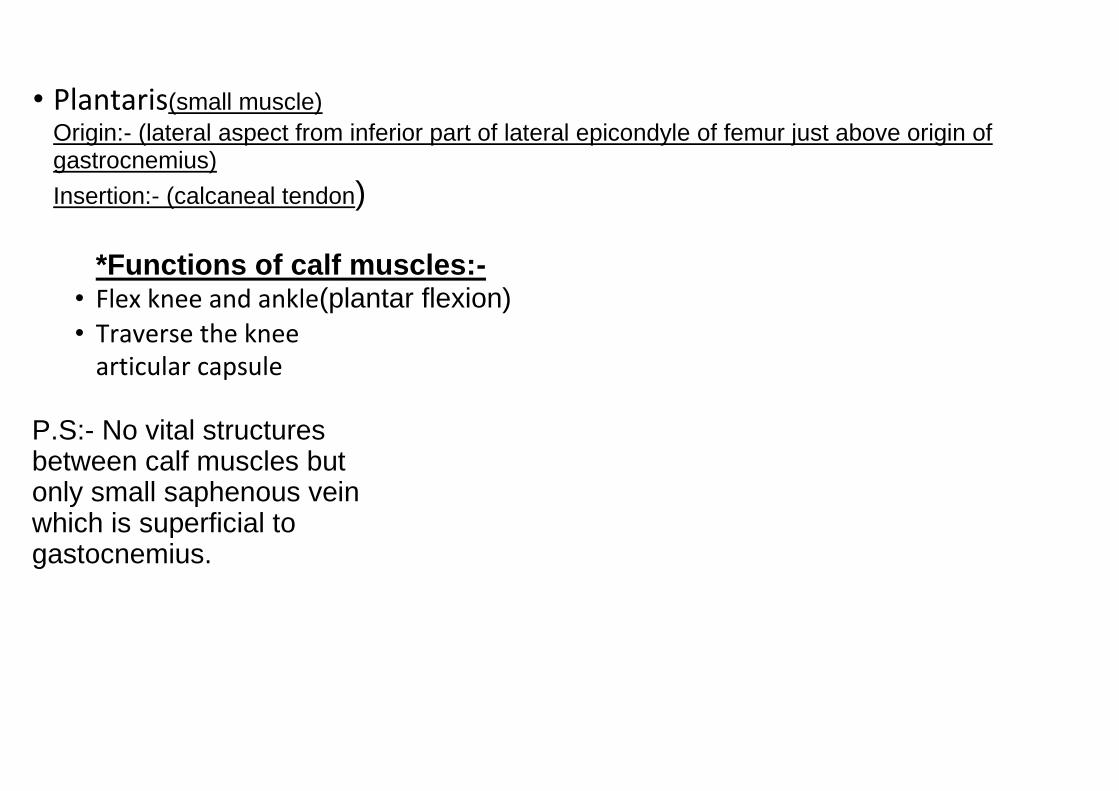
• Plantaris(small muscle) Origin:- (lateral aspect from inferior part of lateral epicondyle of femur just above origin of gastrocnemius)
Insertion:- (calcaneal tendon)
*Functions of calf muscles:- • Flex knee and ankle(plantar flexion) • Traverse the knee
articular capsule
P.S:- No vital structures between calf muscles but only small saphenous vein which is superficial to gastocnemius.

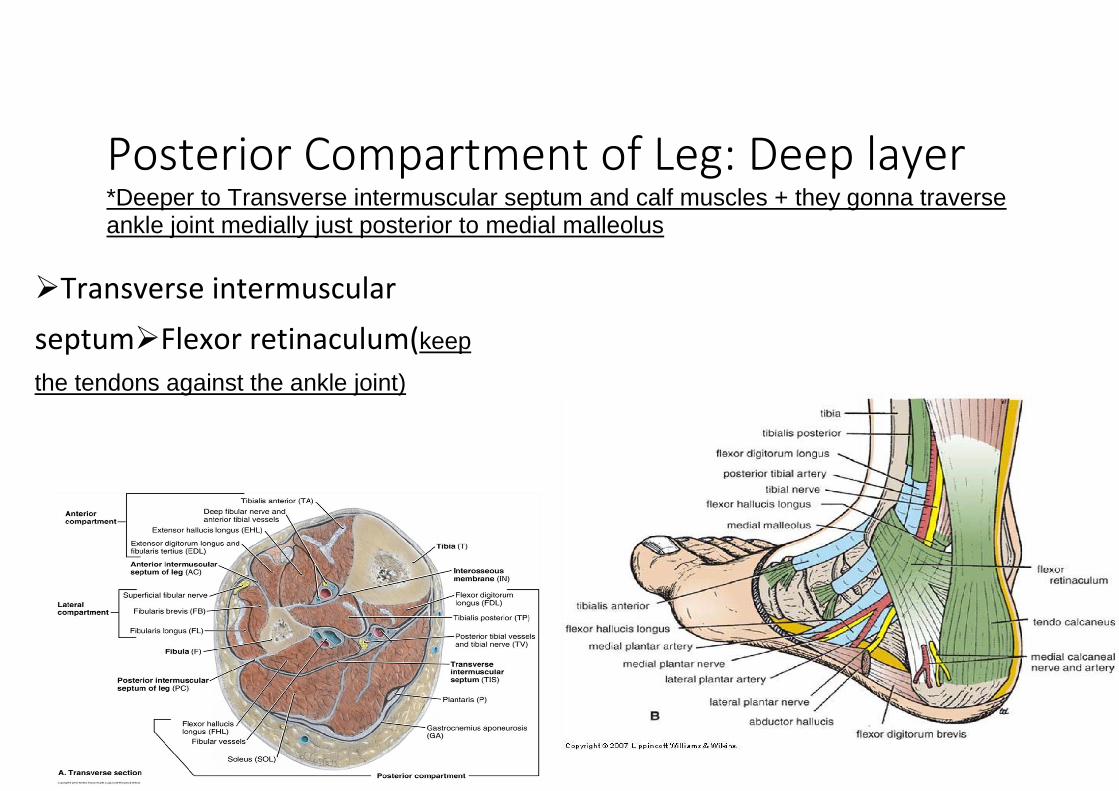
Posterior Compartment of Leg: Deep layer
*Deeper to Transverse intermuscular septum and calf muscles + they gonna traverse ankle joint medially just posterior to medial malleolus
Transverse intermuscular
septumFlexor retinaculum(keep
the tendons against the ankle joint)

Posterior Compartment of Leg:
Deep layer
Popliteus(small short muscle , traverse the knee
capsule posteriorly and has an attachment on lateral meniscus) *Origin:- (lateral condyle of femur) *Insertion:- (posterior superior medial part of tibia) *Function:- (steadying of knee joint while movement of knee joint mainly during the extension).
Flexor hallucis longus *Origin :-middle part of fibula *Insertion:-base of distal phalanx of big toe *Function:-flexion of phalanges
Flexor digitorum longus *Origin:-medial part of tibia *Insertion:-distal phalanx of rest of toes *Function:-flextion of phalanges
Tibialis posterior(same as
fibularis longus going deep) *Origin:-lateral upper posterior part of tibia and interosseous membrane *Insertion:-tarsus and bases of metatarsals,crosses the subtalus and transverse tarsal joints. *Function:-Inversion of foot
*Order from medial to lateral :-Flexor digitorum longus - Tibialis posterior - Flexor hallucis longus.

*Muscular Relations :- *Flexor hallucis longus is the most lateral one but it inserts most medially in the sole so it crosses over superficially to tibialis posterior and deeply to flexor digitorum longus. *Tibialis posterior crosses over deeply to flexor digitorum longus which goes up to the 5th toe but tibialis posterior ends earlier at tarsus *Flexor digitorum longus is the most medial one , its tendon crosses both muscles in order to insertion which is the most lateral one . *Calceneal tendon is the most lateral and posterior to the tendons of 3 muscles so the tendons are surrounded by medial malleolus anteriorly and calceneal tendon posteriorly and you can palpate it.

Deep Layer: Muscular Relation
At Ankle At Leg

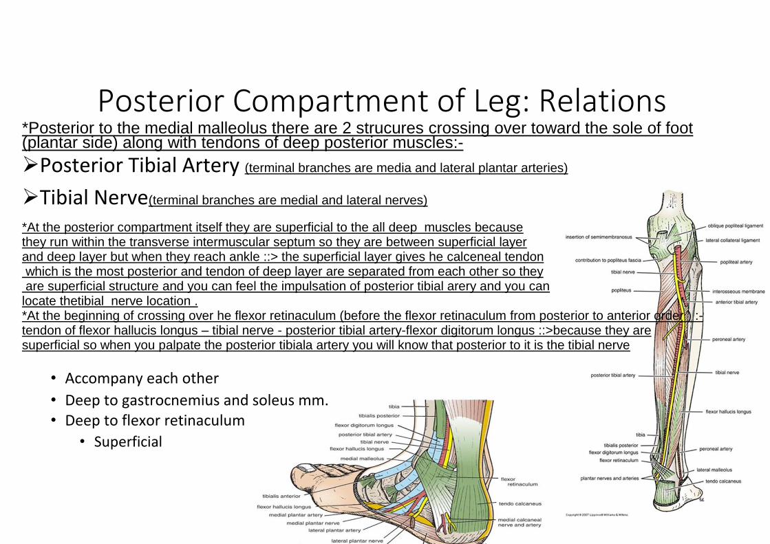
Posterior Compartment of Leg: Relations
*Posterior to the medial malleolus there are 2 strucures crossing over toward the sole of foot (plantar side) along with tendons of deep posterior muscles:-
Posterior Tibial Artery (terminal branches are media and lateral plantar arteries)
Tibial Nerve(terminal branches are medial and lateral nerves)
*At the posterior compartment itself they are superficial to the all deep muscles because they run within the transverse intermuscular septum so they are between superficial layer and deep layer but when they reach ankle ::> the superficial layer gives he calceneal tendon which is the most posterior and tendon of deep layer are separated from each other so they are superficial structure and you can feel the impulsation of posterior tibial arery and you can locate thetibial nerve location . *At the beginning of crossing over he flexor retinaculum (before the flexor retinaculum from posterior to anterior order ) :- tendon of flexor hallucis longus – tibial nerve - posterior tibial artery-flexor digitorum longus ::>because they are superficial so when you palpate the posterior tibiala artery you will know that posterior to it is the tibial nerve
• Accompany each other • Deep to gastrocnemius and soleus mm. • Deep to flexor retinaculum
• Superficial
Related Documents