Muscular adaptations to computer-guided strength training with eccentric overload B. Friedmann, 1 R. Kinscherf, 2 S. Vorwald, 2 H. Mu ¨ ller, 3 K. Kucera, 3 S. Borisch, 1 G. Richter, 4 P. Ba ¨rtsch 1 and R. Billeter 5 1 Department of Sports Medicine, Medical Clinic and Policlinic, University of Heidelberg, Heidelberg, Germany 2 Department of Anatomy and Cell Biology III, University of Heidelberg, Heidelberg, Germany 3 Olympic Training Centre Rhein-Neckar, Heidelberg, Germany 4 Department of Radiology, University of Heidelberg, Heidelberg, Germany 5 School of Biomedical Sciences, University of Leeds, Leeds, UK Received 11 December 2003, accepted 28 April 2004 Correspondence: B. Friedmann, Department of Sports Medicine, Medical Clinic and Policlinic, University of Heidelberg, Im Neuenheimer Feld 710, 69120 Heidelberg, Germany. Abstract Aims: In order to investigate the muscular adaptations to a novel form of strength training, 18 male untrained subjects performed 4 weeks of low resistance–high repetition knee extension exercise. Methods: Nine of them trained on a conventional weight resistance device (Leg curler, CON/ECC group), with loads equivalent to 30% of the con- centric one-repetition maximum (1RM) for both the concentric and eccentric phase of movement. The other nine trained on a newly developed computer- driven device (CON/ECC-OVERLOAD group) with the concentric load equivalent to 30% of the concentric 1RM and the eccentric load equivalent to 30% of the eccentric 1RM. Results: Training resulted in significantly (P £ 0.05) increased peak torque and a tendency (P ¼ 0.092) to increased muscle cross-sectional area for the CON/ECC-OVERLOAD but not the CON/ECC group, while strength endurance capacity was significantly (P £ 0.05) increased in the CON/ECC group only. RT-PCR revealed significantly increased myosin heavy chain (MHC) IIa and lactate dehydrogenase (LDH) A mRNAs, a tendency for increased MHC IIx mRNA (P ¼ 0.056) and high correlations between the changes in MHC IIx and LDH A mRNAs (r ¼ 0.97, P ¼ 0.001) in the CON/ ECC-OVERLOAD group. Conclusions: These results indicate a shift towards a more type II domin- ated gene expression pattern in the vasti laterales muscles of the CON/ECC- OVERLOAD group in response to training. We suggest that the increased eccentric load in the CON/ECC-OVERLOAD training leads to distinct adaptations towards a stronger, faster muscle. Keywords fibre types, gene expression, maximal strength, muscle cross- sectional area, resistance training, strength endurance capacity. Resistance training increases muscular strength, primar- ily due to neuronal adaptations and adaptive changes in the trained skeletal muscles, such as hypertrophy and changes in the myosin heavy chain (MHC) composition of the muscle fibres. The extent and nature of the adaptations to resistance training depend on the specific mode of training. Combinations of concentric and eccentric exercise were found to be most successful for strength gain (Jones & Rutherford 1987, Colliander & Tesch 1990, Dudley et al. 1991a, Hather et al. 1991, O’Hagan et al. 1995, Hortoba ´gyi et al. 2000). In some studies, detectable hypertrophy of muscle fibres, mainly Acta Physiol Scand 2004, 182, 77–88 Ó 2004 Scandinavian Physiological Society 77

Welcome message from author
This document is posted to help you gain knowledge. Please leave a comment to let me know what you think about it! Share it to your friends and learn new things together.
Transcript

Muscular adaptations to computer-guided strength training
with eccentric overload
B. Friedmann,1 R. Kinscherf,2 S. Vorwald,2 H. Muller,3 K. Kucera,3 S. Borisch,1 G. Richter,4
P. Bartsch1 and R. Billeter5
1 Department of Sports Medicine, Medical Clinic and Policlinic, University of Heidelberg, Heidelberg, Germany
2 Department of Anatomy and Cell Biology III, University of Heidelberg, Heidelberg, Germany
3 Olympic Training Centre Rhein-Neckar, Heidelberg, Germany
4 Department of Radiology, University of Heidelberg, Heidelberg, Germany
5 School of Biomedical Sciences, University of Leeds, Leeds, UK
Received 11 December 2003,
accepted 28 April 2004
Correspondence: B. Friedmann,
Department of Sports Medicine,
Medical Clinic and Policlinic,
University of Heidelberg,
Im Neuenheimer Feld 710,
69120 Heidelberg, Germany.
Abstract
Aims: In order to investigate the muscular adaptations to a novel form of
strength training, 18 male untrained subjects performed 4 weeks of low
resistance–high repetition knee extension exercise.
Methods: Nine of them trained on a conventional weight resistance device
(Leg curler, CON/ECC group), with loads equivalent to 30% of the con-
centric one-repetition maximum (1RM) for both the concentric and eccentric
phase of movement. The other nine trained on a newly developed computer-
driven device (CON/ECC-OVERLOAD group) with the concentric load
equivalent to 30% of the concentric 1RM and the eccentric load equivalent
to 30% of the eccentric 1RM.
Results: Training resulted in significantly (P £ 0.05) increased peak torque
and a tendency (P ¼ 0.092) to increased muscle cross-sectional area for the
CON/ECC-OVERLOAD but not the CON/ECC group, while strength
endurance capacity was significantly (P £ 0.05) increased in the CON/ECC
group only. RT-PCR revealed significantly increased myosin heavy chain
(MHC) IIa and lactate dehydrogenase (LDH) A mRNAs, a tendency for
increased MHC IIx mRNA (P ¼ 0.056) and high correlations between the
changes in MHC IIx and LDH A mRNAs (r ¼ 0.97, P ¼ 0.001) in the CON/
ECC-OVERLOAD group.
Conclusions: These results indicate a shift towards a more type II domin-
ated gene expression pattern in the vasti laterales muscles of the CON/ECC-
OVERLOAD group in response to training. We suggest that the increased
eccentric load in the CON/ECC-OVERLOAD training leads to distinct
adaptations towards a stronger, faster muscle.
Keywords fibre types, gene expression, maximal strength, muscle cross-
sectional area, resistance training, strength endurance capacity.
Resistance training increases muscular strength, primar-
ily due to neuronal adaptations and adaptive changes in
the trained skeletal muscles, such as hypertrophy and
changes in the myosin heavy chain (MHC) composition
of the muscle fibres. The extent and nature of the
adaptations to resistance training depend on the specific
mode of training. Combinations of concentric and
eccentric exercise were found to be most successful for
strength gain (Jones & Rutherford 1987, Colliander &
Tesch 1990, Dudley et al. 1991a, Hather et al. 1991,
O’Hagan et al. 1995, Hortobagyi et al. 2000). In some
studies, detectable hypertrophy of muscle fibres, mainly
Acta Physiol Scand 2004, 182, 77–88
� 2004 Scandinavian Physiological Society 77

of type IIA, could only be realized if eccentric work was
included in the strength training regimens (Hather et al.
1991, Hortobagyi et al. 2000). Metabolic stress was
found to be lower in eccentric than in concentric
exercise, thus the greater increase in strength after
combined concentric/eccentric training is accomplished
with reduced additional energy cost of the eccentric
actions (Dudley et al. 1991b, Horstmann et al. 2001).
In most studies involving strength training in a
concentric/eccentric mode, the same absolute load was
used for concentric and eccentric actions, e.g. for lifting
the weight during leg extension (concentric action) and
for lowering it (eccentric action). In this procedure, the
relative workload is smaller for the eccentric exercise,
because the maximal voluntary force is greater in
eccentric than concentric muscle actions (Komi &
Vitasalo 1977). Recently, Hortobagyi et al. (2001) have
shown that significant strength gain could be achieved
after just seven consecutive days of low intensity
strength training with a higher absolute load in the
eccentric than in the concentric movements. The gain in
maximal voluntary isometric and isokinetic eccentric
strength was twofold higher than after standard low
intensity strength training and could be attributed to
neural adaptations.
In the present study, we focused on the muscular
adaptations after a longer period of low resistance–high
repetition strength training. Concentric/eccentric knee
extension exercise was performed during 4 weeks, with
one group (CON/ECC) exercising with the same abso-
lute loads for concentric and eccentric quadriceps
contractions and the other group (CON/ECC-OVER-
LOAD) with similar relative loads (i.e. higher absolute
load during the eccentric contractions). The CON/ECC-
OVERLOAD training was performed on a newly
developed computer-driven resistance device (Motronic;
Schnell, Peutenhausen, Germany), which allows a
selective choice of concentric and eccentric loads for a
muscle group in training. The kinetics of the movement
on these machines are similar to the kinetics on a
conventional leg curler, i.e. constantly changing. This is
different to training on isokinetic dynamometers, which
were designed to keep the velocity of movement
constant.
Low intensity–high repetition strength training is
frequently used in sports practice, because strength
endurance capacity is an important determinant for
performance in a variety of sports, for example, rowing,
canoeing, and wrestling or for physical rehabilitation
(Gullich & Schmidtbleicher 1999). However, only few
studies have investigated the effects of low intensity–
high repetition strength training on structural, cellular
and molecular changes in skeletal muscle. Two systems
are likely to adapt: glycolysis and fiber types (Cadefau
et al. 1990, Sale et al. 1990). Some authors have
described increased enzyme activities of the glycolytic
pathway (Costill et al. 1979) but others, after combined
concentric/eccentric strength training, found no differ-
ences in enzyme activities (Tesch et al. 1990).
Exercise-induced fibre transformations are well
known. In human strength training, transformations
of type IIX into type IIA fibres have been found in the
majority of studies (Hather et al. 1991, Adams et al.
1993, Staron et al. 1994, Carroll et al. 1998, Andersen
& Aagaard 2000, Hortobagyi et al. 2000, Williamson
et al. 2001, Willoughby & Rosene 2001) and also in
response to endurance training (Andersen & Henriks-
son 1977), where transformations to type I were also
found (Howald et al. 1985). Transformations towards a
fast muscle phenotype are difficult to achieve via
training, all-out sprint exercises seem best suited to
induce such transitions (Jacobs et al. 1987). Fibre-type
transitions are linked to changes in the expression of
MHC isoforms, which are the principle marker mole-
cules of muscle fibre types. Changes of fibre types and
MHC mRNAs do not happen in parallel: on the one
hand, it is thought that a distinctly slower turnover of
the MHC proteins in comparison with their mRNAs
can lead to ‘mismatches’ between the MHC mRNAs
and the accumulated protein isoforms in the myofibrils
of a muscle fibre in response to altered muscle load
(Andersen & Schiaffino 1997). On the other hand, there
are indications of altered translation efficiencies in
response to strength training (Welle et al. 1999). How-
ever, the results of other studies suggest that the long-
term steady-state levels of the MHC mRNAs are
reasonably correlated to fibre composition of a muscle
biopsy (Hortobagyi et al. 2000, Friedmann et al. 2003).
We determined the relative levels of a number of
mRNAs that code for the MHC isoforms and for
enzymes of energy metabolism in muscle biopsies taken
before and after a 4-week training period, using them as
indicators of adaptive processes to low resistance–high
repetition strength training with increased eccentric
load. In particular, we hypothesized that training-
induced shifts in fibre-type distribution and increased
activities of glycolytic enzymes would lead to changes in
the steady-state levels of the respective MHC isoform
mRNAs and of the mRNAs of phosphofruktokinase
(PFK), lactate dehydrogenase (LDH) A and B. In
addition, strength endurance capacity, maximal
strength, muscle cross-sectional area (MCSA), fibre-
type distribution, and fibre cross-sectional areas (FCSA)
were investigated. Based on results of recently published
studies, which investigated the effects of eccentric
overload training without providing biopsy data (Hort-
obagyi et al. 2001, Brandenburg & Docherty 2002),
and on observations from coaches and athletes who
have used concentric/eccentric training with the same
relative loads, we expected this recently developed
78 � 2004 Scandinavian Physiological Society
Muscle adaptations to strength training Æ B Friedmann et al. Acta Physiol Scand 2004, 182, 77–88

mode of strength training to induce greater increases in
strength and enhanced muscular adaptations, which
could lead to better performance in sports that require
explosive strength or very high power output.
Methods
Subjects
Eighteen male subjects volunteered for the study. They
were untrained or recreationally active and had not
participated in any systematic strength training for at
least 1 year prior to the study. Written-informed con-
sent was obtained in each case. The study was approved
by the Ethics Committee of the Medical Faculty of the
University of Heidelberg, Germany.
Training protocol
The low resistance–high repetition training performed
in this study was done in series of 25 leg extensions
within 45 s, which is fast for subjects not used to such
kinds of exercise. All the subjects were therefore
familiarized with the movements during a 3-week
lead-in-phase. During this period, they trained to
flawlessly perform 25 repetitions of leg extension within
45 s on a conventional leg curler, using very low
resistance [about 10% of concentric one-repetition
maximum (1RM)] for three times per week. They were
also accustomed to isokinetic testing during one of these
lead-in days. The lead-in training likely induced some of
the neural adaptations found in the early adaptation
phase to strength training (Komi 1986) and ensured that
the movements could be carried out with the correct
technique despite higher fatigue in the ensuing 4-week
resistance training phase (see below). It is possible that
the endurance type activity of the lead-in period could
have induced changes in the levels of the mRNAs
determined, given these were untrained subjects. Con-
sidering the results of low intensity endurance training
with untrained subjects presented by Vogt et al. (2001),
we would expect such changes to be small, however. As
the purpose of this study was to compare the adapta-
tions with two levels of eccentric loads during concen-
tric/eccentric training, it would have been ethically
questionable to monitor also the lead-in phase with
muscle biopsies.
After the lead-in phase, the subjects were randomly
assigned to 4 weeks of low resistance–high repetition
strength training either on a conventional leg curler
(CON/ECC, n ¼ 9, 24.3 � 2.5 years, 179.3 � 8.4 cm,
72.9 � 9.0 kg) or on an corresponding computer-
driven device (CON/ECC-OVERLOAD). Only seven
subjects of the CON/ECC-OVERLOAD group (24.8 �4.2 years, 182.8 � 6.7 cm, 78.3 � 6.4 kg) finished the
study. Two subjects dropped out because of muscle
soreness or injury during the 4-week training period.
The subjects maintained their habitual activity level
during the experimental period.
Both groups performed one-leg knee extension exer-
cise in sitting position three times per week (Monday,
Wednesday, Friday) under continuous supervision of an
experienced strength training coach. Before each train-
ing session, the subjects went through a standardized
warm-up programme. For the training itself, the
subjects of the CON/ECC group used a conventional
leg curler (M3; Schnell, Peutenhausen, Germany). They
completed six sets of 25 repetitions per leg and training
session, taking 45 s per set. Their resistance was set at
30% of their individual 1RM as determined from
concentric contraction. This load was raised (concentric
action) and lowered (eccentric action) for each repeti-
tion. The subjects in the CON/ECC-OVERLOAD
group trained on a computer-driven device (Motronic).
They also raised a load equivalent to 30% of their
concentric 1RM in each repetition, but the eccentric
action was performed with a higher load. The lever arm
of the machine pressed the leg downwards against the
resisting extensors, subjecting them to an eccentric load
equivalent to 30% of the individual eccentric 1RM. In
average, the eccentric training load was 2.32-fold higher
than the concentric training load (equivalent to 70% of
the concentric 1RM). The movements on this machine
are similar to the conventional leg curler, i.e. the
kinetics of movement also constantly change. The
subject’s effort is adjusted through bio-feedback form
an instant display of the force curve. In an attempt to
achieve about the same amount of exertion for both
groups, the CON/ECC-OVERLOAD group performed
only three sets of 25 repetitions per training session.
This is similar to the workloads chosen by Hather et al.
(1991) in their comparison of concentric and combined
concentric/eccentric training and also follows the rec-
ommendations of athletes and coaches who regularly
use CON/ECC-OVERLOAD training. After 2 weeks of
training, 1RM was again determined, in order to adjust
the loads. In order to prevent muscular dysbalance, both
legs were trained in each session, starting with the right
side. The 25-repetition series were separated by 1-min
rest periods, during which the other leg was exercised.
All but one subjects of the CON/ECC group were able
to perform the required six sets of 25 repetitions within
45 s in all training sessions (three sets in the CON/ECC-
OVERLOAD group); this one subject was able to
perform only 22 repetitions in sets 4–6 in three of the 12
training sessions. For the same reasons of preventing
muscular dysbalance, the knee flexors were also trained
after finishing the knee extension exercise, using a
conventional device with 30% of the concentric 1RM
for concentric and eccentric contractions.
� 2004 Scandinavian Physiological Society 79
Acta Physiol Scand 2004, 182, 77–88 B Friedmann et al. Æ Muscle adaptations to strength training

Testing procedures
The 3-week lead-in phase and the 4-week training
period were separated by 1 week, during which strength
tests, magnetic resonance imaging (MRI) and a muscle
biopsy were performed. The biopsy was taken either 4
or 5 days after the last lead-in session. The same routine
was followed in the week after the 4-week training
period, with all the biopsies being taken 5 days after the
last training session.
Strength tests
The strength tests of the quadriceps muscles were
conducted on an isokinetic device (System3Pro; Biodex
Medical Systems, New York, NY, USA), in sitting
position for both legs separately. The subjects were
seated 5� reclined and firmly strapped in at shoulders,
hips and thighs. Maximal concentric strength was
determined as the peak torque in three maximal
attempts at a movement velocity of 60� s)1. After a
short rest, the subjects performed 50 repetitions of
concentric quadriceps contractions, followed by con-
centric hamstring contractions in each cycle at a
movement velocity of 180� s)1. Strength endurance
capacity was measured as the sum of work performed
during the concentric quadriceps contractions. During
these tests, the subjects were verbally encouraged to
exercise with maximal effort. Both tests were performed
within a 90–180� range of limb excursion. The recorded
torque–angle curves were not corrected for the effect of
gravity of the lower leg.
Because all the muscle biopsies were taken from the
right mm. vasti laterales, only the strength data
obtained from the right legs were subjected to statistical
analysis.
Magnetic resonance imaging
MRI scans of both thighs were performed in the supine
position using a 1.5 T system (Symphony; Siemens,
Erlangen, Germany) with a T2-weighted sequence [TSE,
repetition time (TR) ¼ 3100 ms, echo time (TE) ¼119 ms, turbo factor ¼ 17]. The field of view was
45 cm. MCSA of the quadriceps femoris was deter-
mined in the proximal, middle and distal third of both
thighs at 10, 15 and 20 cm from the very distal part of
the os pubis. A computerized digitizer with a trackball
was used to trace each area as displayed on the
computer’s monitor using software provided by the
manufacturer. The measurements were done in rand-
omized order by two investigators; the mean values of
both were used for statistical analysis. Our original
attempt to determine the MCSAs of the vasti laterales
muscles had to be abandoned, because especially in the
proximal axial MRI scans, the fascia boundaries
between the lateral and deep vasti could often not be
identified. Similar findings have recently been reported
elsewhere (Aagaard et al. 2001). Therefore, in the
present study, the MCSAs of the whole quadriceps
muscles were determined.
Muscle biopsy sampling
Muscle biopsy samples were taken from the same region
of the vastus lateralis muscle at mid-thigh level under
local anaesthesia, using the Bergstrom technique (Berg-
strom 1975). The muscle pieces were immediately
frozen in isopentane, cooled by liquid nitrogen, and
then stored at )80 �C. The first biopsy was taken after
the 3-week lead-in phase with training in the same
rhythm but very low resistance loads compared with the
actual training period. The second biopsy was obtained
after 4 weeks of low resistance–high repetition strength
training as described above. The first biopsy was taken
4–5 days after the last training session of the lead-in
phase, all the second biopsies were obtained on day 5
after the last training session. This lag period was
chosen to ensure that we would determine long-term
changes in the steady state of the mRNAs [which
correlate best with the structural and biochemical
changes in response to altered muscle load (Booth &
Baldwin 1996)] and transient regulatory phenomena
would not interfere. Transient increases in transcription
in the hours after a bout of exercise have been described
for some genes (Pilegaard et al. 2000), which partially
resulted in transient increases in mRNA levels. It is
difficult to predict the extent and duration of such
transient changes for the mRNAs under study, since the
molecular events during recovery from exercise are not
well known. We chose a period of 5 days between the
last exercise session and the biopsy, in accordance with
Vestergaard et al. (1994), who recommended a period
of 4–5 days without exercise, based on their experience
on post-exercise modulations of the glucose uptake
system. This is the best-characterized muscular post-
exercise response to date, according to our knowledge.
Histochemistry and morphometry
Serial transverse sections (6 lm) were cut in a cryotome
at )20 �C and stained for myofibrillar ATPase after
preincubations at pH 4.35 (5 min, room temperature),
4.6 (5 min, room temperature) and 10.5 (15 min,
37 �C) (Brooke & Kaiser 1970). Based on their staining
intensities, four fibre types (I, IIA, IIAX, IIX) could be
distinguished after preincubation at pH 4.6 and three
fibre types (I, IIC, II) after preincubation at pH 10.5.
Biopsies with less than 100 fibres were not analysed,
which led to subjects with too small biopsies being
80 � 2004 Scandinavian Physiological Society
Muscle adaptations to strength training Æ B Friedmann et al. Acta Physiol Scand 2004, 182, 77–88
excluded from the statistical analysis. On average,
375 � 219 fibres were classified in each sample. Micro-
scopic images of the ATPase stained cross-sections (pH
4.6) were recorded by a video camera (Olympus HCC-
3600 P high gain; Hamburg, Germany) and digitized by
a personal computer equipped with an image analysis
system developed by our own group (VIBAM 0.0–VFG
1 frame grabber), as described earlier (Kinscherf et al.
1995). FCSAs were determined at a 200-fold magnifi-
cation. As the number of the hybrid fibre types IIC and
IIAX was very small, reliable statistical comparison of
changes in their FCSA was not possible. The statistical
analysis was therefore only performed for the major
fibre types. For the analysis of fibre-type distribution,
the (few) type IIC fibres were added to type I fibres and
the type IIAX to the type IIA. The fibre-type specific
cross-sectional areas could not be determined in all
muscle samples, because in few of them, insufficient
numbers of fibres with perpendicular cross-sections
could be found.
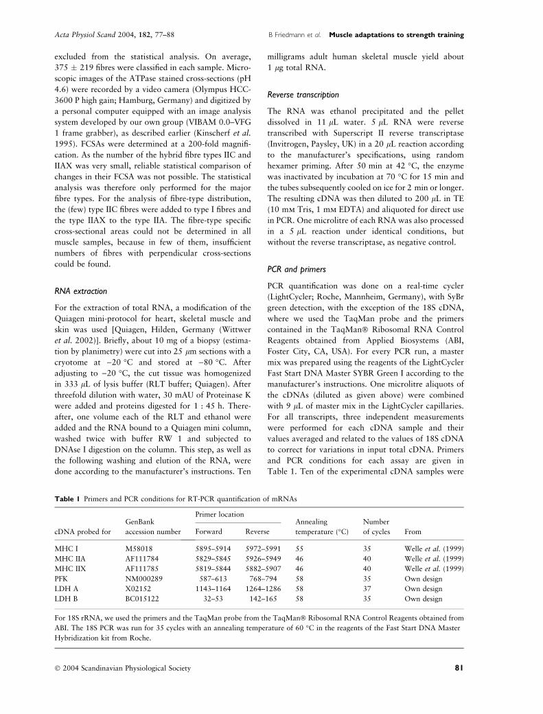
RNA extraction
For the extraction of total RNA, a modification of the
Quiagen mini-protocol for heart, skeletal muscle and
skin was used [Quiagen, Hilden, Germany (Wittwer
et al. 2002)]. Briefly, about 10 mg of a biopsy (estima-
tion by planimetry) were cut into 25 lm sections with a
cryotome at )20 �C and stored at )80 �C. After
adjusting to )20 �C, the cut tissue was homogenized
in 333 lL of lysis buffer (RLT buffer; Quiagen). After
threefold dilution with water, 30 mAU of Proteinase K
were added and proteins digested for 1 : 45 h. There-
after, one volume each of the RLT and ethanol were
added and the RNA bound to a Quiagen mini column,
washed twice with buffer RW 1 and subjected to
DNAse I digestion on the column. This step, as well as
the following washing and elution of the RNA, were
done according to the manufacturer’s instructions. Ten
milligrams adult human skeletal muscle yield about
1 lg total RNA.
Reverse transcription
The RNA was ethanol precipitated and the pellet
dissolved in 11 lL water. 5 lL RNA were reverse
transcribed with Superscript II reverse transcriptase
(Invitrogen, Paysley, UK) in a 20 lL reaction according
to the manufacturer’s specifications, using random
hexamer priming. After 50 min at 42 �C, the enzyme
was inactivated by incubation at 70 �C for 15 min and
the tubes subsequently cooled on ice for 2 min or longer.
The resulting cDNA was then diluted to 200 lL in TE
(10 mm Tris, 1 mm EDTA) and aliquoted for direct use
in PCR. One microlitre of each RNA was also processed
in a 5 lL reaction under identical conditions, but
without the reverse transcriptase, as negative control.
PCR and primers
PCR quantification was done on a real-time cycler
(LightCycler; Roche, Mannheim, Germany), with SyBr
green detection, with the exception of the 18S cDNA,
where we used the TaqMan probe and the primers
contained in the TaqMan� Ribosomal RNA Control
Reagents obtained from Applied Biosystems (ABI,
Foster City, CA, USA). For every PCR run, a master
mix was prepared using the reagents of the LightCycler
Fast Start DNA Master SYBR Green I according to the
manufacturer’s instructions. One microlitre aliquots of
the cDNAs (diluted as given above) were combined
with 9 lL of master mix in the LightCycler capillaries.
For all transcripts, three independent measurements
were performed for each cDNA sample and their
values averaged and related to the values of 18S cDNA
to correct for variations in input total cDNA. Primers
and PCR conditions for each assay are given in
Table 1. Ten of the experimental cDNA samples were
Table 1 Primers and PCR conditions for RT-PCR quantification of mRNAs
cDNA probed for
GenBank
accession number
Primer locationAnnealing
temperature (�C)
Number
of cycles FromForward Reverse
MHC I M58018 5895–5914 5972–5991 55 35 Welle et al. (1999)
MHC IIA AF111784 5829–5845 5926–5949 46 40 Welle et al. (1999)
MHC IIX AF111785 5819–5844 5882–5907 46 40 Welle et al. (1999)
PFK NM000289 587–613 768–794 58 35 Own design
LDH A X02152 1143–1164 1264–1286 58 37 Own design
LDH B BC015122 32–53 142–165 58 35 Own design
For 18S rRNA, we used the primers and the TaqMan probe from the TaqMan� Ribosomal RNA Control Reagents obtained from
ABI. The 18S PCR was run for 35 cycles with an annealing temperature of 60 �C in the reagents of the Fast Start DNA Master
Hybridization kit from Roche.
� 2004 Scandinavian Physiological Society 81
Acta Physiol Scand 2004, 182, 77–88 B Friedmann et al. Æ Muscle adaptations to strength training

chosen as reference standards and were measured in
each run. Relative quantification was performed with
the help of the ‘fit point’ method using the preinstalled
software program of the LightCycler. For each run,
ratios relative to each of the reference standards were
determined based on the respective delta CTs and an
average efficiency (determined graphically, SD 2–3%).
These ratios were then averaged over all three runs and
related to the average content of 18S cDNA in each
sample. The obtained ‘mRNA values’ are therefore
relative values based on total RNA content.
Statistics
Statistical procedures were performed with the software
programs Sigmastat 2.0 and Sigmaplot 2001 for Win-
dows from Jandel Scientific (San Rafael, CA, USA).
Data are presented as mean values � SD for the data
obtained from tests of the right leg only, since the
muscle biopsies were obtained from the right m. vastus
lateralis. Statistical analyses were performed by utilizing
a 2 · 2 repeated measures anova [group (CON/ECC,
CON/ECC-OVERLOAD) · test (pre-training, post-
training)]. Significant between-test differences were
determined involving the post hoc Tukey test. Addi-
tionally, differences between values obtained pre- and
post-training for each group were analysed by using
Student’s paired t-test. Correlations between selected
parameters were computed with the Pearson product–
moment. The level of significance was set at P £ 0.05.
Results
Strength endurance capacity, maximal strength
and muscle cross-sectional area
As all muscle biopsies were taken from the right vastus
lateralis muscle, only the results of the right leg are
presented. Neither significant group · test interactions
nor significant group effects were found for strength
endurance capacity, maximal strength or MCSA. A
significant test effect was observed for strength endur-
ance capacity (F ¼ 5.137, P ¼ 0.040, power ¼ 0.465).
Post hoc testing revealed a statistically significant (P ¼0.028) increase in strength endurance capacity (about
8%) after 4 weeks of CON/ECC training (Fig. 1). The
strength endurance capacity after CON/ECC-OVER-
LOAD training was not significantly different from the
value before training (P ¼ 0.546). For maximal
strength, there was a tendency towards a test effect
(F ¼ 3.180, P ¼ 0.096, power ¼ 0.267), (Fig. 1). The
increase (5% in average) after CON/ECC-OVER-
LOAD training reached statistical significance
(P £ 0.05). No significant difference was found after
CON/ECC training (P ¼ 0.482). A significant test
effect was seen for MCSA (average values for the 10-,
15- and 20-cm axial sites) which was increased by
1.7 � 2.6 cm2 for the collapsed groups (i.e. by an
average of 2%, Fig. 1; F ¼ 7.573, P ¼ 0.016, power ¼0.668). In the CON/ECC-OVERLOAD group, there
was a tendency (P ¼ 0.092) for MCSA to be increased
after training (average increase of 2.5 � 3.3 cm2).
Strength endurance capacity
Wor
k (J
)
0
3500
4000
4500
5000
5500
6000
beforeafter
CON/ECC CON/ECCOVERLOAD
Both groups
* *
Maximal strength
Tor
que
(Nm
)
0
160
180
200
220
240
260
280
CON/ECC CON/ECCOVERLOAD
Both groups
(*) (#)
Muscle cross sectional area
MC
SA
(cm
2 )
0
70
80
90
100
110
CON/ECC CON/ECCOVERLOAD
Both groups
*(§)
Figure 1 Strength endurance capacity, maximal strength, and
muscle cross-sectional area (MCSA): mean values � SD for the
data obtained from the right leg before and after 4 weeks of
concentric/eccentric (CON/ECC) and concentric/eccentric-
overload (CON/ECC-OVERLOAD) training are shown, as
well as mean values � SD for the aggregate of both groups. For
MCSA, average values for the 10-, 15- and 20-cm axial sites
are shown. *P < 0.05; (*): P £ 0.05, (#): P ¼ 0.096, (§):
P ¼ 0.092 compared with the values before training.
82 � 2004 Scandinavian Physiological Society
Muscle adaptations to strength training Æ B Friedmann et al. Acta Physiol Scand 2004, 182, 77–88

Fibre-type distribution and fibre cross-sectional areas
Neither significant group · test interactions, nor signi-
ficant group effects could be detected for the different
fibre types or for FCSA of the different fibre types or for
mean FCSA. A significant test effect was seen for the
percentage of type IIA fibres (F ¼ 5.668, P ¼ 0.039,
power ¼ 0.491). In the CON/ECC-OVERLOAD train-
ing group, the percentage of type IIA fibres was
increased in five subjects and unchanged in one subject.
The statistical treatment gave a tendency for an average
increase (8%, P ¼ 0.084). In the CON/ECC group, the
proportion of type IIA fibres was found to be increased
in four and decreased in two subjects after training
(P ¼ 0.273) (Table 2). The FCSA values were not
significantly different between pre- and post-training,
or between groups (Table 3). In the CON/ECC-OVER-
LOAD group, FCSA could only be determined in the
biopsies of four subjects. Their type IIA fibre FCSAs
correlated significantly with maximal strength after the
training period (r ¼ 0.966, P ¼ 0.03).
mRNAs coding for myosin heavy chain isoforms I, IIa, IIx
The relative contents of all the mRNAs determined
showed considerable inter-individual variation. Sub-
stantial variability was also seen when the values
obtained from the biopsies before and after the training
period were compared. This is illustrated in Figures 2
and 3. Neither a significant group · test interaction, nor
significant group or test effects could be detected for
MHC I mRNA. For MHC IIa mRNA, a significant
group · test interaction was observed (F ¼ 8.322, P ¼0.016, power ¼ 0.689, Fig. 2). There were no signifi-
cant group or test effects. MHC IIa mRNA was
increased after CON/ECC-OVERLOAD training in
each subject, between 4 and 84%. The resulting average
increase (30%) was statistically significant (P ¼ 0.026).
In the CON/ECC group, MHC IIa mRNA decreased in
five subjects between 22 and 37%, and increased by
54% in one subject. The average value after the training
period was 25% lower, but this was not statistically
significant (P ¼ 0.186) (Fig. 2). The individual values
for MHC IIx mRNA were increased in five subjects
after CON/ECC-OVERLOAD training between 36 and
463%. In one subject, the value had decreased by 7%.
The resulting average increase (320%) approached
statistical significance (P ¼ 0.056). In the CON/ECC
group, the MHC IIx mRNA values had increased in
four subjects, between 32 and 634% and decreased in
the other two, between 78 and 98%. The after-training
average was 24% lower, but this was statistically not
significant (Fig. 2).
mRNAs coding for glycolytic enzymes (PFK, LDH A
and LDH B)
For PFK mRNA, neither a significant group · test
interaction, nor significant group or test effects could
be detected. For LDH A mRNA, a significant group ·
Table 2 Fibre-type distribution. The data
are shown as mean values � SD before
and after 4 weeks of concentric/eccentric
or concentric/eccentric-overload training
Fibre types (%) Test CON/ECC-OVERLOAD CON/ECC Both groups
Type I Before 56.3 � 12.0 50.1 � 12.4 53.4 � 11.9
After 48.3 � 15.5 50.6 � 9.4 49.4 � 12.3
Type IIC Before 0.2 � 0.3 0.1 � 0.2 0.1 � 0.2
After 0.8 � 1.2 0.4 � 2.9 0.4 � 0.9
Type IIA Before 30.4 � 8.8 36.6 � 6.5 33.5 � 8.0
After 38.2 � 10.5* 38.7 � 6.6 38.4 � 8.4**
Type IIAX Before 1.1 � 1.9 2.9 � 3.4 2.3 � 2.9
After 0.1 � 0.4 1.3 � 1.6 0.5 � 1.1
Type IIX Before 13.0 � 8.3 13.4 � 9.9 13.2 � 8.7
After 13.5 � 9.7 10.8 � 7.1 12.2 � 8.2
*P ¼ 0.084, **P ¼ 0.039 compared with values before training.
Table 3 Fibre cross-sectional areas
(FCSA). The data are shown as mean
values � SD before and after 4 weeks of
concentric/eccentric or concentric/eccen-
tric-overload training
FCSA (lm2) Test CON/ECC-OVERLOAD CON/ECC Both groups
Type I Before 4182 � 933 4298 � 1710 4252 � 1385
After 4820 � 637 5525 � 1428 5243 � 1184
Type IIA Before 5118 � 1541 5458 � 2556 5322 � 2096
After 6475 � 1718 6193 � 1598 6306 � 1557
Type IIX Before 4190 � 1364 4282 � 2218 4241 � 1778
After 4720 � 1125 4804 � 720 4772 � 812
� 2004 Scandinavian Physiological Society 83
Acta Physiol Scand 2004, 182, 77–88 B Friedmann et al. Æ Muscle adaptations to strength training

test interaction was obtained (F ¼ 10.224, P ¼ 0.010,
power ¼ 0.791). No group or test effects were found.
LDH A mRNA was significantly increased (P ¼ 0.01)
after 4 weeks of CON/ECC-OVERLOAD training, by
about 70%. Each subject of this group had increased
LDH A mRNA values, between 20 and 122%. In the
CON/ECC group, the individual LDH A mRNA values
were decreased in half the subjects, between 17 and
66%, and increased in the other half, between 6 and
58% (Fig. 3). For LDH B mRNA, neither a significant
group · test interaction nor significant group or test
effects were observed (Fig. 3). A statistically significant
MHC I mRNA
MH
CI/1
8s
0.4
0.6
0.8
1.0
1.2
1.4
1.6
1.8
2.0
2.2
MHC IIa mRNA
MH
CIIa
/18s
0.4
0.6
0.8
1.0
1.2
1.4
1.6
1.8
*
+
MHC IIx mRNA
MH
CIIx
/18s
0
2
4
6
8
10
(*)
before after
Figure 2 mRNAs coding for myosin heavy chain isoforms
(MHC) I, IIa, IIx: individual values and mean � SD before and
after 4 weeks of concentric/eccentric ( ) and concentric/
eccentric-overload (•) training are shown to illustrate the inter-
individual variability. *: P ¼ 0.026, (*): P ¼ 0.056 compared
with value before training; +: P ¼ 0.016 compared with CON/
ECC (change in mRNA level).
PFK
PF
K/1
8s
0.2
0.4
0.6
0.8
1.0
1.2
1.4
1.6
1.8
2.0
LDH A
LDH
A/1
8s
0.2
0.4
0.6
0.8
1.0
1.2
1.4
1.6
1.8
2.0 **++
LDH B
LDH
B/1
8s
0.0
0.5
1.0
1.5
2.0
2.5
before after
Figure 3 mRNAs coding for the glycolytic enzymes phos-
phofruktokinase (PFK), lactate dehydrogenase (LDH) A and B:
individual values and means � SD before and after 4 weeks of
concentric/eccentric ( ) and concentric/eccentric-overload (•)training are shown. **: P ¼ 0.01 compared with value before
training; ++: P ¼ 0.01 compared with concentric/eccentric
training group (change in mRNA level).
84 � 2004 Scandinavian Physiological Society
Muscle adaptations to strength training Æ B Friedmann et al. Acta Physiol Scand 2004, 182, 77–88

positive correlation was observed between the relative
individual changes in MHC IIx mRNA and LDH A
mRNA in the CON/ECC-OVERLOAD group (Fig. 4).
In the CON/ECC group, the changes of these two
mRNAs were not significantly correlated (r ¼ )0.405,
P ¼ 0.426).
Discussion
This study investigated structural and gene expression
changes in response to two modes of concentric/eccen-
tric strength training in human m. vastus lateralis: low
resistance–high repetition knee extension exercise with
equal absolute loads in the concentric and eccentric
phases (CON/ECC) and low resistance–high repetition
knee extension exercise with equal relative loads in the
concentric and eccentric phases (CON/ECC-OVER-
LOAD).
Functional tests and structural analyses performed
before and after a 4-week training period indicate that
the two modes give rise to different adaptations:
Strength endurance capacity increased significantly only
in the CON/ECC group (by about 8%), while a
significant increase in maximal strength (about 5%)
was found in the CON/ECC OVERLOAD group only
(Fig. 1). It cannot be ruled out that slightly different
results might have been observed if the strength data
had been gravity corrected. However, the functional
differences were paralleled by differences in the expres-
sion of marker mRNAs: MHC IIa and LDH A mRNA
were significantly increased in the biopsies after CON/
ECC-OVERLOAD training, but not after CON/ECC
training (Figs 2 and 3). For the mRNA of MHC IIx we
found a tendency towards an increase (P ¼ 0.056) in
the CON/ECC-OVERLOAD group, but not in the
CON/ECC group. In addition, the post- to pre-training
ratios of MHC IIx mRNA were highly correlated with
LDH A mRNA, but in the CON/ECC-OVERLOAD
group only (Fig. 4).
These data indicate a shift in gene expression towards
the RNA pattern of a more type II dominated muscle in
response to CON/ECC-OVERLOAD, but not to CON/
ECC training. Such a shift did not become manifest as a
significant increase in the proportions of IIA and IIX
fibre types measured by ATPase histochemistry, rather
as tendency towards increased type IIA percentage
(P ¼ 0.084). It is possible that this is indicative of a
transient state, with MHC isoform switches detectable
at the level of mRNAs but not yet at the level of protein
incorporated in the myofibrils. This would correspond
to previous reports that show changes in MHC isoform
mRNAs to precede changes in the histochemical fibre
type, because the myosin proteins are thought to have
much slower turnover rates than their mRNAs (Ander-
sen & Schiaffino 1997). It should also be pointed out
that ATPase histochemistry is a relatively crude
indicator of the MHC composition of a given fibre. In
fibres containing several isoforms, the minor ones are
often not detected (Klitgaard et al. 1990, Staron 1991,
Andersen et al. 1994). Thus this discrepancy could be
the product of the additional fast MHC proteins being
present but not detected by the histochemical stain
because they are not the major isoform in all the
adapting fibres. We cannot, however, exclude that this
discrepancy arose from differential modulation of the
translation efficiencies of the MHC mRNAs which has
been described by Welle et al. (1999). They found an
increase in myofibrillar synthesis, e.g. an increase in the
synthesis rate of the aggregate of numerous myofibrillar
proteins while relative mRNA levels of the MHC
isoforms remained unchanged after three sessions of
resistance training. At present, the relative importance
of transcriptional and translational mechanisms for
training-induced changes in the synthesis of muscle
proteins still remains to be elucidated. At least, to our
knowledge, there has been no study so far in which an
adaptive shift in the levels of MHC isoform mRNAs
was shown to be accompanied by an opposing trans-
lational modulation.
The increase in the mRNAs for the type II MHC
isoforms as well as the LDH A isoform differs from the
adaptations described in most strength training studies,
which – considering mostly the levels of accumulated
proteins – almost all point towards transformation of
(more glycolytic) type IIX to (less glycolytic) IIA fibres
(Colliander & Tesch 1990, Adams et al. 1993, Staron
et al. 1994, Carroll et al. 1998, Andersen & Aagaard
2000, Hortobagyi et al. 2000, Williamson et al. 2001,
Willoughby & Rosene 2001). In the present study,
the lack of even slight a trend towards a decrease
in the proportion of type IIX fibres in the
MHC IIx-mRNA (post-/pre-training ratio)
LDH
A-m
RN
A (
post
-/pr
e-tr
aini
ng r
atio
)
0 1 2 3 4 51.0
1.2
1.4
1.6
1.8
2.0
2.2
2.4
r = 0.971, P = 0.001
Figure 4 Correlation between the post- to pre-training ratios
of myosin heavy chain (MHC) IIx mRNA and lactate
dehydrogenase (LDH) A mRNA in the concentric/eccentric-
overload (CON/ECC-OVERLOAD) training group.
� 2004 Scandinavian Physiological Society 85
Acta Physiol Scand 2004, 182, 77–88 B Friedmann et al. Æ Muscle adaptations to strength training

CON/ECC-OVERLOAD group gives additional sup-
port to the suggestion that this particular training mode
induces unique adaptations.
There are few studies on strength training and MHC
mRNA levels in humans. They either showed no
significant changes in MHC mRNA levels (Welle et al.
1999, Hortobagyi et al. 2000), an increase in MHC I
and MHC IIa mRNA, but no change in MHC IIx
mRNA (Willoughby & Rosene 2001), or even an
overall shift towards slow MHC expression (Balagopal
et al. 2001) after high resistance–low repetition CON/
ECC strength training. The only regimen leading to an
increase in MHC IIx mRNA was high resistance–low
repetition training with creatine loading, which led to
relative increases in all MHC mRNA isoforms (Will-
oughby & Rosene 2001). No study so far has described
a pattern matching the one found in our study
(significant increase in MHC IIa mRNA with a tendency
towards an increased MHC IIx mRNA in the CON/
ECC-OVERLOAD group).
To our knowledge, mRNA levels of glycolytic
enzymes have not been described in connection with
strength training in human muscles. In addition, there
are few data on enzyme activities from muscle homo-
genates. Tesch et al. (1990) did not find significant
changes in the activities of glycolytic enzymes after
concentric or combined concentric/eccentric strength
training. Our LDH mRNA data, with the significant
increases in relative LDH A mRNA levels after CON/
ECC-OVERLOAD but not after CON/ECC training
also suggest that the CON/ECC-OVERLOAD regimen
leads to unique adaptations in the glycolytic pathway.
The mRNA estimates in the present study showed
remarkable inter-individual variability (Figs 2 and 3).
The figures overrepresent the variability to a small
extent, because the scatter introduced by the RT
reaction cannot be accounted for. The standard devi-
ation of RT is 10–20% in our laboratory, as derived
from cDNA array studies (Wittwer et al. 2002 and
other unpublished results). The high variability in our
mRNA data is in agreement with many studies on gene
expression in human muscle biopsies, which found such
variability, e.g. for MHC mRNAs (derived from
estimates of the standard deviations in Welle et al.
1999) or for PFK mRNA (Vestergaard et al. 1994).
Recent results from microarray studies confirm these
observations: human muscle biopsy samples show
remarkably variable patterns of gene expression, not
only just between individuals, but also within a large
biopsy (Bakay et al. 2002, Wittwer et al. 2004). Nev-
ertheless, consistent differences between biopsies can be
detected, provided they are large enough (Bakay et al.
2002).
In our CON/ECC training group, we found an
increase in strength endurance capacity (Fig. 1), but no
statistically significant changes in any of the mRNAs
determined. It is possible that there were systematic
changes in some of these mRNAs, but they were too
small to be detected in the scatter of the individual
results. However, it could also be that translational
modulations and/or adaptations in systems other than
those tested with the mRNA markers in this study were
primarily responsible for the improved test results, either
in the muscles themselves or in the nervous system.
The higher eccentric component of the CON/ECC-
OVERLOAD training is likely responsible for the
significant increase in maximal strength and the trend
towards enhanced MCSA. Our results correspond well
to the findings of Brandenburg & Docherty (2002) and
Hortobagyi et al. (2001) who reported greater increases
in maximal strength after comparable concentric/eccen-
tric-overload strength training than after concentric/
eccentric strength training. The higher intramuscular
pressure due to the higher tension during the eccentric
phase in the CON/ECC-OVERLOAD exercise probably
impedes blood flow to a greater extent than during
CON/ECC exercise, possibly leading to enhanced hyp-
oxia, which could induce different adaptations. Hyp-
oxia during endurance training has been shown to cause
specific adaptations in muscle gene expression, such as
increased mRNAs of vascular endothelial growth factor
(VEGF) or myoglobin (Vogt et al. 2001). While myo-
globin mRNA was not significantly changed after
4 weeks of low resistance–high repetition strength
training in neither group, we even found a decrease in
VEGF mRNA (P £ 0.05) in the CON/ECC-OVER-
LOAD group (data not shown). It is, therefore, not
likely that local hypoxia was a decisive factor. Another
possibility is that these adaptations towards a faster,
more glycolytic mRNA pattern were due to differential
recruitment of fibre types. The combination of concen-
tric with higher eccentric load in the CON/ECC-
OVERLOAD strength training could have led to
enhanced recruitment of IIA and IIX fibres, the
enhanced glycolytic metabolism of which is more suited
to deal with reduced blood flow. This is supported by
the data of Hortobagyi et al. (2001) who used training
modes similar to the ones of the present study and found
increased EMG activities in the group following the
concentric/eccentric-overload training regimen. It is
possible that the high total eccentric workload of the
CON/ECC-OVERLOAD training regimen stimulated
overall MHC II synthesis (due to recruitment of
additional units or other mechanisms) but the drop in
MHC IIx mRNA, which usually occurs with concentric
training, was prevented by the low proportion of
concentric training to the total workout.
CON/ECC-OVERLOAD strength training is becom-
ing an increasingly popular component in the prepara-
tion of athletes for sports involving explosive strength
86 � 2004 Scandinavian Physiological Society
Muscle adaptations to strength training Æ B Friedmann et al. Acta Physiol Scand 2004, 182, 77–88

and/or very high power output. The protocols used in
this study are very similar to the regimens used in
training practice in terms of loads and length of the
training period. The changes in our marker mRNAs,
which are compatible with a shift towards a faster,
more glycolytic muscle after CON/ECC-OVERLOAD
training are therefore in good agreement with the
enhanced athletic performance observed.
It is possible that the RNA results of the current study
were influenced by the muscle activity during the high
repetition–very low resistance conventional strength
training of the lead-in period, which would have set
an endurance type stimulus, albeit a low one. Previously
observed changes in mRNAs of energy metabolism
enzymes after low intensity endurance training were
small (Vogt et al. 2001), thus it is not likely that the
results of this study were decidedly influenced by the
lead-in phase. Nevertheless, the results of the present
study deserve to be checked by further research on the
effects of CON/ECC-OVERLOAD training, e.g. with
athletes familiar with strength training, who do not
need a lead-in phase, and also with additional strength
tests on conventional devices besides isokinetic testing
because isovelocity muscle actions are not natural to
most activities.
In summary, we found different functional and
cellular adaptations in the muscles of subjects training
concentrically and eccentrically with the same absolute
loads compared with subjects training with the same
relative loads (eccentric-overload). Significant increases
in the mRNAs coding for MHC IIa and LDH A and a
strong tendency towards an increase in MHC IIx
mRNA in the group following the concentric/eccen-
tric-overload training regimen indicate a shift towards a
more type II dominated mRNA pattern specific for this
type of training.
The authors thank Judith Schoenith for excellent assistance
with RT-PCR and Gunther Erb for assistance on the measure-
ment of muscle cross-sectional area in the MRI scans. We
would also like to thank the unknown reviewer of a previous
version of this manuscript for the thorough review and the
many helpful suggestions.
This investigation was supported by grants from the Deut-
sche Forschungsgemeinschaft (FR 1262/3-1) and from the
Bundesinstitut fur Sportwissenschaft (VF 0407/01/04/98).
References
Aagaard, P., Andersen, J.L, Dyhre-Poulsen, P. et al. 2001. A
mechanism for increased contractile strength of human
pennate muscle in response to strength training: changes in
muscle architecture. J Physiol 534, 613–623.
Adams, G.R, Hather, B.M, Baldwin, K.M & Dudley, G.A
1993. Skeletal muscle myosin heavy chain composition and
resistance training. J Appl Physiol 74, 911–915.
Andersen, J.L & Aagaard, P. 2000. Myosin heavy chain IIX
overshoot in human skeletal muscle. Muscle Nerve 23,
1095–1104.
Andersen, P. & Henriksson, J. 1977. Training induced changes
in the subgroups of human type II skeletal muscle fibres.
Acta Physiol Scand 99, 123–125.
Andersen, J.L & Schiaffino, S. 1997. Mismatch between
myosin heavy chain mRNA and protein distribution in hu-
man skeletal muscle fibres. Am J Physiol 272, C1881–
C1889.
Andersen, J.L, Klitgaard, H. & Saltin, B. 1994. Myosin heavy
chain isoforms in single fibres form m. vastus lateralis of
sprinters: influence of training. Acta Physiol Scand 151, 135–
142.
Bakay, M., Chen, Y.-W., Borup, R., Zhao, P., Nagaraju, K. &
Hofmann, E.P 2002. Sources of variability and effect of
experimental approach on expression profiling data inter-
pretation. BMC Bioinformatics 3, 4.
Balagopal, P., Schimke, J.C, Ades, P., Adey, D. & Nair, K.S
2001. Age effect on transcript levels and synthesis rate of
muscle MHC and response to resistance exercise. Am J
Physiol 280, E203–E208.
Bergstrom, J. 1975. Percutaneous needle biopsy of skeletal
muscle in physiological and clinical research. Scand J Clin
Lab Invest 35, 609–616.
Booth, F.W & Baldwin, K.M 1996. Muscle plasticity: energy
demand and supply process. In: L.B. Rowell & J.T. Shepherd
(eds) The Handbook of Physiology. Exercise: Regulation
and Integration of Multiple Systems, pp. 1075–1123.
American Physiological Society Sect. 12, Bethesda, MD,
USA.
Brandenburg, J.P & Docherty, D. 2002. The effects of accen-
tuated eccentric loading on strength, muscle hypertrophy,
and neural adaptations in trained individuals. J Strength
Cond Res 16, 25–32.
Brooke, M.H & Kaiser, K.K 1970. Muscle fibre types: how
many and what kind?. Arch Neurol 23, 369–379.
Cadefau, J.A, Casademont, J., Grau, J.M et al. 1990. Bio-
chemical and histochemical adaptation to sprint training in
young athletes. Acta Physiol Scand 140, 341–351.
Carroll, T.J, Abernethy, P.J, Logan, P.A, Barber, M. &
McEniery, M.T 1998. Resistance training frequency:
strength and myosin heavy chain responses to two
and three bouts per week. Eur J Appl Physiol 78, 270–
275.
Colliander, E.B & Tesch, P.A 1990. Effects of eccentric and
concentric muscle actions in resistance training. Acta Physiol
Scand 140, 31–39.
Costill, D.L, Coyle, E.F, Fink, W.F, Lesmes, G.R & Witzmann,
F.A 1979. Adaptations in skeletal muscle following strength
training. J Appl Physiol 46, 96–99.
Dudley, G.A, Tesch, P.A, Miller, B.J & Buchanan, P. 1991a.
Importance of eccentric actions in performance adaptations
to resistance training. Aviat Space Environ Med 62, 543–
550.
Dudley, G.A, Tesch, P.A, Harris, R.T, Golden, C.L &
Buchanan, P. 1991b. Influence of eccentric actions on the
metabolic cost of resistance exercise. Aviat Space Environ
Med 62, 678–682.
� 2004 Scandinavian Physiological Society 87
Acta Physiol Scand 2004, 182, 77–88 B Friedmann et al. Æ Muscle adaptations to strength training

Friedmann, B., Kinscherf, R., Borisch, S., Richter, G., Bartsch,
P. & Billeter, R. 2003. Effects of low resistance–high repe-
tition strength training in hypoxia on muscle structure and
gene expression. Pflugers Arch 446, 742–751.
Gullich, A. & Schmidtbleicher, D. 1999. Structure of motor
strength and training methods. Dtsch Z Sportmed 50, 223–
234.
Hather, B.M, Tesch, P.A, Buchanan, P. & Dudley, G.A 1991.
Influence of eccentric actions on skeletal muscle adaptations
to resistance training. Acta Physiol Scand 143, 177–185.
Horstmann, T., Mayer, F., Maschmann, J., Niess, A., Roecker,
K. & Dickhuth, H.-H. 2001. Metabolic reaction after con-
centric and eccentric endurance-exercise of the knee and
ankle. Med Sci Sports Exerc 33, 791–795.
Hortobagyi, T., Dempsey, L., Fraser, D. et al. 2000. Changes in
muscle strength, muscle fibre size and myofibrillar gene
expression after immobilization and retraining in humans.
J Physiol 524, 293–304.
Hortobagyi, T., Devita, P., Money, J. & Barrier, J. 2001.
Effects of standard and eccentric overload strength training
in young women. Med Sci Sports Exerc 33, 1206–1212.
Howald, H., Hoppeler, H., Claassen, H., Mathieu, O. &
Straub, R. 1985. Influence of endurance training on the
ultrastructural composition of the different muscle fibre
types in humans. Pflugers Arch 403, 369–376.
Jacobs, I., Esbjornsson, M., Sylven, C., Holm, I. & Jansson, E.
1987. Sprint training effects on muscle myoglobin, enzymes,
fibre types, and blood lactate. Med Sci Sports Exerc 19, 368–
374.
Jones, D.A & Rutherford, O.M 1987. Human muscle strength
training: the effects of three different regimes and the nature
of the resultant changes. J Physiol 391, 1–11.
Kinscherf, R., Kohler, C., Kreuter, C., Pill, J. & Metz, J. 1995.
Hypercholesterolemia increases manganese superoxide dis-
mutase immunoreactive macrophages in myocardium. His-
tochem Cell Biol 104, 295–300.
Klitgaard, H., Bergman, O., Betto, R. et al. 1990. Co-Existence
of myosin heavy chain I and IIA isoforms in human skeletal
muscle fibres with endurance training. Pflugers Arch 416,
470–472.
Komi, P.V 1986. Training of muscle strength and power:
interaction of neuromotoric, hypertrophic, and mechanical
factors. Int J Sports Med 7, S10–S15.
Komi, P.V & Vitasalo, J.T 1977. Changes in motor unit
activity and metabolism in human skeletal muscle during
and after repeated eccentric and concentric contractions.
Acta Physiol Scand 100, 246–254.
O’Hagan, F.T, Sale, D.G, MacDougall, J.D & Garner, S.H
1995. Comparative effectiveness of accommodating and
weight resistance training modes. Med Sci Sports Exerc 27,
1210–1219.
Pilegaard, H., Ordway, G.A, Saltin, B. & Neufer, P.D 2000.
Transcriptional regulation of gene expression in human
skeletal muscle during recovery from exercise. Am J Physiol
279, E806–E814.
Sale, D.G, MacDougall, J.D, Jacobs, I. & Garner, S. 1990.
Interaction between concurrent strength and endurance
training. J Appl Physiol 68, 260–270.
Staron, R.S. 1991. Correlation between myofibrillar ATPase
activity and myosin heavy chain composition in single
human muscle fibres. Histochemistry 96, 21–24.
Staron, R.S, Karapondo, D.L, Kraemer, W.J et al. 1994.
Skeletal muscle adaptations during early phase of heavy-
resistance training in men and women. J Appl Physiol 76,
1247–1255.
Tesch, P.A, Thorsson, A. & Colliander, E.B 1990. Effects of
eccentric and concentric resistance training on skeletal
muscle substrates, enzyme activities and capillary supply.
Acta Physiol Scand 140, 575–580.
Vestergaard, H., Andersen, P.H, Lund, S., Schmity, O., Junker,
S. & Pedersen, O. 1994. Pre- and posttranslational upregu-
lation of muscle-specific glycogen synthase in athletes. Am J
Physiol 266, E92–E101.
Vogt, M., Puntschart, A., Geiser, J., Zuleger, C., Billeter, R. &
Hoppeler, H. 2001. Molecular adaptations in human skele-
tal muscle to endurance training under simulated hypoxic
conditions. J Appl Physiol 91, 173–182.
Welle, S., Bhatt, K. & Thornton, C.A 1999. Stimulation of
myofibrillar synthesis by exercise is mediated by more effi-
cient translation of mRNA. J Appl Physiol 86, 1220–1225.
Williamson, D.L, Gallagher, P.M, Carroll, C.C, Raue, U. &
Trappe, S.W 2001. Reduction in hybrid single muscle fiber
proportions with resistance training in humans. J Appl
Physiol 91, 1955–1961.
Willoughby, D. & Rosene, J. 2001. Effects of oral creatine and
resistance training on myosin heavy chain expression. Med
Sci Sports Exerc 33, 1674–1681.
Wittwer, M., Fluck, M., Hoppeler, H., Muller, S., Desp-
lanches, D. & Billeter, R. 2002. Prolonged unloading of rat
soleus muscle causes distinct adaptations of the gene profile.
FASEB J 16, 884–886.
Wittwer, M., Billeter, R., Hoppeler, H. & Fluck, M. 2004.
Regulatory gene expression in skeletal muscle of highly en-
durance trained humans. Acta Physiol Scand 180, 217–227.
88 � 2004 Scandinavian Physiological Society
Muscle adaptations to strength training Æ B Friedmann et al. Acta Physiol Scand 2004, 182, 77–88
Related Documents