Muscle Disease: When is it not myositis? Justin Kwan, M.D. Associate Professor Department of Neurology Disclosure Statement • I have no conflicts of interest to report • I do not have financial or other relationships with any commercial interest • I will not be discussing off label use of pharmaceuticals or devices 1 2

Welcome message from author
This document is posted to help you gain knowledge. Please leave a comment to let me know what you think about it! Share it to your friends and learn new things together.
Transcript

Muscle Disease:When is it not myositis?
Justin Kwan, M.D.
Associate Professor
Department of Neurology
Disclosure Statement
• I have no conflicts of interest toreport
• I do not have financial or otherrelationships with any commercialinterest
• I will not be discussing off labeluse of pharmaceuticals or devices
1
2
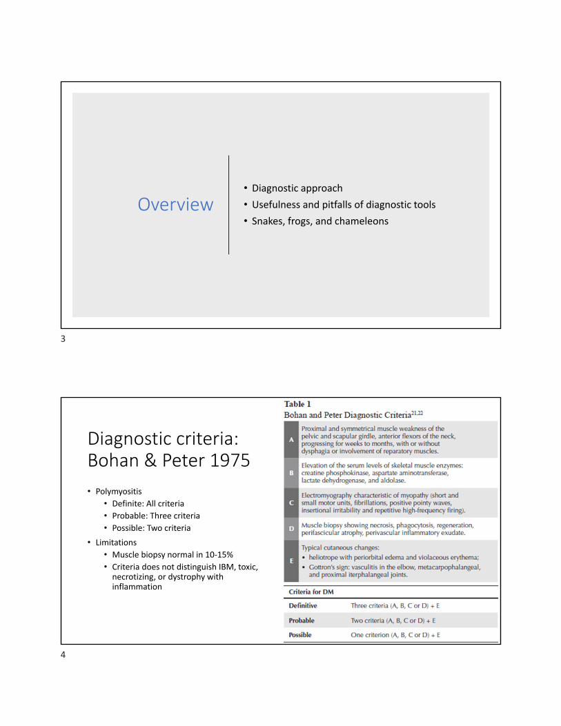
Overview• Diagnostic approach
• Usefulness and pitfalls of diagnostic tools
• Snakes, frogs, and chameleons
Diagnostic criteria: Bohan & Peter 1975
• Polymyositis
• Definite: All criteria
• Probable: Three criteria
• Possible: Two criteria
• Limitations
• Muscle biopsy normal in 10‐15%
• Criteria does not distinguish IBM, toxic,necrotizing, or dystrophy withinflammation
3
4

Proposed diagnostic criteria
• ENMC proposed major subtypes of IIM (2004):1. Inclusion body myositis
2. Polymyositis
3. Dermatomyositis
4. Non‐specific myositis (non‐specific perimysial/perivascular infiltrates)
5. Immune mediated necrotizing myopathy
6. Amyopathic DM
7. Possible DM sine dermatitis
ENMC proposed classification criteria, 2004 (exclude IBM)
• Inclusion: age > 18 (except DM or non‐specificmyositis), subacute, symmetric proximal > distal, neckflexor > neck extensor, rash typical of DM
• Exclusion: clinical features of IBM; ocular or isolatedbulbar weakness; neck extensor > flexor weakness,toxic, endocrine, amyloid myopathy; family historymuscular dystrophy or proximal motor neuropathies
Clinical criteria
Elevated CK
• Electromyography
• MRI: diffuse or patchy increased signal on STIR
• MSA in serum
Other laboratory criteria
5
6
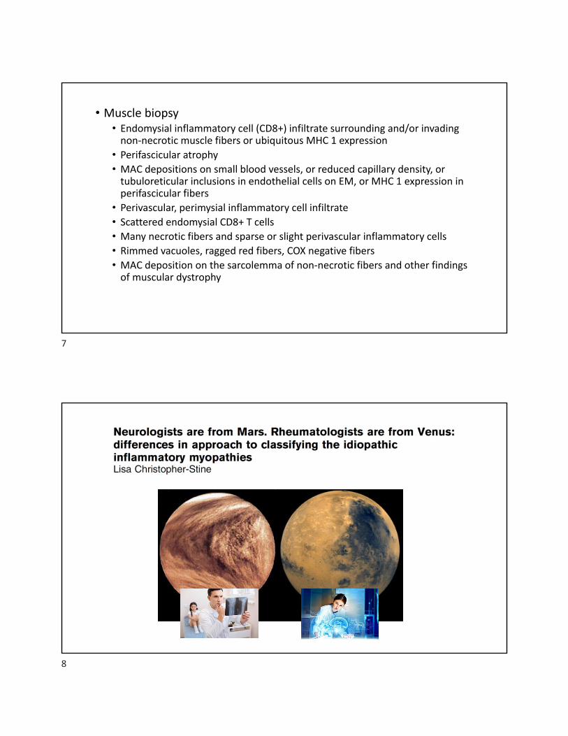
• Muscle biopsy• Endomysial inflammatory cell (CD8+) infiltrate surrounding and/or invadingnon‐necrotic muscle fibers or ubiquitous MHC 1 expression
• Perifascicular atrophy• MAC depositions on small blood vessels, or reduced capillary density, ortubuloreticular inclusions in endothelial cells on EM, or MHC 1 expression inperifascicular fibers
• Perivascular, perimysial inflammatory cell infiltrate
• Scattered endomysial CD8+ T cells
• Many necrotic fibers and sparse or slight perivascular inflammatory cells
• Rimmed vacuoles, ragged red fibers, COX negative fibers
• MAC deposition on the sarcolemma of non‐necrotic fibers and other findingsof muscular dystrophy
7
8
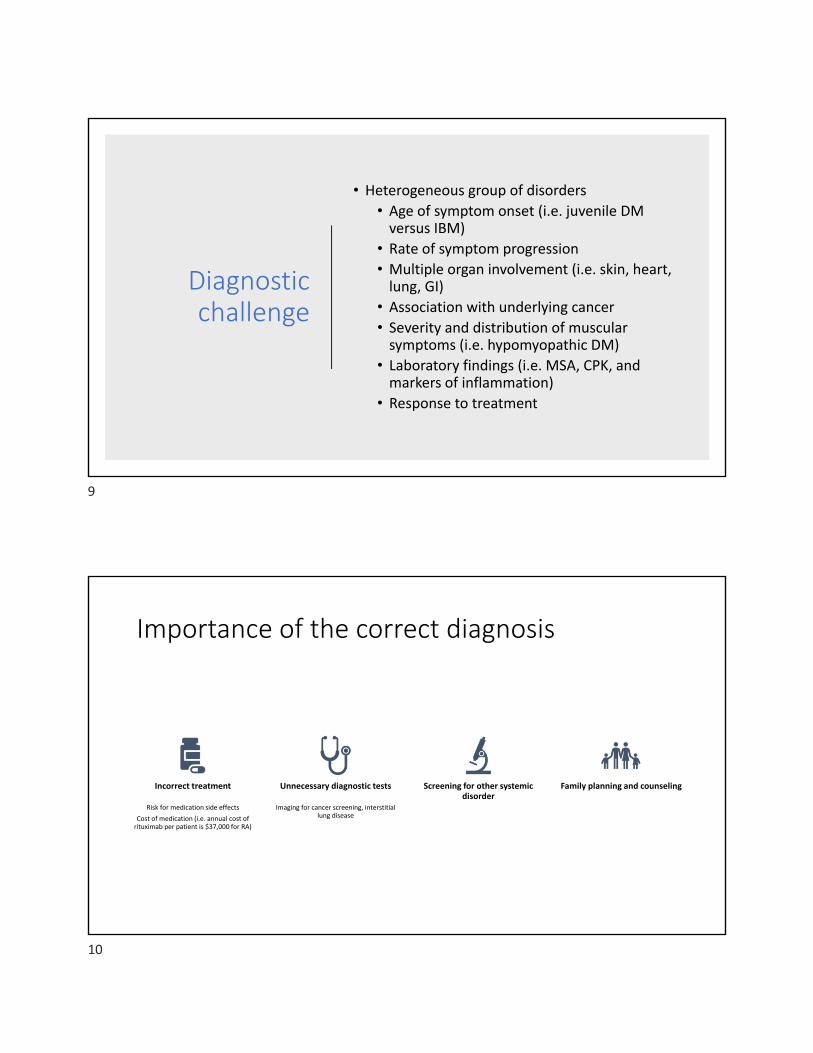
Diagnostic challenge
• Heterogeneous group of disorders• Age of symptom onset (i.e. juvenile DMversus IBM)
• Rate of symptom progression
• Multiple organ involvement (i.e. skin, heart,lung, GI)
• Association with underlying cancer• Severity and distribution of muscularsymptoms (i.e. hypomyopathic DM)
• Laboratory findings (i.e. MSA, CPK, andmarkers of inflammation)
• Response to treatment
Importance of the correct diagnosis
Incorrect treatment
Risk for medication side effects
Cost of medication (i.e. annual cost of rituximab per patient is $37,000 for RA)
Unnecessary diagnostic tests
Imaging for cancer screening, interstitial lung disease
Screening for other systemic disorder
Family planning and counseling
9
10

Approach to a patient with muscle weakness
• Clinical history and examination
• Laboratory studies
• Electrophysiological testing
• Imaging (ultrasound and MRI)
• Pathology
• How helpful are these studies in makingthe diagnosis of inflammatory myopathy?
Case 1
• 48‐year‐old black man with a history of osteoarthritis was noted tohave CPK of 1280 IU/L
• He took ibuprofen as needed for his joint pains
• Examination findings:• General examination was normal
• Neurological examination showed normal muscle bulk and give way weaknessat the hip flexors due to hip pain
• Sensory examination and reflexes were normal
• He was told that he had “inflammation of the muscle” and to see aneurologist
11
12
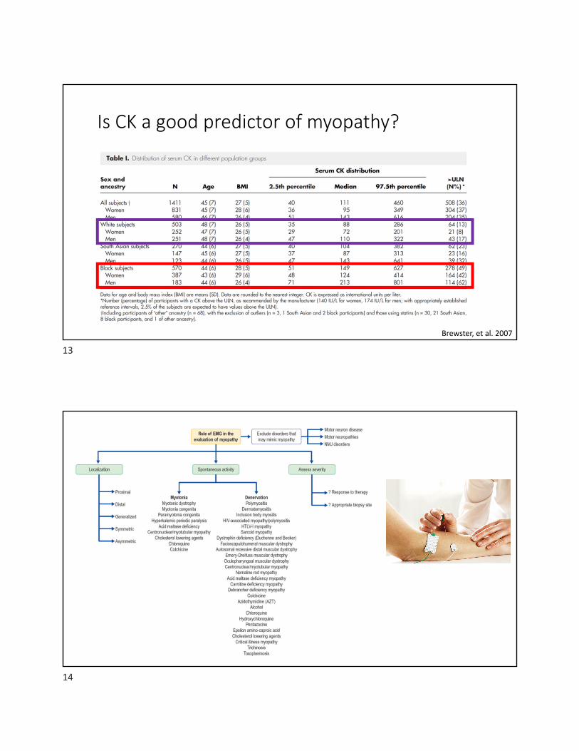
Is CK a good predictor of myopathy?
Brewster, et al. 2007
13
14
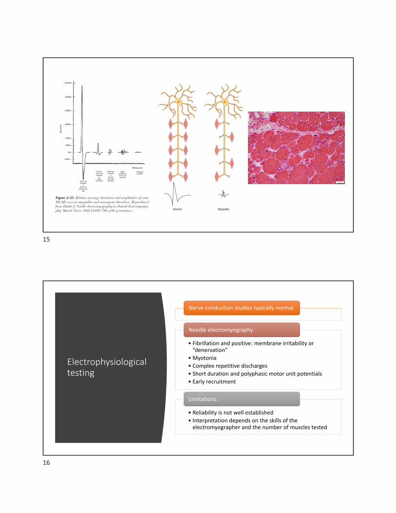
Electrophysiological testing
Nerve conduction studies typically normal
• Fibrillation and positive: membrane irritability or“denervation”
• Myotonia
• Complex repetitive discharges
• Short duration and polyphasic motor unit potentials
• Early recruitment
Needle electromyography
• Reliability is not well established
• Interpretation depends on the skills of theelectromyographer and the number of muscles tested
Limitations:
15
16
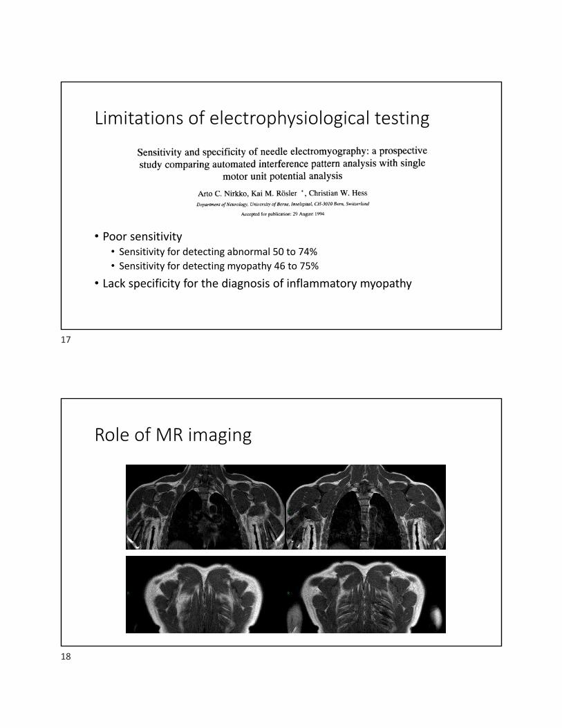
Limitations of electrophysiological testing
• Poor sensitivity• Sensitivity for detecting abnormal 50 to 74%
• Sensitivity for detecting myopathy 46 to 75%
• Lack specificity for the diagnosis of inflammatory myopathy
Role of MR imaging
17
18
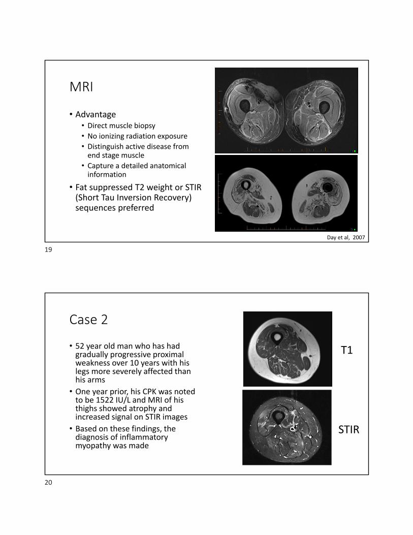
MRI
• Advantage• Direct muscle biopsy
• No ionizing radiation exposure• Distinguish active disease fromend stage muscle
• Capture a detailed anatomicalinformation
• Fat suppressed T2 weight or STIR(Short Tau Inversion Recovery)sequences preferred
Day et al, 2007
Case 2
• 52 year old man who has hadgradually progressive proximalweakness over 10 years with hislegs more severely affected thanhis arms
• One year prior, his CPK was notedto be 1522 IU/L and MRI of histhighs showed atrophy andincreased signal on STIR images
• Based on these findings, thediagnosis of inflammatorymyopathy was made
T1
STIR
19
20
MRI limitations
• Muscle T2 hyperintensity is non‐specific: trauma, myonecrosis,infection, denervation, rhabdomyolysis, interstitial fluid overload andnon‐inflammatory myopathies all have similar appearance
• No objective quantification of disease
• MR imaging is not a feature of most diagnostic criteria
• Others: cost, availability, contraindications
Vlekkert et al, 2015
21
22
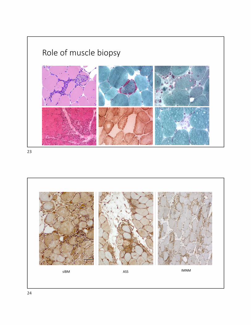
Role of muscle biopsy
sIBM ASS IMNM
23
24
Role of muscle biopsy
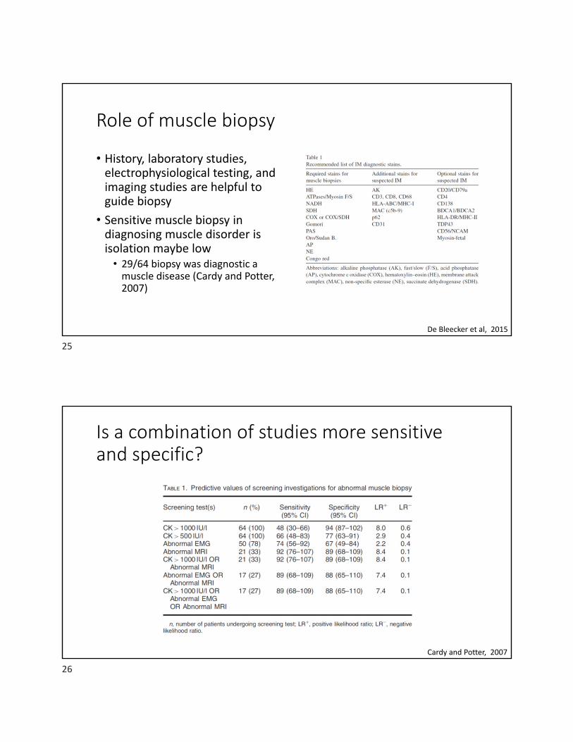
• History, laboratory studies,electrophysiological testing, andimaging studies are helpful toguide biopsy
• Sensitive muscle biopsy indiagnosing muscle disorder isisolation maybe low• 29/64 biopsy was diagnostic amuscle disease (Cardy and Potter,2007)
De Bleecker et al, 2015
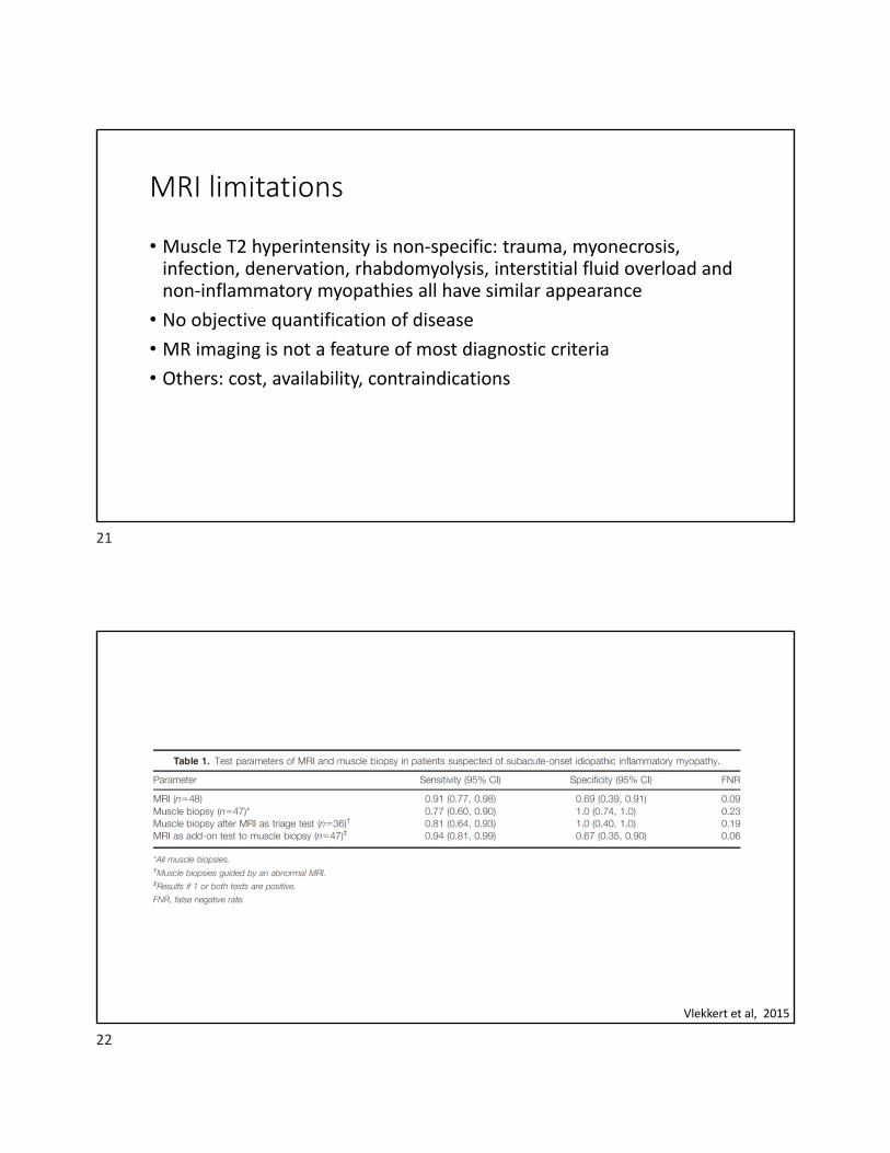
Is a combination of studies more sensitive and specific?
Cardy and Potter, 2007
25
26

http://ngm.nationalgeographic.com/2015/09/chameleons/edmonds‐text
Case 3
• A 19 year old young man was found to have an elevated CK of 8950IU/L after he complained of not being able to keep up with his peerswhile running
• He was treated with a course of oral prednisone with noimprovement in his symptoms and his CK decreased to high 6000’s
• Physical examination showed mild weakness in the hamstring musclesand asymmetric moderate weakness of the gastrocnemius muscle,with the left more severely affected than the right
27
28
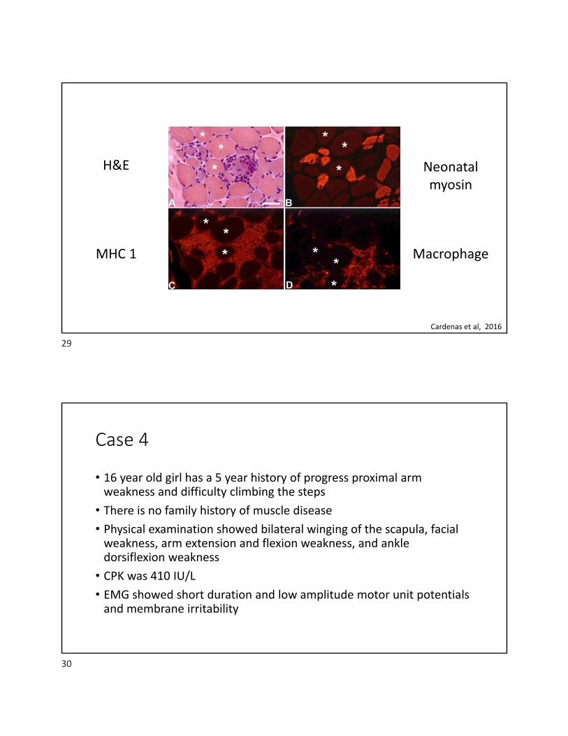
H&E Neonatal myosin
MHC 1 Macrophage
Cardenas et al, 2016
Case 4
• 16 year old girl has a 5 year history of progress proximal armweakness and difficulty climbing the steps
• There is no family history of muscle disease
• Physical examination showed bilateral winging of the scapula, facialweakness, arm extension and flexion weakness, and ankledorsiflexion weakness
• CPK was 410 IU/L
• EMG showed short duration and low amplitude motor unit potentialsand membrane irritability
29
30
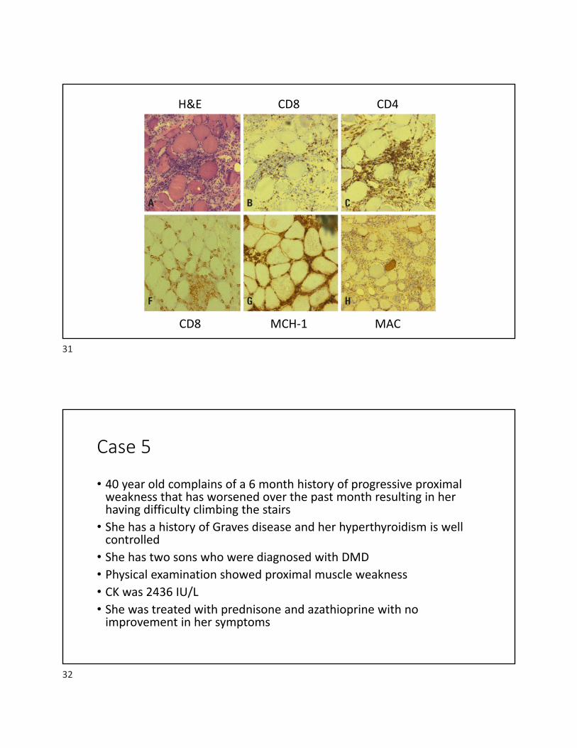
H&E CD8 CD4
CD8 MCH‐1 MAC
Case 5
• 40 year old complains of a 6 month history of progressive proximalweakness that has worsened over the past month resulting in herhaving difficulty climbing the stairs
• She has a history of Graves disease and her hyperthyroidism is wellcontrolled
• She has two sons who were diagnosed with DMD
• Physical examination showed proximal muscle weakness
• CK was 2436 IU/L• She was treated with prednisone and azathioprine with noimprovement in her symptoms
31
32

Yoon et al, 2011
Case 6
• 49 year old woman has a 10 year history of progressive proximalmuscle weakness and occasional myalgias and muscle cramps
• Physical examination showed proximal (3/5) more than distalweakness (4/5)
• Electrophysiological testing showed widespread myopathic changesand her CPK was 660 IU/L
33
34
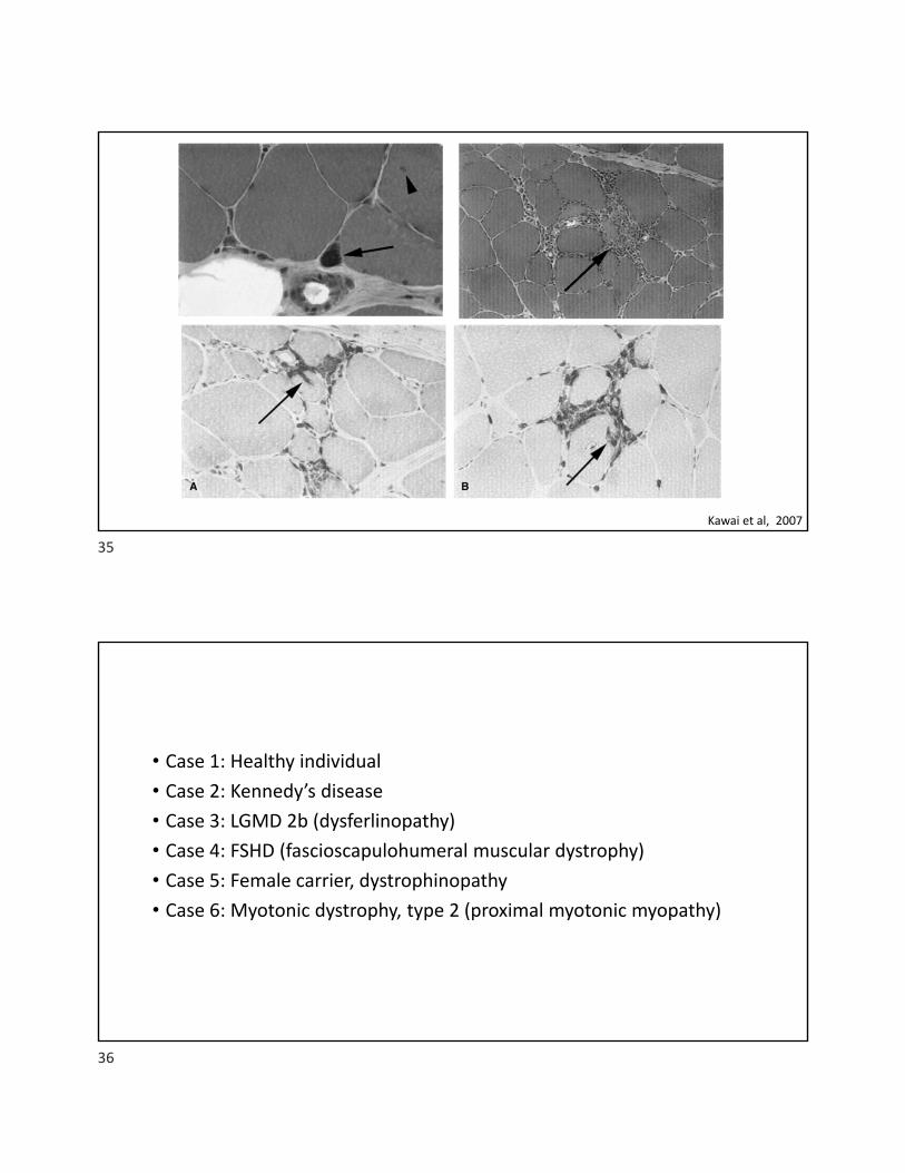
Kawai et al, 2007
• Case 1: Healthy individual
• Case 2: Kennedy’s disease
• Case 3: LGMD 2b (dysferlinopathy)
• Case 4: FSHD (fascioscapulohumeral muscular dystrophy)
• Case 5: Female carrier, dystrophinopathy
• Case 6: Myotonic dystrophy, type 2 (proximal myotonic myopathy)
35
36
Differential diagnosis to consider
• Inherited myopathy• Metabolic myopathy
• Muscular dystrophy
• Toxic myopathy
• Endocrine myopathy
• Can IIM mimic muscular dystrophy?• 17 of 51 (33%) anti‐HMGCR myopathy initially suspected of having limb girdlemuscular dystrophy
• All patient underwent genetics testing for LGMD (dystrophin, dysferlin,sarcoglycans, calpain‐3, caveolin‐3, anoctamin 5, fukutin‐related protein) andother inherited myopathies (acid maltase deficiency)
• All characterized by prolonged disease course, asymptomatic oroligosymptomatic hyperCKemia, exercise intolerance, or myalgia
37
38
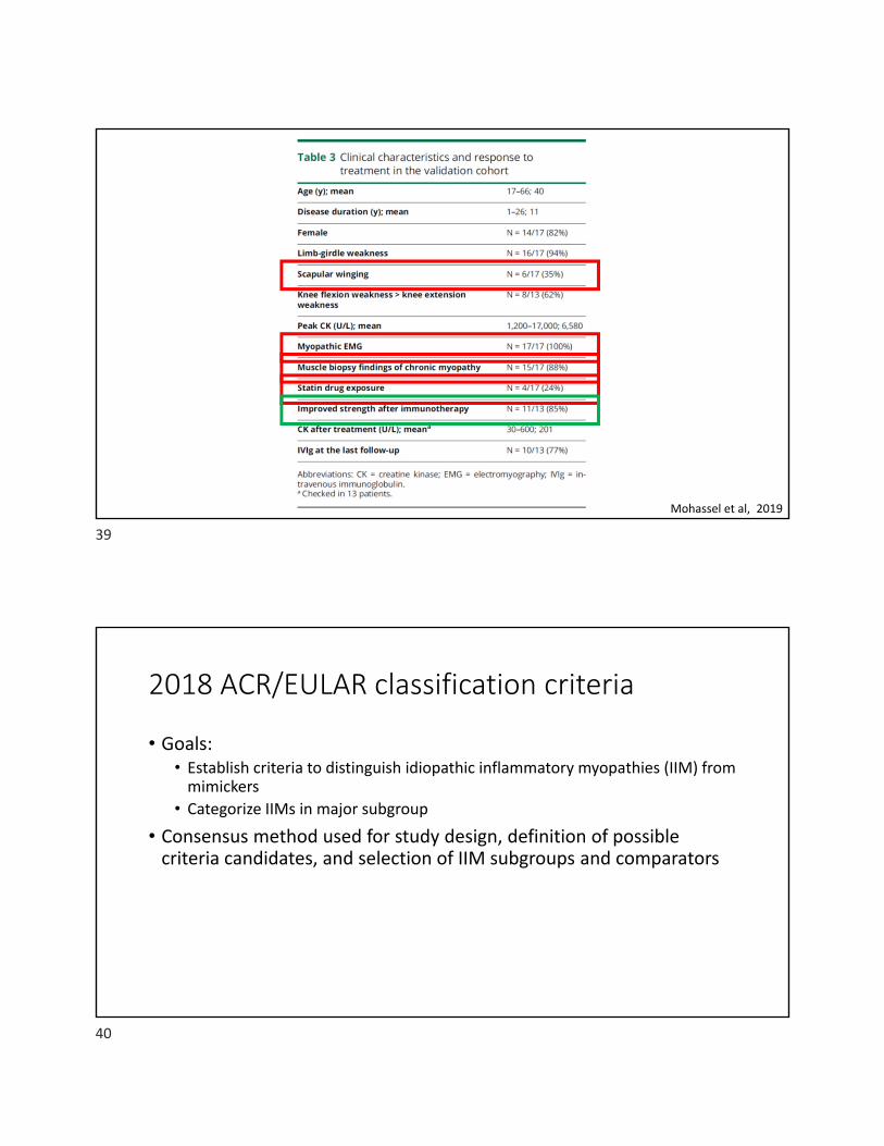
Mohassel et al, 2019
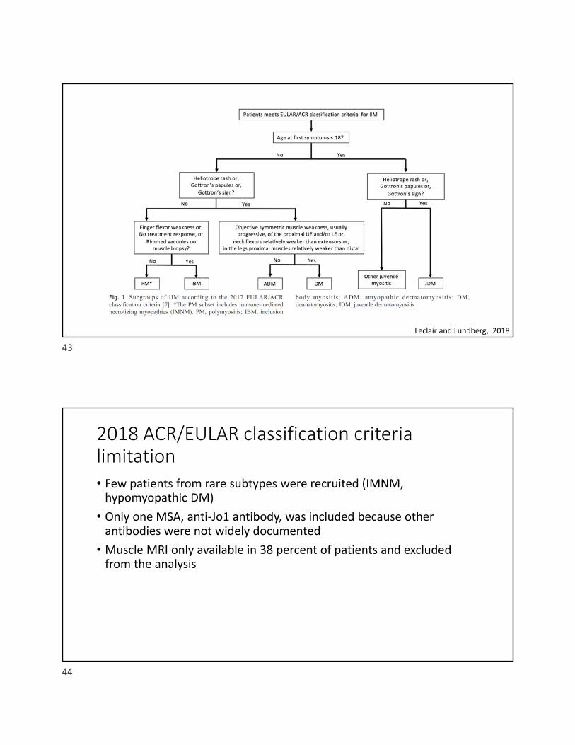
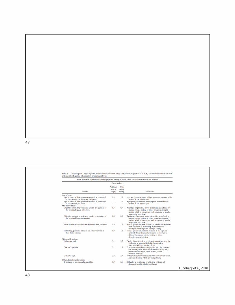
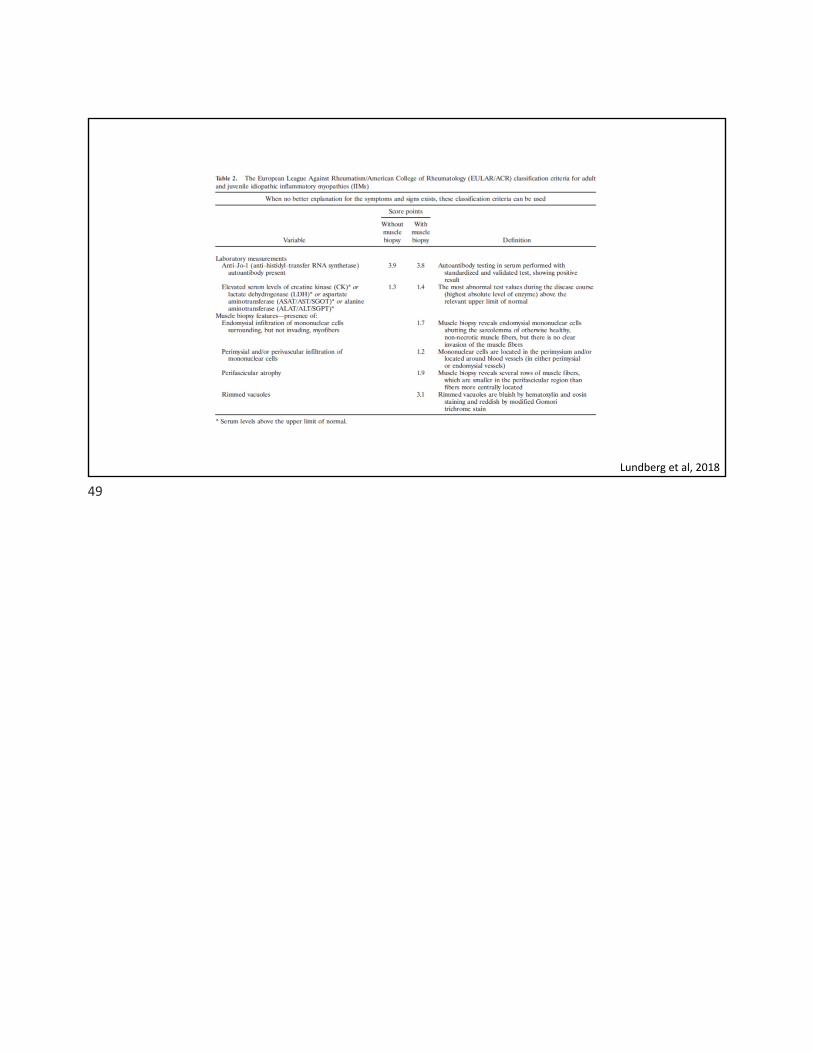
2018 ACR/EULAR classification criteria
• Goals:• Establish criteria to distinguish idiopathic inflammatory myopathies (IIM) frommimickers
• Categorize IIMs in major subgroup
• Consensus method used for study design, definition of possiblecriteria candidates, and selection of IIM subgroups and comparators
39
40
2018 ACR/EULAR classification criteria
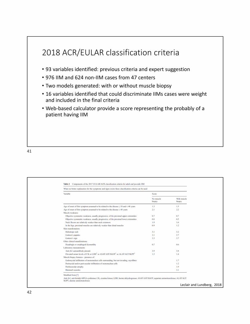
• 93 variables identified: previous criteria and expert suggestion
• 976 IIM and 624 non‐IIM cases from 47 centers
• Two models generated: with or without muscle biopsy
• 16 variables identified that could discriminate IIMs cases were weightand included in the final criteria
• Web‐based calculator provide a score representing the probably of apatient having IIM
Leclair and Lundberg, 2018
41
42
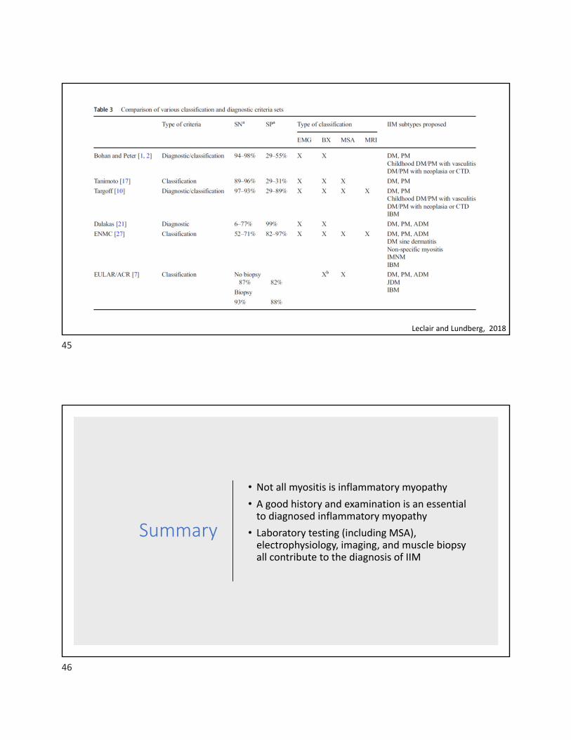
Leclair and Lundberg, 2018
2018 ACR/EULAR classification criteria limitation• Few patients from rare subtypes were recruited (IMNM,hypomyopathic DM)
• Only one MSA, anti‐Jo1 antibody, was included because otherantibodies were not widely documented
• Muscle MRI only available in 38 percent of patients and excludedfrom the analysis
43
44
Leclair and Lundberg, 2018
Summary
• Not all myositis is inflammatory myopathy
• A good history and examination is an essentialto diagnosed inflammatory myopathy
• Laboratory testing (including MSA),electrophysiology, imaging, and muscle biopsyall contribute to the diagnosis of IIM
45
46
Lundberg et al, 2018
47
48
Lundberg et al, 2018
49
Related Documents