JOURNAL OF VIROLOGY, Jan. 2010, p. 280–290 Vol. 84, No. 1 0022-538X/10/$12.00 doi:10.1128/JVI.01772-09 Copyright © 2010, American Society for Microbiology. All Rights Reserved. Murine Hepatitis Virus Nonstructural Protein 4 Regulates Virus-Induced Membrane Modifications and Replication Complex Function Mark J. Gadlage, 2,3 Jennifer S. Sparks, 2,3 Dia C. Beachboard, 2 Reagan G. Cox, 2 Joshua D. Doyle, 2,3 Christopher C. Stobart, 2,3 and Mark R. Denison 1,2,3,4 * Departments of Pediatrics, 1 Microbiology and Immunology, 2 The Elizabeth Lamb Center for Pediatric Research, 3 and the Monroe Carroll, Jr., Children’s Hospital, 4 Vanderbilt University Medical Center, Nashville, Tennessee Received 21 August 2009/Accepted 13 October 2009 Positive-strand RNA viruses induce modifications of cytoplasmic membranes to form replication complexes. For coronaviruses, replicase nonstructural protein 4 (nsp4) has been proposed to function in the formation and organization of replication complexes. Murine hepatitis virus (MHV) nsp4 is glycosylated at residues Asn176 (N176) and N237 during plasmid expression of nsp4 in cells. To test if MHV nsp4 residues N176 and N237 are glycosylated during virus replication and to determine the effects of N176 and N237 on nsp4 function and MHV replication, alanine substitutions of nsp4 N176, N237, or both were engineered into the MHV-A59 genome. The N176A, N237A, and N176A/N237A mutant viruses were viable, and N176 and N237 were glycosylated during infection of wild-type (wt) and mutant viruses. The nsp4 glycosylation mutants exhibited impaired virus growth and RNA synthesis, with the N237A and N176A/N237A mutant viruses demonstrating more profound defects in virus growth and RNA synthesis. Electron microscopic analysis of ultrastructure from infected cells demonstrated that the nsp4 mutants had aberrant morphology of virus-induced double-membrane vesicles (DMVs) compared to those infected with wt virus. The degree of altered DMV morphology directly correlated with the extent of impairment in viral RNA synthesis and virus growth of the nsp4 mutant viruses. The results indicate that nsp4 plays a critical role in the organization and stability of DMVs. The results also support the conclusion that the structure of DMVs is essential for efficient RNA synthesis and optimal replication of coronaviruses. Positive-strand RNA viruses rely on host intracellular mem- branes to form replication complexes, defined as sites of viral RNA synthesis (11, 34, 40–42). These virus-induced membrane modifications are crucial for creating an environment that sup- ports viral RNA synthesis, as well as protecting newly synthe- sized viral RNA. For many positive-strand RNA viruses, spe- cific replicase proteins, often containing multiple hydrophobic domains, have been implicated in targeting to and modifying host membranes, ultimately leading to the formation of repli- cation complexes. The coronavirus murine hepatitis virus (MHV) is an envel- oped, positive-strand RNA virus that contains a 31.4-kb ge- nome, consisting of seven open reading frames (ORFs). ORF1 encodes the replicase/transcriptase polyprotein, while ORFs 2 to 7 encode structural and accessory proteins. ORF1 comprises approximately two-thirds of the genome and is translated as either polyprotein 1a (pp1a) or, due to a 1 ribosomal frame- shift, pp1ab (3, 5, 6, 28, 34). pp1a and pp1ab are processed by three virus-encoded proteases to yield 16 nonstructural pro- teins (nsp1 to 16) (Fig. 1A) (1, 3, 13, 21, 32, 48). Analysis of nsp3, nsp4, and nsp6 amino acid sequences and in vitro bio- chemical studies have shown that these three nsp’s all have transmembrane domains that are likely important for virus- induced membrane modifications (2, 23, 28). MHV nsp4 is processed by papain-like protease 2 (PLP2) at its amino ter- minus, resulting in an nsp4-to-10 precursor, and after this ini- tial processing event, nsp5 (3Clpro) mediates processing at the carboxy terminus of nsp4 (15, 17, 21, 22, 24). The predicted molecular mass of nsp4 is 56 kDa, but it is detected as a 44-kDa protein by sodium dodecyl sulfate-polyacrylamide gel electro- phoresis (SDS-PAGE) (22, 31). All tested coronavirus nsp’s localize to replication complexes that are located on virus-induced double-membrane vesicles (DMVs), and nsp4 has been proposed to play roles in the formation, organization, and function of these virus replication complexes (15, 38). nsp4 has been shown to associate with membrane fractions of infected cells and is resistant to mem- brane extraction following Triton X-114 treatment, indicating that nsp4 is an integral membrane protein (15). Bioinformatics of the MHV nsp4 amino acid sequence predicted that nsp4 has four transmembrane domains (TM1 to 4). MHV nsp4 has also been shown to be required for rescue of infectious virus (45), as have TM1 to 3, but TM4 is dispensable for recovery of infectious virus in culture. Charge-to-alanine substitutions be- tween TM1 and TM2 of nsp4 result in viruses with phenotypes ranging from nonrecoverable to viruses that exhibit reduced virus growth, RNA synthesis, and protein processing (45). Analysis of nsp4 from multiple coronaviruses across all coro- navirus groups predicts N-linked glycosylation sites for all tested nsp4 sequences. The glycosylation sites, or sequons, Asn-X-Ser, Asn-X-Thr, and rarely Asn-X-Cys, are amino acid sequences * Corresponding author. Mailing address: Department of Pediatrics, Vanderbilt University Medical Center, D6217 MCN, 1161 21st Ave. S., Nashville, TN 37232-2581. Phone: (615) 343-9881. Fax: (615) 343-9723. E-mail: [email protected]. Published ahead of print on 21 October 2009. 280

Welcome message from author
This document is posted to help you gain knowledge. Please leave a comment to let me know what you think about it! Share it to your friends and learn new things together.
Transcript
JOURNAL OF VIROLOGY, Jan. 2010, p. 280–290 Vol. 84, No. 10022-538X/10/$12.00 doi:10.1128/JVI.01772-09Copyright © 2010, American Society for Microbiology. All Rights Reserved.
Murine Hepatitis Virus Nonstructural Protein 4 RegulatesVirus-Induced Membrane Modifications and Replication
Complex Function�
Mark J. Gadlage,2,3 Jennifer S. Sparks,2,3 Dia C. Beachboard,2 Reagan G. Cox,2 Joshua D. Doyle,2,3
Christopher C. Stobart,2,3 and Mark R. Denison1,2,3,4*Departments of Pediatrics,1 Microbiology and Immunology,2 The Elizabeth Lamb Center for Pediatric Research,3 and
the Monroe Carroll, Jr., Children’s Hospital,4 Vanderbilt University Medical Center, Nashville, Tennessee
Received 21 August 2009/Accepted 13 October 2009
Positive-strand RNA viruses induce modifications of cytoplasmic membranes to form replication complexes.For coronaviruses, replicase nonstructural protein 4 (nsp4) has been proposed to function in the formation andorganization of replication complexes. Murine hepatitis virus (MHV) nsp4 is glycosylated at residues Asn176(N176) and N237 during plasmid expression of nsp4 in cells. To test if MHV nsp4 residues N176 and N237 areglycosylated during virus replication and to determine the effects of N176 and N237 on nsp4 function and MHVreplication, alanine substitutions of nsp4 N176, N237, or both were engineered into the MHV-A59 genome. TheN176A, N237A, and N176A/N237A mutant viruses were viable, and N176 and N237 were glycosylated duringinfection of wild-type (wt) and mutant viruses. The nsp4 glycosylation mutants exhibited impaired virus growthand RNA synthesis, with the N237A and N176A/N237A mutant viruses demonstrating more profound defectsin virus growth and RNA synthesis. Electron microscopic analysis of ultrastructure from infected cellsdemonstrated that the nsp4 mutants had aberrant morphology of virus-induced double-membrane vesicles(DMVs) compared to those infected with wt virus. The degree of altered DMV morphology directly correlatedwith the extent of impairment in viral RNA synthesis and virus growth of the nsp4 mutant viruses. The resultsindicate that nsp4 plays a critical role in the organization and stability of DMVs. The results also support theconclusion that the structure of DMVs is essential for efficient RNA synthesis and optimal replication ofcoronaviruses.
Positive-strand RNA viruses rely on host intracellular mem-branes to form replication complexes, defined as sites of viralRNA synthesis (11, 34, 40–42). These virus-induced membranemodifications are crucial for creating an environment that sup-ports viral RNA synthesis, as well as protecting newly synthe-sized viral RNA. For many positive-strand RNA viruses, spe-cific replicase proteins, often containing multiple hydrophobicdomains, have been implicated in targeting to and modifyinghost membranes, ultimately leading to the formation of repli-cation complexes.
The coronavirus murine hepatitis virus (MHV) is an envel-oped, positive-strand RNA virus that contains a 31.4-kb ge-nome, consisting of seven open reading frames (ORFs). ORF1encodes the replicase/transcriptase polyprotein, while ORFs 2to 7 encode structural and accessory proteins. ORF1 comprisesapproximately two-thirds of the genome and is translated aseither polyprotein 1a (pp1a) or, due to a �1 ribosomal frame-shift, pp1ab (3, 5, 6, 28, 34). pp1a and pp1ab are processed bythree virus-encoded proteases to yield 16 nonstructural pro-teins (nsp1 to 16) (Fig. 1A) (1, 3, 13, 21, 32, 48). Analysis ofnsp3, nsp4, and nsp6 amino acid sequences and in vitro bio-chemical studies have shown that these three nsp’s all havetransmembrane domains that are likely important for virus-
induced membrane modifications (2, 23, 28). MHV nsp4 isprocessed by papain-like protease 2 (PLP2) at its amino ter-minus, resulting in an nsp4-to-10 precursor, and after this ini-tial processing event, nsp5 (3Clpro) mediates processing at thecarboxy terminus of nsp4 (15, 17, 21, 22, 24). The predictedmolecular mass of nsp4 is 56 kDa, but it is detected as a 44-kDaprotein by sodium dodecyl sulfate-polyacrylamide gel electro-phoresis (SDS-PAGE) (22, 31).
All tested coronavirus nsp’s localize to replication complexesthat are located on virus-induced double-membrane vesicles(DMVs), and nsp4 has been proposed to play roles in theformation, organization, and function of these virus replicationcomplexes (15, 38). nsp4 has been shown to associate withmembrane fractions of infected cells and is resistant to mem-brane extraction following Triton X-114 treatment, indicatingthat nsp4 is an integral membrane protein (15). Bioinformaticsof the MHV nsp4 amino acid sequence predicted that nsp4 hasfour transmembrane domains (TM1 to 4). MHV nsp4 has alsobeen shown to be required for rescue of infectious virus (45),as have TM1 to 3, but TM4 is dispensable for recovery ofinfectious virus in culture. Charge-to-alanine substitutions be-tween TM1 and TM2 of nsp4 result in viruses with phenotypesranging from nonrecoverable to viruses that exhibit reducedvirus growth, RNA synthesis, and protein processing (45).
Analysis of nsp4 from multiple coronaviruses across all coro-navirus groups predicts N-linked glycosylation sites for all testednsp4 sequences. The glycosylation sites, or sequons, Asn-X-Ser,Asn-X-Thr, and rarely Asn-X-Cys, are amino acid sequences
* Corresponding author. Mailing address: Department of Pediatrics,Vanderbilt University Medical Center, D6217 MCN, 1161 21st Ave. S.,Nashville, TN 37232-2581. Phone: (615) 343-9881. Fax: (615) 343-9723.E-mail: [email protected].
� Published ahead of print on 21 October 2009.
280
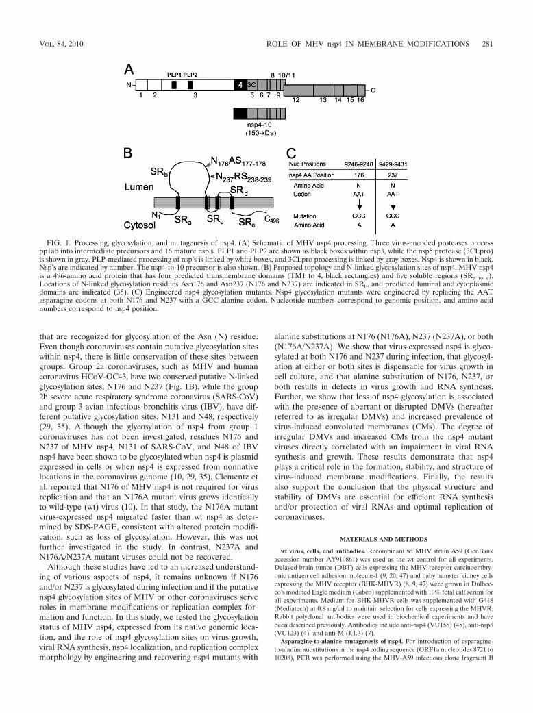
that are recognized for glycosylation of the Asn (N) residue.Even though coronaviruses contain putative glycosylation siteswithin nsp4, there is little conservation of these sites betweengroups. Group 2a coronaviruses, such as MHV and humancoronavirus HCoV-OC43, have two conserved putative N-linkedglycosylation sites, N176 and N237 (Fig. 1B), while the group2b severe acute respiratory syndrome coronavirus (SARS-CoV)and group 3 avian infectious bronchitis virus (IBV), have dif-ferent putative glycosylation sites, N131 and N48, respectively(29, 35). Although the glycosylation of nsp4 from group 1coronaviruses has not been investigated, residues N176 andN237 of MHV nsp4, N131 of SARS-CoV, and N48 of IBVnsp4 have been shown to be glycosylated when nsp4 is plasmidexpressed in cells or when nsp4 is expressed from nonnativelocations in the coronavirus genome (10, 29, 35). Clementz etal. reported that N176 of MHV nsp4 is not required for virusreplication and that an N176A mutant virus grows identicallyto wild-type (wt) virus (10). In that study, the N176A mutantvirus-expressed nsp4 migrated faster than wt nsp4 as deter-mined by SDS-PAGE, consistent with altered protein modifi-cation, such as loss of glycosylation. However, this was notfurther investigated in the study. In contrast, N237A andN176A/N237A mutant viruses could not be recovered.
Although these studies have led to an increased understand-ing of various aspects of nsp4, it remains unknown if N176and/or N237 is glycosylated during infection and if the putativensp4 glycosylation sites of MHV or other coronaviruses serveroles in membrane modifications or replication complex for-mation and function. In this study, we tested the glycosylationstatus of MHV nsp4, expressed from its native genomic loca-tion, and the role of nsp4 glycosylation sites on virus growth,viral RNA synthesis, nsp4 localization, and replication complexmorphology by engineering and recovering nsp4 mutants with
alanine substitutions at N176 (N176A), N237 (N237A), or both(N176A/N237A). We show that virus-expressed nsp4 is glyco-sylated at both N176 and N237 during infection, that glycosyl-ation at either or both sites is dispensable for virus growth incell culture, and that alanine substitution of N176, N237, orboth results in defects in virus growth and RNA synthesis.Further, we show that loss of nsp4 glycosylation is associatedwith the presence of aberrant or disrupted DMVs (hereafterreferred to as irregular DMVs) and increased prevalence ofvirus-induced convoluted membranes (CMs). The degree ofirregular DMVs and increased CMs from the nsp4 mutantviruses directly correlated with an impairment in viral RNAsynthesis and growth. These results demonstrate that nsp4plays a critical role in the formation, stability, and structure ofvirus-induced membrane modifications. Finally, the resultsalso support the conclusion that the physical structure andstability of DMVs are essential for efficient RNA synthesisand/or protection of viral RNAs and optimal replication ofcoronaviruses.
MATERIALS AND METHODS
wt virus, cells, and antibodies. Recombinant wt MHV strain A59 (GenBankaccession number AY910861) was used as the wt control for all experiments.Delayed brain tumor (DBT) cells expressing the MHV receptor carcinoembry-onic antigen cell adhesion molecule-1 (9, 20, 47) and baby hamster kidney cellsexpressing the MHV receptor (BHK-MHVR) (8, 9, 47) were grown in Dulbec-co’s modified Eagle medium (Gibco) supplemented with 10% fetal calf serum forall experiments. Medium for BHK-MHVR cells was supplemented with G418(Mediatech) at 0.8 mg/ml to maintain selection for cells expressing the MHVR.Rabbit polyclonal antibodies were used in biochemical experiments and havebeen described previously. Antibodies include anti-nsp4 (VU158) (45), anti-nsp8(VU123) (4), and anti-M (J.1.3) (7).
Asparagine-to-alanine mutagenesis of nsp4. For introduction of asparagine-to-alanine substitutions in the nsp4 coding sequence (ORF1a nucleotides 8721 to10208), PCR was performed using the MHV-A59 infectious clone fragment B
FIG. 1. Processing, glycosylation, and mutagenesis of nsp4. (A) Schematic of MHV nsp4 processing. Three virus-encoded proteases processpp1ab into intermediate precursors and 16 mature nsp’s. PLP1 and PLP2 are shown as black boxes within nsp3, while the nsp5 protease (3CLpro)is shown in gray. PLP-mediated processing of nsp’s is linked by white boxes, and 3CLpro processing is linked by gray boxes. Nsp4 is shown in black.Nsp’s are indicated by number. The nsp4-to-10 precursor is also shown. (B) Proposed topology and N-linked glycosylation sites of nsp4. MHV nsp4is a 496-amino acid protein that has four predicted transmembrane domains (TM1 to 4, black rectangles) and five soluble regions (SRa to e).Locations of N-linked glycosylation residues Asn176 and Asn237 (N176 and N237) are indicated in SRb, and predicted luminal and cytoplasmicdomains are indicated (35). (C) Engineered nsp4 glycosylation mutants. Nsp4 glycosylation mutants were engineered by replacing the AATasparagine codons at both N176 and N237 with a GCC alanine codon. Nucleotide numbers correspond to genomic position, and amino acidnumbers correspond to nsp4 position.
VOL. 84, 2010 ROLE OF MHV nsp4 IN MEMBRANE MODIFICATIONS 281
(pCR-XL-pSMART B) as a template. Fragment B of the MHV-A59 clonecontains MHV ORF1a nsp4 nucleotides 8721 to 9555 (47). Asparagine-to-ala-nine codon changes were introduced using the ExSite/QuikChange mutagenesiskit (Stratagene) with the primers listed in Table 1. Changes to the manufacturer’sprotocol included the use of Pfu Turbo and Pfu Ultra instead of the ExSite DNApolymerase blend. PCR was performed using the following parameters: initialdenaturation at 95°C once for 2 min, denaturation at 95°C for 1 min, annealingat various temperatures depending on the primers for 1 min, extension at 72°Cfor 7 min, and repeating of the denaturing, annealing, and extension steps for atotal of 40 cycles. Products were ligated and sequenced across the MHV genome-containing region of fragment B to ensure that PCR amplification did not introduceany unintended mutations. For introduction of both N176A and N237A, restrictionendonuclease EcoN I was used to digest both single nsp4 glycosylation mutantplasmids, and ligation was used to introduce both mutations into the same plasmid.
Generation of MHV nsp4 glycosylation mutant viruses. Viruses containing theengineered mutations within nsp4 were produced using the infectious cDNAassembly strategy for MHV-A59 that has previously been described by Yountet al. (47) and modified by Denison et al. (12) and Sparks et al. (45). Briefly,plasmids containing the seven cDNA cassettes that make up the MHV genomewere digested using the appropriate restriction enzymes. The correct restrictionfragments were gel purified and ligated together overnight at 16°C. The ligatedDNA was purified, in vitro transcribed, and electroporated with N gene tran-scripts into BHK-MHVR cells. The electroporated cells were then laid over alayer of 2.5 � 106 uninfected DBT cells in a 75-cm2 flask and incubated at 37°C.Virus viability was determined by cytopathic effect, in this case syncytium for-mation, in the electroporated cell culture. Progeny virus in the cell culture mediumof electroporated cells (passage 0 [P0]) was passaged onto uninfected DBT cells(P1), and the virus released from cells in the culture medium was designated P1stock, the titer was determined, and it was used for all experiments.
RT-PCR and sequencing of recovered viruses. Total intracellular RNA washarvested from P1-infected cells using TRIzol (Invitrogen) according to themanufacturer’s protocol. Extracted RNA was used as a template for reversetranscription (RT)-PCR. RT was performed using Superscript III reverse tran-scriptase (Invitrogen) and random hexamers (Roche). Primers complementaryto genome nucleotides 8486 to 8502 (sense) and 10361 to 10345 (antisense) werethen used to amplify the nsp4 coding region by PCR. These PCR products weresequenced to confirm the retention of the engineered mutations and the absenceof additional mutations in the nsp4 coding sequence.
Protein immunoprecipitations. For radiolabeling of proteins and immunopre-cipitations, cells were grown on 60-mm dishes and infected at a multiplicity ofinfection (MOI) of 10 PFU/cell with wt or nsp4 glycosylation mutant viruses andincubated at 37°C. At 4 h postinfection (p.i.), medium was aspirated and replacedwith medium lacking methionine and cysteine and supplemented with actinomy-cin D (Act D; Sigma) at a final concentration of 20 �g/ml. At 5 h p.i., cells wereradiolabeled with [35S]methionine-cysteine ([35S]Met-Cys) at a concentration of0.08 mCi/ml. When cells reached �90% involvement in syncytia, radiolabeledcells were washed once in phosphate-buffered saline (PBS), and then lysed in 1ml of lysis buffer lacking SDS (1% NP-40, 0.5% sodium deoxycholate, 150 mMNaCl, and 50 mM Tris, pH 8.0). Lysates were then centrifuged at 6,000 � g for3 min to remove cellular debris and nuclei, and the supernatant was collected.Immunoprecipitations were performed in a final volume of 1 ml, using proteinA-Sepharose beads (Sigma), 50 �l of radiolabeled lysate, 1:200 (anti-nsp4) or1:500 (anti-nsp8) dilutions of polyclonal antisera, and proteinase inhibitor(Roche) in lysis buffer. Immunoprecipitations were then performed as previouslydescribed (45). For endoglycosidase H (Endo H) treatment, supernatant wastransferred to a new tube after heating at 70°C for 10 min. Endo H (Sigma) wasadded to the supernatant according to the manufacturer’s protocol, and themixture was incubated at 37°C for 3 h. Proteins were resolved by SDS-PAGE in4 to 12% polyacrylamide gradient Bis-Tris gels (NuPage; Invitrogen) and ana-lyzed by fluorography. 14C-labeled high-molecular-weight markers (NEB) and afull-range rainbow marker were used as molecular weight standards.
Viral growth assays. For viral growth determination (12), DBT cells wereinfected with wt or nsp4 glycosylation mutant viruses at the MOIs indicated.Following a 45-min absorption period at 37°C with periodic swirling, medium wasaspirated, and the cells were washed three times in PBS. Prewarmed 37°Cmedium was then added back to the cells, and the cells were incubated at 37°C.Aliquots of medium were taken from 1 to 30 h p.i., and virus titers weredetermined by plaque assay as previously described (25).
Metabolic labeling of viral RNA. DBT cells were either mock infected orinfected at an MOI of 5 PFU/cell with wt or nsp4 glycosylation mutant viruses insix-well plates. Following a 45-min absorption at 37°C, medium containing viruswas removed, and cells were washed twice in PBS. Cells were then incubated ingrowth medium at 37°C until 30 min prior to labeling, when medium was re-placed with fresh medium containing 20 �g/ml Act D. After this 30-min treat-ment, [3H]uridine was added to a final concentration of 40 �Ci/ml, and cells wereincubated at 37°C for 2-h intervals from 3 to 15 h p.i. At the end of each labelingperiod, cells were lysed in lysis buffer (described above), and nuclei were re-moved by centrifugation at 14,000 � g for 3 min. RNA in 10% of each lysate wasprecipitated with chilled 5% trichloroacetic acid (TCA) onto glass microfiberfilters (Whatman), washed twice in fresh 5% TCA and twice in 95% ethanol, anddried using vacuum filtration. Radiolabel incorporation was quantitated by liquidscintillation counting.
Immunofluorescence assays. DBT cells grown on glass coverslips were in-fected with wt or nsp4 glycosylation mutant viruses at an MOI of 10 PFU/cell. At6 h p.i., medium was aspirated from cells, and cells were fixed in 100% methanolat �20°C. Cells were rehydrated in PBS for 10 min, blocked in PBS containing5% bovine serum albumin, and then aspirated. For indirect immunofluorescence,cells were incubated with primary antibody (anti-nsp4, 1:200; anti-M, 1:1,000) inwash solution (PBS containing 1% bovine serum albumin and 0.05% NP-40) for1 h at room temperature. Cells were washed in wash solution three times for 5min/wash. Cells were then incubated with secondary antibody (goat anti-rabbitAlexa 488, 1:1,000; goat anti-mouse Alexa 546, 1:1,000; Molecular Probes) for 30min at room temperature. Cells were washed again three times for 5 min/wash,subjected to a final wash in PBS, and rinsed with distilled water. For directimmunofluorescence, anti-nsp8 was purified using HiTrap rProtein A FF col-umns (GE Life Sciences) for fast protein liquid chromatography. Anti-nsp8 wasdirectly conjugated using the Alexa Fluor 546 protein labeling kit (Invitrogen)according to the manufacturer’s protocol. Cells were incubated with anti-nsp8 ata concentration of 1:500, following the same procedure as above. Coverslips weremounted with Aquapolymount (Polysciences) and visualized using a Zeiss Ax-iovert 200 microscope with a 40� oil immersion lens. Images were processed andmerged using Adobe Photoshop CS3.
TEM analysis. DBT cells were mock infected or infected with wt or nsp4glycosylation mutant viruses at an MOI of 5 PFU/cell in a 60-mm dish andincubated at 37°C. At 6 h p.i., medium was aspirated, and cells were washed oncewith PBS. The cells were then fixed in 2% glutaraldehyde for 10 min, scraped offthe dishes, and centrifuged at 0.5 � g for 3 min. The initial 2% glutaraldehydewas aspirated, fresh 2% glutaraldehyde was added to the fixed cells for 1 h andaspirated, and fresh glutaraldehyde was added to the fixed cells for overnightincubation at 4°C. Cells were washed three times in PBS, transferred to 1%osmium tetroxide in distilled water (diH2O) for 1 h, and washed three times indiH2O. Cells were stained en bloc in 1% aqueous uranyl acetate for 1 h andwashed three times in diH2O. Dehydration of cells was carried out graduallyusing a graded series of ethanol and increasing the times each remained insolution, starting with 30%, followed by 50%, 70%, 95%, and finally absoluteethanol. Propylene oxide was used as a transitional solvent to replace the dehy-dration solution. Cells were transferred to a 1:1 araldite-propylene oxide mixturefor 1 h and then placed in pure araldite in a vacuum oven for another hour tohelp pull resin through the tissue. Pure resin specimens were then transferredinto capsules containing fresh resin and finally placed into an oven overnight topolymerize. Ultra-thin serial sections (50 to 60 nm) from polymerized blockswere obtained using a Leica UCT Ultracut microtome (Leica Microsystems,
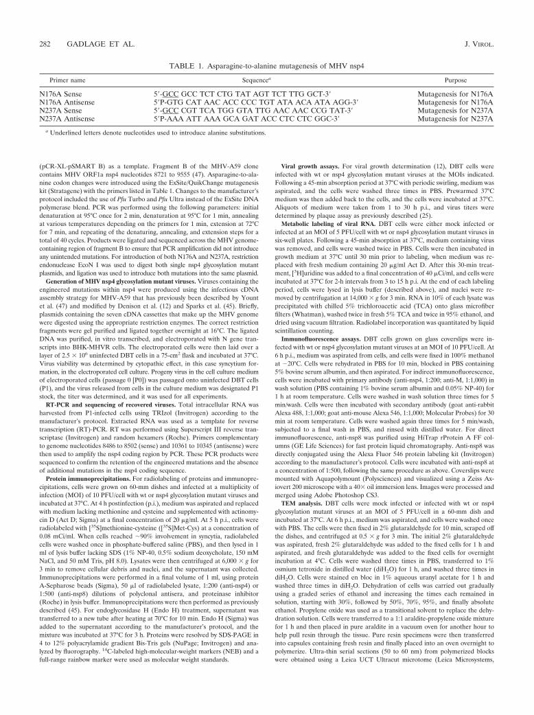
TABLE 1. Asparagine-to-alanine mutagenesis of MHV nsp4
Primer name Sequencea Purpose
N176A Sense 5�-GCC GCC TCT CTG TAT AGT TCT TTG GCT-3� Mutagenesis for N176AN176A Antisense 5�P-GTG CAT AAC ACC CCC TGT ATA ACA ATA AGG-3� Mutagenesis for N176AN237A Sense 5�-GCC CGT TCA TGG GTA TTG AAC AAC CCG TAT-3� Mutagenesis for N237AN237A Antisense 5�P-AAA ATT AAA GCA GAT ACC CTC CTC GGC-3� Mutagenesis for N237A
a Underlined letters denote nucleotides used to introduce alanine substitutions.
282 GADLAGE ET AL. J. VIROL.
Vienna, Austria), transferred to formvar-coated slot grids, and examined using aPhillips CM10 TEM (FEI Company, Hillsboro, OR) equipped with an Advan-tage Plus 2-megapixel digital charge-coupled-device system for CM10 transmis-sion electron microscopy (TEM) (Advanced Microscopy Techniques, Danvers, MA).
Statistical analyses. For statistical analyses, DMVs were characterized intotwo groups, either regular (defined by inner membranes in close approximationwith the outer membrane) or irregular DMVs (defined by moderate to severedisruption or separation of the inner membrane with the outer membrane).Chi-square analysis using contingency tables was performed by comparing thenumber of regularly formed DMVs to irregularly formed DMVs of wt and nsp4glycosylation mutant viruses. Chi-square analysis was also performed to comparethe presence of both CMs and DMVs to the presence of DMVs only. BecauseCMs were found only in the presence of DMVs, the presence of CMs and DMVswas compared to the presence of DMVs alone in a given TEM section. Diam-eters of DMVs were measured using ImageJ 1.40g (http://rsb.info.nih.gov/ij).Diameters were defined by measuring the widest diameter from the outsidemembrane of one side to the outside membrane of the opposite side of a singleDMV. To determine whether there was a statistical difference between thediameters of DMVs, analysis of variance (ANOVA) was used to compare wt andnsp4 glycosylation mutant viruses. Because a statistical difference was indicatedthrough ANOVA, Tukey tests were used to perform pair-wise comparisons of allviruses. P values were determined to indicate significance.
RESULTS
Recovery of nsp4 glycosylation mutant viruses. Group 2acoronaviruses contain conservation of putative glycosylationsites in nsp4 at N176 and N237 (Fig. 1B). To determine if nsp4is glycosylated at residues N176 and N237 in the context ofMHV infection and what roles nsp4 glycosylation may play inthe virus life cycle, viruses were engineered to contain aspar-agine-to-alanine substitutions at either N176, N237, or bothresidues N176 and N237 of nsp4 (Fig. 1C). Cells were electro-porated with genomic RNA for N176A, N237A, or N176A/N237A mutant viruses. All three mutant viruses induced cyto-pathic effect by 36 h postelectroporation, and 90 to 100% ofcells were involved in syncytia by 46 to 50 h postelectropora-tion, similar to wt virus. Viruses were passaged and sequencedacross the nsp4 coding sequence, confirming both the presenceof engineered mutations and lack of any other mutations innsp4. In contrast to previous reports, our results demonstratethat mutants with alanine substitution at N176, N237, or bothare viable, demonstrating that the N176 and N237 residues arenot required for replication in cell culture. To determine ifcompensating mutations occurred outside of the nsp4 sequenceduring recovery of the N237A and N176A/N237A mutant vi-ruses, the complete genome of the N176A/N237A mutant viruswas sequenced, and there were no additional mutations present inthe genome. These results demonstrate that the recovery of theN237A and N176A/N237A mutant viruses was not due to second-site compensating mutations and that the Asn residues are notrequired for virus viability.
nsp4 is glycosylated at both N176 and N237 during MHVinfection. Previous studies have demonstrated that treatmentof lysates with Endo H results in a mobility shift of nsp4expressed from plasmid in HeLa cells (10, 35) or from nsp4-enhanced green fluorescent protein expressed in recombinantvirus from an alternate location (in place of ORF2) (10, 35),consistent with glycosylation of nsp4 with mannose-rich oligo-saccharides in the endoplasmic reticulum (ER) and the lack ofnsp4 trafficking through Golgi. However, there has been nodemonstration of N-linked glycosylation of native nsp4 in wtvirus or identification of specific Asn residues subject to N-linked glycosylation. To test whether natively expressed MHV
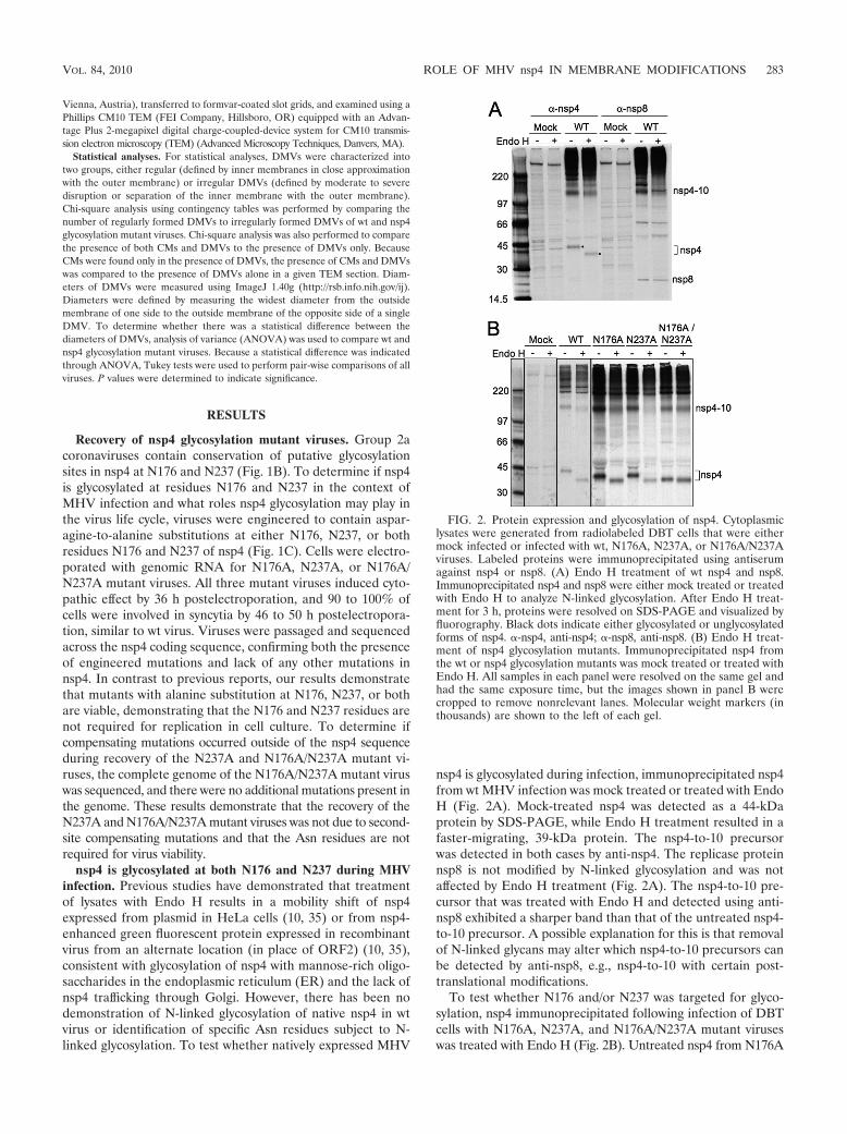
nsp4 is glycosylated during infection, immunoprecipitated nsp4from wt MHV infection was mock treated or treated with EndoH (Fig. 2A). Mock-treated nsp4 was detected as a 44-kDaprotein by SDS-PAGE, while Endo H treatment resulted in afaster-migrating, 39-kDa protein. The nsp4-to-10 precursorwas detected in both cases by anti-nsp4. The replicase proteinnsp8 is not modified by N-linked glycosylation and was notaffected by Endo H treatment (Fig. 2A). The nsp4-to-10 pre-cursor that was treated with Endo H and detected using anti-nsp8 exhibited a sharper band than that of the untreated nsp4-to-10 precursor. A possible explanation for this is that removalof N-linked glycans may alter which nsp4-to-10 precursors canbe detected by anti-nsp8, e.g., nsp4-to-10 with certain post-translational modifications.
To test whether N176 and/or N237 was targeted for glyco-sylation, nsp4 immunoprecipitated following infection of DBTcells with N176A, N237A, and N176A/N237A mutant viruseswas treated with Endo H (Fig. 2B). Untreated nsp4 from N176A
FIG. 2. Protein expression and glycosylation of nsp4. Cytoplasmiclysates were generated from radiolabeled DBT cells that were eithermock infected or infected with wt, N176A, N237A, or N176A/N237Aviruses. Labeled proteins were immunoprecipitated using antiserumagainst nsp4 or nsp8. (A) Endo H treatment of wt nsp4 and nsp8.Immunoprecipitated nsp4 and nsp8 were either mock treated or treatedwith Endo H to analyze N-linked glycosylation. After Endo H treat-ment for 3 h, proteins were resolved on SDS-PAGE and visualized byfluorography. Black dots indicate either glycosylated or unglycosylatedforms of nsp4. �-nsp4, anti-nsp4; �-nsp8, anti-nsp8. (B) Endo H treat-ment of nsp4 glycosylation mutants. Immunoprecipitated nsp4 fromthe wt or nsp4 glycosylation mutants was mock treated or treated withEndo H. All samples in each panel were resolved on the same gel andhad the same exposure time, but the images shown in panel B werecropped to remove nonrelevant lanes. Molecular weight markers (inthousands) are shown to the left of each gel.
VOL. 84, 2010 ROLE OF MHV nsp4 IN MEMBRANE MODIFICATIONS 283
and N237A mutants migrated identically and more rapidly thanuntreated wt nsp4 (42 kDa) but more slowly than wt nsp4treated with Endo H (39 kDa). When nsp4 from N176A andN237A mutant viruses was treated with Endo H, both proteinswere detected at 39 kDa, identical to Endo H-treated wt nsp4.nsp4 from the N176A/N237A mutant virus migrated to 39 kDa,whether untreated or treated with Endo H. The results indi-cate that nsp4 expressed from its native genomic location isspecifically glycosylated at residues N176 and N237 and alsodemonstrate that no other N-linked glycosylation occurs innsp4.
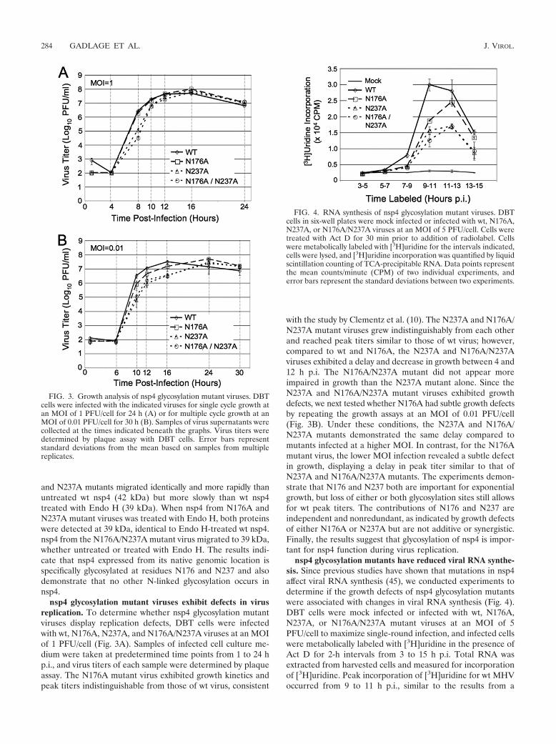
nsp4 glycosylation mutant viruses exhibit defects in virusreplication. To determine whether nsp4 glycosylation mutantviruses display replication defects, DBT cells were infectedwith wt, N176A, N237A, and N176A/N237A viruses at an MOIof 1 PFU/cell (Fig. 3A). Samples of infected cell culture me-dium were taken at predetermined time points from 1 to 24 hp.i., and virus titers of each sample were determined by plaqueassay. The N176A mutant virus exhibited growth kinetics andpeak titers indistinguishable from those of wt virus, consistent
with the study by Clementz et al. (10). The N237A and N176A/N237A mutant viruses grew indistinguishably from each otherand reached peak titers similar to those of wt virus; however,compared to wt and N176A, the N237A and N176A/N237Aviruses exhibited a delay and decrease in growth between 4 and12 h p.i. The N176A/N237A mutant did not appear moreimpaired in growth than the N237A mutant alone. Since theN237A and N176A/N237A mutant viruses exhibited growthdefects, we next tested whether N176A had subtle growth defectsby repeating the growth assays at an MOI of 0.01 PFU/cell(Fig. 3B). Under these conditions, the N237A and N176A/N237A mutants demonstrated the same delay compared tomutants infected at a higher MOI. In contrast, for the N176Amutant virus, the lower MOI infection revealed a subtle defectin growth, displaying a delay in peak titer similar to that ofN237A and N176A/N237A mutants. The experiments demon-strate that N176 and N237 both are important for exponentialgrowth, but loss of either or both glycosylation sites still allowsfor wt peak titers. The contributions of N176 and N237 areindependent and nonredundant, as indicated by growth defectsof either N176A or N237A but are not additive or synergistic.Finally, the results suggest that glycosylation of nsp4 is impor-tant for nsp4 function during virus replication.
nsp4 glycosylation mutants have reduced viral RNA synthe-sis. Since previous studies have shown that mutations in nsp4affect viral RNA synthesis (45), we conducted experiments todetermine if the growth defects of nsp4 glycosylation mutantswere associated with changes in viral RNA synthesis (Fig. 4).DBT cells were mock infected or infected with wt, N176A,N237A, or N176A/N237A mutant viruses at an MOI of 5PFU/cell to maximize single-round infection, and infected cellswere metabolically labeled with [3H]uridine in the presence ofAct D for 2-h intervals from 3 to 15 h p.i. Total RNA wasextracted from harvested cells and measured for incorporationof [3H]uridine. Peak incorporation of [3H]uridine for wt MHVoccurred from 9 to 11 h p.i., similar to the results from a
FIG. 3. Growth analysis of nsp4 glycosylation mutant viruses. DBTcells were infected with the indicated viruses for single cycle growth atan MOI of 1 PFU/cell for 24 h (A) or for multiple cycle growth at anMOI of 0.01 PFU/cell for 30 h (B). Samples of virus supernatants werecollected at the times indicated beneath the graphs. Virus titers weredetermined by plaque assay with DBT cells. Error bars representstandard deviations from the mean based on samples from multiplereplicates.
FIG. 4. RNA synthesis of nsp4 glycosylation mutant viruses. DBTcells in six-well plates were mock infected or infected with wt, N176A,N237A, or N176A/N237A viruses at an MOI of 5 PFU/cell. Cells weretreated with Act D for 30 min prior to addition of radiolabel. Cellswere metabolically labeled with [3H]uridine for the intervals indicated,cells were lysed, and [3H]uridine incorporation was quantified by liquidscintillation counting of TCA-precipitable RNA. Data points representthe mean counts/minute (CPM) of two individual experiments, anderror bars represent the standard deviations between two experiments.
284 GADLAGE ET AL. J. VIROL.
previously published report (16). For all three nsp4 mutantviruses, peak incorporation was delayed compared to wt, oc-curring between 11 and 13 h p.i. Delays in the timing of peakviral RNA synthesis displayed by the nsp4 glycosylation mutantviruses were also associated with decreases in the amount ofRNA synthesized over the course of the infection. The N176Amutant virus synthesized approximately 80% of the maximumamount of incorporation seen for wt over a 2-h labeling period.Both the N237A and the N176A/N237A mutant viruses exhib-ited a 50% reduction in peak viral RNA synthesis. These datademonstrate that there is an overall decrease in viral RNA syn-thesis in the nsp4 mutant viruses compared to wt virus. Inaddition, the delay and decrease in RNA synthesis correlatedwith the kinetics and peak titer of infectious viruses, suggestingthat alteration of viral RNA synthesis was responsible for thegrowth defects from the N176A and N237A substitutions.
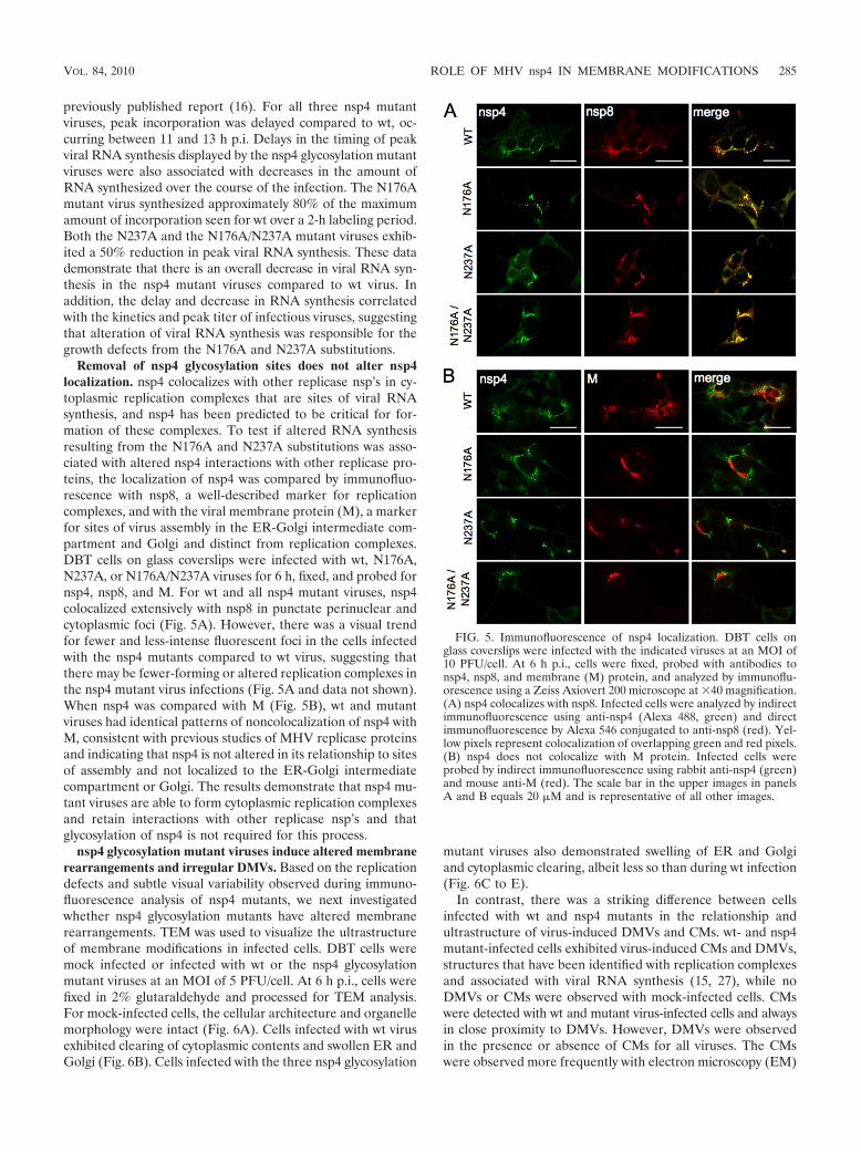
Removal of nsp4 glycosylation sites does not alter nsp4localization. nsp4 colocalizes with other replicase nsp’s in cy-toplasmic replication complexes that are sites of viral RNAsynthesis, and nsp4 has been predicted to be critical for for-mation of these complexes. To test if altered RNA synthesisresulting from the N176A and N237A substitutions was asso-ciated with altered nsp4 interactions with other replicase pro-teins, the localization of nsp4 was compared by immunofluo-rescence with nsp8, a well-described marker for replicationcomplexes, and with the viral membrane protein (M), a markerfor sites of virus assembly in the ER-Golgi intermediate com-partment and Golgi and distinct from replication complexes.DBT cells on glass coverslips were infected with wt, N176A,N237A, or N176A/N237A viruses for 6 h, fixed, and probed fornsp4, nsp8, and M. For wt and all nsp4 mutant viruses, nsp4colocalized extensively with nsp8 in punctate perinuclear andcytoplasmic foci (Fig. 5A). However, there was a visual trendfor fewer and less-intense fluorescent foci in the cells infectedwith the nsp4 mutants compared to wt virus, suggesting thatthere may be fewer-forming or altered replication complexes inthe nsp4 mutant virus infections (Fig. 5A and data not shown).When nsp4 was compared with M (Fig. 5B), wt and mutantviruses had identical patterns of noncolocalization of nsp4 withM, consistent with previous studies of MHV replicase proteinsand indicating that nsp4 is not altered in its relationship to sitesof assembly and not localized to the ER-Golgi intermediatecompartment or Golgi. The results demonstrate that nsp4 mu-tant viruses are able to form cytoplasmic replication complexesand retain interactions with other replicase nsp’s and thatglycosylation of nsp4 is not required for this process.
nsp4 glycosylation mutant viruses induce altered membranerearrangements and irregular DMVs. Based on the replicationdefects and subtle visual variability observed during immuno-fluorescence analysis of nsp4 mutants, we next investigatedwhether nsp4 glycosylation mutants have altered membranerearrangements. TEM was used to visualize the ultrastructureof membrane modifications in infected cells. DBT cells weremock infected or infected with wt or the nsp4 glycosylationmutant viruses at an MOI of 5 PFU/cell. At 6 h p.i., cells werefixed in 2% glutaraldehyde and processed for TEM analysis.For mock-infected cells, the cellular architecture and organellemorphology were intact (Fig. 6A). Cells infected with wt virusexhibited clearing of cytoplasmic contents and swollen ER andGolgi (Fig. 6B). Cells infected with the three nsp4 glycosylation
mutant viruses also demonstrated swelling of ER and Golgiand cytoplasmic clearing, albeit less so than during wt infection(Fig. 6C to E).
In contrast, there was a striking difference between cellsinfected with wt and nsp4 mutants in the relationship andultrastructure of virus-induced DMVs and CMs. wt- and nsp4mutant-infected cells exhibited virus-induced CMs and DMVs,structures that have been identified with replication complexesand associated with viral RNA synthesis (15, 27), while noDMVs or CMs were observed with mock-infected cells. CMswere detected with wt and mutant virus-infected cells and alwaysin close proximity to DMVs. However, DMVs were observedin the presence or absence of CMs for all viruses. The CMswere observed more frequently with electron microscopy (EM)
FIG. 5. Immunofluorescence of nsp4 localization. DBT cells onglass coverslips were infected with the indicated viruses at an MOI of10 PFU/cell. At 6 h p.i., cells were fixed, probed with antibodies tonsp4, nsp8, and membrane (M) protein, and analyzed by immunoflu-orescence using a Zeiss Axiovert 200 microscope at �40 magnification.(A) nsp4 colocalizes with nsp8. Infected cells were analyzed by indirectimmunofluorescence using anti-nsp4 (Alexa 488, green) and directimmunofluorescence by Alexa 546 conjugated to anti-nsp8 (red). Yel-low pixels represent colocalization of overlapping green and red pixels.(B) nsp4 does not colocalize with M protein. Infected cells wereprobed by indirect immunofluorescence using rabbit anti-nsp4 (green)and mouse anti-M (red). The scale bar in the upper images in panelsA and B equals 20 �M and is representative of all other images.
VOL. 84, 2010 ROLE OF MHV nsp4 IN MEMBRANE MODIFICATIONS 285
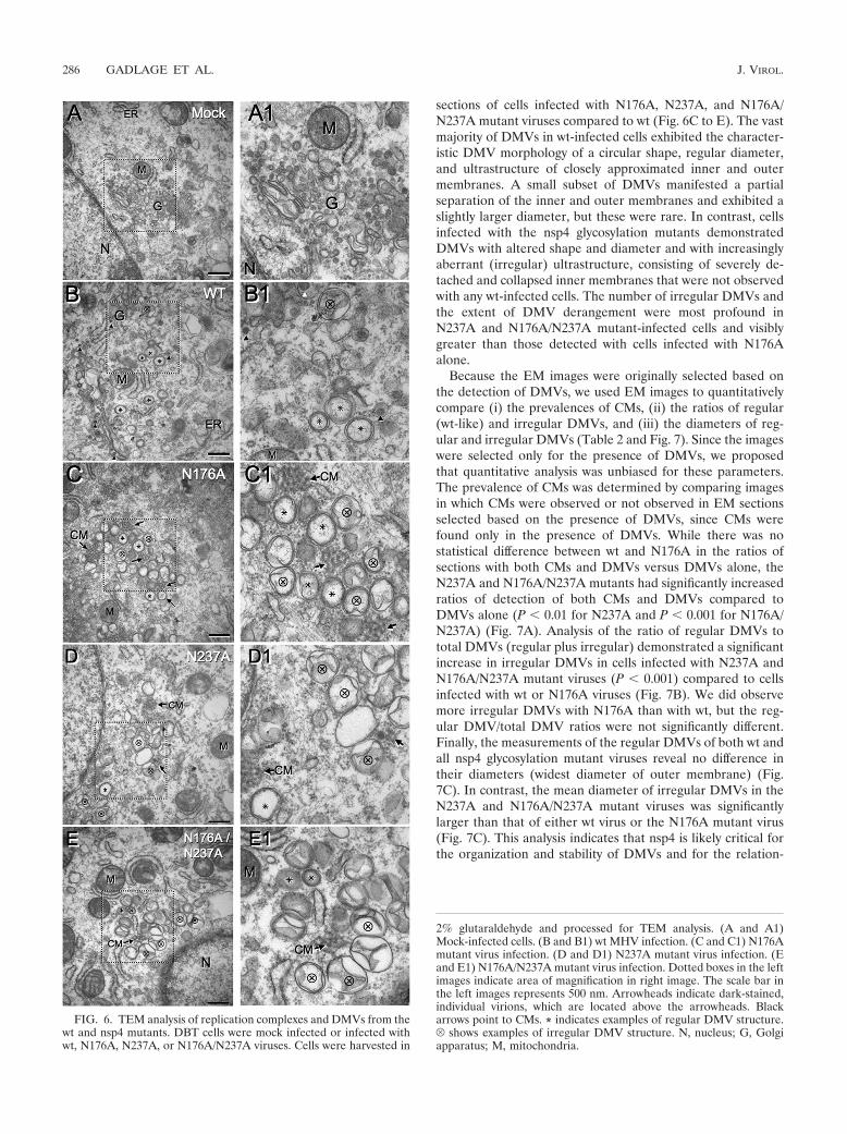
sections of cells infected with N176A, N237A, and N176A/N237A mutant viruses compared to wt (Fig. 6C to E). The vastmajority of DMVs in wt-infected cells exhibited the character-istic DMV morphology of a circular shape, regular diameter,and ultrastructure of closely approximated inner and outermembranes. A small subset of DMVs manifested a partialseparation of the inner and outer membranes and exhibited aslightly larger diameter, but these were rare. In contrast, cellsinfected with the nsp4 glycosylation mutants demonstratedDMVs with altered shape and diameter and with increasinglyaberrant (irregular) ultrastructure, consisting of severely de-tached and collapsed inner membranes that were not observedwith any wt-infected cells. The number of irregular DMVs andthe extent of DMV derangement were most profound inN237A and N176A/N237A mutant-infected cells and visiblygreater than those detected with cells infected with N176Aalone.
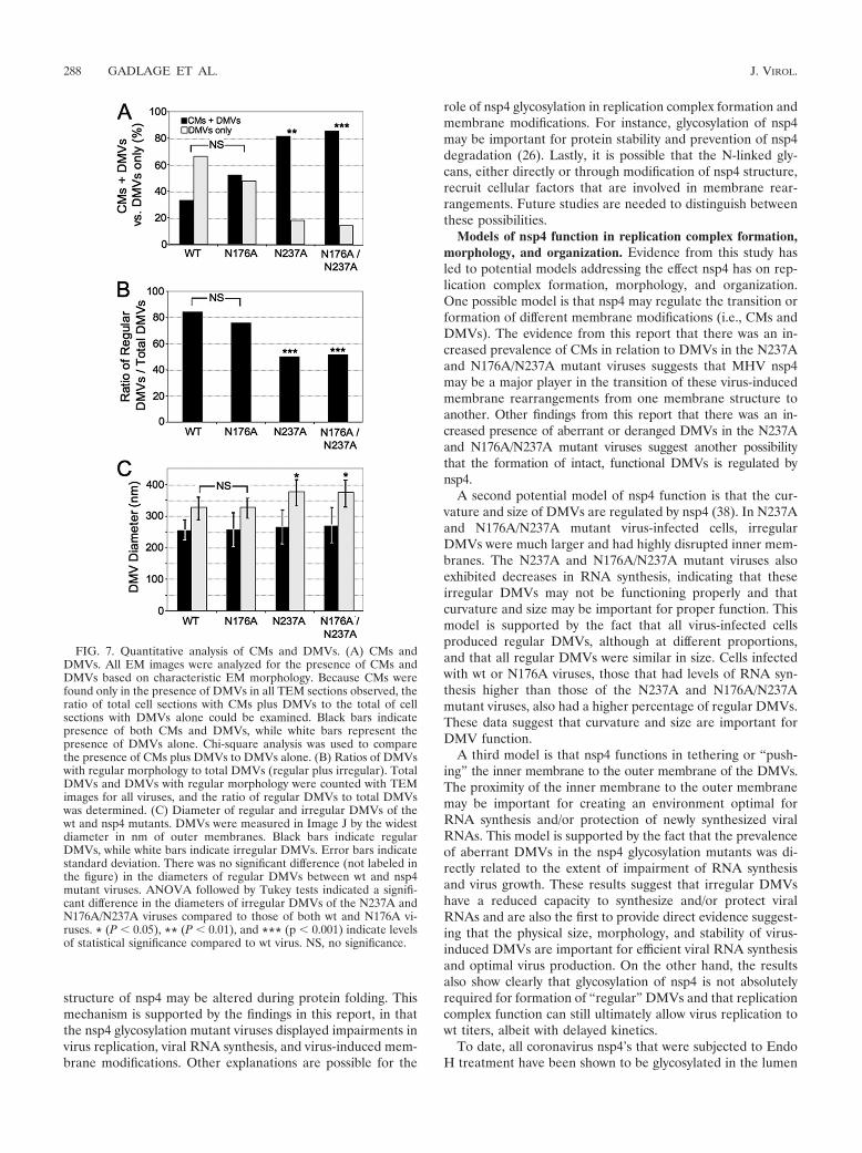
Because the EM images were originally selected based onthe detection of DMVs, we used EM images to quantitativelycompare (i) the prevalences of CMs, (ii) the ratios of regular(wt-like) and irregular DMVs, and (iii) the diameters of reg-ular and irregular DMVs (Table 2 and Fig. 7). Since the imageswere selected only for the presence of DMVs, we proposedthat quantitative analysis was unbiased for these parameters.The prevalence of CMs was determined by comparing imagesin which CMs were observed or not observed in EM sectionsselected based on the presence of DMVs, since CMs werefound only in the presence of DMVs. While there was nostatistical difference between wt and N176A in the ratios ofsections with both CMs and DMVs versus DMVs alone, theN237A and N176A/N237A mutants had significantly increasedratios of detection of both CMs and DMVs compared toDMVs alone (P � 0.01 for N237A and P � 0.001 for N176A/N237A) (Fig. 7A). Analysis of the ratio of regular DMVs tototal DMVs (regular plus irregular) demonstrated a significantincrease in irregular DMVs in cells infected with N237A andN176A/N237A mutant viruses (P � 0.001) compared to cellsinfected with wt or N176A viruses (Fig. 7B). We did observemore irregular DMVs with N176A than with wt, but the reg-ular DMV/total DMV ratios were not significantly different.Finally, the measurements of the regular DMVs of both wt andall nsp4 glycosylation mutant viruses reveal no difference intheir diameters (widest diameter of outer membrane) (Fig.7C). In contrast, the mean diameter of irregular DMVs in theN237A and N176A/N237A mutant viruses was significantlylarger than that of either wt virus or the N176A mutant virus(Fig. 7C). This analysis indicates that nsp4 is likely critical forthe organization and stability of DMVs and for the relation-
FIG. 6. TEM analysis of replication complexes and DMVs from thewt and nsp4 mutants. DBT cells were mock infected or infected withwt, N176A, N237A, or N176A/N237A viruses. Cells were harvested in
2% glutaraldehyde and processed for TEM analysis. (A and A1)Mock-infected cells. (B and B1) wt MHV infection. (C and C1) N176Amutant virus infection. (D and D1) N237A mutant virus infection. (Eand E1) N176A/N237A mutant virus infection. Dotted boxes in the leftimages indicate area of magnification in right image. The scale bar inthe left images represents 500 nm. Arrowheads indicate dark-stained,individual virions, which are located above the arrowheads. Blackarrows point to CMs. * indicates examples of regular DMV structure.V shows examples of irregular DMV structure. N, nucleus; G, Golgiapparatus; M, mitochondria.
286 GADLAGE ET AL. J. VIROL.

ship and evolution of membrane modifications (CMs andDMVs) over the course of infection.
DISCUSSION
Although multiple studies have investigated the roles ofnsp’s in inducing membrane rearrangements, understandingthe role of glycosylation of nsp’s from positive-strand RNAviruses remains limited. A study of the flavivirus yellow fevervirus demonstrated that NS1 glycosylation was important forseveral functions in the virus life cycle (30, 33). NS1 interactswith membranes and is involved in replicase function (30), andremoval of NS1 glycosylation by asparagine-to-alanine substi-tution results in impaired virus growth, RNA synthesis, andpathogenesis (33).
Coronaviruses, like other positive-strand RNA viruses, in-duce the formation of DMVs that serve as scaffolds for repli-cation/transcription complexes. Exogenous expression of thepoliovirus transmembrane proteins 2BC and 3A results inDMVs that are indistinguishable from those formed during wtinfection (43, 46). Equine arteritis virus (EAV), which is clas-sified with coronaviruses in the order Nidovirales, inducesDMVs similar to coronaviruses (37). Exogenous plasmid ex-pression of EAV nsp2 and nsp3 is sufficient to induce mem-brane modifications resulting in membrane structures similarto those seen during EAV infection, and mutations withinEAV nsp3 also result in altered virus-induced membrane re-arrangements (39, 44). EAV nsp3 is a tetra-spanning integralmembrane protein implicated in DMV formation and organi-zation. Of interest, an introduced Asn substitution (T873N) inan EAV nsp3 luminal domain resulted in nsp3 glycosylationin vitro but was highly detrimental when introduced into thegenome and recovered only as a pseudoreversion (N873H)that abolished the glycosylation site. Thus, for another nidovi-rus, the glycosylation status of a membrane-modifying repli-case protein is also important for DMV formation and RNAsynthesis during virus replication.
Our report confirms multiple roles of MHV nsp4 in the viruslife cycle, including optimal virus replication and RNA synthe-sis, as well as its importance in the modification and morphol-ogy of virus-induced membrane structures. In this study, weshow that MHV nsp4 is glycosylated and functions as a mem-brane modification protein that regulates virus-induced mem-brane rearrangements. nsp4 glycosylation mutant viruses dis-play highly irregular DMVs and an increased prevalence ofCMs relative to DMVs alone. The extent of disrupted DMVsin the nsp4 glycosylation mutant viruses correlated directlywith decreases in RNA synthesis and virus replication. These
data suggest that altered membranous structures from the nsp4glycosylation mutants result in a reduced capacity to synthesizeviral RNA and/or protect viral RNA from degradation, ulti-mately leading to impaired virus fitness.
Previous studies have concluded that nsp4 is required forMHV replication and have identified determinants of mem-brane topology, subcellular localization, and function (10, 35,45). This study is the first to recover and characterize theimportance of multiple nsp4 glycosylation events to virus rep-lication, viral RNA synthesis, and virus-induced membranemodifications during coronavirus infection. Clementz et al.recovered an nsp4 N176A mutant but were unable to recoveran N237A or N176A/N237A mutant (10). Their N176A mutantgrew with kinetics similar to those of wt at an MOI of 0.1PFU/cell at 33°C and 39°C but was not further characterized inthat report. In contrast to the results in the previously pub-lished report, we were able to recover and characterize theN237A and N176A/N237A mutant viruses. The reasons for thedifferences in recovery can only be speculated. The back-grounds of cloned MHV genome fragments should be identicalsince the MHV genome fragments were jointly developed byour lab and the lab of Baric and coworkers (47). In addition,we performed RT-PCR sequencing of the complete genomefrom the recovered N176A/N237A mutant virus, which verifiedthe engineered mutations and also confirmed that the rest ofthe genome was identical, with no additional mutations of anykind, to the published recombinant MHV-A59 sequence. Thus,there were no other compensating mutations to account for orconsider for the recovery of the mutant virus. We have expe-rienced occasional mutations in the genome fragments duringpreparation for genome assembly that have prevented recoveryof even known viable mutants and would therefore speculatethat this could account for the nonrecovery of N237A andN176A/N237A mutant viruses by Clementz et al. Our resultsclearly demonstrate that the N176 and N137 residues and theassociated glycosylation events are not required for MHV rep-lication in cell culture. Since no other mutations in the genomeRNA from the recovered N176A/N237A mutant virus wereidentified, we can conclude that the profound and distinctphenotypes in virus replication, RNA synthesis, and virus-in-duced cellular membrane modifications are due to the intro-duced mutations alone.
Potential functions of nsp4 glycosylation. Modification ofproteins by addition of N-linked glycans may result in numer-ous effects on protein functions (14, 19). Therefore, glycosyla-tion of nsp4 may be important for a variety of reasons. Onepotential mechanism of nsp4 glycosylation is proper proteinfolding (18, 36). By removing N-linked glycans, the overall
TABLE 2. Analysis of virus-induced membrane structures
Virus
Total no. of:Avg regularDMV diam
(nm)
Avg irregularDMV diam
(nm)Cell sections with
evidence ofinfection
Sections withCMs and
DMVs
Sections withDMVs only
DMVscounted
RegularDMVs
IrregularDMVs
WT 24 8 16 102 86 16 255.2 � 31.4 323.2 � 35.6N176A 21 11 10 127 96 31 257.0 � 52.3 324.2 � 31.8N237A 11 9 2 72 36 36 264.6 � 53.6 372.5 � 41.6N176A/N237A 21 18 3 117 60 57 270.4 � 56.1 371.3 � 41.6
VOL. 84, 2010 ROLE OF MHV nsp4 IN MEMBRANE MODIFICATIONS 287
structure of nsp4 may be altered during protein folding. Thismechanism is supported by the findings in this report, in thatthe nsp4 glycosylation mutant viruses displayed impairments invirus replication, viral RNA synthesis, and virus-induced mem-brane modifications. Other explanations are possible for the
role of nsp4 glycosylation in replication complex formation andmembrane modifications. For instance, glycosylation of nsp4may be important for protein stability and prevention of nsp4degradation (26). Lastly, it is possible that the N-linked gly-cans, either directly or through modification of nsp4 structure,recruit cellular factors that are involved in membrane rear-rangements. Future studies are needed to distinguish betweenthese possibilities.
Models of nsp4 function in replication complex formation,morphology, and organization. Evidence from this study hasled to potential models addressing the effect nsp4 has on rep-lication complex formation, morphology, and organization.One possible model is that nsp4 may regulate the transition orformation of different membrane modifications (i.e., CMs andDMVs). The evidence from this report that there was an in-creased prevalence of CMs in relation to DMVs in the N237Aand N176A/N237A mutant viruses suggests that MHV nsp4may be a major player in the transition of these virus-inducedmembrane rearrangements from one membrane structure toanother. Other findings from this report that there was an in-creased presence of aberrant or deranged DMVs in the N237Aand N176A/N237A mutant viruses suggest another possibilitythat the formation of intact, functional DMVs is regulated bynsp4.
A second potential model of nsp4 function is that the cur-vature and size of DMVs are regulated by nsp4 (38). In N237Aand N176A/N237A mutant virus-infected cells, irregularDMVs were much larger and had highly disrupted inner mem-branes. The N237A and N176A/N237A mutant viruses alsoexhibited decreases in RNA synthesis, indicating that theseirregular DMVs may not be functioning properly and thatcurvature and size may be important for proper function. Thismodel is supported by the fact that all virus-infected cellsproduced regular DMVs, although at different proportions,and that all regular DMVs were similar in size. Cells infectedwith wt or N176A viruses, those that had levels of RNA syn-thesis higher than those of the N237A and N176A/N237Amutant viruses, also had a higher percentage of regular DMVs.These data suggest that curvature and size are important forDMV function.
A third model is that nsp4 functions in tethering or “push-ing” the inner membrane to the outer membrane of the DMVs.The proximity of the inner membrane to the outer membranemay be important for creating an environment optimal forRNA synthesis and/or protection of newly synthesized viralRNAs. This model is supported by the fact that the prevalenceof aberrant DMVs in the nsp4 glycosylation mutants was di-rectly related to the extent of impairment of RNA synthesisand virus growth. These results suggest that irregular DMVshave a reduced capacity to synthesize and/or protect viralRNAs and are also the first to provide direct evidence suggest-ing that the physical size, morphology, and stability of virus-induced DMVs are important for efficient viral RNA synthesisand optimal virus production. On the other hand, the resultsalso show clearly that glycosylation of nsp4 is not absolutelyrequired for formation of “regular” DMVs and that replicationcomplex function can still ultimately allow virus replication towt titers, albeit with delayed kinetics.
To date, all coronavirus nsp4’s that were subjected to EndoH treatment have been shown to be glycosylated in the lumen
FIG. 7. Quantitative analysis of CMs and DMVs. (A) CMs andDMVs. All EM images were analyzed for the presence of CMs andDMVs based on characteristic EM morphology. Because CMs werefound only in the presence of DMVs in all TEM sections observed, theratio of total cell sections with CMs plus DMVs to the total of cellsections with DMVs alone could be examined. Black bars indicatepresence of both CMs and DMVs, while white bars represent thepresence of DMVs alone. Chi-square analysis was used to comparethe presence of CMs plus DMVs to DMVs alone. (B) Ratios of DMVswith regular morphology to total DMVs (regular plus irregular). TotalDMVs and DMVs with regular morphology were counted with TEMimages for all viruses, and the ratio of regular DMVs to total DMVswas determined. (C) Diameter of regular and irregular DMVs of thewt and nsp4 mutants. DMVs were measured in Image J by the widestdiameter in nm of outer membranes. Black bars indicate regularDMVs, while white bars indicate irregular DMVs. Error bars indicatestandard deviation. There was no significant difference (not labeled inthe figure) in the diameters of regular DMVs between wt and nsp4mutant viruses. ANOVA followed by Tukey tests indicated a signifi-cant difference in the diameters of irregular DMVs of the N237A andN176A/N237A viruses compared to those of both wt and N176A vi-ruses. * (P � 0.05), ** (P � 0.01), and *** (p � 0.001) indicate levelsof statistical significance compared to wt virus. NS, no significance.
288 GADLAGE ET AL. J. VIROL.
of the ER between the first and second predicted transmem-brane domains of nsp4 in exogenous expression experiments,including group 2a MHV nsp4, group 2b SARS-CoV nsp4, andgroup 3 IBV nsp4 (10, 29, 35). It will be interesting to seewhether glycosylation of nsp4 is conserved among other coro-naviruses, specifically group 1 coronaviruses, and what effectthe loss of glycosylation sites has on virus replication, RNAsynthesis, and replication complex morphology.
This study has demonstrated the importance of MHV nsp4glycosylation sites in virus replication, replication complexmorphology and organization, and viral RNA synthesis. Be-cause nsp4 has been shown to have integral membrane char-acteristics and no predicted enzymatic activities, it is rational topropose that nsp4 involvement in viral RNA synthesis is due toreplication complex formation, other possible membrane mod-ifications, and/or protein interactions. The nsp4 glycosylationmutant viruses generated in this study will provide powerfultools to further dissect the definitive mechanisms of nsp4 func-tion on replication complex formation and its roles in the viruslife cycle.
ACKNOWLEDGMENTS
We thank Elvin Woodruff for TEM assistance and image analysis.We also thank Megan Culler and Xiaotao Lu for technical assistance.We thank Michelle Becker and Lance Eckerle for advice and criticalreviews of the manuscript.
Support for this work was provided by National Institutes of Healthgrant R01 AI50083 (M.R.D.) from the National Institute of Allergyand Infectious Diseases. M.J.G. was supported by the Training Grantin Mechanisms of Vascular Disease through the Vanderbilt UniversitySchool of Medicine (T32 HL007751). J.S.S. was supported by PublicHealth Service award T32 CA009385. This work was also supported bythe Elizabeth B. Lamb Center for Pediatric Research.
REFERENCES
1. Baker, S. C., K. Yokomori, S. Dong, R. Carlisle, A. E. Gorbalenya, E. V.Koonin, and M. M. Lai. 1993. Identification of the catalytic sites of a papain-like cysteine proteinase of murine coronavirus. J. Virol. 67:6056–6063.
2. Baliji, S., S. A. Cammer, B. Sobral, and S. C. Baker. 2009. Detection ofnonstructural protein 6 (nsp6) in murine coronavirus-infected cells and anal-ysis of the transmembrane topology using bioinformatics and molecularapproaches. J. Virol. 83:6957–6962.
3. Bonilla, P. J., A. E. Gorbalenya, and S. R. Weiss. 1994. Mouse hepatitis virusstrain A59 RNA polymerase gene ORF 1a: heterogeneity among MHVstrains. Virology 198:736–740.
4. Bost, A. G., E. Prentice, and M. R. Denison. 2001. Mouse hepatitis virusreplicase protein complexes are translocated to sites of M protein accumu-lation in the ERGIC at late times of infection. Virology 285:21–29.
5. Bredenbeek, P. J., C. J. Pachuk, A. F. H. Noten, J. Charite, W. Luytjes, S. R.Weiss, and W. J. M. Spaan. 1990. The primary structure and expression ofthe second open reading frame of the polymerase gene of the coronavirusMHV-A59; a highly conserved polymerase is expressed by an efficient ribo-somal frameshifting mechanism. Nucleic Acids Res. 18:1825–1832.
6. Brierley, I., P. Digard, and S. C. Inglis. 1989. Characterization of an efficientcoronavirus ribosomal frameshift signal: requirement for an RNApseudoknot. Cell 57:537–547.
7. Brockway, S. M., C. T. Clay, X. T. Lu, and M. R. Denison. 2003. Character-ization of the expression, intracellular localization, and replication complexassociation of the putative mouse hepatitis virus RNA-dependent RNApolymerase. J. Virol. 77:10515–10527.
8. Chen, W., and R. S. Baric. 1996. Molecular anatomy of mouse hepatitis viruspersistence: coevolution of increased host cell resistance and virus virulence.J. Virol. 70:3947–3960.
9. Chen, W., V. J. Madden, C. J. Bagnell, and R. S. Baric. 1997. Host-derivedintracellular immunization against mouse hepatitis virus infection. Virology228:318–332.
10. Clementz, M. A., A. Kanjanahaluethai, T. E. O’Brien, and S. C. Baker. 2008.Mutation in murine coronavirus replication protein nsp4 alters assembly ofdouble membrane vesicles. Virology 375:118–129.
11. Denison, M. R. 2008. Seeking membranes: positive-strand RNA virus repli-cation complexes. PLoS Biol. 6:e270.
12. Denison, M. R., B. Yount, S. M. Brockway, R. L. Graham, A. C. Sims, X. Lu,and R. S. Baric. 2004. Cleavage between replicase proteins p28 and p65 ofmouse hepatitis virus is not required for virus replication. J. Virol. 78:5957–5965.
13. Denison, M. R., P. W. Zoltick, S. A. Hughes, B. Giangreco, A. L. Olson, S.Perlman, J. L. Leibowitz, and S. R. Weiss. 1992. Intracellular processing ofthe N-terminal ORF 1a proteins of the coronavirus MHV-A59 requiresmultiple proteolytic events. Virology 189:274–284.
14. Fiedler, K., and K. Simons. 1995. The role of N-glycans in the secretorypathway. Cell 81:309–312.
15. Gosert, R., A. Kanjanahaluethai, D. Egger, K. Bienz, and S. C. Baker. 2002.RNA replication of mouse hepatitis virus takes place at double-membranevesicles. J. Virol. 76:3697–3708.
16. Graham, R. L., and M. R. Denison. 2006. Replication of murine hepatitisvirus is regulated by papain-like proteinase 1 processing of nonstructuralproteins 1, 2, and 3. J. Virol. 80:11610–11620.
17. Harcourt, B. H., D. Jukneliene, A. Kanjanahaluethai, J. Bechill, K. M.Severson, C. M. Smith, P. A. Rota, and S. C. Baker. 2004. Identification ofsevere acute respiratory syndrome coronavirus replicase products and char-acterization of papain-like protease activity. J. Virol. 78:13600–13612.
18. Helenius, A. 1994. How N-linked oligosaccharides affect glycoprotein foldingin the endoplasmic reticulum. Mol. Biol. Cell 5:253–265.
19. Helenius, A., and M. Aebi. 2001. Intracellular functions of N-linked glycans.Science 291:2364–2369.
20. Hirano, N., K. Fujiwara, and M. Matumoto. 1976. Mouse hepatitis virus(MHV-2). Plaque assay and propagation in mouse cell line DBT cells. Jpn.J. Microbiol. 20:219–225.
21. Kanjanahaluethai, A., and S. C. Baker. 2000. Identification of mouse hep-atitis virus papain-like proteinase 2 activity. J. Virol. 74:7911–7921.
22. Kanjanahaluethai, A., and S. C. Baker. 2001. Processing of the replicase ofmurine coronavirus: papain-like proteinase 2 (PLP2) acts to generate p150and p44. Adv. Exp. Med. Biol. 494:267–273.
23. Kanjanahaluethai, A., Z. Chen, D. Jukneliene, and S. C. Baker. 2007. Mem-brane topology of murine coronavirus replicase nonstructural protein 3.Virology 361:391–401.
24. Kanjanahaluethai, A., D. Jukneliene, and S. C. Baker. 2003. Identification ofthe murine coronavirus MP1 cleavage site recognized by papain-like pro-teinase 2. J. Virol. 77:7376–7382.
25. Kim, J. C., R. A. Spence, P. F. Currier, X. T. Lu, and M. R. Denison. 1995.Coronavirus protein processing and RNA synthesis is inhibited by the cys-teine proteinase inhibitor e64dd. Virology 208:1–8.
26. Klausner, R. D., and R. Sitia. 1990. Protein degradation in the endoplasmicreticulum. Cell 62:611–614.
27. Knoops, K., M. Kikkert, S. H. Worm, J. C. Zevenhoven-Dobbe, Y. van derMeer, A. J. Koster, A. M. Mommaas, and E. J. Snijder. 2008. SARS-coro-navirus replication is supported by a reticulovesicular network of modifiedendoplasmic reticulum. PLoS Biol. 6:e226.
28. Lee, H.-J., C.-K. Shieh, A. E. Gorbalenya, E. V. Koonin, N. LaMonica, J.Tuler, A. Bagdzhadhzyan, and M. M. C. Lai. 1991. The complete sequence(22 kilobases) of murine coronavirus gene 1 encoding the putative proteasesand RNA polymerase. Virology 180:567–582.
29. Lim, K. P., L. F. Ng, and D. X. Liu. 2000. Identification of a novel cleavageactivity of the first papain-like proteinase domain encoded by open readingframe 1a of the coronavirus Avian infectious bronchitis virus and character-ization of the cleavage products. J. Virol. 74:1674–1685.
30. Lindenbach, B. D., and C. M. Rice. 1999. Genetic interaction of flavivirusnonstructural proteins NS1 and NS4A as a determinant of replicase function.J. Virol. 73:4611–4621.
31. Lu, X. T., Y. Q. Lu, and M. R. Denison. 1996. Intracellular and in vitrotranslated 27-kDa proteins contain the 3C-like proteinase activity of thecoronavirus MHV-A59. Virology 222:375–382.
32. Lu, Y., X. Lu, and M. R. Denison. 1995. Identification and characterizationof a serine-like proteinase of the murine coronavirus MHV-A59. J. Virol.69:3554–3559.
33. Muylaert, I. R., T. J. Chambers, R. Galler, and C. M. Rice. 1996. Mutagen-esis of the N-linked glycosylation sites of the yellow fever virus NS1 protein:effects on virus replication and mouse neurovirulence. Virology 222:159–168.
34. Novoa, R. R., G. Calderita, R. Arranz, J. Fontana, H. Granzow, and C. Risco.2005. Virus factories: associations of cell organelles for viral replication andmorphogenesis. Biol. Cell 97:147–172.
35. Oostra, M., E. G. te Lintelo, M. Deijs, M. H. Verheije, P. J. M. Rottier, andC. A. M. de Haan. 2007. Localization and membrane topology of coronavirusnonstructural protein 4: involvement of the early secretory pathway in rep-lication. J. Virol. 81:12323–12336.
36. Paulson, J. C. 1989. Glycoproteins: what are the sugar chains for? TrendsBiochem. Sci. 14:272–276.
37. Pedersen, K. W., Y. van der Meer, N. Roos, and E. J. Snijder. 1999. Openreading frame 1a-encoded subunits of the arterivirus replicase induce endo-plasmic reticulum-derived double-membrane vesicles which carry the viralreplication complex. J. Virol. 73:2016–2026.
38. Perlman, S., and J. Netland. 2009. Coronaviruses post-SARS: update onreplication and pathogenesis. Nat. Rev. Microbiol. 7:439–450.
VOL. 84, 2010 ROLE OF MHV nsp4 IN MEMBRANE MODIFICATIONS 289
39. Posthuma, C. C., K. W. Pedersen, Z. Lu, R. G. Joosten, N. Roos, J. C.Zevenhoven-Dobbe, and E. J. Snijder. 2008. Formation of the arterivirusreplication/transcription complex: a key role for nonstructural protein 3 inthe remodeling of intracellular membranes. J. Virol. 82:4480–4491.
40. Restrepo-Hartwig, M. A., and P. Ahlquist. 1996. Brome mosaic virus heli-case- and polymerase-like proteins colocalize on the endoplasmic reticulumat sites of viral RNA synthesis. J. Virol. 70:8908–8916.
41. Salonen, A., T. Ahola, and L. Kaariainen. 2005. Viral RNA replication in associationwith cellular membranes. Curr. Top. Microbiol. Immunol. 285:139–173.
42. Schaad, M. C., P. E. Jensen, and J. C. Carrington. 1997. Formation of plantRNA virus replication complexes on membranes: role of an endoplasmicreticulum-targeted viral protein. EMBO J. 16:4049–4059.
43. Schlegel, A., T. J. Giddings, M. S. Ladinsky, and K. Kirkegaard. 1996.Cellular origin and ultrastructure of membranes induced during poliovirusinfection. J. Virol. 70:6576–6588.
44. Snijder, E. J., H. van Tol, N. Roos, and K. W. Pedersen. 2001. Non-structuralproteins 2 and 3 interact to modify host cell membranes during the formationof the arterivirus replication complex. J. Gen. Virol. 82:985–994.
45. Sparks, J. S., X. Lu, and M. R. Denison. 2007. Genetic analysis of murinehepatitis virus nsp4 in virus replication. J. Virol. 81:12554–12563.
46. Suhy, D. A., T. H. Giddings, and K. Kirkegaard. 2000. Remodeling the ERby poliovirus infection and by individual viral proteins: an autophagy-likeorigin for virus-induced vesicles. J. Virol. 74:8953–8965.
47. Yount, B., M. R. Denison, S. R. Weiss, and R. S. Baric. 2002. Systematicassembly of a full-length infectious cDNA of mouse hepatitis virus strainA59. J. Virol. 76:11065–11078.
48. Ziebuhr, J., E. J. Snijder, and A. E. Gorbalenya. 2000. Virus-encoded pro-teinases and proteolytic processing in the Nidovirales. J. Gen. Virol. 81(4):853–879.
290 GADLAGE ET AL. J. VIROL.
Related Documents