Multiple Membrane Tethers Probed by Atomic Force Microscopy Mingzhai Sun,* John S. Graham,* y Balazs Hegedu ¨ s,* z Franc xoise Marga,* Ying Zhang, § Gabor Forgacs,* { and Michel Grandbois y *Department of Physics, University of Missouri, Columbia, Missouri; y De ´ partement de Pharmacologie, Universite ´ de Sherbrooke, Sherbrooke, Canada; z National Institute of Neurosurgery, Budapest, Hungary; § Department of Physics, University of Indiana, Bloomington, Indiana; and { Department of Biology, University of Missouri, Columbia, Missouri ABSTRACT Using the atomic force microscope to locally probe the cell membrane, we observed the formation of multiple tethers (thin nanotubes, each requiring a similar pulling force) as reproducible features within force profiles recorded on individual cells. Forces obtained with Chinese hamster ovary cells, a malignant human brain tumor cell line, and human endothelial cells (EA hy926) were found to be 28 6 10 pN, 29 6 9 pN, and 29 6 10 pN, respectively, independent of the nature of attachment to the cantilever. The rather large variation of the tether pulling forces measured at several locations on individual cells points to the existence of heterogeneity in the membrane properties of a morphologically homogeneous cell. Measurement of the summary lengths of the simultaneously extracted tethers provides a measure of the size of the available membrane reservoir through which co-existing tethers are associated. As expected, partial disruption of the actin cytoskeleton and removal of the hyaluronan backbone of the glycocalyx were observed to result in a marked decrease (30–50%) in the magnitude and a significant sharpening of the force distribution indicating reduced heterogeneity of membrane properties. Taken together, our results demonstrate the ability of the plasma membrane to locally produce multiple interdependent tethers—a process that could play an important role in the mechanical association of cells with their environment. INTRODUCTION The plasma membrane of mammalian cells is a highly dy- namic structure and its biomechanical properties are vital to the regulation of many cellular functions, such as adhesion, migration, signaling, and morphology (1). One of the most dy- namic processes within these membranes is the formation of tethers or thin nanotubes. These structures have been impli- cated in cell-cell adhesion (2) and recent studies suggest they might also provide a pathway for intracellular and intercel- lular communication (3–7). In vivo, tethers form during the primary adhesion and rolling motion of activated leukocytes on vascular endothe- lial cells or platelets along the walls of blood vessels (2,8,9). Hence, tether formation corresponds to the initial event leading to the extravasation of activated white blood cells at the sites of inflammatory reactions (10). In these systems, membrane tethers originate from pre-existing microvilli through specific selectin/glycoprotein bond formation be- tween cells under hemodynamic conditions (11). Membrane nanotubes have also been observed between liposomes and have been shown to readily form in red blood cells (12,13), neutrophils (14), neurons (15), fibroblasts (16,17), as well as epithelial (18) and endothelial cells (19). Several experimental methods have been used to character- ize the mechanical properties of membrane tethers, such as micropipette aspiration assays (12,13,20–23) and optical tweezers (15,24,25). In these experiments, tethers are ob- served in force-versus-distance curves as well-defined pla- teaus occurring at constant force. The presence of plateaus can be understood in terms of a membrane reservoir being gradually depleted upon pulling on the bilayer (16). These studies also revealed that tether length (i.e., available membrane reservoir) and tether formation force are influ- enced by the various components of the cytoskeleton. On the intracellular side, the membrane is connected to the cyto- skeleton through a variety of proteins and other complexes (26,27) and this association has been proposed to play a major role in cell membrane cohesion. The influence of cy- toskeletal integrity on the force needed to form tethers has been investigated earlier (28). These experiments demon- strated that the disruption of the cytoskeleton leads to a decrease of the force required to extract and elongate tethers. On the extracellular side, the cell membrane is covered by a glycosaminoglycan and proteoglycan network, the glyco- calyx. Whether or not the glycocalyx influences the properties of membrane tether formation has not been explored. Another important question concerns the possible heterogeneity in the interaction of the cytoskeleton/glycocalyx with the mem- brane over a morphologically homogeneous cellular surface. Tether formation in cell motility and cellular adhesion is likely to involve the simultaneous formation of multiple tethers. To our knowledge, the tether pulling experiments performed until now have primarily addressed the forma- tion of single tethers. One recent study explored dual tether extraction using the micropipette aspiration technique. Here the tethers were observed not in force-elongation profiles, but rather through the analysis of the dependence of the pulling force on the growth velocities of the tethers (29). Submitted December 16, 2004, and accepted for publication September 9, 2005. Mingzhai Sun and John S. Graham contributed equally to this work. Address reprint requests to Michel Grandbois, Tel.: 819-820-6868; E-mail: [email protected]. Ó 2005 by the Biophysical Society 0006-3495/05/12/4320/10 $2.00 doi: 10.1529/biophysj.104.058180 4320 Biophysical Journal Volume 89 December 2005 4320–4329

Welcome message from author
This document is posted to help you gain knowledge. Please leave a comment to let me know what you think about it! Share it to your friends and learn new things together.
Transcript
Multiple Membrane Tethers Probed by Atomic Force Microscopy
Mingzhai Sun,* John S. Graham,*y Balazs Hegedus,*z Francxoise Marga,* Ying Zhang,§ Gabor Forgacs,*{
and Michel Grandboisy
*Department of Physics, University of Missouri, Columbia, Missouri; yDepartement de Pharmacologie, Universite de Sherbrooke,Sherbrooke, Canada; zNational Institute of Neurosurgery, Budapest, Hungary; §Department of Physics, University of Indiana,Bloomington, Indiana; and {Department of Biology, University of Missouri, Columbia, Missouri
ABSTRACT Using the atomic force microscope to locally probe the cell membrane, we observed the formation of multipletethers (thin nanotubes, each requiring a similar pulling force) as reproducible features within force profiles recorded on individualcells. Forces obtained with Chinese hamster ovary cells, a malignant human brain tumor cell line, and human endothelial cells(EA hy926) were found to be 28 6 10 pN, 29 6 9 pN, and 29 6 10 pN, respectively, independent of the nature of attachment tothe cantilever. The rather large variation of the tether pulling forces measured at several locations on individual cells points to theexistence of heterogeneity in the membrane properties of a morphologically homogeneous cell. Measurement of the summarylengths of the simultaneously extracted tethers provides a measure of the size of the available membrane reservoir throughwhich co-existing tethers are associated. As expected, partial disruption of the actin cytoskeleton and removal of the hyaluronanbackbone of the glycocalyx were observed to result in a marked decrease (30–50%) in the magnitude and a significantsharpening of the force distribution indicating reduced heterogeneity of membrane properties. Taken together, our resultsdemonstrate the ability of the plasma membrane to locally produce multiple interdependent tethers—a process that could play animportant role in the mechanical association of cells with their environment.
INTRODUCTION
The plasma membrane of mammalian cells is a highly dy-
namic structure and its biomechanical properties are vital to
the regulation of many cellular functions, such as adhesion,
migration, signaling, and morphology (1). One of the most dy-
namic processes within these membranes is the formation of
tethers or thin nanotubes. These structures have been impli-
cated in cell-cell adhesion (2) and recent studies suggest they
might also provide a pathway for intracellular and intercel-
lular communication (3–7).
In vivo, tethers form during the primary adhesion and
rolling motion of activated leukocytes on vascular endothe-
lial cells or platelets along the walls of blood vessels (2,8,9).
Hence, tether formation corresponds to the initial event
leading to the extravasation of activated white blood cells
at the sites of inflammatory reactions (10). In these systems,
membrane tethers originate from pre-existing microvilli
through specific selectin/glycoprotein bond formation be-
tween cells under hemodynamic conditions (11).
Membrane nanotubes have also been observed between
liposomes and have been shown to readily form in red blood
cells (12,13), neutrophils (14), neurons (15), fibroblasts
(16,17), as well as epithelial (18) and endothelial cells (19).
Several experimental methods have been used to character-
ize the mechanical properties of membrane tethers, such as
micropipette aspiration assays (12,13,20–23) and optical
tweezers (15,24,25). In these experiments, tethers are ob-
served in force-versus-distance curves as well-defined pla-
teaus occurring at constant force. The presence of plateaus
can be understood in terms of a membrane reservoir being
gradually depleted upon pulling on the bilayer (16). These
studies also revealed that tether length (i.e., available
membrane reservoir) and tether formation force are influ-
enced by the various components of the cytoskeleton. On the
intracellular side, the membrane is connected to the cyto-
skeleton through a variety of proteins and other complexes
(26,27) and this association has been proposed to play a
major role in cell membrane cohesion. The influence of cy-
toskeletal integrity on the force needed to form tethers has
been investigated earlier (28). These experiments demon-
strated that the disruption of the cytoskeleton leads to a
decrease of the force required to extract and elongate tethers.
On the extracellular side, the cell membrane is covered by
a glycosaminoglycan and proteoglycan network, the glyco-
calyx. Whether or not the glycocalyx influences the properties
of membrane tether formation has not been explored. Another
important question concerns the possible heterogeneity in the
interaction of the cytoskeleton/glycocalyx with the mem-
brane over a morphologically homogeneous cellular surface.
Tether formation in cell motility and cellular adhesion
is likely to involve the simultaneous formation of multiple
tethers. To our knowledge, the tether pulling experiments
performed until now have primarily addressed the forma-
tion of single tethers. One recent study explored dual tether
extraction using the micropipette aspiration technique. Here
the tethers were observed not in force-elongation profiles,
but rather through the analysis of the dependence of the
pulling force on the growth velocities of the tethers (29).
Submitted December 16, 2004, and accepted for publication September 9,
2005.
Mingzhai Sun and John S. Graham contributed equally to this work.
Address reprint requests to Michel Grandbois, Tel.: 819-820-6868; E-mail:
� 2005 by the Biophysical Society
0006-3495/05/12/4320/10 $2.00 doi: 10.1529/biophysj.104.058180
4320 Biophysical Journal Volume 89 December 2005 4320–4329
Other recent work has demonstrated that multiple membrane
tethers can be formed in a minimal system composed of a
giant unilamellar vesicle, kinesin-coated beads, microtubules,
and ATP as energy source (3,5,30). Beyond these examples,
little is known about the behavior of multiple, simultaneously
existing tethers in real cells, and their coupling with the overall
membrane reservoir or their association with each other.
Whether or not multiple membrane tethers can be simulta-
neously extracted from the membranes of living cells is still
a matter of controversy. Indeed, multiple tethers extracted from
close locations along the membrane surface are expected to
rapidly coalesce. In a recent theoretical article, Derenyi et al.
(31) predicted that, in the absence of pinning forces, multiple
membrane tethers coalesce smoothly. However, this study
also points out that in real cells, membrane heterogeneities or
coupling to the cytoskeleton may prevent tether fusion.
To extract multiple tethers, a large initial adhesion force
has to be overcome. For this, a force transducer with the
ability to measure a broad range of forces (such as those
encountered in specific and nonspecific cellular adhesion
events) is needed. The atomic force microscope (AFM) (32)
has proven to be a powerful tool for single molecular in-
vestigations, to observe biological structures and to study
intramolecular and intermolecular interactions (for reviews
on the subject, see (33,34)). Recently, a variety of biologi-
cally relevant binding forces have been characterized by
force spectroscopy including the rupture force of a covalent
bond (35), unfolding forces in individual biomolecules
(36–40), rupture forces between various ligands and recep-
tors (41,42), unbinding forces of cadherins (43), and cell-cell
interaction forces (44,45). The ability of AFM cantilevers to
detect a large range of forces (picoNewtons to nanoNewtons)
provides the opportunity to simultaneously monitor the
formation of individual or multiple tethers and to bring new
insight into the behavior of multipally extracted tethers. In
this study, we used the AFM to extract multiple tethers, us-
ing a variety of cells with different morphology and origin,
including Chinese hamster ovary (CHO) cells, a malignant
human brain tumor cell line (HB), and endothelial cells.
These studies were aimed at demonstrating that formation of
multiple membrane nanotubes is a ubiquitous phenomenon,
largely independent of particular cell type. In particular, we
show that multiple tether formation can be induced locally
through contact of the AFM cantilever with the cell mem-
brane, and that the tethers extracted are interdependent. By
measuring the contributions of both the cytoskeleton and
the glycocalyx to the tether forces, we bring new insights
into the mechanism of membrane tether formation and the
behavior of multiple, simultaneously extracted tethers.
MATERIALS AND METHODS
Cell culture and treatments
Chinese hamster ovary cells (CHO-K1 cells, American Type Culture
Collection, Manassas, VA) and the human brain tumor cell line (HB) (46)
were cultured in DMEM (Invitrogen, Carlsbad, CA) supplemented with
10% fetal bovine serum (Sigma-Aldrich, St. Louis, MO) and 1% penicillin/
streptomycin mixture (Sigma-Aldrich) following standard procedures. The
human endothelial cells EA hy926 were a generous gift of Dr. C-J S. Edgell
(47), and were maintained in HAM’S F-12 supplemented with 20% fetal
bovine serum (Sigma-Aldrich). Cells were plated on glass coverslips (Pierce
Biotechnology, Rockford, IL), placed in 35-mm plastic petri dishes (Techno
Plastic Products, Trasadingen, Switzerland), or plated directly in petri
dishes, and cultured at 37�C in a 5% CO2 incubator typically for 24 h. In
cytoskeletal disruption experiments, the cells were in regular medium
supplemented with latrunculin A (Sigma), a specific actin polymerization
inhibitor (48–50) at various concentrations (0.1, 0.2, 0.5, and 1.0 mM) for 30
min before the measurement. The importance of the glycocalyx in the
fomation of membrane nanotubes was studied through one of its major
components, the glycosaminoglycan hyaluronan (51). The removal of hyal-
uronan from the surface of substrate-attached cells was achieved by a 30-min
incubation in the presence of hyaluronidase (500 IU/ml) (Sigma-Aldrich) in
serum- and polysaccharide-free medium. For surface modification assays,
cantilevers (Veeco, Santa Barbara, CA) and glass coverslips (Pierce Biotech-
nology) were put in the 0.1 mg/ml Poly-L-Lysine solution (Sigma) for 15
min, then rinsed with Milli-Q water (Millipore, Billerica, MA) and air-dried.
The effect of a collagen-coated surface was measured by incubation of
cantilevers in a type I collagen solution (1 mg/ml; Sigma) at 4�C for 60 min,
then rinsing with Milli-Q water and air-drying in a laminar flow hood.
Force spectroscopy measurements
Our in-house-built force measurement device, based on the design and
operation of an AFM, was attached to the stage of an inverted optical micro-
scope (Olympus IX70, Olympus America, Melville, NY). This arrange-
ment allowed for precise positioning of the cantilever on the area of
interest along the cell membrane. Soft silicon nitride cantilevers (Veeco,
Santa Barbara, CA) were cleaned in 70% ethanol, rinsed in Milli-Q water,
and then sterilized with UV light for 15 min. Each cantilever was calibrated
after a given experiment using thermal noise amplitude analysis (52,53). The
measured spring constants were between 8 and 11 mN/m, in agreement with
the nominal spring constant of 10 mN/m.
Cells were placed under the force device in CO2-independent medium
(Invitrogen, Carlsbad, CA) containing 2% fetal bovine serum at room
temperature. A typical experiment was performed as follows: the cantilever
was moved toward the surface until contact with the cell membrane
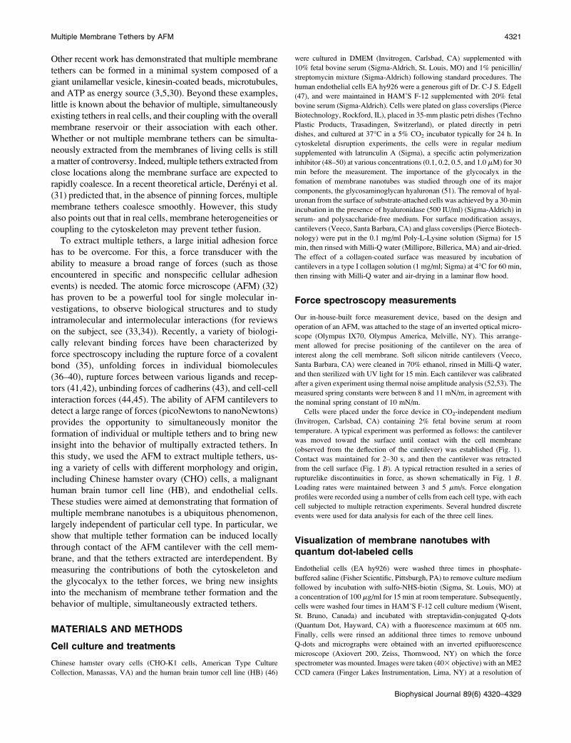
(observed from the deflection of the cantilever) was established (Fig. 1).
Contact was maintained for 2–30 s, and then the cantilever was retracted
from the cell surface (Fig. 1 B). A typical retraction resulted in a series of
rupturelike discontinuities in force, as shown schematically in Fig. 1 B.
Loading rates were maintained between 3 and 5 mm/s. Force elongation
profiles were recorded using a number of cells from each cell type, with each
cell subjected to multiple retraction experiments. Several hundred discrete
events were used for data analysis for each of the three cell lines.
Visualization of membrane nanotubes withquantum dot-labeled cells
Endothelial cells (EA hy926) were washed three times in phosphate-
buffered saline (Fisher Scientific, Pittsburgh, PA) to remove culture medium
followed by incubation with sulfo-NHS-biotin (Sigma, St. Louis, MO) at
a concentration of 100 mg/ml for 15 min at room temperature. Subsequently,
cells were washed four times in HAM’S F-12 cell culture medium (Wisent,
St. Bruno, Canada) and incubated with streptavidin-conjugated Q-dots
(Quantum Dot, Hayward, CA) with a fluorescence maximum at 605 nm.
Finally, cells were rinsed an additional three times to remove unbound
Q-dots and micrographs were obtained with an inverted epifluorescence
microscope (Axiovert 200, Zeiss, Thornwood, NY) on which the force
spectrometer was mounted. Images were taken (403 objective) with an ME2
CCD camera (Finger Lakes Instrumentation, Lima, NY) at a resolution of
Multiple Membrane Tethers by AFM 4321
Biophysical Journal 89(6) 4320–4329
768 3 512 pixels. Membrane tethers were formed by contacting an indi-
vidual cell with the AFM cantilever, followed by a simultaneous vertical and
lateral retraction allowing individual tether visualization.
RESULTS
Multiple tether formation and rupture
Formation of membrane tethers between cells and untreated
AFM cantilevers was demonstrated by labeling the cell sur-
face with fluorescent quantum dots and probing the cell in a
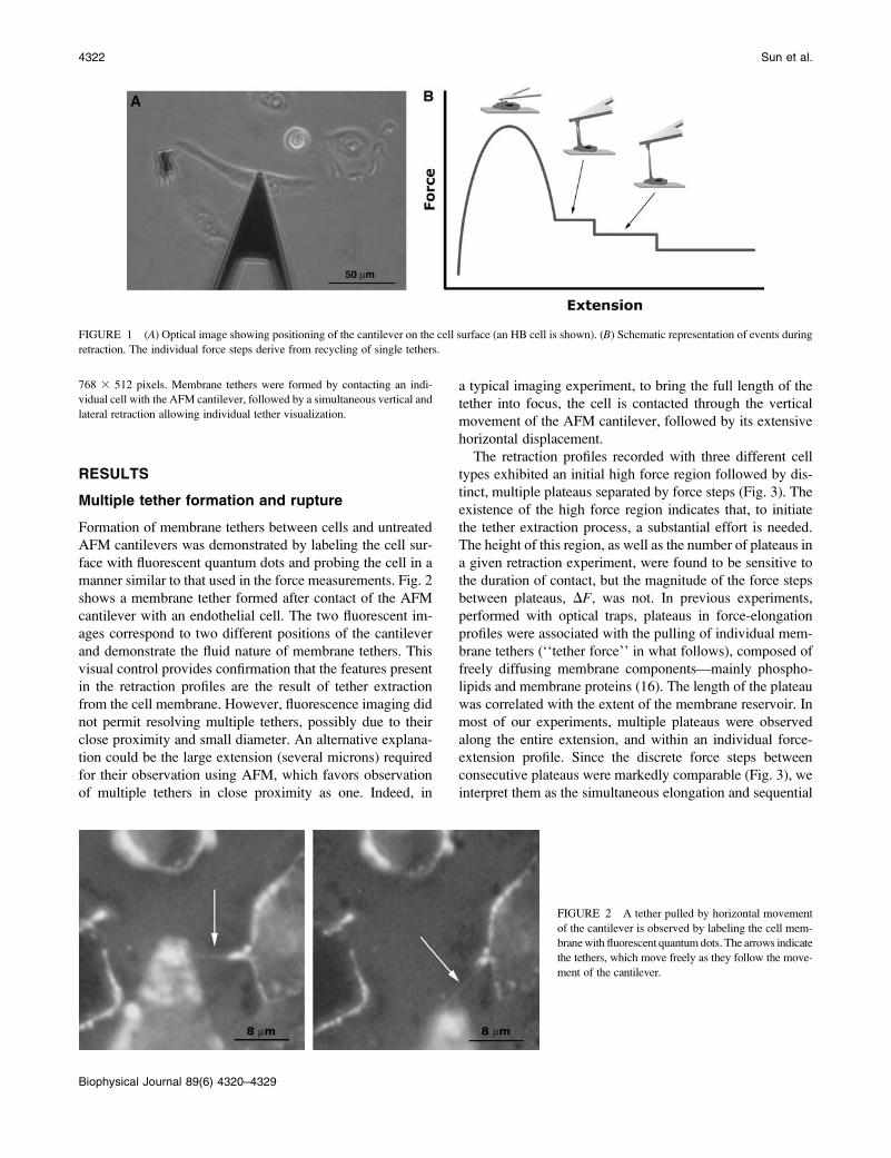
manner similar to that used in the force measurements. Fig. 2
shows a membrane tether formed after contact of the AFM
cantilever with an endothelial cell. The two fluorescent im-
ages correspond to two different positions of the cantilever
and demonstrate the fluid nature of membrane tethers. This
visual control provides confirmation that the features present
in the retraction profiles are the result of tether extraction
from the cell membrane. However, fluorescence imaging did
not permit resolving multiple tethers, possibly due to their
close proximity and small diameter. An alternative explana-
tion could be the large extension (several microns) required
for their observation using AFM, which favors observation
of multiple tethers in close proximity as one. Indeed, in
a typical imaging experiment, to bring the full length of the
tether into focus, the cell is contacted through the vertical
movement of the AFM cantilever, followed by its extensive
horizontal displacement.
The retraction profiles recorded with three different cell
types exhibited an initial high force region followed by dis-
tinct, multiple plateaus separated by force steps (Fig. 3). The
existence of the high force region indicates that, to initiate
the tether extraction process, a substantial effort is needed.
The height of this region, as well as the number of plateaus in
a given retraction experiment, were found to be sensitive to
the duration of contact, but the magnitude of the force steps
between plateaus, DF, was not. In previous experiments,
performed with optical traps, plateaus in force-elongation
profiles were associated with the pulling of individual mem-
brane tethers (‘‘tether force’’ in what follows), composed of
freely diffusing membrane components—mainly phospho-
lipids and membrane proteins (16). The length of the plateau
was correlated with the extent of the membrane reservoir. In
most of our experiments, multiple plateaus were observed
along the entire extension, and within an individual force-
extension profile. Since the discrete force steps between
consecutive plateaus were markedly comparable (Fig. 3), we
interpret them as the simultaneous elongation and sequential
FIGURE 1 (A) Optical image showing positioning of the cantilever on the cell surface (an HB cell is shown). (B) Schematic representation of events during
retraction. The individual force steps derive from recycling of single tethers.
FIGURE 2 A tether pulled by horizontal movement
of the cantilever is observed by labeling the cell mem-
brane with fluorescent quantum dots. The arrows indicate
the tethers, which move freely as they follow the move-
ment of the cantilever.
4322 Sun et al.
Biophysical Journal 89(6) 4320–4329

loss of multiple membrane tethers formed between the cell
and the cantilever. Consequently, DF can be associated with
the force needed to pull a single tether, whereas the total
force at which a given plateau occurs corresponds to the
force necessary to pull all existing tethers.
Our retraction traces did not exhibit the exponential rise of
the force-versus-length observed in optical tweezers experi-
ments at the end of the plateau region that is attributed to
a depleted membrane reservoir (see inset in Fig. 3 (16)). The
length scale (several microns) over which plateaus were
observed, however, is consistent with the earlier finding that
tethers form and grow by the gradual depletion of the cell’s
membrane reservoir (16,18). These observations provide
further evidence that the measured force-steps correspond
to the force required to pull a tether, rather than the force
required in the process by which the tether bridge is lost.
We suggest that the size of the overall membrane reser-
voir, shared by all the tethers, can be characterized by the
length of the last observed tether. Alternatively, the reservoir
size can be evaluated at each discrete force-step by multi-
plying the number of tethers being pulled on a given plateau
before a force drop by the length at which the rupture occurs.
This is true despite the fact that at the force-steps the local
membrane reservoirs are not yet fully depleted. This proce-
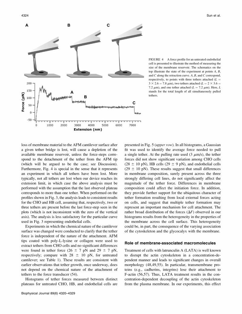
dure is illustrated in Fig. 4, which shows a force-elongation
profile with multiple plateaus corresponding to simulta-
neously pulled tethers and their sequential loss. Using Fig. 4
to evaluate the membrane reservoir in terms of tether length,
we obtain the consistent values of 7.8 mm, 7.2 mm, and 7.2
mm from the last three plateaus, representing three, two, and
a single tether, respectively. This observation is in accord
with the assumption that multiple, simultaneously existing
tethers are interdependent in terms of the cell’s overall mem-
brane reservoir: the material of a ruptured tether is rapidly
recycled and its local membrane reservoir becomes available
for the continued elongation of the still intact ones (15). The
existence of an overall cellular membrane reservoir, com-
posed of interconnected local reservoirs, is also supported
by the earlier finding that—as a consequence of the plasma
membrane’s fluid character, with repeated pull-retract cy-
cles of single tethers—the onset of the exponential regime
is shifted to later times and thus longer plateaus (16). Even
though the result obtained using Fig. 4 is highly suggestive,
it is not fully representative for the following reasons. Any
FIGURE 3 Typical retraction-force curves as a function of cantilever extension for three cell lines. Numbers on the graphs denote the values of the individual
force steps between consecutive plateaus. Note the very similar force drops in the quasi-constant force elongation regime, and that zero force is not reached at
the end of the retractions, indicating tethers are still attached. Inset shows timescale for a typical force drop.
Multiple Membrane Tethers by AFM 4323
Biophysical Journal 89(6) 4320–4329
loss of membrane material to the AFM cantilever surface after
a given tether bridge is lost, will cause a depletion of the
available membrane reservoir, unless the force-steps corre-
spond to the detachment of the tether from the AFM tip
(which will be argued to be the case; see Discussion).
Furthermore, Fig. 4 is special in the sense that it represents
an experiment in which all tethers have been lost. More
typically, not all tethers are lost when our device reaches its
extension limit, in which case the above analysis must be
performed with the assumption that the last observed plateau
corresponds to more than one tether. When performed on the
profiles shown in Fig. 3, the analysis leads to consistent results
for the CHO and HB cell, assuming that, respectively, two or
three tethers are present before the last force-step seen in the
plots (which is not inconsistent with the zero of the vertical
axis). The analysis is less satisfactory for the particular curve
used in Fig. 3 representing endothelial cells.
Experiments in which the chemical nature of the cantilever
surface was changed were conducted to clarify that the tether
force is independent of the nature of the attachment. AFM
tips coated with poly-L-lysine or collagen were used to
extract tethers from CHO cells and no significant differences
were found in tether force (26 6 7 pN and 29 6 7 pN,
respectively; compare with 28 6 10 pN, for untreated
cantilever; see Table 1). These results are consistent with
earlier observations that tether growth, once underway, does
not depend on the chemical nature of the attachment of
tethers to the force transducer (54).
Histograms of tether forces measured between distinct
plateaus for untreated CHO, HB, and endothelial cells are
presented in Fig. 5 (upper row). In all histograms, a Gaussian
fit was used to identify the average force needed to pull
a single tether. At the pulling rate used (3 mm/s), the tether
forces did not show significant variation among CHO cells
(28 6 10 pN), HB cells (29 6 9 pN), and endothelial cells
(29 6 10 pN). These results suggest that small differences
in membrane composition, surely present across the three
strongly differing cell lines, do not significantly affect the
magnitude of the tether force. Differences in membrane
composition could affect the initiation force. In addition,
they provide further support for the ubiquitous character of
tether formation resulting from local external forces acting
on cells, and suggest that multiple tether formation may
represent an important mechanism for cell attachment. The
rather broad distribution of the forces (DF) observed in our
histograms results from the heterogeneity in the properties of
the membrane across the cell surface. This heterogeneity
could be, in part, the consequence of the varying association
of the cytoskeleton and the glycocalyx with the membrane.
Role of membrane-associated macromolecules
Treatment of cells with latrunculin A (LATA) is well known
to disrupt the actin cytoskeleton in a concentration-de-
pendent manner and leads to significant changes in overall
morphology (48,49,55). In particular, transmembrane pro-
teins (e.g., cadherins, integrins) lose their attachment to
F-actin (56,57). Thus, LATA treatment results in the con-
centration-dependent decoupling of the actin cytoskeleton
from the plasma membrane. In our experiments, this effect
FIGURE 4 A force profile for an untreated endothelial
cell is presented to illustrate the method of measuring the
size of the membrane reservoir. The schematics on the
top illustrate the state of the experiment at points A, B,
and C along the retraction curve. A, B, and C correspond,
respectively, to points with three tethers attached (L ¼3 3 2.6 ¼ 7.8 mm), two tethers attached (L ¼ 2 3 3.6 ¼7.2 mm), and one tether attached (L ¼ 7.2 mm). Here, Lstands for the total length of all simultaneously pulled
tethers.
4324 Sun et al.
Biophysical Journal 89(6) 4320–4329
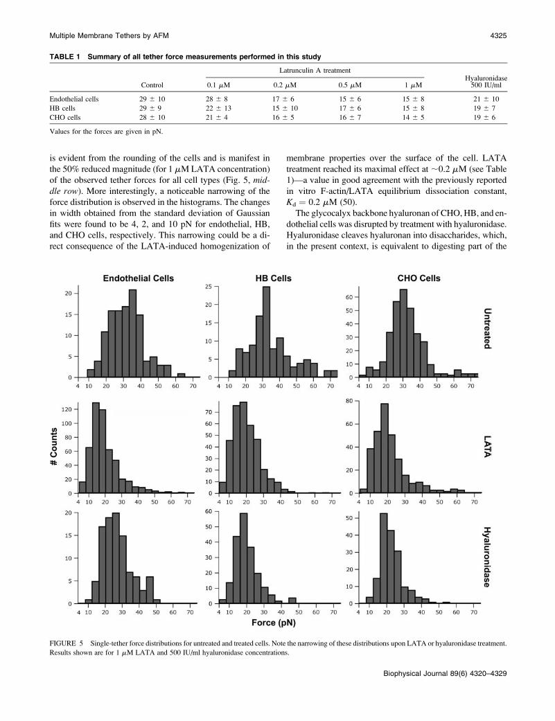
is evident from the rounding of the cells and is manifest in
the 50% reduced magnitude (for 1 mM LATA concentration)
of the observed tether forces for all cell types (Fig. 5, mid-dle row). More interestingly, a noticeable narrowing of the
force distribution is observed in the histograms. The changes
in width obtained from the standard deviation of Gaussian
fits were found to be 4, 2, and 10 pN for endothelial, HB,
and CHO cells, respectively. This narrowing could be a di-
rect consequence of the LATA-induced homogenization of
membrane properties over the surface of the cell. LATA
treatment reached its maximal effect at ;0.2 mM (see Table
1)—a value in good agreement with the previously reported
in vitro F-actin/LATA equilibrium dissociation constant,
Kd ¼ 0.2 mM (50).
The glycocalyx backbone hyaluronan of CHO, HB, and en-
dothelial cells was disrupted by treatment with hyaluronidase.
Hyaluronidase cleaves hyaluronan into disaccharides, which,
in the present context, is equivalent to digesting part of the
TABLE 1 Summary of all tether force measurements performed in this study
Latrunculin A treatmentHyaluronidase
Control 0.1 mM 0.2 mM 0.5 mM 1 mM 500 IU/ml
Endothelial cells 29 6 10 28 6 8 17 6 6 15 6 6 15 6 8 21 6 10
HB cells 29 6 9 22 6 13 15 6 10 17 6 6 15 6 8 19 6 7
CHO cells 28 6 10 21 6 4 16 6 5 16 6 7 14 6 5 19 6 6
Values for the forces are given in pN.
FIGURE 5 Single-tether force distributions for untreated and treated cells. Note the narrowing of these distributions upon LATA or hyaluronidase treatment.
Results shown are for 1 mM LATA and 500 IU/ml hyaluronidase concentrations.
Multiple Membrane Tethers by AFM 4325
Biophysical Journal 89(6) 4320–4329
glycocalyx (51). The tether force measured after hyaluron-
idase treatment was ;30% lower than for untreated cells
(Fig. 5, bottom row and Table 1). As with the experiments
using LATA, one again observes a narrowing in the force
distribution (at least for the HB and CHO cells, with the
respective changes in width of 4 and 8 pN; for the endothelial
cells, the width remained unchanged), suggesting that the
glycocalyx also contributes to the heterogeneity of plasma
membrane properties. Overall, these findings show the impor-
tance of the macromolecular networks on both the intra- and
extracellular sides of the cell membrane in the biomechanical
integrity of the cell.
DISCUSSION
Formation of a tether requires large changes in membrane
curvature. The associated energy cost depends on the bio-
mechanical properties (i.e., bending rigidity, effective surface
tension (58,59)) of the bilayer, as well as its association with
the cytoskeleton and glycocalyx. AFM provides an alterna-
tive means to directly probe these properties through mul-
tiple tether extraction and pulling studies. Among the three
cell lines used in our experiments, at the applied pulling rate,
no major variation in single tether forces was found, imply-
ing that specific local cell membrane characteristics in these
three cell types do not influence the growth of tethers. This
finding also suggests that multiple tether formation is a
ubiquitous phenomenon that can be initiated by nonspecific
binding between a cell and its environment (e.g., other cells),
and it is primarily a property of the plasma membrane itself
that may be utilized in vastly differing circumstances.
Our measured values of tether forces are in good agree-
ment with previously published data ranging from 10 to 60
pN for a variety of immobilized cells. Tether forces similar
to those reported here have been measured at comparable
pulling rates for fibroblasts (16), melanoma cell lines (18),
neuronal growth cone membranes (24), and red blood cells
(21). In contrast to previous studies using optical traps and
micropipette aspiration that have the ability to extract indi-
vidual tethers, with the AFM cantilever we simultaneously
extracted multiple tethers, but retained the sensitivity to
detect the loss of individual tethers as the membrane reser-
voir was gradually depleted. Initiation of multiple tether
extraction requires overcoming a large initial potential barrier
visible in the force profiles in Fig. 3. The force required to
overcome the initial adhesion is typically larger than the
maximum force that can be attained using optical traps or
micropipette aspiration. One must therefore be careful how
much surface area contacts the cell when using these techniques
to ensure that the number of tethers extracted does not exceed
the limiting forces. Attainment of the forces necessary to
initiate extraction of multiple tethers is possible with the AFM.
The observation of discrete force steps in our experiments
raises the question of the origin of these rupturelike events.
First, tethers could snap along their length. However, this
would require overcoming close-to-lytic membrane tensions
and, correspondingly, as experiments (15) and theory (60)
indicate, forces of ;100 pN, considerably higher than our
measured values. Second, it has been suggested that hetero-
geneities within the membrane could locally decrease its
tension and lead to tether fission (61). This effect is expected
to be cell-type-dependent and thus not likely to act in our
experiments, in which no significant differences in tether
force were observed. Third, theory suggests that multiple
tethers extracted locally from the membrane reservoir in
synthetic vesicles could fuse, but that pinning forces may
prevent fusion (31). The glycocalyx and the cytoskeleton,
absent in synthetic vesicles, provide ample possibilities for
local pinning, thus fusion of tethers in living cells is highly
regulated by these structures. Since in our experiments at
least one of these macromolecular networks is always intact,
the probability for fusion should be quite low. Experimental
confirmation of tether fusion in synthetic vesicles has re-
cently been provided (62). They showed that as fusion pro-
ceeds, a sudden change, similar to the discontinuities in Fig.
3, occurs in the components of the pulling force. The compo-
nents parallel and perpendicular to the axis of the fused
tethers respectively decrease and increase abruptly, while the
overall force remains constant (for a given membrane tension).
Interestingly, these results also strongly suggest that, in our
experiments, tethers do not fuse. Fusion of tethers takes time.
In the experiments of Cuvelier et al. (62), under static con-
ditions (no pulling force exerted) the velocity of fusion was
found to be ;80 mm/s, and thus the time for two tethers to
fuse along a 12-mm section to be 150 ms. Fusion in living
cells, if it takes place, should be considerably slower due to
heterogeneities in the membrane. However, as results in Fig.
3 show, force-steps occur much faster at comparable tether
length. In fact, with the time-resolution of our device (,10
ms), we can estimate the time of a force-step to be an order-
of-magnitude less than the time needed for fusion. Finally,
tethers can detach from the cantilever. Considering that we
do not reach the force necessary for tether rupture, and our
force-steps occur on a timescale inconsistent with the fusion
of tethers, we can assume that the force-steps result from
detachment of the tethers from the cantilever.
As we pull groups of tethers with the AFM cantilever,
they typically break off one by one. The extended membrane
is then reincorporated into the membrane reservoir and
the process continues until all of the tethers are released. The
rigidity of the plasma membrane, conferred through the
properties of its intrinsic components and peripherally asso-
ciated macromolecules, defines the limits of the reservoir
available to form tethers. This aspect is best illustrated by the
measurement of the extent of the membrane reservoir as
shown in Fig. 4. When measured within an individual force
elongation profile, the size of the membrane reservoir probed
was found to be approximately constant for each rupture
event between consecutive plateaus. This result suggests
4326 Sun et al.
Biophysical Journal 89(6) 4320–4329
that, when probed locally with an AFM tip, multiple simul-
taneously extracted membrane tethers are equally coupled
to the membrane reservoir of the cell.
Previous studies demonstrated that tether properties de-
pend on both actin microfilaments and microtubules—major
components of the cytoskeleton (16). Our results support
these earlier findings. Indeed, we observed an ;50% decrease
in tether force after the inhibition of actin polymerization,
indicating that cytoskeletal integrity is crucial in the
regulation of the membrane’s biomechanical characteristics.
In contrast to the rather well-established function of the
intracellular cortical cytoskeleton in the biomechanical role
of the cell membrane, relatively little is known about the role
of the proteoglycan complexes covering the extracellular
surface of the phospholipid bilayer. Our finding of a 30%
decrease in single tether force due to the disruption of the
glycocalyx highlights the importance of glycosaminoglycans
on the mechanical properties of the lipid bilayer. Hence the
hyaluronan backbone of the glycocalyx has a distinct effect
on the mechanical properties of the plasma membrane in
intact cells (51).
Multiple tethers are manifest in AFM pulling experi-
ments as plateaus separated by well-defined steps in the force
profile. In this study, we evaluated the force required to pull
individual tethers directly from the measurement of these
force steps. Therefore, each force histogram presented in
Fig. 5 represents an ensemble of measurements performed
at many different positions on many cells. It is expected
that, over the entire cell surface, heterogeneity exists in
the coupling between the glycocalyx, cytoskeleton, and the
membrane. Although the change in the peak values in the
histograms after the two treatments is a specific measure
of the coupling between the membrane and its peripheral
macromolecular networks, the heterogeneity of the coupling
is manifest through the broad distribution in the histograms
for the nontreated cells. The marked narrowing of the histo-
grams in LATA- and hyaluronidase-treated cells illustrates
the reduction of this heterogeneity achieved through dis-
ruption of the cytoskeleton and glycocalyx.
It has been shown that as a first approximation, the total
force, Ft, necessary to extract a tether, can be considered to
be the sum of all macromolecular contributions (18). Follow-
ing this approach, Ft can be expressed in terms of the
individual contributions due to the association between the
cytoskeleton and the membrane (Fc/m), the coupling between
the glycocalyx and the membrane (Fg/m), and the force to
pull a tether composed of a pure cellular membrane (Fm):
Ft ¼ Fc=m 1Fg=m 1Fm: (1)
In our study, the contributions of the cytoskeleton and the
glycocalyx were measured by selectively disrupting these
macromolecular networks. The difference between the tether
forces measured on intact cells (Ft) and on cells depleted of
their cytoskeleton or glycocalyx could provide an estimate
for Fc/m and Fg/m, respectively. Our experiments with the
endothelial, HB, and CHO cells (untreated, 1 mM LATA-,
and 500 IU/ml hyaluronidase-treated) allow us to calculate
Fm values of 7 pN, 5 pN, and 5 pN, respectively. These
values are obtained from Eq. 1, by assuming that LATA and
hyaluronidase treatments results, respectively, in Fc/m ¼ 0 and
Fg/m ¼ 0. An experiment in which cells are treated with both
LATA and hyaluronidase (i.e., Ft � Fm) could have allowed
us to simultaneously eliminate the glycocalyx and the cyto-
skeletal contributions. However, it was not possible to per-
form such an experiment due to extensive desorption of the
cells from the surface. Nevertheless, our calculated estimate
for the Fm component in the total tether force is in good
agreement with the experimental value of 8 pN measured for
a phospholipid membrane decoupled from the cytoskeleton
(18), and the theoretical value of 13 pN for a pure phos-
pholipid vesicle (31).
Based on these findings, we propose that any cellular
process that significantly affects the molecular networks inter-
acting with the phospholipid bilayer influences its effective
mechanical properties, and that this effect can be measured
using atomic force microscopy. Furthermore, our results in-
dicate that living cells can maintain multiple tethers. Local
compositional modifications in the plasma membrane, as well
as its association with the cytoskeleton and glycocalyx,
through heterogeneities, can prevent the fusion of these co-
existing nanotubes and thus control their number. This may
provide living cells with an additional mechanism to regulate
their adhesive properties.
The authors thank Evan Evans for useful discussions and Charles Cuerrier
for help with supplemental experiments.
This study was partially supported by grants from the National Science
Foundation and the National Aeronautics and Space Administration (to G.F.)
and the Natural Sciences and Engineering Research Council (to M.G.).
REFERENCES
1. Sheetz, M. P. 1995. Cellular plasma membrane domains. Mol. Membr.Biol. 12:89–91.
2. Schmidtke, D. W., and S. L. Diamond. 2000. Direct observation ofmembrane tethers formed during neutrophil attachment to platelets orP-selectin under physiological flow. J. Cell Biol. 149:719–730.
3. Roux, A., G. Cappello, J. Cartaud, J. Prost, B. Goud, and P. Bassereau.2002. A minimal system allowing tubulation with molecular motorspulling on giant liposomes. Proc. Natl. Acad. Sci. USA. 99:5394–5399.
4. Iglic, A., H. Hagerstrand, M. Bobrowska-Hagerstrand, V. Arrigler, andV. Kralj-Iglic. 2003. Possible role of phospholipid nanotubes indirected transport of membrane vesicles. Phys. Lett. A. 310:493–497.
5. Koster, G., M. VanDuijn, B. Hofs, and M. Dogterom. 2003. Membranetube formation from giant vesicles by dynamic association of motorproteins. Proc. Natl. Acad. Sci. USA. 100:15583–15588.
6. Rustom, A., R. Saffrich, I. Markovic, P. Walther, and H.-H. Gerdes.2004. Nanotubular highways for intercellular organelle transport.Science. 303:1007–1010.
7. Upadhyaya, A., and M. P. Sheetz. 2004. Tension in tubulovesicularnetworks of Golgi and endoplasmic reticulum membranes. Biophys. J.86:2923–2928.
Multiple Membrane Tethers by AFM 4327
Biophysical Journal 89(6) 4320–4329
8. Giuffre, L., A.-S. Cordy, N. Monai, Y. Tardy, M. Schapira, and O. Spertini.1997. Monocyte adhesion to activated aortic endothelium: roleof L-selectin and heparan sulfate proteoglycans. J. Cell Biol. 136:945–956.
9. Sanders, W. J., E. J. Gordon, O. Dwir, P. J. Beck, R. Alon, and L. L.Kiessling. 1999. Inhibition of L-selectin-mediated leukocyte rolling bysynthetic glycoprotein mimics. J. Biol. Chem. 274:5271–5278.
10. DeGrendele, H. C., P. Estess, L. J. Picker, and M. H. Siegelman. 1996.CD44 and its ligand hyaluronate mediate rolling under physiologicflow: a novel lymphocyte-endothelial cell primary adhesion pathway.J. Exp. Med. 183:1119–1130.
11. Chen, S., and T. A. Springer. 1999. An automatic braking system thatstabilizes leukocyte rolling by an increase in selectin bond number withshear. J. Cell Biol. 144:185–200.
12. Evans, E. A., and R. M. Hochmuth. 1976. Membrane viscoelasticity.Biophys. J. 16:1–12.
13. Evans, E. A., and R. M. Hochmuth. 1976. Membrane viscoplastic flow.Biophys. J. 16:13–26.
14. Zhelev, D. V., and R. M. Hochmuth. 1995. Mechanically stimulated cyto-skeleton rearrangement and cortical contraction. Biophys. J. 68:2004–2014.
15. Dai, J., M. P. Sheetz, X. Wan, and C. E. Morris. 1998. Membranetension in swelling and shrinking molluscan neurons. J. Neurosci. 18:6681–6692.
16. Raucher, D., and M. P. Sheetz. 1999. Characteristics of a membranereservoir buffering membrane tension. Biophys. J. 77:1992–2000.
17. Raucher, D., and M. P. Sheetz. 2001. Phospholipase C activation byanesthetics decreases membrane-cytoskeleton adhesion. J. Cell Sci.114:3759–3766.
18. Dai, J., and M. P. Sheetz. 1999. Membrane tether formation fromblebbing cells. Biophys. J. 77:3363–3370.
19. Girdhar, G., and J. Y. Shao. 2004. Membrane tether extraction fromhuman umbilical vein endothelial cells and its implication in leukocyterolling. Biophys. J. 87:3561–3568.
20. Hochmuth, R. M., N. Mohandas, and P. L. Blackshear. 1973. Mea-surement of the elastic modulus for red cell membrane using a fluidmechanical technique. Biophys. J. 13:747–762.
21. Hwang, W. C., and R. E. Waugh. 1997. Energy of dissociation of lipidbilayer from the membrane skeleton of red blood cells. Biophys. J.72:2669–2678.
22. Shao, J. Y., and R. M. Hochmuth. 1996. Micropipette suction formeasuring picoNewton forces of adhesion and tether formation fromneutrophil membranes. Biophys. J. 71:2892–2901.
23. Waugh, R. E., A. Mantalaris, R. G. Bauserman, W. C. Hwang, andJ. H. D. Wu. 2001. Membrane instability in late-stage erythropoiesis.Blood. 97:1869–1875.
24. Dai, J., and M. P. Scheetz. 1995. Mechanical properties of neuronalgrowth cone membrane studied by tether formation with laser opticaltweezers. Biophys. J. 68:988–996.
25. Li, Z., B. Anvari, M. Takashima, P. Brecht, J. H. Torres, and W. E.Brownell. 2002. Membrane tether formation from outer hair cells withoptical tweezers. Biophys. J. 82:1386–1395.
26. Raucher, D., T. Stauffer, W. Chen, K. Shen, S. Guo, J. D. York, M. P.Sheetz, and T. Meyer. 2000. Phosphatidylinositol 4,5-bisphosphatefunctions as a second messenger that regulates cytoskeleton-plasmamembrane adhesion. Cell. 100:221–228.
27. Sheetz, M. P. 2001. Cell control by membrane-cytoskeleton adhesion.Nat. Rev. Mol. Cell Biol. 2:392–396.
28. Hochmuth, F. M., J. Y. Shao, J. Dai, and M. P. Sheetz. 1996.Deformation and flow of membrane into tethers extracted fromneuronal growth cones. Biophys. J. 70:358–369.
29. Xu, G., and J. Y. Shao. 2005. Double tether extraction from humanneutrophils and its comparison with CD41 T lymphocytes. Biophys. J.88:661–669.
30. Leduc, C., O. Campas, K. B. Zeldovich, A. Roux, P. Jilimaitre, L.Bourel-Bonnet, B. Goud, J.-F. Joanny, P. Bassereau, and J. Prost.
2004. Cooperative extraction of membrane nanotubes by molecularmotors. Proc. Natl. Acad. Sci. USA. 101:17096–17101.
31. Derenyi, I., F. Julicher, and J. Prost. 2002. Formation and interaction ofmembrane tubes. Phys. Rev. Lett. 88:238101.
32. Binnig, G., C. F. Quate, and C. Gerber. 1986. Atomic forcemicroscope. Phys. Rev. Lett. 56:930–933.
33. Clausen-Schaumann, H., M. Seitz, R. Krautbauer, and H. E. Gaub.2000. Force spectroscopy with single biomolecules. Curr. Opin. Chem.Biol. 4:524–530.
34. Zlatanova, J., S. M. Lindsay, and S. H. Leuba. 2000. Single moleculeforce spectroscopy in biology using the atomic force microscope. Prog.Biophys. Mol. Biol. 74:37–61.
35. Grandbois, M., M. Beyer, M. Rief, H. Clausen-Schaumann, andH. E. Gaub. 1999. How strong is a covalent bond? Science. 283:1727–1730.
36. Fernandez, J. M., and H. Li. 2004. Force-clamp spectroscopy monitorsthe folding trajectory of a single protein. Science. 303:1674–1678.
37. Oberhauser, A. F., P. E. Marszalek, H. P. Erickson, and J. M.Fernandez. 1998. The molecular elasticity of the extracellular matrixprotein tenascin. Nature. 393:181–185.
38. Oesterhelt, F., D. Oesterhelt, M. Pfeiffer, A. Engel, H. E. Gaub, and D. J.Muller. 2000. Unfolding pathways of individual bacteriorhodopsins.Science. 288:143–146.
39. Rief, M., M. Gautel, F. Oesterhelt, J. M. Fernandez, and H. E. Gaub.1997. Reversible unfolding of individual titin immunoglobulin do-mains by AFM. Science. 276:1109–1112.
40. Zhuang, X., and M. Rief. 2003. Single-molecule folding. Curr. Opin.Struct. Biol. 13:88–97.
41. Grandbois, M., W. Dettmann, M. Benoit, and H. E. Gaub. 2000.Affinity imaging of red blood cells using an atomic force microscope.J. Histochem. Cytochem. 48:719–724.
42. Moy, V. T., E. L. Florin, and H. E. Gaub. 1994. Intermolecularforces and energies between ligands and receptors. Science. 266:257–259.
43. Baumgartner, W., P. Hinterdorfer, W. Ness, A. Raab, D. Vestweber,H. Schindler, and D. Drenckhahn. 2000. Cadherin interactionprobed by atomic force microscopy. Proc. Natl. Acad. Sci. USA. 97:4005–4010.
44. Benoit, M., D. Gabriel, G. Gerisch, and H. Gaub. 2000. Discreteinteractions in cell adhesion measured by single-molecule force spec-troscopy. Nat. Cell Biol. 2:313–317.
45. Benoit, M., and H. E. Gaub. 2002. Measuring cell adhesion forceswith the atomic force microscope at the molecular level. Cells TissuesOrgans. 172:174–189.
46. Hegedus, B., A. Czirok, I. Fazekas, T. Babel, E. Madarasz, and T.Vicsek. 2000. Locomotion and proliferation of glioblastoma cellsin vitro: statistical evaluation of videomicroscopic observations. J.Neurosurg. 92:428–434.
47. Edgell, C.-J. S., C. C. McDonald, and J. B. Graham. 1983. Permanentcell line expressing human factor VIII-related antigen established byhybridization. Proc. Natl. Acad. Sci. USA. 80:3734–3737.
48. Coue, M., S. L. Brenner, I. Spector, and E. D. Korn. 1987. Inhibition ofactin polymerization by latrunculin A. FEBS Lett. 213:316–318.
49. Spector, I., N. R. Shochet, D. Blasberger, and Y. Kashman. 1989.Latrunculins—novel marine macrolides that disrupt microfilamentorganization and affect cell growth. I. Comparison with cytochalasin D.Cell Motil. Cytoskel. 13:127–144.
50. Yarmola, E. G., T. Somasundaram, T. A. Boring, I. Spector, and M. R.Bubb. 2000. Actin-latrunculin A structure and function. J. Biol. Chem.275:28120–28127.
51. Zaidel-Bar, R., M. Cohen, L. Addadi, and B. Geiger. 2004.Hierarchical assembly of cell-matrix adhesion complexes. Biochem.Soc. Trans. 32:416–420.
52. Butt, H.-J., and M. Jashke. 1995. Thermal noise in atomic forcespectroscopy. Nanotechnology. 6:1–7.
4328 Sun et al.
Biophysical Journal 89(6) 4320–4329
53. Hutter, J. L., and J. Bechhoefer. 1993. Calibration of atomic-forcemicroscope tips. Rev. Sci. Instrum. 64:1868–1873.
54. Girdhar, G., and J.-Y. Shao. 2004. Membrane tether extraction fromhuman umbilical vein endothelial cells and its implication in leukocyterolling. Biophys. J. 87:3561–3568.
55. Rotsch, C., and M. Radmacher. 2000. Drug-induced changes ofcytoskeletal structure and mechanics in fibroblasts: an atomic forcemicroscopy study. Biophys. J. 78:520–535.
56. Chiappuis-Flament, S., E. Wong, L. D. Hicks, C. M. Kay, and B. M.Gumbiner. 2001. Multiple cadherin extracellular repeats mediate homo-philic binding adhesion. J. Cell Biol. 154:231–243.
57. Critchley, D. R. 2000. Focal adhesions—the cytoskeletal connection.Curr. Opin. Cell Biol. 12:133–139.
58. Lipowsky, R. 1995. The morphology of lipid membranes. Curr. Opin.Struct. Biol. 5:531–540.
59. Needham, D., and R. M. Hochmuth. 1992. A sensitive measure ofsurface stress in the resting neutrophil. Biophys. J. 61:1664–1670.
60. Evans, E., and F. Ludwig. 2000. Dynamic strengths of molecularanchoring and material cohesion in fluid bio membranes. J. Phys.Condens. Matter. 12:A315–A320.
61. Allain, J.-M., C. Storm, A. Roux, M. Ben Amar, and J.-F. Joanny.2004. Fission of a multiphase membrane tube. Phys. Rev. Lett. 93:158104.
62. Cuvelier, D., I. Derenyi, P. Bassereau, and P. Nassoy. 2005.Coalescence of membrane tethers: experiments, theory, and applica-tions. Biophys. J. 86:2714–2726.
Multiple Membrane Tethers by AFM 4329
Biophysical Journal 89(6) 4320–4329
Related Documents