Multilocus Sequence Analysis of Xanthomonads Causing Bacterial Spot of Tomato and Pepper Plants Reveals Strains Generated by Recombination among Species and Recent Global Spread of Xanthomonas gardneri Sujan Timilsina, a,b Mustafa O. Jibrin, c Neha Potnis, a Gerald V. Minsavage, a Misrak Kebede, d Allison Schwartz, e Rebecca Bart, f Brian Staskawicz, e Claudine Boyer, g Gary E. Vallad, a,b Olivier Pruvost, g Jeffrey B. Jones, a Erica M. Goss a,h Department of Plant Pathology, University of Florida, Gainesville, Florida, USA a ; Gulf Coast Research and Education Center, University of Florida, Wimauma, Florida, USA b ; Department of Crop Protection, Ahmadu Bello University, Zaria, Nigeria c ; Plant Pathology Department, School of Plant Science, Haramaya University, Dire Dawa, Ethiopia d ; Department of Plant and Microbial Biology, University of California—Berkeley, California, USA e ; Danforth Center, St. Louis, Missouri, USA f ; CIRAD, UMR Peuplements Végétaux et Bioagresseurs en Milieu Tropical CIRAD-Université de la Réunion, Pôle de Protection des Plantes, Saint Pierre, La Réunion, France g ; Emerging Pathogens Institute, University of Florida, Gainesville, Florida, USA h Four Xanthomonas species are known to cause bacterial spot of tomato and pepper, but the global distribution and genetic di- versity of these species are not well understood. A collection of bacterial spot-causing strains from the Americas, Africa, South- east Asia, and New Zealand were characterized for genetic diversity and phylogenetic relationships using multilocus sequence analysis of six housekeeping genes. By examining strains from different continents, we found unexpected phylogeographic pat- terns, including the global distribution of a single multilocus haplotype of X. gardneri, possible regional differentiation in X. vesicatoria, and high species diversity on tomato in Africa. In addition, we found evidence of multiple recombination events be- tween X. euvesicatoria and X. perforans. Our results indicate that there have been shifts in the species composition of bacterial spot pathogen populations due to the global spread of dominant genotypes and that recombination between species has gener- ated genetic diversity in these populations. U nderstanding the evolution and host specificity of plant- pathogenic bacteria is an ongoing challenge. Strains of phy- topathogenic bacteria commonly exhibit high host specificity, with host ranges restricted to one or a few plant species (1, 2). Bacterial plant pathogens also exhibit biogeography, such that species can be limited in their geographic distributions (3). Glob- alization of agriculture has contributed to the dispersal of phyto- pathogenic bacteria, but the geographic ranges of species are not well characterized, in part because of the difficulty in differentiat- ing phylogenetically distinct strains that have similar host speci- ficities (4). Phenotypic characters can sometimes distinguish spe- cies with similar host specificities, but classification by molecular markers is often required due to variation in phenotypic traits within species (5). Phenotypes can also dramatically differ among strains within a species due to acquisition and loss of genes related to pathogenicity and fitness (4). Bacterial evolution is driven by point mutations, variation in gene content, recombination, and selection on the resulting phenotypes (6). Phylogenetic relation- ships among species are defined by point mutations in the genome that accumulate over time; however, these relationships can be obscured by polymorphisms that have been distributed to other closely related species via homologous recombination and hori- zontal gene transfer (7). These events can introduce conflicting phylogenetic signals between genes that have been vertically in- herited versus horizontally acquired (8). The possibility of infec- tion of a single host plant by multiple species may increase the probability of genetic exchange (9). Coinfection by multiple spe- cies may be more common as pathogens are moved out of their native geographic ranges. Multilocus nucleotide-sequence-based approaches help in re- solving phylogenetic relationships of bacteria within and between species (10). Multilocus sequence typing (MLST) and analysis (MLSA) are two approaches used to analyze multiple housekeep- ing genes that are conserved in sequence and present in strains of closely related species. MLST is useful in grouping strains from different groups of species but is limited by the sequence diversity based on allelic mismatches observed within the same species (10). In contrast, MLSA makes use of concatenated nucleotide se- quences of the housekeeping genes for characterization of more diverse strains representing multiple species within a genus by constructing phylogenetic trees (10, 11). Since the method is based on the nucleotide sequence, it provides unambiguous re- sults that are directly comparable, unlike randomly amplified polymorphic DNA (RAPD) or other anonymous marker systems. Since Gevers et al. (10) described the MLSA method; it has been Received 15 September 2014 Accepted 14 December 2014 Accepted manuscript posted online 19 December 2014 Citation Timilsina S, Jibrin MO, Potnis N, Minsavage GV, Kebede M, Schwartz A, Bart R, Staskawicz B, Boyer C, Vallad GE, Pruvost O, Jones JB, Goss EM. 2015. Multilocus sequence analysis of xanthomonads causing bacterial spot of tomato and pepper plants reveals strains generated by recombination among species and recent global spread of Xanthomonas gardneri. Appl Environ Microbiol 81: 1520 –1529. doi:10.1128/AEM.03000-14. Editor: H. Goodrich-Blair Address correspondence to Jeffrey B. Jones, jbjones@ufl.edu. G.E.V., O.P., J.B.J., and E.M.G. are joint last authors. Supplemental material for this article may be found at http://dx.doi.org/10.1128 /AEM.03000-14. Copyright © 2015, American Society for Microbiology. All Rights Reserved. doi:10.1128/AEM.03000-14 1520 aem.asm.org February 2015 Volume 81 Number 4 Applied and Environmental Microbiology on February 11, 2015 by Mustafa Jibrin http://aem.asm.org/ Downloaded from

Welcome message from author
This document is posted to help you gain knowledge. Please leave a comment to let me know what you think about it! Share it to your friends and learn new things together.
Transcript
Multilocus Sequence Analysis of Xanthomonads Causing BacterialSpot of Tomato and Pepper Plants Reveals Strains Generated byRecombination among Species and Recent Global Spread ofXanthomonas gardneri
Sujan Timilsina,a,b Mustafa O. Jibrin,c Neha Potnis,a Gerald V. Minsavage,a Misrak Kebede,d Allison Schwartz,e Rebecca Bart,f
Brian Staskawicz,e Claudine Boyer,g Gary E. Vallad,a,b Olivier Pruvost,g Jeffrey B. Jones,a Erica M. Gossa,h
Department of Plant Pathology, University of Florida, Gainesville, Florida, USAa; Gulf Coast Research and Education Center, University of Florida, Wimauma, Florida, USAb;Department of Crop Protection, Ahmadu Bello University, Zaria, Nigeriac; Plant Pathology Department, School of Plant Science, Haramaya University, Dire Dawa, Ethiopiad;Department of Plant and Microbial Biology, University of California—Berkeley, California, USAe; Danforth Center, St. Louis, Missouri, USAf; CIRAD, UMR PeuplementsVégétaux et Bioagresseurs en Milieu Tropical CIRAD-Université de la Réunion, Pôle de Protection des Plantes, Saint Pierre, La Réunion, Franceg; Emerging PathogensInstitute, University of Florida, Gainesville, Florida, USAh
Four Xanthomonas species are known to cause bacterial spot of tomato and pepper, but the global distribution and genetic di-versity of these species are not well understood. A collection of bacterial spot-causing strains from the Americas, Africa, South-east Asia, and New Zealand were characterized for genetic diversity and phylogenetic relationships using multilocus sequenceanalysis of six housekeeping genes. By examining strains from different continents, we found unexpected phylogeographic pat-terns, including the global distribution of a single multilocus haplotype of X. gardneri, possible regional differentiation in X.vesicatoria, and high species diversity on tomato in Africa. In addition, we found evidence of multiple recombination events be-tween X. euvesicatoria and X. perforans. Our results indicate that there have been shifts in the species composition of bacterialspot pathogen populations due to the global spread of dominant genotypes and that recombination between species has gener-ated genetic diversity in these populations.
Understanding the evolution and host specificity of plant-pathogenic bacteria is an ongoing challenge. Strains of phy-
topathogenic bacteria commonly exhibit high host specificity,with host ranges restricted to one or a few plant species (1, 2).Bacterial plant pathogens also exhibit biogeography, such thatspecies can be limited in their geographic distributions (3). Glob-alization of agriculture has contributed to the dispersal of phyto-pathogenic bacteria, but the geographic ranges of species are notwell characterized, in part because of the difficulty in differentiat-ing phylogenetically distinct strains that have similar host speci-ficities (4). Phenotypic characters can sometimes distinguish spe-cies with similar host specificities, but classification by molecularmarkers is often required due to variation in phenotypic traitswithin species (5). Phenotypes can also dramatically differ amongstrains within a species due to acquisition and loss of genes relatedto pathogenicity and fitness (4). Bacterial evolution is driven bypoint mutations, variation in gene content, recombination, andselection on the resulting phenotypes (6). Phylogenetic relation-ships among species are defined by point mutations in the genomethat accumulate over time; however, these relationships can beobscured by polymorphisms that have been distributed to otherclosely related species via homologous recombination and hori-zontal gene transfer (7). These events can introduce conflictingphylogenetic signals between genes that have been vertically in-herited versus horizontally acquired (8). The possibility of infec-tion of a single host plant by multiple species may increase theprobability of genetic exchange (9). Coinfection by multiple spe-cies may be more common as pathogens are moved out of theirnative geographic ranges.
Multilocus nucleotide-sequence-based approaches help in re-solving phylogenetic relationships of bacteria within and between
species (10). Multilocus sequence typing (MLST) and analysis(MLSA) are two approaches used to analyze multiple housekeep-ing genes that are conserved in sequence and present in strains ofclosely related species. MLST is useful in grouping strains fromdifferent groups of species but is limited by the sequence diversitybased on allelic mismatches observed within the same species (10).In contrast, MLSA makes use of concatenated nucleotide se-quences of the housekeeping genes for characterization of morediverse strains representing multiple species within a genus byconstructing phylogenetic trees (10, 11). Since the method isbased on the nucleotide sequence, it provides unambiguous re-sults that are directly comparable, unlike randomly amplifiedpolymorphic DNA (RAPD) or other anonymous marker systems.Since Gevers et al. (10) described the MLSA method; it has been
Received 15 September 2014 Accepted 14 December 2014
Accepted manuscript posted online 19 December 2014
Citation Timilsina S, Jibrin MO, Potnis N, Minsavage GV, Kebede M, Schwartz A,Bart R, Staskawicz B, Boyer C, Vallad GE, Pruvost O, Jones JB, Goss EM. 2015.Multilocus sequence analysis of xanthomonads causing bacterial spot of tomatoand pepper plants reveals strains generated by recombination among speciesand recent global spread of Xanthomonas gardneri. Appl Environ Microbiol 81:1520 –1529. doi:10.1128/AEM.03000-14.
Editor: H. Goodrich-Blair
Address correspondence to Jeffrey B. Jones, [email protected].
G.E.V., O.P., J.B.J., and E.M.G. are joint last authors.
Supplemental material for this article may be found at http://dx.doi.org/10.1128/AEM.03000-14.
Copyright © 2015, American Society for Microbiology. All Rights Reserved.
doi:10.1128/AEM.03000-14
1520 aem.asm.org February 2015 Volume 81 Number 4Applied and Environmental Microbiology
on February 11, 2015 by M
ustafa Jibrinhttp://aem
.asm.org/
Dow
nloaded from
applied to numerous pathogenic and nonpathogenic bacteria.Housekeeping genes also are subject to homologous recombina-tion and help to approximate the extent and impact of recombi-nation in bacterial evolution (12). As a result, MLSA has been usedto estimate rates of recombination, which vary widely among bac-terial species (13). Because recombination and horizontal genetransfer can result in qualitative differences in phenotype betweenphylogenetically closely related strains, pathogenicity tests andphenotypic assays remain critical to characterizing strains and in-terpreting MLSA results.
The genus Xanthomonas comprises numerous pathogenic spe-cies infecting approximately 400 different host plants (14). Phe-notypic and phylogenetic analyses have shown a wide range ofvariation among Xanthomonas strains that cause bacterial spot oftomato and pepper (15, 16). Bacterial spot is caused by four dif-ferent species: Xanthomonas euvesicatoria, X. vesicatoria, X. per-forans, and X. gardneri (3, 17). Among the four species, X. euvesi-catoria and X. gardneri strains are reported as pathogens of bothtomato and pepper, X. perforans strains are reported only fromtomato, and X. vesicatoria strains primarily infect tomato. Strainsbelonging to X. euvesicatoria and X. vesicatoria have a worldwidedistribution (18). X. perforans and/or X. gardneri strains increas-ingly have been isolated in Canada (19), the United States andSouth America, and regions bordering the Indian Ocean (20–22).These bacterial populations can also change over time. For exam-ple, the bacterial spot pathogen population on tomato in Floridashifted from X. euvesicatoria to X. perforans. Prior to 1991, only X.euvesicatoria strains were found in Florida. In a survey in 2006 and2007, only X. perforans strains were isolated (23), correspondingto a shift in tomato races. The origin of the X. perforans strains nowresponsible for bacterial spot in Florida tomatoes is unknown, inpart because the global distribution of this species is not well char-acterized.
MLSA of Xanthomonas species has been used for phylogeneticsof the genus and to examine evolution via recombination. AnMLSA database of Xanthomonas strains has been created using sixhousekeeping genes (fusA, gapA, gltA, gyrB, lacF, and lepA) (15).MLSA has revealed recombination as a primary factor underlyingthe evolution of X. axonopodis (24). Some xanthomonad popula-tions have been reported as highly clonal, with little variationamong strains collected from geographically distant locations (25,26). MLSA also has been applied to bacterial spot-causing xan-thomonads. Strains causing bacterial spot of tomato and pepperin the southwest Indian Ocean region were examined; all fourspecies were found (22). A recent study found three different spe-cies responsible for bacterial spot of tomato in Ethiopia (27), whileanother study found atypical strains in Grenada and India (28).Findings of species diversity and dynamic shifts in species re-ported from previous regional studies prompted us to applyMLSA and MLST to a collection of bacterial spot-causing xan-thomonads from diverse geographic origins. Bacterial strains rep-resenting four Xanthomonas species associated with bacterial spotof tomato and pepper collected from the Americas, Africa, South-east Asia, and New Zealand were examined using MLSA to under-stand the phylogeographic diversity of bacterial spot pathogens.Our objectives were to determine the geographic distribution ofthe four Xanthomonas species, the extent of diversity within spe-cies, and the role of homologous recombination in generatingdiversity in the bacterial spot pathogens.
MATERIALS AND METHODSBacterial strains. Strains from multiple collections of xanthomonads iso-lated from tomato and pepper exhibiting bacterial spot were used (Table1). The collections were mainly from the United States and Africa, includ-ing the southwest Indian Ocean (SWIO) islands previously reported byHamza et al. (22), with smaller representative samples from elsewhere inthe Americas, India, and New Zealand. These strains were subjected toMLSA using the six housekeeping genes fusA, gapA, gltA, gyrB, lacF, andlepA (16). Sequences were either obtained via Sanger sequencing or ex-tracted from whole genome sequences. The sequenced strains were com-pared with type strains of X. vesicatoria and X. gardneri. Reference strainsfrom X. euvesicatoria (strain 85-10) and X. perforans (strain 91-118) wereused as both these strains have been extensively characterized in previousstudies (16, 29) and have sequences identical to that of the type strain fromtheir respective species for the six housekeeping genes (16).
Phylogenetic analysis. Sequences for six housekeeping genes from theworldwide strains along with reference Xanthomonas strains were alignedusing MUSCLE within MEGA 5.2.1 (30). The alignments were furtherconfirmed via BioEdit software (31). Nucleotide substitution models thatbest fit the aligned sequences were selected using the Akaike InformationCriterion (AIC) within jModeltest 1.1 (32). The general time reversiblemodel with gamma-distributed invariant sites (GTR�G�I) model wasselected and used for construction of phylogenetic trees based on maxi-mum likelihood (ML) and Bayesian inference. Maximum likelihood phy-logenetic trees based on the six housekeeping genes were constructedindividually and using concatenated sequences. The maximum likelihoodtree, with 1,000 bootstrap samples, inferred using RaxML was comparedto ML, maximum parsimony (MP), and neighbor-joining (NJ) trees con-structed using the GTR�G�I model in MEGA 5.2.1 (29). MrBayes v.3.2(33) was used for the Bayesian phylogeny, using the same substitutionmodel with 1,000,000 Markov chain Monte Carlo (MCMC) steps, sam-pled every 500 steps. A burn-in period of 88,500 steps was used for theconcatenated data set and 56,000 steps for the individual genes (33, 34).Consensus trees obtained from MrBayes were visualized using FigTreeversion 1.4 (Institute of Evolutionary Biology, University of Edinburgh[http://tree.bio.ed.ac.uk/software/figtree/]). Results from the phyloge-netic analyses, along with phenotypic assays from previous studies, wereused to delineate groups of strains into species.
Analysis of diversity and recombination. Nucleotide diversity, thenumber of haplotypes, and the minimum number of recombinationevents were determined using DnaSP 5.0 (35). DnaSP also was used forcalculating class I neutrality tests (Tajima’s D and Fu and Li’s D* and F*)for detecting departure from the mutation/drift equilibrium (36, 37). Forthese calculations, all strains were considered together and by species.
Multiple methods were used to detect recombination. Splits-decom-position trees were constructed (38), and the pairwise homoplasy index(PHI) was calculated using SplitsTree version 4.13.1 (39). These calcula-tions used the concatenated genes for both the entire data set and a subsetof the data that included only X. perforans and X. euvesicatoria strains. TheRecombination Detection Program (RDP) version 4 combines sevennonparametric detection programs (3Seq, Chimaera, RDP, GENECONV,MaxChi, BootScan, and SiScan) to detect recombination and estimatebreakpoints (40). The default settings and a Bonferroni step-down cor-rection method with a P value cutoff of 0.05 were applied in the analysis ofthe concatenated data set. Recombination breakpoints also were identi-fied using GARD (Genetic Algorithm for Recombination Detection) (41).
After detection of recombination, the concatenated data were used toreconstruct a nonrecombinant coalescent-based genealogy usingClonalFrame 1.1 with the default settings (42). The MCMC used aburn-in period of 50,000 steps sampled every 100th step. Mutational rate(�), intragenic recombination rate (R), average length of recombinationevent (�), and the rate of new polymorphism generated due to recombi-nation were estimated along with time to most recent common ancestor(TMRCA) for all strains and for each species group. Outputs were used tocalculate the impact of recombination to mutation (r/m) and to measure
Phylogeny of Xanthomonads from Tomato and Pepper
February 2015 Volume 81 Number 4 aem.asm.org 1521Applied and Environmental Microbiology
on February 11, 2015 by M
ustafa Jibrinhttp://aem
.asm.org/
Dow
nloaded from
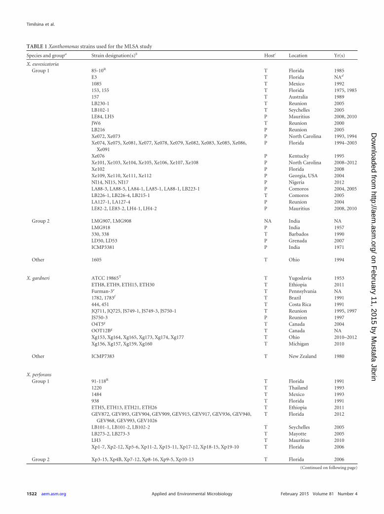
TABLE 1 Xanthomonas strains used for the MLSA study
Species and groupa Strain designation(s)b Hostc Location Yr(s)
X. euvesicatoriaGroup 1 85-10R T Florida 1985
E3 T Florida NAd
1085 T Mexico 1992153, 155 T Florida 1975, 1985157 T Australia 1989LB230-1 T Reunion 2005LB102-1 T Seychelles 2005LE84, LH5 P Mauritius 2008, 2010JW6 T Reunion 2000LB216 P Reunion 2005Xe072, Xe073 P North Carolina 1993, 1994Xe074, Xe075, Xe081, Xe077, Xe078, Xe079, Xe082, Xe083, Xe085, Xe086,
Xe091P Florida 1994–2003
Xe076 P Kentucky 1995Xe101, Xe103, Xe104, Xe105, Xe106, Xe107, Xe108 P North Carolina 2008–2012Xe102 P Florida 2008Xe109, Xe110, Xe111, Xe112 P Georgia, USA 2004NI14, NI15, NI17 P Nigeria 2012LA88-3, LA88-5, LA84-1, LA85-1, LA88-1, LB223-1 P Comoros 2004, 2005LB226-1, LB226-4, LB215-1 T Comoros 2005LA127-1, LA127-4 P Reunion 2004LE82-2, LE83-2, LH4-1, LH4-2 P Mauritius 2008, 2010
Group 2 LMG907, LMG908 NA India NALMG918 P India 1957330, 338 T Barbados 1990LD50, LD53 P Grenada 2007ICMP3381 P India 1971
Other 1605 T Ohio 1994
X. gardneri ATCC 19865T T Yugoslavia 1953ETH8, ETH9, ETH15, ETH30 T Ethiopia 2011Furman-3e T Pennsylvania NA1782, 1783f T Brazil 1991444, 451 T Costa Rica 1991JQ711, JQ725, JS749-1, JS749-3, JS750-1 T Reunion 1995, 1997JS750-3 P Reunion 1997O4T5g T Canada 2004OOT12Bg T Canada NAXg153, Xg164, Xg165, Xg173, Xg174, Xg177 T Ohio 2010–2012Xg156, Xg157, Xg159, Xg160 T Michigan 2010
Other ICMP7383 T New Zealand 1980
X. perforansGroup 1 91-118R T Florida 1991
1220 T Thailand 19931484 T Mexico 1993938 T Florida 1991ETH5, ETH13, ETH21, ETH26 T Ethiopia 2011GEV872, GEV893, GEV904, GEV909, GEV915, GEV917, GEV936, GEV940,
GEV968, GEV993, GEV1026T Florida 2012
LB101-1, LB101-2, LB102-2 T Seychelles 2005LB273-2, LB273-3 T Mayotte 2005LH3 T Mauritius 2010Xp1-7, Xp2-12, Xp5-6, Xp11-2, Xp15-11, Xp17-12, Xp18-15, Xp19-10 T Florida 2006
Group 2 Xp3-15, Xp4B, Xp7-12, Xp8-16, Xp9-5, Xp10-13 T Florida 2006
(Continued on following page)
Timilsina et al.
1522 aem.asm.org February 2015 Volume 81 Number 4Applied and Environmental Microbiology
on February 11, 2015 by M
ustafa Jibrinhttp://aem
.asm.org/
Dow
nloaded from
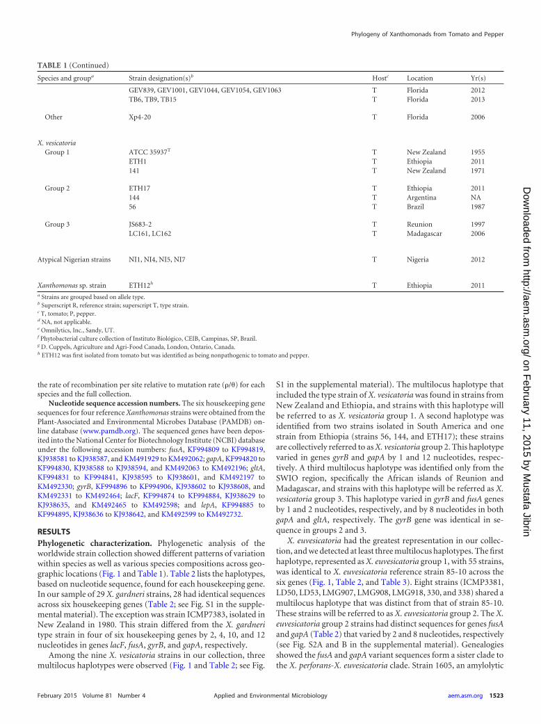
the rate of recombination per site relative to mutation rate (�/�) for eachspecies and the full collection.
Nucleotide sequence accession numbers. The six housekeeping genesequences for four reference Xanthomonas strains were obtained from thePlant-Associated and Environmental Microbes Database (PAMDB) on-line database (www.pamdb.org). The sequenced genes have been depos-ited into the National Center for Biotechnology Institute (NCBI) databaseunder the following accession numbers: fusA, KF994809 to KF994819,KJ938581 to KJ938587, and KM491929 to KM492062; gapA, KF994820 toKF994830, KJ938588 to KJ938594, and KM492063 to KM492196; gltA,KF994831 to KF994841, KJ938595 to KJ938601, and KM492197 toKM492330; gyrB, KF994896 to KF994906, KJ938602 to KJ938608, andKM492331 to KM492464; lacF, KF994874 to KF994884, KJ938629 toKJ938635, and KM492465 to KM492598; and lepA, KF994885 toKF994895, KJ938636 to KJ938642, and KM492599 to KM492732.
RESULTSPhylogenetic characterization. Phylogenetic analysis of theworldwide strain collection showed different patterns of variationwithin species as well as various species compositions across geo-graphic locations (Fig. 1 and Table 1). Table 2 lists the haplotypes,based on nucleotide sequence, found for each housekeeping gene.In our sample of 29 X. gardneri strains, 28 had identical sequencesacross six housekeeping genes (Table 2; see Fig. S1 in the supple-mental material). The exception was strain ICMP7383, isolated inNew Zealand in 1980. This strain differed from the X. gardneritype strain in four of six housekeeping genes by 2, 4, 10, and 12nucleotides in genes lacF, fusA, gyrB, and gapA, respectively.
Among the nine X. vesicatoria strains in our collection, threemultilocus haplotypes were observed (Fig. 1 and Table 2; see Fig.
S1 in the supplemental material). The multilocus haplotype thatincluded the type strain of X. vesicatoria was found in strains fromNew Zealand and Ethiopia, and strains with this haplotype willbe referred to as X. vesicatoria group 1. A second haplotype wasidentified from two strains isolated in South America and onestrain from Ethiopia (strains 56, 144, and ETH17); these strainsare collectively referred to as X. vesicatoria group 2. This haplotypevaried in genes gyrB and gapA by 1 and 12 nucleotides, respec-tively. A third multilocus haplotype was identified only from theSWIO region, specifically the African islands of Reunion andMadagascar, and strains with this haplotype will be referred as X.vesicatoria group 3. This haplotype varied in gyrB and fusA genesby 1 and 2 nucleotides, respectively, and by 8 nucleotides in bothgapA and gltA, respectively. The gyrB gene was identical in se-quence in groups 2 and 3.
X. euvesicatoria had the greatest representation in our collec-tion, and we detected at least three multilocus haplotypes. The firsthaplotype, represented as X. euvesicatoria group 1, with 55 strains,was identical to X. euvesicatoria reference strain 85-10 across thesix genes (Fig. 1, Table 2, and Table 3). Eight strains (ICMP3381,LD50, LD53, LMG907, LMG908, LMG918, 330, and 338) shared amultilocus haplotype that was distinct from that of strain 85-10.These strains will be referred to as X. euvesicatoria group 2. The X.euvesicatoria group 2 strains had distinct sequences for genes fusAand gapA (Table 2) that varied by 2 and 8 nucleotides, respectively(see Fig. S2A and B in the supplemental material). Genealogiesshowed the fusA and gapA variant sequences form a sister clade tothe X. perforans-X. euvesicatoria clade. Strain 1605, an amylolytic
TABLE 1 (Continued)
Species and groupa Strain designation(s)b Hostc Location Yr(s)
GEV839, GEV1001, GEV1044, GEV1054, GEV1063 T Florida 2012TB6, TB9, TB15 T Florida 2013
Other Xp4-20 T Florida 2006
X. vesicatoriaGroup 1 ATCC 35937T T New Zealand 1955
ETH1 T Ethiopia 2011141 T New Zealand 1971
Group 2 ETH17 T Ethiopia 2011144 T Argentina NA56 T Brazil 1987
Group 3 JS683-2 T Reunion 1997LC161, LC162 T Madagascar 2006
Atypical Nigerian strains NI1, NI4, NI5, NI7 T Nigeria 2012
Xanthomonas sp. strain ETH12h T Ethiopia 2011a Strains are grouped based on allele type.b Superscript R, reference strain; superscript T, type strain.c T, tomato; P, pepper.d NA, not applicable.e Omnilytics, Inc., Sandy, UT.f Phytobacterial culture collection of Instituto Biológico, CEIB, Campinas, SP, Brazil.g D. Cuppels, Agriculture and Agri-Food Canada, London, Ontario, Canada.h ETH12 was first isolated from tomato but was identified as being nonpathogenic to tomato and pepper.
Phylogeny of Xanthomonads from Tomato and Pepper
February 2015 Volume 81 Number 4 aem.asm.org 1523Applied and Environmental Microbiology
on February 11, 2015 by M
ustafa Jibrinhttp://aem
.asm.org/
Dow
nloaded from
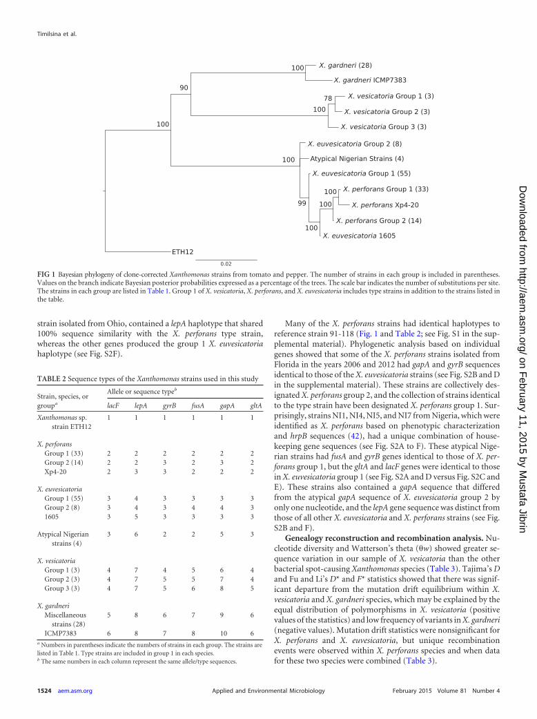
strain isolated from Ohio, contained a lepA haplotype that shared100% sequence similarity with the X. perforans type strain,whereas the other genes produced the group 1 X. euvesicatoriahaplotype (see Fig. S2F).
Many of the X. perforans strains had identical haplotypes toreference strain 91-118 (Fig. 1 and Table 2; see Fig. S1 in the sup-plemental material). Phylogenetic analysis based on individualgenes showed that some of the X. perforans strains isolated fromFlorida in the years 2006 and 2012 had gapA and gyrB sequencesidentical to those of the X. euvesicatoria strains (see Fig. S2B and Din the supplemental material). These strains are collectively des-ignated X. perforans group 2, and the collection of strains identicalto the type strain have been designated X. perforans group 1. Sur-prisingly, strains NI1, NI4, NI5, and NI7 from Nigeria, which wereidentified as X. perforans based on phenotypic characterizationand hrpB sequences (42), had a unique combination of house-keeping gene sequences (see Fig. S2A to F). These atypical Nige-rian strains had fusA and gyrB genes identical to those of X. per-forans group 1, but the gltA and lacF genes were identical to thosein X. euvesicatoria group 1 (see Fig. S2A and D versus Fig. S2C andE). These strains also contained a gapA sequence that differedfrom the atypical gapA sequence of X. euvesicatoria group 2 byonly one nucleotide, and the lepA gene sequence was distinct fromthose of all other X. euvesicatoria and X. perforans strains (see Fig.S2B and F).
Genealogy reconstruction and recombination analysis. Nu-cleotide diversity and Watterson’s theta (�w) showed greater se-quence variation in our sample of X. vesicatoria than the otherbacterial spot-causing Xanthomonas species (Table 3). Tajima’s Dand Fu and Li’s D* and F* statistics showed that there was signif-icant departure from the mutation drift equilibrium within X.vesicatoria and X. gardneri species, which may be explained by theequal distribution of polymorphisms in X. vesicatoria (positivevalues of the statistics) and low frequency of variants in X. gardneri(negative values). Mutation drift statistics were nonsignificant forX. perforans and X. euvesicatoria, but unique recombinationevents were observed within X. perforans species and when datafor these two species were combined (Table 3).
FIG 1 Bayesian phylogeny of clone-corrected Xanthomonas strains from tomato and pepper. The number of strains in each group is included in parentheses.Values on the branch indicate Bayesian posterior probabilities expressed as a percentage of the trees. The scale bar indicates the number of substitutions per site.The strains in each group are listed in Table 1. Group 1 of X. vesicatoria, X. perforans, and X. euvesicatoria includes type strains in addition to the strains listed inthe table.
TABLE 2 Sequence types of the Xanthomonas strains used in this study
Strain, species, orgroupa
Allele or sequence typeb
lacF lepA gyrB fusA gapA gltA
Xanthomonas sp.strain ETH12
1 1 1 1 1 1
X. perforansGroup 1 (33) 2 2 2 2 2 2Group 2 (14) 2 2 3 2 3 2Xp4-20 2 3 3 2 2 2
X. euvesicatoriaGroup 1 (55) 3 4 3 3 3 3Group 2 (8) 3 4 3 4 4 31605 3 5 3 3 3 3
Atypical Nigerianstrains (4)
3 6 2 2 5 3
X. vesicatoriaGroup 1 (3) 4 7 4 5 6 4Group 2 (3) 4 7 5 5 7 4Group 3 (3) 4 7 5 6 8 5
X. gardneriMiscellaneous
strains (28)5 8 6 7 9 6
ICMP7383 6 8 7 8 10 6a Numbers in parentheses indicate the numbers of strains in each group. The strains arelisted in Table 1. Type strains are included in group 1 in each species.b The same numbers in each column represent the same allele/type sequences.
Timilsina et al.
1524 aem.asm.org February 2015 Volume 81 Number 4Applied and Environmental Microbiology
on February 11, 2015 by M
ustafa Jibrinhttp://aem
.asm.org/
Dow
nloaded from

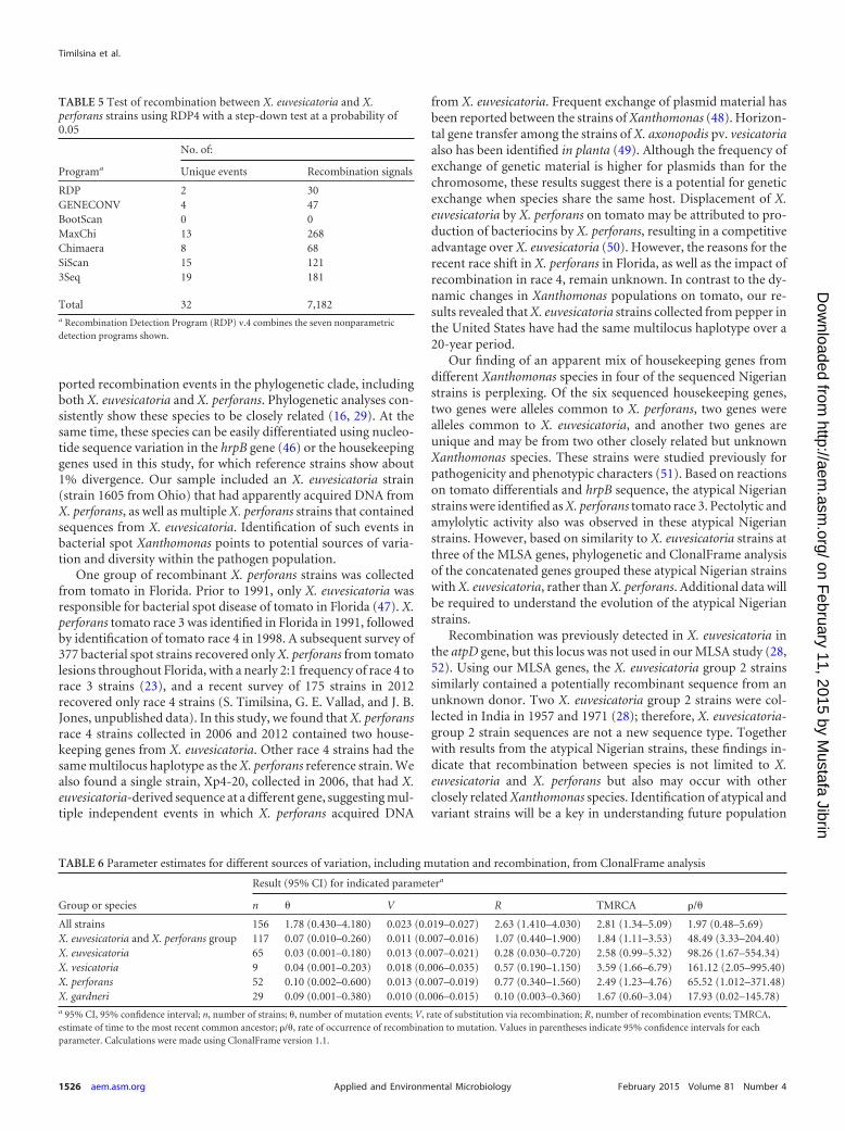
Phylogenetic networks were generated because individualmaximum likelihood phylogenies showed incompatible topolo-gies, suggesting recombination. The splits-decomposition phylo-genetic tree inferred from concatenated housekeeping gene se-quences confirmed incompatibilities and recombination withinX. euvesicatoria and X. perforans (Fig. 2). The pairwise homoplasyindex (PHI) rejected the hypothesis of no recombination in thewhole set of strains, and the same result was obtained when only X.euvesicatoria and X. perforans were considered (Table 4). GeneticAlgorithm for Recombination Detection (GARD) found evidencefor 3 recombination breakpoints in the concatenated genes of thewhole data set. The Kishino-Hasegawa test of tree congruencyindicated one significant breakpoint in the lacF gene. The Recom-bination Detection Program (RDP) detected recombination be-tween X. euvesicatoria and X. perforans in 6 out of 7 methods(Table 5). The genes lepA, fusA, and gapA were identified as po-tential recombinants, but the different algorithms varied in thestrains identified as probable recombinants, which included X.euvesicatoria 1605, the X. perforans group 2 strains, and the atyp-ical Nigerian strains. The X. euvesicatoria group 2 strains wereidentified as potential recombinants by only 2 algorithms (datanot shown).
The program ClonalFrame was used to calculate the mean rel-ative impact of recombination to mutation (r/m) on sequence
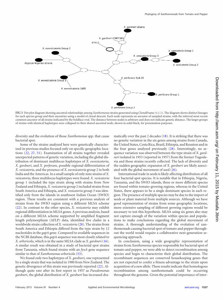
variation. Recombination had 18 times higher impact than pointmutation (r/m � 18.516) for the whole data set, but the rate ofoccurrence of recombination was only slightly higher than the rateof mutation (�/�) at 1.97, The mean tract length of recombinantsequence (�) was 843 bp (95% confidence interval [CI], 602 to1,099 bp), which is approximately the length of two arbitrarilyconcatenated genes. When species groups were considered sepa-rately, recombination was more frequent than mutation withinspecies (Table 6). A dot plot of the 50% consensus tree showsinferred ancestral relationships under clonal descent, meaningthat it attempts to remove the effects of recombination (Fig. 3).The X. perforans group 2 strains share an ancestor with the X.perforans group 1 strains, as would be expected if the variant geneswere introduced via homologous recombination. In contrast, theatypical Nigerian strains (NI1, NI4, NI5, and NI7) and X. euvesi-catoria group 2 share an ancestor that is distinct from the ancestryof the X. euvesicatoria reference strain.
DISCUSSION
Bacterial spot of pepper and tomato is caused by four differentXanthomonas species with dynamic global distributions. We char-acterized strains collected from different geographical locationsby MLSA and found that recombination between species is shap-ing the diversity of some bacterial spot pathogen populations.Homologous recombination should be more likely betweenclosely related strains due to sequence similarity (43). However,recombination is difficult to detect when it occurs between highlysimilar sequences; thus, some sequence divergence is required toidentify recombinant sequences (44). In Xanthomonas, recombi-nation in housekeeping genes has been observed when there issequence variation within a species (45) and among closely relatedpathovars in a species complex (23). We found statistically sup-
TABLE 4 Test of recombination based on pairwise homoplasy index(�w)
Sequence setNo. of polymorphicsites
Mean�w P value
Xanthomonascollection
229 0.0944 �0.0001
X. euvesicatoria andX. perforans
32 0.32056 �0.0005
TABLE 3 Sequence variation statistics for the collection of Xanthomonas strainsa
Sequence set
Diversity parameterb Neutrality test
n H S ND �w NM NSM Tajima’s D Fu and Li’s D* Fu and Li’s F* Rc
All strains 156 13 283 0.02554 50.379 307 61 0.545 (NS)d 0.32 (NS) 0.107 (NS) 40X. euvesicatoria 65 3 18 0.00099 3.087 18 8 1.06 (NS) 1.78 (NS) 1.81 (NS) 0X. perforans 52 4 27 0.00194 5.975 27 3 0.61 (NS) 0.91 (NS) 0.44 (NS) 2X. vesicatoria 9 3 21 0.00419 7.727 21 0 1.77 (NS) 1.57 (P � 0.02) 1.81541 (P � 0.02) 0X. gardneri 29 2 21 0.00058 5.347 21 21 2.57 (P � 0.001) 4.57 (P � 0.02) 4.63 (P � 0.02) 0X. euvesicatoria and
X. perforans group117 7 32 0.00483 12.067 32 0 3.04 1.99 (P � 0.02) 2.89 (P � 0.02) 4
a All calculations were made using DNAsp v.5 software. While calculating different parameters for the whole set, strain ETH12 was also used, but since it was not possible to define aspecies group for that strain, it was not included in other parameter calculations.b n, number of strains; H, number of haplotypes; S, total number of segregating sites; ND, nucleotide diversity; �w, Watterson’s theta; NM, number of mutations; NSM, number ofsingleton mutations.c R, minimum number of recombination events.d NS, not significant.
FIG 2 Splits decomposition tree of the subset of strains from the X. euvesica-toria and X. perforans species groups. Parallel lines indicate conflicting phylo-genetic relationships.
Phylogeny of Xanthomonads from Tomato and Pepper
February 2015 Volume 81 Number 4 aem.asm.org 1525Applied and Environmental Microbiology
on February 11, 2015 by M
ustafa Jibrinhttp://aem
.asm.org/
Dow
nloaded from
ported recombination events in the phylogenetic clade, includingboth X. euvesicatoria and X. perforans. Phylogenetic analyses con-sistently show these species to be closely related (16, 29). At thesame time, these species can be easily differentiated using nucleo-tide sequence variation in the hrpB gene (46) or the housekeepinggenes used in this study, for which reference strains show about1% divergence. Our sample included an X. euvesicatoria strain(strain 1605 from Ohio) that had apparently acquired DNA fromX. perforans, as well as multiple X. perforans strains that containedsequences from X. euvesicatoria. Identification of such events inbacterial spot Xanthomonas points to potential sources of varia-tion and diversity within the pathogen population.
One group of recombinant X. perforans strains was collectedfrom tomato in Florida. Prior to 1991, only X. euvesicatoria wasresponsible for bacterial spot disease of tomato in Florida (47). X.perforans tomato race 3 was identified in Florida in 1991, followedby identification of tomato race 4 in 1998. A subsequent survey of377 bacterial spot strains recovered only X. perforans from tomatolesions throughout Florida, with a nearly 2:1 frequency of race 4 torace 3 strains (23), and a recent survey of 175 strains in 2012recovered only race 4 strains (S. Timilsina, G. E. Vallad, and J. B.Jones, unpublished data). In this study, we found that X. perforansrace 4 strains collected in 2006 and 2012 contained two house-keeping genes from X. euvesicatoria. Other race 4 strains had thesame multilocus haplotype as the X. perforans reference strain. Wealso found a single strain, Xp4-20, collected in 2006, that had X.euvesicatoria-derived sequence at a different gene, suggesting mul-tiple independent events in which X. perforans acquired DNA
from X. euvesicatoria. Frequent exchange of plasmid material hasbeen reported between the strains of Xanthomonas (48). Horizon-tal gene transfer among the strains of X. axonopodis pv. vesicatoriaalso has been identified in planta (49). Although the frequency ofexchange of genetic material is higher for plasmids than for thechromosome, these results suggest there is a potential for geneticexchange when species share the same host. Displacement of X.euvesicatoria by X. perforans on tomato may be attributed to pro-duction of bacteriocins by X. perforans, resulting in a competitiveadvantage over X. euvesicatoria (50). However, the reasons for therecent race shift in X. perforans in Florida, as well as the impact ofrecombination in race 4, remain unknown. In contrast to the dy-namic changes in Xanthomonas populations on tomato, our re-sults revealed that X. euvesicatoria strains collected from pepper inthe United States have had the same multilocus haplotype over a20-year period.
Our finding of an apparent mix of housekeeping genes fromdifferent Xanthomonas species in four of the sequenced Nigerianstrains is perplexing. Of the six sequenced housekeeping genes,two genes were alleles common to X. perforans, two genes werealleles common to X. euvesicatoria, and another two genes areunique and may be from two other closely related but unknownXanthomonas species. These strains were studied previously forpathogenicity and phenotypic characters (51). Based on reactionson tomato differentials and hrpB sequence, the atypical Nigerianstrains were identified as X. perforans tomato race 3. Pectolytic andamylolytic activity also was observed in these atypical Nigerianstrains. However, based on similarity to X. euvesicatoria strains atthree of the MLSA genes, phylogenetic and ClonalFrame analysisof the concatenated genes grouped these atypical Nigerian strainswith X. euvesicatoria, rather than X. perforans. Additional data willbe required to understand the evolution of the atypical Nigerianstrains.
Recombination was previously detected in X. euvesicatoria inthe atpD gene, but this locus was not used in our MLSA study (28,52). Using our MLSA genes, the X. euvesicatoria group 2 strainssimilarly contained a potentially recombinant sequence from anunknown donor. Two X. euvesicatoria group 2 strains were col-lected in India in 1957 and 1971 (28); therefore, X. euvesicatoria-group 2 strain sequences are not a new sequence type. Togetherwith results from the atypical Nigerian strains, these findings in-dicate that recombination between species is not limited to X.euvesicatoria and X. perforans but also may occur with otherclosely related Xanthomonas species. Identification of atypical andvariant strains will be a key in understanding future population
TABLE 5 Test of recombination between X. euvesicatoria and X.perforans strains using RDP4 with a step-down test at a probability of0.05
Programa
No. of:
Unique events Recombination signals
RDP 2 30GENECONV 4 47BootScan 0 0MaxChi 13 268Chimaera 8 68SiScan 15 1213Seq 19 181
Total 32 7,182a Recombination Detection Program (RDP) v.4 combines the seven nonparametricdetection programs shown.
TABLE 6 Parameter estimates for different sources of variation, including mutation and recombination, from ClonalFrame analysis
Group or species
Result (95% CI) for indicated parametera
n � V R TMRCA �/�
All strains 156 1.78 (0.430–4.180) 0.023 (0.019–0.027) 2.63 (1.410–4.030) 2.81 (1.34–5.09) 1.97 (0.48–5.69)X. euvesicatoria and X. perforans group 117 0.07 (0.010–0.260) 0.011 (0.007–0.016) 1.07 (0.440–1.900) 1.84 (1.11–3.53) 48.49 (3.33–204.40)X. euvesicatoria 65 0.03 (0.001–0.180) 0.013 (0.007–0.021) 0.28 (0.030–0.720) 2.58 (0.99–5.32) 98.26 (1.67–554.34)X. vesicatoria 9 0.04 (0.001–0.203) 0.018 (0.006–0.035) 0.57 (0.190–1.150) 3.59 (1.66–6.79) 161.12 (2.05–995.40)X. perforans 52 0.10 (0.002–0.600) 0.013 (0.007–0.019) 0.77 (0.340–1.560) 2.49 (1.23–4.76) 65.52 (1.012–371.48)X. gardneri 29 0.09 (0.001–0.380) 0.010 (0.006–0.015) 0.10 (0.003–0.360) 1.67 (0.60–3.04) 17.93 (0.02–145.78)a 95% CI, 95% confidence interval; n, number of strains; �, number of mutation events; V, rate of substitution via recombination; R, number of recombination events; TMRCA,estimate of time to the most recent common ancestor; �/�, rate of occurrence of recombination to mutation. Values in parentheses indicate 95% confidence intervals for eachparameter. Calculations were made using ClonalFrame version 1.1.
Timilsina et al.
1526 aem.asm.org February 2015 Volume 81 Number 4Applied and Environmental Microbiology
on February 11, 2015 by M
ustafa Jibrinhttp://aem
.asm.org/
Dow
nloaded from
diversity and the evolution of those Xanthomonas spp. that causebacterial spot.
Some of the strains analyzed here were genetically character-ized in previous studies focused only on specific geographic loca-tions (22, 27, 51). Examination of all strains together revealedunexpected patterns of genetic variation, including the global dis-tribution of dominant multilocus haplotypes of X. euvesicatoria,X. gardneri, and X. perforans, possible regional differentiation ofX. vesicatoria, and the presence of X. euvesicatoria group 2 in bothIndia and the Americas. In a small sample of only nine strains of X.vesicatoria, three multilocus haplotypes were found: X. vesicatoriagroup 1 included the type strain along with strains from NewZealand and Ethiopia, X. vesicatoria group 2 included strains fromSouth America and Ethiopia, and X. vesicatoria group 3 was iden-tified only from the islands in southwest Indian Ocean (SWIO)region. These results are consistent with a previous analysis ofstrains from the SWIO region using a different MLSA scheme(22). In contrast to the other species, X. vesicatoria may exhibitregional differentiation in MLSA genes. A previous analysis, basedon a different MLSA scheme supported by amplified fragmentlength polymorphism (AFLP) data, identified five clades in aworldwide strain collection of X. vesicatoria (28). The strains fromSouth America and Ethiopia differed from the type strain by 12nucleotides in the gapA gene. Compared to available sequences inthe NCBI database, this gapA sequence was most closely related toX. arboricola, which is in the same MLSA clade as X. gardneri (16).A similar result was obtained in a study of bacterial spot strainsfrom Tanzania, which found strains with an fyuA gene sequencesimilar to that of Xanthomonas arboricola (53).
We found only two haplotypes of X. gardneri, one representedby a single strain that was isolated in 1980 from New Zealand. Thegenetic divergence of this strain was previously reported (22). Al-though quite rare after its first report in 1957 as Pseudomonasgardneri, the global distribution of X. gardneri has increased dra-
matically over the past 2 decades (18). It is striking that there wasno genetic variation in the six genes among strains from Canada,the United States, Costa Rica, Brazil, Ethiopia, and Reunion and inthe four genes analyzed previously (28). Interestingly, no se-quence variation was observed between the type strain of X. gard-neri isolated in 1953 (reported in 1957) from the former Yugosla-via and those strains recently collected. The lack of diversity andthe sudden geographic expansion of X. gardneri are likely associ-ated with the global movement of seed (26).
International trade in seeds is likely affecting distribution of allfour bacterial spot species. It is notable that in Ethiopia, Nigeria,Tanzania, and the SWIO islands, three or more different speciesare found within tomato-growing regions, whereas in the UnitedStates, there appears to be a single dominant species in each re-gion. The presence of multiple species may be due to the import ofseeds or plant material from multiple sources. Although we havegood representation of strains from some geographic locations,more extensive sampling of different growing regions would benecessary to test this hypothesis. MLSA using six genes also maynot capture enough of the variation within species and popula-tions to make conclusions regarding the global movement ofstrains. A thorough understanding of the evolution of xan-thomonads causing bacterial spot of tomato and pepper through-out the world would require a collaborative next-generation se-quencing approach.
In conclusion, using a wide geographic representation ofstrains from Xanthomonas species responsible for bacterial spot oftomato and pepper, we were able to detect recombination amongspecies and begin to characterize their global distribution. Therecombinant sequences are conserved housekeeping genes thatare not expected to confer a fitness advantage to the strain uponacquisition of a new allele. These results indicate that homologousrecombination among xanthomonads could be occurringthroughout the genome. Given the potential importance of inter-
FIG 3 Dot plot diagram showing ancestral relationships among Xanthomonas strains generated using ClonalFrame (v.1.1). The diagram shows distinct lineagesfor each species group and their ancestries using a model of clonal descent. Each node represents an ancestor of sampled strains, with the inferred most recentcommon ancestor of all strains indicated by the boldface oval. The distance between nodes is arbitrary and does not indicate genetic distance. The larger groupsof strains with identical haplotypes were collapsed to their shared ancestral node, shown in solid black, for presentation purposes.
Phylogeny of Xanthomonads from Tomato and Pepper
February 2015 Volume 81 Number 4 aem.asm.org 1527Applied and Environmental Microbiology
on February 11, 2015 by M
ustafa Jibrinhttp://aem
.asm.org/
Dow
nloaded from
specific recombination in shaping diversity of bacterial spotpathogen populations, it should be determined if genetic ex-change among species has introduced variation in pathogenicityand other fitness-associated genes.
ACKNOWLEDGMENTS
We thanks Sally Miller and David Ritchie for the X. gardneri and X. eu-vesicatoria strains provided, respectively.
This research was supported in part with funds from a Specialty CropBlock Grant, award 18015, to G. E. Vallad and J. B. Jones from the FloridaDepartment of Agriculture and Consumer Services and administered bythe Florida Specialty Crop Foundation. This work was also financiallysupported in part by the European Union (ERDF), Conseil Régional de LaRéunion, and CIRAD.
REFERENCES1. Jones JB, Lacy GH, Bouzar H, Stall RE, Schaad NW. 2004. Reclassifi-
cation of the xanthomonads associated with bacterial spot disease of to-mato and pepper. Syst Appl Microbiol 27:755–762. http://dx.doi.org/10.1078/0723202042369884.
2. Schaad NW, Postnikova E, Lacy G, Sechler A, Agarkova I, StrombergPE, Stromberg VK, Vidaver AK. 2006. Emended classification of xan-thomonad pathogens on citrus. Syst Appl Microbiol 29:690 – 695. http://dx.doi.org/10.1016/j.syapm.2006.08.001.
3. Martiny JBH, Bohannan BJM, Brown JH, Colwell RK, Fuhrman JA,Green JL, Horner-Devine MC, Kane M, Krumins JA, Kuske CR, MorinPJ, Naeem S, �vreås L, Reysenbach AL, Smith VH, Staley JT. 2006.Microbial biogeography: putting microorganisms on the map. Nat RevMicrobiol 4:102–112. http://dx.doi.org/10.1038/nrmicro1341.
4. Martinez JL, Baquero F. 2002. Interactions among strategies associatedwith bacterial infection: pathogenicity, epidemicity, and antibiotic resis-tance. Clin Microbiol Rev 15:647– 679. http://dx.doi.org/10.1128/CMR.15.4.647-679.2002.
5. Rosselló-Mora R, Amann R. 2001. The species concept for prokaryotes.FEMS Microbiol Rev 25:39 – 67. http://dx.doi.org/10.1111/j.1574-6976.2001.tb00571.x.
6. Bryant J, Chewapreecha C, Bentley SD. 2012. Developing insights intothe mechanisms of evolution of bacterial pathogens from whole-genomesequences. Future Microbiol 7:1283–1296. http://dx.doi.org/10.2217/fmb.12.108.
7. Wicker E, Lefeuvre P, de Cambiaire J-C, Lemaire C, Poussier S, PriorP. 2012. Contrasting recombination patterns and demographic historiesof the plant pathogen Ralstonia solanacearum inferred from MLSA. ISMEJ 6:961–974. http://dx.doi.org/10.1038/ismej.2011.160.
8. Philippe H, Douady CJ. 2003. Horizontal gene transfer and phylogenet-ics. Curr Opin Microbiol 6:498 –505. http://dx.doi.org/10.1016/j.mib.2003.09.008.
9. Wijayawardena KB, Minchella DJ, DeWoody JA. 2013. Hosts, parasites,and horizontal gene transfer. Trends Parasitol 29:329 –338. http://dx.doi.org/10.1016/j.pt.2013.05.001.
10. Gevers D, Cohan FM, Lawrence JG, Spratt BG, Coenye T, Feil EJ,Stackebrandt E, Van de Peer Y, Vandamme P, Thompson FL, Swings J.2005. Re-evaluating prokaryotic species. Nat Rev Microbiol 3:733–739.http://dx.doi.org/10.1038/nrmicro1236.
11. Martens M, Dawyndt P, Coopman R, Gillis M, De Vos P, Willems A.2008. Advantages of multilocus sequence analysis for taxonomic studies: acase study using 10 housekeeping genes in the genus Ensifer (includingformer Sinorhizobium). Int J Syst Evol Microbiol 58:200 –214. http://dx.doi.org/10.1099/ijs.0.65392-0.
12. Gomez-Valero L, Rusniok C, Jarraud S, Vacherie B, Rouy Z, Barbe V,Medigue C, Etienne J, Buchrieser C. 2011. Extensive recombinationevents and horizontal gene transfer shaped the Legionella pneumophilagenomes. BMC Genomics 12:536. http://dx.doi.org/10.1186/1471-2164-12-536.
13. Vos M, Didelot X. 2009. A comparison of homologous recombinationrates in bacteria and archaea. ISME J 3:199 –208. http://dx.doi.org/10.1038/ismej.2008.93.
14. Hayward AC. 1993. The host of Xanthomonas, p 51–54. In Swings J andCiverolo EL (ed), Xanthomonas. Springer, Dordrecht, Netherlands.
15. Stall RE, Beaulieu C, Egel DS, Hodge NC, Leite RP, Minsavage GV,
Bouzar H, Jones JB, Alvarez AM, Benedict AA. 1994. Two geneticallydiverse groups of strains are included in Xanthomonas campestris pv. vesi-catoria. Int J Syst Bacteriol 44:47–53. http://dx.doi.org/10.1099/00207713-44-1-47.
16. Almeida NF, Yan S, Cai R, Clarke CR, Morris CE, Schaad NW, Schuen-zel EL, Lacy GH, Sun X, Jones JB. 2010. PAMDB, a multilocus sequencetyping and analysis database and website for plant-associated microbes.Phytopathology 100:208 –215. http://dx.doi.org/10.1094/PHYTO-100-3-0208.
17. Jones JB, Bouzar H, Stall RE, Almira EC, Roberts PD, Bowen BW,Sudberry J, Strickler PM, Chun J. 2000. Systematic analysis of xan-thomonads (Xanthomonas spp.) associated with pepper and tomato le-sions. Int J Syst Evol Microbiol 50:1211–1219. http://dx.doi.org/10.1099/00207713-50-3-1211.
18. Jones JB, Lacy GH, Bouzar H, Minsavage GV, Stall R, Schaad N. 2004.Bacterial spot—worldwide distribution, importance and review. 1st Inter-national Symposium on Tomato Diseases. Acta Hortic 695:27–33.
19. Cuppels DA, Louws FJ, Ainsworth T. 2006. Development and evaluationof PCR-based diagnostic assays for the bacterial speck and bacterial spotpathogens of tomato. Plant Dis 90:451– 458. http://dx.doi.org/10.1094/PD-90-0451.
20. Bouzar H, Jones J, Stall R, Louws F, Schneider M, Rademaker J, DeBruijn F, Jackson L. 1999. Multiphasic analysis of xanthomonads causingbacterial spot disease on tomato and pepper in the Caribbean and CentralAmerica: evidence for common lineages within and between countries.Phytopathology 89:328 –335. http://dx.doi.org/10.1094/PHYTO.1999.89.4.328.
21. Bouzar H, Jones JB, Somodi GC, Stall RE, Daouzli N, Lambe RC, FelixGastelum R, Trinidad Correa R. 1996. Diversity of Xanthomonas camp-estris pv. vesicatoria in tomato and pepper fields of Mexico. Can J PlantPathol 18:75–77. http://dx.doi.org/10.1080/07060669609500659.
22. Hamza AA, Robene-Soustrade I, Jouen E, Gagnevin L, Lefeuvre P,Chiroleu F, Pruvost O. 2010. Genetic and pathological diversity amongXanthomonas strains responsible for bacterial spot on tomato and pepperin the southwest Indian Ocean region. Plant Dis 94:993–999. http://dx.doi.org/10.1094/PDIS-94-8-0993.
23. Horvath DM, Stall RE, Jones JB, Pauly MH, Vallad GE, Dahlbeck D,Staskawicz BJ, Scott JW. 2012. Transgenic resistance confers effectivefield level control of bacterial spot disease in tomato. PLoS One 7:e42036.http://dx.doi.org/10.1371/journal.pone.0042036.
24. Mhedbi-Hajri N, Hajri A, Boureau T, Darrasse A, Durand K, Brin C,Fischer-Le Saux M, Manceau C, Poussier S, Pruvost O. 2013. Evolu-tionary history of the plant pathogenic bacterium Xanthomonas axonopo-dis. PLoS One 8:e58474. http://dx.doi.org/10.1371/journal.pone.0058474.
25. Wichmann G, Ritchie D, Kousik CS, Bergelson J. 2005. Reduced geneticvariation occurs among genes of the highly clonal plant pathogen Xan-thomonas axonopodis pv. vesicatoria, including the effector gene avrBs2.Appl Environ Microbiol 71:2418 –2432. http://dx.doi.org/10.1128/AEM.71.5.2418-2432.2005.
26. Parkinson N, Cowie C, Henney J, Stead D. 2009. Phylogenetic structureof Xanthomonas determined by comparison of gyrB seqeuences. Int J SystEvol Microbiol 59:264 –274. http://dx.doi.org/10.1099/ijs.0.65825-0.
27. Kebede MG, Timilsina S, Ayalew A, Admassu B, Potnis N, MinsavageGV, Goss EM, Hong JC, Strayer A, Paret M, Jones JB, Vallad GE. 2014.Molecular characterization of Xanthomonas strains responsible for bacte-rial spot of tomato in Ethiopia. Eur J Plant Pathol 140:677– 688. http://dx.doi.org/10.1007/s10658-014-0497-3.
28. Hamza AA, Robene-Soustrade I, Jouen E, Lefeuvre P, Chiroleu F,Fisher-Le SM, Gagnevin L, Pruvost O. 2012. Multilocus sequence anal-ysis- and amplified fragment length polymorphism-based characteriza-tion of xanthomonads associated with bacterial spot of tomato and pepperand their relatedness to Xanthomonas species. Syst Appl Microbiol 35:183–190. http://dx.doi.org/10.1016/j.syapm.2011.12.005.
29. Potnis N, Krasileva K, Chow V, Almeida NF, Patil PB, Ryan RP,Sharlach M, Behlau F, Dow JM, Momol MT, White FF, Preston JF,Vinatzer BA, Koebni R, Setubal JC, Norman DJ, Staskawicz BJ, JonesJB. 2011. Comparative genomics reveals diversity among xanthomonadsinfecting tomato and pepper. BMC Genomics 12:146. http://dx.doi.org/10.1186/1471-2164-12-146.
30. Tamura K, Peterson D, Peterson N, Stecher G, Nei M, Kumar S. 2011.MEGA5: molecular evolutionary genetics analysis using maximum likeli-hood, evolutionary distance, and maximum parsimony methods. MolBiol Evol 28:2731–2739. http://dx.doi.org/10.1093/molbev/msr121.
Timilsina et al.
1528 aem.asm.org February 2015 Volume 81 Number 4Applied and Environmental Microbiology
on February 11, 2015 by M
ustafa Jibrinhttp://aem
.asm.org/
Dow
nloaded from
31. Hall T. 1999. BioEdit: a user-friendly biological sequence alignment edi-tor and analysis program for Windows 95/98/NT. Nucleic Acids Symp Ser41:95–98.
32. Posada D, Buckley TR. 2004. Model selection and model averaging inphylogenetics: advantages of Akaike information criterion and Bayesianapproaches over likelihood ratio tests. Syst Biol 59:793– 808. http://dx.doi.org/10.1080/10635150490522304.
33. Ronquist F, Huelsenbeck JP. 2003. MrBayes 3: Bayesian phylogeneticinference under mixed models. Bioinformatics 19:1572–1574. http://dx.doi.org/10.1093/bioinformatics/btg180.
34. Ronquist F, Teslenko M, Van der Mark P, Ayres DL, Darling A, HöhnaS, Larget B, Liu L, Suchard MA, Huelsenbeck JP. 2012. MrBayes 3.2:efficient Bayesian phylogenetic inference and model choice across a largemodel space. Syst Biol 61:539 –542. http://dx.doi.org/10.1093/sysbio/sys029.
35. Librado P, Rozas J. 2009. DnaSP v5: a software for comprehensive anal-ysis of DNA polymorphism data. Bioinformatics 25:1451–1452. http://dx.doi.org/10.1093/bioinformatics/btp187.
36. Tajima F. 1989. Statistical method for testing the neutral mutationhypothesis by DNA polymorphism. Genetics 123:585–595.
37. Fu Y-X, Li W-H. 1993. Statistical tests of neutrality of mutations. Genetics133:693–709.
38. Joseph S, Forsythe SJ. 2012. Insights into the emergent bacterial pathogenCronobacter spp., generated by multilocus sequence typing and analysis.Front Microbiol 3:397. http://dx.doi.org/10.3389/fmicb.2012.00397.
39. Huson DH, Bryant D. 2006. Application of phylogenetic networks inevolutionary studies. Mol Biol Evol 23:254 –267. http://dx.doi.org/10.1093/molbev/msj030.
40. Martin DP, Lemey P, Lott M, Moulton V, Posada D, Lefeuvre P. 2010.RDP3: a flexible and fast computer program for analyzing recombination.Bioinformatics 26:2462–2463. http://dx.doi.org/10.1093/bioinformatics/btq467.
41. Pond SLK, Posada D, Gravenor MB, Woelk CH, Frost SDW. 2006.GARD: a genetic algorithm for recombination detection. Bioinformatics22:3096 –3098. http://dx.doi.org/10.1093/bioinformatics/btl474.
42. Didelot X, Falush D. 2007. Inference of bacterial microevolution usingmultilocus sequence data. Genetics 175:1251–1266. http://dx.doi.org/10.1534/genetics.106.063305.
43. Roberts MS, Cohan FM. 1993. The effect of DNA sequence divergence onsexual isolation in Bacillus. Genetics 134:401– 408.
44. Posada D, Crandall KA, Holmes EC. 2002. Recombination in evolution-
ary genomics. Annu Rev Genet 36:75–97. http://dx.doi.org/10.1146/annurev.genet.36.040202.111115.
45. Marcelletti S, Ferrante P, Scortichini M. 2010. Multilocus sequencetyping reveals relevant genetic variation and different evolutionary dy-namics among strains of Xanthomonas arboricola pv. juglandis. Diversity2:1205–1222. http://dx.doi.org/10.3390/d2111205.
46. Obradovic A, Mavridis A, Rudolph K, Janse JD, Arsemijevic M, JonesJB, Minsavage GV, Wang JF. 2004. Characterization of PCR-based typ-ing of Xanthomonas campestris pv. vesicatoria from peppers and tomatoesin Serbia. Eur J Plant Pathol 110:285–292. http://dx.doi.org/10.1023/B:EJPP.0000019797.27952.1d.
47. Astua-Monge G, Minsavage GV, Stall RE, Vallejos CE, Davis MJ, JonesJB. 2000. Xv4-Avrxv4: a new gene-for-gene interaction identified betweenXanthomonas campestris pv. vesicatoria race T3 and the wild tomato rela-tive Lycopersicon pennellii. Mol Plant Microbe Interact 13:1346 –1355.http://dx.doi.org/10.1094/MPMI.2000.13.12.1346.
48. Canteros BI, Minsavage GV, Jones JB, Stall RE. 1995. Diversity ofplasmids in Xanthomonas campestris pv. vesicatoria. Phytopathology 85:1482–1486. http://dx.doi.org/10.1094/Phyto-85-1482.
49. Basim H, Stall RE, Minsavage GV, Jones JB. 1999. Chromosomal genetransfer by conjugation in the plant pathogen Xanthomonas axonopodispv. vesicatoria. Phytopathology 89:1044 –1049. http://dx.doi.org/10.1094/PHYTO.1999.89.11.1044.
50. Hert AP, Roberts PD, Momol MT, Minsavage GV, Tudor-Nelson SM,Jones JB. 2005. Relative importance of bacteriocin-like genes in antago-nism of Xanthomonas perforans tomato race 3 to Xanthomonas euvesica-toria tomato race 1 strains. Appl Environ Microbiol 71:3581–3588. http://dx.doi.org/10.1128/AEM.71.7.3581-3588.2005.
51. Jibrin MO, Timilsina S, Potnis N, Minsavage GV, Shenge KC, Akpa AD,Alegbejo MD, Vallad GE, Jones JB. 2014. First report of Xanthomonaseuvesicatoria causing bacterial spot disease in pepper in northwestern Ni-geria. Plant Dis http://dx.doi.org/10.1094/PDIS-06-14-0586-PDN.
52. Bui Thi Ngoc L, Vernière C, Jouen E, Ah-You N, Lefeuvre P, ChiroleuF, Gagnevin L, Pruvost O. 2010. Amplified fragment length polymor-phism and multilocus sequence analysis-based genotypic relatednessamong pathogenic variants of Xanthomonas citri pv. citri and Xanthomo-nas campestris pv. bilvae. Int J Syst Evol Microbiol 60:515–525. http://dx.doi.org/10.1099/ijs.0.009514-0.
53. Mbega ER, Wulff EG, Mabagala RB, Adriko J, Lund OS, Mortensen CN.2012. Xanthomonads and other yellow-pigmented Xanthomonas-likebacteria associated with tomato seeds in Tanzania. Afr J Biotechnol 11:14303–14312. http://dx.doi.org/10.5897/AJB12.1305.
Phylogeny of Xanthomonads from Tomato and Pepper
February 2015 Volume 81 Number 4 aem.asm.org 1529Applied and Environmental Microbiology
on February 11, 2015 by M
ustafa Jibrinhttp://aem
.asm.org/
Dow
nloaded from
Related Documents