Multigenerational silencing dynamics control cell aging Yang Li a,1 , Meng Jin b,c,1 , Richard O’Laughlin d,1 , Philip Bittihn b,c , Lev S. Tsimring b,c , Lorraine Pillus a,e , Jeff Hasty a,b,c,d , and Nan Hao a,b,c,2 a Section of Molecular Biology, Division of Biological Sciences, University of California, San Diego, La Jolla, CA 92093; b BioCircuits Institute, University of California, San Diego, La Jolla, CA 92093; c The San Diego Center for Systems Biology, La Jolla, CA 92093; d Department of Bioengineering, University of California, San Diego, La Jolla, CA 92093; and e Moores Cancer Center, University of California, San Diego, La Jolla, CA 92093 Edited by Jasper Rine, University of California, Berkeley, Berkeley, CA, and approved September 5, 2017 (received for review February 27, 2017) Cellular aging plays an important role in many diseases, such as cancers, metabolic syndromes, and neurodegenerative disorders. There has been steady progress in identifying aging-related factors such as reactive oxygen species and genomic instability, yet an emerging challenge is to reconcile the contributions of these factors with the fact that genetically identical cells can age at significantly different rates. Such complexity requires single-cell analyses designed to unravel the interplay of aging dynamics and cell-to-cell variability. Here we use microfluidic technologies to track the repli- cative aging of single yeast cells and reveal that the temporal pat- terns of heterochromatin silencing loss regulate cellular life span. We found that cells show sporadic waves of silencing loss in the heterochromatic ribosomal DNA during the early phases of aging, followed by sustained loss of silencing preceding cell death. Isogenic cells have different lengths of the early intermittent silencing phase that largely determine their final life spans. Combining computa- tional modeling and experimental approaches, we found that the intermittent silencing dynamics is important for longevity and is dependent on the conserved Sir2 deacetylase, whereas either sus- tained silencing or sustained loss of silencing shortens life span. These findings reveal that the temporal patterns of a key molecular process can directly influence cellular aging, and thus could provide guidance for the design of temporally controlled strategies to extend life span. replicative aging | single-cell analysis | microfluidics | chromatin silencing | computational modeling C ellular aging is generally driven by the accumulation of ge- netic and cellular damage (1, 2). Although much progress has been made in identifying molecular factors that influence life span, what remains sorely missing is an understanding of how these factors interact and change dynamically during the aging process. This is in part because aging is a complex process wherein isogenic cells have various intrinsic causes of aging and widely different rates of aging. As a result, static population-based ap- proaches could be insufficient to fully reveal sophisticated dynamic changes during aging. Recent developments in single-cell analyses to unravel the interplay of cellular dynamics and variability hold the promise to answer that challenge (3–5). Here we chose the replicative aging of yeast S. cerevisiae as a model and exploited quantitative biology technologies to study the dynamics of molec- ular processes that control aging at the single-cell level. Results Replicative aging of yeast is measured as the number of daughter cells produced before the death of a mother cell (6). The con- ventional method for studying yeast aging requires laborious manual separation of daughter cells from mother cells after each division and does not allow tracking of molecular processes over multiple generations during aging (7). Recent advances in micro- fluidics technology have automated cell separation and enabled continuous single-cell measurements during aging (8–14). Building on these efforts, we developed a microfluidic aging device. The device traps mother cells at the bottom of finger-shaped chambers, allowing them to bud continuously, while daughter cells are re- moved via a waste port. Each chamber also has a small opening at the bottom, allowing daughter removal when mother cells switch budding direction (Fig. 1 A and B and Movie S1). The long trapping chambers allow tracking of each daughter cell during its first several divisions, which is useful for monitoring age-related daughter morphologies. Furthermore, dynamic experiments in- volving precise step changes in media conditions can be conducted using this device. In validating the device, we confirmed that the majority of loaded cells are exponentially growing newborn or young cells, and the replicative life spans (RLS) measured using the device are comparable to those from classical microdissection (15, 16) (SI Appendix, Fig. S1 A–D). Genome instability has been considered a significant causal factor of cell aging (1, 17). A major contributor to the maintenance of genome stability is chromatin silencing, which causes a locally inaccessible heterochromatin structure that represses transcrip- tion, recombination, and DNA damage. The heterochromatic regions in yeast include the silent mating-type (HM) loci, rDNA, and subtelomeric regions (18). Among them, the rDNA region on chromosome XII consists of 100∼200 tandem repeats and is a particularly fragile genomic site, the stability of which closely connects to the RLS (19–21). Previous studies showed that si- lencing loss induced by chemical or genetic perturbations leads to increased recombination at the rDNA region and shorter life spans (22, 23). However, the dynamic changes of rDNA silencing during cell aging remain largely unknown. To measure silencing in single aging cells, we constructed a strain with a fluorescent reporter gene under the control of a constitutive TDH3 promoter at a non- transcribed spacer region (NTS1) of rDNA. Because expression of the reporter gene is repressed by silencing, decreased fluo- rescence indicates enhanced silencing, whereas increased fluorescence Significance Aging is an inevitable consequence of living, and with it comes increased morbidity and mortality. Novel approaches to mitigat- ing age-related chronic diseases demand a better understanding of the biology of aging. Studies in model organisms have iden- tified many conserved molecular factors that influence aging. The emerging challenge is to understand how these factors interact and change dynamically to drive aging. Using multidisciplinary technologies, we have revealed a sirtuin-dependent intermittent pattern of chromatin silencing during yeast aging that is crucial for longevity. Our findings highlight the important role of si- lencing dynamics in aging, which deserves careful consideration when designing schemes to delay or reverse aging by modulating sirtuins and silencing. Author contributions: Y.L., M.J., R.O., L.S.T., L.P., J.H., and N.H. designed research; Y.L. and R.O. performed research; Y.L., M.J., R.O., and P.B. analyzed data; and Y.L., M.J., R.O., L.S.T., L.P., J.H., and N.H. wrote the paper. The authors declare no conflict of interest. This article is a PNAS Direct Submission. Freely available online through the PNAS open access option. 1 Y.L., M.J., and R.O. contributed equally to this work. 2 To whom correspondence should be addressed. Email: [email protected]. This article contains supporting information online at www.pnas.org/lookup/suppl/doi:10. 1073/pnas.1703379114/-/DCSupplemental. www.pnas.org/cgi/doi/10.1073/pnas.1703379114 PNAS | October 17, 2017 | vol. 114 | no. 42 | 11253–11258 SYSTEMS BIOLOGY

Welcome message from author
This document is posted to help you gain knowledge. Please leave a comment to let me know what you think about it! Share it to your friends and learn new things together.
Transcript
Multigenerational silencing dynamics control cell agingYang Lia,1, Meng Jinb,c,1, Richard O’Laughlind,1, Philip Bittihnb,c, Lev S. Tsimringb,c, Lorraine Pillusa,e, Jeff Hastya,b,c,d,and Nan Haoa,b,c,2
aSection of Molecular Biology, Division of Biological Sciences, University of California, San Diego, La Jolla, CA 92093; bBioCircuits Institute, University ofCalifornia, San Diego, La Jolla, CA 92093; cThe San Diego Center for Systems Biology, La Jolla, CA 92093; dDepartment of Bioengineering, Universityof California, San Diego, La Jolla, CA 92093; and eMoores Cancer Center, University of California, San Diego, La Jolla, CA 92093
Edited by Jasper Rine, University of California, Berkeley, Berkeley, CA, and approved September 5, 2017 (received for review February 27, 2017)
Cellular aging plays an important role in many diseases, such ascancers, metabolic syndromes, and neurodegenerative disorders.There has been steady progress in identifying aging-related factorssuch as reactive oxygen species and genomic instability, yet anemerging challenge is to reconcile the contributions of these factorswith the fact that genetically identical cells can age at significantlydifferent rates. Such complexity requires single-cell analysesdesigned to unravel the interplay of aging dynamics and cell-to-cellvariability. Here we use microfluidic technologies to track the repli-cative aging of single yeast cells and reveal that the temporal pat-terns of heterochromatin silencing loss regulate cellular life span.We found that cells show sporadic waves of silencing loss in theheterochromatic ribosomal DNA during the early phases of aging,followed by sustained loss of silencing preceding cell death. Isogeniccells have different lengths of the early intermittent silencing phasethat largely determine their final life spans. Combining computa-tional modeling and experimental approaches, we found that theintermittent silencing dynamics is important for longevity and isdependent on the conserved Sir2 deacetylase, whereas either sus-tained silencing or sustained loss of silencing shortens life span.These findings reveal that the temporal patterns of a key molecularprocess can directly influence cellular aging, and thus could provideguidance for the design of temporally controlled strategies toextend life span.
replicative aging | single-cell analysis | microfluidics | chromatin silencing |computational modeling
Cellular aging is generally driven by the accumulation of ge-netic and cellular damage (1, 2). Although much progress
has been made in identifying molecular factors that influence lifespan, what remains sorely missing is an understanding of howthese factors interact and change dynamically during the agingprocess. This is in part because aging is a complex process whereinisogenic cells have various intrinsic causes of aging and widelydifferent rates of aging. As a result, static population-based ap-proaches could be insufficient to fully reveal sophisticated dynamicchanges during aging. Recent developments in single-cell analysesto unravel the interplay of cellular dynamics and variability holdthe promise to answer that challenge (3–5). Here we chose thereplicative aging of yeast S. cerevisiae as a model and exploitedquantitative biology technologies to study the dynamics of molec-ular processes that control aging at the single-cell level.
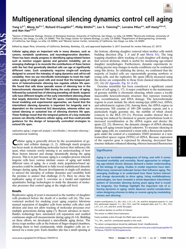
ResultsReplicative aging of yeast is measured as the number of daughtercells produced before the death of a mother cell (6). The con-ventional method for studying yeast aging requires laboriousmanual separation of daughter cells from mother cells after eachdivision and does not allow tracking of molecular processes overmultiple generations during aging (7). Recent advances in micro-fluidics technology have automated cell separation and enabledcontinuous single-cell measurements during aging (8–14). Buildingon these efforts, we developed a microfluidic aging device. Thedevice traps mother cells at the bottom of finger-shaped chambers,allowing them to bud continuously, while daughter cells are re-moved via a waste port. Each chamber also has a small opening at
the bottom, allowing daughter removal when mother cells switchbudding direction (Fig. 1 A and B and Movie S1). The longtrapping chambers allow tracking of each daughter cell during itsfirst several divisions, which is useful for monitoring age-relateddaughter morphologies. Furthermore, dynamic experiments in-volving precise step changes in media conditions can be conductedusing this device. In validating the device, we confirmed that themajority of loaded cells are exponentially growing newborn oryoung cells, and the replicative life spans (RLS) measured usingthe device are comparable to those from classical microdissection(15, 16) (SI Appendix, Fig. S1 A–D).Genome instability has been considered a significant causal
factor of cell aging (1, 17). A major contributor to the maintenanceof genome stability is chromatin silencing, which causes a locallyinaccessible heterochromatin structure that represses transcrip-tion, recombination, and DNA damage. The heterochromaticregions in yeast include the silent mating-type (HM) loci, rDNA,and subtelomeric regions (18). Among them, the rDNA region onchromosome XII consists of 100∼200 tandem repeats and is aparticularly fragile genomic site, the stability of which closelyconnects to the RLS (19–21). Previous studies showed that si-lencing loss induced by chemical or genetic perturbations leads toincreased recombination at the rDNA region and shorter lifespans (22, 23). However, the dynamic changes of rDNA silencingduring cell aging remain largely unknown. To measure silencing insingle aging cells, we constructed a strain with a fluorescent reportergene under the control of a constitutive TDH3 promoter at a non-transcribed spacer region (NTS1) of rDNA. Because expressionof the reporter gene is repressed by silencing, decreased fluo-rescence indicates enhanced silencing, whereas increased fluorescence
Significance
Aging is an inevitable consequence of living, and with it comesincreased morbidity and mortality. Novel approaches to mitigat-ing age-related chronic diseases demand a better understandingof the biology of aging. Studies in model organisms have iden-tified many conserved molecular factors that influence aging. Theemerging challenge is to understand how these factors interactand change dynamically to drive aging. Using multidisciplinarytechnologies, we have revealed a sirtuin-dependent intermittentpattern of chromatin silencing during yeast aging that is crucialfor longevity. Our findings highlight the important role of si-lencing dynamics in aging, which deserves careful considerationwhen designing schemes to delay or reverse aging by modulatingsirtuins and silencing.
Author contributions: Y.L., M.J., R.O., L.S.T., L.P., J.H., and N.H. designed research; Y.L. andR.O. performed research; Y.L., M.J., R.O., and P.B. analyzed data; and Y.L., M.J., R.O.,L.S.T., L.P., J.H., and N.H. wrote the paper.
The authors declare no conflict of interest.
This article is a PNAS Direct Submission.
Freely available online through the PNAS open access option.1Y.L., M.J., and R.O. contributed equally to this work.2To whom correspondence should be addressed. Email: [email protected].
This article contains supporting information online at www.pnas.org/lookup/suppl/doi:10.1073/pnas.1703379114/-/DCSupplemental.
www.pnas.org/cgi/doi/10.1073/pnas.1703379114 PNAS | October 17, 2017 | vol. 114 | no. 42 | 11253–11258
SYST
EMSBIOLO
GY
indicates reduced silencing (24, 25) (Fig. 1C). We observed thatcells with the rDNA reporter gene exhibit weak fluorescence; incontrast, cells carrying the same reporter at the URA3 locus, whichis not subject to silencing, show very high fluorescence. In addition,deletion of SIR2, which is required for rDNA silencing (18), yieldssignificantly increased reporter expression at the rDNA, but not atURA3 (SI Appendix, Fig. S1E).
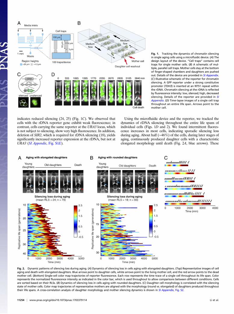
Using the microfluidic device and the reporter, we tracked thedynamics of rDNA silencing throughout the entire life spans ofindividual cells (Figs. 1D and 2). We found intermittent fluores-cence increases in most cells, indicating sporadic silencing lossduring aging. About half (∼46%) of the cells, during later stages ofaging, continuously produced daughter cells with a characteristicelongated morphology until death (Fig. 2A, blue arrows). These
Fig. 1. Tracking the dynamics of chromatin silencingin single aging cells using a microfluidic device. (A) Thedesign layout of the device. “Cell traps” contains celltraps for single mother cells. (B) A schematic of mul-tiple, parallel cell traps. Mother cells stay at the bottomof finger-shaped chambers and daughters are pushedout. Details of the device are provided in SI Appendix.(C) Illustrative schematic of the reporter for chromatinsilencing. A GFP reporter under a strong constitutivepromoter (TDH3) is inserted at an NTS1 repeat withinthe rDNA. Chromatin silencing at the rDNA is reflectedby fluorescence intensity: low, silenced; high, decreasedsilencing. Details of the reporter are provided in SIAppendix. (D) Time-lapse images of a single cell trapthroughout an entire life span. Arrows point to themother cell.
Fig. 2. Dynamic patterns of silencing loss during aging. (A) Dynamics of silencing loss in cells aging with elongated daughters. (Top) Representative images of cellaging and death with elongated daughters. Blue arrows point to daughter cells, white arrows point to the living mother cell, and the red arrow points to the deadmother cell. (Bottom) Single-cell color map trajectories of reporter fluorescence. Each row represents the time trace of a single cell throughout its life span. Colorrepresents the normalized fluorescence intensity as indicated in the color bar, which is used throughout to allow comparisons between different conditions. Cellsare sorted based on their RLSs. (B) Dynamics of silencing loss in cells aging with rounded daughters. (C) Daughter cell morphology is correlated with the silencingstate of mother cells. Color map trajectories of representative mothers are aligned with the morphology (round vs. elongated) of daughters produced throughouttheir life spans. A cross-correlation analysis of daughter morphology and mother silencing dynamics is shown in SI Appendix, Fig. S2.
11254 | www.pnas.org/cgi/doi/10.1073/pnas.1703379114 Li et al.

cells also exhibited a sustained and dramatic increase in fluores-cence, indicating sustained loss of silencing in aged cells (Fig. 2A,color map). In contrast, the other half of the cells, at later phases oftheir life spans, continuously produced small round daughter cells(Fig. 2B, blue arrows) with sharply increased cell cycle length.These cells had a shorter average life span than the other agingtype (with a mean RLS of 18 compared with 24) and did not showsustained silencing loss during aging (Fig. 2B, color map). Thesetwo distinct types of age-associated phenotypic changes suggestdifferent molecular causes of aging in isogenic cells (8, 9, 14).Previous studies showed that the aging phenotype with small rounddaughters could be related to an age-dependent mitochondrialdysfunction (9, 12), but the molecular mechanisms underlying theother aging type characterized by elongated daughters remainlargely unclear. Our results revealed that the sustained rDNA si-lencing loss, which can lead to genome instability (18), is specifi-cally associated with the aging phenotype featured by elongateddaughters. In support of this, young mother cells can also spo-radically produce a few elongated daughters, the occurrence ofwhich correlates with the transient silencing loss during the earlyphases of their life spans (Fig. 2C). In addition, cross-correlationanalysis revealed a ∼140-min time delay between the occurrence ofsilencing loss in mother cells and the production of elongateddaughters (SI Appendix, Fig. S2). This temporal order suggested apotential causal relationship between silencing loss and the elon-gated daughter phenotype. In this work, we focused our analysis onthe dynamics and heterogeneity of the type of aging process withsustained silencing loss and elongated daughters.To exclude the possibility that the observed fluorescence
patterns are caused by age-associated global effects on gene ex-pression (26), we simultaneously monitored two distinguishablefluorescent reporters inserted at the rDNA and at URA3. Whereasthe rDNA reporter showed early sporadic and late sustained in-duction of fluorescence, the reporter at URA3 exhibited relativelyconstant fluorescence during aging (SI Appendix, Fig. S3A). Toconfirm that the observed silencing dynamics are not specific to theNTS1 region, we measured the reporter response at another non-transcribed spacer region of rDNA (NTS2) and observed similardynamic patterns as those found at NTS1 (SI Appendix, Fig. S3B).Together, these results validate the reporter responses during aging.We observed very rare events of recombination or extrachromo-somal rDNA circles of the reporter gene, which can be easily dis-tinguished from silencing loss in single-cell time traces (SI Appendix,Fig. S3C). We have excluded those cells from the analysis.To evaluate how dynamic patterns of chromatin silencing in-
fluence cell aging, we quantitatively analyzed the time traces ofsilencing loss in individual cells producing elongated daughtersbefore death. With very diverse life spans ranging from 9 to48 generations, all the cells show sustained silencing loss towardlater stages of aging. Most cells also exhibit early sporadic wavesof silencing loss, each of which spans multiple cell divisions (Fig.3A and Movie S2). This unprecedented long-wavelength dy-namics is distinct from most previously characterized molecularpulses, which are on timescales faster than or close to a cell cycle(5). We further dissected each single-cell time trace into twophases: an early phase with sporadic silencing loss and a latephase with sustained silencing loss (Fig. 3B, “Intermittent Phase”and “Sustained Phase”). The length of Intermittent Phase (or thenumber of silencing waves) is highly variable among cells (co-efficient of variation, 0.63; SI Appendix, Fig. S4A) and correlatesclosely with final life span, suggesting the longevity of a cell islargely determined by the time it stays in this phase (Fig. 3C, Left).Long-lived cells generally have a longer Intermittent Phase andproduce more silencing waves than short-lived cells (Fig. 3A and SIAppendix, Fig. S4B and D). In contrast, the length of SustainedPhase is more uniform among cells (coefficient of variation, 0.29;SI Appendix, Fig. S4A) and shows little relationship with life span,suggesting sustained silencing loss defines cell death within a relatively
constant period of time (Fig. 3C, Right). We further quantified therise time of each fluorescence increase in single aging cells (theduration of silencing loss; Fig. 3B, t1 and t2) and found a significantdifference between the durations of early sporadic and later sus-tained silencing loss: a sporadic silencing loss on average lasts for∼300 min, whereas sustained silencing loss lasts for ∼1,200 minuntil death (Fig. 3D). Moreover, sporadic waves of silencing lossshow modest effects on the basal silencing level during aging, asindicated by the trough levels of silencing loss pulses in single-celltime traces (SI Appendix, Fig. S4 B and C), yet do not contributeadditively onto sustained silencing loss to inducing cell death (SIAppendix, Fig. S4E).Together, our analyses reveal that cells undergo spontaneous
silencing loss during aging. The early phase of aging features areversible process, in which cells can effectively reestablish si-lencing and produce nondetrimental short waves of silencingloss. The late phase is irreversible: aged cells cannot reestablishsilencing (27), resulting in sustained silencing loss and death.Individual cells may have different intrinsic capacities to main-tain the reversible phase, and thereby the ultimate life span.To provide a quantitative framework for understanding aging
dynamics, we developed a simple phenomenological model. Themodel postulates that an aging cell can be in one of two states:state 0 is the silenced state in which it produces normal daughters,and state 1 is the silencing loss state with elongated daughters (Fig.4A). The transitions between the states are characterized byprobabilities p01 and p10 that depend linearly on the cell age (Fig.4B). We also assume that in the silencing loss state (state 1), adamage factor D accumulates uniformly, and the probability of celldeath is proportional to D, and therefore to the number of gen-erations a cell spends continuously in state 1 (Fig. 4C). In the si-lenced state (state 0), D is set to zero. We fit the model only usingthe experimental data on phenotypic changes and simulated thismodel stochastically. The model reproduced the main statisticalproperties of age-dependent phenotypic changes and RLS re-markably well (Fig. 4 C–F). We also generated individual cell statetrajectories (Fig. 4G) that qualitatively and quantitatively reproducethe data in Fig. 2A. To predict how silencing dynamics influenceaging, we further simulated the effects of an induced silencing loss.Whereas a short pulse of silencing loss does not affect life span, asustained silencing loss dramatically shortens life span (Fig. 4 H andI). To test these predictions, we set out to modify silencing dynamicsusing genetic or chemical perturbations.Chromatin silencing at the rDNA is primarily mediated by the
lysine deacetylase Sir2, encoded by the best-studied longevitygene to date, which is conserved from bacteria to humans (18).To examine the role of Sir2 in regulating silencing dynamics, wemonitored the aging process of sir2Δ cells. We observed thatsir2Δ cells do not exhibit sporadic silencing loss; instead, mostcells show sustained silencing loss throughout their life spans(Fig. 5A), indicating that the intermittent silencing dynamics isdependent on Sir2-mediated silencing reestablishment. Most(∼70%) sir2Δ cells continuously produce elongated daughtersuntil their death, in accordance with the observed correlationbetween silencing loss and elongated daughters. Furthermore, insir2Δ cells, sustained silencing loss leads to cell death within arelatively uniform time frame, strikingly resembling the Sus-tained Phase in WT cells (Fig. 5A, red dashed line). These resultssuggest Sir2 promotes longevity by generating intermittent si-lencing dynamics and delaying entry into the Sustained Phase.We also examined sgf73Δ, a mutant with an extended longevity(28), and observed intermittent silencing dynamics during agingand elongated daughters at the late phase of aging. This long-lived mutant shows more silencing loss pulses than WT, consis-tent with the possibility that the intermittent silencing dynamicspromotes longevity (SI Appendix, Fig. S5).To further test predictions of the model and examine the
causative roles of silencing dynamics on aging, we exposed cells to
Li et al. PNAS | October 17, 2017 | vol. 114 | no. 42 | 11255
SYST
EMSBIOLO
GY

nicotinamide (NAM), an inhibitor of Sir2 (29), to chemically dis-rupt silencing with physiologically relevant durations. In responseto a 1,000-min NAM input mimicking the Sustained Phase, themajority of cells cannot recover from silencing loss (Fig. 5B). All(100%) of the treated cells, although young, continuously produceelongated daughters and die within a similar time frame to sus-tained silencing loss in sir2 mutant or WT cells (Fig. 5B). The cellsalso show an elevated DNA damage response, as reported by theinduction of RNR3 (30, 31) (Fig. 5B, Inset), and have a signifi-cantly shortened life span (mean RLS, 12), comparable to that of
sir2 mutants. These results suggested that sustained silencing losscauses the elongated daughter phenotype and accelerates celldeath in young cells. In contrast, in response to a 240-min NAMinput, mimicking the sporadic silencing loss, most cells exhibit asynchronized silencing loss followed by effective silencing rees-tablishment on the removal of NAM (Fig. 5C). This short-termsilencing loss does not induce the DNA damage response (Fig. 5C,Inset) and does not affect life span, in accord with the sporadicsilencing loss in the Intermittent Phase of naturally aging cells.These perturbation experiments validate the model’s predictions
Fig. 3. A quantitative analysis of silencing loss dy-namics in single cells. (A) Dynamics of silencing loss inrepresentative single cells with different life spans. Foreach cell, (Top) time-lapse images for the cell havebeen shown with (Bottom) the fluorescence time tracethroughout its life span. Vertical dashed line repre-sents each division time of the cell, in which the dis-tance between two adjacent dashed lines indicates thecell cycle length. Reporter fluorescence is normalizedto the baseline level. (B) A schematic illustrates thedissection of two phases based on the silencing lossdynamics. The rise times of early sporadic and finalsustained silencing loss are defined as t1 and t2, re-spectively. (C) Scatter plots showing the relationshipsbetween the length of (Left) Intermittent Phase or(Right) Sustained Phase and life span at the single-celllevel. Single-cell data are from Fig. 2A. Each circlerepresents a single cell. Correlation coefficient (R) iscalculated and shown. (D) Bar graph showing the av-erage durations of (blue) early sporadic and (red) finalsustained silencing loss.
Fig. 4. A phenomenological model of cell aging. (A) Schematic diagram of the model. The circles indicate the cellular states, and the arrows depict transitionsbetween the states. (B) The statistics of silencing state transitions as a function of age. The fraction of all cells at state 0 of a given generation that switch tostate 1 at the next cell cycle (red) and the fraction of the cells at state 1 that return to state 0 at the next cell cycle (blue) have been computed as a function ofage. (C) The transition rates from state 1 to death as a function of the number of consecutive generations in state 1. Blue squares are experimentallymeasured fractions of cells that died exactly after N consecutive generation in state 1 over the total number of cells that lived for at least N generations.Yellow straight line is a linear fit of these data (0 < N < 10). The red line and the error bars indicate the mean and SD of the fraction f1→D from simulations.(D) The average number of cells alive as a function of age and the SD in simulations (red line and error bars) and experimental data (blue line). (E) The averagenumber of cells in state 1 as a function of the age and the SD in simulations (red line and error bars) and experimental data (blue line). (F) The distribution ofthe number of consecutive generations in state 1 until death in simulations (red line and error bars) and experimental data (blue line). All simulation results inC–F were obtained from 200 stochastic simulations of 79 cells. (G) Single-cell state trajectories from a single stochastic simulation of the model with 79 cells attime 0. Each row represents the time trace of a single cell throughout its life span. Blue corresponds to state 0, and red to state 1. Cells are sorted based ontheir RLSs. (H) Single-cell state trajectories simulated using modified transition rates to reflect sustained silencing loss. (I) Single-cell state trajectories simulatedusing modified transition rates for four generations to reflect transient silencing loss. Details of the model are included in SI Appendix.
11256 | www.pnas.org/cgi/doi/10.1073/pnas.1703379114 Li et al.
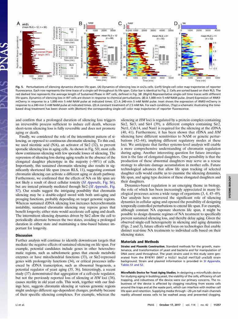
and confirm that a prolonged duration of silencing loss triggersan irreversible process sufficient to induce cell death, whereasshort-term silencing loss is fully reversible and does not promoteaging or death.Finally, we considered the role of the intermittent pattern of si-
lencing, as opposed to continuous chromatin silencing. To this end,we used nicotinic acid (NA), an activator of Sir2 (32), to preventsporadic silencing loss in aging cells. As shown in Fig. 5D, most cellsshow continuous silencing with few sporadic losses of silencing. Therepression of silencing loss during aging results in the absence of theelongated daughter phenotype in the majority (∼98%) of cells.Importantly, this sustained chromatin silencing also results in sig-nificantly shortened life span (mean RLS, 11), suggesting constantchromatin silencing can activate a different aging or death pathway.Furthermore, we confirmed that the effects of NA on life span arenot likely a result of direct cellular toxicity (SI Appendix, Fig. S6),but are instead primarily mediated through Sir2 (SI Appendix, Fig.S7). Our results suggest the intriguing possibility that chromatinsilencing may be a double-edged sword with both anti-aging andproaging functions, probably depending on target genomic regions.Whereas sustained rDNA silencing loss increases heterochromatininstability, sustained chromatin silencing may repress genes thatbenefit longevity; either state would accelerate cell aging and death.The intermittent silencing dynamics driven by Sir2 allow the cell toperiodically alternate between the two states, avoiding a prolongedduration in either state and maintaining a time-based balance im-portant for longevity.
DiscussionFurther analysis will continue to identify downstream targets thatmediate the negative effects of sustained silencing on life span. Forexample, potential candidates include genes in other heterochro-matic regions, such as subtelomeric genes that encode metabolicenzymes or have mitochondrial functions (33), or Sir2-repressedgenes with prolongevity functions (34), or critical processes influ-enced by rDNA transcription, such as ribosomal biogenesis, apotential regulator of yeast aging (35, 36). Interestingly, a recentstudy (37) demonstrated that aggregation of a cell-cycle regulator,but not the previously reported loss of silencing at HM loci (38),causes sterility in old yeast cells. This work, together with our find-ings here, suggests chromatin silencing at various genomic regionsmight undergo different age-dependent changes, probably becauseof their specific silencing complexes. For example, whereas the
silencing at HM loci is regulated by a protein complex containingSir2, Sir3, and Sir4 (39), a different complex containing Sir2,Net1, Cdc14, and Nan1 is required for the silencing at the rDNA(40, 41). Furthermore, it has been shown that rDNA and HMsilencing have different sensitivities to NAM or genetic pertur-bations (42–44), implying different regulatory modes at theseloci. We anticipate that further systems-level analysis will enablea more comprehensive understanding of chromatin regulationduring aging. Another interesting question for future investiga-tion is the fate of elongated daughters. One possibility is that theproduction of these abnormal daughters may serve as a rescuestrategy to alleviate damage accumulation in mother cells. Futuretechnological advances that allow life span tracking of selecteddaughter cells would enable us to examine the silencing dynamics,life span, and aging type decision of these elongated daughters andtheir daughters.Dynamics-based regulation is an emerging theme in biology,
the role of which has been increasingly appreciated in many bi-ological processes across a wide range of organisms (3, 4, 45–49).Our analysis here uncovered the significant role of silencingdynamics in cellular aging and opened the possibility of designingtemporally controlled perturbations to extend life span. For example,although constant NA exposure shortens life span, it might bepossible to design dynamic regimes of NA treatment to specificallyprevent sustained silencing loss, and thereby delay aging. Given theobserved single-cell heterogeneity in silencing and aging dynamics(Figs. 2 and 3), future efforts will focus on technologies that enabledistinct real-time NA treatments to individual cells based on theirsilencing states.
Materials and MethodsStrains and Plasmids Construction. Standard methods for the growth, main-tenance, and transformation of yeast and bacteria and for manipulation ofDNA were used throughout. The yeast strains used in this study were gen-erated from the BY4741 (MAT a his3Δ1 leu2Δ0 met15Δ0 ura3Δ0) strainbackground. Strain and plasmid information is provided in SI Appendix,Tables S1 and S2.
Microfluidic Device for Yeast Aging Studies. In designing a microfluidic devicefor studying aging in budding yeast, the viability of the cells, efficiency of celltrapping, and robustness of the device were our primary concerns. The ro-bustness of the device is affected by clogging resulting from excess cellsaround the traps and at the waste port, which can interfere with mother celllife span and retention. Supplying media through ∼20-μm-tall main channelsreadily allowed excess cells to be washed away and prevented clogging.
Fig. 5. Perturbations of silencing dynamics shorten life span. (A) Dynamics of silencing loss in sir2Δ cells. (Left) Single-cell color map trajectories of reporterfluorescence. Each row represents the time trace of a single cell throughout its life span. Color bar is identical to Fig. 2. Cells are sorted based on their RLS. Thered dashed line represents the average length of Sustained Phase in WT cells, defined in Fig. 3B. (Right) Representative single-cell time traces with differentlife spans. Dynamics of silencing loss in WT cells are shown in response to chemical perturbations. (B) A 1,000-min 5 mM NAM pulse. (Inset) Expression of RNR3-mCherry in response to a 1,000-min 5 mM NAM pulse at indicated times. (C) A 240-min 5 mM NAM pulse. Inset shows the expression of RNR3-mCherry inresponse to a 240-min 5 mM NAM pulse at indicated times. (D) A constant treatment of 2.5 mM NA. For each condition, (Top) a schematic illustrating the time-based drug treatment has been shown with (Bottom) the corresponding single-cell color map trajectories of reporter fluorescence.
Li et al. PNAS | October 17, 2017 | vol. 114 | no. 42 | 11257
SYST
EMSBIOLO
GY
Therefore, a critical feature of our device is its two-layer design, making itextremely robust over the course of our experiments, which takes more than80 h. This is a unique feature compared with recently published devices thatare all single-layer (10, 13, 14). The device was optimized for using contin-uous gravity-driven flow during operation, with the three-inlet design alsofacilitating media switching experiments. Cell loading efficiencies and finalretentions until cell death are ∼93% and 75%, respectively. See SI Appendixfor further details on the device and its validation.
Live-Cell Imaging and Analysis. Time-lapse imaging experiments were per-formed using a Nikon Ti-E inverted fluorescence microscope with PerfectFocus, coupled with an EMCCD camera (Andor iXon ×3 DU897). The lightsource is a spectra X LED system. Images were taken using a CFI plan Apo-chromat Lambda DM 60× oil immersion objective (NA 1.40 WD 0.13MM).The microfluidic device was placed on the motorized microscope stage (withEncoders) within a 30 °C incubator system. The flow of medium in the devicewas maintained by gravity and drove cells into traps. Waste medium wascollected to measure flow rate, which was about 2.5 mL/day. Images wereacquired every 15 min for a total of 80 or more hours. Images were pro-cessed and quantified with a custom MATLAB code. Cell divisions of eachmother cell were manually identified and counted at the time that the nucleiseparated betweenmother and daughter cells. Cells were categorized based on
their aging phenotypes, characterized by the morphologies of later daughtersthey produced. Mothers continually producing elongated daughters at the lastfew generations were categorized as “aging with elongated daughters,”whereas mothers continually producing round daughters at the last few gen-erations were categorized as “aging with rounded daughters.” Dynamic pat-terns of reporter fluorescence have been shown in jet rainbow color maps. Theinferno versions of the color maps to provide continuous luminance visualiza-tion of the data have been included in SI Appendix, Fig. S8.
Detailed methods and the development of the phenomenological modelare given in SI Appendix.
ACKNOWLEDGMENTS. We thank Yuan Zhao (Bioinformatics, University ofCalifornia, San Diego) for helping with developing the image analysis codeand Dr. Philbert Tsai (Physics, University of California, San Diego) for helpingwith the super-resolution confocal microscopy. This work was supported byNational Science Foundation Grant MCB-1616127 (to N.H.); University ofCalifornia Cancer Research Coordinating Committee (L.P.); Department ofDefense, Air Force Office of Scientific Research, National Defense Scienceand Engineering Graduate Fellowship 32 CFR 168a (to R.O.); HumanFrontier Science Program Fellowship LT000840/2014-C (to P.B.); and theSan Diego Center for Systems Biology National Institutes of Health GrantP50-GM085764.
1. McMurray MA, Gottschling DE (2004) Aging and genetic instability in yeast. Curr OpinMicrobiol 7:673–679.
2. López-Otín C, Blasco MA, Partridge L, Serrano M, Kroemer G (2013) The hallmarks ofaging. Cell 153:1194–1217.
3. Purvis JE, Lahav G (2013) Encoding and decoding cellular information through sig-naling dynamics. Cell 152:945–956.
4. Behar M, Hoffmann A (2010) Understanding the temporal codes of intra-cellularsignals. Curr Opin Genet Dev 20:684–693.
5. Levine JH, Lin Y, Elowitz MB (2013) Functional roles of pulsing in genetic circuits.Science 342:1193–1200.
6. Mortimer RK, Johnston JR (1959) Life span of individual yeast cells.Nature 183:1751–1752.7. Steinkraus KA, Kaeberlein M, Kennedy BK (2008) Replicative aging in yeast: The
means to the end. Annu Rev Cell Dev Biol 24:29–54.8. Lee SS, Avalos Vizcarra I, Huberts DH, Lee LP, Heinemann M (2012) Whole lifespan
microscopic observation of budding yeast aging through a microfluidic dissectionplatform. Proc Natl Acad Sci USA 109:4916–4920.
9. Xie Z, et al. (2012) Molecular phenotyping of aging in single yeast cells using a novelmicrofluidic device. Aging Cell 11:599–606.
10. Liu P, Young TZ, Acar M (2015) Yeast replicator: A high-throughput multiplexedmicrofluidics platform for automated measurements of single-cell aging. Cell Reports13:634–644.
11. Zhang Y, et al. (2012) Single cell analysis of yeast replicative aging using a newgeneration of microfluidic device. PLoS One 7:e48275.
12. Fehrmann S, et al. (2013) Aging yeast cells undergo a sharp entry into senescence un-related to the loss of mitochondrial membrane potential. Cell Reports 5:1589–1599.
13. Crane MM, Clark IB, Bakker E, Smith S, Swain PS (2014) A microfluidic system forstudying ageing and dynamic single-cell responses in budding yeast. PLoS One 9:e100042.
14. Jo MC, Liu W, Gu L, Dang W, Qin L (2015) High-throughput analysis of yeast repli-cative aging using a microfluidic system. Proc Natl Acad Sci USA 112:9364–9369.
15. Steffen KK, Kennedy BK, Kaeberlein M (June 25, 2009) Measuring replicative life spanin the budding yeast. J Vis Exp, 10.3791/1209.
16. McCormick MA, et al. (2015) A comprehensive analysis of replicative lifespan in4,698 single-gene deletion strains uncovers conserved mechanisms of aging. CellMetab 22:895–906.
17. Vijg J, Suh Y (2013) Genome instability and aging. Annu Rev Physiol 75:645–668.18. Gartenberg MR, Smith JS (2016) The nuts and bolts of transcriptionally silent chro-
matin in Saccharomyces cerevisiae. Genetics 203:1563–1599.19. Saka K, Ide S, Ganley AR, Kobayashi T (2013) Cellular senescence in yeast is regulated
by rDNA noncoding transcription. Curr Biol 23:1794–1798.20. Lindstrom DL, Leverich CK, Henderson KA, Gottschling DE (2011) Replicative age in-
duces mitotic recombination in the ribosomal RNA gene cluster of Saccharomycescerevisiae. PLoS Genet 7:e1002015.
21. Sinclair DA, Guarente L (1997) Extrachromosomal rDNA circles–a cause of aging inyeast. Cell 91:1033–1042.
22. Kaeberlein M, McVey M, Guarente L (1999) The SIR2/3/4 complex and SIR2 alonepromote longevity in Saccharomyces cerevisiae by two different mechanisms. GenesDev 13:2570–2580.
23. Bitterman KJ, Anderson RM, Cohen HY, Latorre-Esteves M, Sinclair DA (2002) In-hibition of silencing and accelerated aging by nicotinamide, a putative negativeregulator of yeast sir2 and human SIRT1. J Biol Chem 277:45099–45107.
24. Xu EY, Zawadzki KA, Broach JR (2006) Single-cell observations reveal intermediatetranscriptional silencing states. Mol Cell 23:219–229.
25. Dodson AE, Rine J (2015) Heritable capture of heterochromatin dynamics in Saccha-romyces cerevisiae. eLife 4:e05007.
26. Hu Z, et al. (2014) Nucleosome loss leads to global transcriptional up-regulation andgenomic instability during yeast aging. Genes Dev 28:396–408.
27. Dang W, et al. (2009) Histone H4 lysine 16 acetylation regulates cellular lifespan.Nature 459:802–807.
28. McCormick MA, et al. (2014) The SAGA histone deubiquitinase module controls yeastreplicative lifespan via Sir2 interaction. Cell Reports 8:477–486.
29. Kato M, Lin SJ (2014) Regulation of NAD+ metabolism, signaling and compartmen-talization in the yeast Saccharomyces cerevisiae. DNA Repair (Amst) 23:49–58.
30. Elledge SJ, Davis RW (1990) Two genes differentially regulated in the cell cycle and byDNA-damaging agents encode alternative regulatory subunits of ribonucleotide re-ductase. Genes Dev 4:740–751.
31. Xie Z, et al. (2015) Early telomerase inactivation accelerates aging independently oftelomere length. Cell 160:928–939.
32. Evans C, et al. (2010) NAD+ metabolite levels as a function of vitamins and calorierestriction: Evidence for different mechanisms of longevity. BMC Chem Biol 10:2.
33. Ellahi A, Thurtle DM, Rine J (2015) The chromatin and transcriptional landscape ofnative Saccharomyces cerevisiae telomeres and subtelomeric domains. Genetics 200:505–521.
34. Li M, Valsakumar V, Poorey K, Bekiranov S, Smith JS (2013) Genome-wide analysis offunctional sirtuin chromatin targets in yeast. Genome Biol 14:R48.
35. Janssens GE, et al. (2015) Protein biogenesis machinery is a driver of replicative agingin yeast. eLife 4:e08527.
36. Janssens GE, Veenhoff LM (2016) The natural variation in lifespans of single yeast cellsis related to variation in cell size, ribosomal protein, and division time. PLoS One 11:e0167394.
37. Schlissel G, Krzyzanowski MK, Caudron F, Barral Y, Rine J (2017) Aggregation of theWhi3 protein, not loss of heterochromatin, causes sterility in old yeast cells. Science355:1184–1187.
38. Smeal T, Claus J, Kennedy B, Cole F, Guarente L (1996) Loss of transcriptional silencingcauses sterility in old mother cells of S. cerevisiae. Cell 84:633–642.
39. Moazed D, Kistler A, Axelrod A, Rine J, Johnson AD (1997) Silent information regu-lator protein complexes in Saccharomyces cerevisiae: A SIR2/SIR4 complex and evi-dence for a regulatory domain in SIR4 that inhibits its interaction with SIR3. Proc NatlAcad Sci USA 94:2186–2191.
40. Shou W, et al. (1999) Exit from mitosis is triggered by Tem1-dependent release of theprotein phosphatase Cdc14 from nucleolar RENT complex. Cell 97:233–244.
41. Straight AF, et al. (1999) Net1, a Sir2-associated nucleolar protein required for rDNAsilencing and nucleolar integrity. Cell 97:245–256.
42. Gallo CM, Smith DL, Jr, Smith JS (2004) Nicotinamide clearance by Pnc1 directly reg-ulates Sir2-mediated silencing and longevity. Mol Cell Biol 24:1301–1312.
43. Smith JS, et al. (2000) A phylogenetically conserved NAD+-dependent protein de-acetylase activity in the Sir2 protein family. Proc Natl Acad Sci USA 97:6658–6663.
44. Smith JS, Brachmann CB, Pillus L, Boeke JD (1998) Distribution of a limited Sir2 proteinpool regulates the strength of yeast rDNA silencing and is modulated by Sir4p.Genetics 149:1205–1219.
45. Cai L, Dalal CK, Elowitz MB (2008) Frequency-modulated nuclear localization burstscoordinate gene regulation. Nature 455:485–490.
46. Lin Y, Sohn CH, Dalal CK, Cai L, Elowitz MB (2015) Combinatorial gene regulation bymodulation of relative pulse timing. Nature 527:54–58.
47. Hao N, O’Shea EK (2011) Signal-dependent dynamics of transcription factor trans-location controls gene expression. Nat Struct Mol Biol 19:31–39.
48. Purvis JE, et al. (2012) p53 dynamics control cell fate. Science 336:1440–1444.49. Albeck JG, Mills GB, Brugge JS (2013) Frequency-modulated pulses of ERK activity
transmit quantitative proliferation signals. Mol Cell 49:249–261.
11258 | www.pnas.org/cgi/doi/10.1073/pnas.1703379114 Li et al.
Related Documents