9504 | New J. Chem., 2015, 39, 9504--9517 This journal is © The Royal Society of Chemistry and the Centre National de la Recherche Scientifique 2015 Cite this: New J. Chem., 2015, 39, 9504 Multi-nuclear NMR of axially chiral biaryls in polypeptide orienting solvents: spectral discriminations and enantiorecognition mechanisms† Philippe Berdague ´ , a Jose-Enrique Herbert-Pucheta, a Vishwajeet Jha, b Armen Panossian, b Fre ´ de ´ ric R. Leroux b and Philippe Lesot* a Due to the importance of axially chiral biaryl derivatives as chiral auxiliaries and/or ligands for asymmetric synthesis, as well as their structural role in bioactive natural products, continuous efforts have been undertaken to propose efficient methods for their atropo-selective synthesis. As a consequence, proposing robust and reliable analytical tools that are able to discriminate the spectral signal of atropisomeric enantiomers becomes crucial to evaluate the enantiomeric excesses of mixtures. In this work, we show how several multi-nuclear 1D/2D-NMR techniques using homopolypeptide chiral liquid crystals as aligning solvents can provide a panel of analytical possibilities (through differences of chemical shift anisotropies, dipolar and quadrupolar residual couplings) to spectrally discriminate enantiomers of a large collection of trisubstituted axially chiral biphenyls. Approaches involving 31 P, 13 C and 2 H 1D- or 2D-NMR experiments at natural abundance levels are explored. Among noteworthy results, the first examples of spectral enantioseparations using 31 P nuclei as nuclear probe are reported. Finally, the roles of electronic factors and shape anisotropy in the efficiency of chiral discrimination mechanisms are examined and discussed. Molecular modeling calculations were carried out to establish the electronic profile of these analytes in order to understand and rationalize the 13 C–{ 1 H} NMR results. Introduction Axially chiral biaryl derivatives possess a peculiar stereochemical motif able to generate a couple of stereoisomers. The stereogenic structural motif is present in various potentially bioactive natural compounds and exhibits a wide range of biological properties. 1 For instance, one can mention the well-known vancomycin (a clinically used antibiotic glycopeptide) 2 or steganacin (a cyto- toxic tubulin-binding dibenzocyclooctadiene lignan). 3 In fact, the biaryl scaffold is a privileged structure for pharmaceutical research as its incorporation assures frequently high entry rates. 4 In addition, the stereogenic axes provide rigid molecular frame- works for highly efficient tools in asymmetric synthesis. 5 Concomitantly, atropisomeric C 1 -symmetric biaryls play an important and effective role as chiral auxiliaries and/or ligands for asymmetric synthesis. Consequently, continuous efforts have been undertaken by organic chemists to develop efficient methods for the atropo-selective synthesis of ligands based on the biphenyl, binaphthyl, or other biaryl backbones. 6 The con- formational stability of bridged biaryls can be strongly increased by incorporation of ortho-substituents, the associated rotational energy barrier primary depending on their number and their bulkiness. 7 Generally, ortho-trisubstituted biphenyls show no stereo-labile properties, and hence no rapid enantiomerization at room temperature is expected (see Fig. 1a). 8 So far, both chiral supercritical fluid chromatography and chiral gas chromatography have been the main methods used to separate the enantiomers of such peculiar chiral compounds. 8,9 Although adequate in numerous cases, chromatographic approaches present some well-known specific drawbacks for systematic implementation (price of chiral columns, for instance). Furthermore, the determination of experimental conditions leading to enantiomeric resolution is sometimes highly time- consuming. As a consequence, proposing (simple) analytical a Laboratoire de RMN en Milieu Oriente ´, Institut de Chimie Mole ´culaire et des Mate ´riaux d’Orsay (ICMMO), UMR CNRS 8182, Universite ´ Paris-Sud, Universite ´ Paris-Saclay, 91405 Orsay, France. E-mail: [email protected]; Fax: +33 (0)1 69 15 81 05; Tel: +33 (0)1 69 15 47 59 b Laboratoire de Chimie Mole ´culaire, Universite ´ de Strasbourg, UMR CNRS 7509, ECPM, 25 Rue Becquerel, 67087 Strasbourg, France † Electronic supplementary information (ESI) available: A description of experi- mental details and DFT calculation, analytical details, chemical pathway, back- ground of NMR in CLC, further 1D/2D NMR spectra, and NMR assignment data of compound 16 are given. Composition of oriented samples, details on anisotropic NAD NMR, further anisotropic NMR spectra and extra discussion for some compounds. See DOI: 10.1039/c5nj01434d Received (in Montpellier, France) 9th June 2015, Accepted 9th September 2015 DOI: 10.1039/c5nj01434d www.rsc.org/njc NJC PAPER Open Access Article. Published on 11 September 2015. Downloaded on 7/27/2022 2:04:18 AM. This article is licensed under a Creative Commons Attribution 3.0 Unported Licence. View Article Online View Journal | View Issue

Welcome message from author
This document is posted to help you gain knowledge. Please leave a comment to let me know what you think about it! Share it to your friends and learn new things together.
Transcript

9504 | New J. Chem., 2015, 39, 9504--9517 This journal is©The Royal Society of Chemistry and the Centre National de la Recherche Scientifique 2015
Cite this: NewJ.Chem., 2015,
39, 9504
Multi-nuclear NMR of axially chiral biaryls inpolypeptide orienting solvents: spectraldiscriminations and enantiorecognitionmechanisms†
Philippe Berdague,a Jose-Enrique Herbert-Pucheta,a Vishwajeet Jha,b
Armen Panossian,b Frederic R. Lerouxb and Philippe Lesot*a
Due to the importance of axially chiral biaryl derivatives as chiral auxiliaries and/or ligands for asymmetric
synthesis, as well as their structural role in bioactive natural products, continuous efforts have been
undertaken to propose efficient methods for their atropo-selective synthesis. As a consequence, proposing
robust and reliable analytical tools that are able to discriminate the spectral signal of atropisomeric
enantiomers becomes crucial to evaluate the enantiomeric excesses of mixtures. In this work, we show
how several multi-nuclear 1D/2D-NMR techniques using homopolypeptide chiral liquid crystals as aligning
solvents can provide a panel of analytical possibilities (through differences of chemical shift anisotropies,
dipolar and quadrupolar residual couplings) to spectrally discriminate enantiomers of a large collection of
trisubstituted axially chiral biphenyls. Approaches involving 31P, 13C and 2H 1D- or 2D-NMR experiments
at natural abundance levels are explored. Among noteworthy results, the first examples of spectral
enantioseparations using 31P nuclei as nuclear probe are reported. Finally, the roles of electronic factors
and shape anisotropy in the efficiency of chiral discrimination mechanisms are examined and discussed.
Molecular modeling calculations were carried out to establish the electronic profile of these analytes in
order to understand and rationalize the 13C–{1H} NMR results.
Introduction
Axially chiral biaryl derivatives possess a peculiar stereochemicalmotif able to generate a couple of stereoisomers. The stereogenicstructural motif is present in various potentially bioactive naturalcompounds and exhibits a wide range of biological properties.1
For instance, one can mention the well-known vancomycin(a clinically used antibiotic glycopeptide)2 or steganacin (a cyto-toxic tubulin-binding dibenzocyclooctadiene lignan).3 In fact,the biaryl scaffold is a privileged structure for pharmaceuticalresearch as its incorporation assures frequently high entry rates.4
In addition, the stereogenic axes provide rigid molecular frame-works for highly efficient tools in asymmetric synthesis.5
Concomitantly, atropisomeric C1-symmetric biaryls play animportant and effective role as chiral auxiliaries and/or ligandsfor asymmetric synthesis. Consequently, continuous effortshave been undertaken by organic chemists to develop efficientmethods for the atropo-selective synthesis of ligands based onthe biphenyl, binaphthyl, or other biaryl backbones.6 The con-formational stability of bridged biaryls can be strongly increasedby incorporation of ortho-substituents, the associated rotationalenergy barrier primary depending on their number and theirbulkiness.7 Generally, ortho-trisubstituted biphenyls show nostereo-labile properties, and hence no rapid enantiomerizationat room temperature is expected (see Fig. 1a).8
So far, both chiral supercritical fluid chromatography and chiralgas chromatography have been the main methods used to separatethe enantiomers of such peculiar chiral compounds.8,9 Althoughadequate in numerous cases, chromatographic approachespresent some well-known specific drawbacks for systematicimplementation (price of chiral columns, for instance).Furthermore, the determination of experimental conditionsleading to enantiomeric resolution is sometimes highly time-consuming. As a consequence, proposing (simple) analytical
a Laboratoire de RMN en Milieu Oriente, Institut de Chimie Moleculaire et des
Materiaux d’Orsay (ICMMO), UMR CNRS 8182,
Universite Paris-Sud, Universite Paris-Saclay, 91405 Orsay, France.
E-mail: [email protected]; Fax: +33 (0)1 69 15 81 05;
Tel: +33 (0)1 69 15 47 59b Laboratoire de Chimie Moleculaire, Universite de Strasbourg, UMR CNRS 7509,
ECPM, 25 Rue Becquerel, 67087 Strasbourg, France
† Electronic supplementary information (ESI) available: A description of experi-mental details and DFT calculation, analytical details, chemical pathway, back-ground of NMR in CLC, further 1D/2D NMR spectra, and NMR assignment data ofcompound 16 are given. Composition of oriented samples, details on anisotropicNAD NMR, further anisotropic NMR spectra and extra discussion for somecompounds. See DOI: 10.1039/c5nj01434d
Received (in Montpellier, France)9th June 2015,Accepted 9th September 2015
DOI: 10.1039/c5nj01434d
www.rsc.org/njc
NJC
PAPER
Ope
n A
cces
s A
rtic
le. P
ublis
hed
on 1
1 Se
ptem
ber
2015
. Dow
nloa
ded
on 7
/27/
2022
2:0
4:18
AM
. T
his
artic
le is
lice
nsed
und
er a
Cre
ativ
e C
omm
ons
Attr
ibut
ion
3.0
Unp
orte
d L
icen
ce.
View Article OnlineView Journal | View Issue
This journal is©The Royal Society of Chemistry and the Centre National de la Recherche Scientifique 2015 New J. Chem., 2015, 39, 9504--9517 | 9505
alternatives involving other techniques, such as NMR spectro-scopy, to chemists is a valuable task.
In the past, liquid-state NMR methods involving mainlychiral derivatizing agents (MPTA or Mosher’s acid) in combinationwith or without lanthanide shift reagents have been proposed todiscriminate enantiomers of (bridged or not) chiral biaryl atrop-isomers.10 Although successful, these approaches require thepresence of accessible reactive groups (–COOH, –OH, –NH2,. . .)to generate (in situ or not) diastereoisomers.
This prerequisite can be overcome when using NMR inlyotropic chiral liquid crystals (CLC). This breakthroughapproach was revealed to be a powerful and flexible tool,providing a robust and general alternative to the multitude ofisotropic NMR methods for the differentiation of enantiomersof chiral molecules,11 but also enantiotopic elements in pro-chiral molecules.12–14 Compared to classical NMR approachesusing chiral derivatizing or solvating chiral agents, NMR in CLCrequests no specific functional groups inside the analyte,15
while all magnetically active nuclei (even at very low naturalabundance levels) can provide effective probes.
Using a chiral aligned environment, the intermolecular inter-actions between each enantiomer (or each enantiotopic direction)and the CLC differ, and hence the internuclear vectors, i–j, are notoriented the same on average, SS
i–j a SRi–j (or Spro-S
i–j a Spro-Ri–j ).11–13 A
doubling of the spectral information/patterns for a given nuclearsite indicates therefore that the enantiorecognition phenomenonoccurs and is revealed on the NMR spectrum by a difference ofresidual chemical shift anisotropies (CSA), dipolar couplings (D)or quadrupolar couplings (DnQ) (for spin I 4 1/2) (see Fig. S1,ESI†).16,17 As a first example, deuterium NMR in CLC was usedto analyze the intramolecular dynamic processes of chiral andprochiral deuterated ortho-disubstituted biaryls (derivatives of1-(4-methylphenyl)naphthalene).14
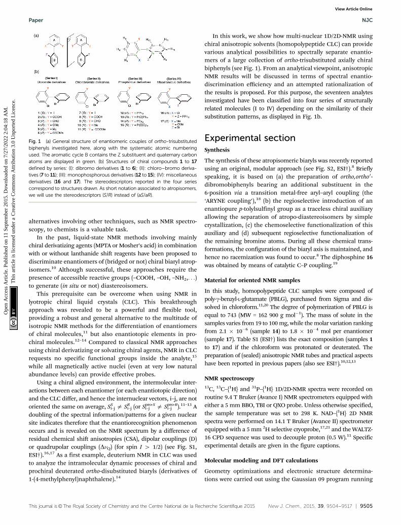
In this work, we show how multi-nuclear 1D/2D-NMR usingchiral anisotropic solvents (homopolypeptide CLC) can providevarious analytical possibilities to spectrally separate enantio-mers of a large collection of ortho-trisubstituted axially chiralbiphenyls (see Fig. 1). From an analytical viewpoint, anisotropicNMR results will be discussed in terms of spectral enantio-discrimination efficiency and an attempted rationalization ofthe results is proposed. For this purpose, the seventeen analytesinvestigated have been classified into four series of structurallyrelated molecules (I to IV) depending on the similarity of theirsubstitution patterns, as displayed in Fig. 1b.
Experimental sectionSynthesis
The synthesis of these atropisomeric biaryls was recently reportedusing an original, modular approach (see Fig. S2, ESI†).8 Brieflyspeaking, it is based on (a) the preparation of ortho,ortho0-dibromobiphenyls bearing an additional substituent in the6-position via a transition metal-free aryl–aryl coupling (the‘ARYNE coupling’),18 (b) the regioselective introduction of anenantiopure p-tolylsulfinyl group as a traceless chiral auxiliaryallowing the separation of atropo-diastereoisomers by simplecrystallization, (c) the chemoselective functionalization of thisauxiliary and (d) subsequent regioselective functionalization ofthe remaining bromine atoms. During all these chemical trans-formations, the configuration of the biaryl axis is maintained, andhence no racemization was found to occur.8 The diphosphine 16was obtained by means of catalytic C–P coupling.19
Material for oriented NMR samples
In this study, homopolypeptide CLC samples were composed ofpoly-g-benzyl-L-glutamate (PBLG), purchased from Sigma and dis-solved in chloroform.11,20 The degree of polymerization of PBLG isequal to 743 (MW = 162 900 g mol�1). The mass of solute in thesamples varies from 19 to 100 mg, while the molar variation rankingfrom 2.1 � 10�6 (sample 14) to 1.8 � 10�4 mol per enantiomer(sample 17). Table S1 (ESI†) lists the exact composition (samples 1to 17) and if the chloroform was protonated or deuterated. Thepreparation of (sealed) anisotropic NMR tubes and practical aspectshave been reported in previous papers (also see ESI†).10,12,13
NMR spectroscopy13C, 13C–{1H} and 31P–{1H} 1D/2D-NMR spectra were recorded onroutine 9.4 T Bruker (Avance I) NMR spectrometers equipped witheither a 5 mm BBO, TBI or QXO probe. Unless otherwise specified,the sample temperature was set to 298 K. NAD–{1H} 2D NMRspectra were performed on 14.1 T Bruker (Avance II) spectrometerequipped with a 5 mm 2H selective cryoprobe,17,21 and the WALTZ-16 CPD sequence was used to decouple proton (0.5 W).11 Specificexperimental details are given in the figure captions.
Molecular modeling and DFT calculations
Geometry optimizations and electronic structure determina-tions were carried out using the Gaussian 09 program running
Fig. 1 (a) General structure of enantiomeric couples of ortho-trisubstitutedbiphenyls investigated here, along with the systematic atomic numberingused. The aromatic cycle B contains the Z substituent and quaternary carbonatoms are displayed in green. (b) Structures of chiral compounds 1 to 17defined by series: (I): dibromo derivatives (1 to 6); (II): chloro–bromo deriva-tives (7 to 11); (III): monophosphorous derivatives (12 to 15); (IV): miscellaneousderivatives (16 and 17). The stereodescriptors reported in the four seriescorrespond to structures drawn. As short notation associated to atropisomers,we will use the stereodescriptors (S/R) instead of (aS/aR).
Paper NJC
Ope
n A
cces
s A
rtic
le. P
ublis
hed
on 1
1 Se
ptem
ber
2015
. Dow
nloa
ded
on 7
/27/
2022
2:0
4:18
AM
. T
his
artic
le is
lice
nsed
und
er a
Cre
ativ
e C
omm
ons
Attr
ibut
ion
3.0
Unp
orte
d L
icen
ce.
View Article Online

9506 | New J. Chem., 2015, 39, 9504--9517 This journal is©The Royal Society of Chemistry and the Centre National de la Recherche Scientifique 2015
on the ‘‘IDA’’ cluster of the University of Paris-Sud.22 Densityfunctional theory (DFT) with self-consistent reaction field (SCRF)Tomasi’s polarized continuum model (PCM) for solvation23 wasused in all calculations to describe implicitly the solvent (chloro-form) for energy minimizations and for description of orbitals.All computations were performed with the hybrid method B3LYP,whereas electronic correlation and exchange were respectivelydescribed by the use of the Becke24 and Lee–Yang–Parr25 func-tionals. Relativistic effective core potentials (ECP) were used todescribe electrons of heavy atoms (Br and Cl) with the valencedouble z quality basis sets Lanl2dz.26 The standard 6-311G(d,p)basis sets were used for the rest of the atomic orbitals’ descrip-tions of H, C, O and P atoms. Local minima conditions permolecule were confirmed with the calculation of harmonicvibrational frequencies of all structures. None of the predictedvibrational spectra has any imaginary frequency (data notshown), implying that the optimized geometry of each of themolecules under study lies at a local point on the potentialenergy surface. The electronic properties such as MolecularElectrostatic Potential (MEP), frontier molecular HOMO–LUMOorbital energies and Mulliken atomic charges have been obtainedwith the same level of theory as previously described.
Results and discussion
For a global view of the results, Table 1 (see also Table S2, ESI†)summarizes the essential data for the analytes and the sets ofexperimental results. As all results were obtained with verysimilar experimental conditions (T E 298 K, W/W of PBLG of14%), we will then follow with their interpretation in terms ofchiral discrimination mechanisms (noted in short as CDMs). Inparticular, for the 13C NMR results, attempts to correlate thenumber of 13C discriminated sites (noted in short as NDS(13C)) andthe possible solute–PBLG electrostatic interactions in combination
with molecular shape recognition effects will be proposedand discussed.
1H 1D-NMR spectroscopy
Even for small-size molecules, the number and the magnitude of(short and long-range) 1H–1H residual dipolar couplings signifi-cantly increase the linewidth to obtain rather low-resolution 1Hspectra where no fine structures clearly emerge. Some exceptionscan be found with molecules possessing isolated methyl groups,for instance. Compounds 4 to 6, 10, 11 and 17 are typicalexamples. Contrarily to isotropic 1H NMR, an uncoupledmethyl group exhibits a triplet structure in a LC (instead of asingle resonance) due to the intramethyl 1H–1H dipolar couplings(see Fig. S3, ESI†). In a CLC, two triplets with different splittings(|3DS
HH| a |3DRHH|) centered on very close 1H chemical shifts (due
to a small difference of 1H CSA) are generally detected.For analytes, 6 and 11, a single triplet (with |3DHH| = 58
and 52 Hz, respectively) is observed, thus revealing no resolveddiscrimination through a difference of DHH. Two reasons mayexplain this absence of enantiodiscriminations: (i) the rather lowsensitivity of D(1H–1H) to a difference of orientational ordering(compared to 2H quadrupolar interaction, for instance); (ii) thecomplex conformational dynamics of ligand Y (here, up to threerotors), which generally leads to averaging down the orderparameters of each internuclear vector along the chain, andsubsequently reduces spectral enantiodifferences. For solutes5, 10 and 17, a symmetric 1H spectral pattern of six lines isobserved for methyl bound to the ring (see Fig. S3, ESI†). Thisstructure can be analyzed as follows: (i) a dedoubled triplet ifthe methyl group is dipolarly coupled with one of the aromaticprotons; (ii) two triplets centered on two 1H chemical shifts dueto a surprisingly large 1H CSA. Two approaches involving thecomponents of mixture can be proposed to assess the originof the spectral pattern. They consist of either recording the
Table 1 Compilation of various solute parameters and the associated number of discriminated sites (racemic series) using proton-decoupled 2H,13C and 31P NMR results in PBLG
Series SoluteMass(mg) 10�5 �nR or S/mole
mmol(scalar)a/D
Hydrogenbond
Oxygenatom
13C–{1H}b
NMR
2H–{1H}b
NMR
31P–{1H}b
NMR
I (Br/Br) 1 (I) 19.9 2.27 2.132 N N 1/12 /c —2 (COOH) 54.5 7.65 2.529 Y Y 12/13 USd —3 (CHO) 61.0 9.00 2.933 N Y 9/13 7/8e —4 (OMe) 100.0 14.60 4.213 N Y 10/13 7/8e —5 (Me) 21.2 3.25 3.104 N N 6/13 NS —6 (COOMe) 72.2 9.75 2.963 N Y 8/13 6/8 —
II (Cl/Br) 7 (I) 21.1 2.68 2.213 N N 3/12 / —8 (COOH) 59.5 9.55 2.357 Y Y 11/13 US —9 (CHO) 62.6 10.60 2.737 N Y 11/13 6/8e —10 (Me) 19.0 3.38 3.179 N Y 6/13 NS —11 (COOMe) 70.1 10.75 3.006 N Y 8/13 6/8 —
III (Cl/Br) 12 (PPh2) 26.0 2.88 4.166 N N 4/24 f / 0/113 (POPh2) 96.2 10.26 4.622 N Y 12/24 f / 0/114 (PCy2) 25.3 2.73 3.048 N N 3/24 f / 1/115 (POCy2) 20.1 2.09 5.146 N Y 2/24 f / 0/1
IV 16 (P2)e 30.4 2.73 5.290 N N 12/36 f / 2/217 (Me/OMe) 100.6 18.20 2.377 N N 6/14 7/9e —
a The molecular electric dipole moment, mmol, is calculated for the lowest-energy conformer. b Number of sites (13C, 2H, 31P) spectrally discriminated.c /: spectrum not recorded. d US: unexpoitable NAD spectrum. e For one site 2H, two interpretations of results are possible. f 13C signals are alsodecoupled from 31P signals.
NJC Paper
Ope
n A
cces
s A
rtic
le. P
ublis
hed
on 1
1 Se
ptem
ber
2015
. Dow
nloa
ded
on 7
/27/
2022
2:0
4:18
AM
. T
his
artic
le is
lice
nsed
und
er a
Cre
ativ
e C
omm
ons
Attr
ibut
ion
3.0
Unp
orte
d L
icen
ce.
View Article Online

This journal is©The Royal Society of Chemistry and the Centre National de la Recherche Scientifique 2015 New J. Chem., 2015, 39, 9504--9517 | 9507
1H spectrum in ALC made of a racemic mixture of PBLG andPBDG (PBLG’s enantiomer)27 or using the enantiopure com-pound (when available) in the CLC. Irrespective of the methodused, it appears here that the doubling of triplets has its originfrom the dipolar coupling with the ortho-position aromaticproton as simply exemplified in the case of 10 (see Fig. S1,ESI†). Spin-manipulation based alternatives for simplifying thecoupling pattern of 1H signals should be possible but they arebeyond the scope of this paper and have not been explored.
1H-(de)coupled 31P NMR spectroscopy
Compounds 12 to 16 possess 31P nuclei, which are both sensitiveand 100% abundant, and hence easy to detect. Spectral enantio-discriminations can be observed through the difference of31P CSA, leading to two independent resonances, one for eachenantiomer, or a difference of 31P–1H RDCs. For the series ofmonophosphorous derivatives, no exploitable spectral separa-tions based on 31P–1H RDC differences were obtained on the31P 1D-NMR spectra, mainly due to the presence of variouslong-range 31P–1H RDCs, which obscure spectra. When protonsare decoupled, spectra are significantly simplified. Contrarily to12, 13 and 15, the presence of two 31P resonances for solute 14indicates that enantiomers are discriminated on the basis of31P CSA differences (|Ds| = 14 Hz, Dn1/2 = 3.5 Hz) (see Fig. 2a).Surprisingly, monophosphine oxide biaryl moieties 13 and 15are not spectrally discriminated despite the expected increase ofthe electronic shielding anisotropy of the phosphorous nucleusdue to the presence of the oxygen atom, which produces bigger31P CSA susceptible to lead (a priori) to larger enantiodiscrimina-tions. Variable temperature 31P NMR experiments (range of 30 K)did not permit enantiodiscriminations neither for 13 nor for 15.The last strongly suggests that the energy gap of the interconver-sion barrier between enantiomers is importantly increased by
the fact that aryls’ free-rotation is sterically hindered by thepresence of the oxide and thus only one enantiomer is favored.
Finally, the case of chiral biaryl-based diphosphane 16 is ratherpeculiar. Indeed in the liquid state, this molecule possesses twoanisochronous 31P atoms (at room temperature) resonating attwo distinct chemical shifts (d(31PA) = �12.0 ppm and d(31PB) =�14.1 ppm) and mutually coupled as first evidenced in 2011.28
This spin–spin coupling finds its origin via a ‘‘through-space’’scalar coupling (noted J(31PA–31PB) = 22.7 Hz) and not via theintramolecular five-bond connectivity, which should lead to asmall scalar 5J(31PA–31PB) coupling. The spectral assignments ofPA and PB atoms derive from the analysis of 1H–31P 2D HMBC,31P 2D J-resolved and 1H–1H 2D COSY experiments shown inESI† (Section SIII). If two doublets (AX spin system) areobserved on the isotropic 31P–{1H} spectrum, four resonances(Dn1/2 o 0.8 Hz) are detected at each 31P site in a racemic series(see Fig. S2a and S4, ESI†). This doubling of lines indicatesenantiodiscrimination. Three spectral situations correspondingto a difference of either 31P CSA or 31P–31P RDC or from bothcontributions can explain the presence of two pairs of doubletsfor each 31P site (see Fig. S5, ESI†). The assignment of 31Presonances was assessed by comparing the 1D spectrum of 16to the one recorded with an enantioenriched mixture (enrichedin R isomer, ee = 51.3%) using similar experimental conditions(sample composition and temperature). As seen in Fig. 2b, thepeak intensity difference between enantiomers allows theirassociated signals to be undoubtedly assigned. Various homo-nuclear 2D experiments confirm qualitatively the presence ofeach enantiomer, regardless of the absolute configuration ofNMR signals (vide infra and ESI†). According to the enantio-assignment made, the analysis of 31P–{1H} 1D spectrum of 16indicates that total couplings between the two 31P nuclei foreach isomer, |TA(31P–31P)| and |TB(31P–31P)| with T = J + 2D, arevery close and equal to 18.7 and 19.1 Hz, respectively while eachdoublet is shifted by 3.9 Hz (31PA) and 4.5 Hz (31PB). Assuming anegative value for TA or B, the magnitude of D(31P–31P) is equalto �20.3 and �20.5 Hz for R and S, respectively. Conversely,if T(31P–31P)A or B is positive, D(31P–31P)A or B becomes equal to�2 or �1.8 Hz, respectively. Among the two anisotropic con-tributions (D and CSA), only the 31P CSA (DDs = 3.9 Hz and4.5 Hz) is the relevant NMR interaction here that can be in fineefficiently exploited to evaluate the ee. As CSA is directly pro-portional to the magnetic field strength, operating with highermagnetic field spectrometers should guarantee larger discrimi-nations. Interestingly, the results obtained here are the two firstexamples of enantiodifferentiation using 31P–{1H} NMR, so far.Previous studies using 31P–{1H} or 31P NMR as the analyticaltechnique had failed in discriminating enantiomers of phos-phorous compounds.29
1H-decoupled 13C 1D-NMR
Although less sensitive compared to 1H or 31P NMR, anisotropic13C–{1H} NMR at natural abundance is an excellent and com-petitive method to discriminate enantiomers, in particularwhen sp2 hybridized carbon atoms are present in the analyte.The gain in sensitivity of commercially available cryogenic
Fig. 2 161.9 MHz 31P–{1H} 1D-NMR spectrum of (a) (R/S)-14 in PBLG/CHCl3 at 335 K and (b) of 16 in racemic series (bottom) and enantio-enriched series (R isomer) (top) in PBLG/CDCl3 at 298 K (ref. d(80% ofH3PO4) = 0 ppm). 1000 scans (a) and 5000 scans (b) are added and softline-sharpened Gaussian filtering is applied. Note the 31P CSA of 3.9 Hz and4.5 Hz for PA and PB of 16. The assignment of PA and PB atoms is given inESI† (see Fig. S14 to S18 in Section SI–SIII). The italicized notations ‘‘A/B’’stand for the chiral stereodescriptors of enantiomers A and B.
Paper NJC
Ope
n A
cces
s A
rtic
le. P
ublis
hed
on 1
1 Se
ptem
ber
2015
. Dow
nloa
ded
on 7
/27/
2022
2:0
4:18
AM
. T
his
artic
le is
lice
nsed
und
er a
Cre
ativ
e C
omm
ons
Attr
ibut
ion
3.0
Unp
orte
d L
icen
ce.
View Article Online

9508 | New J. Chem., 2015, 39, 9504--9517 This journal is©The Royal Society of Chemistry and the Centre National de la Recherche Scientifique 2015
probes with respect to conventional ones (up to a factor of 4to 5) enables the experimental acquisition times of qualitative13C–{1H} NMR to be reduced to less than 1–2 hour(s),30 evenworking with small amounts of solutes or with analytes of highMW. However, when a sufficient amount of solute is available(10–30 mg), depending on the enantiomeric mole number(from 2.27 � 10�5 to 18.20 � 10�5) and the S/N ratio (SNR)desired, all 13C–{1H} 1D spectra were recorded with a moderatemagnetic field (9.4 T) using acquisition times in the range of3–10 hours.
All experimental data related to the number of discrimi-nated 13C sites in PBLG are presented in Table 1, while Table S3(ESI†) lists the value of all d(13C) and the differences of CSA(DDs) for each of them.
For derivatives of series I and II, the 1D analysis of 13C–{1H}NMR spectra in CLC can be easily performed by counting thenumber of 13C lines and then comparing with the isotropic13C–{1H} NMR spectra where no discrimination occurs (seeFig. S6, ESI†). For all of them, several spectral enantiodiscrimi-nations occur both in the CH and quaternary aromatic carbonatoms, but the number of differentiated 13C sites varies fromone (solute 1) to twelve sites (solute 2), with spectral differencefrom 1 Hz (limit of discrimination) to 14 Hz. Note that carbonatoms belonging to the Y substituent (see Fig. 1) can providefurther potential 13C sites for discrimination like in the case of4, 9 and 10, for instance (see Table S3, ESI†).
Two illustrative examples of 13C–{1H} 1D spectra (solutes 2and 8) are given in Fig. 3. The variations of d(13C) between 2 and8 (and between each solute) result in the well-known electroniceffects (+I, �I and +M, �M) of various substituents on the rings(see also Fig. S4, ESI†). For 2 and 8, about 90% of 13C sites arediscriminated, thus affording a multiple choice for measuringenantiomeric excess (ee) of mixture. However, from a quanti-tative viewpoint, the choice of the best sites is clearly governedby three parameters (i) the spectral frequency differencesbetween enantiomer signals; (ii) SNR; and (iii) signal over-lapping of analytes with solvent signals. Typically, for 2 and8, the C-4 atom (SNR E 80–100) provides the best site forquantitative purposes when the PBLG/CHCl3 chiral system isused. Carbon atom C-9 could also provide a discrimination sitebut the signal overlay with very broad resonances of PBLGaromatic signals complicates the evaluation of enantiomericpurity. Finally, the quaternary C-6 and C-8 (for 2) or C-6 (for 8)carbons also provide large separations (E11 Hz), but their lowSNR (E30–35) excludes them from an accurate determinationof large ee’s. The overall interspectral analysis of all analytesindicates that biaryls with an aldehyde (3 and 9), ether (4) andester (6 and 11) substituent possess numerous enantiodiscri-minated sites (60 to 80%), but these sites show smaller spectraldifferences (1 to 3.5 Hz).
For methylated biaryls (5 and 10), the ratio of enantiodiscri-minated sites does not exceed 50% while the lowest number ofsites (o25%) is obtained for the iodo derivatives (1 and 7).13C–{1H} 1D-NMR spectra of 7 and 9 are given in the ESI.†Similar experimental conditions (co-solvent, w/w of polypeptideand temperature) for all solutes allow 13C spectral comparisons.
Furthermore, the 13C CSA is weakly sensitive to small variationsof T or sample concentration.
Due to the diversity of contributing factors/effects to the CDMs(molecular shape, electronic properties and/or conformationaldynamics), the attempt to establish qualitative correlationsbetween the enantiodiscrimination efficiency and molecularproperties is far from being trivial. Nevertheless, it is importantto investigate them in order to rank their role and evaluate theirrespective predominance contributing to the CDMs. This is aprerequisite step toward a global insight of the phenomenon,and subsequently the possibility to predict spectral results forany given analyte.
Considering the rather high degree of structural homology(ortho-three-substituted biaryl) of analytes in series I and II,we have first attempted to simply correlate (and explain) the13C NDS and the range of spectral separations (DDs(13C)) (weak(1–3 Hz), medium (4–8 Hz) or large (4 9 Hz)) to the magnitudeof the global dipolar moment of the molecule, mmol, calculatedby molecular modeling. For this purpose, the dipole moments(in CHCl3) for all biaryls in their lowest-energy conformationhave been calculated using solvent-dependent density func-tional theory (SCRF-DFT) Mulliken charge distribution analysis(see Experimental section for details). The scalar value islisted in Table 1, while the value on the three-axis components(x, y, z) and their vectorial representation are reported inTable S4 (ESI†). A first inspection of the molecular modellingresults shows that: (i) the variation of mmol does not exceed 7%when Br is replaced by the chlorine atom; and (ii) the nature ofsubstituent Y modifies significantly mmol, from 2.1 (1) to 4.2 D (4).
The comparison of NMR results between series I and IIshows rather similar results (NDS(13C) and DDs(13C)), thusindicating that the replacement of Br by Cl in position 6 (ring A)does not change strongly the global molecular properties
Fig. 3 100.4 MHz 13C–{1H} 1D spectra (BBO probe) of (a) (R/S)-2 and(b) (R/S)-8 recorded in PBLG/CHCl3. Only the region ranging from 123 to136 ppm is displayed (carbon atoms C-1/C-7 and from carboxyl group notshown). Note the doubling of numerous 13C signals (compared to isotropicspectra) associated to the spectral discrimination of R/S isomers. The verybroad resonances observed around 132–134 ppm originate from ortho/meta aromatic 13C signals of the benzyl group of the PBLG side chain.
NJC Paper
Ope
n A
cces
s A
rtic
le. P
ublis
hed
on 1
1 Se
ptem
ber
2015
. Dow
nloa
ded
on 7
/27/
2022
2:0
4:18
AM
. T
his
artic
le is
lice
nsed
und
er a
Cre
ativ
e C
omm
ons
Attr
ibut
ion
3.0
Unp
orte
d L
icen
ce.
View Article Online
This journal is©The Royal Society of Chemistry and the Centre National de la Recherche Scientifique 2015 New J. Chem., 2015, 39, 9504--9517 | 9509
(dipole moment and shape) of the solute towards the efficiencyof CDMs. In contrast, in each series, the difference of propertiesof ligand Y has a larger impact on both criteria. Except forsolutes 1 and 7, which both exhibit the smallest values of mmol
and NDS (with small DDs), the analysis of results for otheranalytes indicates that there is no simple dependency (i.e. amonotonous variation) between both spectral criteria and themagnitude of global dipole moment. In clear, the larger NDSdoes not occur for the biggest mmol. This absence of directcorrelation suggests that the nature of substituent Y and theassociated specific electronic properties (presence of labile hydro-gens able to form intermolecular hydrogen bondings (HB) or thepresence of electronegative oxygen in carbon–oxygen doublebond, for instance) play a crucial role in the efficiency of CDMfor this series of biaryls, independently to the global moleculardipole moment. Thus, the best results obtained with acid deri-vatives 2 and 8, and not for the methyl ester analogues (6 and 11),point out that the possibility of forming HB between the sub-stituent Y and the oxygen of the carboxylate group of the PBLGside chain is of primary importance in the CDM. Schematically,the role of HB can be understood as follows. Contrarily toordering mechanisms (mainly due to the coupling between thesolute quadrupole moment and the electric field gradient of thesolvent), the CDMs involve short-range intermolecular inter-actions that derive from the repulsive forces correlated with thesize and shape (and the shape-anisotropy) of the solute.31 Hence,the CDM efficiency is strongly dependent on the average distancebetween the solute and the PBLG chiral helix. In this context,irrespective of the magnitude of the global dipole moment, HBcan be seen as a crucial local electronic interaction susceptible tobring the solute nearest the fibers (at small distances), in turnpromoting better enantiodiscriminations, this effect being parti-cularly strong when the labile hydrogen is highly topologicallyaccessible, as in the case of 2 and 8.
When no HB are possible, other attractive specific electronicinteractions can play important roles in the CDM, in particularto reduce the average solute–PBLG distance. Although probablyless efficient than HB, these (secondary) interactions thenbecome key parameters governing the efficiency of discrimina-tion. Clearly the presence of an electronegative oxygen atom withaccessible lone pairs (aldehydic or ester groups for instance)appears as an important electronic parameter favoring fiber-solute electrostatic interactions (van der Waals type). In contrast(vide infra), it is a priori expected that CDM are much lessefficient for biaryls devoid of any groups susceptible to promoteany attractive intramolecular interactions (case 5 and 10). Thissimply explains why better results are obtained for methoxy,carbonyl or ester groups (NDS varying from 8/13 to 10/13),whereas the situation is much less favorable for methyl group(NDS = 6/13). From the spectral enantiodiscrimination view-point, the case of methoxy or aldehydic derivatives (3, 4, 9, 17)could be qualified as an ‘‘intermediate’’ situation for which onlyenantiodiscriminations with moderate spectral differences areexpected and occur.
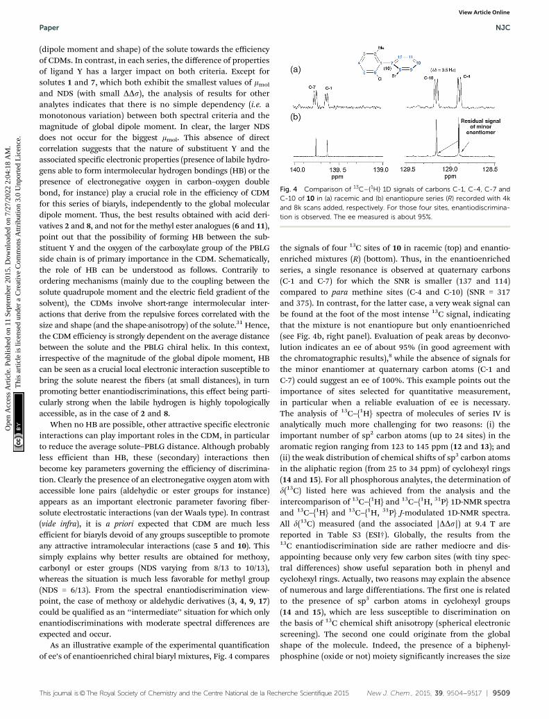
As an illustrative example of the experimental quantificationof ee’s of enantioenriched chiral biaryl mixtures, Fig. 4 compares
the signals of four 13C sites of 10 in racemic (top) and enantio-enriched mixtures (R) (bottom). Thus, in the enantioenrichedseries, a single resonance is observed at quaternary carbons(C-1 and C-7) for which the SNR is smaller (137 and 114)compared to para methine sites (C-4 and C-10) (SNR = 317and 375). In contrast, for the latter case, a very weak signal canbe found at the foot of the most intense 13C signal, indicatingthat the mixture is not enantiopure but only enantioenriched(see Fig. 4b, right panel). Evaluation of peak areas by deconvo-lution indicates an ee of about 95% (in good agreement withthe chromatographic results),8 while the absence of signals forthe minor enantiomer at quaternary carbon atoms (C-1 andC-7) could suggest an ee of 100%. This example points out theimportance of sites selected for quantitative measurement,in particular when a reliable evaluation of ee is necessary.The analysis of 13C–{1H} spectra of molecules of series IV isanalytically much more challenging for two reasons: (i) theimportant number of sp2 carbon atoms (up to 24 sites) in thearomatic region ranging from 123 to 145 ppm (12 and 13); and(ii) the weak distribution of chemical shifts of sp3 carbon atomsin the aliphatic region (from 25 to 34 ppm) of cyclohexyl rings(14 and 15). For all phosphorous analytes, the determination ofd(13C) listed here was achieved from the analysis and theintercomparison of 13C–{1H} and 13C–{1H, 31P} 1D-NMR spectraand 13C–{1H} and 13C–{1H, 31P} J-modulated 1D-NMR spectra.All d(13C) measured (and the associated |DDs|) at 9.4 T arereported in Table S3 (ESI†). Globally, the results from the13C enantiodiscrimination side are rather mediocre and dis-appointing because only very few carbon sites (with tiny spec-tral differences) show useful separation both in phenyl andcyclohexyl rings. Actually, two reasons may explain the absenceof numerous and large differentiations. The first one is relatedto the presence of sp3 carbon atoms in cyclohexyl groups(14 and 15), which are less susceptible to discrimination onthe basis of 13C chemical shift anisotropy (spherical electronicscreening). The second one could originate from the globalshape of the molecule. Indeed, the presence of a biphenyl-phosphine (oxide or not) moiety significantly increases the size
Fig. 4 Comparison of 13C–{1H} 1D signals of carbons C-1, C-4, C-7 andC-10 of 10 in (a) racemic and (b) enantiopure series (R) recorded with 4kand 8k scans added, respectively. For those four sites, enantiodiscrimina-tion is observed. The ee measured is about 95%.
Paper NJC
Ope
n A
cces
s A
rtic
le. P
ublis
hed
on 1
1 Se
ptem
ber
2015
. Dow
nloa
ded
on 7
/27/
2022
2:0
4:18
AM
. T
his
artic
le is
lice
nsed
und
er a
Cre
ativ
e C
omm
ons
Attr
ibut
ion
3.0
Unp
orte
d L
icen
ce.
View Article Online
9510 | New J. Chem., 2015, 39, 9504--9517 This journal is©The Royal Society of Chemistry and the Centre National de la Recherche Scientifique 2015
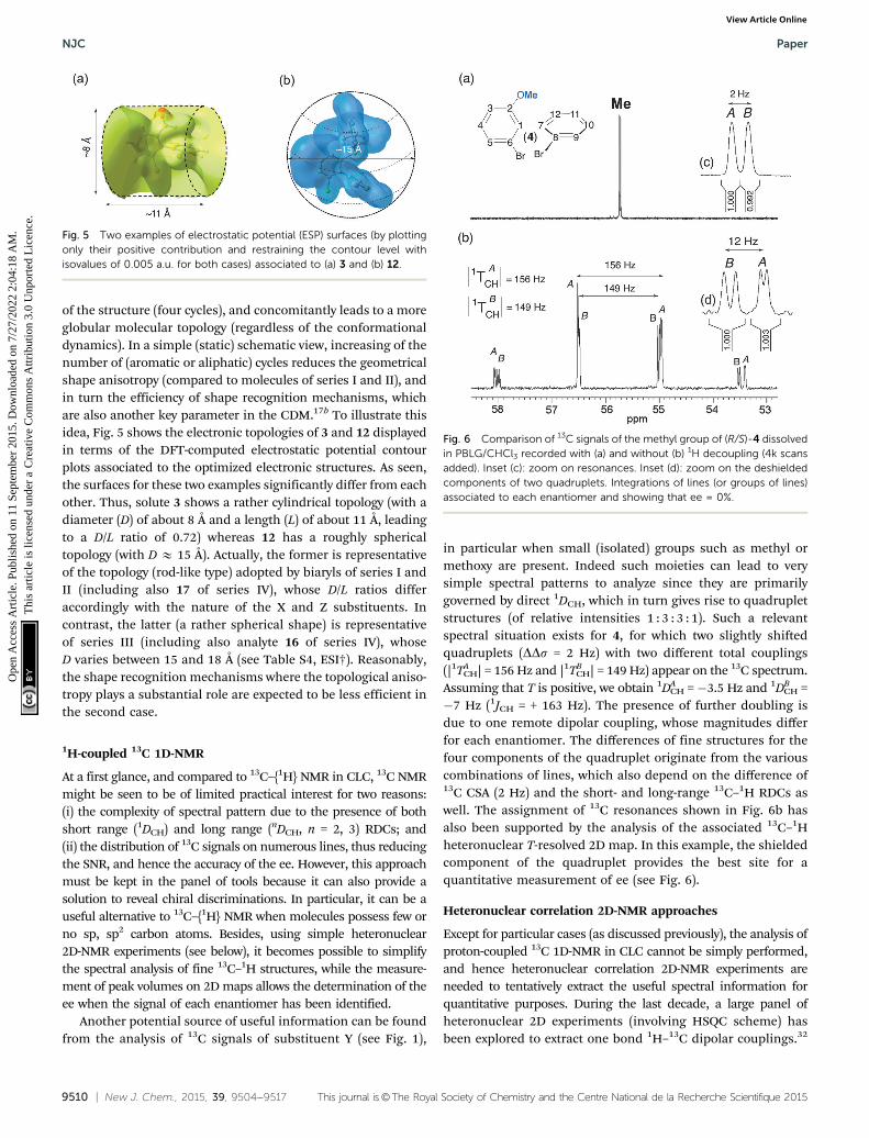
of the structure (four cycles), and concomitantly leads to a moreglobular molecular topology (regardless of the conformationaldynamics). In a simple (static) schematic view, increasing of thenumber of (aromatic or aliphatic) cycles reduces the geometricalshape anisotropy (compared to molecules of series I and II), andin turn the efficiency of shape recognition mechanisms, whichare also another key parameter in the CDM.17b To illustrate thisidea, Fig. 5 shows the electronic topologies of 3 and 12 displayedin terms of the DFT-computed electrostatic potential contourplots associated to the optimized electronic structures. As seen,the surfaces for these two examples significantly differ from eachother. Thus, solute 3 shows a rather cylindrical topology (with adiameter (D) of about 8 Å and a length (L) of about 11 Å, leadingto a D/L ratio of 0.72) whereas 12 has a roughly sphericaltopology (with D E 15 Å). Actually, the former is representativeof the topology (rod-like type) adopted by biaryls of series I andII (including also 17 of series IV), whose D/L ratios differaccordingly with the nature of the X and Z substituents. Incontrast, the latter (a rather spherical shape) is representativeof series III (including also analyte 16 of series IV), whoseD varies between 15 and 18 Å (see Table S4, ESI†). Reasonably,the shape recognition mechanisms where the topological aniso-tropy plays a substantial role are expected to be less efficient inthe second case.
1H-coupled 13C 1D-NMR
At a first glance, and compared to 13C–{1H} NMR in CLC, 13C NMRmight be seen to be of limited practical interest for two reasons:(i) the complexity of spectral pattern due to the presence of bothshort range (1DCH) and long range (nDCH, n = 2, 3) RDCs; and(ii) the distribution of 13C signals on numerous lines, thus reducingthe SNR, and hence the accuracy of the ee. However, this approachmust be kept in the panel of tools because it can also provide asolution to reveal chiral discriminations. In particular, it can be auseful alternative to 13C–{1H} NMR when molecules possess few orno sp, sp2 carbon atoms. Besides, using simple heteronuclear2D-NMR experiments (see below), it becomes possible to simplifythe spectral analysis of fine 13C–1H structures, while the measure-ment of peak volumes on 2D maps allows the determination of theee when the signal of each enantiomer has been identified.
Another potential source of useful information can be foundfrom the analysis of 13C signals of substituent Y (see Fig. 1),
in particular when small (isolated) groups such as methyl ormethoxy are present. Indeed such moieties can lead to verysimple spectral patterns to analyze since they are primarilygoverned by direct 1DCH, which in turn gives rise to quadrupletstructures (of relative intensities 1 : 3 : 3 : 1). Such a relevantspectral situation exists for 4, for which two slightly shiftedquadruplets (DDs = 2 Hz) with two different total couplings(|1TA
CH| = 156 Hz and |1TBCH| = 149 Hz) appear on the 13C spectrum.
Assuming that T is positive, we obtain 1DACH = �3.5 Hz and 1DB
CH =�7 Hz (1JCH = + 163 Hz). The presence of further doubling isdue to one remote dipolar coupling, whose magnitudes differfor each enantiomer. The differences of fine structures for thefour components of the quadruplet originate from the variouscombinations of lines, which also depend on the difference of13C CSA (2 Hz) and the short- and long-range 13C–1H RDCs aswell. The assignment of 13C resonances shown in Fig. 6b hasalso been supported by the analysis of the associated 13C–1Hheteronuclear T-resolved 2D map. In this example, the shieldedcomponent of the quadruplet provides the best site for aquantitative measurement of ee (see Fig. 6).
Heteronuclear correlation 2D-NMR approaches
Except for particular cases (as discussed previously), the analysis ofproton-coupled 13C 1D-NMR in CLC cannot be simply performed,and hence heteronuclear correlation 2D-NMR experiments areneeded to tentatively extract the useful spectral information forquantitative purposes. During the last decade, a large panel ofheteronuclear 2D experiments (involving HSQC scheme) hasbeen explored to extract one bond 1H–13C dipolar couplings.32
Fig. 5 Two examples of electrostatic potential (ESP) surfaces (by plottingonly their positive contribution and restraining the contour level withisovalues of 0.005 a.u. for both cases) associated to (a) 3 and (b) 12.
Fig. 6 Comparison of 13C signals of the methyl group of (R/S)-4 dissolvedin PBLG/CHCl3 recorded with (a) and without (b) 1H decoupling (4k scansadded). Inset (c): zoom on resonances. Inset (d): zoom on the deshieldedcomponents of two quadruplets. Integrations of lines (or groups of lines)associated to each enantiomer and showing that ee = 0%.
NJC Paper
Ope
n A
cces
s A
rtic
le. P
ublis
hed
on 1
1 Se
ptem
ber
2015
. Dow
nloa
ded
on 7
/27/
2022
2:0
4:18
AM
. T
his
artic
le is
lice
nsed
und
er a
Cre
ativ
e C
omm
ons
Attr
ibut
ion
3.0
Unp
orte
d L
icen
ce.
View Article Online

This journal is©The Royal Society of Chemistry and the Centre National de la Recherche Scientifique 2015 New J. Chem., 2015, 39, 9504--9517 | 9511
However, the control of 1H–13C polarization transfer efficiency(for quantitative measurement of ee dissolved in CLC) can besubtle and time-consuming for routine NMR users. Consider-ing the framework of this study, we only focused our purposeon 2D experiments based on the well-known heteronuclear‘‘J-resolved’’ schemes.33 As in CLC, the T couplings replacethe J couplings, so the experiments were renamed ‘‘T-resolved’’2D experiments, but the pulse scheme remains identical. Asexpected, the 13C chemical shifts are refocused during t1 whilethe T(13C–1H) couplings are removed during acquisition by 1Hdecoupling. As the 13C–1H T couplings evolve only during half ofthe t1 evolution period (gated-decoupled method), the T valuesare scaled down by a factor of 2. Modified sequences of the basic‘‘T-resolved’’ experiment might be proposed. For instance, withthe view of simplifying the coupling structures in F1, a BIRDcluster can be incorporated to differentiate long-range fromdirect couplings.34 Additionally, the sensitivity could be improvedby incorporating INEPT or DEPT pulse trains as an initial transferstep.35 Nevertheless, one can be faced with either distorted linesor important differences with the transfer efficiency, leading toless accurate ee measurements.
Fig. 7 displays the region of the T-resolved 2D spectrum where1H–13C coupling patterns and 13C chemical shifts associated toC-10 and C-11 atoms of 2 appear in F1 and F2 dimensions,respectively. The analysis of the map allows the relevant informa-tion for each carbon site and each enantiomer (noted A and B) tobe separated. For each of them, the spectral pattern is dominatedby the direct 1TCH coupling, which is different for each enantio-mer (|1TA/B
CH(C-10)| = 459/528 Hz and |1TA/BCH(C-11)| = 106/82 Hz),
as seen on the map. Interestingly, we can measure a largedifference of 1TCH (D1TCH) of about 70 Hz at site C-10. As 1TA/B
CH =1JA/B
CH + 21DA/BCH, the magnitude of 1TA/B
CH(C-11) suggests that thesign of 1DA/B
CH is negative (compared to 1JA/BCH, which is always
positive and ranging from 150–160 Hz for aromatic carbons).36
Note that a similar spectral situation was also observed forthe C-11 site of 3 (see Fig. S5, ESI†). Indeed, here again the1TA/B
CH(C-11) value (66/74 Hz) is smaller than 1JCH(C-11), thusindicating that 1DA/B
CHo0. The further splittings (at C-10) andtriplets (at C-11) observed on the map originate from the nTCH
long-range couplings. For both sites, the separation of couplingpatterns in F1 is facilitated by the 13C chemical shifts differencebetween enantiomers. The presence of two triplets (for eachenantiomer) is quite unusual but can be explained if C-11 iscoupled identically with two inequivalent aromatic protons inthe vicinity of C-11 (62 and 64 Hz for A and B, respectively).
The large magnitude of 1DA/BCH (148 and 170 Hz) for C-10
whereas 1JCH = +164 Hz indicates that the C10–H direction isstrongly ordered. Actually, the analysis of other carbon sites of2 (and 8, also) confirms a rather strong degree of molecularalignment compared to other solutes, leading to large (andunusual) 1DCH values. This result suggests the presence of HB inthe orientation mechanisms of 2 (and 8) leading to a decrease ofthe average distance between the analyte and the fiber, and hencean increase of the average degree of alignment of the solute.Locally, it is expected that the ordering of each C–H vector (SCH),and in turn the associated RDC value, increase.
In a framework of a crude two-site interaction model, we cansimply write that the Sij order parameter is a weighted sum of
Fig. 7 (a) Expanded region (centered on C-10 and C-11 sites) of 13C–1H T-resolved 2D spectra of (R/S)-2 in PBLG/CHCl3 (see Fig. 2a). (b) Two verticalslices extracted from the T-resolved map project two dedoubled triplets, one per enantiomer. Each of the (1 : 2 : 1) intensity patterns in (b) is highlightedwith filled and unfilled circles for visualization purposes. The T values (in Hz) given in (b) are half of the true values.
Paper NJC
Ope
n A
cces
s A
rtic
le. P
ublis
hed
on 1
1 Se
ptem
ber
2015
. Dow
nloa
ded
on 7
/27/
2022
2:0
4:18
AM
. T
his
artic
le is
lice
nsed
und
er a
Cre
ativ
e C
omm
ons
Attr
ibut
ion
3.0
Unp
orte
d L
icen
ce.
View Article Online

9512 | New J. Chem., 2015, 39, 9504--9517 This journal is©The Royal Society of Chemistry and the Centre National de la Recherche Scientifique 2015
two situations corresponding to the case where the solute iseither close to the polypeptide fibers (bonded) or at remotelocation (free):
SObsij = Pbonded(Sbonded
ij ) + Pfree(Sfreeij ) (1)
where Pbonded and Pfree are the normalized population ratios ofsolute (Pbonded + Pfree = 1). From the NMR viewpoint, we cansubsequently write that:
Obsi = Pbonded(Obsbondedi ) + Pfree(Obsfree
i ) (2)
where Obsi stands for NMR observable at site i (Dsi, Dij or DnQi)and is associated to NMR nuclei detected. In this very simpleapproach, the solute is strongly oriented in close vicinity tothe helix, and not oriented (or very weakly oriented) when thesolute is distant from the helix; the respective population andassociated splittings directly depend on the strength of inter-actions between the solute and PBLG.
31P–31P correlation 2D-NMR experiments
As explained previously, the comparison of the 31P–{1H} 1D-NMRspectrum of diphosphino biphenyl 16 recorded in racemic andenantioenriched series leads to a rapid assignment of the abso-lute configuration of lines in the spectrum of the racemic mixture(vide supra). However, this is only possible when an enantio-enriched mixture or an enantiopure compound is available andthe absolute configuration of the major isomer is known. Whenthe racemic mixture is only available, the assignment of various31P peaks of (�)-16 is obviously not straightforward. Indeed thepositions of 31P resonances can be explained by a differenceof 31P CSA or 31P RDC, or both (see Fig. 2). To clear up thisambiguity, various homonuclear 2D-NMR approaches were testedto correlate the 31P resonances to each enantiomer of the mixture:the 31P–31P COSY, T-resolved and INADEQUATE 2D11,37 experi-ments, no knowledge of 31P–31P RDC being requested for the firsttwo. Experimental maps and comments on the experimentalresults are proposed in ESI.†
The NAD 2D-NMR approach
The main feature of NAD NMR is mainly its low sensitivity dueto the very weak natural abundance of deuterium nuclei (1.5 �10�2%), namely 100 fold less than 13C nuclei. Besides usingCLC, the intensity of NAD signals for a given 2H site is reducedby a factor four when spectral discrimination occurs. Indeed,the single 2H peak observed in achiral liquids (see Fig. S2, ESI†)is now split into four resonances (two quadrupolar doublets),hence reducing the SNR, and subsequently the error in the eevalue, in particular when the ee’s are large. Technically, thissituation can be partly overcome using high-field NMR spectro-meters, equipped with cryogenic probes when possible. How-ever, the efficiency/interest of this tool will depend primarily onthe available amount of analyte, concomitantly to its MW. In thisstudy, the MW of the solutes ranges from 282 to 557 g mol�1,while the available amounts vary from 20 to 100 mg (half of thesemasses for each enantiomer). Under these conditions, NAD2D-NMR experiments in CLC have been only recorded for solutes2 to 4, 6, 9, 11 and 17, for which a sufficient mass of solute was
available (see Table 1); this corresponds to a mole numbervarying from 7.65 to 18.2 � 10�5 mol, namely a mole numberof monodeuterated isotopomers [2H] varying from 11.8 to28.2 � 10�9 mol. The number of discriminated 2H sites foreach analyte is reported in Table 1 (see also Table S2, ESI†).
The analytical interest of NAD 2D-NMR is the possibility toseparate the useful information on two spectral dimensions(see Fig. S12 to S14, ESI†). Thus on the tilted NAD Q-COSY Fzmap of 17 (Fig. S12, ESI†), we can easily assess that seven 2H sites(over nine) show spectral discrimination (77%). The presence ofthree DQ (instead of 4) associated to sites 10 and 12 resonating atthe same d(2H) can lead to two possible interpretations from thediscrimination viewpoint: (i) two quadrupolar doublets (QD) forsite 12 and one for site 10; and (ii) two QD for sites 10 and 12,considering that both internal doublets for each site possess thesame splitting. Actually, the second analysis is less probable forthree reasons: (i) the differences of RQCs (DDnQ = |DnR
Q � DnSQ|)
for each aromatic site are quite similar (ranging from 210 to355 Hz for the outer one and from 167 to 278 for the inner one);(ii) in the structure, the C–2H10 and C–2H4 bonds are collinear(para position), and hence the order parameters (and theirassociated DnQ’s) are expected to be similar for both C–2Hvectors (124 and 140 Hz respectively); and (iii) the absence ofdiscrimination at both sites. The analysis of the aliphatic regionindicates that both methyl and methoxy groups are discrimi-nated (until the baseline) with RQCs differences of 24.0 and23.4 Hz, respectively, namely 12 Hz between R and S componentsof doublets. Due to the free rotation of methyl deuterons aroundthe C–C bond (1 rotor) or C–O–C bonds (2 rotors), the RQCvalues are averaged down compared to the RQCs of aromatic 2Hsites (from 3–10 fold less). Interestingly, the contribution of threedeuterons to the NAD signals increases the SNR (162 and 146)compared to the aromatic sites, thus providing the two best sitesto accurately determine the enantiomeric excesses.
Except for analytes 2 and 8 (see below), the distribution inmagnitude of the RQCs of the aromatic 2H QDs observed on theNAD 2D map is globally quite similar for the various analytes(see Fig. S12 and S13, ESI,† for instance). In contrast, the RQCsmeasured for the 2H sites of the flexible Y substituent showlarge variations in magnitude. In order to illustrate this, Fig. 8compares the NAD 1D signals of the aldehyde groups of 3 and 9and methyl groups of 4/17 and 11/17 (see Fig. S15, ESI† for thefull 2D map of 6 and 11). For each case, the spectral enantio-discrimination on Y occurs with differences of RQCs, |DDnQ|,varying from 5 Hz (11) to 35 Hz (4), while the average of thecorresponding RQCs varies from 129 Hz to 28.5 Hz. The largevariation of SNR between the methine and methyl grouporiginates from the number of 2H nuclei contributing to signals(1 to 3) but also the mass (60 mg to 100 mg) and the MW (277 to370) of each sample, and possibly some variations of isotopicratio from one site to another (but not between enantiomers).
Various comments can be made on this series of results,in particular on the variation of RQCs and the magnitude ofenantiodiscriminations for the different 2H sites. Indeed, thecomparison of results indicates that there is not a simplecorrelation between the magnitude of RQCs, |DnQ|, the RQC’s
NJC Paper
Ope
n A
cces
s A
rtic
le. P
ublis
hed
on 1
1 Se
ptem
ber
2015
. Dow
nloa
ded
on 7
/27/
2022
2:0
4:18
AM
. T
his
artic
le is
lice
nsed
und
er a
Cre
ativ
e C
omm
ons
Attr
ibut
ion
3.0
Unp
orte
d L
icen
ce.
View Article Online

This journal is©The Royal Society of Chemistry and the Centre National de la Recherche Scientifique 2015 New J. Chem., 2015, 39, 9504--9517 | 9513
difference between enantiomers, |DDnQ|, and the number ofrotors in the ligand Y (one for 3 and 9), two for 4 and 17, andthree for 6 and 11. Actually, various (sometimes contradictory)effects, such as the difference of electronic properties of eachligand (aldehyde, ether and ester group) and the position(number of rotors) of the 2H site in the ligand can be evokedto explain the results. Thus, the large magnitude of RQCs for 3and 9 associated to a strong degree of alignment of the C–Hdirection could suggest a strong site-specific interaction (charge-transfer interactions) between the carbonyl polar group and thePBLG fiber, despite the free rotation around the C–C bond. Thelarge difference of |DnQ’s| (from 34 to 150) and |DDnQ’s| (from35 to 5) observed for the methyl groups in 4 (17) and 6 (11) ismuch more subtle to explain/understand. They both involvesignificant differences of averaged orientation of the C–D direc-tions, the number of rotors (2 and 3) between the ring and the2H site, and the electronic properties of the ligand. On the basisof the rotor number, it could be expected that the |DnQ’s| for6 and 11 would be larger than 4 and 17. This trend is notexperimentally observed whereas larger |DDnQ’s| are measuredfor those latter compared to 6 and 11. This illustrates thedifference of electronic interaction between ether or carboxylategroups and the PBLG side chain (which can promote a more or
less stronger alignment of the Y ligand), but also the ability ofthe 2H site to sense the chirality of the biaryl skeleton versus itsdistance and the number of rotors in the flexible part. Resultsobtained for the methyl group (6 and 11) suggest that a physicalinteraction between PBLG and the carboxylate group mightgenerate a higher degree of alignment for the COOMe moiety,and hence for the terminal methyl group, but in this case with aweaker enantiodiscrimination efficiency.
Finally, it must be noticed that the NAD 2D-NMR spectra ofthe carboxylic acids (2 and 8) were not analytically exploitablewhereas 13C–{1H} NMR had provided well-resolved spectra withexcellent results in terms of spectral quality and enantiodiscri-minations (see Table 1, Table S3, ESI†). Indeed, 2D maps of 2and 8 are made of weakly resolved NAD QDs of low intensity,not distinctly emerging from noise (even with strong exponen-tial apodisation), and showing splittings ranging from 1500 to2000 Hz (see Fig. S14a/b, ESI†). These magnitudes of RQCs areunusually large for small solutes oriented in weakly aligningmedia as those prepared with the PBLG polymer, whereas thesymmetrical shape, the linewidth (3 Hz) and the splitting(around 500 Hz) of chloroform both indicate a homogeneousand uniform mesophase that complies with standards when w/w(PBLG) = 14%. No significant enhancement was obtained by severalrehomogenizations of the sample (new cycles of centrifugations) orby a sample temperature variation.
Although a priori unexpected, the low quality of NAD NMRspectra for 6 and 8 (due to unusually large RQCs) can beexplained by the presence of HB, and again understood in theframe of the simple model proposed above. Derived from eqn (2),we can write that:
DnObsQ (2H) = Pbonded(Dnbonded
Q (2H)) + Pfree(DnfreeQ (2H)) (3)
Thus, the existence of strong hydrogen bonds may lead to an‘‘aggregation effect’’ of the solute towards PBLG fibers, con-siderably increasing the solute alignment (and the associatedDnQ at each deuterium site), and finally amplifying excessivelythe RQCs. In the case of NAD NMR, this aggregative effect canappear as spectrally unfavorable (as seen for 6 and 8), becausethe larger the 2H splittings are, the larger the linewidths for eachcomponent of DQ are (effect due to the ‘‘disorder’’ of orienta-tional order), and hence the smaller the SNR are. In the case of 2,we can note that the magnitude range of RQCs of DQs observedon the NAD map is rather coherent with the range of RDCsmeasured on the 13C–1H NMR spectra (see the 13C–1H T-resolved2D map in Fig. 7) according to the fact that the ratio ‘‘RQC/RDC’’is equal to 12–14 when sp2 hybridized carbon atoms are involved(this ratio is equal to 11–12 for sp3 ones). This relationshipderives from the fact that 13C–1H and 13C–2H directions (in theassociated isotopomers) are similarly oriented in the mesophaserelative to the magnetic field axis, Bo.38 Finally, the presenceof HB between 2 and 8 and PBLG is simply evidenced whencomparing their NAD spectra with those of ester analogues,6 and 11, which cannot form HB. While the sample compositionis similar to those of the acid derivatives, we obtain exploitableNAD 2D spectra of 11 where the ranges of RQCs are standard(see Fig. S14, ESI†). The analysis of both esters indicates that
Fig. 8 92.1 MHz proton-decoupled NAD 1D-NMR signals of (a and b) themethine of the aldehyde group of (R/S)-3 and (R/S)-9, of (c and d) themethyl of the methoxy group of (R/S)-4 and (R/S)-17 and of (e and f)the methyl of the ester group of (R/S)-6 and (R/S)-11, all of them dissolvedin PBLG/CHCl3 at 295 K. All patterns are extracted from their tilted NADQ-COSY Fz map. Except for e and f (see Fig. S10, ESI†), an exponentialfiltering (LB = 2 Hz) is applied on both dimensions.
Paper NJC
Ope
n A
cces
s A
rtic
le. P
ublis
hed
on 1
1 Se
ptem
ber
2015
. Dow
nloa
ded
on 7
/27/
2022
2:0
4:18
AM
. T
his
artic
le is
lice
nsed
und
er a
Cre
ativ
e C
omm
ons
Attr
ibut
ion
3.0
Unp
orte
d L
icen
ce.
View Article Online

9514 | New J. Chem., 2015, 39, 9504--9517 This journal is©The Royal Society of Chemistry and the Centre National de la Recherche Scientifique 2015
85% (6/7) of the aromatic 2H sites are discriminated (|DDnQ| =20 to 153 Hz) while a small spectral enantiodifference (about3 Hz) is observed on the terminal methyl group.
All these results and arguments point out openly the impor-tant role of HB in the ordering mechanisms of the solute (inparticular the degree of molecular orientation) and the relatedanalytical consequences according to the NMR properties of theobserved nucleus. Thus, the presence of HB involving COOHgroups was a major advantage in terms of 13C enantiodiscrimi-nation (on the basis of 13C CSAs) since 85% of 13C sites showeddiscriminations, but can lead to undesirable effects visible onthe NAD spectra (on basis of 2H RQCs). This beneficial differenceresults from a more complex and ‘‘dilute’’ dependence of the 13CCSA through the electronic screen tensor (for each 13C site)toward the molecular ordering of a solute compared to DnQ. Thisrather contradictory situation associated to 13C and 2H NMRillustrates nicely some versatile aspects of anisotropic NMR aswell as the subtle balance between the orientation and thechiral discrimination.
Importance and role of factors involved in the CDM
In the frame of the understanding and the phenomenologicaldescription of CDMs in polypeptide CLC (and in particular inPBLG), the analysis of the various factors (and their respectiverole) governing or involving the efficiency of CDM is a necessarystep before proposing models describing the phenomenon or astarting point for any computational modeling of the system.A priori, it is difficult to dissociate the topological properties(shape anisotropy) and the electronic profile of a solute due totheir strong inherent intrication. However, for a qualitativedescription of contributive factors, we may propose this artifi-cial separation.
From the analysis of 13C–{1H} NMR results of this study (datafrom seventeen solutes) related to previous studies, we evidencedthat the topological factors are of primary importance.11,16,17b,39
According to the degree of shape anisotropy of solutes (forinstance, spheroid, cylinder, spiral,. . .), the efficiency of globalshape recognition mechanisms (which are closely related tosteric exclusion effects) is basically different, the best situationbeing met with a spiral topology rather than a spheroid one.
As shape recognition mechanisms are short-range inter-actions that are highly active when the solute is in the closestvicinity of the polypeptide, they are strongly dependent onpossible local electronic (electrostatic) interactions betweenthe solute and the chiral fiber, and in particular the flexibleside chain of the polymer. Hence, the electronic local propertiesrelated to the nature of substituents (presence of HB, strengthof C–O dipole moment, steric hindrance) in combination withthe global properties of the analyte (the global dipole moment)play a key role in the solute–PBLG interactions and their cap-ability to ‘‘maintain’’ or not the solute close to the chiral fiber,namely when the CDM are the most efficient.
The role of intermolecular HB. In this series of results, wehave evidenced the importance and the role of HB between thesubstituent and the PBLG fibers in the mechanisms, in parti-cular when the access of the labile hydrogen is easy. As can be
observed in Table 1, solutes 2 and 8 present the highest degreeof discrimination (13C NMR) of all the solutes discussed in thepresent study. Both solutes possess a COOH moiety with alabile proton susceptible to be engaged in HB with PBLG. Once2 and 8 are esterified in order to form 6 and 11, respectively,the enantiodiscrimination efficiency is reduced, thus suggestingthe importance of the carboxyl proton to interact with PBLG.Moreover, when analyzing cylindrical topologies of series Iand II (Table S4, ESI†), it is observed that, independently ofthe distorted cylindrical ESP-topology of each of the solutes,COOH groups in series 2 and 8 present a proper cavity abovethe cylindrical ESP form (around 4 Å of diameter) that canpromote the access of the PBLG basic moiety in order to‘‘orient’’ the HB interaction, regardless of the known PBLG’sconformational dynamics.
The strength of the C–O dipole moment. When HB is notpossible, other electronic factors related to the nature of theY substituent can play a role, and must be taken into account tounderstand the NMR results and explain the mechanisms. Inparticular, the presence of a ‘‘C–O’’ dipole (with an electro-negative oxygen atom with accessible lone pairs) can be seen asan important factor enhancing the PBLG–solute interaction. Todiscuss this point, the local dipolar moment of ‘‘C–O’’ and‘‘CQO’’ bonds in 2, 3, 4 and 6 has been computed by DFTmethod, and an attempt to correlate NDS (and their magnitude)was performed. The correlation curve (mC–O versus% of discri-minated 13C sites) is plotted in Fig. 9. Regardless of the electro-donor or electro-attractor character of each substituent withinthe biaryls, the analysis of mC–O values is informative. Thus, wecan notice that the variation of mC–O trends is rather linear for 2,3 and 4 for series I whereas electro-donor (ED)/electro-attractor(EA) properties towards biaryl electronic density differ fromeach other (EA for 2 and 3, and ED for 4). The divergenceobserved for 6 (compared to the linearity observed for 2, 3 and 4)is rather surprising because mC–O(6) is very similar to mC–O(2).Indeed, for an isovalue of mC–O, we might expect similar NDS.This situation suggests that HB is the primary driving force of 2to interact with PBLG. Once no labile proton is available, solute 6only has the C–O dipole as driving force, such as 3 and 4 in orderto interact with PBLG by means of a positive dipole within themobile arm. This occurrence perfectly illustrates the multivariabledependency of NDS.
Role of p-stacking. Considering the aromatic character ofbiaryls and the terminal benzyl of PBLG side chain, the existenceof non-covalent p–p stacking interactions between these rings isa priori possible. Similarly to other interactions already discussed,the latter can also play a role in the CDM (and subsequently in theNDS) by helping to bring the solute closer to the chiral fiber.31
Theoretically, the strength of this interaction is primarily depen-dent on the energy of the highest occupied molecular orbital(HOMO) of the biaryl (located either on the ring A or B), whichdepends in turns on the activation/deactivation effects of theY substituents in the rings (series I and II).
From the chemical reactivity viewpoint, it is known that we canrelate the Lewis bases (HOMOs with rich electronic density) withthe Lewis acids (LUMOs with deficiency in electronic density).40
NJC Paper
Ope
n A
cces
s A
rtic
le. P
ublis
hed
on 1
1 Se
ptem
ber
2015
. Dow
nloa
ded
on 7
/27/
2022
2:0
4:18
AM
. T
his
artic
le is
lice
nsed
und
er a
Cre
ativ
e C
omm
ons
Attr
ibut
ion
3.0
Unp
orte
d L
icen
ce.
View Article Online

This journal is©The Royal Society of Chemistry and the Centre National de la Recherche Scientifique 2015 New J. Chem., 2015, 39, 9504--9517 | 9515
In our case, the p-stacking interaction (which can be though ofas being a specific kind of dipole–dipole interaction)41 could alsobe explained in terms of frontier molecular orbital approach,and hence regarded as a Lewis base (HOMO) interacting with anacidic moiety (LUMO). In this context, dipole–dipole p-stackinginteractions between biaryls (HOMO) and PBLG benzylic rings(LUMO) can be reasonably proposed.
To understand our purpose, we have determined the loca-tion and the electronic density charge of HOMOs for six modelcompounds of the series I (1, 2, 6) and II (7, 8, 11), and thencorrelated both pieces of information with the NDS revealed by13C NMR. To illustrate our purpose, Fig. 10 proposes such agraphical correlation. As seen in the figure, the resulting effectdue to the various activation/deactivation contributions of thethree substituents leads to the HOMOs of biaryls being locatedeither on ring A (1, 7) or on ring B (2, 6, 8, 11). Note howeverthat the replacement of Br by Cl atom on ring A (series I and II)only slightly modifies the electronic density charge but not theposition of the HOMOs on the rings.
Excluding all electronic effects discussed previously and justlimiting the discussion to the p-stacking interaction, a correla-tion between the NDS(13C) and the location of HOMOs, theirelectronic density charge, and also the steric hindrance on each
ring (number and size of substituents) can be proposed. Thus,biaryl species (1, 7) with a Y substituent (I atom) that localizesthe electronic density of HOMOs within the di-substituted ringA (1 and 7) present the lowest number of discriminated 13Csites. In contrast, once the Y group relocalizes the electronicdensity charge of HOMOs at ring B (mono-substituted), theNDS is considerably enhanced. Besides, Y groups like COOHnot only re-localize the electronic density charge on ring B, butalso increase the electronegativity of HOMOs (observed bynegative red HOMO lobes for COOH-dibromide-biaryl solute2) due to the COOH’s deactivating nature on ring A. The lastcould be conceived as if COOH increases the Lewis basic natureof biaryls and thus increases the ‘‘reactivity’’ towards a Lewisacid (PBLG benzyl ring). Finally, the presence of a lower sterichindrance in ring B (a single mono-atomic ortho-substituent)with respect to ring A (two ortho-substituents with complexdynamics for Y) reinforces the idea that HOMOs at ring B aremore susceptible to establishing p-stacking interactions withthe LUMOs of PBLG rings (compared to HOMOs at site A). Inother words, ring B is more sterically free to form dipole–dipolep-stack interactions with PBLG.
Actually, for these six solutes, the variation of NDS can beglobally understood as follows. For solutes 2 and 8, three highlyfavorable electronic factors enhance the efficiency of CDM (thusleading to a maximal NDS), the presence of HB, the strength ofC–O dipole and the location of HOMOs at ring B. For solutes 6and 11, HB is impossible, and only two favorable electronicfactors exist (the strength of C–O dipole and the location ofHOMOs at ring B), thus reducing the efficiency of CDM (and theNDS). For solutes 1 and 7, the absence of important elements(HB and C–O dipole) on the Y substituent leads to the lessfavorable situation with respect to CDM, thus leading to smallerNDS in the series.
The global dipole moment. In a simple interactional model,it seemed reasonable to correlate the experimental NDS(13C)and the magnitude of the overall molecular dipole moment(mmol) of the minimum-energy structure of a solute within aparticular solvation media. Experimentally, this univocal corre-lation does not fully explain the results observed, mainly for tworeasons: (i) the specific contribution of local, electronic factors(such as those associated with the Y substituents) as discussedabove; and (ii) the too simplified model using a single mmol valueassociated to the most stable conformer instead of describingthe dipole moment distribution (within a dynamic system) as afunction of the conformational freedom, e.g. dependent of inter-ring f angle. To illustrate this idea, the theoretical variation ofmmol by scanning the electronic energy barrier (which can berelated to conformational population) of the rings’ interplanarangle of two model biaryls, 2 and 5, was obtained (see Fig. S16,ESI†). In the first case, it is observed that mmol retains roughlyconstant (variation of mmol versus f is below 10%) regardless theenergetic profile, while in the second one a large variation ofthe overall mmol versus f (up to two fold) exists. This hetero-geneity of distribution illustrates the complexity of the contri-bution of the nature of the Y substituents to the mmol value.It also points out that the mmol value taking into account the
Fig. 9 Variation of the predicted C–O dipole moment (obtained by Mullikencharge distribution analysis, see details in the Experimental Section) as afunction of the percentage of NDS(13C) for biaryl solutes (2, 3, 4 and 6) ofseries I (see Table 1).
Fig. 10 Representation of the highest occupied molecular orbital electro-nic densities of some biaryls of series I (red zone) and series II (grey zone).HOMOs were plotted with the same contour levels and their relativecharges were normalized for all species at the same conditions.
Paper NJC
Ope
n A
cces
s A
rtic
le. P
ublis
hed
on 1
1 Se
ptem
ber
2015
. Dow
nloa
ded
on 7
/27/
2022
2:0
4:18
AM
. T
his
artic
le is
lice
nsed
und
er a
Cre
ativ
e C
omm
ons
Attr
ibut
ion
3.0
Unp
orte
d L
icen
ce.
View Article Online

9516 | New J. Chem., 2015, 39, 9504--9517 This journal is©The Royal Society of Chemistry and the Centre National de la Recherche Scientifique 2015
conformational distribution should be a more reliable parameterin our attempts to explain the NDS (13C) with the exclusive basisof the molecular dipole moment.
Conclusions
New emerging pure enantiomeric structures need robust analyticaltools to determine the enantiopurity of a given class of chiralcompounds, in particular when routine methods failed orrequested instrumentation specific accessories.
The aim of this work was: (i) to propose a panel of simpleand robust 1D/2D-NMR experiments in CLCs to investigate thechiral biaryls, without any need for NMR expertise, (ii) to presentthe relevant analytical subtleties to understand all NMR observ-ables in CLCs and their consequences on spectra; and (iii) toanalyze and correlate all data to provide new insights into thefactors playing a role within the CDMs of polypeptide CLC, thusleading to a better understanding of all interaction mechanismsand enantiorecognition phenomena.
Among the appealing results of the present study, the analysisof NDS and the magnitude of enantiodiscriminations in the 13CNMR spectra revealed the importance of the global molecularshape anisotropy of analytes, the role of (local) electronic proper-ties of substituents (compared to global properties) to maintainthe solute in the vicinity of the PBLG fibers and, last but not least,the subtle balance between the electronic effects favoring inter-action with PBLG and the steric repulsion associated with the sizeof substituents.
From the results reported in this work, it appears that NMRin polypeptide CLC should be considered as a valuable tool toanalyze the enantiopurity of molecular structures belongingto the fascinating family of (bridged and non-bridged) biarylatropisomers.
Acknowledgements
This research work was supported by the CNRS and the MENR.P.L. and F.L. are very much grateful to the CNRS for the conti-nuous financial support of fundamental research. J.-E.H.-P. thanksLabex CHARM3AT and V.J. is very grateful to the IFCPAR/CEFIPRAfor their postdoctoral fellowship.
References
1 G. Bringmann, G. Gunther, M. Ochse, O. Schupp andS. Tasler, in Progress in the Chemistry of Organic NaturalProducts, ed. W. Herz, H. Falk, G. W. Kirby, R. E. Moore andC. Tamm, Springer, New York, 2001, vol. 82, p. 1.
2 (a) A. V. R. Rao, M. K. Gurjar, K. L. Reddy and A. S. Rao, Chem.Rev., 1995, 95, 2135; (b) K. C. Nicolaou, C. N. C. Boddy, S. Braseand N. Winssinger, Angew. Chem., Int. Ed., 1999, 38, 2096;(c) O. Baudoin and F. Gueritte, in Studies in Natural ProductChemistry, ed. Atta-Ur-Rahman, Elsevier, 2003, vol. 29, p. 355;(d) T. Leermann, P. E. Broutin, F. R. Leroux and F. Colobert,Org. Biomol. Chem., 2012, 10, 4095.
3 (a) S. M. Kupchan, R. W. Britton, M. F. Ziegler, C. J. Gilmore,R. J. Restivo and R. F. Bryan, J. Am. Chem. Soc., 1973, 95,1335; (b) R. W.-J. Wang, L. I. Rebhun and S. M. Kupchan,Cancer Res., 1977, 37, 3071; (c) F. Zavala, D. Guenard,J.-P. Robin and E. Brown, J. Med. Chem., 1980, 23, 546;(d) R. Dhal, E. Brown and J.-P. Robin, Tetrahedron, 1983,39, 2787; (e) K. Tomioka, T. Ishiguro, H. Mizuguchi,N. Komeshima, K. Koga, S. Tsukagoshi, T. Tsuruo,T. Tashiro, S. Tanida and T. Kishi, J. Med. Chem., 1991,34, 54; ( f ) D. B. M. Wickramaratne, T. Pengsuparp, W. Mar,H.-B. Chai, T. E. Chagwedera, C. W. W. Beecher, N. R.Farnsworth, A. D. Kinghorn, J. M. Pezzuto and G. A.Cordell, J. Nat. Prod., 1993, 56, 2083; (g) D. L. Sackett, Pharmacol.Ther., 1993, 59, 163; (h) B. Yalcouye, S. Choppin, A. Panossian,F. R. Leroux and F. Colobert, Eur. J. Org. Chem., 2014, 6285.
4 (a) A. A. Patchett and R. P. Nargund, in Annu. Rep. Med.Chem., ed. G. L. Trainor, Academic Press, New York, 2000,section IV, vol. 35, p. 289; (b) F. Leroux, Curr. Med. Chem.,2005, 12, 1623; (c) F. Leroux, ChemBioChem, 2004, 5,644; (d) F. Leroux, T. U. Hutschenreuter, C. Charriere,R. Scopelliti and R. W. Hartmann, Helv. Chim. Acta, 2003,86, 2671; (e) E. Baston and F. R. Leroux, Recent Pat. Anti-Cancer Drug Discovery, 2007, 2, 31.
5 (a) T. P. Yoon and E. N. Jacobsen, Science, 2003, 299, 1691;(b) M. McCarthy and P. J. Guiry, Tetrahedron, 2001, 57, 3809;(c) R. Noyori, Angew. Chem., Int. Ed., 2002, 41, 2008;(d) T. Hayashi, Acc. Chem. Res., 2000, 33, 354; (e) J. M.Brunel, Chem. Rev., 2005, 105, 857; ( f ) O. Baudoin, Eur. J. Org.Chem., 2005, 4223; (g) G. Bringmann, A. J. Price Mortimer,P. A. Keller, M. J. Gresser, J. Garner and M. Breuning, Angew.Chem., Int. Ed., 2005, 44, 5384; (h) J. Wencel-Delord,A. Panossian, F. R. Leroux and F. Colobert, Chem. Soc.Rev., 2015, 44, 3418.
6 (a) G. Bringmann, R. Walter and R. Weirich, Angew. Chem.,Int. Ed., 1990, 29, 977; (b) M. Putala, Enantiomer, 1999,4, 243; (c) P. Lloyd-Williams and E. Giralt, Chem. Soc. Rev.,2001, 30, 145; (d) C. Bolm, J. P. Hildebrand, K. Muniz andN. Hermanns, Angew. Chem., 2001, 113, 3382; (e) Angew. Chem.,Int. Ed., 2001, 40, 3284; ( f ) J. Hassan, M. Sevignon, C. Gozzi,E. Schultz and M. Lemaire, Chem. Rev., 2002, 102, 1359.
7 A. Ahmed, R. A. Bragg, J. Clayden, L. W. Lai, C. McCarthy,J. H. Pink, N. Westlund and S. A. Yasin, Tetrahedron, 1998,54, 13277.
8 F. R. Leroux, A. Berthelot, L. Bonnafoux, A. Panossian andF. Colobert, Chem. – Eur. J., 2012, 18, 14232.
9 (a) J. Graff, T. Debande, J. Praz, L. Guenee and A. Alexakis,Org. Lett., 2013, 15, 4270; (b) Q. Perron and A. Alexakis, Adv.Synth. Catal., 2010, 352, 2611.
10 (a) K. Kabuto, F. Yasuhara and S. Yamaguchi, Tetrahedron Lett.,1980, 21, 307; (b) K. Kabuto, F. Yasuhara and S. Yamaguchi,Tetrahedron Lett., 1981, 22, 659; (c) A. Alexakis, J. C. Frutos andP. Mangeney, Tetrahedron Lett., 1994, 35, 5125.
11 M. Sarfati, P. Lesot, D. Merlet and J. Courtieu, Chem.Commun., 2000, 2069.
12 (a) C. Aroulanda, D. Merlet, J. Courtieu and P. Lesot,J. Am. Chem. Soc., 2001, 123, 12059; (b) P. Lesot, Z. Luz,
NJC Paper
Ope
n A
cces
s A
rtic
le. P
ublis
hed
on 1
1 Se
ptem
ber
2015
. Dow
nloa
ded
on 7
/27/
2022
2:0
4:18
AM
. T
his
artic
le is
lice
nsed
und
er a
Cre
ativ
e C
omm
ons
Attr
ibut
ion
3.0
Unp
orte
d L
icen
ce.
View Article Online
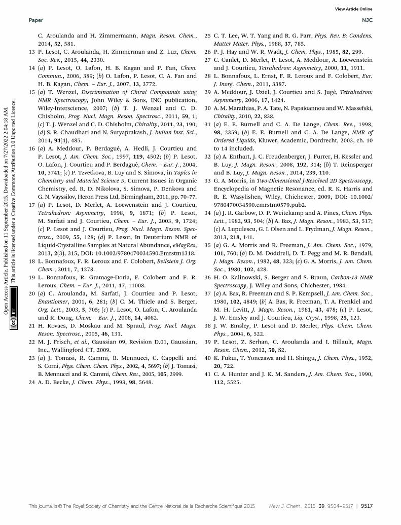
This journal is©The Royal Society of Chemistry and the Centre National de la Recherche Scientifique 2015 New J. Chem., 2015, 39, 9504--9517 | 9517
C. Aroulanda and H. Zimmermann, Magn. Reson. Chem.,2014, 52, 581.
13 P. Lesot, C. Aroulanda, H. Zimmerman and Z. Luz, Chem.Soc. Rev., 2015, 44, 2330.
14 (a) P. Lesot, O. Lafon, H. B. Kagan and P. Fan, Chem.Commun., 2006, 389; (b) O. Lafon, P. Lesot, C. A. Fan andH. B. Kagan, Chem. – Eur. J., 2007, 13, 3772.
15 (a) T. Wenzel, Discrimination of Chiral Compounds usingNMR Spectroscopy, John Wiley & Sons, INC publication,Wiley-Interscience, 2007; (b) T. J. Wenzel and C. D.Chisholm, Prog. Nucl. Magn. Reson. Spectrosc., 2011, 59, 1;(c) T. J. Wenzel and C. D. Chisholm, Chirality, 2011, 23, 190;(d) S. R. Chaudhari and N. Suryaprakash, J. Indian Inst. Sci.,2014, 94(4), 485.
16 (a) A. Meddour, P. Berdague, A. Hedli, J. Courtieu andP. Lesot, J. Am. Chem. Soc., 1997, 119, 4502; (b) P. Lesot,O. Lafon, J. Courtieu and P. Berdague, Chem. – Eur. J., 2004,10, 3741; (c) P. Tzvetkova, B. Luy and S. Simova, in Topics inChemistry and Material Science 5, Current Issues in OrganicChemistry, ed. R. D. Nikolova, S. Simova, P. Denkova andG. N. Vayssilov, Heron Press Ltd, Birmingham, 2011, pp. 70–77.
17 (a) P. Lesot, D. Merlet, A. Loewenstein and J. Courtieu,Tetrahedron: Asymmetry, 1998, 9, 1871; (b) P. Lesot,M. Sarfati and J. Courtieu, Chem. – Eur. J., 2003, 9, 1724;(c) P. Lesot and J. Courtieu, Prog. Nucl. Magn. Reson. Spec-trosc., 2009, 55, 128; (d) P. Lesot, In Deuterium NMR ofLiquid-Crystalline Samples at Natural Abundance, eMagRes,2013, 2(3), 315, DOI: 10.1002/9780470034590.Emrstm1318.
18 L. Bonnafoux, F. R. Leroux and F. Colobert, Beilstein J. Org.Chem., 2011, 7, 1278.
19 L. Bonnafoux, R. Gramage-Doria, F. Colobert and F. R.Leroux, Chem. – Eur. J., 2011, 17, 11008.
20 (a) C. Aroulanda, M. Sarfati, J. Courtieu and P. Lesot,Enantiomer, 2001, 6, 281; (b) C. M. Thiele and S. Berger,Org. Lett., 2003, 5, 705; (c) P. Lesot, O. Lafon, C. Aroulandaand R. Dong, Chem. – Eur. J., 2008, 14, 4082.
21 H. Kovacs, D. Moskau and M. Spraul, Prog. Nucl. Magn.Reson. Spectrosc., 2005, 46, 131.
22 M. J. Frisch, et al., Gaussian 09, Revision D.01, Gaussian,Inc., Wallingford CT, 2009.
23 (a) J. Tomasi, R. Cammi, B. Mennucci, C. Cappelli andS. Corni, Phys. Chem. Chem. Phys., 2002, 4, 5697; (b) J. Tomasi,B. Mennucci and R. Cammi, Chem. Rev., 2005, 105, 2999.
24 A. D. Becke, J. Chem. Phys., 1993, 98, 5648.
25 C. T. Lee, W. T. Yang and R. G. Parr, Phys. Rev. B: Condens.Matter Mater. Phys., 1988, 37, 785.
26 P. J. Hay and W. R. Wadt, J. Chem. Phys., 1985, 82, 299.27 C. Canlet, D. Merlet, P. Lesot, A. Meddour, A. Loewenstein
and J. Courtieu, Tetrahedron: Asymmetry, 2000, 11, 1911.28 L. Bonnafoux, L. Ernst, F. R. Leroux and F. Colobert, Eur.
J. Inorg. Chem., 2011, 3387.29 A. Meddour, J. Uziel, J. Courtieu and S. Juge, Tetrahedron:
Asymmetry, 2006, 17, 1424.30 A. M. Marathias, P. A. Tate, N. Papaioannou and W. Massefski,
Chirality, 2010, 22, 838.31 (a) E. E. Burnell and C. A. De Lange, Chem. Rev., 1998,
98, 2359; (b) E. E. Burnell and C. A. De Lange, NMR ofOrdered Liquids, Kluwer, Academic, Dordrecht, 2003, ch. 10to 14 included.
32 (a) A. Enthart, J. C. Freudenberger, J. Furrer, H. Kessler andB. Luy, J. Magn. Reson., 2008, 192, 314; (b) T. Reinspergerand B. Luy, J. Magn. Reson., 2014, 239, 110.
33 G. A. Morris, in Two-Dimensional J-Resolved 2D Spectroscopy,Encyclopedia of Magnetic Resonance, ed. R. K. Harris andR. E. Wasylishen, Wiley, Chichester, 2009, DOI: 10.1002/9780470034590.emrstm0579.pub2.
34 (a) J. R. Garbow, D. P. Weitekamp and A. Pines, Chem. Phys.Lett., 1982, 93, 504; (b) A. Bax, J. Magn. Reson., 1983, 53, 517;(c) A. Lupulescu, G. L Olsen and L. Frydman, J. Magn. Reson.,2013, 218, 141.
35 (a) G. A. Morris and R. Freeman, J. Am. Chem. Soc., 1979,101, 760; (b) D. M. Doddrell, D. T. Pegg and M. R. Bendall,J. Magn. Reson., 1982, 48, 323; (c) G. A. Morris, J. Am. Chem.Soc., 1980, 102, 428.
36 H. O. Kalinowski, S. Berger and S. Braun, Carbon-13 NMRSpectroscopy, J. Wiley and Sons, Chichester, 1984.
37 (a) A. Bax, R. Freeman and S. P. Kempsell, J. Am. Chem. Soc.,1980, 102, 4849; (b) A. Bax, R. Freeman, T. A. Frenkiel andM. H. Levitt, J. Magn. Reson., 1981, 43, 478; (c) P. Lesot,J. W. Emsley and J. Courtieu, Liq. Cryst., 1998, 25, 123.
38 J. W. Emsley, P. Lesot and D. Merlet, Phys. Chem. Chem.Phys., 2004, 6, 522.
39 P. Lesot, Z. Serhan, C. Aroulanda and I. Billault, Magn.Reson. Chem., 2012, 50, S2.
40 K. Fukui, T. Yonezawa and H. Shingu, J. Chem. Phys., 1952,20, 722.
41 C. A. Hunter and J. K. M. Sanders, J. Am. Chem. Soc., 1990,112, 5525.
Paper NJC
Ope
n A
cces
s A
rtic
le. P
ublis
hed
on 1
1 Se
ptem
ber
2015
. Dow
nloa
ded
on 7
/27/
2022
2:0
4:18
AM
. T
his
artic
le is
lice
nsed
und
er a
Cre
ativ
e C
omm
ons
Attr
ibut
ion
3.0
Unp
orte
d L
icen
ce.
View Article Online
Related Documents