Mucosal malignant melanoma of sinonasal : is it necessary to treat the neck? Shofiah Sari Abstract Primary mucosal melanoma of the paranasal sinuses is a rare tumor of the head and neck. Malignant melanoma had two type; mucosal and cutaneous malignant melanoma. Lymph node of paranasal is drainage to submandibular and Jugulo- omohyoid lymph nodes. This case report is represent to remind us about the important of neck management for malignant melanoma sinonasal with N0. We did the tumour extirpation with nasoendoscopic approach and supra omohyoid selective neck dissection. Key word: malignant melanoma sinonasal, lymph node metastasis, neck dissection Abstrak Melanoma mukosa primer dari sinus paranasal merupakan tumor langka kepala dan leher. Melanoma maligna mempunyai dua tipe; melanoma maligna tipe mukosa dan tipe kutan. Drainase kelenjar limfe dari sinus paranasal mengalir ke kelenjar limfe submandibular dan jugulo-omohioid. Makalah ini dibuat untuk mengingatkan kembali mengenai pentingnya diseksi leher pada melanoma maligna sinonasal dengan N0. Kami lakukan ekstirpasi tumor dengan pendekatan nasoendoskopi dan diseksi leher selektif supra omohyoid. Kata kunci: melanoma maligna sinonasal, metastasis kelenjar getah bening, diseksi leher. Introduction Primary mucosal melanoma of the paranasal sinuses is a rare tumor of the head and neck which can be a devastating disease with poor outcome. Because most of the series extend retrospectively several decades, we sought to determine prognostic factors and outcomes with recent treatment modalities. Melanomas of the sinonasal tract are infrequent and account for less than 1% of all melanomas and up to 4% of all sinonasal malignancies. The rarely of this tumor is because of its origin from melanocytes that have

Welcome message from author
This document is posted to help you gain knowledge. Please leave a comment to let me know what you think about it! Share it to your friends and learn new things together.
Transcript

Mucosal malignant melanoma of sinonasal : is it necessary to treat
the neck?
Shofiah Sari
Abstract
Primary mucosal melanoma of the paranasal sinuses is a rare tumor of the head
and neck. Malignant melanoma had two type; mucosal and cutaneous malignant
melanoma. Lymph node of paranasal is drainage to submandibular and Jugulo-
omohyoid lymph nodes. This case report is represent to remind us about the
important of neck management for malignant melanoma sinonasal with N0. We
did the tumour extirpation with nasoendoscopic approach and supra omohyoid
selective neck dissection.
Key word: malignant melanoma sinonasal, lymph node metastasis, neck
dissection
Abstrak
Melanoma mukosa primer dari sinus paranasal merupakan tumor langka kepala
dan leher. Melanoma maligna mempunyai dua tipe; melanoma maligna tipe
mukosa dan tipe kutan. Drainase kelenjar limfe dari sinus paranasal mengalir ke
kelenjar limfe submandibular dan jugulo-omohioid. Makalah ini dibuat untuk
mengingatkan kembali mengenai pentingnya diseksi leher pada melanoma
maligna sinonasal dengan N0. Kami lakukan ekstirpasi tumor dengan pendekatan
nasoendoskopi dan diseksi leher selektif supra omohyoid.
Kata kunci: melanoma maligna sinonasal, metastasis kelenjar getah bening,
diseksi leher.
Introduction
Primary mucosal melanoma of the
paranasal sinuses is a rare tumor of
the head and neck which can be a
devastating disease with poor
outcome. Because most of the series
extend retrospectively several
decades, we sought to determine
prognostic factors and outcomes with
recent treatment modalities.
Melanomas of the sinonasal tract are
infrequent and account for less than
1% of all melanomas and up to 4%
of all sinonasal malignancies. The
rarely of this tumor is because of its
origin from melanocytes that have

Universitas Indonesia 2
migrated as neuroectodermal
derivatives in the ectodermal
mucosa. Melanomas originating
from the respiratory mucosa and
those originating from the squamous
mucosa have different clinical and
histopathological features, but share
a similar prognosis. The most
common sites for the development of
mucosal melanoma are the nasal
cavity and paranasal sinuses. 1,2,3
Tash and Keskhin9found that face
was the most common anatomic site
of cutaneous (50,53%) which was
followed by scalp (26,28%) and ear
region (18,19%). The most common
histological subtypes of sinonasal
mucosal melanoma include spindle
cell, epitheloid and pleomorphic.9,13
The aggressiveness of mucosal
melanoma may be explained by its
late presentation and delayed
diagnosis, the vascularity of the
mucous membranes, which promotes
hematogenous metastases, or by
cellular and molecular differences
that have been shown to exist
between cutaneous and mucosal
melanoma. The regional metastasis
of mucosal malignant melanoma of
sinonasal is according to sinonasal
lymph node drainage to the neck.
Therefore is important for ENT
doctor do neck dissectionfor mucosal
malignant melanoma of sinonasal
treatment.4
Table 1. Comparison of cutaneous and
mucosal melanoma.5
Cutaneous
melanoma
Mucosal
melanoma
Tissue of
origin
Skin Mucosal surfaces
Mean age at
presentation
55years 67years
Staging American
Joint
comitee on
Cancer
staging
applicable
American Joint
comitee on
Cancer staging
applicable
Presentation Less than
one third
present with
advanced
disease
>50% present
with advanced
disease
(metastasis)
Amelanotic
appearance
1,8%-8,1% 20%-25%
Risk factors Sun
exposure
Unknown
Race White 94%;
Black 0,8%
White 85%
Regional
lymph node
metastasis
5% 32% (head and
neck)
Epidemiology and presentation
Primary mucosal malignant
melanoma of the nasal cavity,
paranasal sinuses and nasopharynx is
rare, accounting for between 0,3%
and 2% of all malignant melanomas
and about 4% of head and neck
melanomas. Overall melanomas
account for 3,5%-7% of all sinonasal
neoplasms. Melanoma is malignancy
of ectodermal origin arising from

Universitas Indonesia 3
melanocytes which are found in
respiratory mucosa. There are
clinical and histopathological
differences between melanomas
originating from the sinonasal
respiratory mucosa and those that
originate from the oral squamous
mucosa. Sinonasal melanomas have
been described to have a lower rate
of regional lymph node metastasis
and the presentation of the tumour
are commonly present ulceration,
necrosis, perineural invasion,
polypoid morphology, and a
pseudopapillary growth pattern.
Despite these well-documented
differences, the prognosis is
comparable to melanomas
originating from squamous oral
cavity mucosa. The most frequent
sites of noncutaneous melanoma are
the eye (5,3%), melanoma of
unknown origin (2,2%) and then
mucosal melanoma.4,5,6
Ciptomangunkusumo hospital in
2013 had 4 case of sinonasal
malignant melanoma and 2 case in
2014. Microscopically, sinonasal
mucosal melanoma (SNMM) is
another “small blue cell tumor” with
similarities to lymphoma,
rhabdomyosarcoma, plasmacytoma,
olfactory neuroblastoma and poorly
differentiated carcinoma. The most
common histological subtypes of
sinonasal melanoma include spindle
cell, epitheloid and pleomorphic.
Regan et all5 found that regional
lymph node metastasis was 32%.4,5,6
Table 2.Patient Demographics for Mucosal
Melanoma of the Head and Neck.3
Characteristic Number Presentation
Sex
Male
Female
Race
White
BlackAmericanIndian/Alaska
Native
Asian or Pacific Islander
Age, yr
0–49
50–64
65–74
75
Site
Nasal cavity
Maxillary sinus
Ethmoid sinus
Accessory sinuses
133
171
276
13
0
15
24
61
69
150
199
46
27
32
43.8
56.3
90.8
4.3
0.0
4.9
7.9
20.1
22.7
49.3
65.5
15.1
8.9
10.5

Universitas Indonesia 4
Histological appearance
This is the histopathology figures of
malignant melanoma.6
Figure 1. A to F: Mucosal malignant
melanoma (A) partially denuded respiratory
epithelium (arrowheads) with underlying
malignant spindle cells exhibiting marked
cytologic atypia (H and E stain: original
magnification , 400c).(B) these cells
infiltrate deep into the submusosa and
surround the seromucous gland (asterisks)
(H and E stain; original magnification 200x).
(C) Scattered malignant cells demonstrate
intranuclear inclusion (arrows) (H and E
stain, original magnification 400x). (D) in
some areas, the malignant cells appear
epitheloid with prominentmacronucleoli
(arrows). (E) HMB 45. (F) MART 1 imm
exhibit strong, diffuse, cytoplasmic
positivity within malignant cells(original
magnification, 400x ).6
Staging
In contrast to cutaneous melanomas,
there widely classification and
staging system and evidence-based
treatment concept specifically
focused on SNMM.
Ballantyne7describe a simplified
staging system for head and neck
mucosal melanomas in 1970, which
continues to be the most widely
utilized. This system classifies
tumors in the following three stages:
stage 1 for localized lesions; stage II
for cervical lymph node metastasis
and stage III for distant metastasis.
The advantage of this system are its
simplicity and the fact that it can be
used for oral and mucosal primaries.
Unfortunately, since the vast
majority of patient present with
local disease only (stage 1), the
prognostic value of this staging
system is relatively limited and it
appears that the current AJCC
sinonasal staging system to be
useful.6,7
The 7th
edition of the American Joint
Committee on Cancer (AJCC)
staging manual contains an entirely
new chapter focusing on the staging
of mucosal melanoma of the head
and neck. The utility of this primarily
arises from the difficulty of staging
melanoma, essentially a cutaneous
disease, using the classification
schemes for mucosal head and neck
cancer. The difficulty arises from the
fact that the former is almost
exclusively tumor thickness specific,
and the latter is a combination of
Universitas Indonesia 5
tumor size and tumor site. What is
less clear is whether the changes in
the classification of head and neck
mucosal melanoma allow for the
useful delineation of survival by
staging group. The American Joint
Committee on Cancer (AJCC) in
2013 made staging of mucosal
melanoma of head and neck in this
table below.3
Table 3. NCNN staging of mucosal
melanoma.8
Survival
The 5-year overall survival range
from 20% to 35% among different
series. Recently, Lund et al6
reported 28% and 20% 5-year and
10-year overall survival rate
respectively, with a 21-month
median survival. After a mean
follow-up of almost 14 years, 35%
of the patients in the study were
alive or died without evidence of
disease.6
Tash and keshkin9 found an
estimated 1- and 5-year overall
survival rates in 41 Sinonasal
Mucosal Melanoma (SNMM)
patients were 81% and 58%,
respectively. The 1- and 5-year
survival rates were 84% and 64% for
patients with sinonasal and 79% and
53% for oral cavity. Advanced
disease (stage II and III) at
presentation was the significant
prognostic factor for outcome, age
and gender of SNMM patients did
not affect survival. 9
Lester et all10
found that 40 patients
of SNMM who were without
evidence of disease at last follow-up
had a mean follow-up of 13.9 years.
These results yield a raw 5-year
survival of 42.6% and a raw 10-year
survival of 24.3%. This contrasts to a
disease free 5-year survival of 31.3%
and a disease-free 10-year survival of
22.6%. Cheng et al10
found the mean
5-year overall survival rate was only
15.65%.10
Universitas Indonesia 6
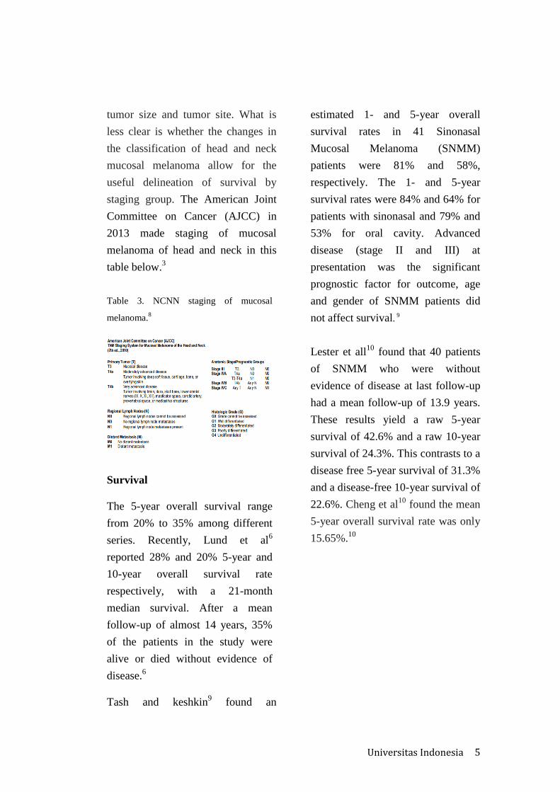
Lymph node
Figure 2: Level of the cervical lymph
node.11
The American Academy of
Otolaryngology–Head and Neck
Surgery has modified the system
used to assign level by subdividing
levels I, II and V into A and B
designation for each levels. Level I
divide into level IA and IB, the
submental group of level IA include
is the lymph nodes between the
anterior belly of the digastric
muscle and cephalad to the hyoid
bone. The submandibular group of
level IB includes lymph nodes in the
triangular area bounded by the
anterior and posterior bellies of the
digastric muscle and the inferior
border of the body of the mandible
also the lymph nodes adjacent to the
mandibular salivary gland and along
the facial artery.
Level II is the midjugular group,
which includes lymph nodes around
the upper portion of the internal
jugular vein and the upper part of
the spinal accessory nerve,
extending from the base of the skull
up to the bifurcation of the carotid
artery or the hyoid bone. The
posterior limit for this level is the
posterior border of the
sternocleidomastoid muscle, and the
anterior border is the lateral limit of
the sternohyoid muscle. Lymph
nodes anterior the spinal accessory
nerve are designated level IIA, and
those posterior to it are designated
level IIB.11
Level III is the midjugular group,
which includes lymph nodes around
the middle third of the internal
jugular vein from the hyoid bone up
to the inferior border of cricoid
cartilage. The anterior and posterior
borders are the same as those for
level II. Level IV is the lower
jugular group, which includes
lymph nodes around the lower
portion of the spinal accessory
nerve and along the transverse
cervical vessels. It is bounded by
triangle formed by the clavicle, the
posterior border of the
sternocleidomastoid muscle, and the
anterior border of the trapezius
muscle. Level V is divided into two
Universitas Indonesia 7
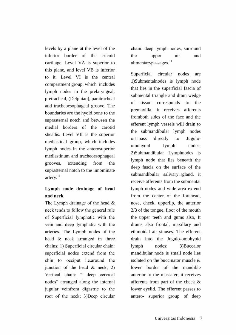
levels by a plane at the level of the
inferior border of the cricoid
cartilage. Level VA is superior to
this plane, and level VB is inferior
to it. Level VI is the central
compartment group, which includes
lymph nodes in the prelaryngeal,
pretracheal, (Delphian), paratracheal
and tracheoesophageal groove. The
boundaries are the hyoid bone to the
suprasternal notch and between the
medial borders of the carotid
sheaths. Level VII is the superior
mediastinal group, which includes
lymph nodes in the anterosuperior
mediastinum and tracheoesophageal
grooves, extending from the
suprasternal notch to the innominate
artery.11
Lymph node drainage of head
and neck
The Lymph drainage of the head &
neck tends to follow the general rule
of Superficial lymphatic with the
vein and deep lymphatic with the
arteries. The Lymph nodes of the
head & neck arranged in three
chains; 1) Superficial circular chain:
superficial nodes extend from the
chin to occiput i.e.around the
junction of the head & neck; 2)
Vertical chain: “ deep cervical
nodes” arranged along the internal
jugular veinfrom digastric to the
root of the neck; 3)Deep circular
chain: deep lymph nodes, surround
the upper air and
alimentarypassages.11
Superficial circular nodes are
1)Submentalnodes is lymph node
that lies in the superficial fascia of
submental triangle and drain wedge
of tissue corresponds to the
premaxilla, it receives afferents
fromboth sides of the face and the
efferent lymph vessels will drain to
the submandibular lymph nodes
orpass directly to Jugulo-
omohyoid lymph nodes;
2)Submandibular Lymphnodes is
lymph node that lies beneath the
deep fascia on the surface of the
submandibular salivarygland, it
receive afferents from the submental
lymph nodes and wide area extend
from the center of the forehead,
nose, cheek, upperlip, the anterior
2/3 of the tongue, floor of the mouth
the upper teeth and gums also, It
drains also frontal, maxillary and
ethmoidal air sinuses. The efferent
drain into the Jugulo-omohyoid
lymph nodes; 3)Buccalor
mandibular node is small node lies
isolated on the buccinator muscle &
lower border of the mandible
anterior to the massater, it receives
afferents from part of the cheek &
lower eyelid. The efferent passes to
antero- superior group of deep

Universitas Indonesia 8
cervical nodes; 4)Preauricular nodes
(parotid) is lymph node that lies on
& within the parotid gland. Some of
these lymph nodes are located
around the external jugular vein. It
receives afferents from the temple,
vertex, eyelids, orbit, & external
acoustic meatus.The efferent passes
to the deep cervical lymph nodes; 5)
Occipitalnodes are few nodes lie at
the apex of posterior triangle &
mastoid process.It receives afferents
from the posterior part of the scalp
& auricle.The efferent passes to the
supraclavicular lymph nodes.Lym
phatic drainage of the face are the
central part i.e. the chin and tongue
tip drain to submental lymphnodes
and wedge of tissue above includes
central forehead and frontal
sinuses,anterior half of the nose &
maxillary sinuses, side of the tongue
and floor ofthe mouth drain into
submandibular nodes.Lateral part of
the forehead, temple, orbital
contents & cheek drain to
thepreauricular group.11
The deep circular lymph nodes are
lymph node that surround the
larynx, trachea & pharynx. It is a
collection of scattered lymph
nodes.Classified as:1. Pretracheal
nodes: drain part of lower larynx,
trachea & thyroid isthmus; 2.
Retropharyngeal nodes: drain the
soft palate, posterior part of hard
palate, nose& pharynx; 3.
Paratracheal. The vertical chain
arranged around the whole length of
internal jugular vein.The nodes are
arranged in demonstrated groups,
superior and inferior, anterior and
posterior.From these groups two
nodes are well known: 1) The
Jugulo digastric that below the
posterior belly of digastric, between
the angle of the mandible and
anterior border of Sternomastoid, it
is a member ofantero-superior
group; 2) Jugulo omohyoid is lies
above the inferior belly of
Omohyoid, behind the jugularvein
undercover of sternomastoid, it is a
member of postero-inferior group.11
Mucosal Melanoma regional
lymph node metastasis
Regional lymph node metastasis was
defined as histologically proven
disease in the primary draining
lymph node basin for each primary
site. These sites included cervical
nodes (sinonasal primary), pelvic
nodes (vaginal and rectal primary)
and inguinal nodes (vaginal and anal
primary). In patients without regional
lymph node metastasis at
presentation, the time to
development of regional lymph node
involvement was recorded, in other
research by Regan et all5 found that

Universitas Indonesia 9
regional lymph node metastasis of
Head and Neck Mucosal Melanoma
was 32%.5
The biological behavior of mucosal
melanoma is not well understood and
explanations for its more aggressive
behavior have been proposed,
including delays in clinical
presentation and diagnosis, the rich
lymphatic and vascular supply of the
mucosal surfaces, and an inherent
more aggressive biology.4
Distant Metastasis
O’regan et all5 research about distant
metastasis of mucosal melanoma,
they found that the commonest initial
site of distant metastasis was the
lungs (66%), followed by the liver
(50%) and peritoneum (33%).
Peritoneal metastasis was more
common in patients with vaginal
melanoma. Sinonasal melanoma
metastasized to the liver in all cases
and peritoneal disease was seen in
50% of patients. No CNS metastasis
was seen in patients with sinonasal
primaries. Metastasis to the
musculoskeletal soft tissues (e.g.
skin and muscle) was also infrequent
(16%). Regional lymph node
metastasis preceded distant
metastatic disease in 33% of patients,
with a median time interval from
lymph node involvement to distant
metastasis of 13 months (range 4-36
months). 5
Imaging Findings
CT appearance of sinonasal
melanomas is nonspecific. Density
values and enhancement pattern do
not provide key information. Bone
destruction is observed in most
malignant tumors. The site of origin
may offer more substantial clues for
the differential diagnosis whenever
the lesion arises from the anterior
nasal septum or middle/inferior
turbinate. MRI (Magnetic
Resonance Imaging) findings
largely depend upon the histological
features of the lesion. Melanotic
melanomas exhibit a peculiar
pattern - consisting of hypo intense
signal on T2 and spontaneous
hyperintensity on T1 – as the result
of paramagnetic properties of
melanin. More in detail,
paramagnetic effect could be due
either to metal ions bound to
melanin, or to free radicals
formation. Conversely, amelanotic
variant displays a less specific
pattern: hyper intense on T2, hypo
intense on plain T1.MRI findings of
amelanotic melanoma are
nonspecific as they are shared by a
longer and wider list of different

Universitas Indonesia 10
lesions, including squamous cell
carcinoma, adenocarcinoma, minor
salivary glands neoplasms, olfactory
neuroblastoma, and fibro-osseous
lesions.12
Figure 2. Melanoma of the left nasal cavity.
Axial CT (a), TSE T2 (b), unenhanced (c),
and enhanced (d) T1. The tumor presents as
a polypoid mass, filling the nasal cavity,
without specific signal features.12
Diagnosis
The diagnosis of mucosal melanoma
is usually made on the basis of
histology and immune
histochemistry. Radiologic
evaluation is important for the
purposes of staging, operative
planning and monitoring of patients
with metastatic disease undergoing
systemic treatment. Computed
tomography (CT) and Positron
Emission Tomography (PET)/CT are
of relatively limited value in local
disease evaluation but are primarily
used to detect clinically unsuspected
metastatic disease. The role of
fluorodeoxyglucose (FDG)-PET/CT
in cutaneous melanoma management
is well established. Its role in
mucosal melanoma is less well
investigated, but given the high
metabolic activity of these tumors, it
is likely to provide similar staging
information but this requires
validation in large-scale trials.4
The NCNN guidelines
madeguidelines for diagnosis of
mucosal malignant melanoma such
as complete head and neck
examination, mirror and fiberoptic
examination as clinically indicated,
verification of pathology using
appropriate staining (HMB-45, S-
100, Melan-A), CT-scan and/or MRI
to determine anatomic extent of
disease, particulary for sinus disease,
chest imaging as indicated and
consider PET-CT scan to rule out
metastatic disease. Histologic
diagnosis was confirmed in all cases
by means of fine-needle aspirate,
sentinel node biopsy, and/or surgical
lymph node resection. In patients
without regional lymph node
metastasis at presentation, the time to
development of regional lymph node
involvement had to be recorded.8
Treatment
All patients were treated by partial or
complete surgical excision; complete
surgical removal of the tumor was
not always possible as a result of the
complex anatomy of the nasal cavity,

Universitas Indonesia 11
paranasal sinuses, and nasopharynx.
NCNN 2013 made guidelines for
mucosal melanoma of paranasal,
stage III treated by wide surgical
resection of primary tumour and
strongly consider postoperative
radiotherapy to primary site. T4a
with No treated by wide surgical
resection and postoperative
radiotherapy to primary site. T3-T4a
with N1 treated by wide surgical
resection and neck dissection of
positive neck also postoperative
radiotherapy to primary site and
neck. Stage IVB treated by primary
radiotherapy or systemic therapy, the
last is stage IV C treated by best
supportive care or primary
radiotherapy or systemic therapy.
Lester et all10
research to compare
the succeed between surgery alone
and with radiotherapy or
chemotherapy, specific type of
therapy did not seem to influence the
overall patient outcome, as there was
no statistically significant difference
between patients managed by
surgery alone, surgery with
chemotherapy, surgery with radiation
therapy, or surgery and combination
therapy.8,10
Complete tumor excision is widely
accepted as the standard of care for
treatment of patients with mucosal
melanomas of the sinonasal tract.
The anatomical complexity of the
sinonasal passages and proximity to
vital structures makes a complete
resection difficult in most cases.
Even more, radical procedures do not
appear to be justified when there is
evidence suggesting that >50% of
the patients who achieve local
control with surgery will ultimately
develop distant metastasis. In this
context, the surgeon’s experience
and thorough knowledge of the
anatomy are key to achieve a
successful oncologic resection with a
minimum cosmetic and functional
impact.8,10
Tash and Keshkin9 researchfound
that all the patient except 2 patients
underwent surgery with curative
intent; the type of surgery and
surgical approach were based on the
location and extension of the tumor.
Patients who underwent endoscopic
resection had better survival.
Surgical margins were described as
microscopically positive or close
(within 1 mm) in 20% of patients.
Bachar et al9 has reported where
margin status did not affect overall
survival. In contrast, Penel et al9 has
reported recently that margin status
affects overall survival. The potential

Universitas Indonesia 12
benefits of negative margins in terms
of local control and overall outcome
may be concealed by the
development of distant metastasis in
a significant number of patients with
a profound negative impact on
survival.9
Neck Treatment
Moreno and Hanna13
found elective
treatment of the neck is usually not
performed, as the incidence of nodal
disease at the time of presentation is
relatively low, ranging from 6 to
25%. However, the incidence of
nodal disease is higher in oral cavity
than in sinonasal melanomas, both at
presentation and during the course of
the disease. Medina et al13
has
recommended elective treatment of
the neck in patients with melanomas
of oral origin, although most authors
would still recommend a
conservative approach. In a recent
report of 74 patients treated for Head
and Neck Mucosal Melanoma
(HNMM), Krengli et al13
described a
77% regional recurrence rate for
HNMM arising in the oral cavity,
suggesting a potential advantage for
prophylactic neck treatment in this
subset of patients.13
Selective Neck dissections (SND)
The SND consist of the removal of
only the lymph node groups at the
highest risk containing metastases
according to the location of the
primary tumor, preserving the spinal
accessory nerve, the IJV, and the
sternocleidomastoid muscle. There
are four main types of SND such as;
1) SND of Level I to II
(supraomohyoid neck dissection)
commonly used in the treatment of
patients with squamous cell
carcinoma of oral cavity. The lymph
node that removed are in the
submental and submandibular
triangle (level I), the upper jugular
region (level II) and the midjugular
region (level III). The posterior limit
is cutaneous branches of the cervical
plexus and the posterior border of the
sternocleidomastoid muscle, the
inferior limit is the omohyoid muscle
as it crosses the IJV. 2) SND of level
II (Lateral neck dissection).
Commonly used in the treatment of
patients with squamous cell
carcinoma of the larynx, oropharynx
and hypopharynx. Removed of the
upper (level II), middle (level III)
and lower (level IV) jugular lymph
nodes. The superior limit is the
digastric muscle and the mastoid tip.
The inferior limit is clavicle. The
posterior limit is cutaneous branches
of the cervical plexus and the
posterior border of the
Universitas Indonesia 13
sternocleidomastoid muscle. 3) SND
of level VI (anterior or central
compartment) is used for patient with
cancer of the midline structures of
the anterior inferior aspect of the
neck and thoracic inlet such as the
pyriform sinus and the cervical
esophagus and trachea. Removal of
the prelaryngeal, pretracheal and
paratracheal bilateral lymph node. 4)
SND for cutaneous malignancies of
the head and neck. The extent of the
regional node dissection in patient
with cutaneous malignancies
depends on location of primary
lesion. Skin cancers originating from
the posterior scalp and the upper-
lateral aspect of the neck commonly
used SND levels II-V, retroauricular,
suboccipital (posterolateral neck
dissection). The superior limit of this
dissection is the posterior belly of the
digastric muscle and the mastoid tip
anterior laterally and the nuchal
line/ridge posteriorly. The inferior
limit is the clavicle. The anterior-
medial is the lateral border of the
sternohyoid muscle. The
posteriolateral limit of the dissection
is marked by the anterior border of
the trapezius muscle inferiorly and
the pposterior midline of the neck
superiorly. The regional node
dissection often performed for
cutaneous malignancies originating
from the periauricular skin, anterior
sclap and temporal region is a SND
(parotid, facial and external jugular
nodes, level II, III, VA).14
Radiation Therapy.
The available evidence from
radiation postoperative studies
suggests that the use of adjuvant
radiation therapy improves
locoregional control, although there
is no evidence of benefit in overall
survival. Some authors report the
use of postoperative radiation more
commonly in sinonasal versus oral
cavity melanomas, probably
because of the inherent difficulty of
obtaining negative margins in the
nasal cavity. To date, there is no
consensus regarding the indications
for postoperative radiation therapy,
although most authors agree
regarding its use in patients with
positive and close margins,
especially as these have been
recently identified as negative
prognostic factors.2
Mereno et al2 study find the use of
postoperative radiation improved
locoregional control but only when
a total dose greater than 54 Gy was
used. It is difficult to determine
whether the lack of improvement
for the group that received a lower
dose was determined by total

Universitas Indonesia 14
radiotherapy (RT) dose,
hypofractionation, or a combination
of both. In contrast with these
findings, other authors have
reported that hypofractionation
might improve local control and
overall survival in headand neck
mucosal melanomas. The optimal
RT dose and fractionation schedule
for mucosal melanomas of the
sinonasal tract remains
undetermined. 2
NCNN made guidelines for
radiation therapy, radiotherapy for
unresectable locally advanced
melanoma treated by 66 Gy to 72
Gy and palliative radiotherapy dose
and schedule may be considered.
Post operative (after primary site
resection) got radiotherapy to
primary site +2-3cm margins or to
anatomic compartment. Primary and
neck melanoma (high-risk site) got
60-66Gy or 70Gy for gross disease,
if the melanoma was low risk,
undissected or uninvolved portions
of neck got 50-6-Gy.8
Follow Up
NCNN made guidelines for follow
up mucosal melanoma; head and
neck examination every 1-3 months
in first year, every 2-6 months in
second year, every 4-8 months year
3-5 and every 12 months after year 5.
Post-treatment baseline imaging of
primary (and neck if treated)
recommended within 6 months of
treatment.8
CASE
Patient male, 62 years old, with
bleeding from right nasal cavity
since 4 months prior to admission.
He also complain about right nasal
blockage, bloody discharge, post
nasal drip, headache and smell
disturbance on the right nose.
Patient went to public hospital in
Bogor and referred to ENT
oncology division outpatient clinic
of Ciptomangunkusumo Hospital.
Physical examination within normal
limit. Nasoendoscopy examination
from the right nasal cavity showed a
narrow nasal cavity, there was mass
covered nasal cavity until 1/3
anterior nasal cavity, craggy and
bleed easily, inferior turbinate,
middle meatus and middle turbinate
could not be evaluated.
Nasoendoscopy of the left nasal
cavity showed a wide nasal cavity,
inferior turbinate was eutrophy,
middle turbinate was eutrophy, the
middle meatus was open, there was
deviated septum at 1/3 middle nasal
cavity and there were no mass at the
nasopharynx.
Universitas Indonesia 15
CT-scan revealed a mass suggestive
malignant that involve the inferior
turbinate and spread to right
maxillary sinus, there were also had
veiled at bilateral ethmoid sinus
suspicious sinusitis. Biopsy result
was malignant melanoma nodular
type. Patient then diagnosed with
malignant melanoma T3N0M0. The
patient had already been discussed
in tumor meeting of ENT and plan
for re-evaluation of biopsy result.
The review of biopsy results was
malignant melanoma nodular type
and plan for mass extirpation with
nasoendoscopy approach and
selective neck dissection.
Patient was hospitalized one day
before the surgery, Cefazolin
2grams as a prophylaxis antibiotic
and methylprednisolone 250mg
once daily also omeprazole 40mg
once daily was given. Mass
extirpation and concotomy with
nasoendoscopic approach and
selective radical neck dissection was
performed under general anesthesia.
Lidocaine and adrenaline tampon
was applied in both nasal cavities.
Evaluation of the right nasal cavity,
showed that right nasal cavity full of
mass until 1/3 anterior nasal cavity,
craggy and bleed easily which
attach to middle part of inferior
turbinate, 1/3 anterior until 1/3
middle of nasal septum and inferior
part of middle turbinate also lateral
nasal wall. Mass attachment at
inferior turbinate and attachment at
inferior part of middle turbinate was
released with suction cauter and
bipolar cauter. The sphenopalatine
foramen was infiltration with
lidocain and adrenaline continued
with cauter the sphenopalatine
artery. The left side of nasal septum
was infiltration with lidocain and
adrenaline continued with
hemitransfiction incision and
elevation of mucopericondrium
until nasal base, lateral nasal wall
and posterior part of posterior
fontanel continued with incision of
mucoperiosteal flap. The
spongostan was applied at nasal
base, septum and lateral nasal wall
also at sphenopalatine foramen. The
whole tumour mass was
extirpatedfrom the nose.Net cell
was applied at right nasal cavity.
The operation was continued with
neck dissection. Site marking was
from region submental along the
mandible until infra auricular area at
the point below the mastoid process.
The incision was done at the site
marking area until platisma muscle
was seen. Platisma was aside until

Universitas Indonesia 16
mylohyoid and sternocleidomastoid
muscle was seen. Lymph node at
level I,II and III was identified and
separated from its surrounding
structures. The Marginal
Mandibularis and Accecorius
nervehas been preserved after that
all of the lymph node level I, II and
III was excluded from the neck. The
patient got ceftriaxone 1mg twice
daily as antibiotic post operative, he
also got ranitidine 50mg twice daily,
tranexamic acid 500mg three times
daily and ketorolac 30mg three
times daily. Two day after the
surgery the patient hemoglobin was
8,9 g/dL and has been PRC
transfusion 250cc and the
hemoglobin increase become 11,0
g/dL. One week after surgery the
wound was healed and patient was
discharge from Cipto
Mangunkusumo hospital.
Follow up at outpatient clinic one
week post surgery, from
nasoendoscopy examination, there
was no inferior and middle turbinate
and there were crusting covered the
middle meatus. The right colli region
the wound was healedand there was
no pus, all the stitches was removed.
We gaveCefixime 200mg twice
daily, paracetamol plus tramadol
three times daily, nasal wash 30cc
twice daily and mometashone furoat
nasal spray 2 puff twice daily was
prescribed. One week after that the
patient came for follow up, from
nasoendoscopic examination there
was still crusting covered the middle
meatus and the uncinate process.
there was serous post nasal drip,
nasal wash was continued. Result
from the histopathology the tumor
was malignant melanoma noduler
type and there was no locoregional
lymph node metastasis. Follow up
two months post operation there was
reccurent mass at the left nasal cavity
that craggy and bleed easily.
Discussion
The subject of this case was a male
patient 62 years old with bleeding
from right nose since 4 months prior
to admission, right nasal blockage,
bloody discharge, post nasal drip,
headache and smell disturbance on
the right nasal cavity. The
nasoendoscopic examination from
the right nasal cavity showed a
narrow nasal cavity, there was mass
covering nasal cavity until 1/3
anterior nasal cavity, craggy and
bleeding easily, inferior turbinate,
middle meatus and middle turbinate
could not be evaluated. This patient
had mucosal malignant melanoma at
sinonasal that is the most common
Universitas Indonesia 17
site of MM at the head and neck. The
patient’s age was 62 years old that
included in the third highest
presentation group of age. This case
showedthe tumor was on the right
nasal cavity originating from
mucosal surface of the nasal cavity
and was in accordance with mucosal
malignant melanoma type.2,15
The result from the histopathology
was malignant melanoma nodular
type and there was no locoregional
lymph node metastasis and assessed
with Melanoma malignant T3N0M0.
Vijay et al said that mucosal
melanoma had 3 subtypes including
spindle cell, epitheloid and
pleomorphic. This was not in
accordance with the result of
histopathology of this patient. The
result was malignant melanoma
nodular type. Malignant melanoma
nodular type is in accordance with
cutaneous malignant melanoma
subtype.6
NCNN 20138 made guidelines for
mucosal melanoma of paranasal,
stage III was treated by wide surgical
resection of primary tumor and
strongly considers postoperative
radiotherapy to primary site.8
Mucosal malignant melanoma
patients was treated by partial or
complete surgical excision; complete
surgical removal of the tumor was
not always possible as a result of the
complex anatomy of the nasal cavity,
paranasal sinuses, and nasopharynx
so that we could not have free
margin to eradicate complete
removal.
This patient had selective neck
dissection with Supraomohyoid neck
dissection which was dissection of
the neck that removed lymph node at
level I, II and III in accordance with
the flow of the lymph node from
sinonasal area.The supraomohyoid
neck dissection was performed for
prevention to cervical lymph node
metastasis. Elective treatment of the
neck was usually not performed
although from literature there were
still debating about whether it was
necessary for the neck dissection of
mucosal malignant melanoma.11
Before the neck dissectionive we did
nasoendoscopic approach with the
sentripetal dissection that dissection
was find the outer border of the
tumor, after found all the border and
separated it from its surroundings
structure, we elevated the tumor and
release whole tumor from its
surroundings structure. Intra
Universitas Indonesia 18
operative we found that the tumor
origin form inferior turbinate and we
did the concotomy. We did the
nasoendoscopic approach because
the advantages from nasoendocopic
approachwas the margins were one
of the strongest predictors of disease
control, and thus this remained a
critical factor in the assessment of
evolving techniques. Advances in
tumor imaging, surgical techniques,
appropriate patient selection and the
ability to apply combined
endoscopic/craniofacial approaches
to difficult cases. An endoscopic
approach should be considered for
tumors which occupies the central
nasal cavity between the frontal and
sphenoidal sinuses but do not extend
to the lateral lamella of the pterygoid
bone. 5,16
Tumor invasion of the nasal bones,
anterior/posterior table of the frontal
sinus or frank orbital invasion are
considered contraindications.
Posteriorly, it is important to assess
invasion of the carotid & cavernous
sinus. If tumor is noted tracking
along nerves (most importantly
trigeminal), these are also relative
contraindications to an endoscopic
approach. Malignant tumor types that
have been resected with favourable
results include adenocarcinoma,
adenoid cystic carcinoma, chordoma,
malignantmelanoma, olfactory
neuroblastoma, osteosarcoma and
squamous cell carcinoma.The most
common serious complication of
nasoendoscopic approach is CSF
leak.Tash and Keshkin9 found
patients who underwent endoscopic
resection had better survival.5,9, 15,16
In this patient we did the CT-Scan
because CT-Scan represented the
best modality with which to assess
for the presence of bony remodelling
or bony invasion, for example of the
sinus walls, orbital margins, and the
floor of the anterior cranial fossa. We
wanted to know the spreading of the
tumor that occured in the sinus wall
and metastatic desease. 5,16
This patient didn’t get the
radiotherapy because according to
Lester et al10
that the specific type of
therapy did not seem to influence the
overall patient outcome, as there was
no statistically significant difference
between patients managed by
surgery alone, surgery with
chemotherapy, surgery with radiation
therapy, or surgery and combination
therapy.
Two months follow upat this patient
post operation we found that this

Universitas Indonesia 19
disease was recurrent (reoccurred)
and plan for maxillectomy. We
should evaluate the neck area
whether there was lymph node
enlargement during follow up and
the Ct-Scan examination.5,8
The conclusion of this case was that
we had to do the neck surgery
although we didn’t find any regional
lymph node metastasis.
REFFERENCE
1. Gasparyan A, Amiri F, Safdieh J,
Reid V, Cirincione E, Shah
D.Malignant mucosal melanoma of
the paranasal sinuses: Two case
presentations. World J Clin Oncol
2011; 2(10): 344-347
2. Moreno et all. Mucosal Melanoma
of the Nose and Paranasal Sinuses; A
Contemporary Experience From The
M. D. Anderson Cancer Center.
Wiey Inter Science 2010;
(116):2215-23.
3. J Thomas, Silver N, Huang B.
Demographics and Treatment Trends
in Sinonasal Mucosal Melanoma.
Laryngoscope 2011;(121); 2026–33.
4. Seetharamu N, Otta A. Mucosal
melanoma review of literature. The
oncologist 2010;(98); 15: 772-81.
5. Regan K et all. Metastatic mucosal
melanoma: imaging patterns of
metastasis and recurrence. Cancer
Imaging 2013; 13(4): 626-632
6. Vijay R, Jeffrey D. Malignant.
Sinonasal Tumor. In Chiuca G, Vijay
R, Jeffrey D,Ed. Sinonasal Tumors
books. Second Edition. Jaypee Bros.
Medical Publishers India. New
Delhi. 2012. 212-28.
7. Ballantyne AJ. Malignant
melanoma of the skin of the head and
neck: an analysis of 405 cases. Am J
Surg 1970;120:425–31.
8. NCCN Guidline Version 2.2013
Head and Neck Cancer. 2013.
9. Tas F, Keskin S. Melanoma in the
Head and Neck Region: Different
Clinical Features and Same Outcome
to Cutaneous Melanoma. Hindawi
2013.1-6
10. Lester D, Thompson R,
Jacqueline A, Markku M. Sinonasal
Tract and Nasopharyngeal
Melanomas A Clinicopathologic
Study of 115 Cases With a Proposed
Staging System. The American
Journal of Surgical Pathology 2003;
27(5): 594-611.
11. Shah J. Cervical Lymph Node.In
Shah J, Patel S, Singh B,Ed.
Headand neck surgery and oncology.

Universitas Indonesia 20
Fourth Edition. Elsivier health
sciences.New York. 2012. 426-60.
12. Maroldi R, Lombardi D, Farina
D, Nicolai F, Moraschi I. Malignant
Neoplasma. InMaroldi R, Nicolai
P,Ed.Imaging in treatment Planning
for Sinonasal Diseases. First
Edition.Springer Berlin Heidelberg.
New York. 2012. 159-260.
13. Moreno M, Hanna Y.
Management of mucosal melanomas
of the head and neck: did we make
any progress? Current Opinion in
Otolaryngology & Head and Neck
Surgery 2010; (18):101–6.
14. Bailey B, Johnson J, Jonas T.
Neck Dissection. In Bailey B,
Johnson J, Jonas T, Newland S,Ed.
Head and Neck Surgery-
Otolaryngology. Fourth Edition.
Lippincott Williams and Wilkins.
Texas. 2006. 1585-1609.
15. Schuchter L. Melanoma and Non
Melanoma Skin Cancer. Chapter
210;1329-34.
16. Dmytriw AA, Witterick IJ, Yu E.
Endoscopic resection of malignant
sinonasal tumours: Current trends
and imaging workup. OA Minimally
Invasive Surgery 2013;(1): 1-6

Universitas Indonesia 21
Related Documents