PLEASE SCROLL DOWN FOR ARTICLE This article was downloaded by: [Alsarra, Ibrahim A.] On: 21 February 2009 Access details: Access Details: [subscription number 908872393] Publisher Informa Healthcare Informa Ltd Registered in England and Wales Registered Number: 1072954 Registered office: Mortimer House, 37-41 Mortimer Street, London W1T 3JH, UK Drug Development and Industrial Pharmacy Publication details, including instructions for authors and subscription information: http://www.informaworld.com/smpp/title~content=t713597245 Mucoadhesive Polymeric Hydrogels for Nasal Delivery of Acyclovir Ibrahim A. Alsarra a ; Amel Y. Hamed a ; Gamal M. Mahrous a ; Gamal M. El Maghraby a ; Abdulrahman A. Al- Robayan b ; Fars K. Alanazi a a Kayyali Chair for Pharmaceutical Industry, Pharmaceutical Technology Center, College of Pharmacy, King Saud University, Riyadh, Kingdom of Saudi Arabia b Gastroenterology Division, Department of Medicine, Riyadh Military Hospital, Riyadh, Saudi Arabia First Published:March2009 To cite this Article Alsarra, Ibrahim A., Hamed, Amel Y., Mahrous, Gamal M., El Maghraby, Gamal M., Al-Robayan, Abdulrahman A. and Alanazi, Fars K.(2009)'Mucoadhesive Polymeric Hydrogels for Nasal Delivery of Acyclovir',Drug Development and Industrial Pharmacy,35:3,352 — 362 To link to this Article: DOI: 10.1080/03639040802360510 URL: http://dx.doi.org/10.1080/03639040802360510 Full terms and conditions of use: http://www.informaworld.com/terms-and-conditions-of-access.pdf This article may be used for research, teaching and private study purposes. Any substantial or systematic reproduction, re-distribution, re-selling, loan or sub-licensing, systematic supply or distribution in any form to anyone is expressly forbidden. The publisher does not give any warranty express or implied or make any representation that the contents will be complete or accurate or up to date. The accuracy of any instructions, formulae and drug doses should be independently verified with primary sources. The publisher shall not be liable for any loss, actions, claims, proceedings, demand or costs or damages whatsoever or howsoever caused arising directly or indirectly in connection with or arising out of the use of this material.

Welcome message from author
This document is posted to help you gain knowledge. Please leave a comment to let me know what you think about it! Share it to your friends and learn new things together.
Transcript
PLEASE SCROLL DOWN FOR ARTICLE
This article was downloaded by: [Alsarra, Ibrahim A.]On: 21 February 2009Access details: Access Details: [subscription number 908872393]Publisher Informa HealthcareInforma Ltd Registered in England and Wales Registered Number: 1072954 Registered office: Mortimer House,37-41 Mortimer Street, London W1T 3JH, UK
Drug Development and Industrial PharmacyPublication details, including instructions for authors and subscription information:http://www.informaworld.com/smpp/title~content=t713597245
Mucoadhesive Polymeric Hydrogels for Nasal Delivery of AcyclovirIbrahim A. Alsarra a; Amel Y. Hamed a; Gamal M. Mahrous a; Gamal M. El Maghraby a; Abdulrahman A. Al-Robayan b; Fars K. Alanazi a
a Kayyali Chair for Pharmaceutical Industry, Pharmaceutical Technology Center, College of Pharmacy, KingSaud University, Riyadh, Kingdom of Saudi Arabia b Gastroenterology Division, Department of Medicine,Riyadh Military Hospital, Riyadh, Saudi Arabia
First Published:March2009
To cite this Article Alsarra, Ibrahim A., Hamed, Amel Y., Mahrous, Gamal M., El Maghraby, Gamal M., Al-Robayan, Abdulrahman A.and Alanazi, Fars K.(2009)'Mucoadhesive Polymeric Hydrogels for Nasal Delivery of Acyclovir',Drug Development and IndustrialPharmacy,35:3,352 — 362
To link to this Article: DOI: 10.1080/03639040802360510
URL: http://dx.doi.org/10.1080/03639040802360510
Full terms and conditions of use: http://www.informaworld.com/terms-and-conditions-of-access.pdf
This article may be used for research, teaching and private study purposes. Any substantial orsystematic reproduction, re-distribution, re-selling, loan or sub-licensing, systematic supply ordistribution in any form to anyone is expressly forbidden.
The publisher does not give any warranty express or implied or make any representation that the contentswill be complete or accurate or up to date. The accuracy of any instructions, formulae and drug dosesshould be independently verified with primary sources. The publisher shall not be liable for any loss,actions, claims, proceedings, demand or costs or damages whatsoever or howsoever caused arising directlyor indirectly in connection with or arising out of the use of this material.
Drug Development and Industrial Pharmacy, 35:352–362, 2009Copyright © Informa UK, Ltd.ISSN: 0363-9045 print / 1520-5762 online DOI: 10.1080/03639040802360510
352
LDDIMucoadhesive Polymeric Hydrogels for Nasal Delivery of Acyclovir
Polymeric Hydrogels for Nasal Delivery of AcyclovirIbrahim A. Alsarra, Amel Y. Hamed, Gamal M. Mahrous, and Gamal M. El MaghrabyKayyali Chair for Pharmaceutical Industry, Pharmaceutical Technology Center, College of Pharmacy, King Saud University, Riyadh, Kingdom of Saudi Arabia
Abdulrahman A. Al-RobayanGastroenterology Division, Department of Medicine, Riyadh Military Hospital, Riyadh, Saudi Arabia
Fars K. AlanaziKayyali Chair for Pharmaceutical Industry, Pharmaceutical Technology Center, College of Pharmacy, King Saud University, Riyadh, Kingdom of Saudi Arabia
The study evaluated different mucoadhesive polymeric hydro-gels for nasal delivery of acyclovir. Gels containing poly-N-vinyl-2-pyrrolidone (PVP) were prepared with crosslinking achieved byirradiation with a radiation dose of 15 kGy being as efficient as20 kGy. Gels containing chitosan and carbopol were also evaluated.The mucoadhesive properties of gels were measured by a modifi-cation of a classical tensile experiment, employing a tensile testerand using freshly excised sheep nasal mucosa. Considering themucoadhesive force, chitosan gel and gel prepared with 3% PVPin presence of polyethylene glycol (PEG) 600 were the most effi-cient. The in vitro drug release depended on the gel composition.Higher release rates were obtained from PVP gels compared tochitosan or carbopol gels. The release rate of drug from PVP gelswas increased further in presence of PEG or glycerol. Histopatho-logical investigations proved that the PVP was a safe hydrogel tobe used for mucosal delivery. The PEG in gel formulations causedless damages to the nasal mucosal compared to formulation con-taining glycerol.
Keywords acyclovir nasal; nasal mucoadhesion; poly-N-vinyl-2-pyrrolidone hydrogels; chitosan nasal; carbopol nasal
INTRODUCTIONMucoadhesive polymers are synthetic or natural macromol-
ecules that are capable of attaching to mucosal surfaces. Thehistory of pharmaceutical mucoadhesives started more than 40years ago. Mucoadhesion is widely investigated as a promising
strategy for prolonged and enhanced drug delivery throughvarious membranes (Grabovac, Guggi, & Bernkop-Schnurch,2005).
Nasal route of administration has gained great interest. Itprovides the formulator with a large absorptive surface withhigh vascularity, ensuring rapid absorption of drugs bypassingthe hepatic first-pass elimination. Unfortunately, the nasalmucosa poses a permeation barrier for drugs, especially high-molecular weight therapeutics, such as peptides and proteins.In addition, the mucociliary clearance provides another addi-tion to the barrier nature by reducing the contact time afternasal application (Mattrin, Schipper, Verhoef, & Merkus,1998). Accordingly, improving nasal drug delivery can beachieved by enhancing the permeation and/or prolonging thecontact time. The tight junctions that form this barrier to para-cellular drug delivery can be reversibly and safely opened. Anabsorption-enhancing effect has been recorded for some poly-mers and was attributed to widening of tight junctions (Guoet al., 2004). Gels can be employed for nasal delivery, becausethey give a high drug transport often provided by the longerresidence time of the formulation at the site of absorption(Ugwoke, Van den Mooter, Verbeke, & Kinget, 1999). Bioad-hesive polymers can be used to achieve a more intimate contactwith nasal mucosa, which results in higher concentration gradi-ent and subsequently increasing absorption (Critchley, Davis,Farraj, & Illum, 1994). The advantages of a nasal gel includethe reduction of postnasal drip due to high viscosity, reductionof anterior leakage of the formulation, reduction of irritation byusing soothing/emollient excipients, and target delivery tomucosa for better absorption. Nasal mucoadhesive gel can pro-vide a viable alternative to the conventional nasal formulations
Address correspondence to Ibrahim A. Alsarra, Kayyali Chair forPharmaceutical Industry, Pharmaceutical Technology Center, Collegeof Pharmacy, King Saud University, P.O. Box 2457, Riyadh 11451,Kingdom of Saudi Arabia. E-mail: [email protected]
Downloaded By: [Alsarra, Ibrahim A.] At: 07:57 21 February 2009
POLYMERIC HYDROGELS FOR NASAL DELIVERY OF ACYCLOVIR 353
as they provide a high drug transport often provided by thelonger residence time of the formulation at the site of absorp-tion (Behl, Pimplaskar, Sileno, deMeireles, & Romeo, 1998).Polymer gels and mucoadhesive polymers have been studiedfor the mucosal delivery of various compounds ranging fromsmall molecule to macromolecular drugs. Various bioadhesivepolymers such as polyacrylic acids (carbopol 971P) and chito-san have been used in gel forms for nasal delivery.
Chitosan is a cationic polysaccharide obtained fromdeacetylation of chitin, a structural polymer abundant in creta-ceous animals such as crabs and shrimps. Because of its bio-compatibilty, biodegradability, and low toxicity, chitosanrepresents an attractive biopolymer for a variety of pharmaceu-tical applications (Paul & Sharma, 2000). Chitosan may be agood option in nasal delivery as it binds to the nasal mucosalmembrane with an increased retention time, and it is also agood absorption enhancer (Varshosaz, Sadrai, & Heidar,2006). Successful mucosal delivery of insulin has beenrecorded after nasal application of mucoadhesive gel compris-ing 2% medium molecular weight chitosan with ethylenedi-aminetetraacetic acid (EDTA) to diabetic rats. This formulationenhanced the absorption of insulin and reduced the blood glu-cose by 46% compared to intravenous route (Varshosaz et al.,2006).
Poly-N-vinyl-2-pyrrolidone (PVP) is an example of poly-mer applied for the synthesis of hydrogel to be used in differentmedical applications. In recent years, PVP hydrogels haveemerged as an important class of biomaterials as they possessexcellent biocompatibility (Rosiak, Ulanski, Pajewnski,Yoshii, & Makuuchi, 1995). PVP hydrogels have a wide rangeof applications including transdermal systems as wound dress-ings (Benamer, Mahlous, Boukrif, Mansouri, & Youcef, 2006),nasal drug delivery systems (Bertram & Bodmeier, 2006), andeye preparation (Yanai et al., 2006). Roos, Creton, Novikov,and Feldstein (2002) investigated a series of blends of highmolecular weight PVP (K-90) and polyethylene glycol (PEG)600. PVP/PEG blends were referred to in the literature as thepressure-sensitive adhesives (PSAs). In some applications,such as wound dressing and transdermal drug delivery systems,PSAs act as adhesives and reservoirs for the drug to be deliv-ered. Crosslinking transforms a linear polymer into a three-dimensional (3D) molecule, resulting in a significant increasein molecular mass with improved mechanical properties. High-energy radiation in particular gamma rays can be used to poly-merize unsaturated compounds by converting water-solublepolymers into hydrogels (Rosiak & Yoshii, 1999).
Acyclovir was chosen for intranasal delivery in this study asit does not undergo degradation and metabolism in the nasalcavity. The currently available dosage regimens of acyclovirhave a number of limitations including the following: (a) vari-able bioavailability by oral administration, (b) poor percutane-ous absorption, and (c) thrombophlebitis after intravenousbolus injection. Earlier attempts in literature for improving thebioavailability of acyclovir were based on chemical modification
(amino-acid ester pro-drug), very limited work has beenreported on modification, which includes transbuccal deliveryand ocular delivery.
The objectives of this study were to (a) investigate themucoadhesive properties of different hydrogels (PVP, chito-san, and carbopol), (b) study drug release profile of acyclovir,and (c) evaluate the safety of the prepared hydrogels on nasalmucosa.
MATERIALS AND METHODS
MaterialsAcyclovir was kindly provided from Riyadh Pharma Phar-
maceutical Company (Riyadh, Saudi Arabia). Chitosanmedium molecular weight (103.200 g/mol) with degree ofdeacetylation of 76.6% was purchased from Sigma-AldrichCompany (St. Louis, MO, USA). Carbopol 974 was obtainedfrom BF Goodrich (Cleveland, OH, USA). PVP known asLuviskol® K-90 was purchased from Aktiengesellschaft(Ludwigshafen, Germany). PEG 600 was purchased fromMerck-Schuchardt (Hohenbrunn, Germany). Glycerin BP waspurchased from Pacegrove Chemical and BiopharmaceuticalChemicals (Leicestershine, UK). Lactic acid and glutaralde-hyde 50% were purchased from BDH Laboratory Supplies(Poole, England). Acetonitrile and triethanolamine were pur-chased from Sigma-Aldrich, Chemie GmbH. All chemicalsand organic solvents used were of high-performance liquidchromatography (HPLC) grade and were used as received.
Preparation of Different HydrogelsPreparation of PVP Gels
PVP gels (3 and 6%, wt/vol) with and without glycerol orPEG 600 were prepared in double distilled water according tothe composition presented in Table 1. The polymer was addedto the vortex of continuously stirred solvent. Mixing was con-tinued for 3 min before adjusting the pH to 6.4 ± 0.05 using tri-ethanolamine. The mixtures were then filled into glass bottles.Crosslinking was achieved by gamma irradiation. The irradia-tion of PVP hydrogels was carried out using gamma irradiation(Gamma Irradiation Facility, King Faisal Specialist Hospitaland Research Center, Riyadh, Saudi Arabia) with 60Co sourceat room temperature at radiation doses of 15 and 20 kGy (pre-liminary study indicated that these doses are appropriate dosesfor PVP crosslinking). The irradiation time was calculatedbased on the radiation source dosing rate (51.71 Gy/min). Thedrug was homogeneously dispersed in the gel after crosslink-ing in an amount that is sufficient to provide final drug concen-tration of 20 mg/g.
Preparation of Chitosan GelChitosan solution (2%, wt/vol) was prepared in 1% lactic
acid (n = 3). The solution was kept for 4 h for the mixture toequilibrate, and the entrapped air was removed, then two drops
Downloaded By: [Alsarra, Ibrahim A.] At: 07:57 21 February 2009
354 I. A. ALSARRA ET AL.
of glutaraldehyde have been added and left for 24 h to allowcrosslinking process to take place, the pH value of gel wasadjusted to 5.6 ± 0.05 using triethanolamine. Lactic acid wasused to dissolve chitosan. Lactic acid was found to be biologi-cally safe (nonirritant) and appeared to be a universal solventfor chitosan (Knapezyk, 1993). The drug was incorporated asbefore.
Preparation of Carbopol GelCarbopol gel 2% (wt/vol) was prepared in double distilled
water (n = 3). The polymer was added to the vortex of continu-ously stirred water. The mixture was neutralized using trietha-nolamine until transparent gel appeared (pH value was 5.6 ±0.05). The drug was incorporated as before.
Mucoadhesion Performance EvaluationThe mucoadhesive performance of gels was determined by
measuring the force required to detach the formulation fromnasal mucosal tissue; sheep was selected as an animal modelfor mucoadhesion performance evaluation. Majithiya, Ghosh,Umrethia, and Murthy (2006) used the sheep mucosa in theevaluation of mucoadhesion of the thermosensitive gellingpolymer, Pluronic F127 (PF127) and the mucoadhesive poly-mer Carbopol 934P. The sheep nasal mucosa was prepared tostudy mucoadhesion characteristics adopting the proceduredescribed by Gavini, Rassu, Sanna, Cossu, and Giunchedi(2005). Immediately after killing the animal, the nasal mucosawas carefully removed from the nasal cavity of the sheep.Longitudinal incisions were made through the septum wall, andthe nose was kept in ice until removing the mucosa. After expos-ing the nasal cavity on each side of the septum, the mucosalcavity (i.e., the mucous membrane covering the turbinates) wascarefully removed using forceps and dissecting scissors. Theremoval of mucosa was completed within 1 h after killing the
animal. The mucosa was cut into small pieces taking into con-sideration to avoid the areas that were held by forceps.
The mucoadhesive properties of the prepared gels weremeasured by modification of a classical tensile experiment,using a tensile tester Instron® Texture analyzer model no. 8500Digital control, with Instron® Series IX automated materialtester software, version 8.32.00 (Instron Co., Norwood, MA,USA), equipped with a 5-kg cell. In this test, two equal cylin-drical metallic supports with a circular surface of 1.2-cm diam-eter were constructed; one was connected to the upper movableprobe and the other was connected to the lower stationaryprobe of the apparatus. The mucosa was attached to both upperand lower metal support using cyanoacrylate adhesive (keep-ing the mucosal side exposed). A 250-mg sample of the gelwas placed on the lower mucosa. The upper mucosa was thenlowered to the gel surface until contact was achieved betweenthe two surfaces. During the contact phase, the thickness of thegel layer was kept constant (0.5 mm), then the two phases werebrought immediately into direct contact with an initial force of0.5 N for 2 min. The upper mucosa was withdrawn upward at aspeed of 5 mm/s until failure occurred between the surfaces.The whole experiment was preformed at room temperature anda relative humidity of 50%. This method determines the mini-mum force needed to separate two surfaces in intimate contact(Bonacucina, Cespi, Misici-Falzi, & Palmieri, 2006). Duringthe experiment, the force was recorded as a function of detach-ment stress (N) until the break point. Data collection and calcu-lation were preformed by the dedicated software, and eachmeasurement was repeated at least five times.
In Vitro Release StudiesVertical jacketed Franz diffusion cell, 15 mm diameter and
12 mL capacity (Crown Glass Co. Inc., Somerville, NJ, USA),was used for in vitro study. Cellophane membrane Spectra Por®
TABLE 1 Compositions of Various PVP Hydrogels (% wt/vol in water) and Doses of Radiation (kGy)
Gels CodePVP
ConcentrationPolyethylene
Glycol GlycerolRadiation
Dose
A 3 — — 15B 3 — — 20BG1 3 — 1 20BG2 3 — 2 20BP1 3 1 — 20BP2 3 2 — 20C 6 — — 15D 6 — — 20DG1 6 — 1 20DG2 6 — 2 20DP1 6 1 — 20DP2 6 2 — 20
Downloaded By: [Alsarra, Ibrahim A.] At: 07:57 21 February 2009
POLYMERIC HYDROGELS FOR NASAL DELIVERY OF ACYCLOVIR 355
dialysis membrane with a molecular weight cut-off of12.000–14.000 was mounted between the receiver and thedonor compartments of the diffusion cells. The receptorcompartment was filled with 12 mL of phosphate buffer of pH5.5 and the system was maintained at 37°C. The receptorcompartment was stirred continuously at a controlled speedusing a magnetic stirrer. The test formulations (0.5 g) were loadedinto the donor compartments before occluding the donorcompartments with parafilm. Receptor samples were taken atpredetermined time intervals (30, 60, 120, 150, 180, 210, and240 min). These samples were replaced with equal volumes ofbuffer to maintain the sink conditions. All in vitro drug releasestudies were performed in triplicates, and samples wereassayed using HPLC (Jain et al., 2005).
The analysis employed an HPLC system consisted of Intel-ligent Shimadzu two pumping systems LC-10AT VP,equipped with a data processor Shimadzu chromatographysoftware class-VP, version 6.12 SP1, Shimadzu Corporation(Kyoto, Japan). Separation was achieved using a C18 μ-Bonda-pak column (300 × 3.9 mm i.d.), packed with 5-μm particles(Waters Inc., Bedford, MA, USA). This was maintained at25°C (column oven). The mobile phase comprised 0.02 Mpotassium dihydrogen phosphate (pH 3.5) (99%) and acetoni-trile (1%), filtered through 0.45-μm Millipore filter anddegassed. The mobile phase was freshly prepared beforeeach experiment. The mobile phase was flowing at a rate of1.5 mL/min with detection being preformed at 254 nm afterinjecting 20 μL into the HPLC.
Histopathological Estimation of Nasal MucosaTo evaluate the effect of the prepared hydrogels on the nasal
mucosa, histopathological examination of the sheep nasalmucosa incubated in different gels was performed. The sheep’snasal mucosa was pretreated in the same manner as formucoadhesion measurement. The mucosal tissues were incu-bated in different gel formulations for 5 h at 25°C. The
mucosal tissue was fixed in 10% formalin solution for 48 h andthen embedded in paraffin wax. Paraffin sections (7 μm) werecut on glass slides and stained with hematoxylin and eosin(HX–E). Sections were examined for any changes under a lightmicroscope connected to a digital camera. Mucosal tissuesincubated in isotonic phosphate buffer saline for 5 h were usedas a control for comparison.
Statistical AnalysisData were expressed as a mean of three experiments ± the
SD. Data were analyzed by one-way analysis of variance(ANOVA) using SPSS® statistical package (version 13, 2001,SPSS Inc., Chicago, IL, USA). Statistical differences yieldingp ≤ 0.05 were considered significant. Tukey’s multiple-com-parison post hoc tests were applied.
RESULTS AND DISCUSSION
Mucoadhesion Performance EvaluationMost in vitro mucoadhesion studies have provided valuable
information on the force of adhesion by measuring tensilestrength or calculating the detachment force and work of adhe-sion. Methods using tensile strength usually examine the forcenecessary for separation of the two surfaces after establishmentof mucoadhesion. This force is recorded as a function of theelongation observed at polymer-mucus interfacial (Ponchel,Lejoyeux, & DucheÃne, 1991). Depending on the direction inwhich the adhesive is being separated from the substrate (peel),the shear and tensile force necessary to detach a polymer for-mulation from mucosa can be measured (Hägerström &Edsman, 2003).
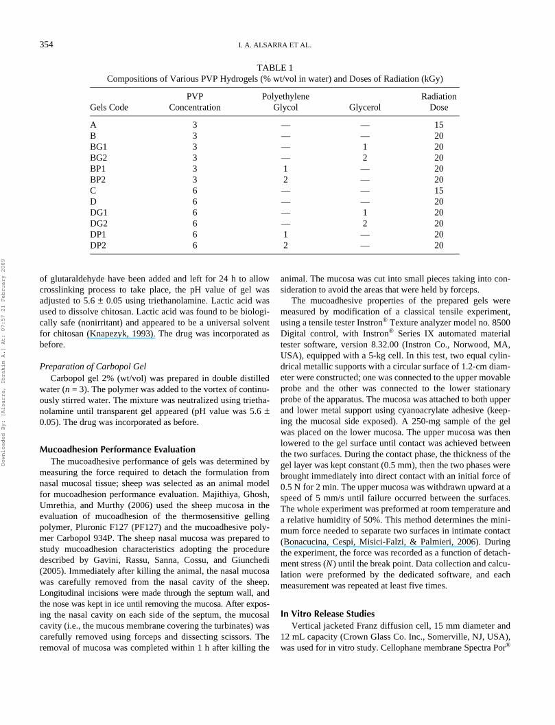
Figures 1–3 illustrate the mucoadhesion performance of theprepared gels. Considering the overall adhesion force at differ-ent contact time values, chitosan gel and gels prepared withPVP (3%) in presence of PEG showed the greatest force of
FIGURE 1. Force of adhesion of chitosan gel and carbopol gel.
0
2
4
6
8
10
12
14
30 120 300 600 1200Contact time (s)
Max
imum
forc
e of
adh
esio
n (N
)
Carbopol
Chitosan
Downloaded By: [Alsarra, Ibrahim A.] At: 07:57 21 February 2009
356 I. A. ALSARRA ET AL.
adhesion. Gels can be arranged according to their adhesivenessin descending order as 3% PVP with PEG ≈ chitosan > 3%PVP at 15 kGy > carbopol > 3% PVP at 20 kGy > 3% PVPwith glycerol > 6% PVP with PEG > 6% PVP with glycerol >
6% PVP at 15 kGy > 6% PVP at 20 kGy. An electron transferprocess has been previously adopted to explain mucoadhesion(Peppas & Sahlin, 1996). According to this process, electrontransfer occurs on contact between adhering surfaces resulting
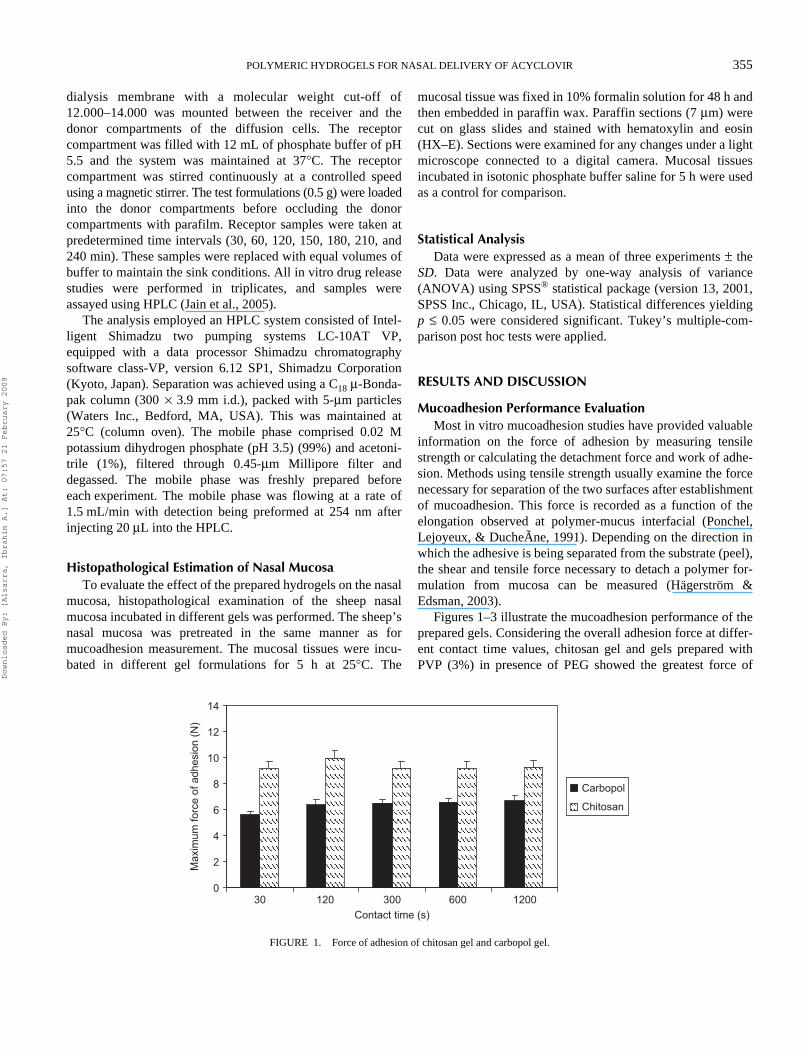
FIGURE 2. Effect of radiation dose and PVP concentration on the mucoadhesive effect of PVP gel.
0
2
4
6
8
10
12
14
30 120 300 600 1200Contact time (s)
Max
imum
forc
e of
adh
esio
n (N
) 3% PVP + 15 kGy
3% PVP + 20 kGy
6% PVP + 15 kGy
6% PVP + 20kGy
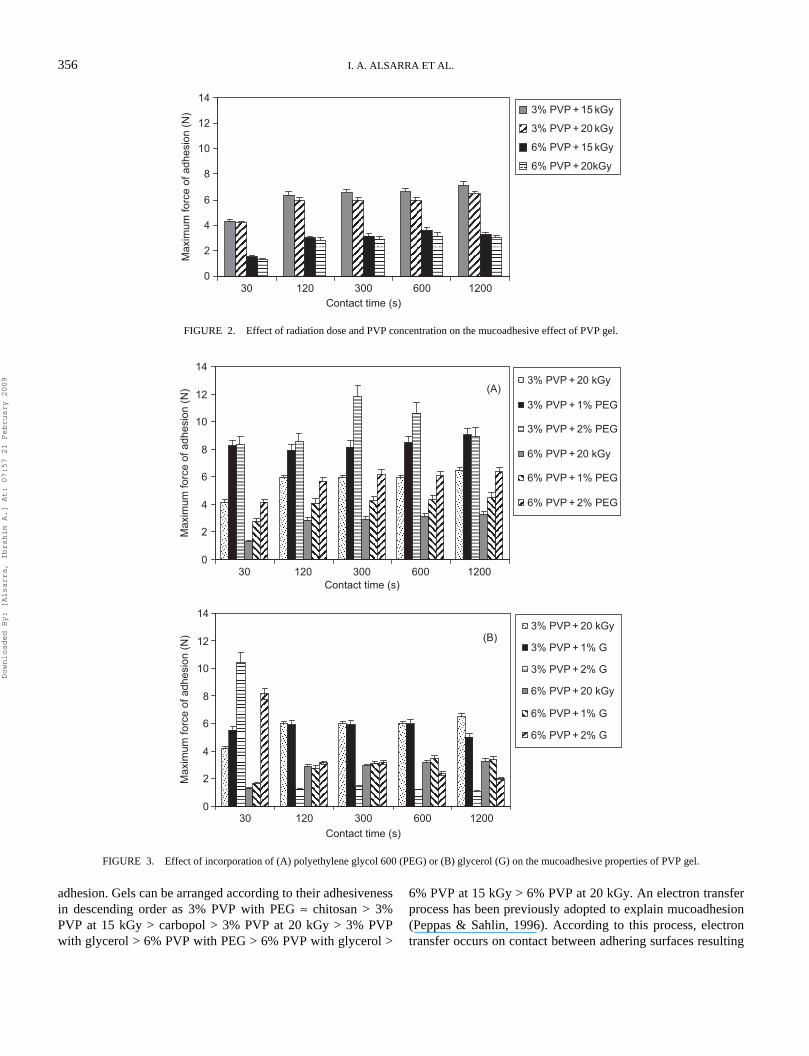
FIGURE 3. Effect of incorporation of (A) polyethylene glycol 600 (PEG) or (B) glycerol (G) on the mucoadhesive properties of PVP gel.
(A)
0
2
4
6
8
10
12
14
30 120 300 600 1200Contact time (s)
Max
imum
forc
e of
adh
esio
n (N
)
3% PVP + 20 kGy
3% PVP + 1% PEG
3% PVP + 2% PEG
6% PVP + 20 kGy
6% PVP + 1% PEG
6% PVP + 2% PEG
(B)
0
2
4
6
8
10
12
14
30 120 300 600 1200Contact time (s)
Max
imum
forc
e of
adh
esio
n (N
)
3% PVP + 20 kGy
3% PVP + 1% G
3% PVP + 2% G
6% PVP + 20 kGy
6% PVP + 1% G
6% PVP + 2% G
Downloaded By: [Alsarra, Ibrahim A.] At: 07:57 21 February 2009
POLYMERIC HYDROGELS FOR NASAL DELIVERY OF ACYCLOVIR 357
in the formation of an electrical double layer at the interface,with subsequent adhesion because of attractive forces. Studieshave shown that the mucoadhesive properties of polymers con-taining ionizable groups are affected by the pH of the sur-rounding media. For cationic polymers like chitosan, thepositive charge will favor binding to negatively charged groups(such as carboxyl or sulphate) on the mucin or cell surface(Fiebrig, Harding, Rowe, Hyman, & Davis, 1995).
The results shown here are in good agreement with the workof Lehr, Bouwstra, Schacht, and Junginger (1992). Thus, atnasal pH (5.5–6.5), the cationic polymer, chitosan, exists in theionized form. The NH3
+ groups on the deacetylated N-acetylgroups can thus undergo electrostatic interaction with the nega-tively charged mucus. It should be noted that the mucoadhe-sion of chitosan polymers may be weakened by its solubility inthat acidic medium; however, this effect can be neutralized bychemical crosslinking (Lehr et al., 1992).
On the other hand, the adhesion behavior of carbopol gelwas significantly lower than that of chitosan gel (p < 0.05)(Figure 1). This can be returned to the experimental pH (nasalpH). A previous study showed that carbopol did not adherewhen applied as gel to buccal mucosa in organ culture,whereas, chitosan gel showed up to 4 days retention (Needleman& Smales, 1995).
As poly(acrylic acid) hydrogels contain coiled macromole-cules, it is unable to form an elastic polymer network due to therepulsion of negative charges; many of the active groups areshielded inside the coils and do not actively participate in theadhesion process (Tamburic & Craig, 1997). As carbopol isstrongly coiled into a spiral form in dry powder state, when dis-persed in water, the molecules become hydrated and slowlyunwind, generating an increase in viscosity. To ensure maxi-mum viscous effects, the molecules must unwind completely.The most common method to unwind the molecules involvesneutralization of the negative charges along the polymer chainswith appropriate base. Repulsion between these charges con-tributes to unfold the structure while intertwining of chainsyields a 3D matrix. The result is the instantaneous formation ofa highly viscous gel (Hernández, Pellicer, Delegido, & Dolz,1998). Triethanolamine was preferred as a neutralizing agent,since relatively higher viscosity could be obtained using organicamines than using inorganic bases. This effect was describedpreviously by Tamburic and Craig (1995a) with the cationsgenerated by amines, resulting in a greater expansion of poly-mer molecules than the smaller sodium cations generated byinorganic bases as sodium hydroxide, hence mono-valiant saltcould generally lead to lower hydration of poly(acrylic acid).
Figure 2 shows the effect of radiation dose and the PVPconcentration on the mucoadhesive efficiency of PVP gels.The data revealed no significant differences between gels con-taining the same concentration of polymer and radiated withdifferent radiation doses. The results revealed that 3% PVPgels have significantly higher forces of adhesion than 6% PVPgels (p < 0.05).
The physical and mechanical characteristics of the PVP gelscan depend on the radiation dose as well as the presence ofadditives in solution. The irradiation causes crosslinkingbetween the PVP chains and consequently results in the forma-tion of a polymer network that influences the mechanicalbehavior of the resultant product (Güner & Ataman, 1997).The effect of radiation dose on crosslinking density has beenstudied and showed that increasing of crosslinking density byincreasing the radiation dose can result in lower maximumswelling percent (Can, Denizli, Kavlak, & Guner, 2005). Wateruptake is higher when the network is connected by relativelylow number of intermolecular bonds, and it decreases as thecrosslinking density increases. Therefore, the swelling ofcrosslinked hydrogels depends on the absorbed irradiation doseand the polymer concentration. Also, the swelling degreedepends on the polymer concentration; the higher the polymerconcentration, the lower is the equilibrium water uptake(Benamer et al., 2006). In agreement with this, our studyrevealed reduced mucoadhesion with increasing PVP concen-tration. However, increasing the radiation dose from 15 to 20kGy did not produce significant effect on the adhesion force.This can be explained taking into consideration that exposingthe system to 15 kGy of radiation could have produced themaximum crosslinking with any further increase in the radia-tion dose producing no significant effect.
PVP hydrogel itself is of limited adhesiveness; conse-quently, blending PVP with other polymers has a significantrole in a series of PVP hydrogels as biomedical materials.Figure 3 also shows the effect of PEG on the adhesionperformance of PVP hydrogels. It is clear that the forces ofadhesion of PVP gels at different time intervals increase asthe concentration of PEG increases (p < 0.05). PEG is anadditive that is typically used in the case of PVP hydrogelsfor biomedical applications. It also allows changes in therheological characteristics of PVP hydrogels. The presence ofPEG usually increases the elasticity, adhesion, and tackyproperties of the hydrogels because of its plasticizing effect.The PEG chains settle among those of PVP avoidingcrosslinking and decreasing the physical interactions betweenPVP chains (Sen & Avc, 2005). The increase in the force ofadhesion of PVP hydrogels containing PEG can be returnedto the ability of PEG to form hydrogen bonds with mucosalsurfaces.
The presence of glycerol decreases the crosslinking densityof the PVP network leading to increase in the expected bioad-hesive performance owing to the flexibility of polymer chainsallowing interpenetration and entanglement of the hydrogel inthe substrate (Rosiak & Yoshii, 1999). Furthermore, it wasreported that glycerol has three folds the hygroscopicity ofPEG 600. Consequently, glycerol increases the fluidity of thePVP hydrogel (Rosiak & Yoshii, 1999). As a result of thehydration, adhesion might be promoted by tendency to dehy-drate and consolidate an intermediate mucin layer. Accord-ingly, at a contact of 30 s, the order of adhesion was 3% PVP
Downloaded By: [Alsarra, Ibrahim A.] At: 07:57 21 February 2009
358 I. A. ALSARRA ET AL.
with 2% glycerol > 3% PVP with 1% glycerol > 3% PVP.However, excessive hydration of 3% PVP with 2% glycerol ata contact time of 120 s with mucus membrane resulted in a sig-nificant drop in the adhesive strength as compared with 3%PVP with 1% glycerol and 3% PVP without glycerol (p < 0.05)(Figure 3). This is clearly an indication of disentanglement atthe hydrocolloid-tissue interface due to slippage of the macro-molecular chains of the formulations, leading eventually to theseparation of the adhesive polymer and the substrate (Eouani,Piccerelle, Prinderre, Bourret, & Joachim, 2001). Ponchel et al.(1991) described a model, whereby the strength of mucoadhe-sive bond is considered to be a function of both the interactionenergy (adhesion work) between the mucoadhesive polymer/mucosa and the viscoelastic properties at the interfacial layerformed between the two surfaces. The results of force ofdetachment tests within polymer–mucin systems are thereforealmost certainly a function of viscoelastic moduli of the poly-mers themselves (Eouani et al., 2001; Tamburic & Craig,1995b).
The mechanical theory of adhesion assumes that adhesionarises from an interlocking of a liquid adhesive (on setting)into the irregularities present on the surface. The process bywhich polymers spread and retain on mucosa will depend prin-cipally on the spreadability along with the flowability of theliquid (Peppas & Sahlin, 1996). The dispersion will need to besufficiently mobile to allow spreading and interaction, but notbeing so mobile to be readily dislodged. Following themechanical theory of adhesion, only 3% PVP gel with 1% PEGand all 6% PVP gels, which exhibited rheopectic behavior, canbe the candidates for mechanical adherence to mucous mem-branes. As the gel able to thin after being exposed to a shearingforce (shear thinning) that facilitates the flow of the formula-tion (easily spread) over mucosal surface during instillation,followed by a fast tendency to thicken when stress is removed(rheopectic behavior).
In Vitro Release StudiesThe amount of drug available at the site of application
depends on the release from formulation toward the underlyingtissues and on the loss (because of diffusion of the drug anderosion of the formulation) toward the external environment(Bonferoni, Rossi, Ferrari, & Caramella, 1999). There areimportant factors to be considered in the evaluation of the drugpenetration across an artificial membrane: the pH value of thevehicle and drug solubility in vehicle. The release profile ofacyclovir from gels through cellulose membrane previouslydemonstrated that this type of membrane does not exert anyeffect on release (Diez-Sales et al., 2005). The pH value of thebuffer used in release study was chosen to guarantee the solu-bility of acyclovir as well as to resemble the normal pH rangeof nasal cavity.
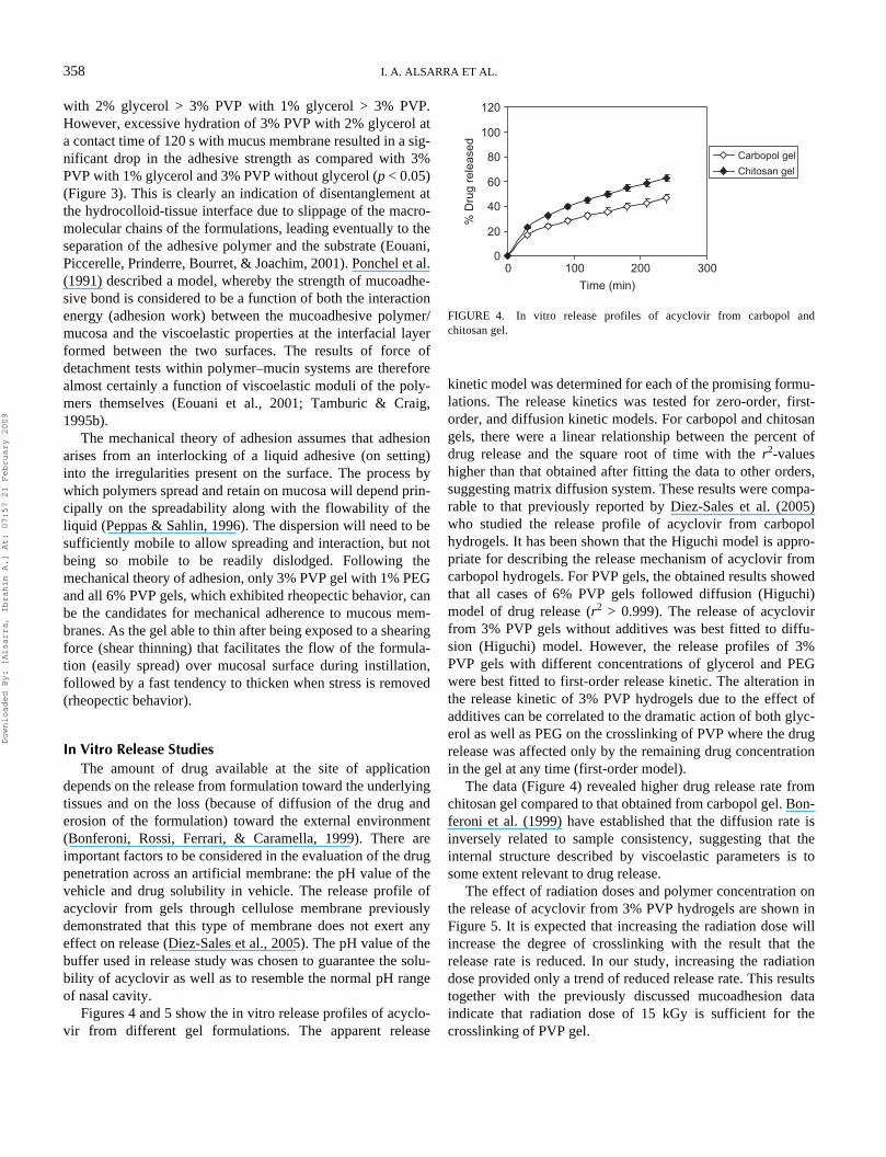
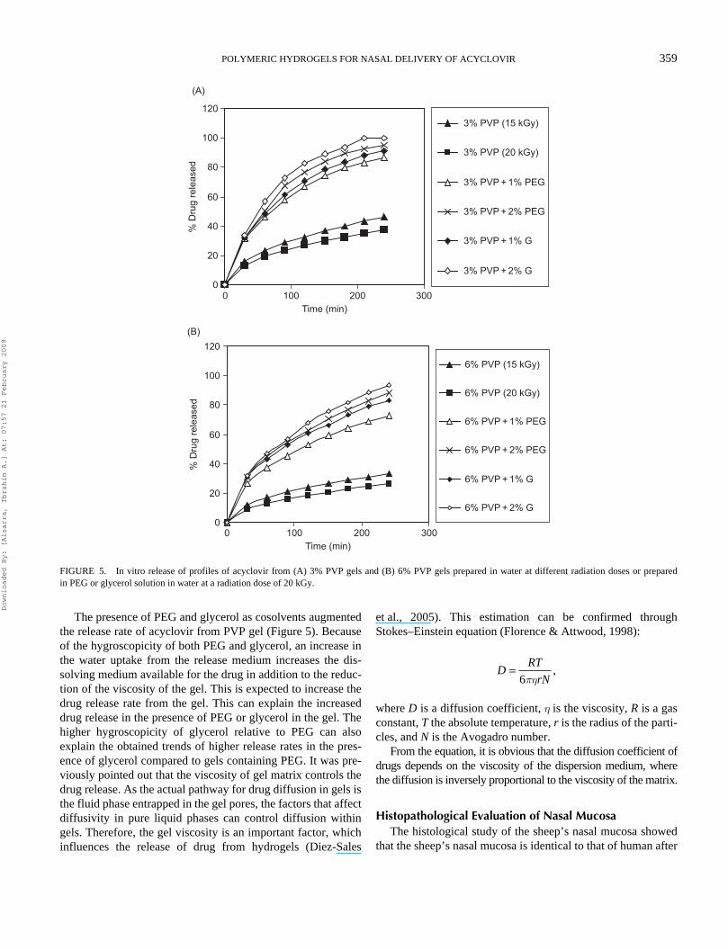
Figures 4 and 5 show the in vitro release profiles of acyclo-vir from different gel formulations. The apparent release
kinetic model was determined for each of the promising formu-lations. The release kinetics was tested for zero-order, first-order, and diffusion kinetic models. For carbopol and chitosangels, there were a linear relationship between the percent ofdrug release and the square root of time with the r2-valueshigher than that obtained after fitting the data to other orders,suggesting matrix diffusion system. These results were compa-rable to that previously reported by Diez-Sales et al. (2005)who studied the release profile of acyclovir from carbopolhydrogels. It has been shown that the Higuchi model is appro-priate for describing the release mechanism of acyclovir fromcarbopol hydrogels. For PVP gels, the obtained results showedthat all cases of 6% PVP gels followed diffusion (Higuchi)model of drug release (r2 > 0.999). The release of acyclovirfrom 3% PVP gels without additives was best fitted to diffu-sion (Higuchi) model. However, the release profiles of 3%PVP gels with different concentrations of glycerol and PEGwere best fitted to first-order release kinetic. The alteration inthe release kinetic of 3% PVP hydrogels due to the effect ofadditives can be correlated to the dramatic action of both glyc-erol as well as PEG on the crosslinking of PVP where the drugrelease was affected only by the remaining drug concentrationin the gel at any time (first-order model).
The data (Figure 4) revealed higher drug release rate fromchitosan gel compared to that obtained from carbopol gel. Bon-feroni et al. (1999) have established that the diffusion rate isinversely related to sample consistency, suggesting that theinternal structure described by viscoelastic parameters is tosome extent relevant to drug release.
The effect of radiation doses and polymer concentration onthe release of acyclovir from 3% PVP hydrogels are shown inFigure 5. It is expected that increasing the radiation dose willincrease the degree of crosslinking with the result that therelease rate is reduced. In our study, increasing the radiationdose provided only a trend of reduced release rate. This resultstogether with the previously discussed mucoadhesion dataindicate that radiation dose of 15 kGy is sufficient for thecrosslinking of PVP gel.
FIGURE 4. In vitro release profiles of acyclovir from carbopol andchitosan gel.
0
20
40
60
80
100
120
0 100 200 300Time (min)
% D
rug
rele
ased
Carbopol gelChitosan gel
Downloaded By: [Alsarra, Ibrahim A.] At: 07:57 21 February 2009
POLYMERIC HYDROGELS FOR NASAL DELIVERY OF ACYCLOVIR 359
The presence of PEG and glycerol as cosolvents augmentedthe release rate of acyclovir from PVP gel (Figure 5). Becauseof the hygroscopicity of both PEG and glycerol, an increase inthe water uptake from the release medium increases the dis-solving medium available for the drug in addition to the reduc-tion of the viscosity of the gel. This is expected to increase thedrug release rate from the gel. This can explain the increaseddrug release in the presence of PEG or glycerol in the gel. Thehigher hygroscopicity of glycerol relative to PEG can alsoexplain the obtained trends of higher release rates in the pres-ence of glycerol compared to gels containing PEG. It was pre-viously pointed out that the viscosity of gel matrix controls thedrug release. As the actual pathway for drug diffusion in gels isthe fluid phase entrapped in the gel pores, the factors that affectdiffusivity in pure liquid phases can control diffusion withingels. Therefore, the gel viscosity is an important factor, whichinfluences the release of drug from hydrogels (Diez-Sales
et al., 2005). This estimation can be confirmed throughStokes–Einstein equation (Florence & Attwood, 1998):
where D is a diffusion coefficient, h is the viscosity, R is a gasconstant, T the absolute temperature, r is the radius of the parti-cles, and N is the Avogadro number.
From the equation, it is obvious that the diffusion coefficient ofdrugs depends on the viscosity of the dispersion medium, wherethe diffusion is inversely proportional to the viscosity of the matrix.
Histopathological Evaluation of Nasal MucosaThe histological study of the sheep’s nasal mucosa showed
that the sheep’s nasal mucosa is identical to that of human after
FIGURE 5. In vitro release of profiles of acyclovir from (A) 3% PVP gels and (B) 6% PVP gels prepared in water at different radiation doses or preparedin PEG or glycerol solution in water at a radiation dose of 20 kGy.
0
20
40
60
80
100
120
0 100 200 300Time (min)
% D
rug
rele
ased
3% PVP (15 kGy)
3% PVP (20 kGy)
3% PVP + 1% PEG
3% PVP + 2% PEG
3% PVP + 1% G
3% PVP + 2% G
(A)
0 100 200 300Time (min)
(B)
0
20
40
60
80
100
120
% D
rug
rele
ased
6% PVP (15 kGy)
6% PVP (20 kGy)
6% PVP + 1% PEG
6% PVP + 2% PEG
6% PVP + 1% G
6% PVP + 2% G
DRT
rN=
6ph,
Downloaded By: [Alsarra, Ibrahim A.] At: 07:57 21 February 2009
360 I. A. ALSARRA ET AL.
extensive investigation of all possible models (Illum, 1996;Shaw, Cowin, & Wormald, 2001).
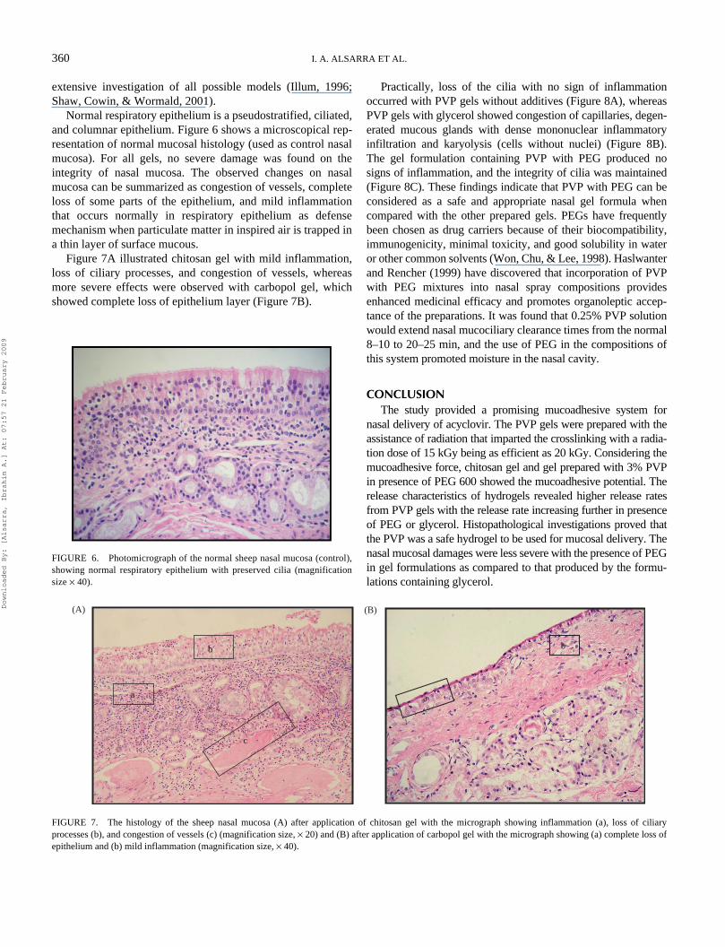
Normal respiratory epithelium is a pseudostratified, ciliated,and columnar epithelium. Figure 6 shows a microscopical rep-resentation of normal mucosal histology (used as control nasalmucosa). For all gels, no severe damage was found on theintegrity of nasal mucosa. The observed changes on nasalmucosa can be summarized as congestion of vessels, completeloss of some parts of the epithelium, and mild inflammationthat occurs normally in respiratory epithelium as defensemechanism when particulate matter in inspired air is trapped ina thin layer of surface mucous.
Figure 7A illustrated chitosan gel with mild inflammation,loss of ciliary processes, and congestion of vessels, whereasmore severe effects were observed with carbopol gel, whichshowed complete loss of epithelium layer (Figure 7B).
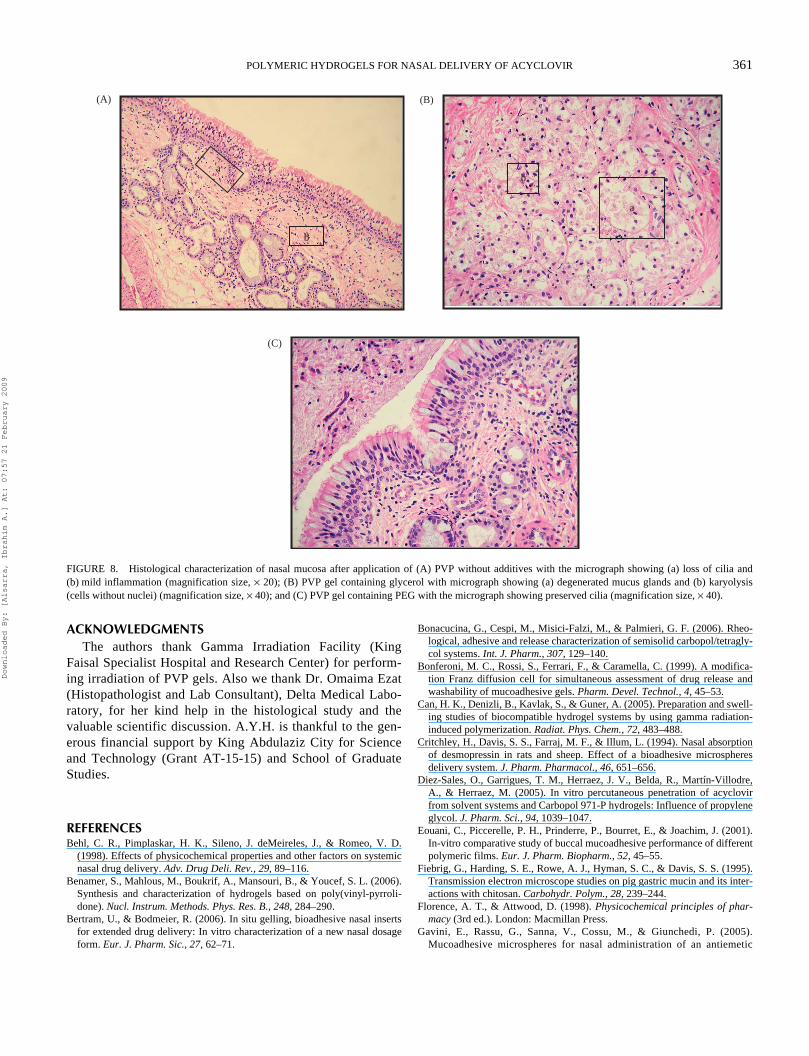
Practically, loss of the cilia with no sign of inflammationoccurred with PVP gels without additives (Figure 8A), whereasPVP gels with glycerol showed congestion of capillaries, degen-erated mucous glands with dense mononuclear inflammatoryinfiltration and karyolysis (cells without nuclei) (Figure 8B).The gel formulation containing PVP with PEG produced nosigns of inflammation, and the integrity of cilia was maintained(Figure 8C). These findings indicate that PVP with PEG can beconsidered as a safe and appropriate nasal gel formula whencompared with the other prepared gels. PEGs have frequentlybeen chosen as drug carriers because of their biocompatibility,immunogenicity, minimal toxicity, and good solubility in wateror other common solvents (Won, Chu, & Lee, 1998). Haslwanterand Rencher (1999) have discovered that incorporation of PVPwith PEG mixtures into nasal spray compositions providesenhanced medicinal efficacy and promotes organoleptic accep-tance of the preparations. It was found that 0.25% PVP solutionwould extend nasal mucociliary clearance times from the normal8–10 to 20–25 min, and the use of PEG in the compositions ofthis system promoted moisture in the nasal cavity.
CONCLUSIONThe study provided a promising mucoadhesive system for
nasal delivery of acyclovir. The PVP gels were prepared with theassistance of radiation that imparted the crosslinking with a radia-tion dose of 15 kGy being as efficient as 20 kGy. Considering themucoadhesive force, chitosan gel and gel prepared with 3% PVPin presence of PEG 600 showed the mucoadhesive potential. Therelease characteristics of hydrogels revealed higher release ratesfrom PVP gels with the release rate increasing further in presenceof PEG or glycerol. Histopathological investigations proved thatthe PVP was a safe hydrogel to be used for mucosal delivery. Thenasal mucosal damages were less severe with the presence of PEGin gel formulations as compared to that produced by the formu-lations containing glycerol.
FIGURE 6. Photomicrograph of the normal sheep nasal mucosa (control),showing normal respiratory epithelium with preserved cilia (magnificationsize × 40).
FIGURE 7. The histology of the sheep nasal mucosa (A) after application of chitosan gel with the micrograph showing inflammation (a), loss of ciliaryprocesses (b), and congestion of vessels (c) (magnification size, × 20) and (B) after application of carbopol gel with the micrograph showing (a) complete loss ofepithelium and (b) mild inflammation (magnification size, × 40).
c
b
a
b
a
(A) (B)Downloaded By: [Alsarra, Ibrahim A.] At: 07:57 21 February 2009
POLYMERIC HYDROGELS FOR NASAL DELIVERY OF ACYCLOVIR 361
ACKNOWLEDGMENTSThe authors thank Gamma Irradiation Facility (King
Faisal Specialist Hospital and Research Center) for perform-ing irradiation of PVP gels. Also we thank Dr. Omaima Ezat(Histopathologist and Lab Consultant), Delta Medical Labo-ratory, for her kind help in the histological study and thevaluable scientific discussion. A.Y.H. is thankful to the gen-erous financial support by King Abdulaziz City for Scienceand Technology (Grant AT-15-15) and School of GraduateStudies.
REFERENCESBehl, C. R., Pimplaskar, H. K., Sileno, J. deMeireles, J., & Romeo, V. D.
(1998). Effects of physicochemical properties and other factors on systemicnasal drug delivery. Adv. Drug Deli. Rev., 29, 89–116.
Benamer, S., Mahlous, M., Boukrif, A., Mansouri, B., & Youcef, S. L. (2006).Synthesis and characterization of hydrogels based on poly(vinyl-pyrroli-done). Nucl. Instrum. Methods. Phys. Res. B., 248, 284–290.
Bertram, U., & Bodmeier, R. (2006). In situ gelling, bioadhesive nasal insertsfor extended drug delivery: In vitro characterization of a new nasal dosageform. Eur. J. Pharm. Sic., 27, 62–71.
Bonacucina, G., Cespi, M., Misici-Falzi, M., & Palmieri, G. F. (2006). Rheo-logical, adhesive and release characterization of semisolid carbopol/tetragly-col systems. Int. J. Pharm., 307, 129–140.
Bonferoni, M. C., Rossi, S., Ferrari, F., & Caramella, C. (1999). A modifica-tion Franz diffusion cell for simultaneous assessment of drug release andwashability of mucoadhesive gels. Pharm. Devel. Technol., 4, 45–53.
Can, H. K., Denizli, B., Kavlak, S., & Guner, A. (2005). Preparation and swell-ing studies of biocompatible hydrogel systems by using gamma radiation-induced polymerization. Radiat. Phys. Chem., 72, 483–488.
Critchley, H., Davis, S. S., Farraj, M. F., & Illum, L. (1994). Nasal absorptionof desmopressin in rats and sheep. Effect of a bioadhesive microspheresdelivery system. J. Pharm. Pharmacol., 46, 651–656.
Diez-Sales, O., Garrigues, T. M., Herraez, J. V., Belda, R., Martín-Villodre,A., & Herraez, M. (2005). In vitro percutaneous penetration of acyclovirfrom solvent systems and Carbopol 971-P hydrogels: Influence of propyleneglycol. J. Pharm. Sci., 94, 1039–1047.
Eouani, C., Piccerelle, P. H., Prinderre, P., Bourret, E., & Joachim, J. (2001).In-vitro comparative study of buccal mucoadhesive performance of differentpolymeric films. Eur. J. Pharm. Biopharm., 52, 45–55.
Fiebrig, G., Harding, S. E., Rowe, A. J., Hyman, S. C., & Davis, S. S. (1995).Transmission electron microscope studies on pig gastric mucin and its inter-actions with chitosan. Carbohydr. Polym., 28, 239–244.
Florence, A. T., & Attwood, D. (1998). Physicochemical principles of phar-macy (3rd ed.). London: Macmillan Press.
Gavini, E., Rassu, G., Sanna, V., Cossu, M., & Giunchedi, P. (2005).Mucoadhesive microspheres for nasal administration of an antiemetic
FIGURE 8. Histological characterization of nasal mucosa after application of (A) PVP without additives with the micrograph showing (a) loss of cilia and(b) mild inflammation (magnification size, × 20); (B) PVP gel containing glycerol with micrograph showing (a) degenerated mucus glands and (b) karyolysis(cells without nuclei) (magnification size, × 40); and (C) PVP gel containing PEG with the micrograph showing preserved cilia (magnification size, × 40).
b
a
(A) (B)
(C)
b
a
Downloaded By: [Alsarra, Ibrahim A.] At: 07:57 21 February 2009
362 I. A. ALSARRA ET AL.
drug, metaclopramide: In-vivo/ex-vivo studies. J. Pharm. Pharmacol.,57, 287–294.
Grabovac, V., Guggi, D., & Bernkop-Schnurch, A. (2005). Mucoadhesivepolymers: Strategies, achievements and future challenges. Adv. Drug Deliv.Rev., 57, 1713–1723.
Güner, A., & Ataman, M. (1997). Effects of inorganic salts on the properties ofaqueous PVP solutions. Colloid. Polym. Sci., 272, 175–180.
Guo, J., Ping, Q., Jiang, G., Dong, J. Q. S., Feng, L., Li, Z., & Li, C. (2004).Transport of leuprolide across rat intestine, rabbit intestine and Caco-2 cellmonolayer. Int. J. Pharm., 278, 415–422.
Hägerström, H., & Edsman, K. (2003). Limitations of the rheological mucoad-hesion method: The effect of the choice of conditions and the rheologicalsynergism parameter. Eur. J. Pharm. Sci., 18, 349–357.
Haslwanter, J. A., & Rencher, W. F. (1999). Nasal spray compositions exhibitingincreased retention in the nasal cavity, U.S. Patent No. 5897858, April 27.
Hernández, M. J., Pellicer, J., Delegido, I., & Dolz, M. (1998). Rheologicalcharacterization of easy to disperse (ETD) carbopol hydrogels. J. Disper.Sci. Technol., 19, 31–42.
Illum, L. (1996). Nasal delivery. The use of animal models to predict perfor-mance in man. J. Drug Target., 3, 427–442.
Jain, S. K., Jain, R. K., Chourasia, M. K, Jain, A. K., Chalasani, K. B., Soni,V., & Jain, A. (2005). Design and development of multivesicular liposomaldepot delivery system for controlled systemic delivery of acyclovir sodium.AAPS PharmSciTech, 6, 35–41.
Knapezyk, J., (1993). Chitosan hydrogel as a base for semisolid drug forms.Int. J. Pharm., 93, 233–237.
Lehr, C. M., Bouwstra, J. A., Schacht, E. H., & Junginger, H. E. (1992). In-vitro evaluation of mucoadhesive properties of chitosan and some other nat-ural polymers. Int. J. Pharm., 78, 43–48.
Majithiya, R. J., Ghosh, P. K., Umrethia, M. L., & Murthy, R. S. (2006). Ther-moreversible-mucoaadhesive gel for nasal delivery of sumatriptan. AAPS.PharmSciTech, 7, 1–7.
Mattrin, E., Schipper, N. G. M., Verhoef, J. C., & Merkus, F. W. (1998). Nasalmucociliary clearance as a factor for nasal drug delivery. Adv. Drug Deliv.Rev., 29, 13–38.
Needleman, I. G., & Smales, F. C. (1995). In vitro assessment of bioadhesionfor periodontal and buccal drug delivery. Biomaterials, 16, 617–624.
Paul, W., & Sharma, C. P. (2000). Chitosan, a drug carrier for the 21st century:A review. S.T.P. Pharm. Sci., 10, 5–22.
Peppas, N. A., & Sahlin, J. J. (1996). Hydrogels as mucoadhesive and bioadhe-sive materials: a review. Biomaterials, 17, 1553–1561.
Ponchel, G., Lejoyeux, F., & DucheÃne, D. (1991). Bioadhesion of poly(acrylic acid) containing systems. Thermodynamics and rheological aspects.Proceedings of International Symposium on Control Release BioactiveMaterials, 18, 111–112.
Roos, A., Creton, C., Novikov, M. B., & Feldstein, M. M. (2002). Viscoelas-ticity and tack of poly(vinyl pyrrolidone)–poly(ethylene glycol) blends. J.Polym. Sci. Part. B Polym. Phys., 40, 2395–2409.
Rosiak, J. M., Ulanski, P., Pajewnski, L. A., Yoshii, F., & Makuuchi, K.(1995). Radiation formation of hydrogels for biomedical purpose. Someremarks and comments. Radiat. Phys. Chem., 46, 161–168.
Rosiak, J. M., & Yoshii, F. (1999). Hydrogels and their medical applications.Nucl. Instr. Methods Phys. Res. B., 151, 56–64.
Sen, M., & Avc, E. N. (2005). Radiation synthesis of poly(N-vinyl-2-pyr-rolidone)–k-carrageenan hydrogels and their use in wound dressingapplications. I. Preliminary laboratory tests. J. Biomed. Mater. Res. A.,1, 187–196.
Shaw, C. K. L., Cowin, A., & Wormald, P. J. (2001). Standardization of thesheep as a suitable animal model for studying endoscopic sinus surgery.Aust. J. Otolaryngol., 4, 23–26.
Tamburic, S., & Craig, D. Q. M., (1995a). An investigation into the rheologi-cal, dielectric and mucoadhesive properties of poly (acrylic acid) gel system.J. Control Release, 37, 59–68.
Tamburic, S., & Craig, D. Q. M. (1995b). Rheological evaluation of poly-acrylic acid hydrogels. Pharm. Sci., 1, 107–109.
Tamburic, S., & Craig, D. Q. M. (1997). A comparison of different in vitromethod for measuring mucoadhesive performance. Eur. J. Pharm. Biop-harm., 44, 159–167.
Ugwoke, M. I., Van den Mooter, G., Verbeke, N., & Kinget, R. (1999). Nasalmucoadhesion delivery systems of the antiparkinsonain drug, apomorphine;influence of drug loading on in-vitro and in-vivo release in rabbits. Int. J.Pharm., 181, 125–138.
Varshosaz, J., Sadrai, H., & Heidar, A. (2006). Nasal delivery of insulin usingbioadhesive chitosan gels. Drug Deliv., 13, 31–38.
Won, C. Y., Chu, C. C., & Lee, J. D. (1998). Novel biodegradable copolymerscontaining pendant amine functional groups based on aspartic acid andpoly(ethyleneglycol). Polymer, 25, 6677–6681.
Yanai, R., Yamada, N., Ueda, K., Tajiri, M., Matsumoto, T., Kido, K.,Nakamura, S., Saito, F., & Nishida, T. (2006). Evaluation of povidone–iodine as a disinfectant solution for contact lenses: Antimicrobial activityand cytotoxicity for corneal epithelial cells. Cont Lens. Anterior. Eye., 29,85–91.
Downloaded By: [Alsarra, Ibrahim A.] At: 07:57 21 February 2009
Related Documents