Research Article Mre11 exonuclease activity promotes irreversible mitotic progression under replication stress Yoshitami Hashimoto , Hirofumi Tanaka Mre11 is a versatile exo-/endonuclease involved in multiple as- pects of DNA replication and repair, such as DSB end processing and checkpoint activation. We previously demonstrated that forced mitotic entry drives replisome disassembly at stalled replication forks in Xenopus egg extracts. Here, we examined the effects of various chemical inhibitors using this system and discovered a novel role of Mre11 exonuclease activity in pro- moting mitotic entry under replication stress. Mre11 activity was necessary for the initial progression of mitotic entry in the presence of stalled forks but unnecessary in the absence of stalled forks or after mitotic entry. In the absence of Mre11 ac- tivity, mitotic CDK was inactivated by Wee1/Myt1–dependent phosphorylation, causing mitotic exit. An inhibitor of Wee1/Myt1 or a nonphosphorylatable CDK1 mutant was able to partially bypass the requirement of Mre11 for mitotic entry. These results suggest that Mre11 exonuclease activity facilitates the processing of stalled replication forks upon mitotic entry, which attenuates the inhibitory pathways of mitotic CDK activation, leading to irreversible mitotic progression and replisome disassembly. DOI 10.26508/lsa.202101249 | Received 29 September 2021 | Revised 2 March 2022 | Accepted 2 March 2022 | Published online 15 March 2022 Introduction Eukaryotic DNA replication is initiated at replication origins during the S-phase of the cell cycle, forming replication forks to which the replisome, a multi-protein complex, binds and conducts DNA replication. The replisome contains the Cdc45/MCM2–7/GINS helicase complex and the replicative DNA polymerases Polα/δ/ε, as well as many other accessory factors with specialized functions (Burgers & Kunkel, 2017). When two progressing forks converge, DNA replication is locally terminated, the daughter strands are deca- tenated by topoisomerase II, and the replisomes are disassembled by p97 ATPase that recognizes MCM7 poly-ubiquitylated by cullin E3 ligase (CRL2 Lrr1 in metazoa) (Maric et al, 2014; Moreno et al, 2014; Dewar et al, 2017; Sonneville et al, 2017). However, replication forks can often encounter obstacles for their progression, such as DNA lesions, nucleotide reduction, secondary DNA structures, DNA–RNA hybrids, and transcriptional machinery (Zeman & Cimprich, 2014; Gaillard et al, 2015). These replication stress factors cause stalled replication forks, which are stabilized for replication restart until the stress is relieved. Recently, increasing evidence suggests that stalled forks are regularly remodeled into reversed forks in which the two parental strands reanneal, and the two nascent strands also anneal to form a chicken footlike four-way junction (fork re- versal) (Neelsen & Lopes, 2015; Poole & Cortez, 2017). Reversed forks are believed to contribute to fork stabilization and repair, and stalled forks (or reversed forks) have been shown to activate ATR- Chk1 checkpoint signaling to down-regulate CDK1/2 activities via Cdc25 inactivation, suppressing de novo initiation of DNA repli- cation and cell cycle progression into G2/M phases (Saldivar et al, 2017). Without replication stress, Cdc25 usually activates Cdk1/2 during S to G2/M phase transition to drive replication initiation and cell cycle progression by removing Wee1/Myt1 kinase–mediated inhibitory phosphorylation of Thr14 and Tyr15 (Sørensen & Sylju ˚ asen, 2012; Crncec & Hochegger, 2019; Elbæk et al, 2020). Mre11, a versatile nuclease with both exo- and endonuclease activities, functions within the Mre11–Rad50–Nbs1 (MRN) complex that is involved in many aspects of DNA replication and repair, such as DSB end resection for homologous recombination-dependent DNA repair, ATM activation in response to DSBs, and DNA tether- ing between sister chromatids (Paull, 2018; Syed & Tainer, 2018; Reginato & Cejka, 2020). In addition to these physiological roles, pathological fork resection by Mre11 has also been reported, re- vealing its role in a fork protection mechanism that involves BRCA2, Rad51, and other DNA damage response proteins (Pasero & Vindigni, 2017; Rickman & Smogorzewska, 2019). A reversed fork comprises a regressed arm with two nascent DNA strands of dif- ferent lengths, exposing a single-stranded DNA (ssDNA) region. BRCA2 promotes the assembly of filament Rad51 onto this ssDNA region to stabilize the reversed fork under replication stress. On the contrary, in BRCA2-deficient cells, this unprotected ssDNA region serves as an entry point for Mre11 to bring about uncontrolled fork degradation (Schlacher et al, 2011; Kolinjivadi et al, 2017; Taglialatela et al, 2017). The fork stabilization system ensures that replisomes are stably maintained at stalled forks (or maybe reversed forks) under rep- lication stress, which enables replication restart without de novo School of Life Sciences, Tokyo University of Pharmacy and Life Sciences, Hachioji, Japan Correspondence: [email protected] © 2022 Hashimoto and Tanaka https://doi.org/10.26508/lsa.202101249 vol 5 | no 6 | e202101249 1 of 12 on 30 March, 2022 life-science-alliance.org Downloaded from http://doi.org/10.26508/lsa.202101249 Published Online: 15 March, 2022 | Supp Info:

Welcome message from author
This document is posted to help you gain knowledge. Please leave a comment to let me know what you think about it! Share it to your friends and learn new things together.
Transcript
Research Article
Mre11 exonuclease activity promotes irreversible mitoticprogression under replication stressYoshitami Hashimoto , Hirofumi Tanaka
Mre11 is a versatile exo-/endonuclease involved in multiple as-pects of DNA replication and repair, such as DSB end processingand checkpoint activation. We previously demonstrated thatforced mitotic entry drives replisome disassembly at stalledreplication forks in Xenopus egg extracts. Here, we examined theeffects of various chemical inhibitors using this system anddiscovered a novel role of Mre11 exonuclease activity in pro-moting mitotic entry under replication stress. Mre11 activity wasnecessary for the initial progression of mitotic entry in thepresence of stalled forks but unnecessary in the absence ofstalled forks or after mitotic entry. In the absence of Mre11 ac-tivity, mitotic CDK was inactivated by Wee1/Myt1–dependentphosphorylation, causing mitotic exit. An inhibitor of Wee1/Myt1or a nonphosphorylatable CDK1 mutant was able to partiallybypass the requirement of Mre11 for mitotic entry. These resultssuggest that Mre11 exonuclease activity facilitates the processingof stalled replication forks upon mitotic entry, which attenuatesthe inhibitory pathways of mitotic CDK activation, leading toirreversible mitotic progression and replisome disassembly.
DOI 10.26508/lsa.202101249 | Received 29 September 2021 | Revised 2 March2022 | Accepted 2 March 2022 | Published online 15 March 2022
Introduction
Eukaryotic DNA replication is initiated at replication origins duringthe S-phase of the cell cycle, forming replication forks to which thereplisome, a multi-protein complex, binds and conducts DNAreplication. The replisome contains the Cdc45/MCM2–7/GINShelicase complex and the replicative DNA polymerases Polα/δ/ε, aswell as many other accessory factors with specialized functions(Burgers & Kunkel, 2017). When two progressing forks converge, DNAreplication is locally terminated, the daughter strands are deca-tenated by topoisomerase II, and the replisomes are disassembledby p97 ATPase that recognizes MCM7 poly-ubiquitylated by cullin E3ligase (CRL2Lrr1 in metazoa) (Maric et al, 2014; Moreno et al, 2014;Dewar et al, 2017; Sonneville et al, 2017). However, replication forkscan often encounter obstacles for their progression, such as DNAlesions, nucleotide reduction, secondary DNA structures, DNA–RNA
hybrids, and transcriptional machinery (Zeman & Cimprich, 2014;Gaillard et al, 2015). These replication stress factors cause stalledreplication forks, which are stabilized for replication restart untilthe stress is relieved. Recently, increasing evidence suggests thatstalled forks are regularly remodeled into reversed forks in whichthe two parental strands reanneal, and the two nascent strandsalso anneal to form a chicken footlike four-way junction (fork re-versal) (Neelsen & Lopes, 2015; Poole & Cortez, 2017). Reversed forksare believed to contribute to fork stabilization and repair, andstalled forks (or reversed forks) have been shown to activate ATR-Chk1 checkpoint signaling to down-regulate CDK1/2 activities viaCdc25 inactivation, suppressing de novo initiation of DNA repli-cation and cell cycle progression into G2/M phases (Saldivar et al,2017). Without replication stress, Cdc25 usually activates Cdk1/2during S to G2/M phase transition to drive replication initiation andcell cycle progression by removing Wee1/Myt1 kinase–mediatedinhibitory phosphorylation of Thr14 and Tyr15 (Sørensen &Syljuasen, 2012; Crncec & Hochegger, 2019; Elbæk et al, 2020).
Mre11, a versatile nuclease with both exo- and endonucleaseactivities, functions within the Mre11–Rad50–Nbs1 (MRN) complexthat is involved in many aspects of DNA replication and repair, suchas DSB end resection for homologous recombination-dependentDNA repair, ATM activation in response to DSBs, and DNA tether-ing between sister chromatids (Paull, 2018; Syed & Tainer, 2018;Reginato & Cejka, 2020). In addition to these physiological roles,pathological fork resection by Mre11 has also been reported, re-vealing its role in a fork protection mechanism that involves BRCA2,Rad51, and other DNA damage response proteins (Pasero &Vindigni, 2017; Rickman & Smogorzewska, 2019). A reversed forkcomprises a regressed arm with two nascent DNA strands of dif-ferent lengths, exposing a single-stranded DNA (ssDNA) region.BRCA2 promotes the assembly of filament Rad51 onto this ssDNAregion to stabilize the reversed fork under replication stress. On thecontrary, in BRCA2-deficient cells, this unprotected ssDNA regionserves as an entry point for Mre11 to bring about uncontrolled forkdegradation (Schlacher et al, 2011; Kolinjivadi et al, 2017; Taglialatelaet al, 2017).
The fork stabilization system ensures that replisomes are stablymaintained at stalled forks (or maybe reversed forks) under rep-lication stress, which enables replication restart without de novo
School of Life Sciences, Tokyo University of Pharmacy and Life Sciences, Hachioji, Japan
Correspondence: [email protected]
© 2022 Hashimoto and Tanaka https://doi.org/10.26508/lsa.202101249 vol 5 | no 6 | e202101249 1 of 12
on 30 March, 2022life-science-alliance.org Downloaded from http://doi.org/10.26508/lsa.202101249Published Online: 15 March, 2022 | Supp Info:
assembly of replisomes in the S-phase (Bellelli & Boulton, 2021;Guilliam, 2021). However, these stabilized replisomes are dis-assembled by increasing mitotic CDK activity that leads to cell cycleprogression into mitosis (Hashimoto & Tanaka, 2018). The RING E3ligase TRAIP mediates the poly-ubiquitylation of MCM7, whichpromotes p97-dependent replisome disassembly in the M-phase(Deng et al, 2019; Moreno et al, 2019; Sonneville et al, 2019). ExploitingXenopus egg extracts with interphase and mitotic activities, wepreviously established an in vitro system that reconstitutes mitoticreplisome disassembly (MRD) under replication stress (Hashimoto &Tanaka, 2018). In this study, we further investigated the molecularmechanism of MRD using this system and chemical inhibitors. Un-expectedly, we found that Mre11 exonuclease activity is required for
mitotic entry under replication stress, possibly through the pro-cessing of stalled or reversed forks, which in turn promotes MRD.
Results
Mre11 exonuclease activity is required for mitotic entry andreplisome disassembly in the presence of stalled replicationforks
We previously demonstrated that mitotic entry drives replisomedisassembly at stalled replication forks in Xenopus egg extract(Hashimoto & Tanaka, 2018) (Fig 1A). In this system, sperm nuclei are
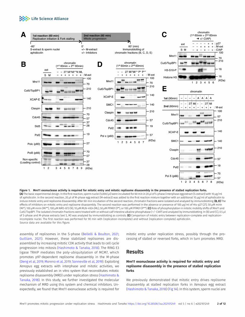
Figure 1. Mre11 exonuclease activity is required for mitotic entry and mitotic replisome disassembly in the presence of stalled replication forks.(A) The basic experimental design. In thefirst reaction, spermnuclei (5,000/μl) were incubated for 60min in 20μl of S-phase/interphase egg extract (S-extract) with 10μg/mlof aphidicolin. In the second reaction, 20 μl of M-phase egg extract (M-extract) was added to the first reaction mixture together with an additional 10 μg/ml of aphidicolin toinducemitotic entry and replisomedisassembly. After 60-min incubation of the second reaction, chromatin fractionswere isolated and analyzed by immunoblotting. (B, D) Theeffects of inhibitors on mitotic entry and replisome disassembly. The second reaction was performed in the absence or presence of 100 μg/ml of His-p27 (27), 50 μMmirin(Mi*), 100 μMmirin (Mi**), 100 μMNMS-873 (N), 10 μMMLN-4924 (ML), 50 μMPFM01 (P*), or 100 μMPFM01 (P**). (C) Role of phosphorylation inmitoticmobility shifts of Mre11 andCut5/TopBP1. The isolated chromatin fractions were treatedwith or without calf intestine alkaline phosphatase (+/−CIAP) and analyzed by immunoblotting. In (B) and (C), 0.5 μlof S-phase and M-phase extracts (ext S, M) was analyzed by immunoblotting as controls. (E) Comparison of mitotic entry between replication-complete and replication-incomplete nuclei. The first reaction was performed for 90 min with (replication-incomplete) and without (replication-complete) aphidicolin.Source data are available for this figure.
Mre11 promotes mitotic progression under replication stress Hashimoto and Tanaka https://doi.org/10.26508/lsa.202101249 vol 5 | no 6 | e202101249 2 of 12
first incubated in S-phase (interphase) egg extract (S-extract) in thepresence of aphidicolin, an inhibitor of replicative DNA polymer-ases α/δ/ε, which allows initiation but not elongation and causesall replication forks to be stalled. An equal volume of M-phaseextract (M-extract) is then supplemented to induce mitotic entryand replisome disassembly. MRD has been shown to be mecha-nistically different from interphase replisome disassembly, whichoccurs during replication termination or replication fork collapse(Wu et al, 2021).
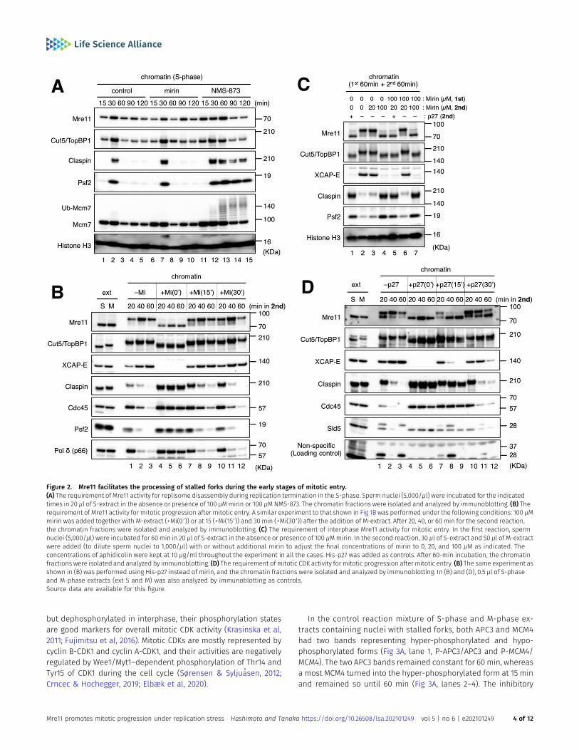
We first tested various inhibitors for their effects on mitotic entryand MRD by the immunoblotting of the chromatin fractions (Fig 1B).Major replisome components such as claspin, GINS subunits (Sld5and Psf2), and Polδ/ε were stably maintained on chromatin untilthe end of the second incubation without the induction of mitoticentry, whereas they largely dissociated from chromatin during thesecond incubation with induction of mitotic entry (Fig 1B, lanes 3and 4), indicating MRD had occurred. Mitotic entry was confirmed bychromatin binding of XCAP-E, a common subunit of condensin I/II.The mobility shifts of Mre11 and Cut5/TopBP1 were also observedupon mitotic entry. Mre11 reverted to its original state after treatingchromatin with alkaline phosphatase (Fig 1C), demonstrating thatthe mobility shift of Mre11 on chromatin was exclusively due tomitotic phosphorylation. The mobility of Cut5/TopBP1 on chromatindid not fully revert to its interphase state but shifted closer to itscytosolic state in M-extract after phosphatase treatment (Fig 1C),suggesting a partial role of phosphorylation in the mobility shift. Itis also possible that mitotic Cut5/TopBP1 undergoes additionalmodification other than phosphorylation. Inactivation of mitoticCDK activity with recombinant p27 nullified the effect of the additionof M-extract for mitotic entry and MRD (Fig 1B, lane 5; Fig 1C lanes 5and 6). NMS873 (an inhibitor of p97 ATPase) partly inhibited MRD butdid not inhibit mitotic entry (Fig 1B, lane 8). MLN4924 (an inhibitor ofcullin Ub ligases) did not suppress mitotic entry or MRD (Fig 1B, lane9). These results were in accordance with our previous results(Hashimoto & Tanaka, 2018). Intriguingly, we found that mitoticentry and MRD were both suppressed by the presence of mirin, aninhibitor of the exonuclease activity of Mre11 (Fig 1B, lanes 6 and 7)(Dupre et al, 2008). In contrast, PFM01, an inhibitor of the endo-nuclease activity of Mre11, did not have any inhibitory effect (Fig 1D)(Moiani et al, 2018). We then examined whether the effect of mirin isrestricted to situations of replication stress. When the first incu-bation was extended to 90 min and performed without aphidicolinto allow for the completion of DNA replication, mirin did not inhibitmitotic entry, and a small amount of residual replisome com-ponents were dissociated from chromatin (Fig 1E, lanes 1, 2, and4). In the presence of aphidicolin, mirin inhibited mitotic entryand MRD even after the longer first incubation (Fig 1E, lanes 5, 6,and 8). By contrast, recombinant p27 inhibited mitotic entry andMRD irrespective of the presence of aphidicolin (Fig 1E, lanes 3and 7). In addition, mirin did not inhibit interphase replisomedisassembly in the absence of aphidicolin, as opposed to NMS873which caused persistent chromatin binding of claspin, Psf2, andpoly-ubiquitylated Mcm7 at later time points (Fig 2A). These re-sults suggest that the exonuclease activity of Mre11 is required formitotic entry and MRD in the presence of stalled replication forksbut not when DNA replication is nearly complete and withoutexogenous inhibition.
Mre11 facilitates the processing of stalled forks during the earlystages of mitotic entry
As mirin was used together with M-extract in our experimentsdescribed so far, Mre11 could have the potential to act on stalledforks during interphase before mitotic induction. However, ourresults show that such activity, if any, was not sufficient to promotemitotic entry. Alternatively, Mre11 activity may be suppressed duringinterphase, possibly through a fork protection mechanism (Pasero& Vindigni, 2017; Rickman & Smogorzewska, 2019). To clarify thetiming at which Mre11 activity is required, we addedmirin at differenttime points on and after mitotic induction and monitored chromatinassociation of replisome-related factors. Whenmirin was added at 15or 30min after mitotic induction, the appearance of themitotic formsof Mre11 and Cut5/TopBP1 were not affected, and XCAP-E accumu-lated on, and the replisome components claspin/Cdc45/Ps2/Polδdisappeared from chromatin over time in a similar fashion to thecase without mirin (Fig 2B, lanes 1–3 and 7–12), indicating that mirinhad no inhibitory effect after the initiation of mitotic entry. Whenadded at 0min, mirin completely inhibitedmitotic entry andMRD (Fig2B, lanes 4–6). These results suggest that Mre11 acts on stalled forksduring an initial 15-min period after mitotic induction and that Mre11activity is required for subsequent mitotic progression.
We then examined whether interphase Mre11 activity is alsoinvolved in mitotic entry and MRD (Fig 2C). To suppress Mre11 ac-tivity only in the interphase, we added mirin at 100 μM in the firstincubation and diluted it by one-fifth (to 20 μM) with 1.5 volumes ofS-extract and 2.5 volumes of M-extract in the second incubation. Inthis condition, mirin hardly inhibited mitotic entry and MRD as withthe case that 20 μM of mirin was added only in the second incu-bation (Fig 2C, lanes 3 and 6). When 100 μMofmirin was added in thesecond incubation, mitotic entry and MRD were inhibited irre-spective of the presence or absence of mirin in the first incubation(Fig 2C, lanes 4 and 7). These results suggest that interphase Mre11activity is not required for mitotic entry and MRD. On the otherhand, the addition of recombinant p27 at 15 or 30 min stoppedmitotic progression as indicated by the unloading of XCAP-E and themobility shift of Mre11 to an interphase form (Fig 2D, lanes 7–12).MRD was also inhibited by p27 when added at 15 min but not at30 min. These results show that Mre11-dependent processing ofstalled forks during the early stages of mitotic entry is necessaryand sufficient for mitotic progression, whereas mitotic CDK activityis continuously required for mitotic progression.
Mitotic CDK activity is inactivated without Mre11-dependentprocessing of stalled forks
We then assessed whether the inhibition of Mre11 activity wouldaffect the overall activity of mitotic CDK by the immunoblotting ofwhole extracts containing both nuclei and cytosol (Fig 3A and B).APC3 is a subunit of the anaphase-promoting complex/cyclosomeE3 ubiquitin ligase responsible for cyclin B degradation (Fujimitsuet al, 2016), and Mcm4 is a subunit of the minichromosomemaintenance complex MCM2-7 that constitutes the replicativehelicase Cdc45/MCM2-7/GINS complex together with Cdc45 andGINS at the replication fork (Burgers & Kunkel, 2017). Because bothAPC3 andMcm4 are highly phosphorylated bymitotic CDK in mitosis
Mre11 promotes mitotic progression under replication stress Hashimoto and Tanaka https://doi.org/10.26508/lsa.202101249 vol 5 | no 6 | e202101249 3 of 12
but dephosphorylated in interphase, their phosphorylation statesare good markers for overall mitotic CDK activity (Krasinska et al,2011; Fujimitsu et al, 2016). Mitotic CDKs are mostly represented bycyclin B-CDK1 and cyclin A-CDK1, and their activities are negativelyregulated by Wee1/Myt1–dependent phosphorylation of Thr14 andTyr15 of CDK1 during the cell cycle (Sørensen & Syljuasen, 2012;Crncec & Hochegger, 2019; Elbæk et al, 2020).
In the control reaction mixture of S-phase and M-phase ex-tracts containing nuclei with stalled forks, both APC3 and MCM4had two bands representing hyper-phosphorylated and hypo-phosphorylated forms (Fig 3A, lane 1, P-APC3/APC3 and P-MCM4/MCM4). The two APC3 bands remained constant for 60 min, whereasa most MCM4 turned into the hyper-phosphorylated form at 15 minand remained so until 60 min (Fig 3A, lanes 2–4). The inhibitory
Figure 2. Mre11 facilitates the processing of stalled forks during the early stages of mitotic entry.(A) The requirement of Mre11 activity for replisome disassembly during replication termination in the S-phase. Sperm nuclei (5,000/μl) were incubated for the indicatedtimes in 20 μl of S-extract in the absence or presence of 100 μMmirin or 100 μM NMS-873. The chromatin fractions were isolated and analyzed by immunoblotting. (B) Therequirement of Mre11 activity for mitotic progression after mitotic entry. A similar experiment to that shown in Fig 1B was performed under the following conditions: 100 μMmirin was added together with M-extract (+Mi(09)) or at 15 (+Mi(159)) and 30 min (+Mi(309)) after the addition of M-extract. After 20, 40, or 60 min for the second reaction,the chromatin fractions were isolated and analyzed by immunoblotting. (C) The requirement of interphase Mre11 activity for mitotic entry. In the first reaction, spermnuclei (5,000/μl) were incubated for 60 min in 20 μl of S-extract in the absence or presence of 100 μMmirin. In the second reaction, 30 μl of S-extract and 50 μl of M-extractwere added (to dilute sperm nuclei to 1,000/μl) with or without additional mirin to adjust the final concentrations of mirin to 0, 20, and 100 μM as indicated. Theconcentrations of aphidicolin were kept at 10 μg/ml throughout the experiment in all the cases. His-p27 was added as controls. After 60-min incubation, the chromatinfractions were isolated and analyzed by immunoblotting. (D) The requirement of mitotic CDK activity for mitotic progression after mitotic entry. (B) The same experiment asshown in (B) was performed using His-p27 instead of mirin, and the chromatin fractions were isolated and analyzed by immunoblotting. In (B) and (D), 0.5 μl of S-phaseand M-phase extracts (ext S and M) was also analyzed by immunoblotting as controls.Source data are available for this figure.
Mre11 promotes mitotic progression under replication stress Hashimoto and Tanaka https://doi.org/10.26508/lsa.202101249 vol 5 | no 6 | e202101249 4 of 12
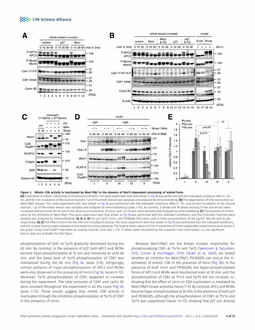
phosphorylation of CDK1 at Tyr15 gradually decreased during the60 min. By contrast, in the presence of p27, both APC3 and MCM4became hypo-phosphorylated at 15 min and remained so until 60min, and the basal level of Tyr15 phosphorylation of CDK1 wasmaintained during the 60 min (Fig 3A, lanes 5–8). Intriguingly,similar patterns of hypo-phosphorylation of APC3 and MCM4were also observed in the presence of mirin (Fig 3A, lanes 9–12).Moreover, Tyr15 phosphorylation of CDK1 appeared to increaseduring the experiment. The total amounts of CDK1 and cyclin B2were constant throughout the experiment in all the cases (Fig 3A,lanes 1–12). These results suggest that mitotic CDK activity isinactivated through the inhibitory phosphorylation at Tyr15 of CDK1in the presence of mirin.
Because Wee1/Myt1 are the known kinases responsible forphosphorylating CDK1 at Thr14 and Tyr15 (Sørensen & Syljuasen,2012; Crncec & Hochegger, 2019; Elbæk et al, 2020), we testedwhether an inhibitor for Wee1/Myt1, PD166285 can rescue the in-activation of mitotic CDK in the presence of mirin (Fig 3B). In thepresence of both mirin and PD166285, the hyper-phosphorylatedforms of APC3 and MCM4 were maintained even at 20 min, and thephosphorylation of CDK1 at Thr14 and Tyr15 did not increase, in-dicating that the effect of mirin on CDK inactivation is mediated byWee1/Myt1 kinase activities (lanes 7–9). By contrast, APC3 and MCM4became hypo-phosphorylated at 10 min in the presence of both p27and PD166285, although the phosphorylation of CDK1 at Thr14 andTyr15 was suppressed (lanes 13–15), showing that p27 can directly
Figure 3. Mitotic CDK activity is inactivated by Wee1/My1 in the absence of Mre11-dependent processing of stalled forks.(A) Inactivation of mitotic CDK activity in the presence of mirin. The same experiment with that shown in Fig 1B was performed with the indicated conditions. After 0-, 15-,30-, and 60-min incubation of the second reaction, 1 μl of the whole extract was sampled and analyzed by immunoblotting. (B) The dependence of CDK inactivation onWee1/Myt1 kinases. The same experiment with that shown in Fig 1B was performed with the indicated conditions. After 0-, 10-, and 20-min incubation of the secondreaction, 1 μl of the whole extract was sampled and analyzed by immunoblotting (lanes 1–15). As controls, S-phase and M-phase extracts (S-ext and M-ext) wereincubated without nuclei for 40 min in the absence and presence of mirin, and 1 μl of each were sampled and analyzed by immunoblotting. (C) The recovery of mitoticentry by the inhibition of Wee1/Myt1. The same experiment with that shown in Fig 1B was performed with the indicated conditions, and the chromatin fractions wereisolated and analyzed by immunoblotting. (A, B, C, D) His-p27 (p27), mirin, and PD166285 (PD) were used at final concentrations of 100 μg/ml, 100 μM, and 10 μM,respectively. (D, E) The effect of mirin for the ATR-Chk1 checkpoint activity. The same experiment with that shown in Fig 1B was performed with the indicated conditions,and the nuclear fractions were isolated and analyzed by immunoblotting. The relative mean values of Chk1-P intensities of three independent experiments were shown inthe graph using Cut5/TopBP1 intensities as loading controls. Error bar, ± S.D. P-values were calculated by the unpaired t test (one-tailed). ns, not significant.Source data are available for this figure.
Mre11 promotes mitotic progression under replication stress Hashimoto and Tanaka https://doi.org/10.26508/lsa.202101249 vol 5 | no 6 | e202101249 5 of 12
inactivate CDK activity irrespective of its phosphorylation status.When S-extract or M-extract was incubated without nuclei, theaddition of mirin did not show any inhibitory effect on APC3, MCM4,or CDK1 (Fig 3B, lanes 16–19), eliminating the possibility that mirindirectly inhibits CDK. Furthermore, immunoblotting of the chro-matin fractions showed that the addition of PD166285 nullified theinhibitory effect of mirin on mitotic entry and MRD (Fig 3C, lanes3–6). These results suggest that when stalled replication forks areleft without Mre11-dependent processing during the early stages ofmitotic entry, Wee1/Myt1 activities remain high to inhibit the es-tablishment of a mitotic state by inactivating mitotic CDK activity,causing the cell cycle to return to interphase.
We also examined the ATR-Chk1 checkpoint activity by immu-noblotting of nuclear fractions (Fig 3D and E). During interphase, theaddition of aphidicolin led to the mobility shift and phosphory-lation of Chk1, indicating the activation of the ATR-Chk1 pathway(lanes I and II). After the addition of M-extract, the active form ofChk1 almost disappeared at 40 min (lanes III). Without mitotic in-duction, the addition of mirin in the second incubation significantlysuppressed further accumulation of the active form of Chk1 at 20 min(lanes IV, P = 0.0426), suggesting the role of Mre11 in checkpoint ac-tivation during interphase as previously described (Lee & Dunphy,2013). However, the remaining active formof Chk1wasmaintained evenafter mitotic induction (lanes V). This suggests that Mre11 exonucleaseactivity is necessary for attenuation of the ATR-Chk1 pathway uponmitotic entry. As Chk1 suppresses Cdc25-mediated removal of theinhibitory phosphorylation of CDK1 Thr14 and Tyr15, it is likely that bothATR-Chk1 and Wee1/Myt1 pathways cooperate in the inactivation ofmitotic CDK in the absence of Mre11 exonuclease activity.
A nonphosphorylatable CDK1 mutant can promote mitotic entrywithout Mre11 exonuclease activity
To further clarify the role of ATR-Chk1 and Wee1/Myt1 pathways ininactivating mitotic CDK in the absence of Mre11 exonuclease ac-tivity, we used wild type and a nonphosphorylatable mutant ofrecombinant GST-CDK1. In the mutant, Thr14 and Tyr15 weresubstituted with Ala and Phe, respectively (designated as CDK1-AFin comparison with wild–type CDK1-WT). These CDK1 proteins werepreincubated with recombinant protein MBP–cyclin B1-ΔN90 (anondegradable mutant from the deletion of the N-terminal 90-amino acid region that contains the destruction box) to form activeCDK1–cyclin B1 complexes.
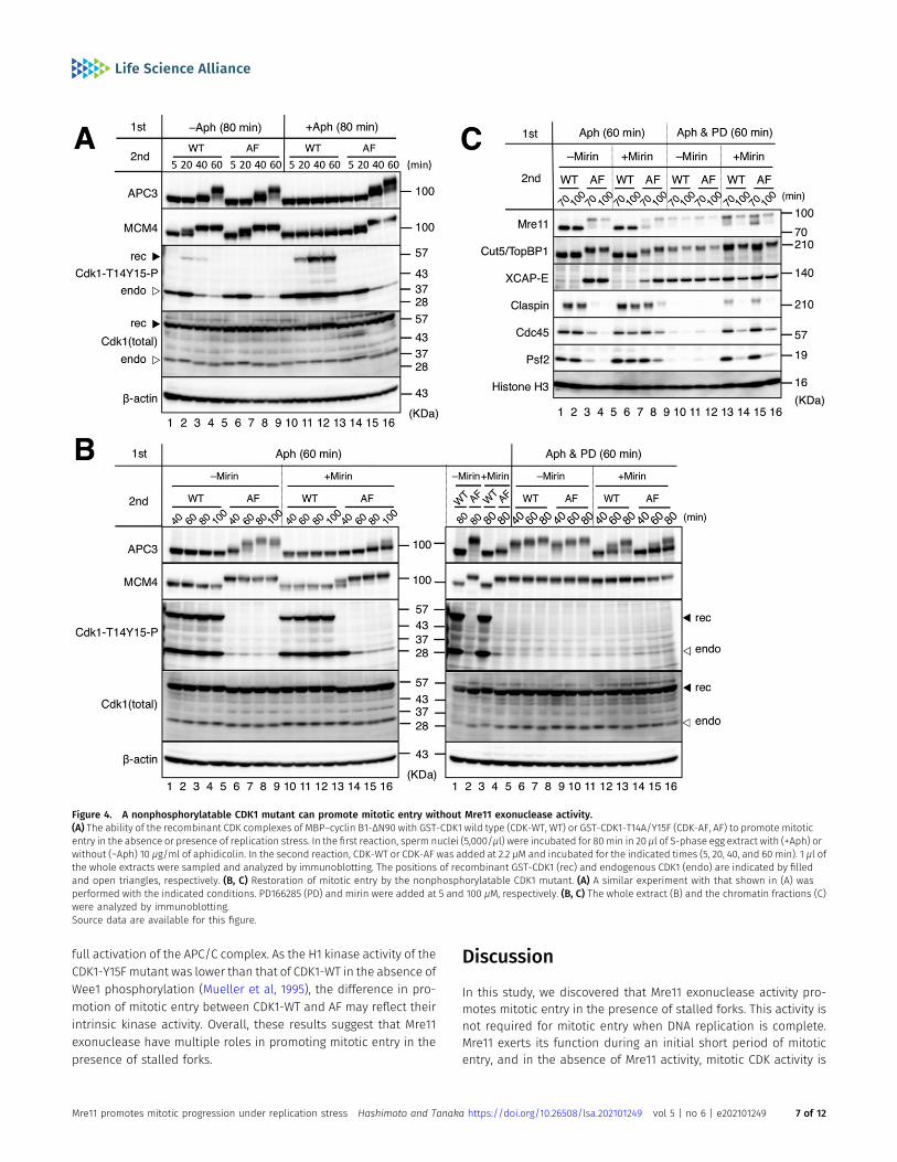
The time course of mitotic entry and progression was monitoredby the immunoblotting of the whole extracts (Fig 4A and B). WhenDNA replication was almost complete without aphidicolin (Fig 4A,−Aph 80 min), both CDK1-WT and CDK1-AF were able to pro-mote mitotic entry as indicated by the appearance of hyper-phosphorylated forms of APC3 and MCM4 at 40 or 60 min and thedisappearance of Thr14 and Tyr15 phosphorylation of endogenousCDK1 at 40 and 60 min (Fig 4A, lanes 1–8). GST-CDK1-WT was slightlyand transiently phosphorylated at 20 min but was dephos-phorylated together with endogenous CDK1 at later time points.By contrast, in the presence of aphidicolin, CDK1-WT could notpromote mitotic entry, and both recombinant and endogenousCDK1 were more strongly phosphorylated at later time points,whereas CDK1-AF was able to promote mitotic entry in a similar
fashion to the case without aphidicolin (Fig 4A, lanes 9–16).These results show that inhibitory phosphorylation of CDK1Thr14 and Tyr15 is predominant in the presence of stalledreplication forks and that the nonphosphorylatable mutant CDK1can overcome this inhibitory effect.
We then tried to induce mitotic entry using CDK1-WT/AF in thepresence of mirin and aphidicolin (Fig 4B). CDK1-WT was unable toinduce mitotic entry in the presence or absence of mirin (Fig 4B left,lanes 1–4, lanes 9–12). CDK1-AF seemed to partly promote mitoticentry even in the presence of mirin as indicated by the observationthat APC3 became gradually phosphorylated and nearer to thehyper-phosphorylated state during the experiment, whereas mostMCM4 belatedly turned into the hyper-phosphorylated form at 60min (Fig 4B left, lanes 5–8, lanes 13–16). The phosphorylation ofendogenous CDK1 remained at 40min but disappeared at later timepoints, providing additional evidence of mitotic entry. Immuno-blotting of the chromatin fractions also showed that CDK1-AF, butnot CDK1-WT, was able to promote mitotic entry and MRD even inthe presence of mirin, although both processes were less efficientthan in the absence of mirin (Fig 4C, lanes 1–8). These resultssuggest that the inhibitory phosphorylation of CDK1 Thr14 and Tyr15is largely responsible for the inability of CDK1-WT to promote mi-totic entry without Mre11 exonuclease activity and that mirin hasadditional inhibitory effects on mitotic entry and progression otherthan the phosphorylation of CDK1 Thr14 and Tyr15.
Finally, we examined the effect of PD166285 treatment onrecombinant CDK1–cyclin B1–driven mitotic entry in the presence ofaphidicolin with or without mirin. Because mitotic induction by therecombinant proteins took longer thanM-extract, we added PD166285at the beginning of the first incubation. Immunoblotting of wholeextracts (Fig 4B) and chromatin fractions (Fig 4C) showed thatthe PD166285 treatment accelerated the timing of APC3 hyper-phosphorylation by CDK1-AF and enabled CDK1-WT to induce mi-totic entry andMRDat a comparable level with CDK1-AF in the absenceof mirin (Fig 4B right, lanes 5–10 versus left, lanes 5–8; Fig 4C, lanes9–12 versus lanes 1–4). Thus, inhibition of Wee1/Myt1 was sufficient topromote mitotic entry and MRD when Mre11 exonuclease was func-tional. Importantly, PD166285 enabled CDK1-WT to promote mitoticentry and MRD even in the presence of mirin (Fig 4B right, lanes 11–13versus left, lanes 9–12; Fig 4C, lanes 13–14 versus lanes 5–6). The timingof APC3 hyper-phosphorylation by CDK1-AF was also accelerated byPD166285 (Fig 4B right, lanes 14–16 versus left, lanes 13–16). Thissuggests that Wee1/Myt1–dependent inhibitory phosphorylation ofCDK1 Thr14 and Tyr15 plays a significant role in suppressing mitoticentry and MRD when Mre11 exonuclease is not functional.
However, it should be noted that both mitotic entry and MRDwere less efficient in the presence of mirin than in the absence ofmirin (Fig 4B right, lanes 11–16 versus lanes 5–10; Fig 4C, lanes 13–16versus lanes 9–12). Specifically, APC3 was not fully phosphorylatedin the presence of mirin even with CDK1-AF or PD166285. In addition,CDK1-AF was less active than CDK1-WT in the presence of bothPD166285 and mirin (Fig 4B right lanes 11–13 versus lanes 14–16; Fig4C, lanes 13–14 versus lanes 15–16). It is possible that unprocessedstalled forks without Mre11 exonuclease continue activating ATR-Chk1 or other pathways to suppress mitotic entry independently ofthe regulation of CDK1 phosphorylation. Alternatively, Mre11 exo-nuclease may be involved in another process that is required for
Mre11 promotes mitotic progression under replication stress Hashimoto and Tanaka https://doi.org/10.26508/lsa.202101249 vol 5 | no 6 | e202101249 6 of 12
full activation of the APC/C complex. As the H1 kinase activity of theCDK1-Y15F mutant was lower than that of CDK1-WT in the absence ofWee1 phosphorylation (Mueller et al, 1995), the difference in pro-motion of mitotic entry between CDK1-WT and AF may reflect theirintrinsic kinase activity. Overall, these results suggest that Mre11exonuclease have multiple roles in promoting mitotic entry in thepresence of stalled forks.
Discussion
In this study, we discovered that Mre11 exonuclease activity pro-motes mitotic entry in the presence of stalled forks. This activity isnot required for mitotic entry when DNA replication is complete.Mre11 exerts its function during an initial short period of mitoticentry, and in the absence of Mre11 activity, mitotic CDK activity is
Figure 4. A nonphosphorylatable CDK1 mutant can promote mitotic entry without Mre11 exonuclease activity.(A) The ability of the recombinant CDK complexes of MBP–cyclin B1-ΔN90 with GST-CDK1 wild type (CDK-WT, WT) or GST-CDK1-T14A/Y15F (CDK-AF, AF) to promote mitoticentry in the absence or presence of replication stress. In the first reaction, sperm nuclei (5,000/μl) were incubated for 80 min in 20 μl of S-phase egg extract with (+Aph) orwithout (−Aph) 10 μg/ml of aphidicolin. In the second reaction, CDK-WT or CDK-AF was added at 2.2 μM and incubated for the indicated times (5, 20, 40, and 60 min). 1 μl ofthe whole extracts were sampled and analyzed by immunoblotting. The positions of recombinant GST-CDK1 (rec) and endogenous CDK1 (endo) are indicated by filledand open triangles, respectively. (B, C) Restoration of mitotic entry by the nonphosphorylatable CDK1 mutant. (A) A similar experiment with that shown in (A) wasperformed with the indicated conditions. PD166285 (PD) and mirin were added at 5 and 100 μM, respectively. (B, C) The whole extract (B) and the chromatin fractions (C)were analyzed by immunoblotting.Source data are available for this figure.
Mre11 promotes mitotic progression under replication stress Hashimoto and Tanaka https://doi.org/10.26508/lsa.202101249 vol 5 | no 6 | e202101249 7 of 12
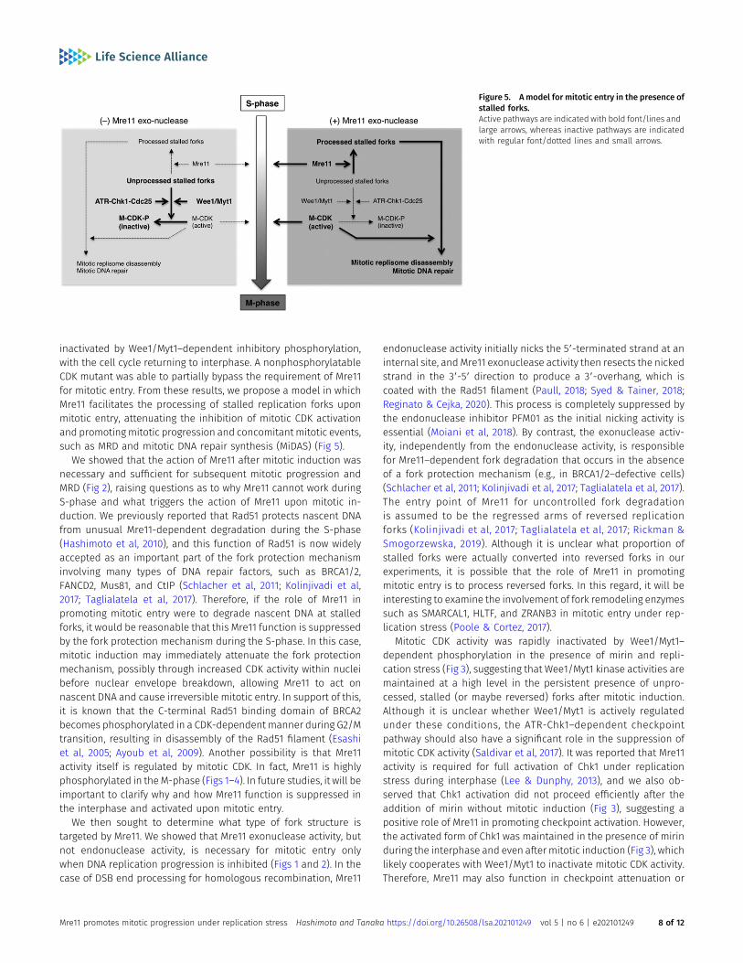
inactivated by Wee1/Myt1–dependent inhibitory phosphorylation,with the cell cycle returning to interphase. A nonphosphorylatableCDK mutant was able to partially bypass the requirement of Mre11for mitotic entry. From these results, we propose a model in whichMre11 facilitates the processing of stalled replication forks uponmitotic entry, attenuating the inhibition of mitotic CDK activationand promotingmitotic progression and concomitant mitotic events,such as MRD and mitotic DNA repair synthesis (MiDAS) (Fig 5).
We showed that the action of Mre11 after mitotic induction wasnecessary and sufficient for subsequent mitotic progression andMRD (Fig 2), raising questions as to why Mre11 cannot work duringS-phase and what triggers the action of Mre11 upon mitotic in-duction. We previously reported that Rad51 protects nascent DNAfrom unusual Mre11-dependent degradation during the S-phase(Hashimoto et al, 2010), and this function of Rad51 is now widelyaccepted as an important part of the fork protection mechanisminvolving many types of DNA repair factors, such as BRCA1/2,FANCD2, Mus81, and CtIP (Schlacher et al, 2011; Kolinjivadi et al,2017; Taglialatela et al, 2017). Therefore, if the role of Mre11 inpromoting mitotic entry were to degrade nascent DNA at stalledforks, it would be reasonable that this Mre11 function is suppressedby the fork protection mechanism during the S-phase. In this case,mitotic induction may immediately attenuate the fork protectionmechanism, possibly through increased CDK activity within nucleibefore nuclear envelope breakdown, allowing Mre11 to act onnascent DNA and cause irreversible mitotic entry. In support of this,it is known that the C-terminal Rad51 binding domain of BRCA2becomes phosphorylated in a CDK-dependent manner during G2/Mtransition, resulting in disassembly of the Rad51 filament (Esashiet al, 2005; Ayoub et al, 2009). Another possibility is that Mre11activity itself is regulated by mitotic CDK. In fact, Mre11 is highlyphosphorylated in theM-phase (Figs 1–4). In future studies, it will beimportant to clarify why and how Mre11 function is suppressed inthe interphase and activated upon mitotic entry.
We then sought to determine what type of fork structure istargeted by Mre11. We showed that Mre11 exonuclease activity, butnot endonuclease activity, is necessary for mitotic entry onlywhen DNA replication progression is inhibited (Figs 1 and 2). In thecase of DSB end processing for homologous recombination, Mre11
endonuclease activity initially nicks the 59-terminated strand at aninternal site, andMre11 exonuclease activity then resects the nickedstrand in the 39-59 direction to produce a 39-overhang, which iscoated with the Rad51 filament (Paull, 2018; Syed & Tainer, 2018;Reginato & Cejka, 2020). This process is completely suppressed bythe endonuclease inhibitor PFM01 as the initial nicking activity isessential (Moiani et al, 2018). By contrast, the exonuclease activ-ity, independently from the endonuclease activity, is responsiblefor Mre11–dependent fork degradation that occurs in the absenceof a fork protection mechanism (e.g., in BRCA1/2–defective cells)(Schlacher et al, 2011; Kolinjivadi et al, 2017; Taglialatela et al, 2017).The entry point of Mre11 for uncontrolled fork degradationis assumed to be the regressed arms of reversed replicationforks (Kolinjivadi et al, 2017; Taglialatela et al, 2017; Rickman &Smogorzewska, 2019). Although it is unclear what proportion ofstalled forks were actually converted into reversed forks in ourexperiments, it is possible that the role of Mre11 in promotingmitotic entry is to process reversed forks. In this regard, it will beinteresting to examine the involvement of fork remodeling enzymessuch as SMARCAL1, HLTF, and ZRANB3 in mitotic entry under rep-lication stress (Poole & Cortez, 2017).
Mitotic CDK activity was rapidly inactivated by Wee1/Myt1–dependent phosphorylation in the presence of mirin and repli-cation stress (Fig 3), suggesting that Wee1/Myt1 kinase activities aremaintained at a high level in the persistent presence of unpro-cessed, stalled (or maybe reversed) forks after mitotic induction.Although it is unclear whether Wee1/Myt1 is actively regulatedunder these conditions, the ATR-Chk1–dependent checkpointpathway should also have a significant role in the suppression ofmitotic CDK activity (Saldivar et al, 2017). It was reported that Mre11activity is required for full activation of Chk1 under replicationstress during interphase (Lee & Dunphy, 2013), and we also ob-served that Chk1 activation did not proceed efficiently after theaddition of mirin without mitotic induction (Fig 3), suggesting apositive role of Mre11 in promoting checkpoint activation. However,the activated form of Chk1 was maintained in the presence of mirinduring the interphase and even after mitotic induction (Fig 3), whichlikely cooperates with Wee1/Myt1 to inactivate mitotic CDK activity.Therefore, Mre11 may also function in checkpoint attenuation or
Figure 5. A model for mitotic entry in the presence ofstalled forks.Active pathways are indicated with bold font/lines andlarge arrows, whereas inactive pathways are indicatedwith regular font/dotted lines and small arrows.
Mre11 promotes mitotic progression under replication stress Hashimoto and Tanaka https://doi.org/10.26508/lsa.202101249 vol 5 | no 6 | e202101249 8 of 12
recovery by degrading the fork structure necessary for ATR acti-vation during an initial period of mitotic entry. In light of thefindings that inhibition of Wee1/Myt1 and recombinant CDK1-AF canbypass the requirement of Mre11 for both mitotic entry and MRD(Figs 3 and 4), it is likely that the primary role of Mre11 exonucleaseactivity is to regulate mitotic CDK activity and that this exonucleaseactivity itself is not required for MRD, although there is still thepossibility that the exonuclease activity of Mre11 is necessary forsubsequent MiDAS. We previously detected MiDAS-like activity in theexperiment in which replication forks stalled by ara-CTP were re-leased by excess dCTP aftermitotic entry (Hashimoto& Tanaka, 2021).Although we performed this experiment repeatedly, the addition ofexcess dCTP often causedmitotic exit in the presence ofmirin but notin the absence of mirin (data not shown). This mitotic exit was notsuppressed by PD166285 or CDK1-AF (data not shown). Therefore, it iscurrently unclear whether Mre11 exonuclease promotes MiDAS.
We observed that both CDK1-WT/PD166285 and CDK1-AF were notable to fully phosphorylate APC3 in the presence of mirin (Fig 4).This raises several possibilities. First, unprocessed stalled forks (orreversed forks) may affect other regulatory processes for CDK1beyond inhibitory phosphorylation. Second, Mre11 has specificmitotic roles other than DNA processing, which may affect theobserved inefficient phosphorylation of APC3 in the presence ofmirin. It was reported that the MRN complex regulates spindleassembly (Rozier et al, 2013) and spindle dynamics in mitosis (Xuet al, 2018). Specifically, the former function is dependent on Mre11exonuclease activity, which is required for RCC1 chromatin asso-ciation and the subsequent establishment of a RanGTP gradientaround chromatin (Rozier et al, 2013). Therefore, the spindle as-sembly checkpoint pathway may be activated to suppress the fullactivation of APC3 in the presence of mirin.
Finally, we utilized a unique system based on Xenopus egg ex-tracts to reveal a novel role of Mre11 for mitotic entry under rep-lication stress. In the future, it will be important to examine whetherthis function of Mre11 is conserved among other species, especiallyin mammals. It is possible that Mre11-dependent fork processingmay frequently occur upon mitotic entry during unperturbed cellcycles at difficult-to-replicate regions such as common fragile sitesand centromeres, and its failure may promote genomic instability.
Material and Methods
Xenopus laevis egg extracts, chromatin fractions, nuclearfractions, and chemicals
S-phase (interphase) and M-phase (CSF-arrested) egg extracts andde-membranated sperm nuclei were prepared as previously de-scribed (Murray & Kirschner, 1989; Kubota & Takisawa, 1993). Two orthree different preparations were mixed to normalize the activitiesof egg extracts. In all of the experiments using egg extract, thereaction temperature was 23°C, and the concentration of spermnuclei was 5,000 nuclei/μl in the first reaction with the S-phaseextract. This was diluted to 2,500 nuclei/μl in the second reactionwhen mitotic entry was induced with an equal volume of theM-phase extract. In the experiment of Fig 2C as an exception, thefirst reaction was diluted with 1.5 volumes of S-phase extract and
2.5 volumes of M-phase extract to produce 1,000 nuclei/μl in thesecond reaction.
To isolate chromatin fractions, 20 μl (S-phase) or 40 μl (S-phaseand M-phase) of egg extract (containing a total of 100,000 nuclei)was diluted with 700 μl of EB buffer (100 mM KCl, 2.5 mM MgCl2, and50 mM Hepes–KOH, pH 7.5) containing 0.2% NP40 and layered onto200 μl of a 30% (wt/vol) sucrose cushionmade with the same buffer.The chromatin was centrifuged at 13,200g for 5 min at 4°C, washedwith 300 μl of EB buffer, and centrifuged again at 20,000g for 1 min.In the experiment of Fig 2C as an exception, 100 μl of egg extract wasinitially diluted with 1 ml of EB + 0.2% NP40, and the chromatin waswashed with 500 μl of EB. The pellet was resuspended in SDS–PAGEsample buffer and analyzed by immunoblotting. To examine thephosphorylation of chromatin–bound proteins (Fig 1C), thechromatin pellet was resuspended in 40 μl of alkaline phos-phatase buffer (1 mM MgCl2, 1 mM PMSF, and 50 mM Tris–HCl, pH9.0) with or without six units of calf intestine alkaline phos-phatase (TOYOBO), incubated for 30 min at 30°C, washed with500 μl of EB buffer, centrifuged again at 20,000g for 1 min, andanalyzed by immunoblotting.
To isolate nuclear fractions, egg extract with 100,000 nuclei wasdiluted with 500 μl of EB buffer and layered onto 300 μl of EB plus30% (wt/vol) sucrose buffer. The nuclei were centrifuged at 6,620gfor 2 min at 4°C, washed with 1 ml of EB plus 30% (wt/vol) sucrosebuffer, and centrifuged again at 6,620g for 2 min. The pellet wasresuspended in SDS–PAGE sample buffer and analyzed byimmunoblotting.
The following chemicals were used at the indicated concen-trations: aphidicolin (10 μg/ml; Sigma-Aldrich), mirin (20, 50 or100 μM; Sigma-Aldrich), PFM01 (50 or 100 μM; Sigma-Aldrich), NMS-873 (100 μM; Sigma-Aldrich), MLN-4924 (10 μM; Sigma-Aldrich), andPD166285 (5 or 10 μM; Sigma-Aldrich).
cDNA cloning and primers
cDNA encoding wild-type full-length X. laevis CDK1B (CDK1-WT) wasfirst amplified by PCR with the primers 59-BamHI-xCDK1B andxCDK1B-HindIII-39 using X. laevis oocyte cDNA as a template. cDNAencoding the CDK1-AF mutant (in which Thr14 and Tyr15 of CDK1Bwere replaced with Ala and Phe, respectively) was then amplified byPCR with primers 59-BamHI-xCDK1B-AF and xCDK1B-HindIII-39 usingthe PCR product of CDK1-WT as a template. cDNA encoding Sac-charomyces cerevisiae Civ1, a CDK-activating kinase, was alsoamplified by PCRwith primers 59-HindIII-EcoRI-Civ1 and Civ1-EagI-39using a plasmid containing the sequence of Civ1 (provided by Dr TSTakahashi, Kyushu University) as a template. The PCR products fromthe reactions for CDK1-WT and CDK1-AF were digested with BamHIand HindIII, whereas the PCR product from the reaction for Civ1 wasdigested with HindIII and EagI. The CDK1-WT and CDK1-AF fragmentswere ligated with the Civ1 fragment in tandem and cloned into pGEX6P-1 (Cytiva) between the BamHI and EagI (NotI) sites (CDK1-WT-Civ1/pGEX 6P-1 and CDK1-AF-Civ1/pGEX 6P-1) for co-expression ofCiv1 with GST-tagged CDK1-WT and CDK1-AF in Escherichia coli.
cDNA-encoding X. laevis cyclin B1 lacking the N-terminal 90amino acids was amplified by PCR with the primers 59-BamHI-xcyclin B1-E91 and xcyclin B1-EcoRI-39 using X. laevis oocytecDNA as a template and cloned into pGEX 6P-1 between the BamHI
Mre11 promotes mitotic progression under replication stress Hashimoto and Tanaka https://doi.org/10.26508/lsa.202101249 vol 5 | no 6 | e202101249 9 of 12
and EcoRI sites (cyclin B1-ΔN90/pGEX 6P-1). The fragment con-taining the cyclin B1-ΔN90 sequence was recovered after BamHIand SalI digestion and cloned into pMAL-C4X (New England Biolabs)between the BamHI and SalI sites (cyclin B1-ΔN90/pMAL-C4X) forexpression of MBP-tagged cyclin B1-ΔN90 in E. coli.
cDNA encoding full-length X. laevis Mre11 was amplified by PCRwith primers 59-BamHI-xMre11 and xMre11-EcoRI-39 using X. laevisoocyte cDNA as a template and cloned into pHEX-1 (amodified pGEX6P-1 vector in which the GST–encoding sequence was replaced withthe His8-encoding sequence: provided by Dr S Mochida, KumamotoUniversity) between the BamHI and EcoRI sites for expression ofHis–tagged Mre11 in E. coli.
The sequences of the PCR primers are as follows (single anddouble underlines show the restriction sites and mutation site ofCDK1-AF, respectively):
59-BamHI-xCDK1B (59-TATGGATCCGATGGACGAGTACACTAAAATAGAG-39)59-BamHI-xCDK1B-AF (59-TATGGATCCGATGGACGAGTACACTAAAATA-GAGAAGATTGGAGAGGGCGCATTTGGGGTCGTG-3’)xCDK1-HindIII-39 (59-TATAAGCTTGTTTTAATTTCTAATCTGATTGGCG-39)59-HindIII-EcoRI-Civ1 (59-TATAAGCTTGAATTCATGAAACTGGATAGTATAG-39)Civ1-EagI-39 (59-TATCGGCCGTTATGGCTTTTCTAATTCTTGCAAG-39)59-BamHI-xcyclin B1-E91 (TATGGATCCGAACCCAGCTCACCAAGCCCAATG)xcyclin B1-EcoRI-39 (59-TATGAATTCTCATACAAGTGGGCGGGCCATTGC-39)59-BamHI-xMre11 (59-TATGGATCCATGAGTTCTTCTAGTAGCTCTCTG-39)xMre11-EcoRI-39 (59-TATGAATTCTTATCTACGGCCCCTTCTGGAAGG-39)
Protein purification
GST-CDK1-WT or GST-CDK1-AF was co-expressed with untagged Civ1in Rossetta(DE3)pLysS (Novagen) to induce CDK1 phosphorylationat Thr161, which is required for CDK1 activity. The GST-tagged proteinswere then purified with Glutathione Sepharose 4B (Cytiva) accordingto the manufacturer’s protocol. MBP–cyclin B1-ΔN90 was expressedin Rossetta(DE3)pLysS and purified with amylose resin (New EnglandBiolabs) according to the manufacturer’s protocol. The purifiedproteins were concentrated and buffer exchanged to PBS(–) using anAmicon Ultra-4 30K (Merck) ultrafiltration device. The protein con-centrations of all samples were then adjusted to 22 μM. GST-CDK1-WTor GST-CDK1-AF was mixed with an equal volume of MBP–cyclin B1-ΔN90 and incubated for 15 min at 23°C to promote the formation ofthe CDK1–cyclin B1 complex. These mixtures were added to the eggextract at 2.2 μM to drive mitotic entry.
His-Mre11 was expressed in BL21-CodonPlus(DE3)-RIPL (AgilentTechnologies) and purified with Ni-NTA agarose (QIAGEN) accordingto the manufacturer’s protocol under denaturing conditions using8 M urea. The purified protein was then dialyzed with PBS(–), andthe resultant precipitate was used as an antigen to immunize rabbits.Anti-Mre11 polyclonal antibodies were affinity purified with theantigen immobilized on Affi-Gel15 (Bio-Rad). His-p27 was preparedas previously described (Hashimoto et al, 2010) and used at 100 μg/ml to inhibit CDK activity.
Immunoblotting and antibodies
SDS–PAGE and immunoblotting were performed according tostandard procedures. The chemiluminescent signals were detected
with ChemiDoc XRS+ (Bio-Rad) and quantifiedwith Image J software.The primary antibodies or antiserum were used at the indicateddilutions as follows: rabbit polyclonal anti-Mre11 antibody (174 μg/ml, 1:200) was prepared in this study; rabbit polyclonal antibodies toPsf2 (183 μg/ml, 1:200) and XCAP-E (936 μg/ml, 1:500) were previ-ously described (Hashimoto & Tanaka, 2018); rabbit antisera toCut5/TopBP1 (1:3,000), Cdc45 (1:2,000), claspin (1:1,000), Polε (p60;1:1,000), Sld5 (1:300), and Chk1 (1:400) were provided by Y. Kubota(Osaka University) and H. Takisawa (Osaka University); rabbit an-tiserum to Polδ (p66) (1:1,000) was provided by S. Waga (JapanWomen’s University); rabbit antiserum to Cdk1 (1:500), mousemonoclonal anti–cyclin B2 antibody (X121, 0.5 mg/ml, 1:200), andmouse monoclonal anti–APC3/CDC27 (610455, 1:200; BD Biosci-ences) were provided by S. Mochida (Kumamoto University). Thefollowing antibodies were obtained from the indicated companies:rabbit polyclonal antibodies to SMC1 (4802, 1:1,000; Cell Signaling);phospho-CDK1 (Tyr15; 9111, 1:500; Cell Signaling); phospho-CDK1(Thr14, Tyr15; 44-686G, 1:500; Thermo Fisher Scientific); phospho-Chk1 (Ser345; 2341, 1:300; Cell Signaling); MCM4 (ab4459, 1:2,000;Abcam); rabbit monoclonal anti-histone H3 antibody (4499, 1:800;Cell Signaling); mouse monoclonal antibodies to MCM7 (sc-9966,1:2,500; Santa Cruz); and β-actin (ab8224, 1:5,000; Abcam).
Supplementary Information
Supplementary Information is available at https://doi.org/10.26508/lsa.202101249.
Acknowledgements
We thank H Takisawa, Y Kubota, S Waga, S Mochida, and TS Takahashi forproviding antibodies/antisera (to Cut5/TopBP1, claspin, Cdc45, Chk1, Polε[p60], Polδ [p66], APC3, and cyclin B2), pHEX-1 vector, and Civ1 cDNA. Thiswork was supported by JSPS KAKENHI grants to Y Hashimoto (19K06617 and15K06855) from the Ministry of Education, Cultures, Sports, Science andTechnology (MEXT) in Japan.
Author Contributions
Y Hashimoto: conceptualization, data curation, funding acquisition,investigation,methodology, project administration, andwriting—originaldraft, review, and editing.H Tanaka: supervision.
Conflict of Interest Statement
The authors declare that they have no conflict of interest.
References
Ayoub N, Rajendra E, Su X, Jeyasekharan AD, Mahen R, Venkitaraman AR(2009) The carboxyl terminus of Brca2 links the disassembly of Rad51complexes to mitotic entry. Curr Biol 19: 1075–1085. doi:10.1016/j.cub.2009.05.057
Mre11 promotes mitotic progression under replication stress Hashimoto and Tanaka https://doi.org/10.26508/lsa.202101249 vol 5 | no 6 | e202101249 10 of 12
Bellelli R, Boulton SJ (2021) Spotlight on the replisome: Aetiology of DNAreplication-associated genetic diseases. Trends Genet 37: 317–336.doi:10.1016/j.tig.2020.09.008
Burgers PMJ, Kunkel TA (2017) Eukaryotic DNA replication fork. Annu RevBiochem 86: 417–438. doi:10.1146/annurev-biochem-061516-044709
Crncec A, Hochegger H (2019) Triggering mitosis. FEBS Lett 593: 2868–2888.doi:10.1002/1873-3468.13635
Deng L, Wu RA, Sonneville R, Kochenova OV, Labib K, Pellman D, Walter JC(2019) Mitotic CDK promotes replisome disassembly, fork breakage,and complex DNA rearrangements. Mol Cell 73: 915–929.e6.doi:10.1016/j.molcel.2018.12.021
Dewar JM, Low E, Mann M, Raschle M, Walter JC (2017) CRL2Lrr1 promotesunloading of the vertebrate replisome from chromatin duringreplication termination. Genes Dev 31: 275–290. doi:10.1101/gad.291799.116
Dupre A, Boyer-Chatenet L, Sattler RM, Modi AP, Lee JH, Nicolette ML,Kopelovich L, Jasin M, Baer R, Paull TT, et al (2008) A forward chemicalgenetic screen reveals an inhibitor of the Mre11-Rad50-Nbs1 complex.Nat Chem Biol 4: 119–125. doi:10.1038/nchembio.63
Elbæk CR, Petrosius V, Sørensen CS (2020) WEE1 kinase limits CDK activities tosafeguard DNA replication and mitotic entry. Mutat Res 819-820:111694. doi:10.1016/j.mrfmmm.2020.111694
Esashi F, Christ N, Gannon J, Liu Y, Hunt T, Jasin M, West SC (2005) CDK-dependent phosphorylation of BRCA2 as a regulatory mechanism forrecombinational repair. Nature 434: 598–604. doi:10.1038/nature0340418.10.064
Fujimitsu K, Grimaldi M, Yamano H (2016) Cyclin-dependent kinase 1-dependent activation of APC/C ubiquitin ligase. Science 352: 1121–1124.doi:10.1126/science.aad3925
Gaillard H, Garcıa-Muse T, Aguilera A (2015) Replication stress and cancer.Nat Rev Cancer 15: 276–289. doi:10.1038/nrc3916
Guilliam TA (2021) Mechanisms for maintaining eukaryotic replisomeprogression in the presence of DNA damage. Front Mol Biosci 8: 712971.doi:10.3389/fmolb.2021.712971
Hashimoto Y, Ray Chaudhuri A, Lopes M, Costanzo V (2010) Rad51 protectsnascent DNA from Mre11-dependent degradation and promotescontinuous DNA synthesis. Nat Struct Mol Biol 17: 1305–1311.doi:10.1038/nsmb.1927
Hashimoto Y, Tanaka H (2018) Mitotic entry drives replisome disassembly atstalled replication forks. Biochem Biophys Res Commun 506: 108–113.doi:10.1016/j.bbrc.2018.10.064
Hashimoto Y, Tanaka H (2021) Ongoing replication forks delay the nuclearenvelope breakdown upon mitotic entry. J Biol Chem 296: 100033.doi:10.1074/jbc.RA120.015142
Kolinjivadi AM, Sannino V, De Antoni A, Zadorozhny K, Kilkenny M, Techer H,Baldi G, Shen R, Ciccia A, Pellegrini L, et al (2017) Smarcal1-Mediatedfork reversal triggers mre11-dependent degradation of nascent DNAin the absence of Brca2 and stable Rad51 nucleofilaments.Mol Cell 67:867–881.e7. doi:10.1016/j.molcel.2017.07.001
Krasinska L, Domingo-Sananes MR, Kapuy O, Parisis N, Harker B, Moorhead G,Rossignol M, Novak B, Fisher D (2011) Protein phosphatase 2A controlsthe order and dynamics of cell-cycle transitions. Mol Cell 44: 437–450.doi:10.1016/j.molcel.2011.10.007
Kubota Y, Takisawa H (1993) Determination of initiation of DNA replicationbefore and after nuclear formation in Xenopus egg cell free extracts. JCell Biol 123: 1321–1331. doi:10.1083/jcb.123.6.1321
Lee J, Dunphy WG (2013) The Mre11-Rad50-Nbs1 (MRN) complex has a specificrole in the activation of Chk1 in response to stalled replication forks.Mol Biol Cell 24: 1343–1353. doi:10.1091/mbc.E13-01-0025
Maric M, Maculins T, De Piccoli G, Labib K (2014) Cdc48 and a ubiquitin ligasedrive disassembly of the CMG helicase at the end of DNA replication.Science 346: 1253596. doi:10.1126/science.1253596
Moiani D, Ronato DA, Brosey CA, Arvai AS, Syed A, Masson JY, Petricci E, TainerJA (2018) Targeting allostery with avatars to design inhibitors assessedby cell activity: Dissecting MRE11 endo- and exonuclease activities.Methods Enzymol 601: 205–241. doi:10.1016/bs.mie.2017.11.030
Moreno SP, Bailey R, Campion N, Herron S, Gambus A (2014)Polyubiquitylation drives replisome disassembly at the terminationof DNA replication. Science 346: 477–481. doi:10.1126/science.1253585
Mueller PR, Coleman TR, Dunphy WG (1995) Cell cycle regulation of a Xenopuswee1-like kinase. Mol Biol Cell 6: 119–134. doi:10.1091/mbc.6.1.119
Murray AW, Kirschner MW (1989) Cyclin synthesis drives the early embryoniccell cycle. Nature 339: 275–280. doi:10.1038/339275a0
Neelsen KJ, Lopes M (2015) Replication fork reversal in eukaryotes: From deadend to dynamic response. Nat Rev Mol Cell Biol 16: 207–220.doi:10.1038/nrm3935
Pasero P, Vindigni A (2017) Nucleases acting at stalled forks: How to rebootthe replication program with a few shortcuts. Annu Rev Genet 51:477–499. doi:10.1146/annurev-genet-120116-024745
Paull TT (2018) 20 Years of Mre11 biology: No end in sight.Mol Cell 71: 419–427.doi:10.1016/j.molcel.2018.06.033
Poole LA, Cortez D (2017) Functions of SMARCAL1, ZRANB3, and HLTF inmaintaining genome stability. Crit Rev Biochem Mol Biol 52: 696–714.doi:10.1080/10409238.2017.1380597
Moreno SP, Jones RM, Poovathumkadavil D, Scaramuzza S, Gambus A (2019)Mitotic replisome disassembly depends on TRAIP ubiquitin ligaseactivity. Life Sci Alliance 2: e201900390. doi:10.26508/lsa.201900390
Reginato G, Cejka P (2020) The MRE11 complex: A versatile toolkit for therepair of broken DNA. DNA Repair (Amst) 91-92: 102869. doi:10.1016/j.dnarep.2020.102869
Rickman K, Smogorzewska A (2019) Advances in understanding DNAprocessing and protection at stalled replication forks. J Cell Biol 218:1096–1107. doi:10.1083/jcb.201809012
Rozier L, Guo Y, Peterson S, Sato M, Baer R, Gautier J, Mao Y (2013) The MRN-CtIP pathway is required for metaphase chromosome alignment. MolCell 49: 1097–1107. doi:10.1016/j.molcel.2013.01.023
Saldivar JC, Cortez D, Cimprich KA (2017) The essential kinase ATR: Ensuringfaithful duplication of a challenging genome. Nat Rev Mol Cell Biol 18:622–636. doi:10.1038/nrm.2017.67
Schlacher K, Christ N, Siaud N, Egashira A, Wu H, Jasin M (2011) Double-strandbreak repair-independent role for BRCA2 in blocking stalledreplication fork degradation by MRE11. Cell 145: 529–542. doi:10.1016/j.cell.2011.03.041
Sonneville R, Bhowmick R, Hoffmann S, Mailand N, Hickson ID, Labib K (2019)TRAIP drives replisome disassembly and mitotic DNA repair synthesisat sites of incomplete DNA replication. Elife 8: e48686. doi:10.7554/eLife.48686
Sonneville R, Moreno SP, Knebel A, Johnson C, Hastie CJ, Gartner A, Gambus A,Labib K (2017) CUL-2LRR-1 and UBXN-3 drive replisome disassemblyduring DNA replication termination and mitosis. Nat Cell Biol 19:468–479. doi:10.1038/ncb3500
Sørensen CS, Syljuasen RG (2012) Safeguarding genome integrity: Thecheckpoint kinases ATR, CHK1 and WEE1 restrain CDK activity duringnormal DNA replication. Nucleic Acids Res 40: 477–486. doi:10.1093/nar/gkr697
Syed A, Tainer JA (2018) The MRE11-RAD50-NBS1 complex conducts theorchestration of damage signaling and outcomes to stress in DNAreplication and repair. Annu Rev Biochem 87: 263–294. doi:10.1146/annurev-biochem-062917-012415
Taglialatela A, Alvarez S, Leuzzi G, Sannino V, Ranjha L, Huang JW, Madubata C,Anand R, Levy B, Rabadan R, et al (2017) Restoration of replication forkstability in BRCA1- and BRCA2-deficient cells by inactivation of SNF2-family fork remodelers. Mol Cell 68: 414–430.e8. doi:10.1016/j.molcel.2017.09.036
Mre11 promotes mitotic progression under replication stress Hashimoto and Tanaka https://doi.org/10.26508/lsa.202101249 vol 5 | no 6 | e202101249 11 of 12

Wu RA, Pellman DS, Walter JC (2021) The ubiquitin ligase TRAIP: Double-edgedsword at the replisome. Trends Cell Biol 31: 75–85. doi:10.1016/j.tcb.2020.11.007
Xu R, Xu Y, Huo W, Lv Z, Yuan J, Ning S, Wang Q, Hou M, Gao G, Ji J, et al (2018)Mitosis-specific MRN complex promotes a mitotic signalingcascade to regulate spindle dynamics and chromosome segregation.Proc Natl Acad Sci U S A 115: E10079–E10088. doi:10.1073/pnas.1806665115
Zeman MK, Cimprich KA (2014) Causes and consequences of replicationstress. Nat Cell Biol 16: 2–9. doi:10.1038/ncb2897
License: This article is available under a CreativeCommons License (Attribution 4.0 International, asdescribed at https://creativecommons.org/licenses/by/4.0/).
Mre11 promotes mitotic progression under replication stress Hashimoto and Tanaka https://doi.org/10.26508/lsa.202101249 vol 5 | no 6 | e202101249 12 of 12
Related Documents