MRI Physics 101 Prof. Sairam Geethanath, Ph.D. Medical Imaging Research Centre Dayananda Sagar Institutions 1 st October 2016 A"enua’on (E/E0) (µV) γ 2 G 2 δ 2 (∆- δ/3) (10 9 sm -2 )

Welcome message from author
This document is posted to help you gain knowledge. Please leave a comment to let me know what you think about it! Share it to your friends and learn new things together.
Transcript

MRI Physics 101
Prof. Sairam Geethanath, Ph.D. Medical Imaging Research Centre
Dayananda Sagar Institutions 1st October 2016
A"en
ua'o
n(E/E0)
(µV)
γ2G2δ2(∆-δ/3)(109sm-2)

Declara'on
• Ihavenoconflictsofinteresttodeclare,withrespecttothecontentsofthispresenta'onanddatashownhereisalreadyinpublicdomain
• WiproGEHealthcarefundspartofmyresearchaspartofaDSTTechnologysystemsdevelopmentgrant
MIRC 2

Outline• Highschoolphysics–relevanttoMRI
• OverviewoftheMRIsystem
• PrecessionandthePeltonWheel
• Relaxa'on'mes:T1andT2
• Basisforcontrastgenera'on
• SpinEchosequence:Anexampleofimagegenera'on• Spa'allocaliza'onthroughmagne'cfieldgradient• k-spaceanditstraversal
• Toolstogetstarted• Pulseq-Acquisi'on• GPI-Reconstruc'on
• DIYassignmentsMIRC 3

ImportanthighschoolPhysics
4
Lenz’slaw Reciprocitytheorem BiotSavart’sLaw Faraday’slaw
Vineet,MIRC Abitha,MIRC Aparana,MIRC Anusha,MIRC

Gradientcoils
Subject
Radiofrequency
coil
Magnet
OverviewofaMRIsystem
Imagecourtesy:MRIscannercutaway:Colinmcnulty.com
BOLD
Imagecrea'on
Birdcagecoil
Aliasing
Baselinecorrec'on
Blochequa'on
CPMGSNR Angiography ADC DWI TE/TR Eddycurrent
ContrastbasisSWI
FID
Gyromagne'cra'o
Fouriertransform
Echoes
Keyholeimaging
DCE-MRI
Larmorfrequency
T1/T2relaxa'on
Representa8onofatypicalMRIscannerMIRC 5

Larmor Equation
EffectoftheB0,G.randB1fields
z
x
y
MIRC 6

Whydoweneedto'pthespins?–Understandingprecession

• The application of an RF pulse, causes alignment of spins away from the longitudinal axis (lower energy state) on a transverse plane (higher energy state)
• Spins release the absorbed energy and drop back to their lower energy states • Spin can exchange a quantum of energy with the lattice (also precessing at same frequency)
• Spin transitions from 𝑚=− 1/2 (excited state) to 𝑚= 1/2 (ground state) is accompanied by a transition upwards in energy from some lower lattice state to a higher lattice state h
• Energy transition must be equal ‘ law of conservation of energy’ • Transfer of energy occurs through collisions, rotations, or electromagnetic interactions with the surrounding lattice • This energy loss is unrecoverable and represents the transfer of heat.
h"p://mriques'ons.comMagne'cresonanceimaging:Physicalprinciplesandsequencedesign
𝑚=− 1/2
𝑚= 1/2
ℎ
𝑙
𝑝𝑟𝑜𝑡𝑜𝑛 𝑙𝑎𝑡𝑡𝑖𝑐𝑒
Boltzmannequa'onforpopula'onstates
T1 relaxation
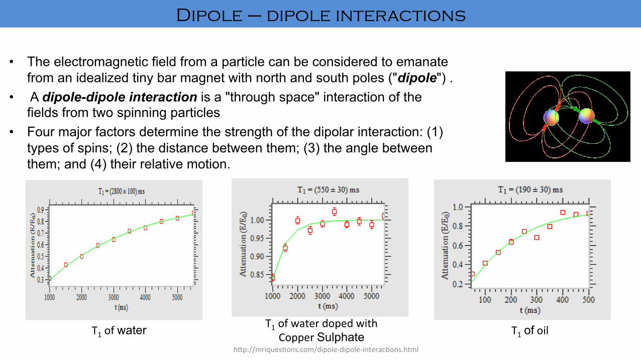
• The electromagnetic field from a particle can be considered to emanate from an idealized tiny bar magnet with north and south poles ("dipole") .
• A dipole-dipole interaction is a "through space" interaction of the fields from two spinning particles
• Four major factors determine the strength of the dipolar interaction: (1) types of spins; (2) the distance between them; (3) the angle between them; and (4) their relative motion.
h"p://mriques'ons.com/dipole-dipole-interac'ons.html
T1ofwater T1ofwaterdopedwithCopperSulphate T1ofoil
Dipole – dipole interactions

h"p://mriques'ons.comMagne'cresonanceimaging:Physicalprinciplesandsequencedesign
0 2 44.0
4.5
5.0
Mz
t
M0
FirstRF SecondRF
TR
T1
0.63M0
• Spinlamceinterac'onresultinre-growthoflongitudinalmagne'za'on
• Rateofchangeoflongitudinalmagne'za'oniscapturedbyanexponen'alrecovery,isacrossproductofmagne'za'onmomentMandtheappliedexternalfieldB
• Synonyms:longitudinalrelaa'on,thermalrelaxa'onandspin-lamcerelaxa'on
T1 recovery

Spin-SpinandEffec-veSpin-Spinrelaxa-on:• NMRsignal–phasecoherenceofnuclearspins&Exponen'alsignaldecay–lossofphasecoherence
• Spin-spinrelaxa'on–dipolecouplingbetweenneighbouringspins
• Sampledsignal,asafunc'onof'meisgivenby, S(t)=S0exp(-t/T2)Where,S0–ini'alsignalmagnitudeatt=0
• Generally,thefieldisnoten'relyhomogeneous
• Phasecoherenceloss–combina'onofspin-spinrelaxa'onandmagne'cfieldinhomogeneity,whichintroducearangeofLarmorfrequencies
• Dispersionoffrequencies–lossofcoherence-signaldecay
Incompletelyhomogeneousfield,thephasecoherencelossisduetospin-spinrelaxa8on
T2 relaxation

• Effec'vespin-spinrelaxa'on'meconstant,T2*isdefinedas,1/T2∗ = 1/T2 +γ∆B0
Where,
γ–gyromagne'cra'ooftheobservednucleus
∆B0–magne'cfieldinhomogeneity• Signalasafunc'onof'meinsitua'onwithfieldinhomogeneityisgivenby,
S(t)=S0exp(-t/T2∗)Where,
S0–ini'alsignalmagnitudeatt=0
Phasecoherencelossduetospin-spinrelaxa8onisirreversible,whereaslossduetofieldinhomogeneitycanbereversedbySpinEcho
(calledasHahnEcho)ErwinHahn
T2* relaxation

Figure1:Spin-Echopulsesequencediagram
• The'mebetweenthe90°pulseandthe180°pulseiscalledTΕ,theecho-8me
• Reversingthede-phasingduetomagne'cfieldinhomogeneityisthegoalofthisbasicspin-echoexperiment
• Onlythede-phasingthatoccurredasaresultofmagne'cfieldinhomogeneitywillbere-focused
spin echo
TerranovastudentGuide,MagritekLimited

• Samplingcommencesatthecentreoftheecho
• Delaybetweenthe180°pulseandthefirstsampleddatapointisTΕ
• TΕmustbechosentobelongenough -toviewtheen'reecho -toallowforthecompleterelaxa'onofthesignalexcited
T2Measurement:• Measuredusingasuccessionofspin-echoexperimentswithincrementallylongerecho'mes
• Theplotofechoamplitudeasafunc'onofecho'mewillbeanexponen'aldecaywithacharacteris'cdecay'meconstant,T2
• Theechoamplitudeisgivenby,
whereE-amplitudeofanechoacquiredwithTΕ E0-echoamplitudeintheabsenceofaT2decay

Relaxa'onprocesses–Blochequa'ons&contrast
MIRC 15
h"ps://www.chemie.uni-hamburg.de/nmr/insensi've/tutorial/en.lproj/vector_model.htmlh"p://www.dayanandasagar.edu/index.php/sharing
Bloch,1946
T1 T2 DIFFUSION

Spa'allocaliza'on–Lauterburgradientexperiment
MIRC 16Lauterbur,Nature,1973

Theory
ThesignalacquiredistheFreeInduc'onDecay(FID)fromallthespinsofthepar'cularslice
TheLarmorfrequencyisgivenby

f
t
A
A
DiscreteFourierTransform

Avisualrepresenta'onofk-space

Topviewofk-space
• Idealk-spaceisHermi'aninnature,discoun'ngerrorsfrommeasurement
• Usedinacquisi'onslikeHASTE

JargonforengineersJ

Frontview Topview
Ananalogyfork-spacetrajectory
Considerahill

20 40 60 80 100 120
50
100
150
200
250
Fewk-spacetrajectoriestrajectories
y
x

Exampleproblem1:DesignCartesiank-spacetrajectory
Givenparameters: ∆x = 1 mm ∆y = 1 mm Lx = 25.6 cm L y = 25.6 cm
Evaluatetheunknowns:
Variable Value
Nx
Ny
256
256 3.9 m-1
3.9 m-1
[-500, 500] m-1
[-500, 500] m-1

kx
ky
RF Pulse
Gz
Gy
Gx
TimingDiagramDepic'ngCartesianSampling
Gx
Gy
Gz
RF Pulse

Reconstruc'on&ar'facts

OpensourcetoolforPSD-Pulseq
MIRC 27
0 0.01 0.02 0.03 0.04 0.05 0.06 0.07 0.08 0.090
500
1000
1500
RF
mag
(Hz)
0 0.01 0.02 0.03 0.04 0.05 0.06 0.07 0.08 0.09t (s)
0
2
4
RF
ph (r
ad)
0 0.01 0.02 0.03 0.04 0.05 0.06 0.07 0.08 0.09t (s)
-500
0
500
Gz
(kH
z/m
)
0 0.01 0.02 0.03 0.04 0.05 0.06 0.07 0.08 0.09-600
-400
-200
0
Gy
(kH
z/m
)
0 0.01 0.02 0.03 0.04 0.05 0.06 0.07 0.08 0.09-1000
0
1000
Gx
(kH
z/m
)
0 0.01 0.02 0.03 0.04 0.05 0.06 0.07 0.08 0.09-1
0
1
ADC
Source:h"p://pulseq.github.io/(NeedsMatlabinstalled)Laytonet.al.,MRM2016

Opensourcetoolforstudentstouse–recon-GPI
MIRC 28
• h"p://gpilab.com/2016/09/gpi-on-windows/
• Simula'on
• Reconstruc'on
• ImageProcessing
• Visualiza'on
• MacOSX/Ubuntu/Windows
Zwartet.al.,MRM,2016

DIYassignments
MIRC 29
Courseassignments(andsolu'ons):1. Introduc'onandpreliminaries2. Physics3. WORKINPROGRESS(IntroductoryPSDandrecon)4. FastImaging5. MRapplica'ons

? MIRC 30
[email protected] http://dayanandasagar.edu/index.php/mirc http://dayanandasagar.edu/index.php/sharing

Prof. Sairam Geethanath, Ph.D. Medical Imaging Research Centre Dayananda Sagar Institutions 1st October 2016

¡ Head¡ Tumors,aneurysms,bleedinginthebrain,¡ Nerveinjury,damagecausedbystroke.
¡ Spine¡ Discs and nerves of the spine for conditions such
asspinalstenosis,discbulges,andspinaltumors.
¡ Chest:Heart,thevalves,andcoronarybloodvessels
¡ Bloodvesselsand?low–Dr.RameshVenkatesan’stalk
¡ Abdomenandpelvis¡ Belly,liver,gallbladder,pancreas,kidneys,andbladder
¡ Bonesandjoints¡ Arthritis,problemswithjoints,bonemarrowproblems,¡ Bonetumors,cartilageproblems,¡ Tornligamentsortendons,infection.


¡ DWIisacommonlyusedMRIsequenceforevaluationofacuteischaemicstroke
¡ Sensitiveindetectionofsmallandearlyinfarcts
¡ Noninvasivewayofunderstandingbrainstructuralconnectivityandmacroscopicaxonalorganization
¡ DWimageisgeneratedbasedonthedirectionalrateofdiffusionofwatermoleculesinsidethebrainduetoBrownianmotion
¡ Imageintensitiesinverselyrelatedtotherelativemobilityofwatermoleculesintissueandthedirectionofthemotion

Result Interpretation
Quantitative analysis
Segmentation
Feature Extraction
Visualization (may include generating FA,ADC, Tractograpghy)
Processing (may include DWI enhancement using Super resolution techniques )
Preprocessing (may include registration,skull stripping,normalization motion , denoisng of low field MR
image/DWI)

¡ Dark regions – water diffusing slower, more obstacles to movement OR increased viscosity
¡ Bright regions – water diffusing faster DWI
¡ Bright regions – decreased water diffusion
¡ Dark regions – increased water diffusion
Figure Source: www.radiopaedia.org
ADC
Matlab Code available with Arush, MIRC

COLOUR FA MAP
TRACTOGRAPHY
• According to the principal direction of diffusion, colour coding of the diffusion data is done
• Red - transverse axis (x-axis) • Blue – superior-inferior (z –axis) • Green – anterior-posterior axis (y-
axis)
• I n t en s i t y o f t he c o l ou r i s proportional to the fractional anisotropy
• It is 3D modeling technique used to visually represent neural tracts using data collected by diffusion tensor imaging (DTI)
• Voxels are connected based upon
similarit ies in the maximum diffusion direction.
Figure Source: www.radiopaedia.org
Matlab Code available with Arush, MIRC

ADC map computa'on
b=0 b=100 b=200 b=500 b=1000
Signalintensitydecreasingwithincreaseinb-value ADCmapscanner ADCmapmatlab

FAmap MeanDiffusivitymap
Scanner
MATLAB
ColorcodedFAmap
Frac-onalAnisotropymap

FA ColFA
Nofilter
PMfilterusingfixedKappa
PMfilterwithAutomatedKappa
Scannerresults
DiffusionWeightedimagesdenoisingforFAmapcomputa-on

Figure source: Nucifora et al. Radiology 245:2 (2007)
Corticospinal Tracts -Probabilistic Corticospinal Tracts - Streamline
1.Streamline tractography • Connects neighbouring voxels
from user defined voxels (seed regions)
• Tracts are traced until termination criteria are met.
2.Probabilistic tractography • Value of each voxel in the map is the
probability the voxel which is in the diffusion path between the ROIs.
• It provides quantitative probability of connection at each voxel
• Allows tracking into regions where there is low anisotropy.

Degree of anisotropy Streamline tractography Probabilistic tractography
Figure source :Nucifora et al. Radiology 245:2 (2007)
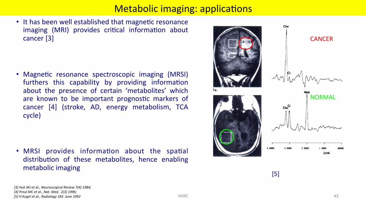
• Ithasbeenwellestablishedthatmagne'cresonanceimaging (MRI) provides cri'cal informa'on aboutcancer[3]
• Magne'c resonance spectroscopic imaging (MRSI)furthers this capability by providing informa'onabout the presence of certain ‘metabolites’ whichare known to be important prognos'c markers ofcancer [4] (stroke, AD, energy metabolism, TCAcycle)
• MRSI provides informa'on about the spa'aldistribu'on of these metabolites, hence enablingmetabolicimaging
[3]HukWJetal.,NeurosurgicalReview7(4)1984;[4]PreulMCetal.,Nat.Med.2(3)1996;
Metabolicimaging:applica'ons
CANCER
NORMAL
[5]HKugeletal.,Radiology183June1992 MIRC 43
[5]

• 3D- PRESS makes it possible to localize the signal in the voxel formed by the intersection of the three slices
Figure7:Displayofthevolumeofinterest(voxel)locatedattheintersec8onoftheslices
[3]*

SpectroscopicImagingMethods• SpectroscopicImagingmethodsmapspa'aldistribu'onofcomponentswithdifferentchemicalshi}s• Thisimagingisacombina'onofspa'aladspectralimaging• Goal:ToobtainNMRspectrumateachspa'alposi'on/todisplayanimageofeachchemicalshi}1.3DFourierTransformSpectroscopicImaging:
RF
GZ
Gy
Gx
DAQ
Figure4:3DFourierTransformSpectroscopicImagingSequence,Phaseencodinginxandyisfollowedbydataacquisi8on(DAQ)withgradients
turnedoff
1*
RF
GZ
Gy
Gx
DAQ
TE
90 180
Figure5:3DFTSpinEchoSpectroscopicImagingSequence

2.SpectroscopicImagingwithTime–VaryingGradients• Signalisreadoutinpresenceofgradientrota'ngatangularfrequencyΩasinfigure6
• Usingthesegradients,datacanbeacquiredoverarangeoffrequenciesthusavoidingaliasing
Figure6:SpectroscopicImagingSequencewithRota8ngGradients
tRF
GZ
Gy
Gx
DAQ
sin Ωt
cos Ωt
1*

• Longacquisi'on'mesforMRSI• AtypicalMRSIprotocol(32X32X512)takes~10-12minutes• Difficulttomaintainanatomicalpostureforlong'me• Increases pa'ent discomfort, likelihood of early termina'on ofstudy
• Discouragesrou'neclinicaluseofthispowerfulMRItechnique
• Toincreasethroughput(decreasedscanner'me,technician'me)
• Reduc'on of acquisi'on 'me is usually accomplished byunder-samplingmeasureddata(k-space)
• Limita'onsofShannon-Nyquistcriterion
• Compressed sensingprovidesa framework toachieve sub-Nyquistsamplingrateswithgooddatafidelity
CS-MRSI:Needforaccelera'on
MIRC 47
kx
ky
x
y
3DFT

Brain - normal (N=6)
Brain - cancer (N=2)
Prostate -cancer (N=2)
MRSI data Scanner TR(ms) TE(ms) # Averages Grid Size FOV (mm3)
Brain - normal (N=6)
Siemens 3.0T Trio Tim 1700 270 4 16 x 16 x 1024 100 x 100 x 15
Brain cancer (N=2) Philips 3.0T Achieva 1000 112
112 2 2
18 x 21 x 1024 19 x 22 x 1024
180 x 210 x15 190 x 220 x 15
Prostate cancer (N=2) Philips 3.0T Achieva 1200
1000 140 140
1 1
14 x 10 x 1024 16 x 12 x 1024
25 x 50 x 33 20 x 51 x 26
Insilicoandinvitrophantomstudiesreportedin[6]Geethanathet.al.,SPIEMedicalImaging2010[7]Geethanathetal.,Radiology.2012
MRSI:acquisi'onparameters
MIRC 48
[7]

Applica'onofCStoMRSI
MIRC 49
• Signalmodelofafree-induc'ondecaywithN(3inthiscase)metabolites
• ThesparselymeasuredFourierdataisrepresentedbyy,Objecttobees'matedisin(x,y,f)spaceism
• Undersamplinginx-ydimensionsvsx-fdimension• Problemdefini'on:
• FindthesparsesttransformcoefficientsofmthatprovidesfordataconsistencybetweenFouriercoefficientsmeasuredandes'mated,atsampledloca'ons
(2) argminm∥Fu(m)-y∥2
2+λ∥ψ(m)∥1 (3)
(1)

Processing So=wares: [1] jMRUI:
It is a software that can be used to process MRSI data.
The spectra are typically subjected to the following processing steps in jMRUI [5]:
(a) Apodization to remove existing truncation artifacts,
(b) baseline correction,
(c) time-domain Hankel-Lanczos singular value decomposition filtering of residual water and fat peaks,
(d) Phase Correction,
(e) Frequency Shift.
5*

[2] VeSPA:
It is a open source software for MRS applications. It supports four applications:
1. RFPulse (for RF pulse design),
2. Simulation (for spectral simulation),
3. Priorset (for creating simulated MR spectroscopic data)
4. Analysis (for spectral data processing and analysis)
6*
Cr23.916
Cho3.186
Cr3.03
NAA2.008
Lipids0.9-1.4
Gln,Glu,GABA2.12-2.42
Figure8:SpectrasimulatedusingVeSPAsoVwarewithmajormetabolitesofbrain

[7]Geethanathetal.,Radiology.2012
MRSI:invivonormalbrain
1X
NAA Cr Cho
5X
MIRC 52

[7]Geethanathetal.,Radiology.2012
Brain cancer
1X
2X
5X
10X
Prostate cancer Normal Cancer Normal Cancer
NAA Cr Cho NAA
Cr Cho Cr2 Cr2 Cr
Cho Cit Cit
Cho + Cr
CS-MRSI:Cancerresults
MIRC 53

Brain - cancer
Prostate - cancer
Brain - Normal
Brain - Normal Brain - cancer
Prostate - cancer
CS-MRSI:Metabolitemaps
MIRC 54[7]Geethanathetal.,Radiology.2012

Limita'ons of PRESS • Conventional slice-selective 180 refocusing pulses do not have particularly good slice profiles, leading to
non-uniform metabolite excitation and signal generation from outside PRESS box
• By definition, it restricts excitation to a rectangular volume, but brain has a curved, elliptical shape – difficult to obtain signal from cortical regions close to the skull
• 3DPRESS-MRSI sequence - scan-time becomes very long if high spatial resolution in all three directions is required (number of PE gradients to be recorded becomes very high since encoding is performed in all three directions, hence giving long scan times)
• Difficulty of obtaining sufficient magnetic field homogeneity for large spatial coverages
[4]*
Schizophrenia: - In a study [7], it is shown that using proton MRSI, in case of patients with schizophrenia, there will be a relative loss of signal from N-acetyl- containing compounds (NAA)
- Patients with schizophrenia, when compared as a group to normal controls, show a consistent 1H-MRSI pattern of group differences, i.e., bilateral reductions of NAA/CRE and NAA/CHO in HIPPO and DLPFC;
- 1H-MRSI data in both patients and controls do not show significant changes over a period of 90 days; however, absolute metabolite ratios in individuals show low predictability over this time interval;
- 1H-MRSI data show relatively low variability (as measured by the coefficients of variation (CVs)) both in patients and normal controls, especially for NAA/ CRE and CHO/CRE.
7*
Mild Cogni've Impairment: - Mild cognitive impairment (MCI) is a clinical state between normal aging and Alzheimer's disease (AD)
- In a study [8], 1H-MRS findings were compared in the superior temporal lobe, posterior cingulate gyri and medial occipital lobe among 21 patients with MCI, 21 patients with probable AD, and 63 elderly controls
- Results showed that, NAA /Cr ratios were significantly lower in AD patients compared to both MCI and normal control subjects in the left superior temporal and the posterior cingulate VOI
- Myoinositol (MI) /Cr ratios measured from the posterior cingulate VOI were significantly higher in both MCI and AD patients than controls
- Cho /Cr ratios measured from the posterior cingulate VOI were higher in AD patients compared to both MCI and control subjects
8*

• Increasedinforma'oncontentbutatcostofincreasedacquisi'on'me
• Provides richer insight into the pathophysiology and direct impact on therapeu'cdesign
• Mul'pleopen-sourcetoolsavailable–jMRUIandVespa
• Increasedclinicalresearchinneuro-,breast,prostate,cardiac(murine)andliver
• Ac'veareaofresearch–developmentofPSDandrecon
Summary
MIRC 58

References [1] Nishimura, Dwight George. Principles of magnetic resonance imaging. Stanford University, 1996.
[2] SG Dissertation
[3] Theoretical background: MRI and MRS
[4] Peter B. Barker et al., “In vivo proton MR spectroscopy of the human brain”, Progress in Nuclear Magnetic Resonance Spectroscopy 49 (2006) 99–128
[5] A. Naressi, et al. Java-based graphical user interface for MRUI, a software package for quantitation of in vivo/medical magnetic resonance spectroscopy signals. Computers in Biology and Medicine, 31(4), 269-286 (2001)
[6] https://scion.duhs.duke.edu/vespa/
[7] Alessandro Bertolino et al., “Reproducibility of Proton Magnetic Resonance Spectroscopic Imaging in Patients with Schizophrenia”, Neuropsychopharmacology 1998
[8] K. Kantarci et al., “Regional Metabolic Patterns In Mild Cognitive Impairment And Alzheimer's Disease A 1h Mrs Study”, Neurology. 2000

Acknowledgement• People
§ Prof.VikramD.Kodibagkar§ Prof.JosephV.Hajnal§ ColleaguesatUTSouthwestern§ ColleaguesatICL§ StudentsatMIRC&radiologistsatSagarHospital
§ Collaboratorsfrom§ UMN§ Oxford§ GEHealthcare(Dr.RameshVenkatesan)§ ASU§ IISc§ NIMHANS§ PhilipsHealthcare
• Funding
§ Pilotgrant(PI:Kodibagkar)fromUL1RR024982,(PI:MiltonPacker)
§ ARP#010019-0056-2007(PI:Kodibagkar)§ R21CA132096-01A1(PI:Kodibagkar)§ W81XWH-05-1-0223(PI:Kodibagkar)§ R21CA139688(PI:Corum)§ S10RR023730(PI:Garwood)§ P41RR008079(PI:Garwood)§ MRIIndiaNa'onalMissiongrant–SCANERA(co-PI:Geethanath)fromDEITY§ DST-TSDgrantandWiproGEHealthcare(PI:Geethanath)§ KFISTgrant(PI:Geethanath)
MIRC 60

? MIRC 61
[email protected] http://dayanandasagar.edu/index.php/mirc http://dayanandasagar.edu/index.php/sharing
References
• [1]M.Kassetal.Interna'onalJournalofComputerVision,1988.• [2]HandbookofMRIPulseSequences,M.A.Bernstein• [3]M.GrantandS.Boyddisciplinedconvexprogramming.• [4]M.Lus'nget.al.MRM,2007• [5]A.S.Konaretal.,JournalofIndianIns'tuteofScience,2014.
MIRC 62

Acknowledgement• People
§ Dr.VikramD.Kodibagkar§ Dr.JosephV.Hajnal§ StudentsatMIRC
§ MRSIproject§ HyeonmanBaek,Ph.D.
§ Ma"hewLewis,Ph.D.§ SandeepK.Ganji,B.
Tech.§ YaoDing,M.S.§ RobertD.Sims,M.D.
§ ChanghoChoi,Ph.D.§ ElizabethMaher,M.D.,
Ph.D.
• Funding
§ Pilotgrant(PI:Kodibagkar)fromUL1RR024982,(PI:MiltonPacker)
§ ARP#010019-0056-2007(PI:Kodibagkar)§ R21CA132096-01A1(PI:Kodibagkar)§ W81XWH-05-1-0223(PI:Kodibagkar)§ R21CA139688(PI:Corum)§ S10RR023730(PI:Garwood)§ P41RR008079(PI:Garwood)§ MRIIndiaNa'onalMissiongrant–SCANERA(co-PI:Geethanath)§ DST-TSDgrantandWiproGEHealthcare(PI:Geethanath)§ KFISTgrant(PI:Geethanath)
§ ROICSproject
§ AmareshKonar,M.Tech
§ Shashikala,M.Tech§ Shivaraj,M.Tech§ RashmiRao,B.E.§ BarjorGimi,Ph.D.§ SteenMoeller,Ph.D.§ JuliannaCzum,MD
§ RUSL/Go-Ac'veproject
§ AmareshKonar,M.Tech
§ PavanPoojar,M.Tech§ NutanDev,B.E.§ SmeraLingesh,M.Tech§ RameshVenkatesan,
D.Sc§ Smt.Prema,B.A.§ ShilpaD.,M.S.
MIRC 63

MIRC 64
Related Documents