Michael Brant-Zawadzki1 George Fein 2 ,3 Craig Van Dyke 2 ,3 Ralph Kiernan 2 Linda Davenpore Jack de Groot 4 Received January 8, 1985; accepted after revi- sion April 11 , 1985. Presented at the annual meeting of the American Society of Neuroradiology, New Orleans, February 1985. This work was supported in part by National Institute of Aging grants BRSG S07-RR-5755 and R01 AG03334 . 'Department of Radiology, C309, University of California, San Francisco, San Francisco, CA 94143. Address reprint requests to M. Brant-Za- wadzki. ' Department of Psychiatry, University of Califor- nia ' San Francisco , CA 94143 . 3Department of Psychiatry, Veterans Administra- tion Medical Center, San Francisco, CA 94121. 'Department of Anatomy, University of Califor- nia ' San Francisco, CA 94143. AJNR 6:675-682, September/October 1985 0195-6108/85/0605-0675 © American Roentgen Ray Society 675 MR Imaging of the Aging Brain: Patchy White-Matter Lesions and Dementia Magnetic resonance (MR) imaging studies of the brain in five elderly patients with non-Alzheimer dementia were compared with those in two groups of nondemented control subjects. Group 1 included five subjects aged 59-66; group 2 included nine subjects aged 74-81. In all of the demented patients and in three of the subjects in the older control group, MR showed diffuse, patchy white-matter lesions. A rating scale was used to grade the severity of the changes, The results suggest a higher incidence of white-matter lesions in elderly patients with non-Alzheimer dementia and in cognitively normal elderly with advancing age. The unprecedented sensitivity of magnetic resonance (MR) imaging in detecting pathologic alterations of the central nervous system [1-5] has yielded disquieting results when applied to the aging brain. For instance, 20%-30 % of patients over the age of 65 demonstrate patchy, deep white-matter foci of abnormal signal intenSity when examined by MR [6] . Because these foci appear unrelated to the clinical indication for the MR study, they present a diagnostic dilemma. Since these foci are virtually absent in normal individuals under age 50, they are clearly related to the aging process. One possibility is that they are of little or no cl ini cal significance. Another alternative is that the foci are both pathologic and clinically significant, since they match the distribution of peri ventricular lucencies seen less often with computed tomography (CT) in aged patients with atrophic changes [7] and in patients with multiinfarct dementia [8-11]. If these deep white-matter foci are pathologic, it is possible that they represent edema and/or demyelination secondary to ischemia. There is a known association of subcortical arteriosclerotic encephalopathy (SAE) as well as multiple foci of infarction with aging and dementia [7 , 9, 10, 12, 13]. The foci of abnormal signal intensity in the elderly discussed above resemble those seen in known cerebral infarction on MR [14]. The high incidence of patchy white- matter lesions (PWMLs) in the aging brain may be due to ischemia. The possibility that these lesions are an MR marker of cognitive change is the subject of this analysis. It is currently estimated that of the more than 24 million elderly Americans, 5% have severe dementia and 10% have mild to moderate dementia [15-17]. It is likely that the number of elderly with dementia will increase in the near future as the median age of our population increases. Dementia can be defined as a dysfunction in more than one of the four major cognitive ability areas: language, memory, reasoning, and construction. It must be differentiated from the subtle decrements in cognitive abilities of norma/ aging. Diagnostic imaging methods, including CT, have generated controversy regarding their ability to image specific parenchymal changes due to normal senescence [18-21]. Likewise, the correlation between those changes observed with CT (e.g., en larged ventricles) and detectable loss of cognitive function has been unconvincing [22-27]. It is currently believed that 50% of senile dementi as are of Alzheimer type

Welcome message from author
This document is posted to help you gain knowledge. Please leave a comment to let me know what you think about it! Share it to your friends and learn new things together.
Transcript
Michael Brant-Zawadzki1 George Fein2,3
Craig Van Dyke2,3
Ralph Kiernan2
Linda Davenpore Jack de Groot4
Received January 8, 1985; accepted after revision April 11 , 1985.
Presented at the annual meeting of the American Society of Neuroradiology, New Orleans, February 1985.
This work was supported in part by National Institute of Aging grants BRSG S07-RR-5755 and R01 AG03334.
'Department of Radiology, C309, University of California, San Francisco, San Francisco, CA 94143. Address reprint requests to M. Brant-Zawadzki.
' Department of Psychiatry, University of California ' San Francisco, CA 94143 .
3Department of Psychiatry, Veterans Administration Medical Center, San Francisco, CA 94121.
'Department of Anatomy, University of California ' San Francisco, CA 94143.
AJNR 6:675-682, September/October 1985 0195-6108/85/0605-0675 © American Roentgen Ray Society
675
MR Imaging of the Aging Brain: Patchy White-Matter Lesions and Dementia
Magnetic resonance (MR) imaging studies of the brain in five elderly patients with non-Alzheimer dementia were compared with those in two groups of nondemented control subjects. Group 1 included five subjects aged 59-66; group 2 included nine subjects aged 74-81. In all of the demented patients and in three of the subjects in the older control group, MR showed diffuse, patchy white-matter lesions. A rating scale was used to grade the severity of the changes, The results suggest a higher incidence of white-matter lesions in elderly patients with non-Alzheimer dementia and in cognitively normal elderly with advancing age.
The unprecedented sensitivity of magnetic resonance (MR) imaging in detecting pathologic alterations of the central nervous system [1-5] has yielded disquieting results when applied to the aging brain. For instance, 20%-30% of patients over the age of 65 demonstrate patchy, deep white-matter foci of abnormal signal intenSity when examined by MR [6] . Because these foci appear unrelated to the clinical indication for the MR study, they present a diagnostic dilemma. Since these foci are virtually absent in normal individuals under age 50, they are clearly related to the aging process. One possibility is that they are of little or no cl inical significance. Another alternative is that the foci are both pathologic and clinically significant, since they match the distribution of peri ventricular lucencies seen less often with computed tomography (CT) in aged patients with atrophic changes [7] and in patients with multiinfarct dementia [8-11] . If these deep white-matter foci are pathologic, it is possible that they represent edema and/or demyelination secondary to ischemia.
There is a known association of subcortical arteriosclerotic encephalopathy (SAE) as well as multiple foci of infarction with aging and dementia [7 , 9, 10, 12, 13] . The foci of abnormal signal intensity in the elderly discussed above resemble those seen in known cerebral infarction on MR [14]. The high incidence of patchy whitematter lesions (PWMLs) in the aging brain may be due to ischemia. The possibility that these lesions are an MR marker of cognitive change is the subject of this analysis.
It is currently estimated that of the more than 24 million elderly Americans, 5% have severe dementia and 10% have mild to moderate dementia [15-17] . It is likely that the number of elderly with dementia will increase in the near future as the median age of our population increases. Dementia can be defined as a dysfunction in more than one of the four major cognitive ability areas: language, memory, reasoning, and construction . It must be differentiated from the subtle decrements in cognitive abilities of norma/ aging . Diagnostic imaging methods, including CT, have generated controversy regarding their ability to image specific parenchymal changes due to normal senescence [18-21]. Likewise, the correlation between those changes observed with CT (e.g., enlarged ventricles) and detectable loss of cognitive function has been unconvincing [22-27].
It is currently believed that 50% of senile dementi as are of Alzheimer type
676 BRANT-ZAWADZKI ET AL. AJNR :6, Sept/Oct 1985
TABLE 1: Summary of Patients in MRI of Aging Brain Study
Study Graup: Case No.
(age, gender)
Dementia patients: 1 (66 ,M) 2 (75 ,F) .
3 (79 ,M) 4 (65,M)
5 (70,F) .
Risk Factors
? Previous SAH
HBP PVD, CBVD,
HBP, COPD
CVD Group 1 controls (59-66 years old):
6 (63,M) MildMI 7 (59 ,F) . 8 (61 ,M) 9 (63 ,M)
10 (66,M) Group 2 controls (74-81 years old):
11 (74,F) . Mild HBP
12 (79,F) ..
13 (80 ,F) . 14 (78,M) 15 (75 ,F) 16 (76,F) . 17 (74,M) 18 (81 ,F) . 19 (75 ,F) .
MRI Score
4
4 3
4 2
0 1 1 0 0
3
4
4 0 0 0 1 1 2
Anatomic Findings
CSO, OR, TEMP
CSO,OR , V CSO, OR
CSO, OR, BG, V
CSO, V CSO, OR, EC, IC,
BG CSO, OR, EC, IC,
BG
Note.-SAH = subarachnoid hemorrhage: HBP = high blood pressure; PVD = peripheral vascular disease; CBVD = cerebravasular disease; COPD = chronic obstructive pulmonary disease; CVD = cardiovascular disease; MI = myocardial infarct: CSO = centrum semiovale; OR = optic radiations; TEMP = temporal white matter; V = borderline ventricular enlargement ; BG = basal ganglia: EC = external capsule; IC = internal capsule.
(SDAT), 20% result from cerebral arteriosclerosis , and another 20% are combinations of both these conditions [11 , 28-30] . There is no method to prevent or arrest the progression of SDA T. In contrast , dementia of vascular etiology can potentially be prevented or arrested . Such a potential makes early diagnosis of vascular dementia essential. Our initial assessment of the relation between PWMLs on MR imaging and cognitive function in elderly individuals is the subject of this report. The purpose of this ongoing investigation is to determine the functional significance of these lesions in the aged. We plan to address the association of both PWMLs and cognitive changes with cerebral vascular disorders.
Subjects and Methods
Subjects
In this initial study we compared five patients with nonAlzheimer dementia with 14 nondemented, elderly controls . Of the patients with dementia, two aged 64 and 74 were scanned by MR for suspected central nervous system disease . Their scans evidenced moderate to severe PWMLs, and they were referred for neuropsychologic assessment. Three patients aged 66- 79 , in whom neuropsychologic testing had indicated dementia, were referred for MR imaging.
The cognitive and imaging studies of these five patients
were compared with those of two control groups. Group 1 consisted of five nonretired professionals aged 59-66, who had been prescreened to include only those in excellent health and whose test performance showed no evidence of cognitive deficit. (A sixth candidate for this group, who had met the medical criteria, was excluded on the basis of subtle signs of cognitive loss.) Group 2 consisted of nine retired professionals aged 74-81 . Subjects in this group were in good health , with no medical conditions associated with cognitive loss. The single exception was a woman whose mild hypertenSion was well controlled with thiazide diuretics. (These medications do not affect cognition.) The age, gender, and medical conditions of these subjects are listed in table 1 .
MR Methodology
The MR imager used was a Diasonics MT/S system operating at 0.35 T. The spin-echo technique of image acquisition was used, with repetition time (TR) settings of 2.0 sec and echo time (TE) settings of 28 and 56 msec. This technique is the most sensitive for the detection of cerebral tissue abnormality [1). Slice thickness was 7 mm, with 2.6 mm gap between adjacent sections; in-plane pixel resolution was 1.7 mm. Twenty consecutive axial sections through the brain were obtained in a single image acquisition lasting 17 min. Other technical aspects of the instrument have been described [31] .
The MRI studies were graded for the extent and severity of PWMLs on a 0-4 scale. A score of zero was assigned when no such lesions were evident. A score of 1 indicated punctate foci of high signal intensity in the white matter immediately at the tip of the frontal horns of the lateral ventricles. A score of 2 was assigned when the white-matter lesions were seen elsewhere but remained confined to the immediate subependymal region of the ventricles. A score of 3 indicated periventricular as well as separate, discrete, deep white-matter foci of signal abnormality. A score of 4 was reserved for those cases in which the discrete white-matter foci had become large and coalescent. In addition to grading the extent and severity of the PWMLs, we categorized their anatomic locations: centrum semiovale, optic radiations, external capsule, internal capsule, basal ganglia. Finally, we judged the size of the ventricles as either normal, possibly enlarged, or definitely enlarged. The detection, grading, and anatomic localization of the PWMLs and the assessment of ventricular size were performed by two experienced observers (M . B-Z., J. dG.).
Psychometric Testing
Four of the five patients with dementia and all of the control subjects were given a battery of psychometric tests chosen for their sensitivity to the cognitive impairments of dementia. The other patient was so evidently impaired that he was given a dementia screening test , the Neurobehavioral Mental Status Examination [32], to avoid subjecting him to the demands of the more extensive battery.
The standard battery consisted of the following tests:

AJNR :6, Sept/Oct 1985 MR OF THE AGING BRAIN 677
A B
c D
Wechsler Adult Intelligence Scale, revised (WAIS-R) [33] : vocabulary , comprehension, digit span , block design , and object assembly subtests; Wechsler Memory Scale [34], Russell adaptation [35]: logical memory and visual reproduction subtests, including a half-hour delayed recall; Knox cubes from point scale of performance tests [36] ; selective reminding tests [37]; and the controlled word association test from the Benton Multilingual Aphasia Examination [38] .
Results
Four of our five patients with documented dementia had diffuse PWMLs (score 3 or 4) (table 1, figs. 1 and 2). The other patient showed only subependymal PWMLs (score 2).
In the younger controls , group 1, scores were all 1 or O. In the older controls, group 2, three of nine subjects exhibited PWMLs with scores of 3 or 4 (fig . 3). None of the three showed evidence of dementia on the battery of cognitive tests. One of them did score substantially below the other controls on picture arrangement; however, the other two subjects achieved average or better scores on all tests.
Fig. 1.-Case 1, dementia patien t. Uneventful medical dementia on cognitive testing. A and B, CT scans at level of lateral ventricles and centrum semiovale. Vague low density in right hemispheric white matter. C, 28, and D, 56 msec TE images at same level as A. Marked, coalescent foci of abnormal signal intensity in white matter. E, 56 msec TE image at higher section (compared with B) shows similar abnormalities.
E
Another control subject with a rating of zero scored in the range of the demented patients in both subtests of the Wechsler Memory Scale and in block design. His scores on the rest of the tests were comparable to the other controls, however, and he was successfully operating a postretirement real estate business.
The distribution of PWMLs (table 1) indicates that the deep white matter of the centrum semiovale and that of the optic radiations was most commonly affected in subjects whose lesions were separate from the immediate subependymal regions (scores of 3 or 4). In these subjects, the basal ganglia were involved in three cases . Since ventricular dilatation was not a specific focus of the current analysis , traditional indices such as the Evans index were not used. Rather, two experienced observers judged the ventricular volume as possibly enlarged in three. All three of these subjects had MRI scores of 3 or 4, and two were in the demented group.
Of interest in the subjects with MRI scores of 2 or less , the most common focus of abnormality was the discrete region of the white matter immediately adjacent to the tips of the frontal horns of the lateral ventricles (fig . 4) . In fact , this
678 BRANT-ZAWADZKI ET AL. AJNR:6, Sept/Oct 1985
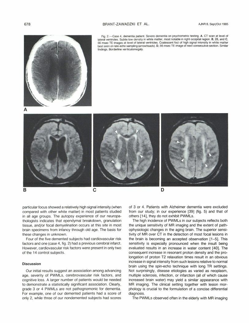
Fig . 2.-Case 4, dementia patient . Severe dementia on psychometric testing. A, CT scan at level of lateral ventricles. Subtle low density in white matter, most notable in right occipital region. B, 28, and C, 56 msec TE images at level of lateral ventricles. Coalescent foci of high signal intensity in white matter best seen on late echo sampling (arrowheads) . D, 56 msec TE image of next consecutive section. Similar findings . Borderline venticulomegaly.
A
8 c
particular focus showed a relatively high signal intensity (when compared with other white matter) in most patients studied in all age groups. The autopsy experience of our neuropathologists indicates that ependymal breakdown, granulation tissue, and/or focal demyelination occurs at this site in most brain specimens from infancy through old age. The basis for these changes in unknown.
Four of the five demented subjects had cardiovascular risk factors and one (case 4, fig . 2) had a previous cerebral infarct. However, cardiovascular risk factors were present in only two of the 14 control subjects.
Discussion
Our initial results suggest an association among advancing age, severity of PWMLs, cerebrovascular risk factors , and cognitive loss. A larger number of patients would be needed to demonstrate a statistically significant association. Clearly, grade 3 or 4 PWMLs are not pathognomonic for dementia. For example, one of our demented patients had a score of only 2, while three of our nondemented subjects had scores
o
of 3 or 4. Patients with Alzheimer dementia were excluded from our study; in our experience [39] (fig. 5) and that of others [14], they do not exhibit PWMLs.
The high incidence of PWMLs in our subjects reflects both the unique sensitivity of MR imaging and the extent of pathophysiologic changes in the aging brain. The superior sensitivity of MR over CT in the detection of most focal lesions in the brain is becoming an accepted observation [1-5]. This sensitivity is especially pronounced when the insult being evaluated results in an increase in water content [40]. The consequent increase in resonant proton density and the prolongation of proton T2 relaxation times result in an obvious increase in signal intensity from such lesions relative to normal brain using the spin-echo technique with long TR settings. Not surprisingly, disease etiologies as varied as neoplasm, multiple sclerosis , infection, or infarction (all of which cause increased brain water) may yield a similar appearance with MR imaging. The clinical setting together with lesion morphology is crucial to the formulation of a concise differential diagnosis.
The PWMLs observed often in the elderly with MR imaging

AJNR :6, Sept/Oct 1985 MR OF THE AGING BRAIN 679
Fig. 3.-Case 12, control patient. Uneventful medical history . A, 28, and B, 56 msec TE images at two consecutive sections. DiHuse, coalescent periventicular and isolated areas of high signal intensity .
A
probably reflect the so-called peri ventricular low-density foci variably noted on CT scans in this population [7-9, 14]. This finding on CT has been variously ascribed to pathologic entities such as hydrocephalus (when seen in conjunction with ventriculomegaly), the lacunar state, Binswanger disease, SAE, and white-matter atrophy. Previous reports have also documented a progressive decrease in Hounsfield number values in the white matter with advancing age in normal subjects [18].
MR adds to the weight of pathologic, physiologic, and neurochemical data suggesting that PWMLs are due to foci of increased water content in the aging brain, most likely on an ischemic basis. Autopsy studies reflect a high incidence (20%-50%) of deep, focal regions of brain softening consistent with punctate infarction in elderly subjects [11 , 41-43] . Radionuclide studies of regional cerebral blood flow (rCBF) consistently indicate progressive decrease in cerebral perfusion with advancing age and with dementia [44-48] . This loss of rCBF is aggravated by the well known risk factors of hypertension, smoking, diabetes, and hyperlipidemia [45]. These observations have led to the following speculation:
B
Because the white matter lies in the border zones and end zones of arterial perfusion, it experiences a lower regional perfusion than does cortical gray matter. This white matter may be more vulnerable to the effects of a global decrease in cerebral perfusion with advancing age. In the presence of arteriosclerotic changes with rr ial hypertrophy of deep perforating arteries supplying the periventricular regions, as seen with SAE, the white matter is especially at risk for ischemia. Such low perfusion of the white matter may adversely affect the maintenance of myelin, as evidenced by the increased evidence of myelin degradation products seen in elderly patients [41 , 49] . Of course, generalized decrease in cerebral perfusion lowers the threshold for frank infarction that may occur during hypotensive periods or with emboli . Both senescent demyelination and multiple foci of infarction could explain the observations that the water content of the brain progressively increases with advancing age [41] .
This scenario presumes an ischemic basis for the high incidence of PWMLs on MR imaging of elderly subjects. The relation of such lesions to cognitive loss and frank dementia in the elderly is less direct, as evidenced by the presence of

680
A B
A B
BRANT-ZAWADZKI ET AL.
c
AJNR:6, Sept/Oct 1985
Fig. 4.-Case 18, 81-year-old woman, control subject, MRI score = 1. Spin-echo sequence, 2000 msec TR . A, 28, and B, 56 msec TE images at level of frontal horns and lateral ventricles. Punctate focus of high signal intensity in white matter immediately at tip of frontal horns (arrowheads) . C, 28, and B, 56 msec TE images at level of centrum semiovale and body of caudate nucleus. No abnormalities in white matter. Caudate nucleus, being gray matter, shows relatively higher signal intensity than white matter.
Fig. 5.-64-year-old man with known Alzheimer disease. A, CT scan. Ventriculomegaly; no focal lesion. B, 28, and C, 56 msec TR , 2000 msec TE images. No abnormality of white matter; ventriculomegaly is again evident.

AJNR:6, Sept/Oct 1985 MR OF THE AGING BRAIN 681
PWMLs in three of our non demented controls . Certainly, given sufficient progressive insult to the associational white-matter tracts , dementia can occur, as evidenced by those patients studied who have multiinfarct dementia or SAE [7 , 9, 12-14). Given a sufficient severity in the number and perhaps distribution of PWMLs in any patient, the likelihood of dementia may be increased. Clearly, a larger study is needed. Further investigation is also suggested in correlating the MR frequency of PWMLs and dementia in patients with diffuse whitematter disease (e.g. , multiple sclerosis) and in patients with other causes of small vessel disease (e.g., diabetes).
REFERENCES
1. Brant-Zawadzki M, Norman 0 , Newton TH , et al. Magnetic resonance of the brain: the optimal screening technique. Radiology 1984 ;152 :71-77
2. Bradley WG, Waluch V, Yadley RA, Wycoff RR. Comparison of CT and MR in 400 patients with suspected disease of the brain and cervical spinal cord . Radiology 1984; 152 : 695-702
3. Bydder GM, Steiner RE, Young IR, et al. Clinical NMR imaging of the brain: 140 cases. AJNR 1982;3: 459-480, AJR 1982; 139 : 215-236
4. Brant-Zawadzki M, Badami JP, Mills CM , Norman 0 , Newton TH. Primary intracranial tumor imaging: a comparison of magnetic resonance and CT. Radiology 1984; 150: 435-440
5. Bryan RN, Willcott MR , Schneiders NJ, Ford JJ , Derman HS. Nuclear magnetic resonance evaluation of stroke. Radiology 1983;149: 189-192
6. Bradley WG, Waluch V, Brant-Zawadzki M, Yadley RA, Wycoff RR . Patchy, periventricular white matter lesions in the elderly: common observation during NMR imaging. Noninvasive Med Imaging 1984; 1 : 35-41
7. Valentine AR , Moseley IF, Kendall BE. White matter abnormality in cerebral atrophy: clinicoradiological correlations. J Neurol Neurosurg Psychiatry 1980;43 : 139-142
8. Mori K, Handa H, Murata T, Nakano Y. Periventricular lucency in computed tomography of hydrocephalus and cerebral atrophy. J Comput Assist Tomogr 1980;4 : 204-209
9. Zeumer H, Schon sky B, Sturm KW. Predominant white matter involvement in subcortical arteriosclerotic encephalopathy (Binswanger disease). J Comput Assist Tomogr 1980;4: 14-19
10. Goto K, Ishii N, Fukasawa H. Diffuse white-matter disease in the geriatric population. Radiology 1981 ;141 : 687-695
11. Tomlinson BE, Blessed G, Roth M. Observations on the brains of non-demented old people . J Neurol Sci 1968;7 : 331-356
12. Caplan LR , Schoene WC. Clinical features of subcortical arteriosclerotic encephalopathy (Binswanger disease). Neurology (NY) 1978;28 : 1206-1215
13. Loeb C, Gandolfo C. Diagnostic evaluation of degenerative and vascular dementia. Stroke 1983; 14 : 399-401
14. Erkinjuntti T, Sipponen JT, livanainen M, Ketonen L, Sulkava R, Sepponen RE. Cerebral NMR and CT imaging in dementia. J Comput Assist Tomogr 1984;8 : 614-618
15. Mortimer JA, Schuman LM, French LR. Epidemiology of dementing illness. In: Mortimer JA, Schuman LM, eds. The epidemiology of dementia. New York: Oxford University, 1981: 3-23
16. Terry RD. Dementia. Arch Neuro/1976 ;33 : 1-4 17. United States Bureau of Census. Demographic aspects of aging
and the older population in the United States. In Current population reports , special studies series , series P-23, no. 59. Washington, DC: U.S. Government Printing Office, 1976
18. Zatz LM, Jernigan TL, Ahumada AJ . White matter changes in cerebral computed tomography related to aging. J Comput Assist Tomogr 1982;6 : 19-23
19. Gado M, Danziger WL, Chi 0 , Hughes CP, Coben LA. Brain parenchymal density measurements by CT in demented subjects and normal controls. Radiology 1983 ;147 :703-710
20. Naeser MA, Gebhardt C, Levine HL. Decreased computeri zed tomography numbers in patients with presenile dementia. Arch Neurol 1980;37 : 401-409
21. George AE, de Leon MJ , Ferris SH, Kricheff II. Parenchymal CT correlates of senile dementia (Alzheimer disease): loss of graywhite matter discriminability. AJNR 1981;2 : 205-213
22. Brinkman SO, Sarwar M, Levin HS, Morris HH. Quantitative indexes of computed tomography in dementia and normal aging. Radiology 1981; 138 : 89-92
23. Laffey PA, Peyster RG, Nathan R, Haskin ME , McGinley JA. Computed tomography and aging: results in a normal elderly population. Neuroadiology 1984;26 : 273- 278
24. Hughes CP, Gado M. Computed tomography and aging of the brain. Radiology 1981; 139: 391-396
25. Zatz LM , Jernigan TL, Ahumada AJ . Changes on computed cranial tomography with aging: intracranial fluid volume. AJNR 1982;3 : 1-11
26. Gado M, Hughes CP, Danziger W, Chi 0 , Jost G, Berg L. Volumetric measurements of the cerebrospinal fluid spaces in demented subjects and controls. Radiology 1982; 144 : 535- 538
27. George AE, de Leon MJ , Rosenbloom S, et al. Ventricular volume and cognitive deficit: a computed tomographic study. Radiology 1983;149 : 493-498
28. Todorov AB, Go RC, Constantinidis J, Elston RC. Specificity of the clinical diagnosis of dementia. J Neurol Sci 1975;26: 81 - 98
29. Tomlinson BE, Blessed G, Roth M. Observations on the brains of demented old people . J Neurol Sci 1968; 11 : 205-242
30. Sourander P, Sjogren H. The concept of Alzheimer's disease and its clinical implications. In: Wolstenholme G, O'Connor M, eds. Alzheimer disease and related conditions. London: Churchill Livingstone, 1970: 11-36
31 . Crooks LE, Mills CM , David PL, et al. Visualization of cerebral and vascular abnormalities by NMR imaging. The effects of imaging parameter on contrast. Radiology 1982; 144 : 843-852
32. Kiernan R, Langston W, Mueller J. A brief yet specific and quantitative mental status examination. Presented at annual meeting of American Psychological Association , Anaheim, CA, August 1983
33. Wechsler D. Wechsler adult intelligence scale-revised manual. New York: Psychology Corp., 1981
34. Wechsler D. A standardized memory scale for clinical use. J Psycho/1945 ;19 :87-95
35. Russell E. A multiple scoring method for the assessment of complex memory functions. J Consult Clin Psychol 1975 ; 43 :800-809
36. Arthur GA. A point scale of performance tests. Revised form II. New York: Psychological Corp. 1947
37. Buschke H, Fuld PA. Evaluating storage, retention , and retrieval in disordered memory and learning. Neurology (NY) 1974;24 : 1019-1025
38. Benton AL, Hamsher K, eds. Multilingual aphasia examination , revised ed. Iowa City: University of Iowa, 1978
39. Friedland RP, Budinger TF, Brant-Zawadzki M, Jagust WJ . The diagnosis of Alzheimer-type dementia: a preliminary comparison of PET and proton NMR imaging. JAMA 1985;252 : 2750- 2752
40. Brant-Zawadzki M, Bartkowski HM, Ortendahl DA, et al. NMR in experimental and clinical cerebral edema. Noninvasive Med Imaging 1984;1 : 43-47

682 BRANT-ZAWADZKI ET AL. AJNR :6, Sept/Oct 1985
41 . Rolsten C, Samorajski C. Age and regional differences in the chemical composition of brains of mice, monkeys and humans. In: Ford DE, ed. Neurobiological aspects of maturation and aging. Prog Brain Res 1973;40 : 253-265
42 . Wisniewski HM, Terry RD. Morphology of the aging brain , human and animal. In: Ford DE, ed. Neurobiological aspects of maturation and aging. Prog Brain Res 1973;40 : 167-186
43 . Peress NS, Kane WC, Aronson SM. Central nervous system findings in a tenth decade autopsy population. In: Ford DE, ed. Neurobiological aspects of maturation and aging. Prog Brain Res 1973;40: 482-483
44. Melamed E, Lavy S, Bentin S, Cooper YR , Rinot Y. Reduction in regional cerebral blood flow during normal aging in man. Stroke 1980; 11 : 31-36
45 . Shaw TG, Mortel KF , Meyer JS, Rogers RL, Hardenberg J, Cutaia MM. Cerebral blood flow changes in benign aging and cerebrovascular disease. Neurology (NY) 1984;34: 855-862
46. Naritomi H, Meyer JS, Sakai F, Yamaguchi F, Shaw T. Effects of advancing age on regional cerebral blood flow. Arch Neurol 1979;36 : 41 0-416
47. Yamaguchi F, Meyer JS, Yamamoto M, Sakai F, Shaw T. Noninvasive regional cerebral blood flow measurements in dementia. Arch Neuro/1980 ;37 :410-418
48. Melamed E, Lavy S, Siew F, Bentin S, Cooper G. Correlation between regional cerebral blood flow and brain atrophy in dementia. J Neurol Neurosurg Psychiatry 1978;41 : 894-899
49 . Ansari KA, Loch J. Decreased myelin basic protein content of the aged human brain . Neurology (NY) 1975;25: 1045-1050
Related Documents