AUTHOR COPY Restorative Neurology and Neuroscience 32 (2014) 755–765 DOI 10.3233/RNN-130366 IOS Press 755 Motor recovery from constraint induced movement therapy is not constrained by extent of tissue damage following stroke Lynne V. Gauthier a,∗ , Victor W. Mark b,c,d , Edward Taub b , Adrianne McCullars f , Ameen Barghi b , Tyler Rickards b , Jarrod Hicks b and Gitendra Uswatte b,e a Department of Physical Medicine and Rehabilitation, The Ohio State University, Columbus, OH, USA b Department of Psychology, University of Alabama at Birmingham, Birmingham, AL, USA c Departments of Physical Medicine and Rehabilitation, University of Alabama at Birmingham, Birmingham, AL, USA d Department of Neurology, University of Alabama at Birmingham, Birmingham, AL, USA e Department of Physical Therapy, University of Alabama at Birmingham, Birmingham, AL, USA f Department of Psychology, University of South Alabama, Mobile, AL, USA Abstract. Purpose: The purpose of this study was to delineate the relationship between several types of T1-weighted MRI pathology and motor rehabilitation potential following Constraint Induced Movement therapy (CI therapy) in chronic stroke. Methods: Stepwise regression was employed (n = 80) to identify predictors of motor recovery (prior to therapy) and of response to Constraint-Induced Movement therapy [measured via the Wolf Motor Function Test (WMFT) and Motor Activity Log (MAL)] from among the following: age, side of motor deficit, chronicity, gender, lesion volume, peri-infarct damage volume, white matter hypointensity volume, ventricular asymmetry, and lesion location. Results: Although extent of total stroke damage weakly correlated with poorer performance on the WMFT prior to therapy, this relationship was mediated by the location of the damage. No metric of tissue damage examined here was associated with real-world arm use at baseline (MAL at pre-treatment) or with CI therapy-induced improvement in either best motor performance upon request (WMFT) or spontaneous arm use for daily activities (MAL). Conclusions: In sum, the extent of brain tissue damage of any type examined here poorly predicted motor function and response to rehabilitation in chronic stroke. Keywords: Stroke, rehabilitation, CI therapy, lesion volume, infarct volume, motor 1. Introduction Motor rehabilitation potential following stroke is anticipated to relate to the size and functional anatomic location of injury. Although there is fairly consistent evidence that lesion location influences subsequent ∗ Corresponding author: Lynne V. Gauthier, Ph.D., Department of Physical Medicine and Rehabilitation, The Ohio State University, 480 Medical Center Drive, Dodd Hall, 2nd Floor, USA. Tel.: +1 614 293 3480; Fax: +1 614 293 4870; E-mail: lynne.gauthier@ osumc.edu. recovery of motor function (Gauthier et al., 2009; Lindenberg et al., 2010; Lo et al., 2010; Zhu et al., 2010), the literature portrays an inconsistent and over- all weak relationship between lesion volume and motor ability in both acute and chronic stroke (Lauretani et al., 2010; Lindberg et al., 2007; Mark et al., 2008b; Mori et al., 1994; Page et al., 2013; Riley et al., 2011; Sterr et al., 2010). The largest lesion study (n = 513), which followed individuals from the acute phase through one-year follow-up, showed that total anterior circulation 0922-6028/14/$27.50 © 2014 – IOS Press and the authors. All rights reserved

Welcome message from author
This document is posted to help you gain knowledge. Please leave a comment to let me know what you think about it! Share it to your friends and learn new things together.
Transcript
AU
THO
R C
OP
Y
Restorative Neurology and Neuroscience 32 (2014) 755–765DOI 10.3233/RNN-130366IOS Press
755
Motor recovery from constraint inducedmovement therapy is not constrained byextent of tissue damage following stroke
Lynne V. Gauthiera,∗, Victor W. Markb,c,d, Edward Taubb, Adrianne McCullarsf , Ameen Barghib,Tyler Rickardsb, Jarrod Hicksb and Gitendra Uswatteb,e
aDepartment of Physical Medicine and Rehabilitation, The Ohio State University, Columbus, OH, USAbDepartment of Psychology, University of Alabama at Birmingham, Birmingham, AL, USAcDepartments of Physical Medicine and Rehabilitation, University of Alabama at Birmingham, Birmingham,AL, USAdDepartment of Neurology, University of Alabama at Birmingham, Birmingham, AL, USAeDepartment of Physical Therapy, University of Alabama at Birmingham, Birmingham, AL, USAf Department of Psychology, University of South Alabama, Mobile, AL, USA
Abstract.Purpose: The purpose of this study was to delineate the relationship between several types of T1-weighted MRI pathology andmotor rehabilitation potential following Constraint Induced Movement therapy (CI therapy) in chronic stroke.Methods: Stepwise regression was employed (n = 80) to identify predictors of motor recovery (prior to therapy) and of responseto Constraint-Induced Movement therapy [measured via the Wolf Motor Function Test (WMFT) and Motor Activity Log (MAL)]from among the following: age, side of motor deficit, chronicity, gender, lesion volume, peri-infarct damage volume, white matterhypointensity volume, ventricular asymmetry, and lesion location.Results: Although extent of total stroke damage weakly correlated with poorer performance on the WMFT prior to therapy,this relationship was mediated by the location of the damage. No metric of tissue damage examined here was associated withreal-world arm use at baseline (MAL at pre-treatment) or with CI therapy-induced improvement in either best motor performanceupon request (WMFT) or spontaneous arm use for daily activities (MAL).Conclusions: In sum, the extent of brain tissue damage of any type examined here poorly predicted motor function and responseto rehabilitation in chronic stroke.
Keywords: Stroke, rehabilitation, CI therapy, lesion volume, infarct volume, motor
1. Introduction
Motor rehabilitation potential following stroke isanticipated to relate to the size and functional anatomiclocation of injury. Although there is fairly consistentevidence that lesion location influences subsequent
∗Corresponding author: Lynne V. Gauthier, Ph.D., Department ofPhysical Medicine and Rehabilitation, The Ohio State University,480 Medical Center Drive, Dodd Hall, 2nd Floor, USA. Tel.: +1614 293 3480; Fax: +1 614 293 4870; E-mail: [email protected].
recovery of motor function (Gauthier et al., 2009;Lindenberg et al., 2010; Lo et al., 2010; Zhu et al.,2010), the literature portrays an inconsistent and over-all weak relationship between lesion volume and motorability in both acute and chronic stroke (Lauretani etal., 2010; Lindberg et al., 2007; Mark et al., 2008b;Mori et al., 1994; Page et al., 2013; Riley et al., 2011;Sterr et al., 2010).
The largest lesion study (n = 513), which followedindividuals from the acute phase through one-yearfollow-up, showed that total anterior circulation
0922-6028/14/$27.50 © 2014 – IOS Press and the authors. All rights reserved
AU
THO
R C
OP
Y
756 L.V. Gauthier et al. / Stroke damage and motor recovery
infarcts resulted in greater initial impairment (lowerBarthel index) and longer periods of rehabilitationthan partial or lacunar anterior circulation infarctsubtypes, but extent of recovery was indistinguishablebetween subtypes by the chronic phase (Lauretaniet al., 2010). Another large sample study (n = 300)found that infarct volume only weakly predictedchronic motor outcomes (as measured by Brunstrommstaging, Mori et al., 1994). These studies were limited,however, by imprecise measurement of motor ability(e.g., Brunnstrom staging, Barthel index) (Lauretaniet al., 2010; Mori et al., 1994), and categoricalclassification of lesion size (Lauretani et al., 2010).
A number of smaller-sample studies employed moresensitive measures of motor function and lesion vol-ume. The great majority of these did not show ameaningful relationship between lesion volume andspontaneous motor recovery (Lindberg et al., 2007;Mark et al., 2008b; Page et al., 2013; Riley et al.,2011; Sterr et al., 2010) or response to rehabilitation inchronic stroke (Mark et al., 2008b; Riley et al., 2011;Sterr et al., 2010), albeit with some exceptions (Craftonet al., 2003; Cramer et al., 2007). Consensus acrossstudies may be limited by the very small sample sizesemployed in some investigations (Crafton et al., 2003;Cramer et al., 2007; Lindberg et al., 2007; Riley et al.,2011; Sterr et al., 2010), different measures of motorfunction, and inconsistent inclusion/exclusion criteriabetween studies. A shared limitation of this prior work,however, is the exclusion of other MRI pathology frominvestigation.
Given the diverse pathology following stroke, con-current examination of several types of MRI pathology,in conjunction with lesion location, is needed to clar-ify the relationship between tissue pathology andmotor function. For example, the accuracy of mea-surements of lesion volume in the chronic phase maybe compromised by marked atrophy of the damagedparenchyma (Bigler and Maxwell, 2011) and accom-panying ex vacuo ventricular enlargement. It is thusimportant to include extent of hydrocephalus ex vacuoin the analysis. Another type of (often overlooked)tissue damage is reduced tissue integrity surround-ing the infarct. Preserved tissue adjacent to the infarctoften has diminished integrity and exhibits abnormalmetabolism (Beaulieu et al., 1999). Although this tis-sue remains somewhat structurally intact, it is unclearto what extent it is functioning effectively, and thus towhat extent this damage contributes to lasting motordeficit. A third type of commonly observed tissue dam-
age not previously accounted for in studies relatingMRI pathology to motor function is reduced whitematter integrity secondary to small vessel disease.Small vessel disease is a common risk factor for strokeand often produces tissue damage that can be cap-tured via MRI (e.g., hypointensities in white matteron T1-weighted images or FLAIR hyperintensities,most commonly observed in periventricular regions).It is currently unknown whether this damage increasestotal lesion burden in individuals who have experi-enced stroke. Finally, predictive relationships betweenbrain tissue damage and motor function found in somestudies (Chen et al., 2000; Crafton et al., 2003; Crameret al., 2007) may be better explained by lesion locationeffects, but few studies have examined these factorstogether.
Examined here is the relationship between severaldifferent types of pathological MRI findings on motorrecovery following usual care and response to a spe-cific, carefully controlled form of rehabilitation (CItherapy). The latter has particular basic significancebecause the effectiveness of CI therapy has been linkedto neuroplasticity, a process that could theoreticallybe constrained by inflammatory response in survivingdamaged tissue (Hughes et al., 2010; Jin et al., 2014;Price et al., 2006). Prior work shows that CI therapyhas the potential to alter brain structure (Gauthier etal., 2008; Sterling et al., 2013), cortical excitability(Liepert et al., 2000), neurogenesis and gene expres-sion (in animal models) (Maier et al., 2008; Schneideret al., 2014; Zhao et al., 2009). Moreover, grey mat-ter density in nondamaged regions has been shown topredict CI therapy response (Gauthier et al., 2012).These findings raise the question of whether a greaterextent of damage to non-infarcted regions could inter-fere with the brain’s ability to reorganize, and hencethe effectiveness of CI therapy.
Based on previous work that showed large clinicalgains from CI therapy irrespective of damage to thecorticospinal tract (Rickards et al., 2014), we hypothe-sized that rehabilitation response would be imperviousto pathology in remaining brain tissue (e.g., hydro-cephalus ex vacuo and peri-infarct tissue damage).Alternatively, extensive damage to non-infarcted tissuecould interfere with CI therapy response by reducingthe potential for neuroplasticity, a likely mechanism forthe effectiveness of CI therapy (Gauthier et al., 2008;Liepert et al., 2000). Utilizing retrospective imagingdata acquired during several previous clinical trials(Taub et al., 2005, 2006, 2013), this study is the first
AU
THO
R C
OP
Y
L.V. Gauthier et al. / Stroke damage and motor recovery 757
to concurrently model the effects of several differenttypes of pathological MRI findings on motor ability inthe chronic phase of stroke and response to CI therapy.
2. Methods
2.1. Participants
Participants consist of 80 individuals (40 females, 40males) with chronic stroke and mild-moderate upperextremity motor deficit who were enrolled from 1997to 2009 in various comparative effectiveness trials con-ducted at the University of Alabama at Birminghamand for whom good quality volumetric MRI data wasobtained. Participants had been randomized to receiveeither Constraint-Induced Movement therapy (CI ther-apy) or alternative (control) interventions (Taub et al.,2005, 2006, 2013). Individuals were recruited withoutrespect to lesion etiologies (e.g., cortical vs. subcor-tical, ischemic vs. hemorrhagic infarcts, lacunes) tomaximize the external validity of the findings. Allparticipants provided signed informed consent to par-ticipate. Inclusion criteria consisted of: post-strokeinterval of more than 10 months; ability to initiatewrist extension, thumb abduction, and extension ofat least 2 additional fingers; substantial nonuse of themore-affected arm (i.e., score ≤2.5 out of 5 on theMotor Activity Log), general medical stability, abilityto follow basic verbal commands, absence of currentpharmacological treatment of their motor disability(e.g., no botulinum toxin injections within the past 3months), and no history of having received Constraint-Induced Movement therapy.
Participants received volumetric T1-weighted MRIof their brain prior to receiving any study-related inter-vention. The most recent participants (n = 49) were
scanned on a Phillips (Intera) 3 Tesla machine, lessrecent participants on either a Philips (ACS) 1.5 Teslamachine (n = 14) or a GE (Signa) 1.5 Tesla machine(n = 17). Image acquisition parameters for the GE 1.5Tesla MRI included axial 3D spoiled gradient recalled(SPGR) acquisition with 1.5 mm slice thickness, in-plane resolution of 0.90 mm × 0.90 mm, repetitiontime (TR) = 30, echo time (TE) = 6, field of view(FOV) = 250, flip angle ( �) = 45. Acquisition param-eters for the Philips 1.5 Tesla were identical exceptTR = 20 and � = 8. Image acquisition parameters forthe Phillips 3 Tesla MRI included axial 3D Turbo-FieldEcho (TFE) acquisition with 1 mm slice thickness,in-plane resolution of 0.94 mm × 0.94 mm, TR = 9.8,TE = 4.6, FOV = 250, � = 8. T2 FLAIR images werealso obtained for a subset of participants (n = 38). Forthe 3 Tesla MRI, the FLAIR sequence was acquiredaxially in 4 mm slices with 1 mm gaps, TR = 11,000,TE = 120, inversion time (TI) = 2,800, � = 90. FLAIRparameters for the GE 1.5 T were axial 4 mm slices withno gap, TR = 9,002, TE = 142.5, TI = 2,200, � = 90.Participants ranged in age from 38 to 87 years (mean,62.9 ± 12.3). Stroke onset was 4.3 ± 4.0 years previ-ously. Thirty-nine exhibited right hemiparesis; 71 wereright-handed prior to stroke. See Table 1 for a break-down of patient characteristics.
2.2. Assessment of pre-treatment motorfunctioning
Motor functioning was assessed before and aftertreatment with the Motor Activity Log Arm Use scale(MAL) and the Wolf Motor Function Test (WMFT),both of which have established reliability and validity.The MAL quantifies how well and how often activi-ties of daily living are carried out spontaneously using
Table 1
Patient characteristics prior to CI therapy (n = 80)
Mean ± SD Min Max
Age 62.9 ± 12.3 38 87Chronicity (years) 4.3 ± 4.0 0.8 20Percentage with right hemiparesis 48.8Percentage of purely subcortical infarcts 51.3Infarction volume (% of ICV) 0.8 ± 1.7* 0.002 9.1Stroke damaged volume (% of ICV) 1.8 ± 2.6* 0.002 11.9White matter hypointensity volume (% of ICV) 0.2 ± 0.4* 0.0 2.0Ventricular asymmetry ratio (1 = ventricles of equal volume) 1.4 ± 0.6* 0.7 3.8Motor Activity Log Quality of Movement scale (0–5 points) 1.0 ± 0.7 0.0 2.6Wolf Motor Function Test Log2 Performance Time 1.3 ± 1.0 0.1 3.8∗positively skewed distribution, ICV = intracranial volume.
AU
THO
R C
OP
Y
758 L.V. Gauthier et al. / Stroke damage and motor recovery
the more-affected arm outside the laboratory (Taubet al., 1993; Uswatte et al., 2006; van der Lee et al.,2004). The WMFT measures how rapidly participantsperform standardized tests that they are required tocarry out with the more-affected arm inside the labo-ratory (Morris et al., 2001; Wolf et al., 2001). The twomeasures thus assess distinct domains of motor ability(Taub et al., 2006; Uswatte and Taub, 1999). Perfor-mance time on the WMFT was recorded as a log2 trans-formation of the mean time in seconds to more accu-rately portray patient progress (Mark et al., 2008b).
2.3. Interventions
Forty-three of 80 participants received CI therapy,a motor rehabilitation therapy with strong evidenceof efficacy for treating hemiparesis in chronic stroke(Morris et al., 2006; Taub et al., 1993, 2006; Wolf et al.,2006). The other participants were randomly assignedto other interventions. Only those participants assignedto CI therapy (n = 43), the most effective intervention,were retained for the analysis examining predictors oftherapy outcome. The demographics of those receivingCI therapy were similar to those of the entire sam-ple. Participants receiving CI therapy had a mean ageof 62.1 ± 12.2 years and mean stroke chronicity of4.3 ± 3.9 years; 23 were women, 22 exhibited righthemiparesis, and 41 were right-handed prior to stroke.
CI therapy is characterized by at least 30 hours ofhigh-repetition in-clinic training of the more impairedarm on functional tasks, restraint of the less-impairedarm for a target 90% of waking hours, and a number ofbehavioral techniques termed the “transfer package”lasting an additional 0.5 hours in the laboratory (Taubet al., 1999). The transfer package, designed to facili-tate transfer of therapeutic gains to real world activities,includes daily monitoring of life situation use of themore affected arm in several ways, problem-solvingwith a therapist to overcome perceived barriers tousing the extremity, and a behavioral contract, amongother techniques (Morris et al., 2006; Taub et al.,2013). Although some characteristics of the CI ther-apy protocol differed across studies (e.g., sling versusmitt restraint), comparable treatment outcomes wereobserved across studies (Taub et al., 1999).
2.4. MRI Analysis
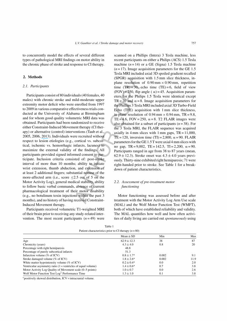
The following types of tissue damage werequantified on T1-weighted MRI images: infarcts [cere-
brospinal fluid (CSF) filled space left behind bystroke], peri-infarct damage, and (non-CSF) whitematter hypointensities. Brain voxels with T1-weightedhypointensity were classified either as peri-infarct tis-sue damage when no normal-appearing tissue waspresent between the CSF-filled infarct and the voxelsin question, or as non-specific white matter hypointen-sity when it appeared remote from an infarct. Infarcts(cerebrospinal fluid filled space left behind by stroke),peri-infarct damage, and white matter hypointensitieswere separately traced by four blinded raters under thesupervision of a neurologist (V.W. Mark) using MRI-cro/MRIcron software (see Fig. 1 for delineation ofthese regions). Tracings were then refined using semi-automated intensity thresholding (of only the tracedregion). The thresholding procedure involved identify-ing an intensity cutoff that best distinguished damagedfrom visually-intact tissue and applying this thresholdto the entire manual trace. Although this threshold-ing step did not greatly alter the resultant volumes, itexpedited tracing [by allowing for less precise (slightlyover-inclusive) delineation of the lesion border] andreduced the potential for human error. Each rater per-formed tracings on a minimum of 20 scans. Interraterand intrarater reliability of the thresholded tracingswas excellent (>.99), as assessed via intraclass corre-lation on an overlapping subset of 10 images for eachrater or rater-pair. Individuals performing the tracingwere unaware of participants’ motor status. To con-trol for variability in brain size, thresholded tracingsof damaged tissue were expressed as a percentage of
A B
Fig. 1. Several different types of damage were quantified withineach individual’s scan. A: Coronal reconstruction from an originalaxial acquisition. B: The infarct is depicted in blue, peri-infarct tis-sue damage in red, and white matter hypointensities in green. Notealso the size discrepancy between the right ventricle (adjacent to thelesion) and the contralateral ventricle.
AU
THO
R C
OP
Y
L.V. Gauthier et al. / Stroke damage and motor recovery 759
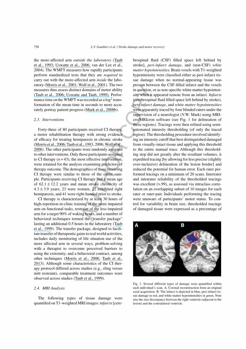
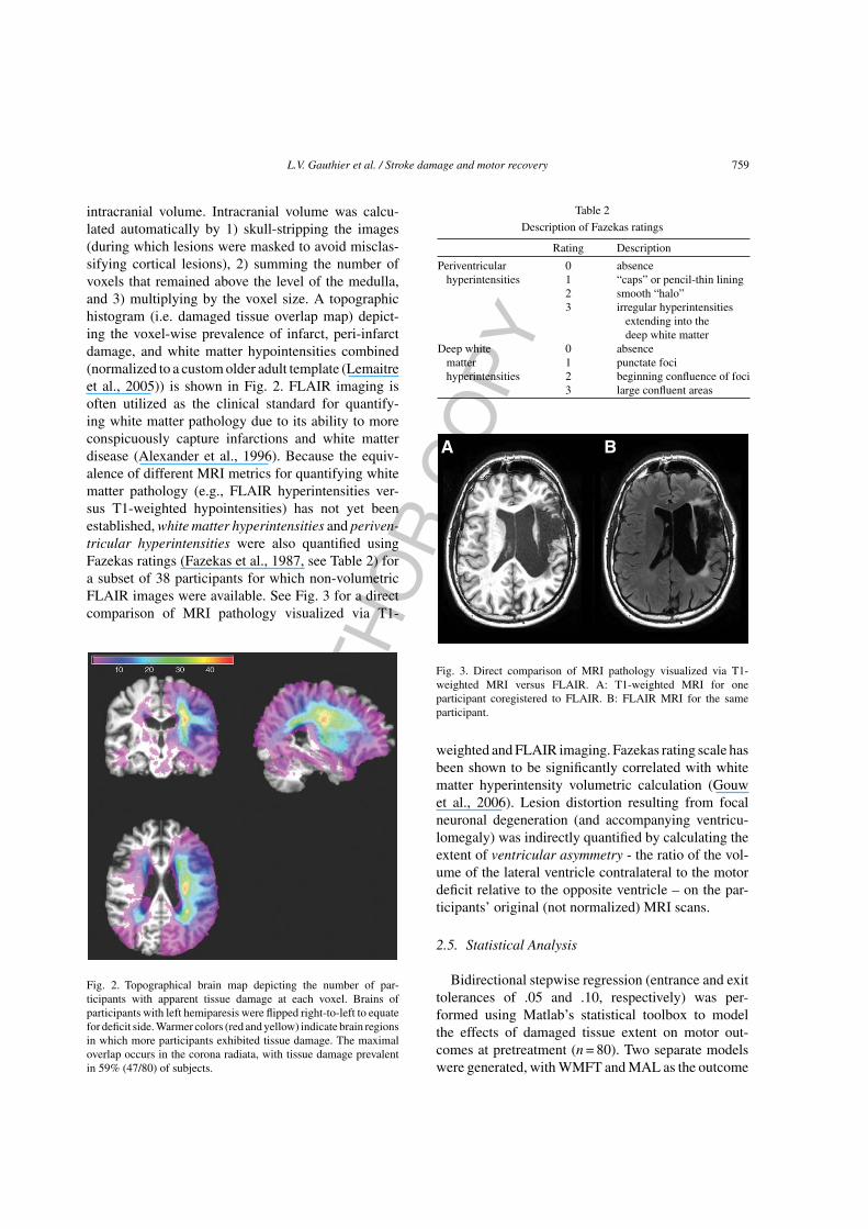
intracranial volume. Intracranial volume was calcu-lated automatically by 1) skull-stripping the images(during which lesions were masked to avoid misclas-sifying cortical lesions), 2) summing the number ofvoxels that remained above the level of the medulla,and 3) multiplying by the voxel size. A topographichistogram (i.e. damaged tissue overlap map) depict-ing the voxel-wise prevalence of infarct, peri-infarctdamage, and white matter hypointensities combined(normalized to a custom older adult template (Lemaitreet al., 2005)) is shown in Fig. 2. FLAIR imaging isoften utilized as the clinical standard for quantify-ing white matter pathology due to its ability to moreconspicuously capture infarctions and white matterdisease (Alexander et al., 1996). Because the equiv-alence of different MRI metrics for quantifying whitematter pathology (e.g., FLAIR hyperintensities ver-sus T1-weighted hypointensities) has not yet beenestablished, white matter hyperintensities and periven-tricular hyperintensities were also quantified usingFazekas ratings (Fazekas et al., 1987, see Table 2) fora subset of 38 participants for which non-volumetricFLAIR images were available. See Fig. 3 for a directcomparison of MRI pathology visualized via T1-
Fig. 2. Topographical brain map depicting the number of par-ticipants with apparent tissue damage at each voxel. Brains ofparticipants with left hemiparesis were flipped right-to-left to equatefor deficit side. Warmer colors (red and yellow) indicate brain regionsin which more participants exhibited tissue damage. The maximaloverlap occurs in the corona radiata, with tissue damage prevalentin 59% (47/80) of subjects.
Table 2
Description of Fazekas ratings
Rating Description
Periventricular 0 absencehyperintensities 1 “caps” or pencil-thin lining
2 smooth “halo”3 irregular hyperintensities
extending into thedeep white matter
Deep white 0 absencematter 1 punctate focihyperintensities 2 beginning confluence of foci
3 large confluent areas
Fig. 3. Direct comparison of MRI pathology visualized via T1-weighted MRI versus FLAIR. A: T1-weighted MRI for oneparticipant coregistered to FLAIR. B: FLAIR MRI for the sameparticipant.
weighted and FLAIR imaging. Fazekas rating scale hasbeen shown to be significantly correlated with whitematter hyperintensity volumetric calculation (Gouwet al., 2006). Lesion distortion resulting from focalneuronal degeneration (and accompanying ventricu-lomegaly) was indirectly quantified by calculating theextent of ventricular asymmetry - the ratio of the vol-ume of the lateral ventricle contralateral to the motordeficit relative to the opposite ventricle – on the par-ticipants’ original (not normalized) MRI scans.
2.5. Statistical Analysis
Bidirectional stepwise regression (entrance and exittolerances of .05 and .10, respectively) was per-formed using Matlab’s statistical toolbox to modelthe effects of damaged tissue extent on motor out-comes at pretreatment (n = 80). Two separate modelswere generated, with WMFT and MAL as the outcome
AU
THO
R C
OP
Y
760 L.V. Gauthier et al. / Stroke damage and motor recovery
measures, respectively, age, side of motor deficit,chronicity, and gender as potential nuisance covari-ates, and lesion volume, total stroke damage volume,T1-weighted white matter hypointensity volume, andventricular asymmetry as potential predictor variables.Where significant effects were found, lesion location(whether or not the stroke-damaged region overlappedwith the intersection of the corona radiata and corpuscallosum - a region found to be associated with greaterimpairment on the WMFT in this sample of partici-pants (Gauthier et al., 2009) and linked to greater motorimpairment in other studies (Lindenberg et al., 2012;Lo et al., 2010) was tested as a potential mediatingvariable through use of the Sobel procedure.
To ensure consistent results when white matter dam-age is quantified via FLAIR hyperintensities versusT1-weighted hypointensities, a similar bidirectionalstepwise regression was run incorporating Fazekasperiventricular hyperintensity ratings and Fazekasdeep white matter hyperintensity ratings in lieu ofT1-weighted white matter hypointensity volume forthe subset of 38 participants for which Fazekasscores could be obtained. Fazekas ratings for bothperiventricular hyperintensity and deep white matterhyperintensity were dichotomized in the model (rat-ings of 0 and 1 were grouped together and ratings of 2and 3 were grouped together), given the low prevalenceof 0 and 2 Fazekas ratings in our dataset.
To examine the effects of damaged tissue onresponse to CI therapy, identical stepwise regressionprocedures were used, with the exception that post-treatment WMFT or MAL score served as the outcomemeasure, respectively, and pre-treatment WMFT orMAL score served as an additional covariate. Theinitial model was constrained such that pre-treatmentWMFT or MAL score entered into the model first. SeeTable 3 for a depiction of the statistical models used.
The positively skewed volumetric measurementsin this dataset violated some parametric assumptions(non-normal distribution of residuals for white mat-ter hypointensity volume and chronicity). Bootstrapanalysis revealed that the resultant models were notsignificantly affected by these violations.
3. Results
3.1. Clinical response to CI therapy
CI therapy recipients showed large significantimprovements on the MAL (t(42) = 15.89; P < 0.0001;
d’ = 2.41) and the WMFT (t(42) = 4.04; P = 0.0002;d’ = 0.62).
3.2. MRI predictors of motor performance
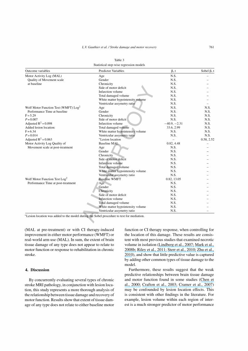
Consistent with prior work (Taub et al., 1999,2006; Wolf et al., 2006), age, side of greatest motorimpairment, chronicity, and gender were not signif-icant predictors in any of the models. The modelrelating characteristics of tissue damage to WMFTperformance at baseline yielded two significant pre-dictors: total stroke damage volume (p < 0.004) andinfarct volume (p = 0.023, see Table 3). Participantswith larger total stroke damage performed significantlyslower on the WMFT. It should be noted, however,that less than 10% of the variance in WMFT scores(Adjusted R2 = 0.098) was accounted for by volume ofstroke-damaged tissue. Furthermore, when location ofdamage was added to the model, it was found to fullymediate effects that were initially attributed to volumeof damage [i.e. greater volume of tissue damage wasassociated with lesions at the intersection of the coronaradiata and corpus callosum (p < 0.001), lesions in thislocation predicted poorer motor function (p = 0.014),and lesion location replaced volume of stroke-damagedtissue as the sole predictor of WMFT performancewhen entered into the full model (p = 0.014, AdjustedR2 = 0.063)]. Similar results were obtained whenFazekas scores were utilized in lieu of peri-infarct andT1-weighted white matter hypointensity volume ona subset of 38 participants. More severe deep whitematter hypointensity Fazekas ratings (largely influ-enced by the extent of damaged tissue surroundingthe infarct) predicted poorer WMFT scores (p = 0.005),but this relationship disappeared entirely after account-ing for lesion location. The consistency in resultsregardless of how tissue damage was quantified (i.e.,via Fazekas score or volume traced on T1-weightedscan) is not surprising given excellent correspondencebetween the two metrics [t = 5.8, p < 0.0001, effectsize = 2.6 for comparison of T1-weighted white mat-ter hypointensity volume between those who receivedlower (<2) and higher Fazekas scores (≥2)]. Theseresults indicate that regardless of how damaged tis-sue is measured (i.e., using T1-weighted or FLAIRimages), volume of stroke-damaged tissue is not a use-ful predictor of motor dysfunction after accountingfor more salient predictors (lesion location). Extent oftissue damage (irrespective of how it was quantified)was not associated with real-world arm use at baseline
AU
THO
R C
OP
Y
L.V. Gauthier et al. / Stroke damage and motor recovery 761
Table 3
Statistical step-wise regression models
Outcome variables Predictor Variables �, t Sobel �, t
Motor Activity Log (MAL) Age N.S. –Quality of Movement scale Gender N.S. –at baseline Chronicity N.S. –
Side of motor deficit N.S. –Infarction volume N.S. –Total damaged volume N.S. –White matter hypointensity volume N.S. –Ventricular assymetry ratio N.S. –
Wolf Motor Function Test (WMFT) Log2 Age N.S. N.S.Performance Time at baseline Gender N.S. N.S.
F = 5.29 Chronicity N.S. N.S.P = 0.007 Side of motor deficit N.S. N.S.Adjusted R2 = 0.098 Infarction volume −40.9, −2.31 N.S.Added lesion location: Total damaged volume 33.6, 2.99 N.S.F = 6.34 White matter hypointensity volume N.S. N.S.P = 0.014 Ventricular assymetry ratio N.S. N.S.Adjusted R2 = 0.063 ∗Lesion location – 0.58, 2.52Motor Activity Log Quality of Baseline MAL 0.82, 4.48 –
Movement scale at post-treatment Age N.S. –Gender N.S. –Chronicity N.S. –Side of motor deficit N.S. –Infarction volume N.S. –Total damaged volume N.S. –White matter hypointensity volume N.S. –Ventricular assymetry ratio N.S. –
Wolf Motor Function Test Log2 Baseline WMFT 0.82, 13.05 –Performance Time at post-treatment Age N.S. –
Gender N.S. –Chronicity N.S. –Side of motor deficit N.S. –Infarction volume N.S. –Total damaged volume N.S. –White matter hypointensity volume N.S. –Ventricular assymetry ratio N.S. –
∗Lesion location was added to the model during the Sobel procedure to test for mediation.
(MAL at pre-treatment) or with CI therapy-inducedimprovement in either motor performance (WMFT) orreal-world arm use (MAL). In sum, the extent of braintissue damage of any type does not appear to relate tomotor function or response to rehabilitation in chronicstroke.
4. Discussion
By concurrently evaluating several types of chronicstroke MRI pathology, in conjunction with lesion loca-tion, this study represents a more thorough analysis ofthe relationship between tissue damage and recovery ofmotor function. Results show that extent of tissue dam-age of any type does not relate to either baseline motor
function or CI therapy response, when controlling forthe location of this damage. These results are consis-tent with most previous studies that examined necroticvolume in isolation (Lindberg et al., 2007; Mark et al.,2008b; Riley et al., 2011; Sterr et al., 2010; Zhu et al.,2010), and show that little predictive value is capturedby adding other common types of tissue damage to themodel.
Furthermore, these results suggest that the weakpredictive relationships between brain tissue damageand motor function found in some studies (Chen etal., 2000; Crafton et al., 2003; Cramer et al., 2007)may be confounded by lesion location effects. Thisis consistent with other findings in the literature. Forexample, lesion volume within each region of inter-est is a much stronger predictor of motor performance
AU
THO
R C
OP
Y
762 L.V. Gauthier et al. / Stroke damage and motor recovery
than lesion volume in isolation (Chen et al., 2000). Inother accounts, lesion volume appears to lose its pre-dictive validity when extent of lesion overlap with thecorticospinal tract or the motor map or is accounted for(Crafton et al., 2003; Zhu et al., 2010).
Although lesion location is a more robust predictorof motor ability in the chronic phase of stroke than vol-ume of tissue damage, it should be noted that it still onlyexplains a small portion of the variance in motor status(less than 8%). This is consistent with the hypothe-sis that the brain compensates for the effects of theinitial damage by harnessing alternative motor path-ways, thereby lessening the impact of the initial infarct.This hypothesis garners support from prior researchthat showed that significant recovery can occur by thechronic phase even with complete disruption of the cor-ticospinal tract, despite greater deficits initially (Janget al., 2008; Swayne et al., 2008). Moreover, the pre-dictive validity of corticospinal disruption on motorstatus diminishes over time, while the inverse is true ofalternate motor fibers (Lindenberg et al., 2010; Swayneet al., 2008). Also in support of this hypothesis isthe observation that infarcts impacting multiple motorpathways have a more lasting detrimental effect (Frieset al., 1993; Lindenberg et al., 2010).
The present investigation builds substantially uponprior lesion studies in that it additionally investigatedthe effect of different types of brain damage on indi-viduals’ ability to benefit from CI therapy. CI therapyis an intensive intervention for hemiparesis secondaryto stroke, TBI, cerebral palsy, and multiple sclerosis(Choudhary et al., 2012; Mark et al., 2008a; Shawet al., 2005; Sterling et al., 2013; Taub, 2004, 1993)that is known to promote neuroplasticity (Gauthier etal., 2008; Liepert et al., 2000; Sterling et al., 2013).As such, it provides an excellent model for evaluat-ing clinical response/ rehabilitation potential (becausethe majority of patients show large clinical gains inresponse to the therapy) (Wolf et al., 2006). However,despite its wide presence in the literature, there exist nopublished accounts relating the different types of braindamage studied in this paper to CI therapy outcome.The finding that CI therapy outcome was independentof (all types of) MRI pathology measurable via rou-tine clinical scans suggests that rehabilitation-inducedneuroplasticity is unaffected by both lesion extent andextensive damage to non-infarcted tissue.
It has been hypothesized that similar neural recoverymechanisms operate during a targeted intervention asduring recovery in the absence of intervention (Cramer,
2008). The present findings, taken together with otheraccounts in the literature, support an alternate view thatCI therapy targets different or additional neurorecoveryprocesses. Reported here is the finding that location ofstroke damage predicted motor ability prior to receiv-ing CI therapy (recovery in the chronic phase followingusual care), but not response to CI therapy, evidencethat the neural constraints on recovery following usualcare can be overcome by treatment with CI therapy.A similar dissociation was observed by Rickards andcolleagues when examining the effect of disruptionof the corticospinal tract on both initial motor statusand response to CI therapy (Rickards et al., 2014).The inverse has also been shown whereby grey mat-ter density in brain regions that are not predominantlyassociated with motor function predicted CI therapyresponse, but not pre-treatment motor ability. The com-bination of these findings reveals a double-dissociationthat raises the possibility that CI therapy harnesses dif-ferent neural substrates than recovery following usualcare. It appears that the recovery mechanisms har-nessed by CI therapy are not hindered by the initialdamage that produced the motor deficit, but are depen-dent, in part, on the density of undamaged corticaltissue (Gauthier et al., 2012).
This study has a few limitations that are worthy ofmention. A retrospective dataset of patients who wererecruited for CI therapy treatment studies was utilized.As such, conventional MRI acquisition sequenceswere employed on the select group of chronic strokepatients that met enrollment criteria for these CI ther-apy studies, namely patients with mild to moderatehemiparesis. MRIs from this dataset were obtainedusing only commonly available scanning strengths andacquisition sequences, potentially limiting the sensi-tivity of our analyses. However, given that extent oftissue damage was not even marginally predictive ofmotor function after controlling for lesion location (ps>0.32), it is unlikely that enhanced sensitivity (e.g.,from high-resolution FLAIR imaging) would impactour overall findings. A final limitation of these findingswas that participants had infarcts of various etiologiesthat could not be elucidated by the research team (giventhat individuals with chronic stroke were recruited forthis study), and thus could not be controlled for in dataanalysis. To our knowledge, no data exists regardingwhether stroke etiology results in chronic tissue differ-ences between hemorrhagic versus ischemic lesions.Additional research is needed to determine whetherlesion etiology may impact these findings.
AU
THO
R C
OP
Y
L.V. Gauthier et al. / Stroke damage and motor recovery 763
In sum, extent of tissue damage is a relatively unso-phisticated predictor of motor dysfunction that carrieslittle value. Lesion location (Gauthier et al., 2009;Lindenberg et al., 2010; Lo et al., 2010; Zhu et al.,2010) and integrity of remaining tissue (Gauthier etal., 2012) have shown more promise for predictinglasting motor deficit; however, these measures are stillunable to quantify recovery trajectories of individualpatients or the extent to which motor networks as awhole are affected. The motor network is a compli-cated system of disparate brain regions that work inconcert to execute motor movements. Research study-ing network-based models of motor deficit is thus morelikely to yield a greater depth of knowledge than canbe gained by studying facets of damage in isolation.For example, neuroplasticity may allow for recoveryof function after stroke by strengthening alternativepathways (Zaaimi et al., 2012). Recovery of functionmay therefore depend in large part on the viability ofremaining pathways and the extent to which they areable to reproduce the lost function (Lindenberg et al.,2012). More research along this line is needed to betterunderstand the effects of stroke on interconnected com-ponents of our complex motor circuitry and, ultimately,motor function.
Acknowledgments
This work was supported by the National Institutesof Health [HD34273] and the American Heart Associ-ation [0815065E].
References
Alexander, J.A., Sheppard, S., Davis, P.C., & Salverda, P.(1996). Adult cerebrovascular disease: Role of modified rapidfluid-attenuated inversion-recovery sequences. AJNR Am JNeuroradiol, 17(8), 1507-1513.
Beaulieu, C., de Crespigny, A., Tong, D.C., Moseley, M.E., Albers,G.W., & Marks, M.P. (1999). Longitudinal magnetic resonanceimaging study of perfusion and diffusion in stroke: Evolutionof lesion volume and correlation with clinical outcome. AnnNeurol, 46(4), 568-578.
Bigler, E.D., & Maxwell, W.L. (2011). Neuroimaging and neu-ropathology of TBI. Neurorehabilitation, 28(2), 63-74.
Chen, C.L., Tang, F.T., Chen, H.C., Chung, C.Y., & Wong, M.K.(2000). Brain lesion size and location: Effects on motor recov-ery and functional outcome in stroke patients. Arch Phys MedRehabil, 81(4), 447-452.
Choudhary, A., Gulati, S., Kabra, M., Singh, U.P., Sankhyan, N.,Pandey, R.M., & Kalra, V. (2012). Efficacy of modified con-straint induced movement therapy in improving upper limbfunction in children with hemiplegic cerebral palsy: A random-ized controlled trial. Brain Dev, 35(9), 870-876.
Crafton, K.R., Mark, A.N., & Cramer, S.C. (2003). Improvedunderstanding of cortical injury by incorporating measures offunctional anatomy. Brain, 126(Pt 7), 1650-1659.
Cramer, S.C. (2008). Repairing the human brain after stroke: I.Mechanisms of spontaneous recovery. Ann Neurol, 63(3), 272-287.
Cramer, S.C., Parrish, T.B., Levy, R.M., Stebbins, G.T., Ruland,S.D., Lowry, D.W., & Himes, D.M. (2007). Predicting func-tional gains in a stroke trial. Stroke, 38(7), 2108-2114.
Fazekas, F., Chawluk, J.B., Alavi, A., Hurtig, H.I., & Zimmerman,R.A. (1987). MR signal abnormalities at 1.5 T in Alzheimer’sdementia and normal aging. AJR Am J Roentgenol, 149(2), 351-356.
Fries, W., Danek, A., Scheidtmann, K., & Hamburger, C. (1993).Motor recovery following capsular stroke. Brain, 116, 369-382.
Gauthier, L.V., Taub, E., Mark, V.W., Barghi, A., & Uswatte, G.(2012). Atrophy of spared gray matter tissue predicts poorermotor recovery and rehabilitation response in chronic stroke.Stroke, 43(2), 453-457.
Gauthier, L.V., Taub, E., Mark, V.W., Perkins, C., & Uswatte, G.(2009). Improvement after constraint-induced movement ther-apy is independent of infarct location in chronic stroke patients.Stroke, 40(7), 2468-2472.
Gauthier, L.V., Taub, E., Perkins, C., Ortmann, M., Mark, V.W.,& Uswatte, G. (2008). Remodeling the brain: Plastic struc-tural brain changes produced by different motor therapies afterstroke. Stroke, 39(5), 1520-1525.
Gouw, A.A., Van der Flier, W.M., van Straaten, E.C., Barkhof, F.,Ferro, J.M., & Baezner, H. et al. (2006). Simple versus complexassessment of white matter hyperintensities in relation to phys-ical performance and cognition: The LADIS study. J Neurol,253(9), 1189-1196.
Hughes, J., Beech, J., Jones, P.S., Wang, D., Menon, D., & Baron,J. (2010). Mapping selective neuronal loss and microglialactivation in the salvaged neocortical penumbra in the rat. Neu-roimage, 49(1), 19-31.
Jang, S.H., Bai, D., Son, S.M., Lee, J., Kim, D., & Sakong, J. etal. (2008). Motor outcome prediction using diffusion tensortractography in pontine infarct. Ann Neurol, 64(4), 460-465.
Jin, Q., Cheng, J., Liu, Y., Wu, J., Wang, X., & Wei, S. et al. (2014).Improvement of functional recovery by chronic metformintreatment is associated with enhanced alternative activation ofmicroglia/macrophages and increased angiogenesis and neu-rogenesis following experimental stroke, Brain Behav Immun,epub ahead of print.
Lauretani, F., Saccavini, M., Zaccaria, B., Agosti, M., Zampolini, M.,& Franceschini, M. (2010). Rehabilitation in patients affectedby different types of stroke. A one-year follow-up study. Eur JPhys Rehabil Med, 46(4), 511-516.
Lemaitre, H., Crivello, F., Grassiot, B., Alperovitch, A., Tzourio,C., & Mazoyer, B. (2005). Age- and sex-related effects on theneuroanatomy of healthy elderly. Neuroimage, 26(3), 900-911.
AU
THO
R C
OP
Y
764 L.V. Gauthier et al. / Stroke damage and motor recovery
Liepert, J., Bauder, H., Wolfgang, H.R., Miltner, W.H., Taub, E., &Weiller, C. (2000). Treatment-induced cortical reorganizationafter stroke in humans. Stroke, 31, 1210-1216.
Lindberg, P.G., Skejo, P.H., Rounis, E., Nagy, Z., Schmitz, C., &Wernegren, H. et al. (2007). Wallerian degeneration of the corti-cofugal tracts in chronic stroke: A pilot study relating diffusiontensor imaging, transcranial magnetic stimulation, and handfunction. Neurorehabil Neural Repair, 21(6), 551-560.
Lindenberg, R., Renga, V., Zhu, L.L., Betzler, F., Alsop, D., &Schlaug, G. (2010). Structural integrity of corticospinal motorfibers predicts motor impairment in chronic stroke. Neurology,74(4), 280-287.
Lindenberg, R., Zhu, L.L., Ruber, T., & Schlaug, G. (2012). Predict-ing functional motor potential in chronic stroke patients usingdiffusion tensor imaging. Hum Brain Mapp, 33(5), 1040-1051.
Lo, R., Gitelman, D., Levy, R., Hulvershorn, J., & Parrish, T. (2010).Identification of critical areas for motor function recovery inchronic stroke subjects using voxel-based lesion symptom map-ping. Neuroimage, 49(1), 9-18.
Maier, I.C., Baumann, K., Thallmair, M., Weinmann, O., Scholl, J., &Schwab, M.E. (2008). Constraint-Induced Movement Therapyin the Adult Rat after Unilateral Corticospinal Tract Injury. JNeurosci, 28, 9386-9403.
Mark, V.W., Taub, E., Bashir, K., Uswatte, G., Delgado, A., &Bowman, M.H. et al. (2008a). Constraint-Induced Movementtherapy can improve hemiparetic progressive multiple sclerosis.Preliminary findings. Mult Scler, 14, 992-994.
Mark, V.W., Taub, E., Perkins, C., Gauthier, L., & Uswatte, G.(2008b). MRI infarction load and CI therapy outcomes forchronic post-stroke hemiparesis. Restor Neurol Neurosci, 26(1),13-33.
Mori, S., Sadoshima, S., Ibayashi, S., Lino, K., & Fujishima, M.(1994). Relation of cerebral blood flow to motor and cognitivefunctions in chronic stroke patients. Stroke, 25(2), 309-317.
Morris, D.M., Taub, E., & Mark, V.W. (2006). Constraint-inducedmovement therapy: Characterizing the intervention protocol.Eura Medicophys, 42(3), 257-268.
Morris, D.M., Uswatte, G., Crago, J.E., Cook, E.W., 3rd, & Taub,E. (2001). The reliability of the wolf motor function test forassessing upper extremity function after stroke. Arch Phys MedRehabil, 82(6), 750-755.
Page, S.J., Gauthier, L.V., & White, S. (2013). Size doesn’t mat-ter: Cortical stroke lesion volume is not associated with upperextremity motor impairment and function in mild, chronichemiparesis. Arch Phys Med Rehabil, 94(5), 817-821.
Price, C.J., Wang, D., Menon, D.K., Guadagno, J.V., Cleij, M., Fryer,T., & Warburton, E.A. (2006). Intrinsic activated microglia mapto the peri-infarct zone in the subacute phase of ischemic stroke.Stroke, 37(7), 1749-1753.
Rickards, T., Sterling, C., Taub, E., Perkins-Hu, C., Gauthier, L., Gra-ham, M., & Uswatte, G. (2014). Diffusion tensor imaging studyof the response to constraint-induced movement therapy of chil-dren with hemiparetic cerebral palsy and adults with chronicstroke. Arch Phys Med Rehabil, 95(3), 506-514.e1.
Riley, J.D., Le, V., Der-Yeghiaian, L., See, J., Newton, J.M., Ward,N.S., & Cramer, S.C. (2011). Anatomy of stroke injury predictsgains from therapy. Stroke, 42(2), 421-426.
Schneider, A., Rogalewski, A., Wafzig, O., Kirsch, F., Gretz, N.,Kruger, C., & Schabitz, W.R. (2014). Forced arm use is superiorto voluntary training for motor recovery and brain plasticityafter cortical ischemia in rats. Exp Transl Stroke Med., 6(1),3-7378-6-3.
Shaw, S.E., Morris, D.M., Uswatte, G., McKay, S., Meythaler,J.M., & Taub, E. (2005). Constraint-induced movement ther-apy for recovery of upper-limb function following traumaticbrain injury. J Rehabil Res Dev, 42, 769-778.
Sterling, C., Taub, E., Davis, D., Rickards, T., Gauthier, L.V., Griffin,A., & Uswatte, G. (2013). Structural neuroplastic change afterconstraint-induced movement therapy in children with cerebralpalsy. Pediatrics, 131(5), e1664-e1669.
Sterr, A., Shen, S., Szameitat, A.J., & Herron, K.A. (2010). The roleof corticospinal tract damage in chronic motor recovery andneurorehabilitation: A pilot study. Neurorehabil Neural Repair,24(5), 413-419.
Swayne, O.B., Rothwell, J.C., Ward, N.S., & Greenwood, R.J.(2008). Stages of motor output reorganization after hemisphericstroke suggested by longitudinal studies of cortical physiology.Cereb Cortex, 18(8), 1909-1922.
Taub, E. (2004). Harnessing brain plasticity through behavioral tech-niques to produce new treatments in neurorehabilitation. AmPsychol, 59, 692-704.
Taub, E., Lum, P.S., Hardin, P., Mark, V.W., & Uswatte, G. (2005).AutoCITE: Automated delivery of CI therapy with reducedeffort by therapists. Stroke, 36, 1301-1304.
Taub, E., Miller, N.E., Novack, T.A., Cook, E.W., 3rd, Fleming,W.C., & Nepomuceno, C.S. et al. (1993). Technique to improvechronic motor deficit after stroke. Arch Phys Med Rehabil,74(4), 347-354.
Taub, E., Uswatte, G., King, D.K., Morris, D., Crago, J.E., & Chatter-jee, A. (2006). A placebo-controlled trial of constraint-inducedmovement therapy for upper extremity after stroke. Stroke,37(4), 1045-1049.
Taub, E., Uswatte, G., Mark, V.W., & Morris, D.M. (2006). Thelearned nonuse phenomenon: Implications for rehabilitation.Eura Medicophys, 42(3), 241-256.
Taub, E., Uswatte, G., Mark, V.W., Morris, D.M., Barman, J., Bow-man, M.H., & Bishop-McKay, S. (2013). Method for enhancingreal-world use of a more affected arm in chronic stroke: Trans-fer package of constraint-induced movement therapy. Stroke,44(5), 1383-1388.
Taub, E., Uswatte, G., & Pidikiti, R. (1999). Constraint-InducedMovement Therapy: A new family of techniques with broadapplication to physical rehabilitation-a clinical review. J Reha-bil Res Dev, 36, 237-251.
Uswatte, G., & Taub, E. (1999). Constraint-induced movementtherapy: New approaches to outcome measurement in rehabili-tation. In D. T. Stuss, G. Winocur & I. H. Robertson (Eds.),Cognitive neurorehabilitation: A comprehensive approach.Cambridge University Press, 215-229.
Uswatte, G., Taub, E., Morris, D., Light, K., & Thompson, P.A.(2006). The Motor Activity Log-28: Assessing daily use of thehemiparetic arm after stroke. Neurology, 67(7), 1189-1194.
van der Lee, J.H., Beckerman, H., Knol, D.L., de Vet, H.C., & Bouter,L.M. (2004). Clinimetric properties of the motor activity log for
AU
THO
R C
OP
Y
L.V. Gauthier et al. / Stroke damage and motor recovery 765
the assessment of arm use in hemiparetic patients. Stroke, 35(6),1410-1414.
Wolf, S.L., Catlin, P.A., Ellis, M., Archer, A.L., Morgan, B., &Piacentino, A. (2001). Assessing Wolf motor function test asoutcome measure for research in patients after stroke. Stroke,32(7), 1635-1639.
Wolf, S.L., Winstein, C.J., Miller, J.P., Taub, E., Uswatte, G., &Morris, D. et al. (2006). Effect of constraint-induced movementtherapy on upper extremity function 3 to 9 months after stroke:The EXCITE randomized clinical trial. Jama, 296(17), 2095-2104.
Zaaimi, B., Edgley, S.A., Soteropoulos, D.S., & Baker, S.N. (2012).Changes in descending motor pathway connectivity after cor-ticospinal tract lesion in macaque monkey. Brain, 135(Pt 7),2277-2289.
Zhao, C., Wang, J., Zhao, S., & Nie, Y. (2009). Constraint-inducedmovement therapy enhanced neurogenesis and behavioralrecovery after stroke in adult rats. Tohoku J Exp Med, 218(4),301-308.
Zhu, L.L., Lindenberg, R., Alexander, M.P., & Schlaug, G. (2010).Lesion load of the corticospinal tract predicts motor impairmentin chronic stroke. Stroke, 41(5), 910-915.
Related Documents