Motor Proteins - Introduction Part 1 Biochemistry 4000 Dr. Ute Kothe

Motor Proteins - Introduction Part 1 Biochemistry 4000 Dr. Ute Kothe.
Dec 15, 2015
Welcome message from author
This document is posted to help you gain knowledge. Please leave a comment to let me know what you think about it! Share it to your friends and learn new things together.
Transcript
Motor Proteins- Introduction Part 1
Biochemistry 4000
Dr. Ute Kothe
Motor Proteins
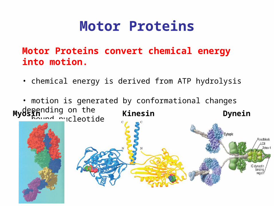
Motor Proteins convert chemical energy into motion.
• chemical energy is derived from ATP hydrolysis
• motion is generated by conformational changes depending on the bound nucleotide
Myosin Kinesin Dynein
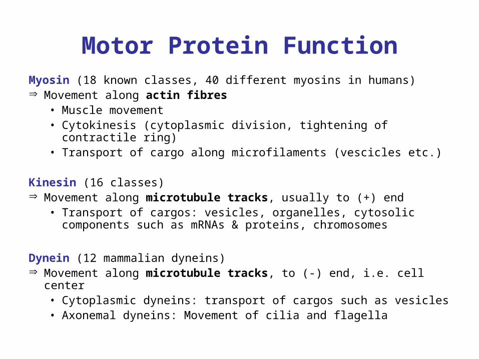
Motor Protein FunctionMyosin (18 known classes, 40 different myosins in humans) Movement along actin fibres
• Muscle movement• Cytokinesis (cytoplasmic division, tightening of contractile ring)• Transport of cargo along microfilaments (vescicles etc.)
Kinesin (16 classes) Movement along microtubule tracks, usually to (+) end
• Transport of cargos: vesicles, organelles, cytosolic components such as mRNAs & proteins, chromosomes
Dynein (12 mammalian dyneins) Movement along microtubule tracks, to (-) end, i.e. cell center
• Cytoplasmic dyneins: transport of cargos such as vesicles• Axonemal dyneins: Movement of cilia and flagella
Tubulin
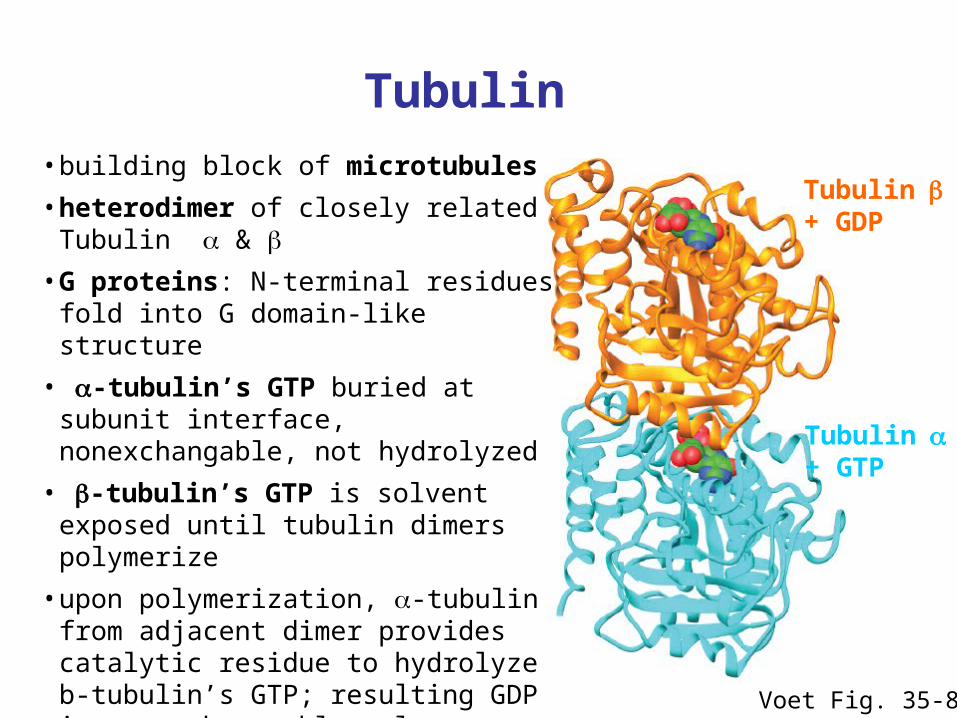
Voet Fig. 35-89
Tubulin+ GTP
Tubulin+ GDP
• building block of microtubules
• heterodimer of closely related Tubulin &
• G proteins: N-terminal residues fold into G domain-like structure
• -tubulin’s GTP buried at subunit interface, nonexchangable, not hydrolyzed
• -tubulin’s GTP is solvent exposed until tubulin dimers polymerize
• upon polymerization, -tubulin from adjacent dimer provides catalytic residue to hydrolyze b-tubulin’s GTP; resulting GDP is nonexchangeable unless tubulin dissociates from microtubule
Microtubules
Voet Fig. 35-92
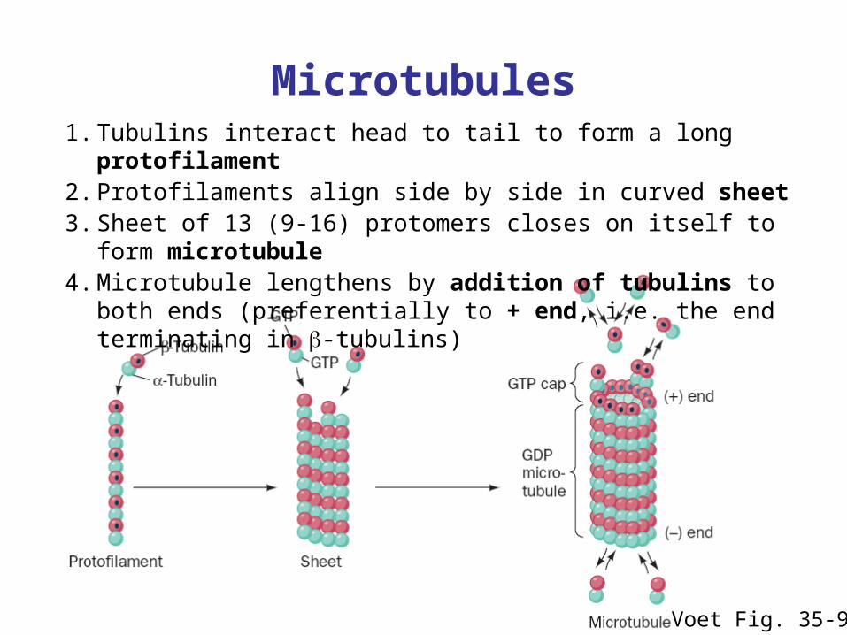
1. Tubulins interact head to tail to form a long protofilament2. Protofilaments align side by side in curved sheet3. Sheet of 13 (9-16) protomers closes on itself to form microtubule4. Microtubule lengthens by addition of tubulins to both ends
(preferentially to + end, i.e. the end terminating in -tubulins)
Structure of the Axoneme
Voet Fig. 35-102
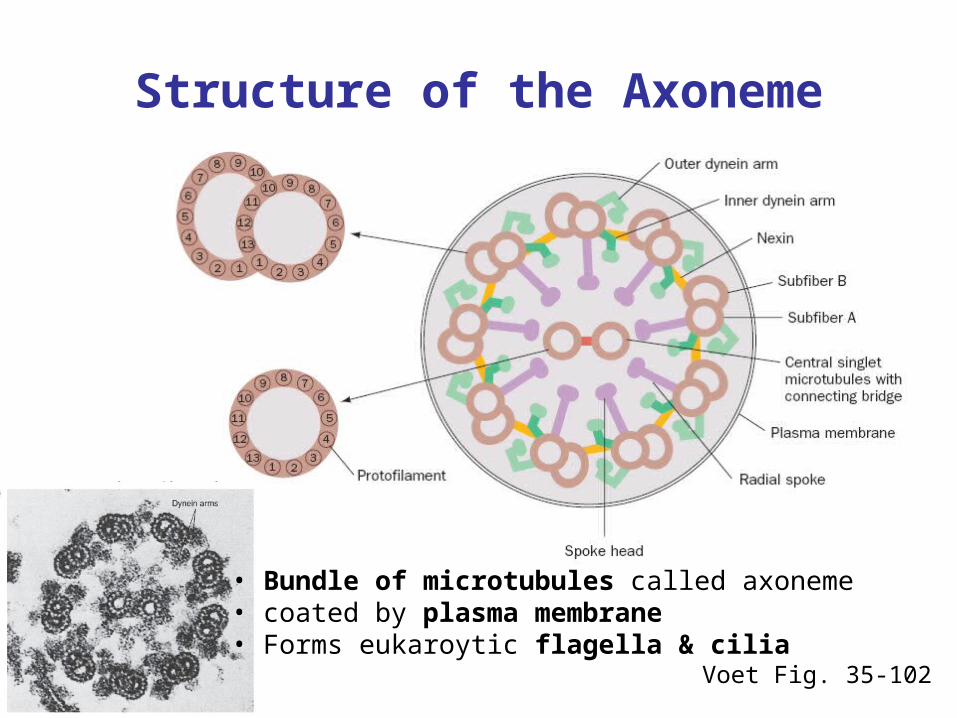
• Bundle of microtubules called axoneme• coated by plasma membrane• Forms eukaroytic flagella & cilia
Dynein
Valle, Cell 2003; Voet Fig. 35-107
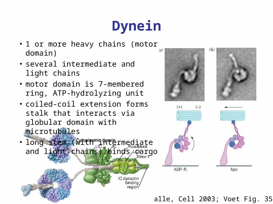
• 1 or more heavy chains (motor domain)• several intermediate and light chains• motor domain is 7-membered ring, ATP-
hydrolyzing unit• coiled-coil extension forms stalk that
interacts via globular domain with microtubules
• long stem (with intermediate and light chains) binds cargo
Conventional Kinesin
Voet Fig. 35-94
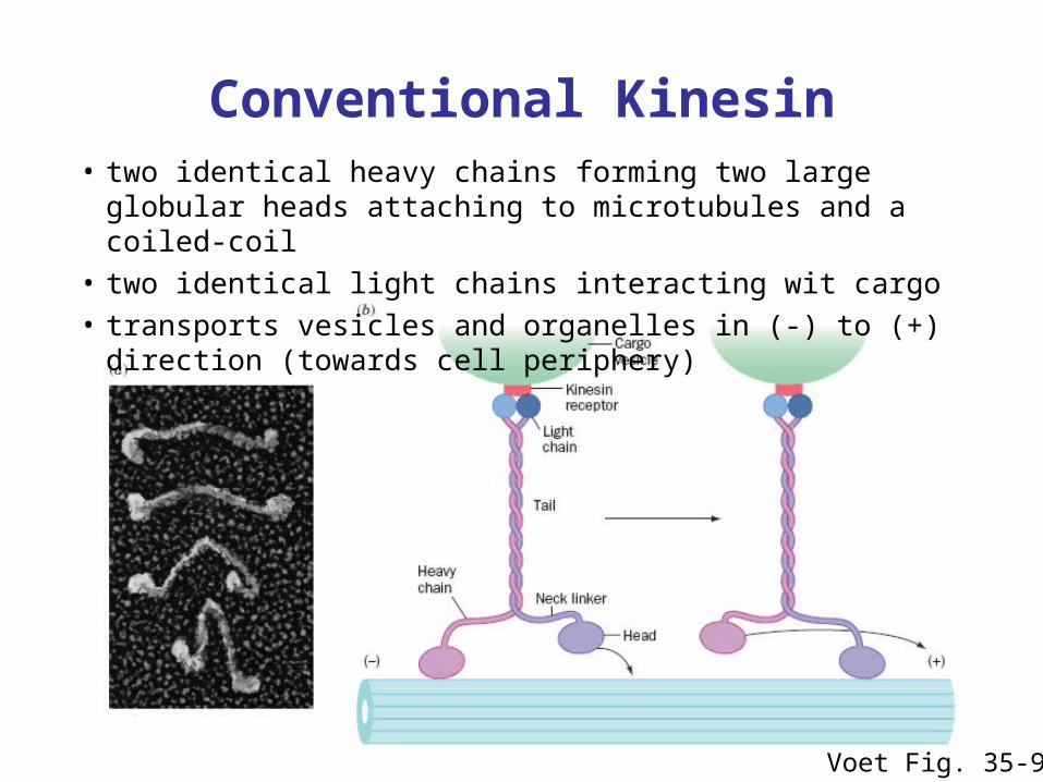
• two identical heavy chains forming two large globular heads attaching to microtubules and a coiled-coil
• two identical light chains interacting wit cargo• transports vesicles and organelles in (-) to (+) direction (towards cell
periphery)
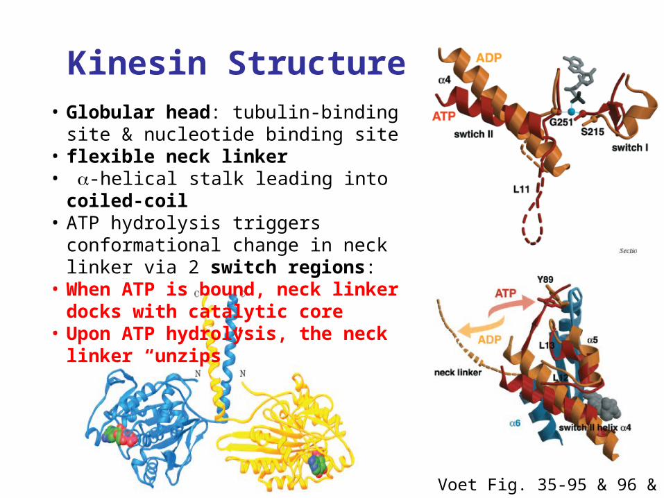
Kinesin Structure
Voet Fig. 35-95 & 96 & 97
• Globular head: tubulin-binding site & nucleotide binding site
• flexible neck linker• -helical stalk leading into coiled-coil• ATP hydrolysis triggers conformational
change in neck linker via 2 switch regions: • When ATP is bound, neck linker docks
with catalytic core• Upon ATP hydrolysis, the neck linker
“unzips”
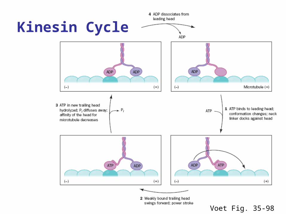
Voet Fig. 35-98
Kinesin Cycle
Hand-over-Hand Mechanism
ATP-bound state: strong microtuble bindingADP-bound state: weak microtubule binding
1. ATP binds to leading head - globular kinesin head which is already bound to the microtubule and oriented towards (+) end
2. Neck linker of leading head “zips up” agains catalytic core 3. trailing head is thrown forward (trailing head has bound ADP and
reduced affinity to microtubule): 4. Trailing head swings by ~ 160 Å, net movement of dimeric kinesin
is ~ 80 Å = length of one microtubule dimer5. ATP in new trailing head is hydrolyzed & phosphate released:
affinity for microtuble decreases6. ADP in new leading head dissociates
Two heads work in a coordinated fashion ATP binding to leading head induces power stroke
Processivity
Kinesin is highly processive: it takes several 100 steps on a microtubule without detaching or sliding backwards!
How?
• coordinated, but out of phase ATP cycle in both heads
• one head is always firmly attached to microtubule
Movie demonstrating kinesins processivity:http://www.proweb.org/kinesin/axonemeMTs.html

Dynein
Valle, Cell 2003; Voet Fig. 35-107
• 1 or more heavy chains (motor domain)• several intermediate and light chains• motor domain is 7-membered ring, ATP-
hydrolyzing unit• coiled-coil extension forms stalk that
interacts via globular domain with microtubules
• long stem (with intermediate and light chains) binds cargo
Related Documents