Brain Injury, July 2006; 20(8): 807–824 Motor deficits and recovery during the first year following mild closed head injury MARCUS H. HEITGER 1,2 , RICHARD D. JONES 1,2,3 , JOHN C. DALRYMPLE-ALFORD 1,4 , CHRIS M. FRAMPTON 1,2 , MICHAEL W. ARDAGH 1,5 , & TIM J. ANDERSON 1,2,6 1 Van der Veer Institute for Parkinson’s & Brain Research, Christchurch, New Zealand, 2 Department of Medicine, Christchurch School of Medicine & Health Sciences, Christchurch, New Zealand, 3 Department of Medical Physics and Bioengineering, Christchurch Hospital, Christchurch, New Zealand, 4 Department of Psychology, University of Canterbury, Christchurch, New Zealand, 5 Emergency Department, and 6 Department of Neurology, Christchurch Hospital, Christchurch, New Zealand (Received 8 August 2005; accepted 2 March 2006) Abstract Objective: This study examined motor impairments over 1 year following mild closed head injury (CHI). It is the first study to serially assess long-term oculomotor and upper-limb visuomotor function following mild head trauma. Methods: Thirty-seven patients with mild CHI and 37 matched controls were compared at 1 week, 3 months and 6 months and 31 available pairs at 12 months post-injury on measures of saccades, oculomotor smooth pursuit, upper-limb visuomotor function and neuropsychological performance. Symptomatic recovery was sampled using the Rivermead Postconcussion Symptoms Questionnaire. Results: At 1 week, the group with CHI reported high levels of post-concussional symptoms and exhibited prolonged saccade latencies, increased directional errors, decreased saccade accuracy and impaired fast sinusoidal smooth pursuit concomitant with increased arm movement reaction time, decreased arm movement speed and decreased motor accuracy on upper-limb visuomotor tracking tasks. Neuropsychological testing identified deficits only in verbal learning and speed of processing while attention, short-term/working memory and general cognitive performance were preserved. At 3 and 6 months, the group with CHI continued to show deficits on several oculomotor and upper-limb visuomotor measures in combination with some deficits on verbal learning and improved, yet abnormal, levels of post-concussional symptoms. At 12 months, the group with CHI had no cognitive impairment but residual deficits in eye and arm motor function and continued to show elevated levels of post-concussional symptoms. Conclusions: The findings indicate that multiple motor systems are measurably impaired up to 12 months following mild CHI and that instrumented motor assessment may provide sensitive and objective markers of cerebral dysfunction during recovery from mild head trauma independent of neuropsychological assessment and patient self-report. Keywords: Closed head injury, recovery, saccades, oculomotor smooth pursuit, upper-limb Introduction Mild closed head injury (CHI) is a substantial cause of morbidity world-wide. Approximately 80% of head injury admissions are categorized as mild with 100 to 300 cases per year per 100 000 popula- tion [1, 2]. It follows that mild CHI is one of the commonest causes of emergency department visits and hospital admissions [3] and the source of substantial related costs for health care providers [4–6]. There is increasing evidence that even mild head trauma is associated with a physical impact on the brain that can result in neuronal injury and subse- quent cerebral dysfunction [7–11]. This physical Correspondence: Marcus Heitger, Van der Veer Institute for Parkinson’s & Brain Research, 16 St Asaph Street, Christchurch, New Zealand. Tel: þ64 3 378 6074. Fax: þ64 3 378 6080. E-mail: [email protected] ISSN 0269–9052 print/ISSN 1362–301X online ß 2006 Informa UK Ltd. DOI: 10.1080/02699050600676354

Welcome message from author
This document is posted to help you gain knowledge. Please leave a comment to let me know what you think about it! Share it to your friends and learn new things together.
Transcript
Brain Injury, July 2006; 20(8): 807–824
Motor deficits and recovery during the first year following mildclosed head injury
MARCUS H. HEITGER1,2, RICHARD D. JONES1,2,3, JOHN C. DALRYMPLE-ALFORD1,4,CHRIS M. FRAMPTON1,2, MICHAEL W. ARDAGH1,5, & TIM J. ANDERSON1,2,6
1Van der Veer Institute for Parkinson’s & Brain Research, Christchurch, New Zealand, 2Department of Medicine,
Christchurch School of Medicine & Health Sciences, Christchurch, New Zealand, 3Department of Medical Physics
and Bioengineering, Christchurch Hospital, Christchurch, New Zealand, 4Department of Psychology, University of
Canterbury, Christchurch, New Zealand, 5Emergency Department, and 6Department of Neurology, Christchurch
Hospital, Christchurch, New Zealand
(Received 8 August 2005; accepted 2 March 2006)
AbstractObjective: This study examined motor impairments over 1 year following mild closed head injury (CHI). It is the first studyto serially assess long-term oculomotor and upper-limb visuomotor function following mild head trauma.Methods: Thirty-seven patients with mild CHI and 37 matched controls were compared at 1 week, 3 months and 6 monthsand 31 available pairs at 12 months post-injury on measures of saccades, oculomotor smooth pursuit, upper-limbvisuomotor function and neuropsychological performance. Symptomatic recovery was sampled using the RivermeadPostconcussion Symptoms Questionnaire.Results: At 1 week, the group with CHI reported high levels of post-concussional symptoms and exhibited prolonged saccadelatencies, increased directional errors, decreased saccade accuracy and impaired fast sinusoidal smooth pursuit concomitantwith increased arm movement reaction time, decreased arm movement speed and decreased motor accuracy on upper-limbvisuomotor tracking tasks. Neuropsychological testing identified deficits only in verbal learning and speed of processingwhile attention, short-term/working memory and general cognitive performance were preserved. At 3 and 6 months,the group with CHI continued to show deficits on several oculomotor and upper-limb visuomotor measures in combinationwith some deficits on verbal learning and improved, yet abnormal, levels of post-concussional symptoms. At 12 months,the group with CHI had no cognitive impairment but residual deficits in eye and arm motor function and continued toshow elevated levels of post-concussional symptoms.Conclusions: The findings indicate that multiple motor systems are measurably impaired up to 12 months following mildCHI and that instrumented motor assessment may provide sensitive and objective markers of cerebral dysfunction duringrecovery from mild head trauma independent of neuropsychological assessment and patient self-report.
Keywords: Closed head injury, recovery, saccades, oculomotor smooth pursuit, upper-limb
Introduction
Mild closed head injury (CHI) is a substantial causeof morbidity world-wide. Approximately 80% ofhead injury admissions are categorized as mildwith 100 to 300 cases per year per 100 000 popula-tion [1, 2]. It follows that mild CHI is one ofthe commonest causes of emergency department
visits and hospital admissions [3] and the sourceof substantial related costs for health careproviders [4–6].
There is increasing evidence that even mild headtrauma is associated with a physical impact on thebrain that can result in neuronal injury and subse-quent cerebral dysfunction [7–11]. This physical
Correspondence: Marcus Heitger, Van der Veer Institute for Parkinson’s & Brain Research, 16 St Asaph Street, Christchurch, New Zealand.Tel: þ64 3 378 6074. Fax: þ64 3 378 6080. E-mail: [email protected]
ISSN 0269–9052 print/ISSN 1362–301X online � 2006 Informa UK Ltd.DOI: 10.1080/02699050600676354
impact is thought to contribute to post-concussionalsymptoms of both a physical and cognitive nature[9, 12–14]. Within the first 6–12 months followingmild CHI, an estimated 30–40% of patients are likelyto show ongoing health problems [15–20].
Despite this potentially debilitating impact ofmild CHI and the subsequent demand for objectivetools to confirm injury status and track functionalrecovery, there are currently no assessment optionsthat are equally accurate and cost-efficient inobjectively quantifying the long-term presence ofresidual brain dysfunction after mild CHI. Imagingis expensive and does not provide a detailed pictureof functional aspects of the injured brain [9, 21–23].The application of neuropsychological testing toconfirm injury status and track recovery in patientswith mild CHI has been hampered by its suscept-ibility to pre-morbid factors such as age, IQ andsocioeconomic status [24–26]. External factorsare likely to outweigh the head-trauma-effect onneuropsychological tests and the likelihood thatabnormal neuropsychological performance indicatesthe presence of a pathological condition has beenestimated to be under 50% in cases with mild headtrauma [27]. Similarly, the use of biochemicalmarkers to quantify the impact of mild CHI on thebrain has been unsatisfactory [28–30].
Based on earlier findings of oculomotor andupper-limb visuomotor impairment at 1 weekafter mild CHI [31–33], it was considered thatthere may be measurable long-term abnormalitiesin eye and arm motor function after mild CHI andthat motor testing may be a useful supplementarytool in objectively quantifying residual brain dys-function during recovery from mild head trauma.The approach of examining motor performance asan independent functional biomarker for the adverseimpact of mild CHI takes advantage of the estab-lished knowledge on motor control, its complexfunctional neuroanatomy and the availability ofestablished tools and paradigms to assess motorfunction [34–40]. Whilst it was the principal aimof this study to establish whether there are long-termmotor impairments within the first year followingmild CHI, the authors also wanted to determineif the recovery of these deficits parallels overallpatient recovery.
Saccadic deficits have been described after severehead trauma [41] and several studies have examinedupper-limb motor deficits and recovery of arm/handmotor function after moderate-to-severe headtrauma using tasks such as finger tapping [42–45],visuomotor tracking tasks [46, 47], video games[48], reaching [49, 50] and grasping and reactiontime tests [50]. However, this study is the first to
serially assess long-term deficits in saccades andoculomotor smooth pursuit following head injuryand to measure long-term upper-limb visuomotorfunction specifically after mild head trauma. It isalso unique in taking a multi-modality approachof gauging ongoing cerebral dysfunction after mildCHI by tracking the recovery of motor performance,neuropsychological function and self-reportedsymptom status in a homogeneously mild CHIpatient group.
Participants and methods
Participants
Thirty-seven subjects (13 female and 24 male)with mild CHI (Glasgow Coma Scale [GCS] score13: four cases, 14: 12 cases, 15: 21 cases) wererecruited from patients presenting with acute headinjury to Christchurch Hospital (the principalhospital for a regional population of over 400 000).The GCS used was the score on first assessment(i.e. the first recorded GCS post-injury). In mostcases, this was at time of admission to theEmergency Department. In some cases, the GCSwas first assessed by the ambulance team prior toarrival at the hospital. Patients had to have a score onGCS of between 13–15 on first assessment, withoutfalling below 13 at any consecutive assessment at thehospital. At the time of recruitment, no standardizedmethod was in practice to assess duration of post-traumatic amnesia (PTA) in patients presenting withmild head injury to the Emergency Department atChristchurch Hospital and PTA duration was notroutinely noted in patient files (apart from briefcomments on the lack of recall of the injury-eventif applicable). Hence, an iterative process was usedto (a) confirm that PTA was less than 24 hours (theonly required screening criterion) and (b) providean approximation of length of PTA (the GCS scorewas the principal factor for CHI classification).At the initial pre-recruitment interview at thehospital patients were asked about their firstmemory following the injury. If the rememberedevent fell within a 24 hour period, it was assessedwhether the patient remembered being at the sceneafter the accident/regaining consciousness, beinghelped by others (e.g. extraction from a vehicle,somebody clearing their bike off the street orcalling an ambulance), the arrival of the ambulance(if applicable, standard response time of ambulanceswithin city boarders taken as time approximation),being in the ambulance, arriving at the hospital(time was recorded on admission sheet as was timeof accident in the case of MVAs and most sportaccidents), treatment events for which the time
808 M. H. Heitger et al.

was noted on the patient chart, being served a meal(usually dinner or breakfast for patients who stayedovernight).
All patients had experienced PTA rangingbetween �2 minutes and 22 hours (mean¼ 2 hours40 minutes) and 32 patients had a confirmedloss of consciousness (mean¼3.96 minutes, range0.5–15 minutes). Mean age was 29.1 years (SD12.7, range 15–56 years) and mean years ofeducation was 13.6 (SD 2.56, range 8–19 years).All patients were either employed or attendedinstitutions for secondary or tertiary educationand none was involved in litigation. Other potentialparticipants were excluded if there was evidenceof any influence of alcohol or psychoactive drugs attime of injury, regular intake of psychoactive drugsor history of drug abuse, central neurologicaldisorder or psychiatric condition, structural braindamage or haematoma on CT head scan (whereobtained), oculomotor or somatomotor deficits uponclinical examination, presence of strabismus, visualacuity of worse than 6/12, skull fractures or priorhistory of mild, moderate or severe head injury withpersisting symptoms or complaints.
The control group consisted of subjects withno history of mild, moderate or severe head injurywith persisting symptoms or complaints, no centralneurological disorder or psychiatric condition andno regular intake of psychoactive drugs or history ofdrug abuse. The controls were individually matchedto each CHI case with respect to age (within 3 yearsfor patients >18 years, within 1 year for patients <18years), gender and years of formal education (within2 years for patients >18, within 1 year for subjects<18). The mean age for the control group was 29.2years (SD 12.6, range 15–57 years) and mean yearsof formal education was 13.7 (SD 2.71, range 9–19years). The head injured group and the controlshad equivalent IQ (mean IQ CHI group at 1 week:110.8� 11.6 vs. controls: 112.4� 10.5). Controlswere recruited via a volunteer database madeavailable by the Department of Psychology atthe University of Canterbury, Christchurch,New Zealand. These volunteers are interested intaking part in research studies and have agreed to becontacted for this purpose. In cases where a subjectwith head injury could not be matched with a controlfrom the database, controls were recruited amongstsiblings, relatives or friends of the head injuredparticipant (patients were happy to suggest andcontact potential controls having been explainedthe necessity for having a control and the matchingcriteria).
Throughout the study, none of the participantswas hospitalized or developed secondary health
problems related to other causes which could haveaffected any of the measures. Subjects were offeredcompensation for travel costs to attend the testingat the hospital but received no other payment.The project was approved by the CanterburyEthics Committee and written consent was obtainedfrom all participants.
Oculomotor assessment
The assessment methods, paradigm parametersand key measures of motor performance wereidentical to an earlier study and have been describedin detail elsewhere [31, 32]. This study incorporatedparadigms for reflexive saccades (‘looking at thestimulus’, 44 saccades, stimuli jumping randomlyby 5, 10, 15, 20, 25 or 30� in a horizontal direction,at intervals varying pseudorandomly between1.0–1.6 seconds), anti-saccades (‘looking awayfrom the stimulus to its mirror-location on theopposite side of the screen’, 32 anti-saccades,stimuli at 5 and 15� off centre, at intervalsvarying pseudorandomly between 1.0–1.6 seconds,balanced for left and right), memory-guidedsequences of saccades (‘performing a memorizedsequence of saccades’, six different sequences,each with four steps, duration of 1.0 second perstep, each sequence practised five times, thenperformed once, followed by presentation of thenext sequence), self-paced saccades (‘do-as-many-as-possible’-self-pacing for 30 seconds betweentwo stationary targets, �15� off centre) as well assine and random oculomotor smooth pursuit(‘tracking a continuously moving target’, sine at40 and 60� s�1 peak velocity and random smoothpursuit, mean peak velocity 80� s�1, each task40 second duration). The only change from anearlier study was the presentation of the anti-saccadeparadigm by way of video screen using differentcolours for fixation (red) and anti-saccade stimuli(green), rather than by red LED (light-emittingdiode) bar.
Eye movements were recorded using an IRISinfrared limbus tracker (Skalar Medical, BV, Delft,The Netherlands) [51]. Eye position signals werelow-pass filtered at 100 Hz, sampled and digitizedat 200 Hz and recorded for off-line analysis.Subjects were seated in a darkened room. Headmovements were stabilized via a wax bite-bar.Eye movements were elicited by instructing thesubject to follow a computer-generated stimulus(for reflexive and anti-saccades: a red/green squaretarget, subtending 0.75�, front-projected onto avideo screen 1.72 m in front of the subject; formemory-guided sequences and self-paced saccades:a LED bar 1.5 m in front of the subject; for
Motor deficits and recovery during the first year following mild closed head injury 809

oculomotor smooth pursuit: a circle with a centredcross, subtending 4.82�, front-projected onto a videoscreen 1.72 m in front of the subject). The testswere generated and controlled by a PC whichalso recorded the data for off-line analysis [52].The equipment was calibrated at the start of thesession and between tests.
Mean values of the key measures over all trials ina particular test paradigm were used in analyses.Before the test proper, subjects were shown anexample of each paradigm in order to familiarizethem with the task requirements. Key measures (perparadigm) were saccade latency (ms) (reflexive, anti-and self-paced saccades), saccade velocity (� s�1)(reflexive, anti- and self-paced saccades), numberof self-paced saccades within 30 s, directional errors(anti-saccades, memory-guided sequences), meanabsolute position error of the final eye position andgain (eye position/stimulus position) of the primarysaccade and final eye position [32]. An ‘absolutetime index’ (ATI¼ subject’s total response time/duration of the sequence) and ‘inter-response-index’(IRI, measure for the subject’s ability to maintaina constant rhythm during a sequence, centredaround the optimum of zero) [31] was calculatedfor memory-guided sequences. Key measures foroculomotor smooth pursuit were the average eyepeak velocity (oS�1) after removal of all saccadesfrom the tracking performance and the trackinglag (ms).
Upper-limb visuomotor testing
The test set-up and paradigm parameters wereidentical to a previous study [32], comprising testsof visual acuity, visual perception and resolution,basic arm motor function (movement speed andsteadiness) and several one-dimensional (1-D)visuomotor tracking tasks (sine and randompreview/non-preview and step tracking) [32, 35].Subjects were seated in front of a colour monitor(312�234 mm) with an eye-to-screen distanceof 132 cm. The 1-D tracking tasks used a steeringwheel (395 mm diameter) as subjects’ output sensor,which moved a vertical white arrow (16 mm high,11 mm wide) horizontally on a black background(top of arrow 58 mm from bottom of the screen).Subjects placed their preferred hand at a fixedposition on the steering wheel (10 o’clock markfor left handers and 2 o’clock mark for right-handersor, for tests of arm speed, reaction time andsteadiness, the hand was placed at the 12 o’clockmark). Key measures for all 1-D tracking tasks werethe mean absolute error (mm; horizontal distancebetween arrow head and target) and trackinglag (ms) [32].
Neuropsychological tests
Attention, working memory, episodic memoryand speed of information processing was assessedusing the Paced Auditory Serial Addition Task(PASAT) [53], the California Verbal LearningTest I (CVLT) [54], Symbol Digit Modalities Test(SDMT) [55] and the Trail Making Test AþB(TMT AþB) [56]. General cognitive performancewas evaluated with the Vocabulary Test and MatrixReasoning sub-tests of the Wechsler AbbreviatedScale of Intelligence (WASI) [57]. Standardizedinstructions were followed for all tests.
Symptom evaluation: Rivermead Post-concussion
Symptoms Questionnaire
The presence of post-concussional symptoms andcomplaints was assessed using the Rivermead Post-concussion Symptoms Questionnaire (RPSQ) [58].This questionnaire was developed and standardizedby the Oxford Head Injury Service (Oxford, UK)and is a widely used tool to assess and quantify thepresence of post-concussional complaints in headtrauma patients, rating 16 symptoms on a scale from0 (i.e. not experienced at all) to 4 (i.e. a severeproblem). The key measure was the cumulative totalof all symptoms.
General procedures
Patients were assessed within 1 week of injury (mean5.5�3.0 days) and then at 3 months (90�5.5days), 6 months (182� 15 days) and 12 months(365� 14 days). Controls completed the samenumber of assessments at the same time intervals.Patients completed the RPSQ in each session whilecontrols completed it once to establish a controlbase-line.
Statistical analysis
Most measures were not normally distributed and,therefore, non-parametric Wilcoxon MatchedPairsstatistics were used to compare patients and controlsat 1 week and at 3, 6 and 12 months post-injury.The analysis at 12 months included only 31 matchedpairs, due to six patients not returning for their1-year follow-up. Differences between groups wereconsidered significant at a two-tailed p value �0.05.No formal adjustment was made to preserve theType I error rate as this study was primarily viewedas a screening study identifying putative deficitmeasures which may be further examined for clinicaluse. Based on a previous work in the area of eyeand arm motor function after mild CHI [31, 32],37 subject pairs were considered sufficient to provideadequate power (>80%) to ensure detection ofgroup differences on measures of clinical relevance
810 M. H. Heitger et al.
(an effect size of 0.5 or more at a two-tailed� ¼ 0.05).
Results
Eye movement performance
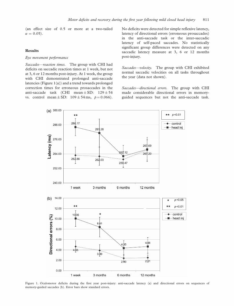
Saccades—reaction times. The group with CHI haddeficits on saccadic reaction times at 1 week, but notat 3, 6 or 12 months post-injury. At 1 week, the groupwith CHI demonstrated prolonged anti-saccadelatencies (Figure 1(a)) and a trend towards prolongedcorrection times for erroneous prosaccades in theanti-saccade task (CHI mean�SD: 129� 54vs. control mean�SD: 109� 54 ms, p¼ 0.066).
No deficits were detected for simple reflexive latency,latency of directional errors (erroneous prosaccades)in the anti-saccade task or the inter-saccadiclatency of self-paced saccades. No statisticallysignificant group differences were detected on anysaccadic latency measure at 3, 6 or 12 monthspost-injury.
Saccades—velocity. The group with CHI exhibitednormal saccadic velocities on all tasks throughoutthe year (data not shown).
Saccades—directional errors. The group with CHImade considerable directional errors in memory-guided sequences but not the anti-saccade task.
Figure 1. Oculomotor deficits during the first year post-injury: anti-saccade latency (a) and directional errors on sequences ofmemory-guided saccades (b). Error bars show standard errors.
Motor deficits and recovery during the first year following mild closed head injury 811
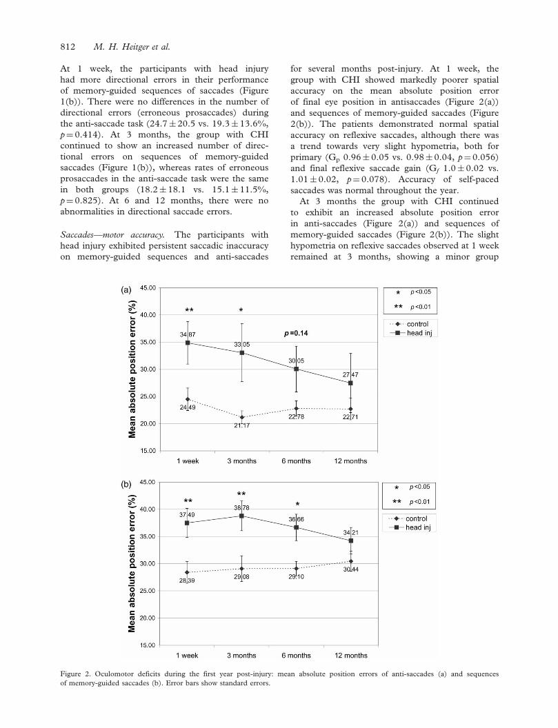
At 1 week, the participants with head injuryhad more directional errors in their performanceof memory-guided sequences of saccades (Figure1(b)). There were no differences in the number ofdirectional errors (erroneous prosaccades) duringthe anti-saccade task (24.7�20.5 vs. 19.3�13.6%,p¼ 0.414). At 3 months, the group with CHIcontinued to show an increased number of direc-tional errors on sequences of memory-guidedsaccades (Figure 1(b)), whereas rates of erroneousprosaccades in the anti-saccade task were the samein both groups (18.2�18.1 vs. 15.1� 11.5%,p¼ 0.825). At 6 and 12 months, there were noabnormalities in directional saccade errors.
Saccades—motor accuracy. The participants withhead injury exhibited persistent saccadic inaccuracyon memory-guided sequences and anti-saccades
for several months post-injury. At 1 week, thegroup with CHI showed markedly poorer spatialaccuracy on the mean absolute position errorof final eye position in antisaccades (Figure 2(a))and sequences of memory-guided saccades (Figure2(b)). The patients demonstrated normal spatialaccuracy on reflexive saccades, although there wasa trend towards very slight hypometria, both forprimary (Gp 0.96� 0.05 vs. 0.98� 0.04, p¼0.056)and final reflexive saccade gain (Gf 1.0� 0.02 vs.1.01�0.02, p¼ 0.078). Accuracy of self-pacedsaccades was normal throughout the year.
At 3 months the group with CHI continuedto exhibit an increased absolute position errorin anti-saccades (Figure 2(a)) and sequences ofmemory-guided saccades (Figure 2(b)). The slighthypometria on reflexive saccades observed at 1 weekremained at 3 months, showing a minor group
Figure 2. Oculomotor deficits during the first year post-injury: mean absolute position errors of anti-saccades (a) and sequencesof memory-guided saccades (b). Error bars show standard errors.
812 M. H. Heitger et al.

difference on primary (Gp 0.95� 0.04 vs.0.98� 0.04, p¼ 0.034) and final reflexive saccadegain (Gf 0.99� 0.02 vs. 1.01� 0.01, p¼ 0.004).At 6 months, a clear deficit remained on the absoluteposition error in sequences of memory-guidedsaccades (Figure 2(b)), but by 12 months, nodeficits in saccadic accuracy were observed onany task.
Saccades—timing and rhythm of memory-guided
sequences. Timing of saccades (absolute timeindex) was normal at 1 week (1.02�0.14 vs.0.99� 0.12, p¼ 0.4) and remained normal through-out the year. Similarly, the ability to keep a steadyrhythm within sequences was not impaired(0.09� 0.04 vs. 0.08� 0.03, p¼0.3) and remainedunimpaired.
Oculomotor smooth pursuit. At 1 week, the groupwith CHI had a slowed average peak trackingvelocity on fast sinusoidal smooth pursuit (60� s�1)(Figure 3(a)) in conjunction with an abnormalpursuit tracking lag on the same task (Figure 3(b)).There were no group differences in average peakvelocity or lag with the slower (i.e. 40� s�1) sinusoidaland (fast) random smooth pursuit tasks but therewas a trend towards a prolonged random trackinglag in the patient group (44.05� 14.8 vs. 37.78�
15.2 ms, p¼ 0.086). Performance on these two tasksremained normal at 3, 6 and 12 months.
By 6 months, the patients continued to haveslowed average peak velocity on 60� s�1 sinusoidalpursuit (Figure 3(a)) and a marginally slowedaverage peak velocity and abnormal lag on this taskat 12 months post-injury (Figure 3(b)).
Figure 3. Oculomotor deficits during the first year post-injury: average peak velocity and lag of fast sinusoidal oculomotor smooth pursuit(a and b, respectively). Error bars show standard errors.
Motor deficits and recovery during the first year following mild closed head injury 813

Upper-limb visuomotor performance
The group with CHI had long-term impairmentsin upper-limb visuomotor function, including poorerupper-limb movement accuracy and slowed ballisticarm movement velocity up to 12 months post-injury.
At 1 week, no deficits were found on visual acuity,visual resolution, static visual perception, dynamicvisual perception or arm movement steadinessand these measures remained normal throughoutthe year.
Conversely, the group with CHI had prolongedarm movement reaction time (Figure 4(a)) andmarkedly reduced (ballistic) arm movement peakvelocity (Figure 4(b)). The patient group showedincreased mean absolute errors on all non-preview1-D tracking tasks, these being sine tracking
(Figure 5(a)), random tracking (Figure 5(b)) andstep tracking (Figure 5(c)), with a trend towardsincreased mean absolute error on random-previewtracking (5.33� 1.6 vs. 4.71� 1.4 mm, p¼ 0.059).Similarly, at 1 week, the patients had prolonged lagon sine tracking (Figure 6(a)), random tracking(Figure 6(b)), random-preview tracking (81.0�63.8vs. 53.8�64 ms, p¼ 0.035) and step tracking(Figure 6(c)). No deficits were found on sine-preview tracking and performance on this taskremained normal at 3, 6 and 12 months.
At 3 months, arm movement reaction time of thegroup with CHI had improved to normal (Figure4(a)), while arm movement velocity remainedimpaired (Figure 4(b)). Similarly, mean absoluteerrors remained impaired on sine tracking
Figure 4. Upper-limb motor measures during the first year post-injury: arm movement reaction time (a) and upper-limb movementpeak velocity (b). Error bars show standard errors.
814 M. H. Heitger et al.
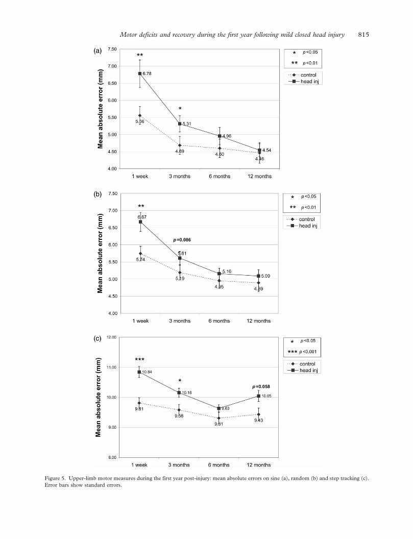
Figure 5. Upper-limb motor measures during the first year post-injury: mean absolute errors on sine (a), random (b) and step tracking (c).Error bars show standard errors.
Motor deficits and recovery during the first year following mild closed head injury 815
(Figure 5(a)) and step tracking (Figure 5(c)).In addition, there was a trend towards a highermean absolute error on random tracking in thepatient group (Figure 5(b)). The patient group alsoexhibited prolonged tracking lags on sine tracking(Figure 6(a)), random tracking (Figure 6(b)) and steptracking (Figure 6(c)).
At 6 months, the group with CHI continuedto have slowed arm movement velocity (Figure 2(b))and prolonged tracking lag on sine tracking(Figure 6(a)) and step tracking (Figure 6(c)). Thedeficits on all other measures of upper-limb motoraccuracy had improved to non-significant levels.
At 12 months, no difference was detected on armmovement peak velocity ( p¼ 0.094) (Figure 4(b))and the deficits on the visuomotor tracking taskshad resolved except for step tracking in which lagwas impaired (Figure 6(c)) and mean absolute errormarginally impaired (Figure 5(c)).
Neuropsychological testing
No deficits were found on the PASAT (2.4, 2.0, 1.6and 1.2 s pacing), TMT A/B time-to-completionor errors and the WASI (including Vocabulary andMatrix Reasoning T-scores) throughout the first yearpost-injury. At 1 week, the group with CHI showeddeficits only on the SDMT (z-score –0.07� 0.93 vs.0.56� 1.04, p¼ 0.007) and CVLT (Figures 7 and8). The impairments of the patient group on theCVLT comprised lower scores on the total standardscore (Figure 7(a)), short delay free recall(Figure 7(b)), short delay cued (Figure 7(c)), longdelay free recall (Figure 8(a)), long delay cued recall(Figure 8(b)) and List A/trial 5 (Figure 8(c)). Thepatients did not differ on serial or semantic clusterscores, meaning that the controls were not betternaming the shopping items in correct order and bothgroups had the same tendency to group the itemsinto their categories (i.e. fruit, tools, spices, etc.)when naming them. The group with CHI showedpreserved item recognition (recognition hits)but showed some difficulty in the differentiationbetween true items and non-items during long-delay recognition (recognition discriminability,�0.38�0.72 vs. �0.11� 0.52, p¼ 0.053).
At 3 months, the patient group showed deficitsonly on the CVLT, comprising lower scores on totalstandard score (Figure 7(a)), short delay free andcued recall (Figures 7(b) and (c)), as well as longdelay free and cued recall (Figures 8(a) and (b)).At 6 months, deficits remained only on CVLT totalstandard score (Figure 7(a)), short delay cued recall(Figure 7(c)) and long delay free recall (Figure 8(a)),while short delay free recall and long delaycued recall showed only marginal differences(Figures 7(b) and 8(b)). At this point, deficits on
List A/trial 5 resurfaced (Figure 8(c)), although thismeasure had been normal at 3 months. The groupwith CHI also showed a lower score on recogni-tion discriminability (–0.11� 0.46 vs. 0.11� 0.31,p¼ 0.017) indicating that the patients still experi-enced problems in differentiating between true itemsand non-items during long-delay recognition,although item recognition (recognition hits) waspreserved (as observed at 1 week and 3 months).At 12 months, no cognitive deficits remained, withthe exception of a marginal group difference on theCVLT total standard score (Figure 7(a)).
Health assessment measures
At 1 week, the patient group had comparatively highscores on the cumulative total of the RivermeadPost-concussion Symptoms Questionnaire (RPSQ)(19.81� 11.72 vs. 4.08� 5.42, p<0.001)(Figure 4). Over 91% of patients reported aninjury-related increase in levels of RPSQ-symptoms(i.e. with a score of 2 or higher it was considered atleast a mild problem by the reporting patient) and nopatient was entirely symptom-free (Table I).
At 3 months, the RPSQ total score had improvedbut remained abnormally high (9.81� 9.89 vs.4.08�5.42, p< 0.001) (Figure 9). Similarly, theCHI group had improved, yet abnormal rates of post-concussional complaints, with 59.5% of patientsreporting symptoms with a score of 2 or higher(i.e. an injury-related increase in symptom levels)(Table I). Only 16.2% were entirely symptom-free.
At 6 months, the average level of post-concussional symptoms was still higher in thegroup with CHI compared to the control baselinelevel (RPSQ total 10.24� 10.65 vs. 4.08� 5.02,p< 0.001) (Figure 9). Over 51% of patients con-tinued to report post-concussional symptoms witha score of 2 or higher, whilst only 13.5% of patientswere entirely symptom-free (Table I).
At 12 months, the RPSQ total of the patient groupremained at an increased level similar to 6 months(10.13� 11.96 vs. 4.39� 5.74, p¼ 0.020) (Figure 9)and more than 50% of the remaining 31 patientscontinued to report scores of 2 or higher on at leastone post-concussional symptom on the RPSQ, withonly 38.7% being entirely symptom-free at 1 yearpost-injury (Table I).
Discussion
The principal and novel finding of this study, thefirst to serially examine oculomotor and upper-limbvisuomotor function after mild CHI, is evidenceof previously unreported persistent eye and armmotor deficits during the first 12 months after mildhead trauma. The group with CHI had prolonged
816 M. H. Heitger et al.

Figure 6. Upper-limb motor measures during the first year post-injury: Lag on sine (a), random (b) and step tracking (c). Error barsshow standard errors.
Motor deficits and recovery during the first year following mild closed head injury 817

Figure 7. California Verbal Learning Test: CVLT total standard score (a), short delay free recall (b) and short delay cued recall (c).Error bars show standard errors.
818 M. H. Heitger et al.

Figure 8. California Verbal Learning Test: Long delay free recall (a), long-delay cued recall (b) and CVLT List A/trial 5 (c).Error bars show standard errors.
Motor deficits and recovery during the first year following mild closed head injury 819

deficits on several motor tasks including anti-saccades, memory-guided sequences, fast sinusoidalsmooth pursuit, upper-limb step, sine and randomtracking as well as ballistic upper-limb movementpeak velocity. Coupled with previous work [31–33],this finding indicates a consistent and reproducibleimpact of mild head trauma on eye and armmotor control.
The finding of motor impairment persistingbeyond 1 week of mild head trauma suggests anunexpectedly profound adverse impact of mild headtrauma on eye and arm motor control and thecerebral components mediating motor function.The profile of motor deficits at 1 week conveyeda picture of multi-focal dysfunction very similar tothat discussed previously [31–33] and consistentwith cerebral abnormalities seen in imaging studiesafter mild CHI [7, 9–12, 59]. The compositionof eye and upper-limb motor deficits persistingbeyond 1 week is consistent with a prolongedimpairment of motor functions originating in frontalcortical areas (i.e. supplementary motor area,primary and pre-motor cortex), posterior parietalcortex and hippocampal formation, areas shownby functional imaging to be adversely affected at3 and more months after mild CHI [9–12, 59, 60].
The deficits in motor function gradually improvedduring the first year post-injury, reflecting anongoing recovery process, parallel to the improvedsymptom profile. Even though a small performanceimprovement on some measures was also apparentin the controls—this indicating the influence ofpractice effects—the group with CHI performedconsistently worse than the controls with statisticallysignificant deficits persisting up to 6 and, in the caseof upper-limb step tracking and fast sinusoidalsmooth pursuit, 12 months post-injury.
It might be argued that these motor impairmentscould equally have been caused by cognitive deficits,in particular factors related to attention, ratherthan disturbances in motor processing due to aphysical impact of head trauma on the networksfor motor control. However, any relevant impair-ment of cognitive/attentional factors should havemanifested in statistically significant group differ-ences on the PASAT, SDMT, TMT (especiallytime-to-completion and errors on the TMT B [61])and the CVLT attention-span factor derived fromfirst recall of List A and List B [62]. Preservedperformance on the PASAT and TMT suggestspreservation of cognitive functions such assustained and divided attention, information
Figure 9. Mean total scores on the Rivermead Post-concussion Symptoms Questionnaire (RPSQ) throughout 1 year post-injury.Error bars show standard errors.
Table I. Recovery throughout the first year.
Rivermead Post-concussion Symptoms Questionnaire 1 week 3 months 6 months 12 months
(a) Patients who are entirely symptom-free (%)* 0.0 16.2 13.5 38.7(b) Patients without injury-related increase in RPSQ symptom levels (%)** 8.1 40.5 48.6 48.4
*This is a sub-group of group B. **No scores of 2 or higher on any symptoms (i.e. no symptom is more pronounced than pre-injury).
820 M. H. Heitger et al.
processing capacity, short-term and workingmemory [41, 63–67]. The group with CHI showedtransient deficits on the SDMT at 1 week, whichsuggests impaired speed of information processing atthis point in time [55], but the SDMT abnormalitieshad recovered by 3 months. The absence of anystatistically significant deficits beyond 1 week on thePASAT, SDMT and TMT, in combination withthe preserved performance on the WASI, indicateslargely normal levels of cognitive functioning,good levels of attention, working memory andspeed of information processing in the patientgroup and supports the conclusion that the impairedmotor performance was not caused by deficits inthese functions.
The inability of neuropsychological testing, withexception of the CVLT, to document a recoveryprofile emphasizes the notion that motor testingmay provide a sensitive and useful marker of residualbrain dysfunction, particularly in patients whopresent with persistent post-concussional symptomsor complaints months after their injury. In contrastto the concomitant occurrence of neuropsychologi-cal impairment (i.e. on tests such as PASAT, SDMTand TMT) and oculomotor deficits (i.e. reflexive,anti- and memory-guided saccades) after severehead injury [41], the findings indicate that deficitsin motor function are still apparent after mildCHI, whilst cerebral dysfunction, with the exceptionof deficits on verbal learning/declarative verbalmemory, may not necessarily manifest in detectableneuropsychological deficits beyond 1 month of mildCHI. These observations are consistent with pre-vious evidence that mild CHI is unlikely to reliablyproduce persisting psychometric abnormalities[24, 68–71]. In addition, the likelihood of abnormalneuropsychological performance indicating thepresence of a pathological condition is low followingmild head trauma [27] due to the susceptibilityof neuropsychological testing to pre-morbid intelli-gence and other factors such as age, level ofeducation, state of employment and socioeconomicstatus [24–26]. The current and previous [32]observations support the conclusion that instrumen-ted motor testing is unaffected by these factorsand can indicate the presence of ongoing dysfunc-tion at a level not easily accessible with conventionalneuropsychological assessment after mild CHI.
Limitations of the current study
Whilst this study uncovered the presence of persis-tent eye and upper-limb visuomotor deficits aftermild CHI, it has to be acknowledged that the level ofimpairment was subtle and that any useful applica-tion of motor testing in a clinical context after mild
CHI should not be based on the assessment of a fewisolated motor measures. The current findings implythat it is unlikely that there are ‘master-measures’(i.e. single measures of motor function that can be asole indicator of pathology in individuals). The stepof an useful transformation of the current (group-level) motor deficits into an application with utilityfor individual patients suffering from mild CHI canonly be achieved by way of regression-based model-ling, ideally using larger groups. Such models shouldbe able to channel the effect detected in the currentstudy into a format that allows the discriminationbetween normal and abnormal motor performanceon an individual level and thereby open a pathwayfor the potential use of motor ability as a marker offunctional impairment in the absence of grossmovement deficits. Preliminary work on this issuesuggests that such models will likely include severalkey measures that manifest deficits at a sample sizeof n¼ 37 in addition to other variables with weakerperformance gradients that do not show significantdeficits in small samples but still contribute in auseful manner to identifying patients with abnorm-alities in brain function after mild CHI. There arealso indications that such modelling may allow thevery efficient use of early motor assessment topredict adverse outcome during the first 6 monthsafter mild head trauma [72].
Conclusion
The results suggest that motor assessment asemployed in the present study may provide a newand objective way of screening for the presence ofresidual brain dysfunction in the months followingmild head trauma. This study has shown thatunsuspected eye and arm motor deficits can persistfor several months post-injury using techniqueswhich can objectively detect and measure suchdeficits. The susceptibility of eye and arm motorfunction to deficits throughout the first year follow-ing mild CHI suggests that the instrumentedassessment of motor function may be useful inproviding sensitive and objective markers of cerebraldysfunction. Such independent markers may be ofvalue in confirming injury status and ongoingcerebral dysfunction in patients who present withreports of persistent post-concussional symptomsand complaints in the months post-injury.
Acknowledgements
The study was hosted by the Canterbury DistrictHealth Board and the Christchurch School of
Motor deficits and recovery during the first year following mild closed head injury 821

Medicine & Health Sciences, University of Otago,New Zealand. Funding for patients’ travel costswas provided by the Neurological Foundation ofNew Zealand (NFNZ grant number 0027/SPG).We would like to thank these institutions for theirsupport of this research.
References
1. Jennett B. Epidemiology of head injury. Journal ofNeurology, Neurosurgery and Psychiatry 1996;60:362–369.
2. Kraus JF, Nourjah P. The epidemiology of mild head injury.In: Levin HS, Eisenberg HM, Benton AL, editors. Mild headinjury. New York: Oxford University Press; 1989. pp 8–22.
3. Jager TE, Weiss HB, Coben JH, Pepe PE. Traumatic braininjuries evaluated in U.S. emergency departments,1992–1994. Academic Emergency Medicine 2000;7:134–140.
4. Max W, MacKenzie EJ, Rice DP. Head injuries: Costs andconsequences. Journal of Head Trauma Rehabilitation1991;6:76–91.
5. Kraus JF, Nourjah P. The epidemiology of milduncomplicated brain injury. Journal of Trauma 1988;28:1637–1643.
6. McGregor K, Pentland B. Head injury rehabilitation in theU.K.: An economic perspective. Social Science and Medicine1997;45:295–303.
7. McAllister TW, Sparling MB, Flashman LA, Saykin AJ.Neuroimaging findings in mild traumatic brain injury.Journal of Clinical and Experimental Neuropsychology2001;23:775–791.
8. Blumbergs PC, Scott G, Manavis J, Wainwright H,Simpson DA, McLean AJ. Staining of amyloid precursorprotein to study axonal damage in mild head injury. Lancet1994;344:1055–1056.
9. Hofman PA, Verhey FR, Wilmink JT, Rozendaal N, Jolles J.Brain lesions in patients visiting a memory clinic withpostconcussional sequelae after mild to moderate braininjury. Journal of Neuropsychiatry and ClinicalNeurosciences 2002;14:176–184.
10. Kant R, Smith-Seemiller L, Isaac G, Duffy J. Tc-HMPAOSPECT in persistent post-concussion syndrome aftermild head injury: Comparison with MRI/CT. Brain Injury1997;11:115–124.
11. Abu-Judeh HH, Parker R, Singh M, el-Zeftawy H, Atay S,Kumar M, Naddaf S, Aleksic S, Abdel-Dayim HM. SPETbrain perfusion imaging in mild traumatic brain injurywithout loss of consciousness and normal computed tomo-graphy. Nuclear Medicine Communications 1999;20:505–510.
12. Jacobs A, Put E, Ingels M, Put T, Bossuyt A. One-yearfollow-up of technetium-99m-HMPAO SPECT in mild headinjury. Journal of Nuclear Medicine 1996;37:1605–1609.
13. McAllister TW, Sparling MB, Flashman LA, Guerin SJ,Mamourian AC, Saykin AJ. Differential working memoryload effects after mild traumatic brain injury. Neuroimage2001;14:1004–1012.
14. Ommaya AK. Head injury mechanisms and the conceptof preventive management: A review and critical synthesis.Journal of Neurotrauma 1995;12:527–546.
15. Centers for Disease Control and Prevention, National Centerfor Injury Prevention and Control. Traumatic brain injuryin the United States—A report to Congress. Atlanta, GA:Centers for Disease Control and Prevention; 1999.
16. Wrightson P, Gronwall D. Mild head injury in New Zealand:Incidence of injury and persisting symptoms. New ZealandMedical Journal 1998;111:99–101.
17. Mallinson AI, Longridge NS. Specific vocalized complaintsin whiplash and minor head injury patients. American Journalof Otology 1998;19:809–813.
18. Bohnen N, Jolles J. Neurobehavioral aspects of postcon-cussive symptoms after mild head injury. Journal of Nervousand Mental Disease 1992;180:683–692.
19. Mittenberg W, Canyock EM, Condit D, Patton D.Treatment of post-concussion syndrome following mildhead injury. Journal of Clinical and ExperimentalNeuropsychology 2001;23:829–836.
20. Bazarian JJ, Wong T, Harris M, Leahey N, Mookerjee S,Dombovy M. Epidemiology and predictors of post-concus-sive syndrome after minor head injury in an emergencypopulation. Brain Injury 1999;13:173–189.
21. Levin HS, Williams DH, Eisenberg HM, High WM, Jr,Guinto FC, Jr. Serial MRI and neurobehavioural findingsafter mild to moderate closed head injury. Journal ofNeurology, Neurosurgery and Psychiatry 1992;55:255–262.
22. Umile EM, Plotkin RC, Sandel ME. Functional assessmentof mild traumatic brain injury using SPECT and neuro-psychological testing. Brain Injury 1998;12:577–594.
23. Radanov BP, Bicik I, Dvorak J, Antinnes J, von SchulthesGK, Buck A. Relation between neuropsychological andneuroimaging findings in patients with late whiplash syn-drome. Journal of Neurology, Neurosurgery and Psychiatry1999;66:485–489.
24. Ponsford J, Willmott C, Rothwell A, Cameron P, Kelly AM,Nelms R, Curan C, Ng K. Factors influencing outcomefollowing mild traumatic brain injury in adults. Journal of theInternational Neuropsychological Society 2000;6:568–579.
25. Dikmen S, Machamer J, Temkin N. Mild head injury: Factsand artifacts. Journal of Clinical and ExperimentalNeuropsychology 2001;23:729–738.
26. Reitan RM, Wolfson D. The significance of sensory-motorfunctions as indicators of brain dysfunction in children.Archives of Clinical Neuropsychology 2003;18:11–18.
27. Binder LM, Rohling ML, Larrabee J. A review of mildhead trauma. Part I: Meta-analytic review of neuropsycho-logical studies. Journal of Clinical and ExperimentalNeuropsychology 1997;19:421–431.
28. De Kruijk JR, Leffers P, Menheere PPCA, Meerhoff S,Rutten J, Twijnstra A. Prediction of post-traumatic com-plaints after mild traumatic brain injury: early symptoms andbiochemical markers. Journal of Neurology, Neurosurgeryand Psychiatry 2002;73:727–732.
29. Savola O, Hillbom M. Early predictors of post-concussionsymptoms in patients with mild head injury. EuropeanJournal of Neurology 2003;10:175–181.
30. Ingebrigtsen T, Waterloo K, Jacobsen EA, Langbakk B,Romner B. Traumatic brain damage in minor head injury:Relation of serum S-100 protein measurements to magneticresonance imaging and neurobehavioral outcome.Neurosurgery 1999;45:468–475; discussion 475–476.
31. Heitger MH, Anderson TJ, Jones RD. Saccadesequences as markers for cerebral dysfunction followingmild closed head injury. Progress in Brain Research2002;140:433–448.
32. Heitger MH, Anderson TJ, Jones RD, et al. Eye movementand visuomotor arm movement deficits following mild closedhead injury. Brain 2004;127:575–590.
33. Heitger MH, MacAskill MR, Jones RD, Dalrymple-Alford JC, Frampton CM, Ardagh MW. The impactof mild closed head injury on involuntary saccadicadaptation – evidence for the preservation of implicitmotor learning. Brain Injury 2005;19:109–117.
822 M. H. Heitger et al.
34. Pierrot-Deseilligny C, Muri RM, Ploner CJ, et al. Corticalcontrol of ocular saccades in humans: A model for motricity.Progress in Brain Research 2003;142:3–17.
35. Jones RD. Measurement of sensory-motor control perfor-mance capacities: Tracking tasks. In: Bronzino JD, editor.The biomedical engineering handbook. 2nd ed. Boca Raton(FL): CRC Press; 2000. pp 1–25.
36. Sweeney JA, Mintun MA, Kwee S, Wiseman MB,Brown DL, Rosenberg DR, Carl JR. Positron emissiontomography study of voluntary saccadic eye movementsand spatial working memory. Journal of Neurophysiology1996;75:454–468.
37. Anderson TJ, Jenkins IH, Brooks DJ, Hawken MB,Franckowian RS. Cortical control of saccades and fixationin man. A PET study. Brain 1994;117:1073–1084.
38. Hamzei F, Dettmers C, Rijntjes M, Glauche V, Kiebei S,Weber B, Weiller C. Visuomotor control within a distributedparieto-frontal network. Experimental Brain Research2002;146:273–281.
39. Ellermann JM, Siegal JD, Strupp JP, Ebner TJ, Ugurbil K.Activation of visuomotor systems during visually guidedmovements: A functional MRI study. Journal of MagneticResonance 1998;131:272–285.
40. Graziano MS, Taylor CS, Moore T, Cooke DF. The corticalcontrol of movement revisited. Neuron 2002;36:349–362.
41. Williams IM, Ponsford JL, Gibson KL, Mulhall LE,Curran CA, Abel LA. Cerebral control of saccades andneuropsychological test results after head injury. Journal ofClinical Neuroscience 1997;4:186–196.
42. Haaland KY, Temkin N, Randahl G, Dikmen S. Recovery ofsimple motor skills after head injury. Journal of Clinicaland Experimental Neuropsychology 1994;16:448–456.
43. Hills EC, Geldmacher DS. The effect of character and arraytype on visual spatial search quality following traumatic braininjury. Brain Injury 1998;12:69–76.
44. Prigatano GP. Impaired awareness, finger tapping, andrehabilitation outcome after brain injury. RehabilitationPsychology 1999;44:145–159.
45. Perbal S, Couillet J, Azouvi P, Pouthas V. Relationshipsbetween time estimation, memory, attention, and processingspeed in patients with severe traumatic brain injury.Neuropsychologia 2003;41:1599–1610.
46. Jones RD, Donaldson IM. Measurement of integratedsensory-motor function following brain damage by a compu-terized preview tracking task. International RehabilitationMedicine 1981;3:71–83.
47. Haggard P, Miall RC, Wade D, Fowler S, Richardson A,Anslow P, Stein J. Damage to cerebellocortical pathways afterclosed head injury: A behavioural and magnetic resonanceimaging study. Journal of Neurology, Neurosurgery andPsychiatry 1995;58:433–438.
48. Sietsema JM, Nelson DL, Mulder RM, Mervau-Scheidel D,White BE. The use of a game to promote arm reach inpersons with traumatic brain injury. American Journal ofOccupational Therapy. 1993;47:19–24.
49. Swaine BR, Sullivan SJ. Relation between clinical andinstrumented measures of motor coordination in traumati-cally brain injured persons. Archives of Physical Medicine &Rehabilitation 1992;73:55–59.
50. Kuhtz-Buschbeck JP, Stolze H, Golge M, Ritz A. Analysesof gait, reaching, and grasping in children after traumaticbrain injury. Archives of Physical Medicine & Rehabilitation2003;84:424–430.
51. Reulen JPH, Marcus JT, Koops D, de Vries FR, Tiesinga G,Boshuizen K, Bos JE. Precise recording of eye movement:The IRIS technique Part 1. Medical and BiologicalEngineering and Computing 1988;26:20–26.
52. Muir SR, MacAskill MR, Herron D, et al. EMMA—an eyemovement measurement and analysis system. AustralasianPhysical and Engineering Sciences in Medicine2003;26:18–24.
53. Gronwall DMA. Paced auditory serial addition test(PASAT). Victoria, Canada: Department of Psychology,University of Victoria; 1977.
54. Delis DC, Kramer JH, Kaplan E, Ober BA. California verballearning test (CVLT). San Antonio: The PsychologicalCorporation�, Harcourt Brace & Company; 1987.
55. Smith A. Symbol digit modalities test. Los Angeles, CA:Western Psychological Services; 1973.
56. Spreen O, Strauss E. A compendium of neuropsychologicaltests. New York: Oxford University Press; 1991.
57. The Psychological Corporation. Wechsler abbreviated scaleof intelligence—manual. San Antonio: The PsychologicalCorporation; 1999.
58. King NS, Crawford S, Wenden FJ, Moss NE, Wade DT.The Rivermead Post Concussion Symptoms Questionnaire:A measure of symptoms commonly experienced afterhead injury and its reliability. Journal of Neurology1995;242:587–592.
59. Chen SH, Kareken DA, Fastenau PS, Trexler LE,Hutchins GD. A study of persistent post-concussion symp-toms in mild head trauma using positron emission tomo-graphy. Journal of Neurology, Neurosurgery and Psychiatry2003;74:326–332.
60. Gray BG, Ichise M, Chung DG, Kirsch JC, Franks W.Technetium-99m-HMPAO SPECT in the evaluation ofpatients with a remote history of traumatic brain injury: Acomparison with x-ray computed tomography. Journal ofNuclear Medicine 1992;33:52–58.
61. Ruffolo LF, Guilmette TJ, Willis GW. Comparison of timeand error rates on the trail making test among patientswith head injuries, experimental malingerers, patients withsuspect effort on testing, and normal controls. ClinicalNeuropsychologist 2000;14:223–230.
62. Wiegner S, Donders J. Performance on the CaliforniaVerbal Learning Test after traumatic brain injury. Journalof Clinical and Experimental Neuropsychology1999;21:159–170.
63. Gronwall D. Cumulative and persisting effects of concussionon attention and cognition. In: Levin HS, Eisenberg HM,Benton AL, editors. Mild head injury. New York: OxfordUniversity Press; 1989. pp 153–162.
64. Sherman EMS, Strauss E, Spellacy F. Validity of the PacedAuditory Serial Addition Test (PASAT) in adults referredfor neuropsychological assessment after head injury. ClinicalNeuropsychologist 1997;11:34–45.
65. Anderson CV, Bigler ED, Blatter DD. Frontal lobelesions, diffuse damage, and neuropsychologicalfunctioning in traumatic brain-injured patients. Journalof Clinical and Experimental Neuropsychology 1995;17:900–908.
66. Arbuthnott K, Frank J. Trail making test, part B as a measureof executive control: Validation using a set-switching para-digm. Journal of Clinical and Experimental Neuropsychology2000;22:518–528.
67. Di Stefano G, Radanov BP. Course of attention and memoryafter common whiplash: A two-years prospective study withage, education and gender pair-matched patients. ActaNeurologica Scandinavica 1995;91:346–352.
68. Dikmen S, McLean A, Temkin N. Neuropsychologicaland psychosocial consequences of minor head injury.Journal of Neurology, Neurosurgery and Psychiatry1986;49:1227–1232.
69. Levin HS, Mattis S, Ruff RM, Eisenberg HM,Marshall LF, Tabaddor K, High WM, Jr,
Motor deficits and recovery during the first year following mild closed head injury 823

Frankowski RF. Neurobehavioral outcome followingminor head injury: A three-center study. Journal ofNeurosurgery 1987;66:234–243.
70. Gentilini M, Nichelli P, Schoenhuber R, et al.Neuropsychological evaluation of mild head injury.Journal of Neurology, Neurosurgery and Psychiatry1985;48:137–140.
71. Richardson JTE. Clinical and neuropsychological aspectsof closed head injury. Brain damage, behaviour and cognitionseries. Hove (UK): Psychology Press; 2000.
72. Heitger MH, Anderson TJ, Jones RD. A role for instrumen-ted motor assessment in clinical assessment and outcomeprediction after mild head injury (Abstract). Brain Injury2005;19(Suppl 1):111.
824 M. H. Heitger et al.
Related Documents