BEHAVIORAL AND NEURAL BIOLOGY 60, 42--51 (1993) Motion Sickness in Amphibians RICHARD J. WASSERSUG,* AKEMI IZUMI-KUROTANI,t MASAMICHI YAMASHITA,t AND TOMIO NAITOH*'1 ~Department of Biology, Shimane University, Matsue 690, Japan; tSpace UtilizationResearch Center, Institute of Space and Astronautical Science, 3-1-1, Yoshino-dai, Sagamihara, Kanagawa 229, Japan We explored the question of whether amphibians get motion sickness by exposing anurans (frogs) and urodeles (salamanders) to the provocative stimulus of parabolic aircraft flight. Animals were fed before flight, and the presence of vomitus in their containers after flight was used to indicate motion-induced emesis. None of the spe- cies that we studied vomited during the 8 to 10 parabolas of each flight. However, at least one specimen from each of the anuran species Rana rugosa, Rana nigromaculata, Hyla japonica, and Rhacophorus schlegelii vomited in a period of 0.5 to 42 h after flight. Some specimens of R. nigromaculata, H. japonica, and R. schlegelii were also observed retching without emesis either during or shortly after exposure to parabolic flight. We were unable to in- 1 We thank the following students at Shimane University and Yokohama City University for their assistance with collecting, maintaining, and observing the amphibians used in this study: Chiharu Suzuki, Shigefumi Yokota, Tetsuya Kitayama, and Ha- jime Koike. Dr. Makato Asashima supplied the larval C. pyr- rhogaster. Drs. Kenichi Ijiri and Michael Wiederhold exhibited great patience and colleagueship in sharing research space and equipment with us during the course of our experiments. The manuscript profited from the critical attention of Monika Fejtek, Ronald Leslie, Kenneth Money, and Scott Pronych. This inves- tigation would not have been possible without extensive help, both in the air and on the ground, from the staff of Diamond Air Service, Nishikasugai-gun, Japan. The flight opportunity on their MU-300 aircraft was funded by the National Space De- velopment Agency of Japan. The research of A. Izumi-Kurotani, M. Yamashita, and T. Naitoh is supported by the Japanese Min- istry of Education, Science and Culture. In addition, A. Izumi- Kurotani and M. Yamashita are supported by the Fund for Basic Experiments Oriented to Space Station Utilization for the In- stitute of Space and Astronautical Science. R. J. Wassersug's participation in this project was made possible by grants from the Natural Science and Engineering Research Council of Can- ada and the Japan Science and Technology Fund (External Af- fairs and International Trade, Canada), plus contractual support from the Canadian Space Agency. Address correspondence and reprint requests to Dr. R. J. Wassersug at present address: Dept. of Anatomy and Neurobiology, Sir Charles Tupper Medical Building, Dalhousie University, Halifax, NS, Canada B3H 4H7.Fax: 902-494-1212; Email address: [email protected] CA.BITNET. duce either emesis or retching behavior in the aquatic frog Xenopus laevis. Among the urodeles studied we saw no signs of motion sickness in either adult or larval Cy- nops pyrrhogaster, but at least one larval Hynobius ne- bulosus vomited shortly after parabolic flight. The am- phibian species that exhibited the most motion sickness were the same ones that showed the greatest amount of tumbling during the microgravity phases of their para- bolic flights. The most distinctive difference between mo- tion sickness in amphibians and mammals that vomit, including man, is the long delay between a provocative stimulus and emesis proper in the amphibians. The retch- ing behavior we induced in the frogs was identical to that described previously for frogs treated with emetic drugs. H. japonica, exposed to extended periods of microgravity on the MIR Space Station, flattened their bellies against the substrate and dorsiflexed their heads in a manner reminiscent of drug-induced nausea. In light of our cur- rent observations of retching behavior in motion sick H. japonica, we suggest that the previously observed be- havior of tree frogs on the MIR Space Station was a man- ifestation of motion sickness. ©1993 Academic Press, Inc. INTRODUCTION Motion sickness is a debilitating condition that limits the capacity for work in environments where changes in acceleration are unavoidable, notably at sea (sailors), in the air (aviators), and in micro- gravity (astronauts). Susceptibility to motion sick- ness varies greatly among individuals (Mirabile, 1990; Reschke, 1990) and among species (see Miller, 1991, for the most recent review). For example, spi- der monkeys are highly susceptible to motion sick- ness, whereas rhesus monkeys are quite resistant (Corcoran, Fox, & Daunton, 1990). Although many factors have been identified that relate to the pro- pensity for some humans to get motion sickness (Crampton, 1990; Miller, 1991), the correlation with any one factor is not high. It remains largely un- known why certain organisms become nauseated by changes in acceleration while others do not. 0163-1047/93 $5.00 Copyright © 1993 by AcademicPress, Inc. All rights of reproduction in any form reserved. 42

Welcome message from author
This document is posted to help you gain knowledge. Please leave a comment to let me know what you think about it! Share it to your friends and learn new things together.
Transcript
BEHAVIORAL AND NEURAL BIOLOGY 6 0 , 4 2 - - 5 1 (1993)
Motion Sickness in Amphibians RICHARD J. WASSERSUG,* AKEMI IZUMI-KUROTANI,t MASAMICHI YAMASHITA,t AND TOMIO NAITOH *'1
~Department of Biology, Shimane University, Matsue 690, Japan; tSpace Utilization Research Center, Institute of Space and Astronautical Science, 3-1-1, Yoshino-dai, Sagamihara, Kanagawa 229, Japan
We explored the question of whether amphibians get motion sickness by exposing anurans (frogs) and urodeles (salamanders) to the provocative stimulus of parabolic aircraft flight. Animals were fed before flight, and the presence of vomitus in their containers after flight was used to indicate motion-induced emesis. None of the spe- cies that we studied vomited during the 8 to 10 parabolas of each flight. However, at least one specimen from each of the anuran species Rana rugosa, Rana nigromaculata, Hyla japonica, and Rhacophorus schlegelii vomited in a period of 0.5 to 42 h after flight. Some specimens of R. nigromaculata, H. japonica, and R. schlegelii were also observed retching without emesis either during or shortly after exposure to parabolic flight. We were unable to in-
1 We thank the following students at Shimane University and Yokohama City University for their assistance with collecting, maintaining, and observing the amphibians used in this study: Chiharu Suzuki, Shigefumi Yokota, Tetsuya Kitayama, and Ha- jime Koike. Dr. Makato Asashima supplied the larval C. pyr- rhogaster. Drs. Kenichi Ijiri and Michael Wiederhold exhibited great patience and colleagueship in sharing research space and equipment with us during the course of our experiments. The manuscript profited from the critical attention of Monika Fejtek, Ronald Leslie, Kenneth Money, and Scott Pronych. This inves- tigation would not have been possible without extensive help, both in the air and on the ground, from the staff of Diamond Air Service, Nishikasugai-gun, Japan. The flight opportunity on their MU-300 aircraft was funded by the National Space De- velopment Agency of Japan. The research of A. Izumi-Kurotani, M. Yamashita, and T. Naitoh is supported by the Japanese Min- istry of Education, Science and Culture. In addition, A. Izumi- Kurotani and M. Yamashita are supported by the Fund for Basic Experiments Oriented to Space Station Utilization for the In- stitute of Space and Astronautical Science. R. J. Wassersug's participation in this project was made possible by grants from the Natural Science and Engineering Research Council of Can- ada and the Japan Science and Technology Fund (External Af- fairs and International Trade, Canada), plus contractual support from the Canadian Space Agency. Address correspondence and reprint requests to Dr. R. J. Wassersug at present address: Dept. of Anatomy and Neurobiology, Sir Charles Tupper Medical Building, Dalhousie University, Halifax, NS, Canada B3H 4H7.Fax: 902-494-1212; Email address: [email protected] CA.BITNET.
duce either emesis or retching behavior in the aquatic frog Xenopus laevis. Among the urodeles studied we saw no signs of motion sickness in either adult or larval Cy- nops pyrrhogaster, but at least one larval Hynobius ne- bulosus vomited shortly after parabolic flight. The am- phibian species that exhibited the most motion sickness were the same ones that showed the greatest amount of tumbling during the microgravity phases of their para- bolic flights. The most distinctive difference between mo- tion sickness in amphibians and mammals that vomit, including man, is the long delay between a provocative stimulus and emesis proper in the amphibians. The retch- ing behavior we induced in the frogs was identical to that described previously for frogs treated with emetic drugs. H. japonica, exposed to extended periods of microgravity on the MIR Space Station, flattened their bellies against the substrate and dorsiflexed their heads in a manner reminiscent of drug-induced nausea. In light of our cur- rent observations of retching behavior in motion sick H. japonica, we suggest that the previously observed be- havior of tree frogs on the MIR Space Station was a man- ifestation of motion sickness. © 1993 Academic Press, Inc.
I N T R O D U C T I O N
Motion sickness is a debi l i ta t ing condit ion t h a t l imits the capaci ty for work in env i ronmen t s where changes in accelerat ion are unavoidable , no tab ly at sea (sailors), in the air (aviators), and in micro- g rav i ty (astronauts) . Suscept ibi l i ty to mot ion sick- ness var ies g rea t ly a m o n g individuals (Mirabile, 1990; Reschke, 1990) and a m o n g species (see Miller, 1991, for the most recent review). For example, spi- der monkeys are h igh ly susceptible to mot ion sick- ness, whereas rhesus monkeys are quite res i s tan t (Corcoran, Fox, & Daun ton , 1990). A l t h o u g h m a n y factors have been identified t h a t re la te to the pro- pens i ty for some h u m a n s to get mot ion sickness (Crampton, 1990; Miller, 1991), the correla t ion wi th any one factor is not high. I t r ema ins la rge ly un- known why cer ta in o rgan i sms become nausea t ed by changes in accelerat ion while o thers do not.
0163-1047/93 $5.00 Copyright © 1993 by Academic Press, Inc. All rights of reproduction in any form reserved.
42
MOTION SICKNESS IN AMPHIBIANS 43
In the present report we explore whether am- phibians experience motion sickness. Our interest in this seemingly esoteric question is prompted by a variety of issues. First, there is an ostensibly prac- tical reason to be concerned about the evolution and neurophysiology of emesis in vertebrates other than mammals. In mammals, including man, emesis is controlled along several neural pathways (see, for example, papers in Crampton, 1990; Davis, Lake- Bakaar, & Grahame-Smith, 1986, for recent re- views). Consequently, drugs that mitigate gastric distress in patients subjected to anticancer chem- otherapy are not necessarily effective against mo- tion-induced nausea (Kohl, 1987; Lucot, 1989; Stott, Barnes, Wright, & Ruddock, 1989), and vice versa (Miller, 1991). At the moment, mammals--often cats, dogs, or monkeys--are the model organisms used in the search for better antiemetic medications. Given their size, costs, and social association with man, these animals are far from ideal test subjects in the necessarily stressful field of emetic research. Efforts are underway to find other model organisms for emetic studies (e.g., Ueno, Matsuki, & Saito, 1988). If it can be shown that the emetic response of amphibians and its neuroregulation are essen- tially the same as in mammals--i.e., evolutionarily conserved--then future work on the development of antiemetic medications could use these lower ver- tebrates in place of mammals.
Second, if amphibians are susceptible to motion sickness, then they offer some advantage over mam- mals in studies directed at understanding environ- mental or "lifestyle" correlates of motion sickness. Does, for example, the propensity toward motion sickness relate to any aspect of the locomotor pat- tern (e.g., arboreal vs terrestrial) or visual realm of organisms? Such questions are not easy to explore with mammals. In contrast, amphibians of similar (compact) size run the gambit from strictly aquatic to strictly fossorial to highly arboreal. Some are diurnal, others are nocturnal. In terms of diet, they range from species with good visual acuity that feed on swift aerial insects, to ones with more limited vision that prey on slower, ground-dwelling ar- thropods, or aquatic and fossorial forms that do not need any visual clues to locate food. If we can doc- ument motion sickness in amphibians, we may be able to use their diversity in habitat and habit to begin to correlate the propensity for motion sickness with the way of life of organisms. Ultimately, un- derstanding the adaptive significance of emesis re- quires elucidating such correlates.
A final reason for exploring motion sickness in amphibians is to evaluate the suggestion that an
unexplained behavior, previously documented for anurans in microgravity (t~G), was actually a man- ifestation of motion sickness. The behavior in ques- tion was exhibited by tree frogs during a 1990 MIR Space Station mission (FRIS Experiment Group, 1991; Izumi-Kurotani, Yamashita, Kawasaki, Ku- rotani, Mogami, Okuno, Akiyama, Oketa, Shiraishi, & Ueda, 1991; Izumi-Kurotani, Yamashita, Ka- wasaki, Kurotani, Mogami, Okuno, Akiyama, Oketa, Shiraishi, Ueda, Wassersug, & Naitoh, 1993). A videotape of those frogs shows a number with hindlimbs retracted, abdomens pressed against the substrate, and heads sharply extended. This pos- ture resembles the retching display induced in frogs by emetic drugs (Naitoh, Wassersug, & Leslie, 1989). We reasoned that if we could provoke the same postural display with a motion stimulus and associate it with emesis, this would offer support for the suggestion that the posture of the frogs on the MIR Space Station was, in fact, a manifestation of motion sickness.
By way of background, it should be noted that pharmaceutically induced emesis in frogs was dem- onstrated well over 100 years ago (Mellinger, 1881). All the frog species used in the current study are known to vomit in response to emetic drugs (Naitoh, Imamura, & Wassersug, 1991). Only recently, how- ever, have salamanders been shown to vomit in re- sponse to the same emetic agents (Naitoh & Was- sersug, 1992). Most frog larvae lack a functional stomach and other anatomical features necessary for emesis (Brown, Wassersug, & Naitoh, 1992; Nai- toh et al., 1989); consequently no tadpoles are known to vomit or exhibit any behavior that could be described as retching.
It is often cited that fish can get motion sickness (e.g., Chinn & Smith, 1955; Miller, 1991; Money, 1970), but this appears to be based on only a few references, largely anecdotal, from the first half of the century (e.g., McKenzie, 1935, for Gadus mor- hua; Suyehiro, 1934, for Sardina melanosticta). We know of no studies on motion sickness for fishes that fully control for the confounding stresses of capture, confinement, or contact with container and conspecifics. In fact, we know of no comparative studies on motion sickness in any poikilothermic vertebrates.
METHODS
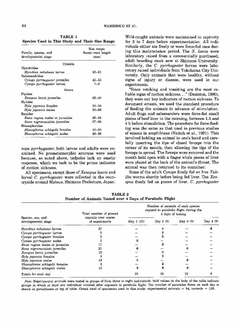
Seven species from six different genera and fam- ilies of amphibians were used in this study (Tables 1 and 2). Both frogs (Anura) and salamanders (Uro- dela) were examined. For one species, the newt Cy-
44 WASSERSUG ET AL.
T A B L E 1 Species Used in This Study and Their Size Range
Family, species, and developmental stage
Size range: Snout-vent length
(ram)
Urodela Hynobiidae
Hynobius nebulosus larvae 15-21 Salamandridae
Cynops pyrrhogaster juveniles 45-55 Cynops pyrrhogaster larvae 7-8
Anura Pipidae
Xenopus laevis juveniles 26-40 Hylidae
Hyla japonica females 34-36 Hyla japonica males 30-36
Ranidae Rana rugosa males or juveniles 26-35 Rana nigromaculata juveniles 27-35
Rhacophoridae Rhacophorus schlegelii females 40-50 Rhacophorus schlegelii males 26-36
nops pyrrhogaster, both larvae and adults were ex- amined. No premetamorphic anurans were used because, as noted above, tadpoles lack an emetic response, which we took to be the prime indicator of motion sickness.
All specimens, except those of Xenopus laevis and larval C. pyrrhogaster were collected in the coun- tryside around Matsue, Shimane Prefecture, Japan.
Wild-caught animals were maintained in captivity for 3 to 7 days before experimentation. All indi- viduals either ate freely or were force-fed once dur- ing this maintenance period. The X. laevis were laboratory raised from a commercially purchased, adult breeding stock now at Shimane University. Similarly, the C. pyrrhogaster larvae were labo- ratory raised individuals from Yokohama City Uni- versity. Only animals that were healthy, without signs of injury or disease, were used in our experiments.
"Since retching and vomiting are the most re- liable signs of motion sickness..." (Daunton, 1990), they were our key indicators of motion sickness. To document emesis, we used the standard procedure of feeding the animals in advance of stimulation. Adult frogs and salamanders were force-fed small pieces of beef liver in the morning, between 1.5 and 4 h before stimulation. The procedure for force-feed- ing was the same as that used in previous studies of emesis in amphibians (Naitoh et al., 1991). This involved holding an animal in one's hand and care- fully inserting the tips of closed forceps into the corner of its mouth, then allowing the tips of the forceps to spread. The forceps were removed and the mouth held open with a finger while pieces of liver were placed at the back of the animal's throat. The animal was then returned to its container.
Some of the adult Cynops freely fed on live Tub- ifex worms shortly before being fed liver. The Xen- opus freely fed on pieces of liver. C. pyrrhogaster
T A B L E 2 Number of Animals Tested over 4 Days of Parabolic Flight
Species, sex, and developmental stage
Total number of ground controls over course
of experiments
Number of animals of each species exposed to parabolic flight during the
4 days of testing
Day 1 (10) Day 2 (8) Day 3 (9) Day 4 (9)
Hynobius nebulosus larvae Cynops pyrrhogaster larvae Cynops pyrrhogaster females Cynops pyrrhogaster males Rana rugosa males or juveniles Rana nigromaculata juveniles Xenopus laevis juveniles Hyla japonica females Hyla japonica males Rhacophorus schlegelii females Rhacophorus schlegelii males
Totals for each day
2 7 - - 4 - - 6
5 - - 6 - - - -
3 - - 5 - - - -
3 6 - - - - - -
1 7 - - 8 - - - -
2 1 6 - - 6 - -
1 5 - - 6 - - - -
3 - - 3 - - - -
19 6 - - 8 - -
3 - - 5 - - - -
I 0 5 6 8 - -
23 4 3 2 2 6
Note. Experimental animals were tested in groups of from three to eight individuals. Bold values in the body of the table indicate groups in which at least one individual vomited after exposure to parabolic flight. The number of parabolas flown on each day is shown in parentheses at top of table. Grand total of specimens used in this study: experimental animals = 94, controls = 126.
MOTION SICKNESS IN AMPHIBIANS 45
and Hynobius nebulosus larvae also fed freely on Tubifex worms. All animals were carefully observed during the half-hour after feeding to ensure that they had swallowed the food.
Extra animals were fed in the manner described and returned to containers to serve as ground con- trols. The number of controls was augmented by additional animals, captured a week after the ex- periments, and maintained and fed as just described. The total number of experimental and control spec- imens used in this study is given in Table 2.
Space and time constraints precluded testing each specimen individually. Consequently, both experi- mental and control specimens were tested in groups separated by species, sex, and developmental stage. Each group had from three to eight specimens (see Table 2). The lower limit was set by the availabili ty of specimens (i.e., we only had a few adult female H. japonica to work with). The upper limit was set by the number of individuals tha t we felt could com- fortably fit into the available test containers and still be easily observed.
All postmetamorphic amphibians, with the ex- ception of the Xenopus, were enclosed in 2.I-liter chambers for testing. These chambers were clear plastic containers with smooth, wet walls, but no measurable volume of free-standing water. The aquatic forms, namely the postmetamorphic Xen- opus and the salamander larvae, were studied in closed containers nearly filled with water. The vol- ume of the container for Xenopus was 1550 ml; for the larval salamanders, 350 ml. The residual air space in these containers was less than 5% of their total volume.
We used parabolic flight, a classical st imulus for provoking motion sickness in man (Reschke, 1990), as the provocative st imulus for our amphibians. Containers with prefed frogs and salamanders were anchored inside a Mitsubishi MU 300 aircraft, which flew 9 -+ 1 consecutive parabolas within an hour period, over a 4-day interval in mid-May 1991. The parabolic maneuvers were performed in a pres- surized cabin between 20,000 and 30,000 feet and during the hours of 10 : 00 AM to 4: 00 PM each day. The sequence of parabolas began with a hyperG "pull-up," and the aircraft achieved approximately 20 s of near zero G on each parabola. Throughout the flight the cabin was pressurized and maintained at a temperature circa 21 _ 1°C; however, over the 4 days of experimentation, animals were exposed to temperatures as low as 19°C and as high as 26°C during brief periods, such as loading. No animal was subjected to more than 1 day's flight. The spe- cies, sample sizes, developmental stage, and sex of
the mature animals flown on each day are given in Table 2.
The containers were examined for the presence of vomitus both before and after the parabolas were flown. They were checked again for vomitus at the time the airplane returned to the hangar and in- termit tent ly in subsequent hours for 1 to 2 days. Because the animals were tested in groups rather than individually, we could not be sure that a single bolus of vomitus was produced by a single indi- vidual. In all cases, the volume of ejected material compared to the amount fed each animal, and its distribution in the containers, set a lower limit on the number of individuals tha t had regurgitated in each group.
The animals in the larger containers, without water, were observed during the flight for signs of vomiting or retching behavior. Videotapes were taken of experimental animals during several pa- rabolas each day, using a hand-held, Sony 8-mm Handycam Hi8 camera (Model CCD-TR705). The animals in water were continuously videotaped with identical, but fix-mounted Sony Hi8 videocameras through the complete series of parabolas on the day they flew.
Not all the animals that we examined have been observed before in t~G. Consequently, in addition to looking for signs of motion sickness, notes were made on their overall behavior during parabolic flights.
RESULTS
Emesis
None of the control animals vomited at any time. No vomitus was found in any of the containers
with amphibians that flew parabolic trajectories, either before or during their flights. However, 1 or at most 2 (out of 10) H. nebulosus larvae, based on the amount of ejected gut contents, vomited within a few minutes after parabolic maneuvers were com- pleted (Table 3). No adults (N = 11) or larvae (N = 6) of the other salamander flown, C. pyrrhogaster, vomited or showed other signs of illness.
A minimum of 9 frogs, from 4 of the 5 species tested, vomited subsequent to parabolic flight. At least 1 individual, possibly 2, in both species of Rana (out of 8 R. rugosa and 12 R. nigromaculata) vom- ited within a few hours after exposure to this pro- vocative stimulus. One male H. japonica (out of 14) vomited, but not until 21 to 28 h postflight. The species which showed the greatest emetic response after parabolic flight was Rhacophorus schlegelii.
46 WASSERSUG ET AL.
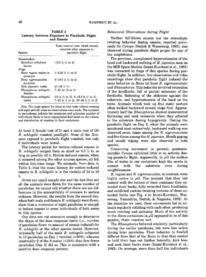
TABLE 3 Latency between Exposure to Parabolic Flight
and Emesis
Species
Time interval over which emesis occurred after exposure to
parabolic flight
Salamanders Hynobius nebulosus
larvae Frogs
Rana rugosa males or juveniles
Rana nigromaculata juveniles
Hyla japonica males Rhacophorus schlegelii
females Rhacophorus schlegelii
males
<0.5 h (1 or 2)
1-2.25 h (1 or 2)
6-19.5 h (1 or 2)
21-28 h (1) 7-21 hr (2 to 4)
0.5 h (1 to 2), 6-19.5 h (1 to 2), 7-21 h (1 to 2), 30-42 h (1 to 2)
Note. The large spread for times in this table reflects evening and night periods when no observations were made. The numbers in parentheses indicate the maximum and minimum number of individuals likely to have regurgitated food based on the volume and distribution of vomitus in their containers.
At least 2 female (out of 5) and 4 male (out of 19) R. schlegelii vomited postflight. None of the Xen- opus exposed to parabolic flight vomited, but only 6 individuals were tested.
The latency period for motion-induced emesis in R. schlegelii ranged from as short as 0.5 h to as long as possibly 42 h. Motion-induced emesis, when it occurred among the other anuran species, all fell within this time range. We estimate, from data in Table 3, that the mean latency for motion-induced emesis in R. schlegelii is in the vicinity of 14 to 16 h.
Given our small sample size and the fact that not all the animals were flown for the same number of parabolas, we cannot tell whether there are sex dif- ferences in the susceptibility of anurans to motion sickness. However, the results from the second day, when both male and female R. schlegelii were flown, show that a minimum of eight parabolas is enough to induce emesis in some individuals of both sexes in this species.
Our data are not extensive enough to determine the shape of the dose-response curve (i.e., number of parabolas flown vs amount of emesis induced) for R. schlegelii or the other species tested. However, minimally half of the male R. schlegelii subjected to 10 parabolas on Day 1 vomited (>~50%), whereas maximally 2 of the 8 males (~<25%) that flew fewer parabolas (Day 3) did so. This is consistent with a positive dose-response pattern.
Behavioral Observations during Flight
Neither full-blown emesis nor the stereotypic twisting behavior during emesis, reported previ- ously for Cynops (Naitoh & Wassersug, 1992), was observed during parabolic flight proper for any of the amphibians.
The previous, unexplained hyperextension of the head and backward walking of H. japonica seen on the MIR Space Station (Izumi-Kurotani et al., 1991) was reenacted by frogs of this species during par- abolic flight. In addition, live observation and video recordings show that parabolic flight induced the same behavior in Rana (at least R. nigromaculata) and Rhacophorus. This behavior involved retraction of the hindlimbs, full or partial extension of the forelimbs, flattening of the abdomen against the substrate, and hyperextension of the head on the torso. Animals which took up this static posture often walked backward several steps first. Approx- imately half the Rhacophorus showed dorsoventral flattening and neck extension when they adhered to the substrate during hypogravity. During the parabolic flight on Day 3, when the animals were monitored most extensively, backward walking was observed seven times among the R. nigromaculata and five times among the H. japonica. Mouth gaping and mouth wiping were also observed in both species.
Concerning movement in general, postmeta- morphic Cynops exhibited little or no activity dur- ing parabolic flight. Apparently, in tLG the surface film of water in our containers kept the newts in contact with the substrate even during weightlessness.
R. rugosa and R. nigromaculata, in contrast, were highly active in t~G. The moment that they lost contact with the bottom of their container they ex- tended their backs, fully extended their hindlimbs, and exhibited various twisting motions of those ex- tended limbs (see Fig. 4 in Izumi-Kurotani, Was- sersug, Yamashita, Naitoh, & Nagaoka, 1992). At the densities we used, their movements led to an- imals regularly colliding with each other, producing much twirling and spinning. Much of the activity in the Rana containers in t~G appeared to be of this passive, chain reaction sort.
The Rhacophorus behaved similarly, particularly during the earlier parabolas, but were less active during later parabolas. Their behavior in freefall differed from that of the Rana in that they tended to hold their legs out further laterally, kick less, and arch their backs more (Izumi-Kurotani et al., 1992). On average, more than half the individuals
MOTION SICKNESS IN AMPHIBIANS 47
were successful in retaining contact with the con- tainer's walls during the hypoG phase of any one parabola. On eight videotape sequences, from zero to three Rhacophorus (N = 5) were floating freely at any one time. Many individuals, which lost con- tact when a parabola began, managed to regain a foothold before the end of the hypoG phase.
Most of the Hyla adhered to the walls during the majority of the parabolas. From zero to four out of eight male Hyla (Day 3) lost contact with the walls on any one parabola. When that happened they promptly took up the extended-leg, arched-back pos- ture just described for Rhacophorus. This extended limb posture was indistinguishable from the posture of free-floating H. japonica in t~G on the MIR Space Station (illustrated in Izumi-Kurotani et al., 1991, 1993).
In tLG, the Xenopus swam sporadically with their hindlimbs and heads extended on their trunk. How- ever, on the power stroke they often adducted their legs past each other, in scissor-kick fashion (see Fig. 5 in Izumi-Kurotani et al., 1992). This foot-crossing maneuver was associated with asymmetrical fore- limb movements. The result was that the frogs spun along their long axis as they swam forward.
Approximately half of the Hynobius larvae ex- hibited long axis rotation during t~G. The smaller Cynops larvae exhibited more irregular movements involving dashing forward and stopping. These movements included both long axis spinning and dorsal deviation from the forward path of motion, again associated with trunk extension. The behavior of our Cynops larvae in tLG was not noticeably dif- ferent from that of unfed larvae of the same de- velopmental stage under the same gravitational re- gime (unpublished observations).
Postflight Behavioral Observations
The general locomotor activity of the frogs and salamanders postflight was indistinguishable from that of controls. The animals in both groups ap- peared equally sensitive to disturbance from people or from other frogs. Mature male H. japonica and R. schlegelii that were in reproductive condition were seen and heard calling shortly after flight. Both control and experimental male frogs produced release calls when other frogs climbed on them. Fe- cal matter was observed in all of the containers 1 to 2 days after feeding, suggesting normal digestive activity among both experimental and control specimens.
Retching displays were noted by two separate ob- servers on two separate occasions postflight. Our
most extensive observations of retching were made on Day 3. For a period of 1.5 h, starting 2 h after the initial parabola was flown, all of the experi- mental R. nigromaculata and H.japonica were care- fully and continuously observed, along with three control specimens from each species. No differences in behavior between experimental and control frogs were identified, except for that of one flown Hyla. This individual showed the retching behavior de- scribed previously for Rana exposed to emetic drugs (Naitoh et al., 1989). The frog held its forelimbs in extension and its hindlimbs retracted. Its vent was against the edge of the container. While in this position the animal went through 5 to 10 episodes of mouth opening. Each time the head was elevated and mouth opened widely. The opening and closing cycle was relatively slow, covering approximately 2 s. Mouth opening was preceded by massive con- tractions of the body wall. Wiping of the mouth with the hand in a caudal to rostral direction was also observed at the end of approximately half of these displays. During three of the cycles, a mucus strand was observed running from mouth to protracted hand. The animal was attempting to wipe this ma- terial away from its mouth.
Although not videotaped, one or two of the Hyla and one Rhacophorus flown on Day 2 made similar retching displays approximately 4 h postflight.
DISCUSSION
Motion-Induced Emesis in Amphibians
The presence of vomitus in the containers of pre- metamorphic Hynobius and postmetamorphic Rana, Hyla, and Rhacophorus subjected to parabolic tra- jectories answers the core question of our investi- gation, namely whether or not motion can provoke emesis in amphibians. However, the fact is that at the most only 17 out of 64 frogs tested (a maximum of 27%) vomited after exposure to 2 h of flight and 8 or more cycles of hyper- and hypoG. Among the species tested, the incidence of motion-induced eme- sis ranged from 0% (for C. pyrrhogaster and X. lae- vis) to possibly as high as 50% (for R. schlegelii). None of the postmetamorphic salamanders vomited and only 1 or 2 Hynobius larvae out of 10 were afflicted with motion sickness. Thus, it seems that frogs are relatively resistant to motion sickness and salamanders even more so. Although none of the Xenopus or Cynops in our study vomited, we cannot yet conclude that they are immune to this malaise. Certainly simple motion and other stresses, such as minor changes in pressure and temperature asso-
48 WASSERSUG ET AL.
ciated with transportation in modern vehicles, are not alone adequate to induce emesis in amphibians. If they were, this fact would have been documented previously, given the enormous numbers of am- phibians shipped commercially worldwide each year.
How does the susceptibility to motion sickness for amphibians compare with that of Homo sapiens and other mammals? Reschke (1990) reviewed the in- cidence of motion sickness for cosmonauts and as- tronauts. During the first 24 flights of the U.S. Space Shuttles, 67% of the astronauts (N = 125 individual exposures) acknowledged some symptoms of motion sickness. Comparable levels were reported from the older Skylab program. Many symptoms of motion sickness were considered in those studies in addition to emesis. If the criteria for motion sickness were restricted to vomiting, this percentage would be lower and closer to that of R. schlegelii. Money (1970) notes that dogs have a similar susceptibility to motion sickness as man; however, this suscep- tibility varies enormously among mammals, de- pending on the provocative stimulus and a mul- titude of other factors.
The biggest difference in motion-induced emesis between mammals and frogs is the long latency pe- riod in the amphibians. In mammals, emesis as a symptom of motion sickness is prompt, typically occurring during or shortly (i.e., minutes) after the animals are subjected to the provocative stimulus. In contrast, the latency for frogs is on the order of several hours to more than a day poststimulation.
Since frogs are poikilothermic, temperature can be a major factor in any physiological response, in- cluding emesis. Although the temperature was con- trolled in the aircraft, the postflight conditions were ambient, but unfortunately neither controllable nor recorded. The temperatures during the day may have climbed a degree or two above 21°C and at night could have fallen as low as 14°C, but no lower. This is a temperature range over which the species we studied are normally active. The fact that our male frogs in reproductive condition persisted in calling further suggests that the long latency is not the result of an overall slowing of metabolism due to hypothermia.
Visceral responses in amphibians are often pro- tracted compared to those of mammals and this may relate to their longer, slower response to emetic stimuli. Notably, the response to emetic drugs is typically many times longer in frogs than in dogs at comparable dosages (cf. data in Naitoh et al., 1991, on Xenopus, with data in Carpenter, Briggs, & Strominger, 1984a,b on Canis). Whereas dogs
respond to injections of the emetic apomorphine in seconds, latencies in excess of 3 h have been reported for frogs.
The long latency does, however, suggest that mo- tion sickness in amphibians involves neurochemical pathways of the autonomic nervous system beyond simple reflexes. If the regulatory pathways that con- trol motion sickness are, in fact, the same in am- phibians as they are in mammals, then this long lag from stimulus to emesis may give frogs some practical advantage in future investigations de- signed to elucidate those pathways.
Other Signs of Motion Sickness
We have now documented that H. japonica in parabolic flight exhibits the same neck extension and backward walking that it exhibited on the MIR Space Station. Neither the neck extension nor the dorsoventral compression that went along with this display were as extreme as that seen on the MIR Space Station. We speculate that this difference in magnitude reflects the different lengths of time that the animals were exposed to t~G. An alternative, but not mutually exclusive, hypothesis is that the differences are due to a greater ability of the an- imals on the MIR Space Station to deform their bodies simply because they had less food in their alimentary tracts to resist abdominal compression and axial extension. In contrast to our well-fed frogs, the MIR specimens had not been fed for 2 weeks (Izumi-Kurotani et al., 1991) and may have had empty alimentary tracts at the time they were vi- deotaped. Both of these hypotheses, among others, are testable with frogs that have varied levels of material preloaded into their stomachs and are then exposed to provocative stimuli of varied duration.
The same extended-neck posture seen in H. ja- ponica on MIR has now been observed in two other genera in ~G: Rana and Rhacophorus. Notably, these are the same genera whose members vomited after we subjected them to parabolic flight.
We believe that frogs on the MIR Space Station had motion sickness and that the behavior they dis- played was essentially the same as the retching display provoked by parabolic flight. Retching in frogs--induced by either drugs or parabolic flight-- and the behavior observed on MIR do differ in minor ways. We did not, for example, observe extensive backward walking during postflight retching in H. japonica. However, backward movements have been reported previously for frogs during emesis (Naitoh et al., 1989), and the H. japonica which made the postflight retching display we observed already had its rump against the side of the container.
MOTION SICKNESS IN AMPHIBIANS 49
Cyclical mouth opening and closing of the frogs on the MIR Space Station was not recorded with videotape, but was observed by T. Akiyama, the Japanese cosmonaut (personal communication). Opening and closing of the mouth by the H. japonica was observed during our parabolic flights.
We anticipate that frogs will be shown to have other, more subtle, physiological signs of motion sickness than only emesis and retching behavior. Increased salivation is, for example, a common symptom of motion sickness in mammals (Daunton, 1990). A hint that this may also be true of motion- sick frogs was seen in the H. japonica which gave the postflight retching display described above. Dur- ing the peak of that display, the frog was observed wiping excess mucus from its mouth.
Increased salivation in motion-sick mammals "is often accompanied by frequent swallowing..." (Daunton, 1990). Similarly, the frogs on the MIR Space Station were seen to close their eyes after they made the mouth-opening and closing displays noted previously. For frogs, closing of the eyes-- that is, retraction of the eye bulbs--is an essential part of swallowing. The backward movement or re- treat in motion-sick frogs itself appears to be the anuran equivalent to the crouched, curled up, and withdrawn posture called the "sopite syndrome" in motion-sick monkeys (Daunton, 1990).
Behavioral Differences among Amphibians in tLG
It may be of significance that the frog species which exhibited the greatest amount of vomiting after parabolic flight were the same ones that ex- perienced the most extreme gyrations during the ItG phases of the parabolas, i.e., the ones that lost contact with the container surfaces during t~G. This suggests that the greater the provocative motion, the greater the chance of inducing motion sickness in anurans. It also raises the question of why the different species responded differently to t~G.
There are alternative, but not necessarily mu- tually exclusive, explanations for the taxonomic pattern we observed. For example, the species that vomited in our study are predominately ranoid frogs and are more closely related to each other than to the other species in our study. However, given our small sample size, it would be premature to conclude that there are familial biases for motion sickness among amphibian species.
Our additional observations of the behavior of anurans in tLG deserve some comment. The ex- tended hindlimb posture of free floating H. japonica on the MIR Space Station (Izumi-Kurotani et al.,
1991, 1993) has been interpreted as a jumping pos- ture, but the combined hyperextension of the torso and the abduction of the fully extended limbs seems to more closely approximate the parachuting or "flying" posture of certain arboreal frogs (Emerson & Koehl, 1990; Stewart, 1985; see also figure in Duellman, 1970). The difference is reflected in the abduction of the limbs and spread of the digits. In parachuting, the limbs and digits are abducted. This posture is one that maximizes the frontal area of the frog to increase drag or generate lift and thus slow descent. In contrast, a frog wishing to max- imize the distance covered in a single jump may maintain a more compact profile that reduces drag (at least until near landing). As illustrated by Gans (1961) and Zug (1986), jumping frogs keep their limbs more adducted, except when lunging at prey or about to land.
The frogs that are best adapted for parachuting or "flying" (Emerson & Koehl, 1990) are also among the most arboreal. Of the amphibians we tested, R. schlegelii is clearly the most arboreal. Interestingly, it was also the species most likely to take up the flying posture during t~G. The other anurans in free- fall took up similar postures, but showed more long axis rotation (see below). Flying in Rana in t~G was more common than in Hyla, not necessarily because Rana is better at parachuting but because it may have no other choice. Rana lacks the digital pads that allowed the majority of our Rhacophorus and Hyla to hold on to the container's wall during freefall.
Clearly, an aquatic frog such as Xenopus would not be expected to parachute under natural cir- cumstances; nor does it exhibit that sort of posture in tLG. The asymmetrical, cross-legged scissor-kick that it does exhibit in ttG causes abrupt, long-axis rotation or rolling. In conjunction with the extension of the torso, this behavior looks identical to the "righting" maneuver used by Xenopus when they are upside down in normal gravity. Indeed, we can easily and instantly provoke the same torso exten- sion and leg-crossing maneuver in X. laevis in 1G by placing them on their backs out of water. We observed long-axis rotation with concurrent trunk extension in t~G, among animals as morphologically dissimilar as postmetamorphic Rana and premeta- morphic Hynobius larvae. In all these cases, the movement appears to be indistinguishable from the normal righting reflex of these animals in 1G.
As a final note, although the stimulus we used to provoke motion sickness was parabolic flight, we have tried to avoid implying that the motion sick- ness we observed is space motion sickness due ex-
50 WASSERSUG ET AL.
plicitly to exposure to /~G. We cannot yet say whether hypoG, hyperG, or the alternation from one to the other was the primary causal factor in in- ducing emesis in frogs. There is no question, how- ever--amphibians do get motion sickness.
Summary
(1) Extreme motions can provoke emesis in cer- tain amphibians.
(2) The individuals which exhibited emesis after parabolic flights were those that experienced the most severe tumbling and rolling during the /~G phase of the parabolas.
(3) The emetic response of these animals is de- layed from approximately 30 min until up to pos- sibly as long as 42 h after exposure to parabolic stimulus.
(4) Our sample sizes were too small to determine if there are sex, size, or species differences in the propensity for motion sickness in anurans. However, the presence of vomitus in containers for both male and female R. schlegelii confirms that both sexes are susceptible to motion sickness.
(5) Motion can induce retching in anurans in the absence of emesis. A strange behavior, reported pre- viously for tree frogs on the MIR Space Station, resembles the retching posture of motion-sick frogs.
REFERENCES
Brown, C. A., Wassersug, R. J., & Naitoh, T. (1992). Metamorphic changes in the vagal innervation to the alimentary tract in the green frog Rana clamitans (Ranidae). Acta Anatomica, 145, 340-344.
Carpenter, D. 0., Briggs, D. B., & Strominger, N. (1984a). Be- havioral and electrophysiological studies of peptide-induced emesis in dogs. Federation Proceedings, 43, 2952-2954.
Carpenter, D. O., Briggs, D. B., & Strominger, N. (1984b). Pep- tide-induced emesis in dogs. Behavioural Brain Research, 11, 277-281.
Corcoran, M. L., Fox, R. A., & Daunton, N. G. (1990). The sus- ceptibility of rhesus monkeys to motion sickness. Aviation, Space and Environmental Medicine, 61, 807-809.
Chinn, H. I., & Smith, P. K. (1955). Motion sickness. Phar- macological Reviews, 7, 33-82.
Crampton, G. H. (Ed.) (1990). Motion and space sickness. Boca Raton, FL: CRC Press.
Daunton, N. G. (1990). Animal models in motion sickness re- search. In G. H. Crampton (Ed.), Motion and space sickness (pp. 87-104). Boca Raton, FL: CRC Press.
Davis, C. J., Lake-Bakaar, G. V., & Grahame-Smith, D. G. (Eds.) (1986). Nausea and vomiting: Mechanisms and treatment (Advances in applied neurological sciences). New York: Springer-Verlag.
Duellman, W. E. (1970). The hylid frogs of Middle America. University of Kansas Museum of Natural History, Mono- graph 1 (2 vol.), pp. 1-753.
Emerson, S. B., & Koehl, M. A. R. (1990). The interaction of behavioral and morphological change in the evolution of a novel locomotor type: "Flying" frogs. Evolution, 44, 1931- 1946.
FRIS Experiment Group (1991). Report of frog experiment on- board space station MIR. Space Utilization Research Center (Ed.). Kanagawa, Japan: Institute of Space and Astronaut- ical Science.
Gans, C. (1961). A bullfrog and its prey. Natural History, 52, 26-37.
Izumi-Kurotani, A., Wassersug, R. J., Yamashita, M., Naitoh, T., & Nagaoka, S. (1992). Frog behavior under microgravity: Possibility of motion sickness in amphibians. ISAS Space Utilization Symposium, 9, 112-114.
Izumi-Kurotani, A., Yamashita, M., Kawasaki, Y., Kurotani, T., Mogami, Y., Okuno, M., Akiyama, T., Oketa, A., Shiraishi, A., & Ueda, K. (1991). Behavior of Japanese tree frog under microgravity. Biological Sciences in Space, 5, 185-189.
Izumi-Kurotani, A., Yamashita, M., Kawasaki, Y., Kurotani, T., Mogami, Y., Okuno, M., Akiyama, T., Oketa, A., Shiraishi, A., Ueda, K., Wassersug, R. J., & Naitoh, T. (1993). Behavior of Japanese tree frogs under microgravity on MIR and in parabolic flight. Advances in Space Research, in press.
Kohl, R. L. (1987). Failure of metoclopramide to control emesis or nausea due to stressful angular or linear acceleration. Aviation, Space and Environmental Medicine, 58, 125-131.
Lucot, J. B. (1989). Blockage of 5-hydroxytryptamine8 receptors prevents cisplatin-induced but not motion- or xylazine-in- duced emesis in the cat. Pharmacology, Biochemistry and Behaviour, 32, 207-210.
McKenzie, R. A. (1935). Codfish in captivity (Note 47). Progress Reports of the Atlantic Biological Station (St. Andrew's, New Brunswick) and the Atlantic Fisheries Experimental Station (Halifax, Nova Scotia), No. 16, pp. 7-10. Biological Board of Canada.
Mellinger, C. (1881). Betr~ige zur Kenntniss des Erbrechens. Ar- chives de Physiologie, 24, 232-245.
Miller, A. D. (1991). Motion-induced nausea and vomiting. In J. Kucharczyk, D. J. Stewart, & A. D. Miller (Eds.), Nausea and vomiting: Recent research and clinical advances (pp. 13- 41). Boca Raton, FL: CRC Press.
Mirabile, C. S. (1990). Motion sickness susceptibility and be- havior. In G. H. Crampton (Ed.), Motion and space sickness (pp. 391-410). Boca Raton, FL: CRC Press.
Money, K. E. (1970). Motion sickness. Physiological Reviews, 50, 1-39.
Naitoh, T., Imamura, M., & Wassersug, R. J. (1991). Interspecific variation in the emetic response of anurans. Comparative Biochemistry and Physiology, 100C, 353-359.
Naitoh, T., & Wassersug, R. J. (1992). The emetic response of urodele amphibians. Zoological Science, 9, 713-718.
Naitoh, T., Wassersug, R. J., & Leslie, R. A. (1989). The phys- iology, morphology, and ontogeny of emetic behavior in an- uran amphibians. Physiological Zoology, 62, 819-843.
Reschke, M. F. (1990). Statistical prediction of space motion sick- ness. In G. H. Crampton (Ed.), Motion and space sickness (pp. 263-316). Boca Raton, FL: CRC Press.
Stewart, M. M. (1985). Arboreal habitat use and parachuting by a subtropical forest frog. Journal of Herpetology, 19, 391- 401.
Stott, J. R. R., Barnes, G. R., Wright, R. J., & Ruddock,

MOTION SICKNESS IN AMPHIBIANS 51
C. J. S. (1989). The effect of motion sickness and oculomotor function of GR 38032F, a 5-HT3 receptor antagonist with antiemetic properties. British Journal of Clinical Phar- macology, 27, 147-157.
Suyehiro, Y. (1934). Doesn't the vomiting center exist in the fish? Bulletin of the Japanese Society of Scientific Fisheries, 2, 307-311.
Ueno, S., Matsuki, N, & Saito, H. (1988). Suncus murinus as a new experimental model for motion sickness. Life Sciences, 43, 413-420.
Zug, G. (1986). Leaps and bounds: Why have frogs lost their tails? In T. Halliday & K. Adler (Eds.), The encyclopedia of reptiles and amphibians (pp. 56-57). New York: Facts on File Inc.
Related Documents