THE JOURNAL OF COMPARATIVE NEUROLOGY 349:343-362 (1994) Morphology of Retinal Axons Induced to Arborize in a Novel Target, the Medial Geniculate Nucleus. I. Comparison With Arbors in Normal Targets S.L. PALLAS, J. HAHM, AND M. SUR Department of Brain and Cognitive Sciences, Massachusetts Institute of Technology, Cambridge, Massachusetts 02139 ABSTRACT Ferret retinal axons can be induced to innervate the medial geniculate nucleus (MGN) by a combination of brain lesions early in development. Our previous work suggests that the retinal ganglion cells responsible for this plasticity are W cells. The present study continues this work with a morphological investigation of normal retinal ganglion-cell axons and retinal ganglion- cell axons induced to arborize in the MGN. Retinal axons were bulk filled with horseradish peroxidase placed in the optic tract, and individual axom were serially reconstructed from sagittal sections. The control population consisted of fine-caliber s o n s arborizing in the superior colliculus (SC) and in the ventral C laminae of the lateral geniculate nucleus (LGN) of normal ferrets. We also compared the axons in the MGN of lesioned ferrets to intracellularly filled X and Y axons from normal ferrets as reported by Roe et al. ([1989] J. Comp. Neurol. 288:208). We have found that the retino-MGN axons in the lesioned ferrets do not resemble X or Y axons in normal ferrets in axon diameter, arbor volume, bouton number, or bouton density. However, they do resemble the fine-caliber, presumed W axons arborizing in the C laminae of the LGN and in the SC of normal ferrets. Thus, this study, in combination with previous studies, suggests strongly that W retinal ganglion cells are responsible for the retinal input to the MGN in lesioned animals. In addition, we find that the retino-MGN axons are of two types, branched and unbranched, which may correspond to different subtypes of retinal W Cek o 1994 Wiley-Liss, Inc. Key words: cross-modal plasticity, ferret, W axon, synaptic specificity, visual development Afferent activity during development plays a major role in the differentiation of target brain areas (for reviews, see Shatz, 1990; Udin and Fawcett, 1988). This phenomenon has generally been studied by manipulating the quantity of activity in a given input pathway. Another approach is to manipulate the source of the input, in which case one can explore the role of the quality of afferent activity. Such a functional mismatch between afferents and their targets can provide a promising tool for studying activity-depen- dent aspects of brain development (Pallas, 1990; Sur et al., 1990). The type of functional mismatch that we have employed in the present study was first described in hamsters. Schneider (1973) and, subsequently, Frost (1981) employed neonatal brain lesions to “rewire” the brain, such that retinal ganglion-cell axons developed projections to nonvi- sual thalamic structures. We have successfully extended the rewiring paradigm to ferrets. Ferrets are altricial, mustelid carnivores with a visual pathway quite similar to that of cats (Jackson and Hickey, 1989; Rockland, 1985; Vitek et al., 1985; Zahs and Stryker, 1985; Law et al., 1988). We have shown previously (Sur et al., 1988) that reduction of the normal targets of retinal ganglion cells and deafferen- tation of the auditory thalamus on the day of birth in ferrets will result in the innervation of the auditory thala- mus by retinal axons. These ectopic axons confer visual Accepted May 27,1994. Sarah L. Pallas is now at the Division of Neuroscience, Baylor College of J. Hahm is now at the Laboratory of Neuropsychology, NIMH, Building Address reprint requests to S.L. Pallas, Division of Neuroscience, Baylor Medicine, One Baylor Plaza, Houston, TX 77030. 49, Room 1B80,9000 Rockville Pike, Bethesda, MD 20892. College of Medicine, One Baylor Plaza, Houston, TX 77030. O 1994 WILEY-LISS, INC.

Welcome message from author
This document is posted to help you gain knowledge. Please leave a comment to let me know what you think about it! Share it to your friends and learn new things together.
Transcript

THE JOURNAL OF COMPARATIVE NEUROLOGY 349:343-362 (1994)
Morphology of Retinal Axons Induced to Arborize in a Novel Target, the
Medial Geniculate Nucleus. I. Comparison With Arbors in Normal Targets
S.L. PALLAS, J. HAHM, AND M. SUR Department of Brain and Cognitive Sciences, Massachusetts Institute of Technology,
Cambridge, Massachusetts 02139
ABSTRACT Ferret retinal axons can be induced to innervate the medial geniculate nucleus (MGN) by a
combination of brain lesions early in development. Our previous work suggests that the retinal ganglion cells responsible for this plasticity are W cells. The present study continues this work with a morphological investigation of normal retinal ganglion-cell axons and retinal ganglion- cell axons induced to arborize in the MGN. Retinal axons were bulk filled with horseradish peroxidase placed in the optic tract, and individual axom were serially reconstructed from sagittal sections. The control population consisted of fine-caliber s o n s arborizing in the superior colliculus (SC) and in the ventral C laminae of the lateral geniculate nucleus (LGN) of normal ferrets. We also compared the axons in the MGN of lesioned ferrets to intracellularly filled X and Y axons from normal ferrets as reported by Roe et al. ([1989] J. Comp. Neurol. 288:208).
We have found that the retino-MGN axons in the lesioned ferrets do not resemble X or Y axons in normal ferrets in axon diameter, arbor volume, bouton number, or bouton density. However, they do resemble the fine-caliber, presumed W axons arborizing in the C laminae of the LGN and in the SC of normal ferrets. Thus, this study, in combination with previous studies, suggests strongly that W retinal ganglion cells are responsible for the retinal input to the MGN in lesioned animals. In addition, we find that the retino-MGN axons are of two types, branched and unbranched, which may correspond to different subtypes of retinal W C e k o 1994 Wiley-Liss, Inc.
Key words: cross-modal plasticity, ferret, W axon, synaptic specificity, visual development
Afferent activity during development plays a major role in the differentiation of target brain areas (for reviews, see Shatz, 1990; Udin and Fawcett, 1988). This phenomenon has generally been studied by manipulating the quantity of activity in a given input pathway. Another approach is to manipulate the source of the input, in which case one can explore the role of the quality of afferent activity. Such a functional mismatch between afferents and their targets can provide a promising tool for studying activity-depen- dent aspects of brain development (Pallas, 1990; Sur et al., 1990).
The type of functional mismatch that we have employed in the present study was first described in hamsters. Schneider (1973) and, subsequently, Frost (1981) employed neonatal brain lesions to “rewire” the brain, such that retinal ganglion-cell axons developed projections to nonvi- sual thalamic structures. We have successfully extended
the rewiring paradigm to ferrets. Ferrets are altricial, mustelid carnivores with a visual pathway quite similar to that of cats (Jackson and Hickey, 1989; Rockland, 1985; Vitek et al., 1985; Zahs and Stryker, 1985; Law et al., 1988). We have shown previously (Sur et al., 1988) that reduction of the normal targets of retinal ganglion cells and deafferen- tation of the auditory thalamus on the day of birth in ferrets will result in the innervation of the auditory thala- mus by retinal axons. These ectopic axons confer visual
Accepted May 27,1994. Sarah L. Pallas is now at the Division of Neuroscience, Baylor College of
J. Hahm is now at the Laboratory of Neuropsychology, NIMH, Building
Address reprint requests to S.L. Pallas, Division of Neuroscience, Baylor
Medicine, One Baylor Plaza, Houston, TX 77030.
49, Room 1B80,9000 Rockville Pike, Bethesda, MD 20892.
College of Medicine, One Baylor Plaza, Houston, TX 77030.
O 1994 WILEY-LISS, INC.
344 S.L. PALLAS ET AL.
responsiveness on cells in the medial geniculate nucleus (MGN) and, via the MGN, on cells in the primary auditory cortex (AI; Sur et al., 1988; Roe et al., 1992).
The retina in ferrets, as in cats, contains at least three types of retinal ganglion cells (X, Y, and W Henderson, 1985; Vitek et al., 1985; Amthor and Jackson, 1986; Roe et al., 1989). Whereas X and Y cells are well-distinguished from each other and from other retinal ganglion cells, W cells constitute a rather heterogeneous mix of cell types (see also Discussion). Roughly 72% of the retinal ganglion cells in ferrets are W cells (Vitek et al., 1985). A number of lines of evidence from our laboratory suggests that the retinal axons that invade the MGN are of the W class rather than X or Y (Sur et al., 1988; Roe, 1991; Roe et al., 1993). This evidence includes the soma sizes of MGN-projecting retinal ganglion cells, the conduction velocities of their axons, and the response features of the MGN cells to which they connect. The present study furthers this line of investiga- tion by examining the terminal arbor morphology of indi- vidual retinal axon arbors in the MGN.
For comparison with the retino-MGN axons, we have also examined the terminal arbors of presumed W axons in the superior colliculus and the ventral C laminae of the lateral geniculate nucleus (LGN) in normal ferrets. There is very little information on W-axon arbor morphology in the literature, particularly for ferrets, and it is our hope that other investigators will find our control study useful as a starting point for further investigations into the morphol- ogy of W axons in normal animals.
In the accompanyingpaper (Pallas and Sur, this issue), in order to address the question of whether the morphology of developing axon arbors is affected by the target, we compare our retino-MGN axons with a population of normal axons that arises from the brachium of the inferior colliculus and arborizes in the MGN. Parts of this work have been reported previously in preliminary form (Pallas et al., 1989, 1991).
MATERIALS AND METHODS Animals
The animals used were pigmented ferrets (Mustela puto- rius furo; family Mustelidae, order Carnivora). Ferret kits used for neonatal manipulations were from dams that were bred by Marshall Farms (North Rose, NY) and shipped to us 2 weeks prior to delivery. Gestation time in ferrets is approximately 42 days. Adult ferrets were maintained on a 12:12 1ight:dark cycle and fed dry cat food and water ad libitum. Pregnant and nursing dams were kept on a 14:lO 1ight:dark cycle and given canned cat food in addition to the regular diet. A total of seven normal and six experimental (lesioned) animals were used in this study.
Abbreviations
AI primary auditory cortex IC inferior colliculus LPiPul lateral posterioripulvinar complex LGN lateral geniculate nucleus MGN medial geniculate nucleus MIN OT optic tract SC superior colliculus SGS so
medial interlaminar nucleus of the LGN
stratum griseum superficiale of the SC stratum opticum of the SC
Neonatal surgery To induce innervation of the MGN by retinal ganglion-
cell axons, the normal targets of the retina were removed or reduced in size, and the MGN was deafferented. All manipu- lations were unilateral and were done within 24 hours of birth. Ferret kits were removed from the nursing dam and anesthetized individually by deep hypothermia. The skull was exposed by an incision in the head, and the flap of skull overlying the colliculi was cut away. The brachium of the inferior colliculus was sectioned at the level of the superior colliculus to deafferent the MGN. To reduce the major targets of the retina, the superior colliculus (SC) and occipital cortex (corresponding to areas 17 and 18) on the left side of the brain were cauterized. The cortical lesion causes a massive retrograde degeneration of the LGN. Following the ablations, the skin was sutured, and the kit was given subcutaneous fluids (5% dextrose) and antibiotic (Amoxicillin, 1 mg), revived by warming, and returned to the dam. Kits were weaned at 8 weeks of age and were reared until adulthood (15 weeks or more) before additional procedures were performed.
Horseradish peroxidase injections When the animals reached adulthood, we examined the
morphology of single axons by the horseradish peroxidase (HRP) bulk-fill method (Mason, 1982; Sachs and Schneider, 1984; Sretavan and Shatz, 1984, 1986; Hahm et al., 1991). Animals were deeply anesthetized with sodium pentobarbi- tal and perfused transcardially with 1 liter of cold (PC), oxygenated, artificial cerebrospinal fluid. The brain was then quickly removed from the skull, the cerebral hemi- spheres were removed for visualization of the thalamus, and the thalamus was bisected along the sagittal plane. Small crystals of HRP (Type VI; Sigma, St. Louis, MO) that had been dried onto the end of a fine micropipette were introduced into the optic tract either ventral to the LGN, anterior to the MGN, or anterior to the superior colliculus, depending on the target to be examined (Fig. 1). This approach avoids inadvertent damage to thalamocortical or corticifugal axons. Three to seven such crystals were in- serted in each optic tract at equally spaced distances. In two cases, we severed the optic tract as it entered the MGN and laid the crystals of HRP on the cut axons. We were able to obtain more filled axons with this method, because we damaged more axons. Care was taken not to insert the HRP into the tissue underlying the fiber tract. The brain tissue was trimmed as much as possible and placed into oxygen- ated artificial cerebrospinal fluid at room temperature for 3-4 hours to allow time for the HRP to transport to the axon terminals. The brain block was fixed by immersion in 1% paraformaldehyde with 2% glutaraldehyde for 2 hours followed by 30% sucrose in phosphate buffer. The tissue was embedded in albumin-gelatin and sectioned frozen a t 100 p,m in the sagittal plane. Sections were treated with diaminobenzidinelcobalt chloride (Adams, 1981) to visual- ize the HRP reaction product.
Axon reconstruction Sections were scanned individually for well-isolated, well-
filled axon arbors. We looked for arbors that had axon trunks extending back into the optic tract and had recogniz- able boutons. There are multiple ways in which retino- MGN axons can enter the MGN (Rocha et al., 19931, but all those that are described here enter from the rostra1 optic tract. The completeness of filling was assessed throughout

RETINAL AXONS IN A NOVEL TARGET I 345
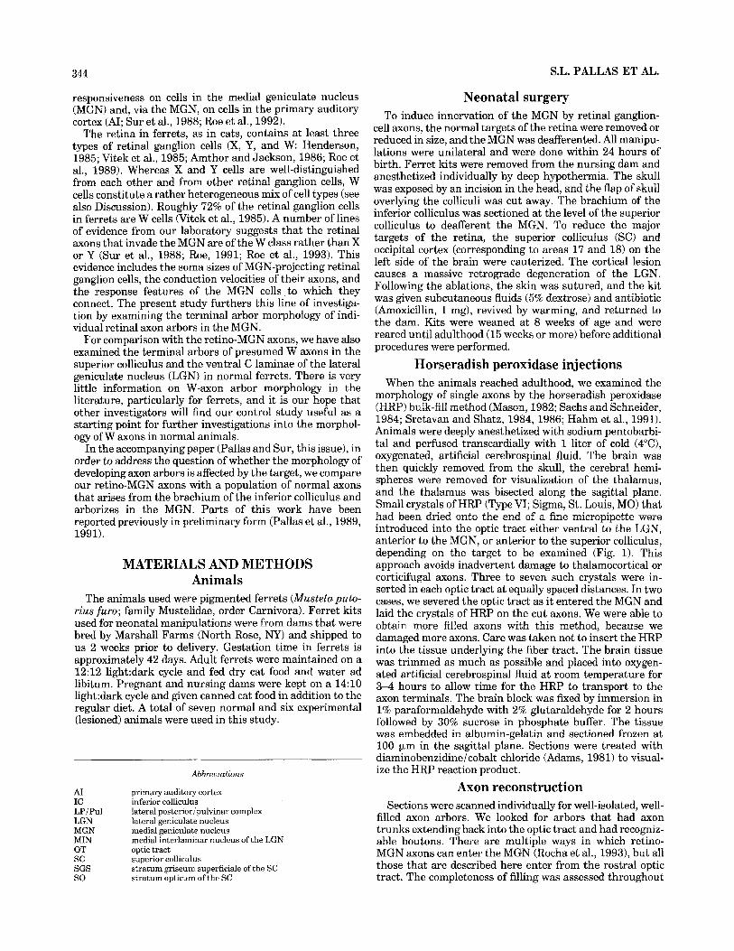
Fig. 1. Method for bulk-fill injections. The drawing at the top (depicting a normal animal) shows where injections were placed for filling retinal axons arborizing in the LGN, the SC, and the MGN. A Photomicrograph of an injection site in the optic tract just outside the LGN in a normal animal. A low-power schematic showing the place-
ment of the injection is shown below the micrograph. B: Photomicro- graph of an injection site in the optic tract just outside MGN in a lesioned animal. The schematic beneath shows the location of the injection. Scale bar = 200 pm.

346 S.L. PALLAS ET AL.
the reconstruction by noting whether branches were well filled and ended in a bouton rather than fading out gradu- ally. Arbors were reconstructed from serial sections with a camera lucida, usually with a x 100 objective. The largest arbors were reconstructed with a x63 objective. Differen- tial interference-contrast microscopy was employed to clarify some details of the arbors during reconstruction. The area of individual arbors was calculated with the aid of a digitizing tablet by drawing a perimeter around the outer- most boutons. Arbor volume was defined as this area multiplied by the section thickness (100 km) and the number of sections in which the arbor was contained. This method will overestimate arbor volume and underestimate bouton density in some cases; importantly, all axons were measured in the same way. Axon diameters were measured directly from the camera lucida, with measurements being taken every 20 pm from the first branch (or the first bouton, in the case of unbranched axons) back toward the optic tract, until the axon could not be traced further or until a distance of 140 pm had been covered. These measurements were then averaged. Note that we did not round off the averages and standard errors, and, thus, the extra decimal places in each value do not represent actual resolution. Individual boutons were counted for each arbor; those with crenelated morphology were counted as single boutons. Bouton density was then calculated by dividing the number of boutons by the arbor volume. Population means were compared with t tests. Some of the axons did not branch in the MGN (“unbranched” type), and quantifi- cation of these was limited to axon diameter. Arbor volume, bouton number, and bouton density were not calculated for unbranched axons.
RESULTS Qua1 i t at ive observations
We have reconstructed a total of 31 retino-MGN axons from six lesioned animals, 15 retino-SC axons from four normal animals, and eight retino-LGN C-lamina axons from three normal animals. In one case, an LGN-projecting axon from the normal (unlesioned) side of the brain of an experimental animal was reconstructed. In addition, we compared our data set with normal physiologically identi- fied ferret retinogeniculate axons reconstructed by Roe et al. (19891, which included seven Y axons and 21 X axons, obtained from eight animals.
The lesions in the experimental animals caused a com- plete loss of the superior colliculus and a partial retrograde degeneration of the LGN. This type of lesion has been described in detail in our previous work. Micrographs and serial reconstructions of the lesion sites can be found in Sur et al., 1988; Pallas et al., 1990; and Roe et al., 1993.
The retinal axons in the MGN were consistently of very fine diameter and consisted of two types, those arborizing within the MGN and those that had no branches but passed through the MGN with en passant boutons only. We were sometimes able to follow individual axons of this unbranched type all the way through the MGN, from the rostroventral part of the optic tract to the dorsal part adjacent to the LGN, suggesting that they continue into the LGN or the SC. We reconstructed five such axons. More commonly, however, the axons were fasciculated, making reconstruction of individual axons difficult. In these cases, the entire bundle could then be followed through the MGN as it coursed medially and dorsally and reentered the optic tract. I t could
Unbranched retino-MGN axons.
clearly be seen that none of the axons in the fascicle branched within the MGN. The fasciculation made it impossible to determine the exact ratio of unbranched to branched axons, but it is our impression that the un- branched axons are much more prevalent than the branched axons. Following axons for any distance within the optic tract was prevented by the large number of labelled axons typical of the bulk-filling method. We partially recon- structed a total of nine unbranched axons as well as several fiber bundles. Our impression is that the individual axons within a bundle follow the same course as the entire bundle. Figure 2A shows an individual axon of the unbranched type, and Figure 2B shows a bundle of unbranched axons. Note the numerous swellings (most of which appear to be en passant boutons) at intervals along the fibers. Figure 3 shows reconstructions of some of these axons. Axons 1 and 2 were reconstructed completely as they passed through the MGN from one part of the optic tract to another. The third axon was reconstructed as far as possible but was lost in a large bundle of axons that reentered the optic tract. The fourth axon was unusual in that it actually ended in the MGN. However, a very small branch can be seen at the rostral end, and it is possible that this axon should be considered “branched.” Because we were interested in comparing axon arbors in the different targets, we concen- trated our analysis on branched axons and measured only axon diameter in the unbranched axons (see below).
Branched retino-MGN axons. The other type of retino- MGN axon formed a terminal arbor within the MGN. We reconstructed a total of 26 axons of this type. These branched axons generally entered the MGN from the optic tract rostral or ventral to the nucleus. They were not observed to exit the MGN, but it is possible that they branch within the optic tract and project to other targets. In bulk-filled material, it is generally not possible to follow axons for any distance within the optic tract. The bouton morphology of these axons was often complex with crene- lated or globose boutons often arranged in strings or clusters (Fig. 4). The individual boutons could be quite large. The arbors, in contrast, were sparse and simple compared to X or Y retinogeniculate axons (Roe et al., 1989) and more closely resembled those seen in the superior colliculus or the ventral C laminae of the LGN (see below).
We have not made an attempt to delineate the borders of the subdivisions of MGN. The absence of a published ferret brain atlas and appropriate connectional studies makes this task difficult, even in counterstained material. I t appears from darkfield examination that the retino-MGN axons lie mainly in the ventral part of MGN, but some also arborize in the dorsal and medial subdivisions. This interpretation is supported by more recent studies (Rocha et al., 1993; Roe et al., 1993). The arbors were variable in appearance, and we have included several camera lucida drawings to demon- strate the range of variability. Most of the arbors have been completely reconstructed. In those cases where branches could not be followed, we have placed an “X” to indicate an incomplete branch. We have grouped the axons loosely according to their degree of branching and appearance of boutons, but we made no attempt to categorize them further.
Figure 5 shows some fairly simple retino-MGN axons that exhibited mainly the beaded string bouton type and many long, straight branches. Axons 1, 2, and 4 also had boutons on their axon trunks, which was not a typical characteristic of most of the branched retino-MGN axons.

RETINAL AXONS IN A NOVEL TARGET I 347
Fig. 2. Photomicrographs of unbranched retino-MGN axons bulk filled with horseradish peroxidase (HRP) in an animal with early lesions diverting retinal axons to the MGN. A The arrowhead shows an en passant bouton from an individual unbranched axon. B: A bundle of unbranched axons is indicated by the arrowhead. These axons continue
through MGN to rejoin the optic tract without branching. Note the numerous en passant boutons. This and subsequent micrographs and reconstructions are from sagittal sections. Rostral is to the left, and dorsal is up. These axons follow approximately the same course as the third and fourth axons in Figure 3. Scale bar = 50 km.
Figures 6 and 7 show axons that had small, branched arbors that were more typical of our sample. Their endings often consisted of clusters of boutons, often with very few boutons located other than at the terminal branches (see, e.g., Fig. 6, axons 1-3; Fig. 7, axons 2,3) . Axons 1 and 4 in Figure 7 had clusters of boutons at their terminals in addition to linearly arranged boutons, and thus may repre- sent yet another morphological variant.
Because several experiments in our laboratory had sug- gested that the retino-MGN axons arise from retinal W cells, we were interested in comparing them with other presumed W-cell axons. Unfortunately, there is very little information available in the literature on the morphology of retinal W-cell terminal arbors in normal targets (however, see Mason and Robson, 1979). We thus undertook a limited study of the axon arbor morphology of fine-caliber (i.e., not X or Y) axons in both the superficial superior colliculus and the ventral C laminae of the LGN in normal ferrets.
Retinal axons terminating in C laminae of the LGN. In addition to the Y axons that terminate in the dorsal part of the C laminae, there are many very fine, small axon arbors more ventrally that are presumably W axons (Fukuda and Stone, 1974; Sur and Sherman, 1982; Leventhal et al., 1985). Figure 8 shows camera lucida reconstructions of four of these axons. They have small, simple arbors with very large and often crenelated boutons, as were seen by Mason and Robson (1979) in cats. They can be easily distinguished from the Y axons in the C laminae by differences in their axon diameter and arbor morphology (see below).
Retinal axons terminating in the superior colliculus. The superficial gray layer of the cat SC contains both Y and W axons (Hoffman, 1973; Cleland and Levick, 1974a,b; Fukuda and Stone, 1974; Wassle and Illing, 1980; Leven- thal et al., 1985; for review, see Huerta and Harting, 1984). These enter the SC via the stratum opticum then turn

348 S.L. PALLAS ET AL.
1 OOFm rostra1
Fig. 3. 1-4: Serial camera lucida reconstructions of four HRP-filled, unbranched retinal axons in the MGN in a rewired ferret. Axon 4 has a small branch and, thus, may be more appropriately considered “branched.” The insets show a parasagittal view of the locations of the axons within the MGN. The optic tract is indicated by the hatching.
dorsally into the superficial gray to elaborate their arbors (Graybiel, 1975; Harting and Guillery, 1976). We recon- structed several retino-SC axons without regard to their
classification and found that their arbor sizes varied over a large range. Figure 9 shows four of the small SC axons. These have small arbors with few branches, but their

RETINAL AXONS IN A NOVEL TARGET I 349
Fig. 4. A-D: Photomicrographs of branched retino-MGN arbors. Boutons were often arranged in grape-like clusters (A,D) or in strings (B,C). Scale bar = 50 Wm.
boutons tend to be small, round, and fewer in number in comparison with the axons in the ventral C laminae of the LGN. The four retino-SC axons shown in Figure 10 are larger than those shown in Figure 9 and may arise from Y axons. They cover a much larger area (note scale bars) and have more complex, densely branched arbors than the axons shown in Figure 9 (see also Bowling and Michael, 1980). Some of the axons with large arbors had thick axons, again suggesting that they were Y axons. However, because the distribution of arbor volumes and axon diameters of the whole population was fairly continuous, and because large- diameter axons did not necessarily correlate with large arbors, we did not try to separate the retino-SC population into large and small axons, and all the retino-SC axons were taken together for quantitative analysis.
Quantitative observations Although it was clear that the retinal ganglion-cell axons
arborizing in MGN did not resemble normal X or Y axons, we decided that a quantitative description would more clearly show whether they resembled presumptive W axons. We thus measured axon diameter, arbor volume, number of boutons, and bouton density (see Materials and Methods). For statistical comparison, we used the more rigorous parametric t test in each case. The number of samples is the number of axons in each group. This assumes that axons of the same class in all animals are independent samples, which we believe is a reasonable assumption. All data on ferret X and Y axons are taken from the published data of Roe et al. (1989) on physiologically identified and intracellu-
larly filled retinal axons. The retino-SC axons were not split into large and small axons, but, rather, the entire popula- tion of retino-SC axons was pooled for comparison with the other axons (see above). With the exception of measure- ments of axon diameter, where data from both branched and unbranched axons was taken, quantitative measures were made only on branched axons. We decided that measures of arbor size and bouton density were not mean- ingful for the unbranched axons, and our main interest in this study was to describe the arborizations of retinal axons in their various targets.
In normal cats and ferrets, Y axons have the largest diameter in both the optic tract and within the LGN, followed by X and then W axons (Friedlander et al., 1981; Fukuda et al., 1984; Roe et al., 1989). Using the bulk-fill method, it is difficult to follow axons into the optic tract because of the large number of filled axons. We measured the axon diameters of our three classes of axons (retino-MGN, retino-LGN C laminae, and retino-SC) by starting at the most distal end of the axon trunk and taking a series of measurements proximally toward the tract (see Materials and Methods) and averaging them. To be more consistent with our measurements, we compared our data to Roe et al.’s (1989) intrageniculate measurements for Y axon diameter after the divergence of the collicular collat- eral in the optic tract.
As was noted in our qualitative observations, the axons of the retino-MGN axons were very fine in diameter. We made a quantitative comparison of the branched and unbranched
Axon diameter.
Comparison with X and Y axons in the LGN.

350 S.L. PALLAS ET AL.
rostra1 T' 50 pm 'f
Fig. 5. 1-4: Serial camera lucida reconstructions of four branched retino-MGN arbors. These axons had mainly the beaded arrangement of boutons with many long, straight branches. The inset shows the
retino-MGN axons and found that the unbranched axons had a mean diameter of 0.44 pm (SE = 0.059, n = 91, and the branched s o n s had a mean diameter of 0.86 pm (SE = 0.086, n = 22). These means are significantly different at the P < 0.005 level. Our entire sample of retino-MGN axons had a mean diameter of 0.74 km (SE = 0.07, n = 31).
The retino-MGN axon population did not overlap at all with the Y axons in axon diameter (mean = 2.37 pm, SE = 0.25, n = 6), and they were also finer than most of the X
approximate locations and sizes of the arbors in parasagittal section (not including the axon trunks) in the MGN. The optic tract is indicated by the hatching.
axons (mean = 1.96 pm, SE = 0.10, n = 21) (Roe et al., 1989). Figure 11A shows the distinct distribution of the three populations. Pairwise comparison with a t test shows that the retino-MGN population means are quite signifi- cantly smaller than both X and Y population means (P < 0.0001 in both cases).
Comparison with axons in the C laminae of the LGN and in SC. The fact that the axon diameters of the retino- MGN cells are so different from X and Y axons suggests that

1
- 50 pm
Fig. 6. 1-4: The retino-MGN axons in these reconstructions and in Figure 7 are somewhat different from those in Figure 5, but are more typical of the population as a whole. The axons are less branched along
their extent, and the arbors are small, with the majority of the boutons clustered at the terminals. The inset shows approximate location and size.

352 S.L. PALLAS ET AL.
Fig. 7. 1-4: Four additional serially reconstructed retino-MGN axons similar to those in Figure 6. Axons 1 and 4 have both en passant boutons (which have been enlarged in the drawing to make them
visible) and terminal boutons on their arbors. The inset shows these axons in their approximate position within the MGN. Axons 1 and 4 overlap somewhat in location.
they arise from W cells, and the comparison with retinal axons in the C laminae of LGN and the SC supports this suggestion (Fig. 11C). In particular, the population means of the retino-MGN axons (mean = 0.74) and the LGN C axons (mean = 0.78 p,m, SE = 0.09, n = 8) are quite similar (P > 0.77). The mean of the SC axon population (mean =
1.06 km, SE = 0.10, n = 15) is significantly larger than that of the retino-MGN axons (P < 0.01), but it is likely that this group includes the SC collaterals of some Y axons.
For comparisons of arbor volume, we considered only the branched retino-MGN axons. Axon arbor volume is a significant morphological criterion in
Arbor volume.

RETINAL AXONS I N A NOVEL TARGET I 353
4
rostral t
rostral n
Fig. 8. 1-4: Serial reconstructions of fine-caliber, presumed retinal W axons terminating in the C laminae of normal ferrets. Note the small, simple arbors with large, complex bouton morphology. The two insets
distinguishing between normal X and Y retinal axons (Sur et al., 1987; Roe et al., 1989). X axons typically have small and clustered arbors, whereas Y axons have much larger arbors, including collaterals in both the LGN and the SC.
We found that the retino-MGN axons had much smaller arbors than either X or Y axons (Fig. 11B). The Y axon population (mean = 9.58 x lo6 km3, SE = 2.02, n = 7) does not overlap at all with the retino-MGN axon population (mean = 0.70 x lo6 km3, SE = 0.10, n = 24) and overlaps very little with the X axon population (mean = 2.56 X lo6 *m3, SE = 0.24,
Comparison with X and Y axons in the LGN.
show the LGN in sagittal section, but rotated so that the orientation matches that of the axon reconstructions. The optic tract is indicated by hatching.
n = 18). Both the X and Y axon arbors are significantly larger than the retino-MGN arbors (P < 0.0001 in both cases). It should be noted that the values given for Y axons include the sum of A and C lamina terminations but not MIN terminations. When considering A laminae termina- tions only, the range for Y axons is 1.54 X lo6 km3 to 8.92 x lo6 +m3 (mean 4.25 x lo6 pm3, SE = 0.91, n = 71, which is still significantly larger than the arbor volume of retino- MGN axons (P < 0.0001).
The X and Y data come from intracellular injection (Roe et al., 1989), whereas the retino-MGN axons were obtained

354 S.L. PALLAS ET AL.
Fig. 9. 1-4: Serial reconstructions of small retinal axon arbors terminating in the superficial gray layer of the SC in normal ferrets. The optic tract is to the right, and the general orientation and position of these axons within the brain is shown in the inset.
from bulk fills, and thus it is possible that the reconstruc- tions of X and Y axons are more complete. However, we believe that our reconstructions of bulk-filled axons in the few cases where they are incomplete have probably omitted only minor parts of the most distal tips of the arbor, because the missing parts were usually adjacent to other terminations. Those rare, small omissions are unlikely to have a significant effect on our statistical comparisons. Also, the majority of our bulk-filled axons were darkly stained, indicating complete filling to the distal ends of the arbor.
Comparison with axons in the C laminae of the LGN and in SC. The comparison of the retino-MGN arbors with those from the ventral C laminae of the LGN and with the SC produced some interesting results (Fig. 11D). The arbor size of the retino-MGN axom (mean = 0.70 x 10⁶ pm3, SE = 0.10, n = 24) fell in between those of the SC (mean = 1.76 x lo6 pm3, SE = 0.90, n = 14) and LGN C-laminae axons (mean = 0.17 x 10⁶ km3, SE = 0.04, n = 8). Surprisingly, even though the SC axon population may
include some Y axons, the mean is not significantly differ- ent from the retino-MGN axon population (P > 0.12). On the other hand, the population of axons in LGN C laminae are smaller in arbor volume (P < 0.005). Some possible reasons for this result will be addressed in the discussion.
Comparisons of bouton number are informative, in that they may illustrate the amount of interaction between an axon and its target. We counted the numbers of boutons on the branched type of retino-MGN axons and compared the data to bouton numbers for normal X, Y, LGN-C, and SC axons. The bouton numbers for the unbranched axons were not analyzed in this study.
Consis- tent with the differences in arbor volume, there is a large difference in total number of boutons between X, Y, and retino-MGN axons (Fig. 12A). The number of boutons in Roe et al.’s (1989) sample of X axons ranged from 286 to 635, with a mean of 455 (SE = 26.7, n = 18). Their sample of Y axons ranged from 320 to 1,542 boutons (mean = 1072, SE = 164.4, n = 71, but this includes boutons from several
Bouton number.
Comparison with X and Y axons in the LGN.

RETINAL AXONS IN A NOVEL TARGET I 355
Fig. 10. 1-4: Reconstructions of larger retina-SC arbors. The optic tract is to the right, and the axons are terminatingin the superficial gray layer of the SC (inset). Note the difference in the scale bar compared to Figure 9.
different laminae in the LGN. When considering the bou- tons strictly within one lamina (A or All, Y axons had from 282 to 947 boutons (mean = 513, SE = 108.6, n = 7). When we counted boutons in our retino-MGN axons, we found a range of 9 to 407, with a mean of 89 (SE = 17.9, n = 24). Two axons were eliminated from this analysis, because they had no clearly definable boutons. This is significantly different from both the X and Y axon populations (P < 0.0001 in both cases). Figure 12B plots bouton number against arbor volume and reveals that the three populations of axons fall into three separate clusters, further supporting our conten- tion that these axon types belong to three distinct groups.
Comparison with mons in the C laminae of the LGN and in SC. When retino-MGN axons are compared to other presumed W type axons, they look similar (Fig. 13A).
Bouton number for LGN C-laminae axons varied from 59 to 74 with a mean of 36 (SE = 8.9, n = 81, whereas SC axons had from 16 to 99 boutons with a mean of 44 (SE = 6.9, n = 14). The mean for the retino-MGN axon population was not significantly different from either the LGN C-laminae group (P > 0.1) or the SC axon group (P > 0.07).
We have plotted bouton number against arbor volume for these three axon groups as seen in Figure 13B. Although there is some overlap, the branched retino-MGN axons tended to have higher numbers of boutons for a given arbor volume (i.e., a higher bouton density) than did the SC axons. The SC axons had extremely bouton-sparse arbors. This measure is quantified separately as bouton density in Figure 14. The LGN C laminae axons had a comparable bouton number over the range of arbor volumes that

A.
rgc
axon
s in
MG
N
0 L
GK
Xax
ons
H L
GN
-Y a
xons
@30
Ua
0
&? 1
0 1 r
o.
..
..
..
..
..
..
,.
..
..
..
Axo
n D
iam
eter
, vm
Fig.
11.
C
ompa
riso
n of
axo
n di
amet
er a
nd a
rbor
vol
ume
for
the
diff
eren
t ty
pes
of r
etin
al a
xons
. A
: A
xon
diam
eter
com
pari
son
of
retin
o-M
GN
axon
s vs.
retin
ogen
icul
ate X
and
Y ax
ons.
B: A
rbor
vol
ume
com
pari
son
of r
etin
o-M
GN
axo
ns v
s. X
and
Y a
xons
. C: A
xon
diam
eter
co
mpa
riso
n of
ret
ino-
MG
N a
xons
and
bot
h re
tino-
LG
N C
-lam
inae
and
C.
W
rgc
axon
s in
MG
N
Gi
rgc
axon
s in
LG
N-C
rg
c ax
ons
in S
C
D. l2
O1
W
rgc
axon
s in
MG
N
IzI
rgc
axon
s in
LG
N-C
rg
c ax
ons
in S
C
Arb
or V
olum
e, 1
06 p
m3
retin
o-SC
axo
ns. D
: A
rbor
vol
ume
com
pari
son
of r
etin
o-M
GN
axo
ns
and
both
retin
o-L
GN
C-l
amin
ae an
d re
tino-
SC ax
ons.
The
retin
o-M
GN
po
pula
tion
is q
uite
dis
tinct
fro
m th
e X
and
Y a
xon
popu
latio
ns, b
ut is
si
mila
r to
the
pre
sum
ed W
-cel
l po
pula
tions
in
the
SC a
nd L
GN
C
lam
inae
.

RETINAL AXONS IN A NOVEL TARGET 1
A. 100 -
80 -
60 -
40 -
rgc axons in MGN 0 X axons in LGN
Y axons in LGN
0 UJ
2 N
357
Number of Boutons
B.
2ooo 1
1000 P Q, 0 1
t t
+ + + +
rgc axons in MGN $ 5OOk;f:: , , . 0 X axons in LGN
+ Y axons in LGN
0 0 5 10 15 20
Arbor Volume, 106 pm3
Fig. 12. A: Comparison of bouton number in retino-MGN vs. retinogeniculate X and Y axons. B: Plot of bouton number vs. arbor volume for the three groups of axons. This plot shows clearly that there are three separate populations of axons.
overlaps with the retino-MGN axons, but the population of retino-MGN axons as a whole has more boutons per arbor volume than the LGN C axons.
To determine how boutons were distrib- uted within the axon arbor, we calculated bouton density for each of the groups of axons.
Bouton density, as suggested by inspection of Figure 12B, is similar for branched retino-MGN, X, and Y axons. Figure 14A shows the distribution of bouton densities for the three groups. The mean density for retino-MGN axons was 1.6 boutons/106 km3 (SE = 0.22, n = 24). This was not significantly different from the X axons (mean = 1.9 boutons/106 pm3, SE = 0.14, n = 18, P > 0.28) or the Y axons (mean = 1.3 boutons/106 hm3, SE = 0.22, n = 7, P >
Bouton density.
Comparison with X and Y axons in the LGN.
0.38, including both A and C terminations). However, there is more variability in bouton number for retino-MGN axons than for the other two groups.
Comparison with axons in the C-laminae of the LGN and in SC. Figure 14B shows bouton density distributions for the branched retino-MGN axons, LGN C-laminae axons, and SC axons. The ventral C-laminae axons tend to have higher bouton densities (mean = 2.7 boutons/106 pm3, SE = 0.65, n = 8) than either the retino-MGN axons (data presented above) or the SC axons (mean = 1.5 boutons/106 bm3, SE = 0.31, n = 14). However, the mean bouton density for the retino-MGN axons is not significantly different from either the LGN C-laminae axons (P > 0.06) or the SC axons (P > 0.65), nor is there a significant difference in bouton density between LGN C-lamina and SC axons (P > 0.074).

358 S.L. PALLAS ET AL.
A. 40
rgc axons in MGN El rgc axons in LGN-C Ei rgc axons in SC
Number of Boutons
B. 500 1 1 ; r g c a x o n s i n ~ ~ ~ 1
rgc axons in LGN-C rgc axons in SC
+ i-
0 , . ' . . I . . . . , . . . . ,
0 5 10
Arbor Volume, 106 pm3 15
Fig. 13. A Bouton number of retina-MGN vs. retino-LGN C-laminae and retino-SC axons. The SC population probably includes some Y axons. B: Plot of bouton number vs. arbor volume for the three axon groups.
DISCUSSION We have shown that retinal ganglion cells can be induced
to form terminal arbors in a novel target, the medial geniculate nucleus. This occurs despite the fact that this nucleus normally processes auditory, not visual, informa- tion. We find that these axom resemble the arbors of presumed retinal W axons much more closely than they do X or Y axons.
Technical considerations The axons in this study have been identified by bulk
labeling them with HRP and not by intracellular injection
following physiological identification. This method was chosen because of its relatively high yield compared to intracellular fills, and because the small size of the W axons makes intracellular injection of a representative sample very difficult. This difficulty may explain in part the relative paucity of information on W-cell axons in the literature. However, the bulk-fill method has certain limitations that must be taken into account when interpreting results.
Unlike intracellular injections where, at most, only a few axons are filled in one animal (see, e.g., Sur et al., 1987; Roe et al., 19891, the bulk-fill method labels hundreds (and perhaps thousands) of axons. This makes it difficult in

RETINAL AXONS IN 359
rgc axons in MGN X axons in LGN
@3 Y axons in LGN
B. 30 1
Bouton Density (# boutondl06 pm3)
I rgc axons in MGN El rgc axons in LGN-C I3 rgc axons in SC
Bouton Density (# boutons/l06 pm3)
Fig. 14. A: Bouton density of retino-MGN axons compared to that of retinogeniculate X and Y axons. There is no significant difference between the three groups. B: Bouton density of retino-MGN s o n s compared to retino-LGN C-laminae and retino-SC axons.
certain circumstances to reconstruct which processes be- long to any one individual axon. To circumvent this prob- lem, we used differential interference-contrast optics to detect whether processes were connected or simply crossed each other. We also took great care in our serial reconstruc- tions, requiring that at least ten points be in alignment between sections, following each branch to its termination wherever possible, and tracing each axon back to the optic tract. There were a few cases where tips of branches could not be located in the next section. This was especially a problem in the C-laminae and SC axons, not due to any inherent difficulty with the axons themselves but because the injections for those animals were relatively large, and more axons were filled. However, it is our impression that the missing parts are not significant components of the arbor (see Results), and we have marked them on the figures with an "X." The expected effect on the data would be a slight increase in the actual arbor size and bouton
counts for those few axons compared to what we have reported.
One tends to choose axons that are well isolated and that have an easily recognizable morphology. The axons that fit these criteria may or may not constitute a representative sample. When making comparisons, this would be more of a concern when comparing s o n s that were bulk-filled to axons that were not. In our case, this type of comparison was made only between the retino-MGN axons and the X and Y axons. Because these populations are so distinct morphologically, this problem is unlikely to be a significant one.
Another limitation is that our axons are not physiologi- cally identified, and the bulk-fill technique can label axons of passage. We do not believe that this was a problem in our study. The location of our injection sites was in the optic tract immediately adjacent to the target nucleus of interest. At the locations where injections were made for filling

360 S.L. PALLAS ET AL.
retinocollicular axons, only retinal and corticotectal axons are present, and the corticotectal axoas are rare and of markedly different morphology than retinal axons (Sachs and Schneider, 1984). Our injections were far from the tectogeniculate pathway, the optic radiations, and the internal capsule. Other inputs to the colliculus and LGN (e.g., parabigeminal nucleus, ventral LGN, brainstem reticu- lar formation) are present in the optic tract or near it but in a more ventral location than our injection sites (Graybiel, 1978; Huerta and Harting, 1984; Reese, 1987; Fitzpatrick et al., 1988, 1989; Harting et al., 1991). We did not note arbors in any nonretinorecipient target structure, nor did we find labelled cells in the parabigeminal nucleus of any of the animals. Other investigators using the bulk-fill tech- nique have also reported that injections confined to the optic tract do not label nonretinal axons (Mason and Robson, 1979; Sachs and Schneider, 1984; Hahm et al., 1991; Xiong et al., 1994).
Branched retino-MGN axons closely resemble normal W-cell axons
The major aim of this study was to determine whether the morphology of the retino-MGN axons in the rewired ferrets is similar to that of normal W-cell axons. There are a number of types of evidence from our laboratory that these axons arise from W cells in the retina. The physiological responses of visual cells in the MGN (Roe et al., 1993) are similar in many ways to those of W cells as recorded in the cat retina (Fukuda et al., 1984; Stanford, 1987), and to W-recipient cells in the superior colliculus (Berson, 1987, 19881, and in the C laminae of the LGN (Sur and Sherman, 1982). In addition to physiological measures, HRP injec- tions in the MGN of rewired animals backfill a high percentage of retinal ganglion cells with small somata (Roe et al., 1993). The present study of the morphology of retino-MGN axon arbors provides further evidence that these axons arise from retinal W cells.
Qualitatively, we find that there are several similarities between the retino-MGN axons and axons in the SC and the ventral C laminae of LGN. All three groups have very fine axons and small, sparse arbors. Clusters of large boutons are typical, as are strings of en passant boutons. Though there is a large amount of variation in these arbors, they are quite distinct from either X or Y axons (Roe et al., 1989).
Quantitatively, using measurements of axon diameter, arbor volume, bouton number, and bouton density, we find a very close resemblance between the retino-MGN axons and retinal axons in the ventral C-laminae and SC. Again, these groups are quite distinct from X and Y axons. It is somewhat surprising that the arbor volume of the retino- MGN axons is more similar to that of the retino-SC axons than to that of the retino-LGN C-laminae axons. The SC sample may contain some Y axons and, thus, might be expected to have a larger mean arbor volume. However, there is very little information about the morphology of Y arbors in the SC of carnivores (but see Bowling and Michael, 1984; Mooney and Rhoades, 1990), and the arbors are likely to be much smaller than Y arbors in the LGN. One possible reason that the arbors of the C-laminae terminating retinal axons appear to be smaller than the arbors of retino-MGN axons is that the ventral C-laminae are smaller in overall size, and target size may, in turn, regulate afferent arbor size (see Xiong et al., 1994). Our sample of C-laminae axons is relatively small; a larger
sample might show the retino-MGN and C-laminae popula- tions to be either more similar or more distinct.
We cannot rule out the possibility that the retino-MGN axons are actually X or Y axons whose morphological and physiological properties have been so altered by their unusual environment in the MGN that they appear similar to W axons. However, given the number of similarities between the retino-MGN axons and the SC and LGN C-laminae axons and the number of differences between them and the X and Y axons, that interpretation seems unlikely. Other studies (Hahm et al. 1989; Weber et al., 1989; Hahm, 1991) suggest that the morphology of X or Y arbors is unaffected by alterations in the size of their major target, the LGN.
Two types of retinal axons innervate the MGN The retinal ganglion cells arborizing in the MGN were of
two types: branched, with terminal arbors within the MGN, and unbranched, with en passant boutons. The unbranched type were much more numerous, although we did not reconstruct many of them here. Why might only some of the axons form arbors in the novel target?
We propose herein a possible explanation for this finding. The branched and unbranched types of arbors in MGN may correspond to two different classes of W cells in the retina. Previous work in the cat by several investigators (Fukuda et al., 1984; Leventhal et al., 1985; Stanford, 1987) showed that there are at least two different physiological and morphological classes of W cell. One class projects to the superior colliculus and has small somata (‘‘g,” cells of Leventhal et al., 1985; “phasic” W cells of Stanford, 19871, and the other projects to the ventral C laminae of the LGN and has medium-sized somata (‘‘gl” cells of Leventhal et al., 1985; “tonic” W cells of Stanford, 1987). We suggest that the branched retino-MGN axons correspond to those W cells that would normally project to the SC, which has been ablated in these experiments, whereas the un- branched axons are those that are on their way to the fragment of the LGN that remains in these animals after the early lesions (Fig. 15). These different responses to the available target space in MGN could be due to intrinsic differences in arborization pattern, to differing responses to the novel target, or to different developmental states of the two types of axons at the time that the lesions are made.
There is some evidence in support of the latter interpreta- tion. The studies of Walsh et al. (1983) and Walsh and Polley (1985) show that medium-sized retinal ganglion cells (which are likely to include the medium-sized W cells as well as X cells) are born early in retinal development, whereas small retinal ganglion cells (including the small W cells) are born throughout the period of retinal ganglion cell generation. It seems reasonable that the differences in projection pattern of the two cell types in our study could arise from a differential capacity for plasticity. If the medium-sized W cells are born earlier than most of the small W cells, they may be further along in their specifica- tion process at the time of our neonatal lesions, which could explain why they do not exhibit branching in the MGN. Another explanation is that the unbranched axons are morphologically modified by the novel target, whereas the branched retino-MGN axons are not. X cells seem not to project to the MGN at all; this may be a result of increased cell death among the normally singly targeted X cells as a result of the cortical ablation, or X cells may be intrinsically different from W cells in their degree of target specificity at

RETINAL AXONS IN A NOVEL TARGET I 361
Fig. 15. Our proposed interpretation of the data. The two types of retino-MGN axon, branched and unbranched, may arise from different types of retinal W cells. These two types differ in soma size and, perhaps, also in birth date (see text for details). In normal animals (top), small W cells (Ws) project to the superior colliculus (SC), whereas medium W cells (W,) project to the lateral geniculate nucleus (LGN). Ablating the SC a t birth, reducing the size of the LGN, and deafferent- ing the MGN causes retinal axons to project to the MGN in rewired animals (bottom). The unbranched axons may represent axons that continue to terminate in the remnant of LGN, while the branched axons may represent axons that normally terminate in the SC.
the time of the lesions. The younger, small W cells, on the other hand, may be undifferentiated enough-when their normal target in the colliculus is eliminated-to respond by sprouting into the MGN. This idea could be tested by an examination of the time of axonal outgrowth and arboriza- tion of the two W-cell populations. Another possibility, which we consider less likely, is that the immature, small cells are more likely to die as a result of the target ablation.
In summary, this study adds to the evidence that retino- MGN axons in rewired ferrets arise from retinal W cells. Furthermore, the similarity of retino-MGN axons to sev-
eral features of W-axon arbors in normal targets supports the suggestion that axon arbor morphology is, at least to some extent, under afferent control. Indeed, if our sugges- tion is correct-that the branched retino-MGN axons are the normal retino-SC axons-axon arbor morphology must be largely under afferent control, because the retino-MGN arbors closely resemble normal retino-SC arbors.
An important and related question is whether and how the retinal ganglion cell arbors in the MGN of rewired ferrets resemble the normal inputs to the MGN from the inferior colliculus. This is addressed in the accompanying paper (Pallas and Sur, this issue).
ACKNOWLEDGMENTS We thank Diana Smetters for helping with some experi-
ments and Barbara Finlay, Reha Erzurumlu, and Jeffrey Wenstrup for valuable discussions. Paul Katz provided helpful criticisms of the manuscript. Teresa Sullivan pro- vided expert histological assistance, and Suzanne Kuffler was indispensable for her technical help. This work was supported by NIH grant EY 07719 to M.S. and NIH postdoctoral grant EY 06121 to S.L.P.
LITERATURE CITED Adams, J.C. (1981) Heavy metal intensification of DAB-based HRP reaction
product. J. Histochem. Cytochem. 29:775. Amthor, F.A., and C.A. Jackson (1986) Staining of retinal neurons in the
isolated eyecup by extracellular horseradish peroxidase injection. Vis. Res. 26.269-274,
Berson, D.M. (1987) Retinal W-cell input to the upper superficial gray layer of the cat’s superior colliculus: A conduction-velocity analysis. J. Neuro- physiol. 58:1035-1051.
Berson, D.M. (1988) Convergence of retinal W-cell and corticotectal input to cells of the cat superior colliculus. J. Neurophysiol. 60:1861-1873.
Bowling, D.B., and C.R. Michael (1980) Projection patterns of single physiologically characterized optic tract fibres in cat. Nature 5776t899- 902.
Bowling, D.B., and C.R. Michael (1984) Terminal patterns of single, physiologically characterized optic tract fibers in the cat’s lateral genicu- late nucleus. J. Neurosci. 4:198-216.
Cleland, B.G., and W.R. Levick (1974a) Brisk and sluggish concentrically organized ganglion cells in the cat’s retina. J. Physiol. (London) 240t421- 456.
Cleland, B.G., and W.R. Levick (1974b) Properties of rarely encountered types of ganglion cells in the cat’s retina and an overall classification. J. Physiol. (London) 240:457492.
Fitzpatrick, D., M. Conley, G . Luppino, M. Matelli, and I.T. Diamond (1988) Cholinergic projections from the midbrain reticular formation and the parabigeminal nucleus to the lateral geniculate nucleus in the tree shrew. J. Comp. Neurol. 272t43-67.
Fitzpatrick, D., I.T. Diamond, and D. Raczkowski (1989) Cholinergic and monoaminergic innervation of the cat’s thalamus: Comparison of the lateral geniculate nucleus with other principal sensory nuclei. J. Comp. Neurol. 2883347-675.
Friedlander, M.J., C.3. Lin, L.R. Stanford, and S.M. Sherman (1981) Morphology of fundionally identified neurons in lateral geniculate nucleus of the cat. J. Neurophysiol. 46r80-129.
Frost, D.O. (1981) Ordered anomalous retinal projections to the medial geniculate, ventrobasal and lateral posterior nuclei. J. Comp. Neurol. 203r227-256.
Fukuda, Y., and J. Stone (1974) Retinal distribution and central projections of Y-, X-, and W-cells of the cat’s retina. J. Neurophysiol. 37:749-772.
Fukuda, Y., C.-F. Hsiao, M. Watanabe, and H. Ito (1984) Morphological correlates of physiologically identified Y-, X- and W-cells in cat retina. J. Neurophysiol. 52t999-1013.
Graybiel, A.M. (1975) Anatomical organization of retinotectal afferents in the cat: An autoradiographic study. Brain Res. 96:l-23.
Graybiel, A.M. (1978) A satellite system of the superior colliculus: The parabigeminal nucleus and its projections to the superficial collicular layers. Brain Res. 145365-374.

362
Hahm, J. (1991) Influence of the target on development of the ferret retinogeniculate projection. Doctoral dissertation, Cambridge, MA, M.I.T.
Hahm, J., A.W. Roe, S.L. Pallas, and M. Sur (1989) Physiologically identified retinogeniculate X axons in ferrets with neonatal ablations of visual cortex: Sizes of terminal arbors depend only partly on target size. SOC. Neurosci. Abstr. 15t495.
Hahm, J., R.B. Langdon, and M. Sur (1991) Disruption of retinogeniculate afferent segregation by antagonists to NMDA receptors. Nature 351:568- 570.
Harting, J.K., and R.W. Guillery (1976) Organization of retinocollicular pathways in cat. J. Comp. Neurol. 166t133-144.
Harting, J.K., D.P. van Lieshout, T. Hashikawa, and J.T. Weber (1991) The parabigeminogeniculate projection: Connectional studies in eight mam- mals. J. Comp. Neurol. 305t559-581.
Henderson, Z. (1985) Distribution of ganglion cells in the retina of adult pigmented ferret. Brain Res. 358:221-228.
Hoffman, K.-P. (1973) Conduction velocity in pathways from retina to superior colliculus in the cat: A correlation with receptive field proper- ties. J. Neurophysiol. 36t409-424.
Huerta, M.F., and J.K. Harting (1984) The mammalian superior colliculus: Studies of its morphology and connections. In H. Vanegas (ed): Compara- tive Neurology of the Optic Tectum. New York: Plenum, pp. 687-773.
Jackson, C.A., J.D. Peduzzi, and T.L. Hickey (1989) Visual cortex develop- ment in the ferret. I. Genesis and migration of visual cortical neurons. J. Neurosci. 9:1242-1253.
Law, M.I., K.R. Zahs, and M.P. Stryker (1988) Organization of primary visual cortex (area 17) in the ferret. J. Comp. Neurol. 278:157-180.
Leventhal, A.G., R.W. Rodieck, and B. Dreher (1985) Central projections of cat retinal ganglion cells. J. Comp. Neurol. 237t216-226.
Mason, C.A. (1982) Development of terminal arbors of retinogeniculate axons in the kitten. Neuroscience 7:541-559.
Mason, C.A., and J.A. Robson (1979) Morphology of retino-geniculate axons in the cat. Neuroscience 4t79-97.
Mooney, R.D., and R.W. Rhoades (1990) Relationships between physiological and morphological properties of retinocollicular axons in the hamster. J. Neurosci. 1 0:3 164-3 17 7.
Pallas, S.L. (1990) Cross-modal plasticity in sensory cortex: Visual responses in primary auditory cortex in ferrets with induced retinal projections to the medial geniculate nucleus. In: B.L. Finlay, G. Innocenti, and H. Scheich (eds): The Neocortex: Ontogeny and Phylogeny. NATO Ad- vanced Research Workshop. New York Plenum, pp. 205-218.
Pallas, S.L., and M. Sur (1994) Morphology of retinal axon arbors induced to arborize in a novel target, the medial geniculate nucleus. 11. Comparison with axons from the inferior colliculus. J. Comp. Neurol. 349t363-376.
Pallas, S.L., J. Hahm, and M. Sur (1989) Retinal axon arbors in a novel target: Morphology of ganglion cell axons induced to arborize in the medial geniculate nucleus of ferrets. SOC. Neurosci. Abstr. 15495.
Pallas, S.L., J. Hahm, and M. Sur (1991) Axonal arhorizations in the medial geniculate nucleus of fei-rets: Comparison of arbors from the inferior colliculus with induced inputs from retinal ganglion cells. Soc. Neurosci. Abstr. 17t304.
Pallas, S.L., A.W. Roe, and M. Sur (1990) Visual projections induced into the auditory pathway of ferrets. I. Novel inputs to primary auditory (AI) from the LPiPulvinar complex and the topography of the MGN-A1 projection. J. Comp. Neurol. 298:50-68.
Reese, B.E. (1987) The position of the crossed and uncrossed optic axons, and the nonoptic axons, in the optic tract of the rat. Neuroscience 22t1025-1039.
Rocha, M., F. Clasca, A. Angelucci, and M. Sur (1993) Experimentally induced retino-thalamic projections in ferrets: Density and distribution of axon arbors. SOC. Neurosci. Abstr. 19:45.
Rockland, K.S. (1985) Anatomical organization of primary visual cortex (area 17) in the ferret. J. Comp. Neurol. 241225-236.
S.L. PALLAS ET AL.
Roe, A.W. (1991) Functional transformations of visual input hy auditory thalamus and cortex: An experimentally induced visual pathway in ferrets. Doctoral dissertation, Cambridge, MA, M.I.T.
Roe, A.W., P.E. Garraghty, and M. Sur (1989) Terminal arbors of single ON-center and OFF-center retinal ganglion cell axons within the ferret’s lateral geniculate nucleus. J. Comp. Nenrol. 288:208-242.
Roe, A.W., S.L. Pallas, Y.H. Kwon, and M. Sur (1992) Visual projections routed to the auditory pathway in ferrets: Receptive fields of visual neurons in primary auditory cortex. J. Neurosci. 123651-3664.
Roe, A.W., P.E. Garraghty, M. Esguerra, and M. Sur (1993) Experimentally induced visual projections to the auditory thalamus in ferrets: Evidence for a W cell pathway. J. Comp. Neurol. 334.263-280.
Sachs, G.M., and G.E. Schneider (1984) The morphology of optic tract axons arborizing in the superior colliculus of the hamster. J. Comp. Neurol. 230:155-167.
Schneider, G.E. (1973) Early lesions of the superior colliculus: Factors affecting the formation of abnormal retinal projections. Brain Behav. Evol. 8:73-109.
Shatz, C.J. (1990) Impulse activity and the patterning of connections during CNS development. Neuron 5345-756.
Sretavan, D.W., and C.J. Shatz (1984) Prenatal development of individual retinogeniculate axons during the period of segregation. Nature 308:845- 848.
Sretavan, D.W., and C.J. Shatz (1986) Prenatal development of retinal ganglion cell axons: Segregation into eye-specific geniculate layers from the intermixed state. J. Neurosci. 6.234-251.
Stanford, L.R. (1987) W-cells in the cat retina: Correlated morphological and physiological evidence for two distinct classes. J. Neurophysiol. 57318- 244.
Sur, M., and S.M. Sherman (1982) Linear and nonlinear W-cells in C- laminae of the cat’s lateral geniculate nucleus. J. Neurophysiol. 47:869- 884.
Sur, M., M. Esguerra, P.E. Garraghty, M.F. Kritzer, and S.M. Sherman (1987) Morphology of pbysiologically identified retinogeniculate X- and Y-sons in the cat. J. Neurophysiol. 58: 1-32.
Sur, M., P.E. Garraghty, andA.W. Roe (1988) Experimentally inducedvisual projections into auditory thalamus and cortex. Science 242r1437-1441.
Sur, M., S.L. Pallas, and A.W. Roe (1990) Cross-modal plasticity in cortical development: Differentiation and specification of sensory neocortex. Trends Neurosci. 13227-233.
Udin, S.B., and J.W. Fawcett (1988) Formation of topographic maps. Annu. Rev. Neurosci. 112289-327.
Vitek, D.J., J.D. Schall, and A.G. Leventhal (1985) Morphology, central projections, and dendritic field orientation of retinal ganglion cells in the ferret. J. Comp. Nenrol. 241:l-11.
Walsh, C., and E.H. Polley (1985) The topography of ganglion cell production in the cat’s retina. J. Neurosci. 55’41-750.
Walsh, C., E.H. Polley, T.L. Hickey, and R.W. Guillery (1983) Generation of cat retinal ganglion cells in relation to central pathways. Nature 302611-614.
W*sle, H., and R:B. Illing (1980) The retinal projection to the superior colliculus in the cat: A quantitative study with HRP. J. Comp. Neurol. 190:333-356.
Weber, A.J., R.E. Kalil, and L.R. Stanford (1989) Morphology of single, physiologically identified retinogeniculate Y-cell axons in the cat follow- ing damage to visual cortex at birth. J. Comp. Neurol. 282t446-455.
Xiong, M., S.L. Pallas, S. Lim, and B.L. Finlay (1994) Regulation of retinal ganglion cell axon arbor size by target availability: Mechanisms of compression and expansion of the retinotectal projection. J. Comp. Neurol. 344t581-597.
Zahs, K.R., and M.P. Stryker (1985) The projection of the visual field onto the lateral geniculate nucleus of the ferret. J. Comp. Nenrol. 241:210- 224.
Related Documents