This content has been downloaded from IOPscience. Please scroll down to see the full text. Download details: IP Address: 116.203.205.246 This content was downloaded on 11/10/2014 at 17:12 Please note that terms and conditions apply. Morphology dependent magnetic properties of -Fe 2 O 3 nanostructures View the table of contents for this issue, or go to the journal homepage for more 2014 Mater. Res. Express 1 046104 (http://iopscience.iop.org/2053-1591/1/4/046104) Home Search Collections Journals About Contact us My IOPscience

Welcome message from author
This document is posted to help you gain knowledge. Please leave a comment to let me know what you think about it! Share it to your friends and learn new things together.
Transcript

This content has been downloaded from IOPscience. Please scroll down to see the full text.
Download details:
IP Address: 116.203.205.246
This content was downloaded on 11/10/2014 at 17:12
Please note that terms and conditions apply.
Morphology dependent magnetic properties of -Fe2O3 nanostructures
View the table of contents for this issue, or go to the journal homepage for more
2014 Mater. Res. Express 1 046104
(http://iopscience.iop.org/2053-1591/1/4/046104)
Home Search Collections Journals About Contact us My IOPscience
Morphology dependent magnetic properties of α-Fe2O3 nanostructures
S Chakrabarty1, T K Jana1, K De2, S Das3, K Dey4 and K Chatterjee11Dept. of Physics and Technophysics, Vidyasagar University, Midnapore—721102, India2NITMAS, Jhinga, D. H. Road, 24 Pgs (S)-743368, India3Dept. of Electronics and Communication Engineering, Guru Ghasidas Vishwavidyalaya,Bilaspur, India4Department of Solid State Physics, Indian Association for the Cultivation of Science, Jadavpur,Kolkata 700 032, IndiaE-mail: [email protected]
Received 17 June 2014, revised 14 August 2014Accepted for publication 11 September 2014Published 10 October 2014
Materials Research Express 1 (2014) 046104
doi:10.1088/2053-1591/1/4/046104
AbstractWell crystalline α-Fe2O3 nanomaterials with a wide range of morphology var-iation have been successfully synthesized by solvothermal route. The synthe-sized products have been characterized for structural and morphological detailsby employing x-ray diffraction patterns, transmission electron microscopy, fieldemission scanning electron microscopy and energy dispersive x-ray spectro-scopy. Various unique shapes of α-Fe2O3 nanocrystal have been modelled on thebasis of their growth evolution. The effect of morphology of α-Fe2O3 nano-crystals on their magnetic behaviour has been studied by investigating tem-perature and field dependence of magnetization. The results are analyzedconsidering all the possible surface anisotropy and lattice strain evolved due totheir surface structure. This comprehensive study of morphology dependentmagnetic behaviour of α-Fe2O3 nanomaterials offers a better opportunity to tunethe materials in the desired technological applications.
Keywords: iron oxide, nanostructures, magnetic properties
1. Introduction
Research on synthesis of nanostructured materials has gained increasing attention due to theirmorphology dependent functional properties such as optical, electrical, magnetic, catalytic,mechanical and chemical [1–5]. Therefore, scientists are actively engaged in the issue ofmorphology controllable synthesis of low dimensional structures. In the past decade iron (III)oxide, especially hematite (α-Fe2O3), has been at the focus of research interest due to its huge
Materials Research Express 1 (2014) 0461042053-1591/14/046104+17$33.00 © 2014 IOP Publishing Ltd
potential for tuning the properties depending upon its morphology [4–10]. α-Fe2O3 isthermodynamically the most stable iron oxide with a band gap of 2.2 eV [11] and the material isbeing investigated extensively for its various technological aspects such as photoinduced watersplitting [12], catalysis [13], gas sensing [14], magnetic recording [5], drug delivery [15], tissuerepair engineering [16], lithium-ion batteries [5], spin electronic devices [17] and pigments [18].Understanding the correlation, between the morphology and the magnetic properties, is theprerequisite for the successful and efficient applications of α-Fe2O3 nanomagnetism in futuretechnology.
Appreciable attention has been paid to synthesizing different morphology of α-Fe2O3 suchas plate-like, stalactite-like, coral-like, hexagonal, nanotube, nanorods, urchin-like, ring-like,nanospheres, flower-like, nanorhombohedral, nanospindle [8, 19–29]. Therefore, it is evidentthat there is a strong thrust on the synthesis of morphologically varied α-Fe2O3 nanostructure.Complex 3D architectures, expecting interesting magnetic properties, have also been attemptedwith great enthusiasm such as dendrite and snowflake-like [30], airplane-like [31], cantaloupe-like [32], shuttle-like [33], nanocages [34], dendritic micro-pines [35], branched topology [36]and so on. However, the studies are in general, bearing limited morphology variations in eachindividual approach and that, in most of the cases, employs complex synthesis mechanism.Integrated version of wide morphology variation is somehow less attempted. Say for example,from nano network systems to descrete nanostructures of α-Fe2O3, in a common pathway, are ofenormous interest owing to their high complexity and high anisotropic surface texture. In ourprevious article [37], we have reported such variation in α-Fe2O3 nanostructures with simple,surfactant free, hydrothermal approach and presented the growth evolution mechanism indetails. Here it opens up a huge possibility of study addressing morphology dependent magneticproperties of α-Fe2O3 nanostructures in a much wider length scale. Actually, the main draggingforce behind the morphology variation of α-Fe2O3 is having to tune the inherentnanomagnetism for the desired technological applications. Magnetic behaviour of α-Fe2O3 isvery much sensitive to its morphology and a decent amount of articles are available reportingmorphology dependent magnetic properties of hematite nanostructures [2, 4, 10, 19, 26–28, 30, 35, 36]. Recently, Bharathi and co-workers [30] have reported controlled growth ofdendrites, single-and double-layered snowflakes of α-Fe2O3 to show shape dependent magneticproperties with variation in coercivity values. Dendritic micropines α-Fe2O3 structure [35],owing to their shape anisotropy and lattice strain, results in lowering of Morin transitiontemperature TM at 216K and coercive force as high as 1510Oe at 300K. Mitra et al [10]prepared nanospindle, nanorhombohedron and nanocube structured α-Fe2O3 and reportedstrong dependence of magnetic behaviour on their morphological aspects. Jagadeesan et al [12]introduced complex shape anisotropy in the form of nanocups in α-Fe2O3 structure to get drasticchange in magnetic results compared to their hollow spherical counterpart. Porous α-Fe2O3
nanostructures with branched topology [36] display two TM, one at 195K and the other at243K. Bo Tang and his team [38] have shown α-Fe2O3 nanorods exhibiting weaklyferromagmetic behaviour at low temperature and superparamagnetic property at roomtemperature. Mandal et al also observed lowering of TM in the structured α-Fe2O3 samplesdepending upon their size, shape and lattice parameters [39]. Hexapods of α-Fe2O3 with armdiameter of 60–80 nm and length of 400–900 nm exhibit TM= 233 and 245K under field cooled(FC) and zero field cooled (ZFC) conditions, respectively [40]. Absence of TM is also reportedin the literature as Mika Sillanpaa et al have synthesized chainlike hematite [41] having no
2
Mater. Res. Express 1 (2014) 046104 S Chakrabarty et al
apparent Morin transition and Zhao et al reported absence of Morin transition in hematitenanorods [42].
Nevertheless, as the synthesis of hematite structure varied in wide range, to address itsmorphology-controlled magnetic behaviour remains a challange. Although several attempts arereported addressing unique structure of α-Fe2O3 and structure dependent magnetic study, anintegrated version of extensive morphology variation to understand the tuning of magneticproperties is still lacking. In particular, network type morphology in α-Fe2O3, to the best of ourknowledge, has not yet been reported. Herein we report the variation of α-Fe2O3 inmorphological aspect, and the wide range of magnetic response originating from differentsamples is presented. Network type systems, in general, show high ferromagnetic response withsuppressed TM, whereas discrete morphological systems reveal strong Morin transition with thepresence of blocking temperature. The samples produced by modified synthesis route show thesignature of either the Morin transition or the blocking temperature. The details of magneticbehaviour of each individual sample is reported and analyzed on the basis of their surfacerelated properties such as surface anisotropy, lattice strain etc.
2. Experimental section
All the reactants are Merck made of analytical grade and used without further purification. Inour synthesis procedure we have adopted simple solvothermal route followed by properannealing treatment and the details of the synthesis technique are described in our previousarticle [37]. To tune the morphology FeCl3 precursor was taken in different organic solventssuch as ethanol amine [EA], ethylene diamine [ED], ethylene glycol [EG], acetic acid [AA],ethanol [EtOH], acetaldehyde [AH] and in inorganic solvent water [H2O] also. Here it is worthmentioning that the solvents are the key factor for modulating the morphology. To expand thedimension of morphology variation we have also altered the reaction time in furnace for thesolvothermal reaction. As was reported earlier [37] the initial reaction time was 18 h and hereanother set of samples have been prepared with 12 h reaction time keeping the other thingsunaltered. Henceforth, we will refer to the samples, treated with 18 h reaction time, by only theirrespective solvent code name such as EA, EG etc, and the samples perpared in 12 h reactiontime as EG12, EA12 etc.
Structural analysis of all the powdered samples was carried out by Rigaku Mini-Flex x-raydiffractometer using Cu Kα radiation (λ= 1.541 78Å) source. Morphological analysis was doneby both JEM 2100 transmission electron microscope (TEM) at an accelerating voltage of200 keV and NEON 40 (CARL ZEISS) scanning electron microscope (SEM). Energydispersive x-ray (EDX) spectrocopy was carried out in an S-4200, Hitachi. DC Magnetizationwas measured in a vibrating sample magnetometer (VSM) with a field range from 0 to 10 T. Inthe case of zero-field-cooled (ZFC) mode the sample was cooled down to the desiredtemperature at zero magnetic fields, while for the field-cooled (FC) mode the sample was cooledin a static magnetic field.
3. Results and discussions
Figure 1 shows powder diffraction pattern for nine different samples. It is obvious from thefigure that the yield materials are well crystalline and well matched with hexagonal α-Fe2O3
3
Mater. Res. Express 1 (2014) 046104 S Chakrabarty et al
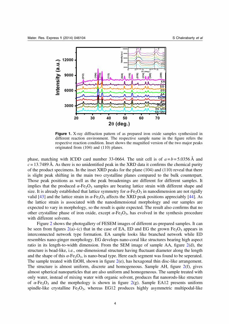
phase, matching with ICDD card number 33-0664. The unit cell is of a = b= 5.0356Å andc = 13.7489Å. As there is no unidentified peak in the XRD data it confirms the chemical purityof the product specimens. In the inset XRD peaks for the plane (104) and (110) reveal that thereis slight peak shifting in the main two crystalline planes compared to the bulk counterpart.Those peak positions as well as the peak broadenings are different for different samples. Itimplies that the produced α-Fe2O3 samples are bearing lattice strain with different shape andsize. It is already established that lattice symmetry for α-Fe2O3 in nanodimension are not rigidlyvalid [43] and the lattice strain in α-Fe2O3 affects the XRD peak positions appreciably [44]. Asthe lattice strain is associated with the nanodimensional morphology and our samples areexpected to vary in morphology, so the result is quite expected. The result also confirms that noother crystalline phase of iron oxide, except α-Fe2O3, has evolved in the synthesis procedurewith different solvents.
Figure 2 shows the photogallery of FESEM images of different as-prepared samples. It canbe seen from figures 2(a)–(c) that in the case of EA, ED and EG the grown Fe2O3 appears ininterconnected network type formation. EA sample looks like branched network while EDresembles nano-ginger morphology. EG develops nano-coral like structures bearing high aspectratio in its length-to-width dimension. From the SEM image of sample AA, figure 2(d), thestructure is bead-like, i.e., one-dimensional structure having fluctuant diameter along the lengthand the shape of this α-Fe2O3, is nano-bead type. Here each segment was found to be seperated.The sample treated with EtOH, shown in figure 2(e), has hexagonal thin disc-like arrangement.The structure is almost uniform, discrete and homogeneous. Sample AH, figure 2(f), givesalmost spherical nanoparticles that are also uniform and homogeneous. The sample treated withonly water, instead of mixing water with organic solvent, produces flat nanorods-like structureof α-Fe2O3 and the morphology is shown in figure 2(g). Sample EA12 presents uniformspindle-like crystalline Fe2O3, whereas EG12 produces highly asymmetric multipodal-like
Figure 1. X-ray diffraction pattern of as prepared iron oxide samples synthesized indifferent reaction environment. The respective sample name in the figure refers therespective reaction condition. Inset shows the magnified version of the two major peaksoriginated from (104) and (110) planes.
4
Mater. Res. Express 1 (2014) 046104 S Chakrabarty et al
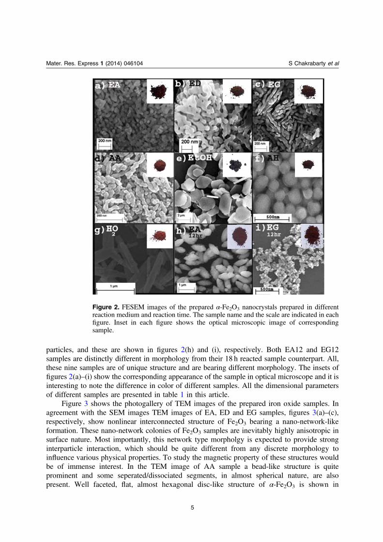
particles, and these are shown in figures 2(h) and (i), respectively. Both EA12 and EG12samples are distinctly different in morphology from their 18 h reacted sample counterpart. All,these nine samples are of unique structure and are bearing different morphology. The insets offigures 2(a)–(i) show the corresponding appearance of the sample in optical microscope and it isinteresting to note the difference in color of different samples. All the dimensional parametersof different samples are presented in table 1 in this article.
Figure 3 shows the photogallery of TEM images of the prepared iron oxide samples. Inagreement with the SEM images TEM images of EA, ED and EG samples, figures 3(a)–(c),respectively, show nonlinear interconnected structure of Fe2O3 bearing a nano-network-likeformation. These nano-network colonies of Fe2O3 samples are inevitably highly anisotropic insurface nature. Most importantly, this network type morpholgy is expected to provide stronginterparticle interaction, which should be quite different from any discrete morphology toinfluence various physical properties. To study the magnetic property of these structures wouldbe of immense interest. In the TEM image of AA sample a bead-like structure is quiteprominent and some seperated/dissociated segments, in almost spherical nature, are alsopresent. Well faceted, flat, almost hexagonal disc-like structure of α-Fe2O3 is shown in
Figure 2. FESEM images of the prepared α-Fe2O3 nanocrystals prepared in differentreaction medium and reaction time. The sample name and the scale are indicated in eachfigure. Inset in each figure shows the optical microscopic image of correspondingsample.
5
Mater. Res. Express 1 (2014) 046104 S Chakrabarty et al
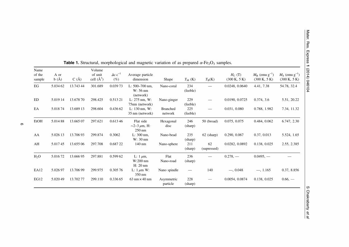
Table 1. Structural, morphological and magnetic variation of as prepared α-Fe2O3 samples.
Nameof thesample
A orb (Å) C (Å)
Volumeof unitcell (Å3)
Δc c−1
(%)Average particle
dimension Shape TM (K) TB(K)HC (T)
(300 K, 5 K)MR (emu g−1)(300 K, 5 K)
MS (emu g−1)(300 K, 5 K)
EG 5.034 62 13.743 44 301.689 0.039 73 L: 500–700 nm,W: 56 nm(network)
Nano-coral 234(feeble)
— 0.0248, 0.0640 4.41, 7.38 54.78, 32.4
ED 5.019 14 13.678 70 298.425 0.513 21 L: 275 nm, W:75nm (network)
Nano-ginger 229(feeble)
— 0.0190, 0.0725 0.374, 3.6 5.51, 20.22
EA 5.018 74 13.689 13 298.604 0.436 62 L: 130 nm, W:35 nm (network)
Branchednetwork
225(feeble)
— 0.031, 0.080 0.788, 1.982 7.34, 11.32
EtOH 5.014 88 13.665 07 297.621 0.613 46 Flat side∼2–3 μm, H:
250 nm
Hexagonaldisc
246(sharp)
50 (broad) 0.075, 0.075 0.484, 0.062 6.747, 2.30
AA 5.026 13 13.706 93 299.874 0.3062 L: 300 nm,W: 30 nm
Nano-bead 235(sharp)
62 (sharp) 0.290, 0.067 0.37, 0.013 5.524, 1.65
AH 5.017 45 13.655 06 297.708 0.687 22 140 nm Nano-sphere 211(sharp)
62(supressed)
0.0282, 0.0892 0.138, 0.025 2.55, 2.385
H2O 5.016 72 13.666 95 297.881 0.599 62 L: 1 μm,W:200 nmH: 20 nm
FlatNano-road
236(sharp)
— 0.278, — 0.0495, — —
EA12 5.026 97 13.706 99 299.975 0.305 76 L: 1 μm W:350 nm
Nano spindle — 140 —, 0.048 —, 1.165 0.37, 8.856
EG12 5.020 49 13.702 77 299.110 0.336 65 63 nm× 40 nm Asymmetricparticle
228(sharp)
— 0.0054, 0.0874 0.138, 0.025 0.66, —
6
Mater.
Res.
Express
1(2014)
046104SChakrabarty
etal
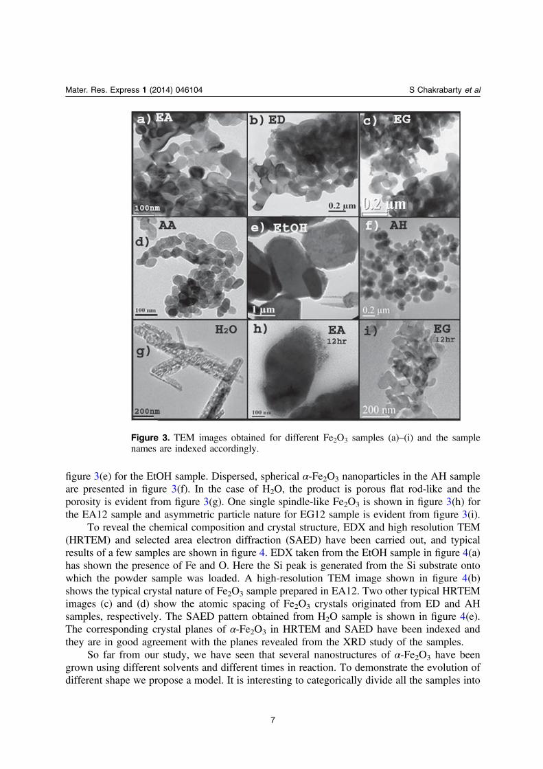
figure 3(e) for the EtOH sample. Dispersed, spherical α-Fe2O3 nanoparticles in the AH sampleare presented in figure 3(f). In the case of H2O, the product is porous flat rod-like and theporosity is evident from figure 3(g). One single spindle-like Fe2O3 is shown in figure 3(h) forthe EA12 sample and asymmetric particle nature for EG12 sample is evident from figure 3(i).
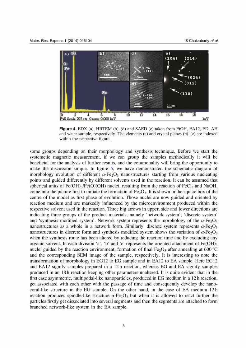
To reveal the chemical composition and crystal structure, EDX and high resolution TEM(HRTEM) and selected area electron diffraction (SAED) have been carried out, and typicalresults of a few samples are shown in figure 4. EDX taken from the EtOH sample in figure 4(a)has shown the presence of Fe and O. Here the Si peak is generated from the Si substrate ontowhich the powder sample was loaded. A high-resolution TEM image shown in figure 4(b)shows the typical crystal nature of Fe2O3 sample prepared in EA12. Two other typical HRTEMimages (c) and (d) show the atomic spacing of Fe2O3 crystals originated from ED and AHsamples, respectively. The SAED pattern obtained from H2O sample is shown in figure 4(e).The corresponding crystal planes of α-Fe2O3 in HRTEM and SAED have been indexed andthey are in good agreement with the planes revealed from the XRD study of the samples.
So far from our study, we have seen that several nanostructures of α-Fe2O3 have beengrown using different solvents and different times in reaction. To demonstrate the evolution ofdifferent shape we propose a model. It is interesting to categorically divide all the samples into
Figure 3. TEM images obtained for different Fe2O3 samples (a)–(i) and the samplenames are indexed accordingly.
7
Mater. Res. Express 1 (2014) 046104 S Chakrabarty et al
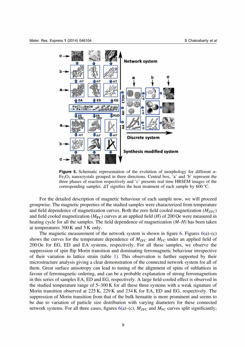
some groups depending on their morphology and synthesis technique. Before we start thesystemetic magnetic measurement, if we can group the samples methodically it will bebeneficial for the analysis of further results, and the commonality will bring the opportunity tomake the discussion simple. In figure 5, we have demonstrated the schematic diagram ofmorphology evolution of different α-Fe2O3 nanostructures starting from various nucleatingpoints and guided differently by different solvents used in the reaction. It can be assumed thatspherical units of Fe(OH)3/Fe(O)(OH) nuclei, resulting from the reaction of FeCl3 and NaOH,come into the picture first to initiate the formation of Fe2O3. It is shown in the square box of thecentre of the model as first phase of evolution. Those nuclei are now guided and oriented byreaction medium and are markedly influenced by the microenvironment produced within therespective solvent used in the reaction. Three big arrows in upper, side and lower directions areindicating three groups of the product materials, namely ‘network system’, ‘discrete system’
and ‘synthesis modified system’. Network system represents the morphology of the α-Fe2O3
nanostructures as a whole in a network form. Similarly, discrete system represents α-Fe2O3
nanostructures in discrete form and synthesis modified system shows the variation of α-Fe2O3
when the synthesis route has been altered by reducing the reaction time and by excluding anyorganic solvent. In each division ‘a’, ‘b’ and ‘c’ represents the oriented attachment of Fe(OH)3nuclei guided by the reaction environment, formation of final Fe2O3 after annealing at 600 °Cand the corresponding SEM image of the sample, respectively. It is interesting to note thetransformation of morphology in EG12 to EG sample and in EA12 to EA sample. Here EG12and EA12 signify samples prepared in a 12 h reaction, whereas EG and EA signify samplesproduced in an 18 h reaction keeping other parameters unaltered. It is quite evident that in thefirst case asymmetric, multipodal-like nanoparticles, produced in EG medium in a 12 h reaction,get associated with each other with the passage of time and consequently develop the nano-coral-like structure in the EG sample. On the other hand, in the case of EA medium 12 hreaction produces spindle-like structure α-Fe2O3 but when it is allowed to react further theparticles firstly get dissociated into several segments and then the segments are attached to formbranched network-like system in the EA sample.
Figure 4. EDX (a), HRTEM (b)–(d) and SAED (e) taken from EtOH, EA12, ED, AHand water sample, respectively. The elements (a) and crystal planes (b)–(e) are indexedwithin the respective figure.
8
Mater. Res. Express 1 (2014) 046104 S Chakrabarty et al
For the detailed description of magnetic behaviour of each sample now, we will proceedgroupwise. The magnetic properties of the studied samples were characterized from temperatureand field dependence of magnetization curves. Both the zero field cooled magnetization (MZFC)and field cooled magnetization (MFC) curves at an applied field (H) of 200Oe were measured inheating cycle for all the samples. The field dependence of magnetization (M–H) has been takenat temperatures 300K and 5K only.
The magnetic measurement of the network system is shown in figure 6. Figures 6(a)–(c)shows the curves for the temperature dependence of MZFC and MFC under an applied field of200Oe for EG, ED and EA systems, respectively. For all these samples, we observe thesuppression of spin flip Morin transition and dominating ferromagnetic behaviour irrespectiveof their variation in lattice strain (table 1). This observation is further supported by theirmicrostructure analysis giving a clear demonstration of the connected network system for all ofthem. Great surface anisotropy can lead to tuning of the alignment of spins of sublattices infavour of ferromagnetic ordering, and can be a probable explanation of strong ferromagnetismin this series of samples EA, ED and EG, respectively. A large field-cooled effect is observed inthe studied temperature range of 5–300K for all these three systems with a weak signature ofMorin transition observed at 225K, 229K and 234K for EA, ED and EG, respectively. Thesuppression of Morin transition from that of the bulk hematite is more prominent and seems tobe due to variation of particle size distribution with varying diameters for these connectednetwork systems. For all three cases, figures 6(a)–(c), MZFC and MFC curves split significantly;
Figure 5. Schematic representation of the evolution of morphology for different α-Fe2O3 nanocrystals grouped in three directions. Central box, ‘a’ and ‘b’ represent thethree phases of reaction respectively and ‘c’ presents real time HRSEM images of thecorresponding samples. ΔT signifies the heat treatment of each sample by 600 °C.
9
Mater. Res. Express 1 (2014) 046104 S Chakrabarty et al
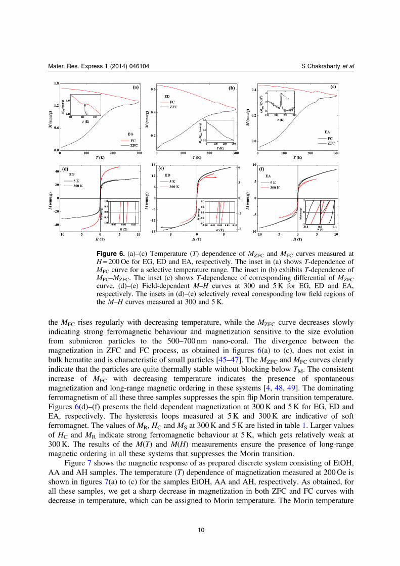
the MFC rises regularly with decreasing temperature, while the MZFC curve decreases slowlyindicating strong ferromagnetic behaviour and magnetization sensitive to the size evolutionfrom submicron particles to the 500–700 nm nano-coral. The divergence between themagnetization in ZFC and FC process, as obtained in figures 6(a) to (c), does not exist inbulk hematite and is characteristic of small particles [45–47]. The MZFC and MFC curves clearlyindicate that the particles are quite thermally stable without blocking below TM. The consistentincrease of MFC with decreasing temperature indicates the presence of spontaneousmagnetization and long-range magnetic ordering in these systems [4, 48, 49]. The dominatingferromagnetism of all these three samples suppresses the spin flip Morin transition temperature.Figures 6(d)–(f) presents the field dependent magnetization at 300K and 5K for EG, ED andEA, respectively. The hysteresis loops measured at 5K and 300K are indicative of softferromagnet. The values of MR, HC and MS at 300K and 5K are listed in table 1. Larger valuesof HC and MR indicate strong ferromagnetic behaviour at 5K, which gets relatively weak at300K. The results of the M(T) and M(H) measurements ensure the presence of long-rangemagnetic ordering in all these systems that suppresses the Morin transition.
Figure 7 shows the magnetic response of as prepared discrete system consisting of EtOH,AA and AH samples. The temperature (T) dependence of magnetization measured at 200Oe isshown in figures 7(a) to (c) for the samples EtOH, AA and AH, respectively. As obtained, forall these samples, we get a sharp decrease in magnetization in both ZFC and FC curves withdecrease in temperature, which can be assigned to Morin temperature. The Morin temperature
Figure 6. (a)–(c) Temperature (T) dependence of MZFC and MFC curves measured atH= 200Oe for EG, ED and EA, respectively. The inset in (a) shows T-dependence ofMFC curve for a selective temperature range. The inset in (b) exhibits T-dependence ofMFC–MZFC. The inset (c) shows T-dependence of corresponding differential of MZFC
curve. (d)–(e) Field-dependent M–H curves at 300 and 5 K for EG, ED and EA,respectively. The insets in (d)–(e) selectively reveal corresponding low field regions ofthe M–H curves measured at 300 and 5 K.
10
Mater. Res. Express 1 (2014) 046104 S Chakrabarty et al
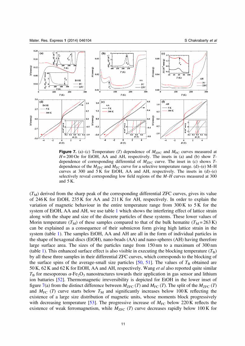
(TM) derived from the sharp peak of the corresponding differential ZFC curves, gives its valueof 246K for EtOH, 235K for AA and 211K for AH, respectively. In order to explain thevariation of magnetic behaviour in the entire temperature range from 300K to 5K for thesystem of EtOH, AA and AH, we use table 1 which shows the interfering effect of lattice strainalong with the shape and size of the discrete particles of these systems. These lower values ofMorin temperature (TM) of these samples compared to that of the bulk hematite (TM=263K)can be explained as a consequence of their submicron form giving high lattice strain in thesystem (table 1). The samples EtOH, AA and AH are all in the form of individual particles inthe shape of hexagonal discs (EtOH), nano-beads (AA) and nano-spheres (AH) having thereforelarge surface area. The sizes of the particles range from 150 nm to a maximum of 300 nm(table 1). This enhanced surface effect is also visible in executing the blocking temperature (TB)by all these three samples in their differential ZFC curves, which corresponds to the blocking ofthe surface spins of the average-small size particles [50, 51]. The values of TB obtained are50K, 62K and 62K for EtOH, AA and AH, respectively. Wang et al also reported quite similarTB for mesoporous α-Fe2O3 nanostructures towards their application in gas sensor and lithiumion battaries [52]. Thermomagnetic irreversibility is depicted for EtOH in the lower inset offigure 7(a) from the distinct difference between MZFC (T) andMFC (T). The split of theMZFC (T)and MFC (T) curve starts below TM and significantly increases below 100K reflecting theexistence of a large size distribution of magnetic units, whose moments block progressivelywith decreasing temperature [53]. The progressive increase of MFC below 220K reflects theexistence of weak ferromagnetism, while MZFC (T) curve decreases rapidly below 100K for
Figure 7. (a)–(c) Temperature (T) dependence of MZFC and MFC curves measured atH= 200Oe for EtOH, AA and AH, respectively. The insets in (a) and (b) show T-dependence of corresponding differential of MZFC curve. The inset in (c) shows T-dependence of the MZFC and MFC curve for a selective temperature range. (d)–(e) M–Hcurves at 300 and 5 K for EtOH, AA and AH, respectively. The insets in (d)–(e)selectively reveal corresponding low field regions of the M–H curves measured at 300and 5 K.
11
Mater. Res. Express 1 (2014) 046104 S Chakrabarty et al
EtOH. For the sample AA with bead like morphology and having moderate strain (0.3%), aconsistent decrease of MZFC and MFC curves down to 100K in figure 1(b), indicatespredominant antiferromagnetic (AFM) ordering in the sublattices of the studied compound.Below the temperature TB the MZFC and MFC curves decrease down to 29K and then decreasedown to 5K. The decrease of MFC curve in 62K⩽ T⩽ 29K is attributed to the AFM like spininteraction between Fe ions. The increase of MFC curve below 29K implies presenceferromagnetic (FM) interaction at T⩽ 29K. For the sample, AH with sphere-like morphology,we get similar behaviour to that of AA down to blocking temperature TB, below which a slowincrease of MZFC and MFC is observed, which is ascribed due to the presence of weak FMinteraction below TB in this sample.
The field (H) dependence of magnetization measured at temperatures 300K and 5K areshown in figures 7(d) to (f) for the samples EtOH, AA and AH, respectively. The existence of ahysteresis loop at 300K, as well as the absence of magnetization saturation at 5K for all thesamples should be noted. The hysteresis behaviours of M–H curves evidence a weakferromagnetic state of the studied samples at room temperature. The M–H curve at higher fieldshows almost linear dependence of magnetization on the applied magnetic field. The remanentmagnetization (MR), coercivity (HC) and saturation magnetization (MS) values (determined byextrapolating 1/H to zero-field in the M versus 1/H plot based on the high field data) of all theabove samples are listed in table 1. For the sample EtOH, the M−H curves show a steep linearincrease with the field at the low-field region (H⩽ 0.35 T) then a downward curvature followedby almost a linear behaviour up to the highest applied field without saturation. Hence there aretwo main contributions to magnetization in this case as given by M =MNC(H) + χH. The initialrapid increase in magnetization is attributed to the weak ferromagnetic behaviour, MNC in thesamples, which tries to saturate the moment. The non-saturation of moments at high field mayprobably be coming due to residual antiferromagnetism in the samples, and expressed by theterm χH, where χ is the magnetic susceptibility and H, the magnetic field. Hence, from M (H)isotherms it is clear that the contribution of ferromagnetic phase at room temperature is strongerthan that at 5K. For sample AA, the magnetization curve at low field region (inset offigure 7(e)) reflects further evidence of a delicate ferromagnetic behaviour at 5K. The shape ofthe loop and the large values of HC, MR and MS indicate strong ferromagnetic behaviour forsample AA at 300K, which becomes relatively weak at 5K due to progressive influence ofantiferromagnetic ordering in the sublattices. It is understood that enhanced surface anisotropyof the studied sample induced larger value of HC, MR and MS. It is worth noting that high valueof HC (≈0.29 T) at 300K can be explained due to the presence of large surface anisotropy ofbead-shaped particle with aspect ratio more than 1:10 [19]. For sample AH, similar to sampleEtOH, a hysteretic behaviour of M–H curve evidences a weak ferromagnetic state of the presentsample at room temperature. A different shaped hysteresis loop is observed at 5K; showing thecoexistence of the AFM and FM phases in the system which bears the maximum strain in lattice(0.68%) in a network of discrete particles of average size 140 nm.
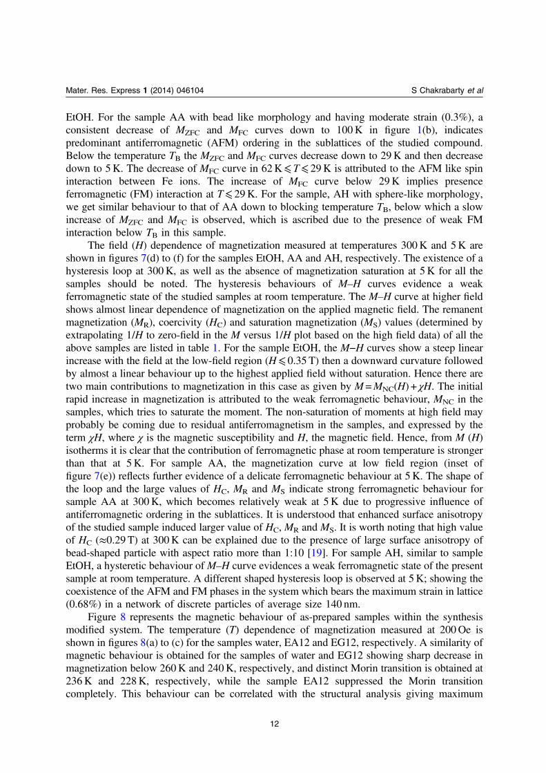
Figure 8 represents the magnetic behaviour of as-prepared samples within the synthesismodified system. The temperature (T) dependence of magnetization measured at 200Oe isshown in figures 8(a) to (c) for the samples water, EA12 and EG12, respectively. A similarity ofmagnetic behaviour is obtained for the samples of water and EG12 showing sharp decrease inmagnetization below 260K and 240K, respectively, and distinct Morin transition is obtained at236K and 228K, respectively, while the sample EA12 suppressed the Morin transitioncompletely. This behaviour can be correlated with the structural analysis giving maximum
12
Mater. Res. Express 1 (2014) 046104 S Chakrabarty et al
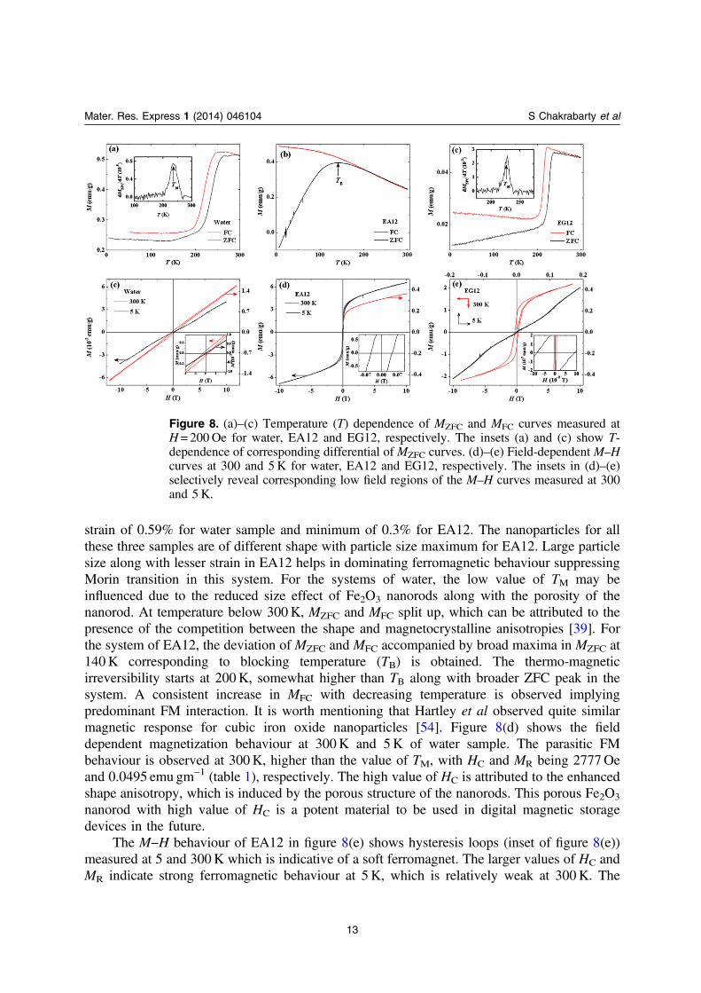
strain of 0.59% for water sample and minimum of 0.3% for EA12. The nanoparticles for allthese three samples are of different shape with particle size maximum for EA12. Large particlesize along with lesser strain in EA12 helps in dominating ferromagnetic behaviour suppressingMorin transition in this system. For the systems of water, the low value of TM may beinfluenced due to the reduced size effect of Fe2O3 nanorods along with the porosity of thenanorod. At temperature below 300K, MZFC and MFC split up, which can be attributed to thepresence of the competition between the shape and magnetocrystalline anisotropies [39]. Forthe system of EA12, the deviation of MZFC and MFC accompanied by broad maxima in MZFC at140K corresponding to blocking temperature (TB) is obtained. The thermo-magneticirreversibility starts at 200K, somewhat higher than TB along with broader ZFC peak in thesystem. A consistent increase in MFC with decreasing temperature is observed implyingpredominant FM interaction. It is worth mentioning that Hartley et al observed quite similarmagnetic response for cubic iron oxide nanoparticles [54]. Figure 8(d) shows the fielddependent magnetization behaviour at 300K and 5K of water sample. The parasitic FMbehaviour is observed at 300K, higher than the value of TM, with HC and MR being 2777Oeand 0.0495 emu gm−1 (table 1), respectively. The high value of HC is attributed to the enhancedshape anisotropy, which is induced by the porous structure of the nanorods. This porous Fe2O3
nanorod with high value of HC is a potent material to be used in digital magnetic storagedevices in the future.
The M−H behaviour of EA12 in figure 8(e) shows hysteresis loops (inset of figure 8(e))measured at 5 and 300K which is indicative of a soft ferromagnet. The larger values of HC andMR indicate strong ferromagnetic behaviour at 5K, which is relatively weak at 300K. The
Figure 8. (a)–(c) Temperature (T) dependence of MZFC and MFC curves measured atH= 200Oe for water, EA12 and EG12, respectively. The insets (a) and (c) show T-dependence of corresponding differential of MZFC curves. (d)–(e) Field-dependent M–Hcurves at 300 and 5K for water, EA12 and EG12, respectively. The insets in (d)–(e)selectively reveal corresponding low field regions of the M–H curves measured at 300and 5 K.
13
Mater. Res. Express 1 (2014) 046104 S Chakrabarty et al
results of the M(T) and M(H) measurements indicate that long-range magnetic ordering occurs,which suppresses the Morin transition.
The magnetic hysteresis of EG12 at 300K and 5K is shown in figure 8(f). A clearhysteresis loop is observed at 300K, which is indicative of the presence of ferromagneticcomponents, however, no saturation in magnetization is observed up to the maximum appliedmagnetic field at 300K showing the influence of other magnetic ordering present in the system.At 5K, a typical superparamagnetic behaviour with small hysteresis loop at low field region isobserved. However, the magnetic behaviour of EG12 needs further investigation to explain itsfield dependence at 5K.
After analysing the magnetic response of three different groups of samples, it is clearlyseen that the network type systems, in general, show high ferromagnetic behaviour whereMorin transiton is almost suppressed. The blocking temperature is also absent, in general, forthe network like α-Fe2O3 nanocrystals. Strong interparticle interaction, in these network-likesamples, is mainly attributed to their dominating ferromagnetic behaviour. Synthesized systemswith discrete morphology and homogeneous surface structure experience strong Morintransition with the presence of blocking temperature. High aspect ratio (in the case of AA andEtOH) and high lattice strain (in the case of EtOH and AH) are believed to be the main factorsin determining their magnetic behaviour. The samples produced by ‘modified synthesis route’show signature of either the Morin transition (in water and EG12) or the blocking temperature(in EA12). In this group, size and shape inhomogeneity of samples play a crucial role for theirmagnetic response.
Finally, all the synthesised α-Fe2O3 nanocrystals with their shape, size and latticeparameters are presented in table 1 to show their major signatures in magnetic behaviour. It isseen from the table that coercivity of AA and water sample at room temperature are even higherthan that measured for highly anisotropic Fe2O3 ‘nanocup’ system [2]. From the coercivityvalue recorded for the network type system it is evident that they are typically soft magnet,which was also seen in branched topology of porous α-Fe2O3 nanostructures [36]. The Morintransition temperature varies in a wide range of temeprature starting from 211K for sphericalAH sample to 246K for hexagonal disc like EtOH sample. It is also noteworthy that thenanocoral shaped EG sample provides excellent magnetic moment at both 300K and 5K.
4. Conclusion
Here we have synthesized α-Fe2O3 nanostructure with different unique morphology by simplesolvothermal technique followed by proper heat treatment. The prepared samples arecharacterized structurally and morphologically to understand their growth evolution. Thearticle reports the effects of morphology in the magnetic behaviour of α-Fe2O3 nanostructures.Magnetic response varies in a wide range for the as-obtained α-Fe2O3 nanocrystals. The resultshave been discussed on the basis of their surface anisotropy and lattice strain. The reportpresents an integrated version of morphology dependent magnetic behaviour of α-Fe2O3
nanocrystals. It allows the creation of numerous recipes of α-Fe2O3 for optimizing and scalingup production towards technological applications.
14
Mater. Res. Express 1 (2014) 046104 S Chakrabarty et al
Acknowledgment
This work was financially supported by the UGC, India [42/1069/2013(SR) & 42-908/2013(SR)] and Special Assistance Program-UGC, India. S Chakrabarty also gratefully acknowledgesher fellowship from UGC-BSR Scheme, India. The authors gratefully acknowledge ProfessorJason Chang, IOP, Academia Sinica, Taiwan for his thoughtful discussions and for providingSEM/EDX facilities.
References
[1] Lian J, Duan X, Ma J, Peng P, Kim T and Zheng W 2009 Hematite (α-Fe2O3) with various morphologies:ionic liquid-assisted synthesis, formation mechanism, and properties ACS Nano 3 3749–61
[2] Jagadeesan D, Mansoori U, Mandal P, Sundaresan A and Eswaramoorthy M 2008 Hollow spheres tonanocups: tuning the morphology and magnetic properties of single-crystalline α-Fe2O3 nanostructuresAngew. Chem. 120 7799–802
[3] Cui Y and Lieber C M 2001 Funtional nanoscale electronic devices assembled using silicon nanowirebuilding blocks Science 291 851–3
[4] Lui L, Kou H-Z, Mo W, Liu H and Wang Y 2006 Surfactant-assisted synthesis of α-Fe2O3 nanotubes andnanorods with shape-dependent magnetic properties J. Phys. Chem. B 110 15218–23
[5] Wu C, Yin P, Zhu X, OuYang C and Xie Y 2006 Synthesis of hematite (α-Fe2O3) nanorods: diameter-sizeand shape effects on their applications in magnetism, lithium ion battery, and gas sensors J. Phys. Chem. B110 17806–12
[6] Garcia-Labato M A, Martinez A I, Castro-Roman M, Falcony C and Escobar-Alarcon L 2011 Correlationbetween structural and magnetic properties of sprayed iron oxide thin films Physica B 406 1496–500
[7] Wang D, Wang Q and Wang T 2011 Controlled synthesis of mesoporous hematite nanostructures and theirapplication as electrochemical capacitor electrodes Nanotechnology 22 135604–15
[8] Wang L and Gao L 2011 Controlled synthesis and tunable properties of hematite hierarchical structures in adual-surfactant system CrystEngComm. 13 1998–2005
[9] Elias V R, Oliva M I, Vaschetto E G, Urreta S E, Eimer G A and Silvetti S P 2010 Magnetic properties of ironloaded MCM-48 molecular sieves J. Magn. Magn. Mater. 322 3438–42
[10] Mitra S, Das S, Mandal K and Chaudhuri S 2007 Synthesis of a α-Fe2O3 nanocrystal in its differentmorphological attributes: growth mechanism, optical and magnetic properties Nanotechnology 18275608–16
[11] Dieckmann R 1993 Point defects and transport in haematite (Fe2O3−ε) Philos. Mag. A 68 725–45[12] Cesar I, Kay A, Martinez J A G and Gratzel M 2006 Translucent thin film Fe2O3 photoanodes for efficient
water splitting by sunlight: nanostructure-directing effect of Si-doping J. Am. Chem. Soc. 128 4582–3[13] Ohmori T, Takahashi H, Mametsuka H and Suzuki E 2000 Photocatalytic oxygen evolution on α-Fe2O3 films
using Fe3+ ion as a sacrificial oxidizing agent Phys. Chem. Chem. Phys. 2 3519–22[14] Gou X, Wang G, Park J, Liu H and Yang J 2008 Monodisperse hematite porous nanospheres: synthesis,
characterization, and applications for gas sensors Nanotechnology 19 125606–11[15] Widder K J, Senyei A E and Scarpelli D G 1978 Magnetic microspheres: a model system of site specific drug
delivery in vivo Proc. Soc. Exp. Biol. Med. 158 141–6[16] Garcon G, Garry S, Gosset P, Zerimech F, Martin A, Hannothiaux M-H and Shirali P 2001 Benzo(a)pyrene-
coated onto Fe2O3 particles-induced lung tissue injury: role of free radicals Cancer Lett. 167 7–15[17] Busch M, Gruyters M and Winter H 2006 Spin polarization and structure of thin iron oxide layers prepared
by oxidation of Fe(110) Surf. Sci. 600 4166–9[18] Walter D 2006 Characterization of synthetic hydrous hematite pigments Thermochim. Acta 445 195–9
15
Mater. Res. Express 1 (2014) 046104 S Chakrabarty et al
[19] Tadić M, Čitaković N, Panjan M, Stojanović Z, Marković D and Spasojević V 2011 Synthesis, morphology,microstructure and magnetic properties of hematite submicron particles J. Alloys Compd. 509 7639–44
[20] Teja A S and Koh P-Y 2009 Synthesis, properties, and applications of magnetic iron oxide nanoparticlesProg. Cryst. Growth Charact. Mater. 55 22–45
[21] Liu Z and Zheng Y 2011 Effect of Fe(II) on the formation of iron oxide synthesized from pyrite cinders byhydrothermal process Powder Technol. 209 119–23
[22] Su C, Wang H and Liu X 2011 Controllable fabrication and growth mechanism of hematite cubes Cryst. Res.Technol. 46 209–14
[23] Wang H, Geng W and Wang Y 2011 Preparation of nanoparticles and hollow spheres of α-Fe2O3 and theirproperties Res. Chem. Intermed. 37 389–95
[24] Wu W, Xiao X, Zhang S, Zhou J, Fan L, Ren F and Jiang C 2010 Large-scale and controlled synthesis of ironoxide magnetic short nanotubes: shape evolution, growth mechanism, and magnetic properties J. Phys.Chem. C 114 16092–103
[25] Tadić M, Kusigerski V, Marković D, Čitaković N, Remškar M and Spasojević V 2009 Morphological,structural and magnetic properties of α-Fe2O3 nanoparticles in an amorphous alumina matrix obtained byaqueous combustion method J. Alloys Compd. 486 839–43
[26] Mandal S and Müller A H E 2008 Facile route to the synthesis of porous α-Fe2O3 nanorods Mater. Chem.Phys. 111 438–43
[27] Liang H-F and Wang Z-C 2010 Template-free synthesis and characterization of snowflake-like α-Fe2O3
microstructures Mater. Lett. 64 2410–2[28] Song F, Guan J, Fan X and Yan G 2009 Single-crystal star-like arrayed particles of hematite: synthesis,
formation mechanism and magnetic properties J. Alloys Compd. 485 753–8[29] Xu X, Cao R, Jeong S and Cho J 2012 Spindle-like mesoporous α-Fe2O3 anode material prepared from MOF
template for high-rate lithium batteries Nano Lett. 12 4988–91[30] Bharathi S, Nataraj D, Seetha M, Mangalaraj D, Ponpandian N, Masuda Y, Senthil K and Yong K 2010
Controlled growth of single-crystalline, nanostructured dendrites and snowflakes of α-Fe2O3: influence ofthe surfactant on the morphology and investigation of morphology dependent magnetic propertiesCrystEngComm. 12 373–82
[31] Li S, Zhang H, Wu J, Ma X and Yang D 2006 Shape-control fabrication and characterization of the airplane-like FeO(OH) and Fe2O3 nanostructures Cryst. Growth Des. 6 351–3
[32] Zhu L-P, Xiao H-M and Fu S-Y 2007 Template-free synthesis of monodispersed and single-crystallinecantaloupe-like Fe2O3 superstructures Cryst. Growth Des. 7 177–82
[33] Zhang Y, Wang S, Li X, Chen L, Qian Y and Zhang Z 2006 CuO shuttle-like nanocrystals synthesized byoriented attachment J. Cryst. Growth 291 196–201
[34] Wang X, Chen X, Ma X, Zheng H, Ji M and Zhang Z 2004 Low-temperature synthesis of α-Fe2O3
nanoparticles with a closed cage structure Chem. Phys. Lett. 384 391–3[35] Cao M, Liu T, Gao S, Sun G, Wu X, Hu C and Wang Z L 2005 Single-crystal dendritic micro-pines of
magnetic α-Fe2O3: large-scale synthesis, formation mechanism, and properties Angew. Chem. Int. Ed. 444197–201
[36] Yang H et al 2010 Porous α-Fe2O3 nanostructures with branched topology: growth, formation mechanism,and properties CrystEngComm. 12 1842–9
[37] Chakrabarty S and Chatterjee K 2013 Oriented growth of α-Fe2O3 nanocrystals with different morphologyand their optical behaviour J. Cryst. Growth 381 107–13
[38] Tang B, Wang G, Zhuo L, Ge J and Cui L 2006 Facile route to α-FeOOH and α-Fe2O3 nanorods andmagnetic property of α-Fe2O3 nanorods Inorg. Chem. 45 5196–200
[39] Mitra S, Das S, Basu S, Sahu P and Mandal K 2009 Shape-and field-dependent Morin transitions instructured α-Fe2O3 J. Magn. Magn. Mater. 321 2925–31
16
Mater. Res. Express 1 (2014) 046104 S Chakrabarty et al
[40] Gou X, Wang G, Kong X, Wexler D, Horvat J, Yang J and Park J 2008 Flutelike porous hematite nanorodsand branched nanostructures: synthesis, characterisation and application for gas-sensing Chem. Eur. J. 145996–6002
[41] Muruganandham M, Amutha R, Sathish M, Singh T S, Suri R P S and Sillanpää M 2011 Facile fabrication ofhierarchical α-Fe2O3: self-assembly and its magnetic and electrochemical properties J. Phys. Chem. C 11518164–73
[42] Zhao Y, Dunnill C W, Zhu Y, Gregory D H, Kockenberger W, Li Y, Hu W, Ahmad I and McCartney D G2007 Low-temperature magnetic properties of hematite nanorods Chem. Mater. 19 916–21
[43] Lu L, Li L, Wang X and Li G 2005 Understanding of the finite size effects on lattice vibrations and electronictransitions of nano α-Fe2O3 J. Phys. Chem. B 109 17151–6
[44] Kleiman-Shwarsctein A, Huda M N, Walsh A, Yan Y, Stucky G D, Hu Y-S, Al-Jassim M M andMcFarland E W 2010 Electrodeposited aluminum-doped α-Fe2O3 photoelectrodes: experiment and theoryChem. Mater. 22 510–7
[45] Li Z, Lai X, Wang H, Mao D, Xing C and Wang D 2009 Direct hydrothermal synthesis of single-crystallinehematite nanorods assisted by 1,2-propanediamine Nanotechnology 20 245603–11
[46] Gupta R K, Ghosh K, Dong L and Kahol P K 2011 Structural and magnetic properties of phase controllediron oxide rods Mater. Lett. 65 225–8
[47] Xia C, Hu C, Xiong Y and Wang N 2009 Synthesis of α-Fe2O3 hexagons and their magnetic propertiesJ. Alloys Compd. 480 970–3
[48] Jiao F, Harrison A, Jumas J-C, Chadwick A V, Kockelmann W and Bruce P G 2006 Ordered mesoporousFe2O3 with crystalline walls J. Am. Chem. Soc. 128 5468–74
[49] Li G, Liu M and Kou H-Z 2011 Mesoporous α-Fe2O3 nanospheres: structural evolution and investigation ofmagnetic properties Chem. Eur. J. 17 4323–9
[50] Cangussu D, Nunes W C, Corrêa H L S, Macedo W A A, Knobel M, Alves O L, Filho A G S and Mazali I O2009 γ-Fe2O3 nanoparticles dispersed in porous Vycor glass: a magnetically diluted integrated systemJ. Appl. Phys. 105 013901–7
[51] Zysler R D, Fiorani D, Testa A M, Suber L, Agostinelli E and Godinho M 2003 Size dependence of the spin-flop transition in hematite nanoparticles Phys. Rev. B 68 212408–11
[52] Sun B, Horvat J, Kim H S, Kim W-S, Ahn J and Wang G 2010 Synthesis of mesoporous α-Fe2O3
nanostructures for highly sensitive gas sensors and high capacity anode materials in lithium ion batteriesJ. Phys. Chem. C 114 18753–61
[53] Mansilla M V, Zysler R, Fiorani D and Suber L 2002 Annealing effects on magnetic properties of acicularhematite nanoparticles Physica B 320 206–9
[54] Zhen G et al 2011 Comparative study of the magnetic behavior of spherical and cubic superparamagnetic Ironoxide nanoparticles J. Phys. Chem. C 115 327–34
17
Mater. Res. Express 1 (2014) 046104 S Chakrabarty et al
Related Documents