Nanotoxicology, 2012; Early Online, 1–13 © 2012 Informa UK, Ltd. ISSN: 1743-5390 print / 1743-5404 online DOI: 10.3109/17435390.2011.652681 Morphological transformation induced by multiwall carbon nanotubes on Balb/3T3 cell model as an in vitro end point of carcinogenic potential Jessica Ponti 1 , Francesca Broggi 1 , Valentina Mariani 1 , Laura De Marzi 1 , Renato Colognato 1 , Patrick Marmorato 1 , Sabrina Gioria 1 , Douglas Gilliland 1 , César Pascual Garcìa 1 , Stefania Meschini 2 , Annarita Stringaro 2 , Agnese Molinari 2 , Hubert Rauscher 1 , & François Rossi 1 1 European Commission, Joint Research Centre, Institute for Health and Consumer Protection, Nanobiosciences Unit, Ispra, (VA), Italy and 2 Istituto Superiore di Sanità, Department of Technology and Health, Rome, Italy Abstract In this work we investigated the toxicological effects of nude and chemically functionalised (-NH 2 , -OH and -COOH groups) multiwall carbon nanotubes (mwCNTs) using immortalised mouse fibroblasts cell line (Balb/3T3) as in vitro model, alternative to the use of animals, to assess basal cytotoxicity, carcinogenic potential, genotoxicity and cell interaction of nanomaterials (NM). Combining in vitro tests such as cell transformation assay and micronucleus with physicochemical and topological analysis, we obtained results showing no cytotoxicity and genotoxicity. Carcinogenic potential and mwCNTs interaction with cells were instead evident. We stressed the importance that different toxicological end points have to be considered when studying NM, therefore, assays able to detect long-term effects, such as carcinogenicity, must be taken into account together with a panel of tests able to detect more immediate effects like basal cytotoxicity or genotoxicity. Keywords: Multiwall carbon nanotubes, morphological transfor- mation, carcinogenic potential, genotoxicity, cytotoxicity, Balb/ 3T3, physicochemical properties, asbestos, electron microscopy Introduction Carbon nanotubes (CNTs) either single-wall or multiwall (mwCNTs) represent, among engineered nanoparticles (NPs), a major interest for many different industrial applica- tions including sensors, drug delivery, biomedical uses and the production of innovative carbon-based intelligent textiles (Kam et al. 2005; Rivas et al. 2007; INTELTEX project web site). The increasing interest in carbon-based materials is mainly due to their exceptional electronic and mechanical properties and their different functionalisation capacity (Rivas et al. 2007). CNTs are already under massive industrial production and novel production methods methods were introduced in the last years, considerably improving their synthesis and yield. Despite this, the information concerning potential hazards related to CNTs exposure remains sparse, incomplete and still under debate (Oberdörster 2004; Lam et al. 2004; Ding et al. 2005; Oberdörster et al. 2005; Ponti et al. 2010; Becker et al. 2011). Many studies report a relationship between exposure to CNTs and health consequences. They come to the conclusion that exposure to CNTs could place workers to a possible risk resulting in cytotoxic response that goes from alterations in cell viability, DNA damage to inflammatory fibrotic lesions in the lung and formation of neoplastic lesions (Donaldson et al. 2006; Rotoli et al. 2008, 2009; Chou et al. 2008; Genaidy et al. 2009; Colognato et al. 2009; Aschberger et al. 2010; Johnston et al. 2010). These effects are also supported by evidence of direct interaction of CNTs with cells (Monteiro-Riviere et al. 2005; Porter et al. 2007; Fenoglio et al. 2008; Muller et al. 2008; Becker et al. 2011; Di Giorgio et al. 2011). Inhaled mwCNTs in mice reached the sub-pleura after only 24 h and after a single inhalation exposure, both pleura and sub-pleura showed fibrotic lesions after 2 and 6 weeks, respectively, inducing inflammation and granulo- mas (Ryman-Rasmussen et al. 2009). In mouse macrophage in vitro model (RAW 264.7), mwCNTs induce necrosis, chromosomal aberrations, oxidative stress but not inflam- mation even if they penetrate the cell membrane within the nuclear envelope (Di Giorgio et al. 2011). Actually no long- term animal studies that allow any conclusion (or that allow to draw any conclusion) about the carcinogenic potential of CNTs following inhalation, together with any epidemiolog- ical data on humans are present. The most important organ to be affected by inflammation and consequent carcinogenic responses after inhalation would/should be the lung since it is the organ where the highest concentration of CNTs would be expected. However, there is the possibility that the CNTs translocate to other locations via the cardiovascular system, Correspondence: Jessica Ponti, European Commission, Joint Research Centre, IHCP, NBS Unit, TP203, via E. Fermi 2749, 21027 Ispra, (VA), Italy. Tel: +39 0332785793. Fax: +39 0332785787. E-mail: [email protected] (Received 17 June 2011; accepted 7 December 2011) Nanotoxicology Downloaded from informahealthcare.com by Commission European Comm on 01/27/12 For personal use only.

Welcome message from author
This document is posted to help you gain knowledge. Please leave a comment to let me know what you think about it! Share it to your friends and learn new things together.
Transcript

Nanotoxicology, 2012; Early Online, 1–13©2012 Informa UK, Ltd.ISSN: 1743-5390 print / 1743-5404 onlineDOI: 10.3109/17435390.2011.652681
Morphological transformation induced by multiwall carbon nanotubeson Balb/3T3 cell model as an in vitro end point of carcinogenic potential
Jessica Ponti1, Francesca Broggi1, Valentina Mariani1, Laura De Marzi1, Renato Colognato1,Patrick Marmorato1, Sabrina Gioria1, Douglas Gilliland1, César Pascual Garcìa1, Stefania Meschini2,Annarita Stringaro2, Agnese Molinari2, Hubert Rauscher1, & François Rossi1
1European Commission, Joint Research Centre, Institute for Health and Consumer Protection, Nanobiosciences Unit, Ispra, (VA),Italy and 2Istituto Superiore di Sanità, Department of Technology and Health, Rome, Italy
AbstractIn this work we investigated the toxicological effects of nude andchemically functionalised (-NH2, -OH and -COOH groups)multiwall carbon nanotubes (mwCNTs) using immortalisedmouse fibroblasts cell line (Balb/3T3) as in vitro model,alternative to the use of animals, to assess basal cytotoxicity,carcinogenic potential, genotoxicity and cell interaction ofnanomaterials (NM). Combining in vitro tests such as celltransformation assay and micronucleus with physicochemicaland topological analysis, we obtained results showing nocytotoxicity and genotoxicity. Carcinogenic potential andmwCNTs interaction with cells were instead evident. We stressedthe importance that different toxicological end points have to beconsidered when studying NM, therefore, assays able to detectlong-term effects, such as carcinogenicity, must be taken intoaccount together with a panel of tests able to detect moreimmediate effects like basal cytotoxicity or genotoxicity.
Keywords: Multiwall carbon nanotubes, morphological transfor-mation, carcinogenic potential, genotoxicity, cytotoxicity, Balb/3T3, physicochemical properties, asbestos, electron microscopy
Introduction
Carbon nanotubes (CNTs) either single-wall or multiwall(mwCNTs) represent, among engineered nanoparticles(NPs), a major interest for many different industrial applica-tions including sensors, drugdelivery, biomedical uses and theproduction of innovative carbon-based intelligent textiles(Kam et al. 2005; Rivas et al. 2007; INTELTEX project website). The increasing interest in carbon-based materials ismainly due to their exceptional electronic and mechanicalproperties and their different functionalisation capacity(Rivas et al. 2007). CNTs are already under massive industrialproduction and novel production methods methods were
introduced in the last years, considerably improving theirsynthesis and yield. Despite this, the information concerningpotential hazards related to CNTs exposure remains sparse,incompleteandstillunderdebate (Oberdörster2004;Lametal.2004; Ding et al. 2005; Oberdörster et al. 2005; Ponti et al. 2010;Becker et al. 2011).
Many studies report a relationship between exposure toCNTs and health consequences. They come to the conclusionthat exposure to CNTs could place workers to a possible riskresulting in cytotoxic response that goes from alterations in cellviability, DNA damage to inflammatory fibrotic lesions in thelung and formation of neoplastic lesions (Donaldson et al. 2006;Rotoli et al. 2008, 2009; Chou et al. 2008; Genaidy et al. 2009;Colognato et al. 2009; Aschberger et al. 2010; Johnston et al.2010). These effects are also supported by evidence of directinteraction of CNTs with cells (Monteiro-Riviere et al. 2005;Porter et al. 2007; Fenoglio et al. 2008; Muller et al. 2008;Becker et al. 2011; Di Giorgio et al. 2011).
Inhaled mwCNTs in mice reached the sub-pleura afteronly 24 h and after a single inhalation exposure, bothpleura and sub-pleura showed fibrotic lesions after 2 and6 weeks, respectively, inducing inflammation and granulo-mas (Ryman-Rasmussen et al. 2009). In mouse macrophagein vitro model (RAW 264.7), mwCNTs induce necrosis,chromosomal aberrations, oxidative stress but not inflam-mation even if they penetrate the cell membrane within thenuclear envelope (Di Giorgio et al. 2011). Actually no long-term animal studies that allow any conclusion (or that allowto draw any conclusion) about the carcinogenic potential ofCNTs following inhalation, together with any epidemiolog-ical data on humans are present. The most important organto be affected by inflammation and consequent carcinogenicresponses after inhalation would/should be the lung since itis the organ where the highest concentration of CNTs wouldbe expected. However, there is the possibility that the CNTstranslocate to other locations via the cardiovascular system,
Correspondence: Jessica Ponti, European Commission, Joint Research Centre, IHCP, NBS Unit, TP203, via E. Fermi 2749, 21027 Ispra, (VA), Italy.Tel: +39 0332785793. Fax: +39 0332785787. E-mail: [email protected]
(Received 17 June 2011; accepted 7 December 2011)
Nan
otox
icol
ogy
Dow
nloa
ded
from
info
rmah
ealth
care
.com
by
Com
mis
sion
Eur
opea
n C
omm
on
01/2
7/12
For
pers
onal
use
onl
y.
or into the pleura. Under these circumstances, additionaltarget organs where the long-term carcinogenic effect can beseen cannot be excluded (Aschberger et al. 2010).
Since CNTs present similarities to asbestos fibres in termsof shape and size and their presumed insolubility in thelungs (Aschberger et al. 2010), long-term studies on theirinhalation are needed to determine the carcinogenic poten-tial. In fact, based on available in vitro/in vivo results, risk forhumans exposed to single or mWCNTs in the workplacecannot be excluded and more data to refine the risk assess-ment or risk reduction measures are required (Ma-Hocket al. 2009; Aschberger et al. 2010).
In thiswork,wepresent an in vitro study onmwCNTs (nudeand functionalised with -OH, -NH2 and -COOH groups) ofindustrial interest, mainly used for the production of smarttextile (INTELTEX project web site), focussing on cytotoxicity,genotoxicity, carcinogenic potential and cell interaction.
Mouse fibroblasts cell line (Balb/3T3) model was selectedamong Syrian Hamster Embryo (SHE) cells, C3H10T1/2 andBhas42 in vitro models (Kerckaert et al. 1996; Isfort &LeBoeuf 1996; Balls & Clothier 2010) alternative to the useof animals able to detect carcinogenic potential induced bychemicals and nanomaterials (NM). These cell models, andthe related in vitromorphological transformation assays, areconsidered comparable with the in vivo 2-year assay used toverify carcinogenicity (Balls & Clothier 2010; Vanparys et al.2010). In addition, Balb/3T3 and SHE models were tested aspart of a pre-validation study and considered scientificallyvalid, to assess the carcinogenic potential of chemicals(Vanparys et al. 2010) Balb/3T3 is already used for NM(Ponti et al. 2009). Balb/3T3 immortalised cells, after expo-sure to carcinogens, become tumourigenic forming morpho-logically transformed colonies (foci type III). Their ability toinduce cancer in vivo is detectable by injecting the trans-formed cells in nude mice and observing their growth assarcomas (Kurzepa et al. 1984; Saffiotti & Ahmed 1995).
The in vitro tests used in this work were the colony-forming efficiency (CFE) to evaluate the cytotoxicity and thecell transformation assay (CTA) to assess concurrently cyto-toxicity and carcinogenic potential (Ponti et al. 2009, 2010).Cytokinesis-block micronucleus assay (MN) was insteadperformed for genotoxicity end point (Ponti et al. 2009).Since the CTA is able to detect both genetic and epigeneticchanges without discriminating between them, we intro-duced the MN assay to understand whether the carcinogenicpotential was due to genotoxic effects or not.
Interaction of nude mwCNTs with cells was previouslyobserved by atomic force microscopy and scanning electronmicroscopy (SEM) (Ponti et al. 2010). In the present work, we
confirmed those results and we investigated the effect of themwCNTs functionalised with -OH, -NH2 and -COOH groupsusing SEM. In addition, the mwCNTs uptake was alsostudied by transmission electron microscopy (TEM).
Amosite, chrysotile and crocidolite asbestos fibreswere used to verify the validity of Balb/3T3 cell model inassessing the morphological transformation as end point ofcarcinogenic potential of asbestos fibres. Since asbestosfibres are well-recognised carcinogens for humans andproduced positive results on Balb/3T3 (Oberdörster2000), we used them as positive control fibres inducingmorphological transformation.
Materials and methods
mwCNTs sample preparation and characterisationmwCNTs (Nanocyl�-7000, nude; Nanocyl�-3101, -COOH;Nanocyl�-3152, -NH2; Nanocyl�-3153, -OH supplied byNanocyl S.A., Belgium) were produced via catalytic carbonvapour deposition process.
Nanocyl-7000 contains 10% (w/v) of metal oxide impu-rities and Nanocyl-3101, Nanocyl-3152, Nanocyl-3153 lessthan 5% (w/v), as declared by the manufacturer after deter-mination by thermogravimetric analysis (TGA) and con-firmed, for the Nanocyl-7000, by inductively coupledplasma mass spectrometry (ICP-MS, Perkin-Elmer SCIEX,Ontario, Canada) in our laboratory. Morphological analysisby TEM showed an average diameter of 10 nm for all thesamples analysed by the manufacturer and length in therange of 1.5 mm for Nanocyl-7000 and Nanocyl-3101 and lessthan 1 mm for Nanocyl-3152 and Nanocyl-3153 (Table I).The percentage of functionalisation <4% for Nanocyl-3101,<0.5% for Nanocyl-3152 and <6% for Nanocyl-3153 wasmeasured by X-ray photoelectron spectroscopy (XPS) bythe supplier.
Stock suspensions containing 10 mg/mL of mwCNTswere prepared in dimethyl sulfoxide (DMSO, Sigma Aldrich,Italy), reported as a solvent that allows the suspension ofCNTs (Soto et al. 2007; Buford et al. 2007) and was selectedas the best dispersant agent under our experimental condi-tions (Ponti et al. 2010).
The stock suspensions were directly diluted in cell culturemedium reaching the concentrations of 1, 10 and 100 mg/mL,keeping the DMSO concentration at 1% (v/v), correspondingto the maximum non-toxic concentration for cells used inthis work.
The size distribution of the mwCNTs suspended in com-plete culture medium at the highest concentration tested(100 mg/mL) was measured by dynamic light scattering
Table I. mwCNTs commercial name and physicochemical characteristics.Product commercialname
Productdescription
Averagediameter (nm)
Averagelength (mm)
Carbon purity% (w/w)
Chemicalfunctionalisationb
Nanocyl�-7000 Thin mwCNTs for industrialapplication
9.5 1.5 90a None
Nanocyl�-3101 Thin mwCNTs 9.5 1.5 >95 -COOH
Nanocyl�-3152 Short thin mwCNTs 9.5 <1 >95 -NH2
Nanocyl�-3153 Short thin mwCNTs 9.5 <1 >95 -OHaAmount of metals in the highest mwCNTs concentration tested (100 mg/mL): Co (1.100 nM); Ni (0.044 nM); Zn (0.075 nM); Pb (0.002 nM); bNanocyl patented process(Nanocyl web site); mwCNTs, multiwall carbon nanotubes.
J. Ponti et al.
Nan
otox
icol
ogy
Dow
nloa
ded
from
info
rmah
ealth
care
.com
by
Com
mis
sion
Eur
opea
n C
omm
on
01/2
7/12
For
pers
onal
use
onl
y.

technique (DLS, Malvern Instruments, UK measurementprotocols ISO 13321: 1996).
The sedimentation of mwCNTs with different types offunctionalisation (nude, -OH, -COOH and -NH2), dispersedin culture medium at a concentration of 100 mg/mL, asdescribed above, was characterised by visible and nearinfrared light absorption (VIS-NIR) with a Thermo NicoletVision 300 spectrometer (Attal et al. 2006). Reproducibility ofthe pipetting was confirmed by VIS-NIR measurements inthe wavelength region between 600 and 1000 nm (Ponti et al.2010). The absorption value at 650 nm was selected toanalyse mwCNTs sedimentation for incubation times upto 72 h. This latter time corresponds to the higher exposuretime used for CFE and CTA. All measurements were carriedout at least in triplicate.
Asbestos fibres sample preparationAsbestos fibres of amosite (CAS n. 12172-73-5), chrysotile(CAS n. 12001-29-5) and crocidolite (CAS n. 12001-28-4)were supplied by the Institute of Occupational Medicine(IOM) (Edinburgh, UK). Stock suspensions of 10 mg/mLwere prepared in Milli-Q water and directly diluted in cellculture medium as described above for mwCNTs, keepingthe Milli-Q water concentration at 1% (v/v).
Cell culture conditionsImmortalised mouse fibroblast cell line (Balb/3T3), stem-ming from the clone A31-1-1, were kindly provided byHatano Research Institute (Japan) in the context of thepre-validation study (Vanparys et al. 2010).
Experimental cultures were prepared from deep-frozenstock vials and always maintained in a sub-confluent state(less than 70% of confluence). They were maintained incomplete culture medium (M10F) made by minimum essen-tial medium (MEM) low glucose added with 10% (v/v) foetalbovine serum (FBS, Australian Origin, Invitrogen, Italy, lot. n.6792117Y) and 1% (v/v) pen/strep (Invitrogen, Italy). Cellswere maintained under standard cell culture conditions (37�C,5% CO2 and 95% humidity, HERAEUS incubator, Germany).
Electron microscopy of mwCNTs interaction with cellsWe investigated mwCNTs interacting with cells, using a SEM(FEI Company, Nova 600I Nanolab) both in reflection andtransmission configurations, and using a TEM (PhilipsEM208S, FEI Company, Hillsboro, OR, USA).
SEM and STEM images were made using low (5 keV) andhigh (30 keV) acceleration voltages to acquire surface (usingreflected and backscattered electrons) and transmissionsignals, respectively. Balb/3T3 cells were seeded on siliconnitride substrate membranes (Silson Ltd., Northampton, UK)contained in 6-wells plate (Corning Costar, Italy) at thedensity of 105 cells/well in 3 mL of culture medium forSEM morphological analysis. Membranes of 100 nm thick-nesses were chosen as a good compromise of the substraterobustness and their transparency needed for the transmis-sion experiments. Cells were cultured for 24 h understandard cell culture conditions (37�C, 5% CO2 and 95%humidity, HERAEUS incubator) and exposed for 72 h to100 mg/mL mwCNTs. After exposure, cells were washed
twice with phosphate buffer solution (PBS) (1X, GIBCO,Italy), fixed for 20 min with 3.7% (v/v) of filtered formalde-hyde solution (Sigma, Milan, Italy) in PBS (1X, GIBCO) andallowed to dry under a biological hood for 2 h.
Exposed and unexposed cells (control) were then directlyanalysed by electron microscopy both in reflection andtransmission configurations.
For TEM analysis, 106 cells were seeded in 75 cm2flasks in
fresh culture medium. Cells were cultured for 24 h understandard cell culture conditions (37�C, 5% CO2 and 95%humidity, HERAEUS incubator) and exposed for 72 h to100 mg/mLmwCNTs (nude, -COOH, -OH, -NH2). After expo-sure cellswerewashed twicewith PBS, detachedusing trypsinEDTA (Invitrogen, Italy) and harvested in fresh culturemedium. Cells were counted using a Bürker chamber thencentrifuged and the pellet was fixed with 2.5% (v/v) of glu-taraldehyde (Sigma, Italy) in fresh culture medium for 2 h.Cells were centrifuged (200� g, 5 min), the supernatant wasremovedand thepelletwashedwith cacodylate buffer 0.2mMprepared inMilli-Qwater. Cells were post-fixedwith 1% (w/v)OsO4 (Merck, KGaA, Darmstact, Germany) in 0.2 M cacody-late buffer (ElectronMicroscopy Sciences, Hatfield, PA, USA)at room temperature for 1 h, and dehydrated through gradedethanol concentrations with propylene oxide (TAAB, Alder-maston Berks, UK, KGaA) final dehydration. Samples wereembedded in Epon 812 resin (Electron Microscopy Science,FortWashington,PA,USA).Ultrathinsections,obtainedwithaLKB Ultratome Nova ultramicrotome (LKB, Bromma, Swe-den), were stained with uranyl acetate and lead citrate (Elec-tron Microscopy Sciences, Hatfield, PA, USA), and examinedwith TEM.
Cell transformation assayThe CTA assay, using Balb/3T3 cells is able to identify, inparallel, both the cytotoxicity and the carcinogenic potential,via the morphological transformation. Cytotoxicity was evalu-ated by counting the number of colonies formed after exposureto NM (CFE) compared with non-treated cell batches. Thetransformation capacity was instead estimated by counting thenumber of morphologically transformed colonies, type III foci.All the above-mentioned biological end points were estimatedby normalising the data to control cell batches and solventused to disperse the mwCNTs. The duration of the test is5 weeks in total: 10 days for the cytotoxicity end point and5 weeks for the morphological transformation capacity.
Colony-forming efficiencyCells were seeded at a density of 200 cells/dish in 3 mLcomplete culture medium (60� 15 mm Petri dish, 19.5 cm2
bottom surface area, Corning Costar) at least in triplicatesfor each treatment. After 24 h, mwCNTs or asbestos fibressuspensions were directly added to the cell culture toobtain the appropriate final concentration of 1, 10 and100 mg/mL.
Cells were exposed to mwCNTs for 24 h, same exposuretime used for the genotoxicity end point, and 72 h, exposuretime used for the CTA and only for 72 h in the case ofasbestos fibres. After exposure, the medium was changed
Carcinogenic potential of mwCNTs
Nan
otox
icol
ogy
Dow
nloa
ded
from
info
rmah
ealth
care
.com
by
Com
mis
sion
Eur
opea
n C
omm
on
01/2
7/12
For
pers
onal
use
onl
y.

with complete fresh culture medium and renewed once/week. After 7 days, cells were fixed for 20 min with 3.7% (v/v)formaldehyde solution (Sigma, Milan, Italy) in PBS (1X,GIBCO). Dishes were stained for 30 min with 4% (v/v)Giemsa solution (GS-500, Sigma, Italy) in Milli-Q water.Colonies were manually scored under a stereomicroscope.Each experiment included a negative control (untreated cellsin culture medium), a positive control (cells exposed tosodium chromate 1000 mM, CAS n. 10034-82-9, Sigma, Italy)and a solvent control (cells exposed to the DMSO solvent 1%(v/v) or to Milli-Q water 1% (v/v)).
The results were normalised to the solvent control andexpressed as CFE (%) ((average of treatment colonies/average of solvent control colonies)�100). The correspondingstandard error mean (SE X ) was calculated for four inde-pendent experiments and at least three replicates for eachexperimental point (SE SDX = / number of replicates ).
The statistically significant differences for CFE valuesversus controls were calculated by the one-way ANOVAanalysis (GraphPad Prism 4 statistical software, GraphPadInc., San Diego, CA, USA).
Morphological transformationCells were seeded at a density of 2� 104 cells/dish in 6 mL ofM10F complete culture medium, as described in the section“cell culture conditions” (100 � 20 mm, 58.9 cm2 bottomsurface area, Corning Costar); five replicates of each treat-ment were prepared. After 24 h, mwCNTs or asbestos fibreswere directly added to the culture medium reaching theexposure concentrations of 1, 10 and 100 mg/mL.
Stock suspension of 3-methylcholanthrene (MCA, Sigma-Aldrich, Milan, Italy), used as positive control for the CTA,was prepared in DMSO at the concentration of 0.8 mg/mLand diluted in culture medium to reach the final testingconcentration of 4 mg/mL. DMSO 1% (v/v) and Milli-Q water1% (v/v) were used as solvent control for mwCNTs.
After 72 h of exposure, the suspensions were changedwith fresh M10F and from day 7 the culture medium wasrenewed twice a week using new culture medium (DF212F)composed by DMEM/F12 (Dulbecco’s modified Eagle’smedium/F12) added with 2 mg/mL insulin and 2% (v/v)FBS till day 24. During the last week of culture the mediumwas not changed. Insulin (Invitrogen, Italy) was dissolved in0.1 N HCl to reach the concentration of 2 mg/mL. Thesolution was filtered with a 0.22 mm filter.
After 31 days, cells were fixed, stained as described in thesection “colony-forming efficiency” and morphologicallytransformed colonies (type III foci) were manually scoredand counted under stereomicroscope as described by theInternational Agency for Research on Cancer (IARC) Work-ing Group (IARC 1985).
Results are expressed as transformation frequency (Tf)(average of four independent experiments) using the followingformula: Tf = [A/(B� C� D)], where A = total number of typeIII foci per treatment, B = CFE (%)/100, C = plating efficiency(%)/100, D = cell seeded � number of plates, where platingefficiency (%) = number of colonies formed in the control� 100/200 and 200 is the total number of cells seeded in oneCFE dish.
The statistical significant data were related to the calcula-tion of the probability of a neoplastic event per surviving cellsanalysed by Fisher’s exact test (http://www.langsrud.com/fisher.htm) and considered statistically significant versus con-trol, or solvent control in the case of asbestos and mwCNTs,respectively only when p < 0.05 (Ponti et al. 2007). The amountof surviving cells fraction was calculated as the percentage ofcells remaining plated after seeding and surviving after tox-icant treatment at the given dose.
Micronucleus testA total of 3 � 105 cells in 4 mL of complete culture medium(M10F) were seeded in each well of a 6-well cell culture plate(9.5 cm2 bottom surface area, Corning Costar). After 24 h,mwCNTs (nude, -COOH, -OH, -NH2) suspensions wereadded to cells for 24 h reaching the concentrations of 1,10 and 100 mg/mL.
Positive control of mitomycin C (MMC; Sigma, Italy) wasprepared in Milli-Q water at the concentration of 127 mManddiluted till the testing concentration of 0.5 mM. DMSO 1%(v/v) was also included in the experiment as solvent control.After 24 h of exposure, cells were washed twice with PBS andincubated with fresh culture medium (M10F) containing4.5 mg/mL of cytochalasin (Sigma, Italy) for 24 h. Then, cellswere washed twice with 4 mL of PBS and 1 mL of Trypsin(Invitrogen, Italy) was added to harvest cells using 5 mLof PBS in a 15 mL tube (FALCON, Italy) and centrifuged at250� g for 5 min. The supernatant was removed and amethanol/acetic acid (3:5) solution was added to the cellpellet that was subsequently centrifuged at 250� g for 5 min.After removal of methanol/acetic acid (3:5) (Sigma, Italy),cells were suspended in 1 mL of methanol and kept at -20�Cfor at least 24 h. Tubes were then centrifuged at 250� g for5 min, the methanol was removed and the pellet suspendedin a solution of methanol/acetic acid (6:1), this last step wasrepeated twice. Cells were spotted on a frozen glass slide, airdried and stained with a 2% (v/v) Giemsa (Sigma, Italy)solution in water for 10 min. Slides were washed twice withMilli-Q water and allowed to dry under biological hood.
The results, average of three independent experiments,were analysed by calculating the binucleated micronu-cleated cells (BNMN) frequency as the number of binu-cleated cells containing one or more micronuclei per1000 binucleated cells.
Statistical analysis was performed by Fisher’s exact test(LANGSRUD web site) and differences versus solvent controlwere considered statistically significant when p < 0.05.
Results
mwCNTs characterisationBefore starting the assessment of mwCNTs biologicaleffect, their physicochemical properties were characterisedin terms of purity, dispersibility, morphology and sedimen-tation. Table I summarises the main characteristics ofmwCNTs used in this work.
The supplier declared 10% (w/w) metal impurities pres-ent in the nude mwCNTs powder, assessed by TGA. This wasalso verified by ICP-MS semi-quantitative analysis in ourlaboratory and results showed, for the highest concentration
J. Ponti et al.
Nan
otox
icol
ogy
Dow
nloa
ded
from
info
rmah
ealth
care
.com
by
Com
mis
sion
Eur
opea
n C
omm
on
01/2
7/12
For
pers
onal
use
onl
y.

tested (100 mg/mL), traces of cobalt (0.068 mg/L correspond-ing to 1.1 � 10-9 M), nickel (0.0026 mg/L corresponding to4.4� 10-11 M), zinc (0.0049 mg/L corresponding to 7.5� 10-11
M) and lead (0.00041 mg/L corresponding to 2.0 � 10-12 M)were identified (Ponti et al. 2010).
For the other mwCNTs used in this study (-COOH, -OH, -NH2), the purity was declared more than 95% (w/w).According to previous published works, the impurities con-centrations measured are too low to induce cytotoxicity andmorphological transformation at least in Balb/3T3 cell model(Ponti 2004; Mazzotti et al. 2002; Ponti et al. 2009); therefore,observed toxic effect should be induced only by mwCNTs.
The size distribution of the mwCNTs suspended in com-plete culture medium at the highest concentration tested(100 mg/mL) was assessed by DLS. With exception of the -NH2 mwCNT, the results obtained appear to show the pres-ence of both small (10–60 nm) and large (200–600 nm)particles/aggregates in all the solutions. It should be notedthat actual results were highly variable from sample to sampleand changed in time as aggregates precipitated. While thelarger peak could be attributed to the length dimension of themwCNT the presence of a peak at smaller size could be theresult of the width dimension of the mwCNT but may equallybe the result of proteins and protein aggregates. In the case ofthe -NH2 mwCNT, two peaks were also observed but withapparent sizes of around 200 and 1000 nm.
In most cases the size distribution data, as determined bythe Malvern software, was indicted as not satisfying theminimum quality criteria for correct assignment of a sizedistribution. Considering the complexity of the culturemedia mixtures and the dimensional variability of themwCNT (width 10 nm and variable length up to1000 nm), the results obtained by a optical scattering tech-nique such as DLS cannot be considered as providingreliable and unequivocal information about the nature ofsuch particles in these solutions.
Since dispersed mwCNTs sediment in culture medium,we evaluated their sedimentation rate. The process wasstudied by light absorption at a fixed wavelength of 650
nm and in the time range from 0 to 3000 min. Two con-centrations (10 and 100 mg/mL) were analysed for each typeof mwCNTs (nude, -COOH, -OH and -NH2 functionalised).For this purpose, absorption spectra were cyclically recordedafter the mwCNTs dilutions were prepared and placed in thespectrometer. Since the absorption spectra did not showpronounced electronic transitions in the investigated spec-tral region, the change of the absorbance at any wavelengthcan be used to follow the sedimentation process. For thisreason, the absorbance value at 650 nm was recorded andthen plotted as a function of time, normalised to the intensityat the beginning of the measurements. Sedimentation of themwCNTs in the dispersions can be seen in the plots as adecrease of the absorbance values as a function of time.Statistical variations in the graph are the results of theanalysis of three independent sedimentation experiments.Figure 1 shows the results of these experiments for a con-centration of 100 mg/mL of mwCNTs dispersed in completeculture medium. Figure 1A shows the initial phase of thesedimentation, whereas Figure 1B shows the sedimentationbehaviour for up to 50 h. Based on our results, we concludethat sedimentation takes place for all four types of mwCNTsprepared in culture medium. -COOH functionalised andnude mwCNTs sediment faster than -OH and -NH2 functio-nalised mwCNTs. The difference in settling behaviourbetween the two groups is most obvious at the initial stage(Figure 1A), but a notable difference in settling can beobserved for up to 36 h of settling time. Whereas an absorp-tion signal close to zero was obtained after approximately24 h for nude and -COOH functionalised mwCNTs, completesettling for -OH and -NH2 functionalised mwCNTs wasachieved only after 36 h.
mwCNTs cell interactionBiopersistence of fibres in cells, tissues and organs is a keyfactor in the onset and progression of neoplastic lesionsmechanisms provoked by asbestos (Nagai & Toyokuni 2010)together with an important role in the mwCNTs toxicologicaleffect both in vitro and in vivo (Thurnherr et al. 2011; Ma-
0 100 200 300 400 500 600 7000.0
0.2
0.4
0.6
0.8
1.0
1.2
No
rmal
ized
ab
sorb
ance
650n
m
Time (s)
-OH-NH2
-COOHNude
Functionalisation:
A
0 1000 2000 3000
0.0
0.2
0.4
0.6
0.8
1.0
1.2
No
rm. A
bs 6
50n
mn
Time (min)
B
Figure 1. Sedimentation behaviour of mwCNTs in complete culture medium monitored by UV-Visible spectroscopy; nominal mwCNT concen-tration at 100 mg/mL. (A) Initial phase of the sedimentation; (B) sedimentation behaviour monitored for up to 50 h.
Carcinogenic potential of mwCNTs
Nan
otox
icol
ogy
Dow
nloa
ded
from
info
rmah
ealth
care
.com
by
Com
mis
sion
Eur
opea
n C
omm
on
01/2
7/12
For
pers
onal
use
onl
y.
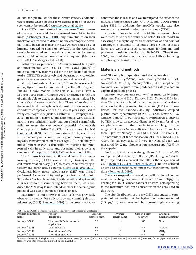
Hock et al. 2009). For this reason, we decided to investigatethe mwCNTs topological interaction and uptake using bothSEM and TEM techniques on Balb/3T3 cells.
Figure 2 shows transmission (STEM on the left) andsurface (SEM, on the right) mode images of Balb/3T3 after72 h of exposure to mwCNTs (nude, -COOH, -OH, -NH2).mwCNTs clusters can be identified as darker or brighterfeatures for STEM or SEM modality of analysis, respectively.mwCNTs clustering was probably due to the different
components present in the cell culture medium (salts, aminoacids, sugars, serum-derived proteins, peptides and growthfactors) that can induce mwCNT bundling together(Raffa et al. 2010; Monteiro-Riviere et al. 2009), thus creating‘sticky aggregates’.
mwCNTs were mostly found forming aggregates offilamentous structures on top of the cells. Each of thesefilaments corresponds to single mwCNTs. In comparisonwith control, cell morphology remains substantially
Nude
-COOH
-OH
-NH2
10 mm
10 mm
10 mm
15 mm
Figure 2. Transmission (STEM on the left) and surface (SEM, on the right) mode images of Balb/3T3 after 72 h of exposure to 100 mg/mL of mwCNTs(nude, -COOH, -OH, -NH2). mwCNTs clusters can be localised with cell as darker or brighter features for STEM or SEM modality of analysis,respectively (see arrows).
J. Ponti et al.
Nan
otox
icol
ogy
Dow
nloa
ded
from
info
rmah
ealth
care
.com
by
Com
mis
sion
Eur
opea
n C
omm
on
01/2
7/12
For
pers
onal
use
onl
y.

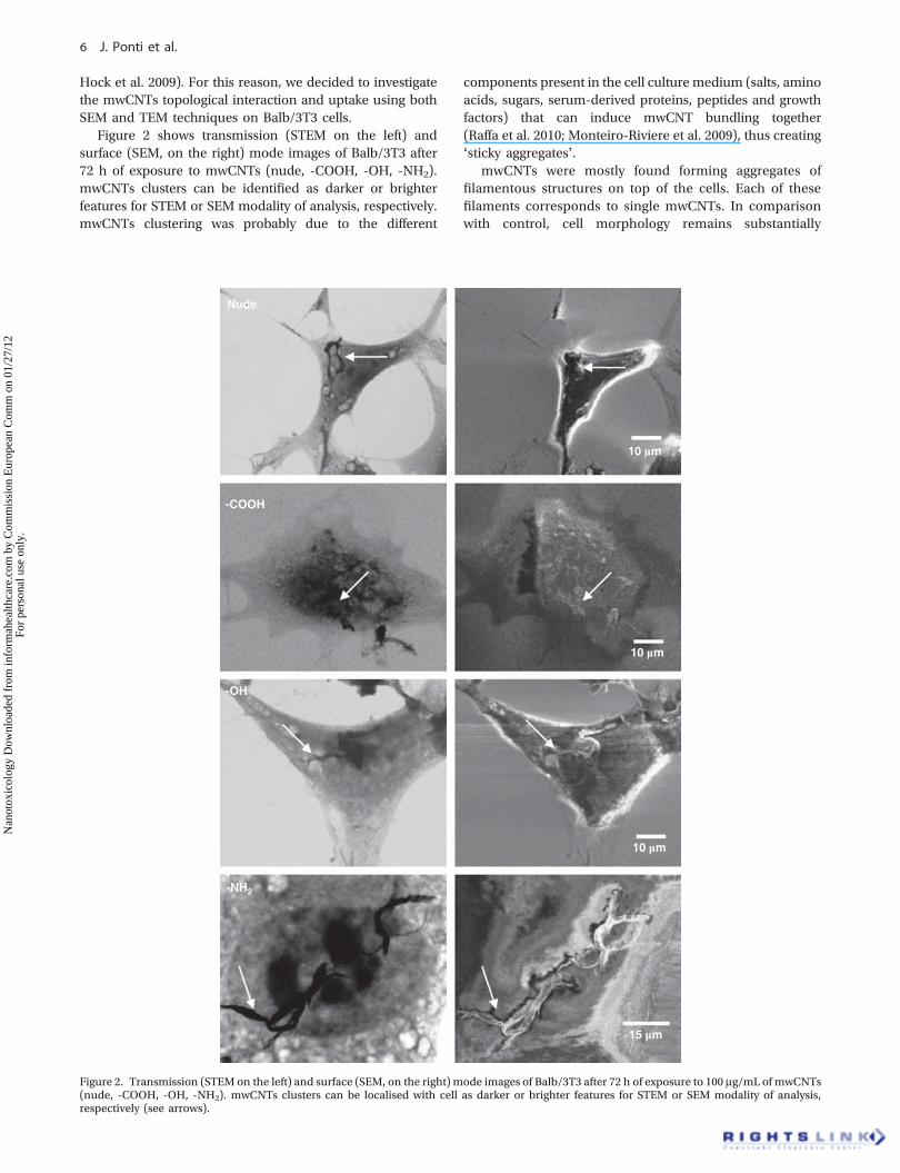
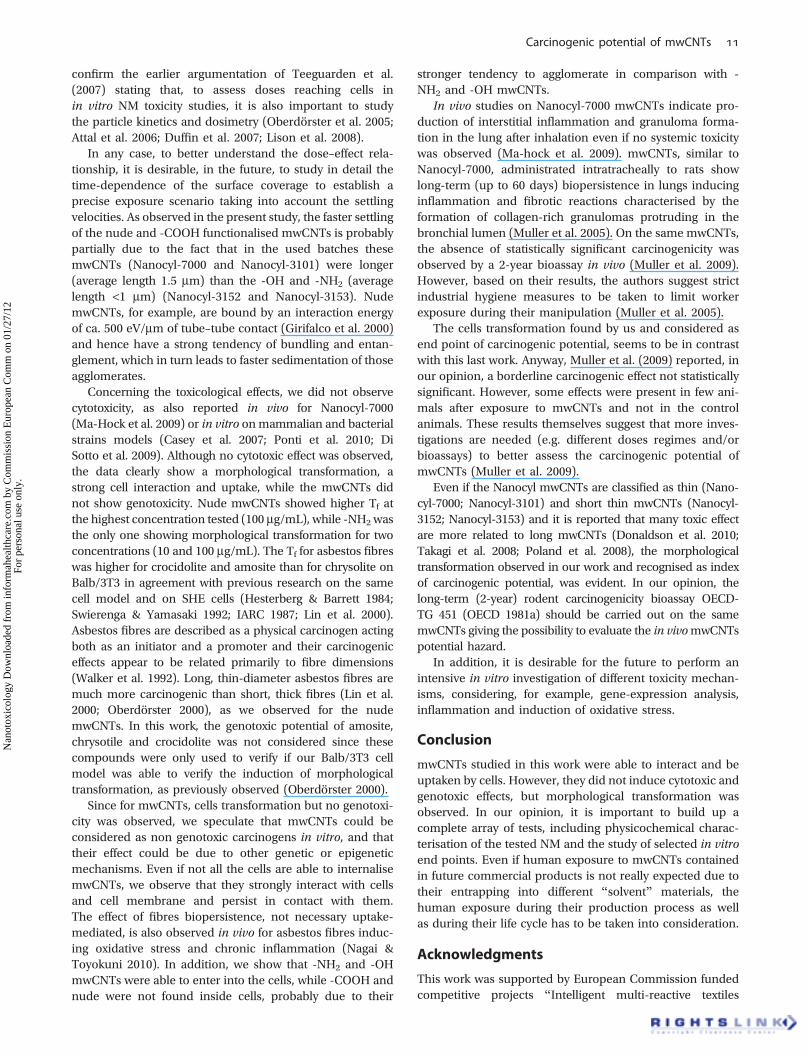
unchanged after exposure. None of the features proper tothe mwCNTs-treated samples was found in the control.Using the SEM and comparing the images from surface andtransmission modes it seems that most of the bundles werepotentially interacting with the cell surface (Figures 2, 3).A proof of this is that after the consecutive washing stepsmade during the sample preparation, most of the mwCNTswere found in contact with cells suggesting a tight inter-action between them and with the cell substrate. The focusdepth of the microscope did not allow us to determine the zposition of the fibres in transmission mode. In surfacemode, because of the low voltages used and the insulatingcharacter of the cell compounds, there was not enoughresolution to determine the edges close to the membrane.Nevertheless, we observed that most of the nanotubes wereoutside the cell (Figure 3). Therefore, it remains unclearusing SEM/STEM analysis if some of the edges of thebundles might enter the cell membrane. To understandif mwCNTs were able to enter into the cells, we analysedsamples by TEM. Generally, the images reveal a stronginteraction of nude and -COOH mwCNTs with cell mem-brane but no uptake, while -NH2 and -OH samples could befound inside the cells. In particular, when observed byTEM, Balb/3T3 control cells displayed polymorphousnuclei and a cytoplasm with a number of organelles andvacuoles (Figure 4A, B). After the interaction with mwCNTsthe cell morphology dramatically changed. Numerousvacuoles containing either mwCNTs or granular materialwere seen inside the cytoplasm of treated cells. Mitochon-dria sometimes appeared modified showing a condensedmatrix and dilated cristae (Figure 4G, H). Long digitationsprotruding from the nuclei were also visible after treatmentwith mwCNTs (Figure 4G, H). Large bundles of CNTs werefound inside the cells and also in proximity of the nucleus(Figure 4C–F). At higher magnification single -OH and -NH2 mwCNTs protruding from the bundles and penetrat-ing into the cytoplasm and nuclei are also visible(Figure 4D–F). By contrast, no cell uptake was found forboth nude and -COOH mwCNTs (Figure 4G–J). Largebundles of CNTs are observable outside Balb/3T3 cellstreated with -COOH samples (Figure 4I–J). The interactionof -COOH mwCNTs with the cell membrane induceda strong activation of cells as demonstrated by thehigh number of cytoplasmic vacuoles as well as surfacefinger-like protrusions (Figure 4J, circle).
Cytotoxicity, morphological transformation andgenotoxicityCell viability was evaluated by CFE assay. Figure 5A and Cshow CFE results obtained after 72 h of exposure to 1, 10 and100 mg/mL of mwCNTs and asbestos fibres. While Figure 7Ashows CFE results obtained after 24 h of cell exposure tomwCNTs, no cytotoxic effect was found for all the mwCNTsconcentrations and functionalisations tested, after 24 and72 h of exposure (Figures 7A and 5B). Dose–effect relation-ship was observed after exposure to asbestos fibres(Figure 5C). In particular, the decrease in cell viability,expressed as the colony number reduction, was statisticallydifferent for exposure to amosite and crocidolite fibres(100 mg/mL; p < 0.001***) when compared with the negativecontrol (Figure 5C). Positive control used for CFE assay(Na2CrO4 1000 mM) induced complete cell death afterboth 24 and 72 h of exposure.
We observed morphological transformation in four inde-pendent experiments and the average of data showed sta-tistically significant results for -NH2 at 1 mg/mL (p < 0.001)and 100 mg/mL (p < 0.01); -COOH at 1 mg/mL (p < 0.05); -OHat 10 mg/mL (p < 0.01); nude at 100 mg/mL (p < 0.001)(Figure 5B). We did not observe a dose-dependent effect,except for nude mwCNTs, which, at the highest concentra-tion tested (100 mg/mL), showed positive results in all theexperiments (Figure 5B).
Figure 6 shows the difference between unexposed dish(Figure 6A) and dish exposed to -NH2 mwCNTs (Figure 6B).Transformed colony appear with a multilayer centre andramifications into the monolayer around (type III foci,Figure 6C) while the untransformed cells grow as contact-inhibited monolayer (Figure 6A).
The Tf obtained after 72 h of exposure to asbestosfibres is reported in Figure 5D. Crocidolite showed thehighest morphological transformation. Statistically signifi-cant results were obtained for all the materials tested at thehighest concentration (100 mg/mL) with a dose-dependenteffect.
In order to understand if the transformation capacity ofmwCNTs is potentially triggered by a genotoxic effect, thecytokinesis-block MN was performed. The data obtained(Figure 7B) showed no statistically significant micronucleusformation for all the different samples tested (Figure 7B)suggesting that the morphological transformation could bedue to the activation of other different molecular pathways.
1 µmNH2 500 nm OH
Figure 3. Surface mode images of Balb/3T3 cell membrane after exposure to 100 mg/mL of mwCNTs (-NH2 and -OH). mwCNTs clusters can belocalised with cell membrane as brighter features (see arrows).
Carcinogenic potential of mwCNTs
Nan
otox
icol
ogy
Dow
nloa
ded
from
info
rmah
ealth
care
.com
by
Com
mis
sion
Eur
opea
n C
omm
on
01/2
7/12
For
pers
onal
use
onl
y.

Discussion
We performed this work in order to verify thepotential cytotoxicity, morphological transformation and
genotoxicity of nude and functionalised mwCNTs onBalb/3T3 mouse fibroblasts cell line. This cell model isconsidered a sensitive in vitro method and it is reported inthe list of accepted methods for Registration Evaluation
CTR
2 µm
A
Nu
CTR
1 µm
B
Nu
Nude
5 µm
G
Nu
-NH2
5 µm
E
Nu
-OH
1 µm
C
Nu
-OH
0.5 µm
D
-COOH
2 µm
I
Nu
NudeH
Nu
2 µm
-COOHJ
2 µm
1 µm
F -NH2
Nu
Figure 4. Transmission electronmicroscope (TEM)analysis of control andmwCNTs-treatedBalb/3T3 cells. Cellswere treatedwith 100mg/mLofmwCNTs(nude, -COOH, -NH2, -OH) for 72 h. Untreated cells (A, B) showed their typical ultra-structural features with well-preserved cytoplasm, indented nucleus(Nu) and dispersed chromatin. At higher magnification (B) numerous regular mitochondria (Mi) are observed. After -OH mwCNTs (C, D) and -NH2
mwCNTs treatments (E,F), a largeamountof electron-densematerialwasvisible inside thecells (seearrows); in cells treatedwithnudemwCNTs (G,H)and-COOHmwCNTs (I, J), no nanomaterial inside the cytoplasm was revealed (see arrows), while surface finger-like protrusions were observed (see circle).
J. Ponti et al.
Nan
otox
icol
ogy
Dow
nloa
ded
from
info
rmah
ealth
care
.com
by
Com
mis
sion
Eur
opea
n C
omm
on
01/2
7/12
For
pers
onal
use
onl
y.

Authorisation of Chemicals for the evaluation of thein vitro carcinogenic potential of chemicals (Madl & Pin-kerton 2009; Ponti et al. 2009; Regulation EC 440/2008).The CTA is also accepted and supported by Organisa-tion for Economic Cooperation and Development andEuropean Commission for the analysis of the in vitrocarcinogenic potential of NM (JRC Projects). To betterunderstand and build a knowledge base for future expla-nation and comparison of the different results obtained onthe potential toxicological effects of mwCNTs, a detailedphysicochemical characterisation including morphology,purity, behaviour in culture medium and solvents wascarried out.
Concerning the presence of metal impurities in thereceived stock preparation, the nude mwCNTs were thosecontaining the higher percentage of metals compared with-COOH, -NH2 and -OH. Nevertheless, we verified that theimpurities were low enough to exclude metal-induced toxicand cell transformation effects on Balb/3T3 (Mazzotti et al.2002; Ponti 2004; Ponti et al. 2009).
The size distribution of mwCNTs suspended in completeculture medium showed poorly reproducible measurements,partially due to the presence of numerous bundles/agglom-erates/aggregates of different sizes in the samples and par-tially to the dynamic sedimentation process which occurswith these mixtures.
This kind of mwCNTs behaviour cannot be avoided sincethe stock compounds are supplied in a powder form, which,although apparently dispersible in DMSO to form a stocksolution, tends to rapidly re-aggregate into bundles/agglom-erates/aggregates when this is further diluted into the celltesting media.
Attempts were made to characterise this behaviour usingDLS but the results obtained were inconclusive as the DLStechnique is fundamentally unsuitable to the characterisa-tion of such complex mixtures of polydispersed, asymmetricparticles and aggregates. In practice, the analysis of NPssuspended in culture media, with irregular shape such asnanorods (Comes Franchini et al. 2010) or NPs mixtures(Calzolai et al. 2011) can result in data which the instrument
120
100
80
60
40
20
120
25
20
15
10
5
0CF
E (
% o
f so
lven
t co
ntr
ol)
Tra
nsf
orm
atio
n f
req
uen
cy (
x10-4
)
**
****
***
***
***
*
100
80
60
40
20
0
Concentrations Concentrations
Controls Control1 µg/ml 1 µg/ml 10 µg/ml 100 µg/ml10 µg/ml 100 µg/ml
0Controls
Concentrations
A B
C D
Concentrations
Tra
nsf
orm
atio
n f
req
uen
cy (
x10-4
) ***
***
***
**
**
*
0
5
10
15
20
25
Negative control
Negative control
Nude
NH2
COOH
OH
Controls
Solvent control (DMSO 1%)
Solvent control (MilliQ water 1%)
Positive control (MCA 4 mg/mL)
Amosite
Crocidolite
Chrysolite
Positive control (MCA 4 µg/mL)C
FE
(%
of
solv
ent
con
tro
l)
**
1 µg/ml 10 µg/ml 100 µg/ml1 µg/ml10 µg/ml 100 µg/ml
Figure 5. Cytotoxicity and carcinogenic potential on Balb/3T3 cells after 72 h of exposure to 1, 10 and 100 mg/mL of mwCNTs (nude, -COOH, -NH2,-OH) and asbestos fibres. Results are expressed as colony-forming efficiency (CFE) percentage of the solvent control (A–C) and transformationfrequency (Tf) (B–D). No cytotoxicity is observed for all the mwCNTs tested (A), while carcinogenic potential (B) resulted statistically significant fornude (100 mg/mL, p < 0.001), -NH2 (1 mg/mL, p < 0.001 and 100 mg/mL, p < 0.01), -COOH (1 mg/mL, p < 0.05) and -OH (10 mg/mL, p < 0.01).Cytotoxicity resulted statistically significant at 100 mg/mL (p < 0.001) for amosite and crocidolite, showing a dose–effect relationship for all the threefibres tested (C). Carcinogenic potential resulted positive for exposure of 100 mg/mL to amosite (p < 0.01), crocidolite (p < 0.01) and chrysolite(p < 0.05). Histogram of the result obtained after cell exposure to the positive control used for CFE assay (Na2CrO4 1000 mM) is not shown sincecomplete cell death was reached.
Carcinogenic potential of mwCNTs
Nan
otox
icol
ogy
Dow
nloa
ded
from
info
rmah
ealth
care
.com
by
Com
mis
sion
Eur
opea
n C
omm
on
01/2
7/12
For
pers
onal
use
onl
y.

software (Malvern Instruments, UK) indicates as beingunsuitable or, in extreme cases, highly misleading due tothe intrinsic bias of this method towards the detection oflarge particles at the expense of smaller particles even inlarger number. In our work, we used DLS only to show howheterogeneous were the suspensions. In our opinion, themore reliable techniques to characterise mwCNTs weremicroscopic analyses such as STEM/TEM.
The results on sedimentation showed that due to settling,the actual exposure to mwCNTs increased continuously from
the start of the experiment until when complete sedimen-tation was achieved. Under our experimental conditions,complete settling was reached after 24 h for nude and -COOH functionalised mwCNTs, and after 36 h for -NH2 and -OH functionalised mwCNTs. This means that for a totalexposure time of 72 h, the cells were exposed to thecompletely settled mwCNTs for two-third of the total expo-sure time for nude and -COOH functionalised mwCNTs andfor half of the total exposure time for -NH2 and -OH func-tionalised mwCNTs in these experiments. These findings
A B
C
5 mm
Figure 6. Pictures of Petri dishes of cell transformation assay (CTA) at the end of the experiment: control dish (A) shows cell monolayer, whileexposure dish (-NH2, 100 mg/mL) shows morphologically transformed colonies (B) (type III foci) characterised by multilayer centre andramifications in the monolayer around (C).
**
0
20
40
60
80
100
120
Controls 1 µg/ml 10 µg/ml 100 µg/ml
ConcentrationsControls 1 µg/ml 10 µg/ml 100 µg/ml
Concentrations
CF
E (
% o
f so
lven
t co
ntr
ol)
0
50
100
150
200
250
300
350
400
BN
MN
cel
ls/1
000
BN
cel
ls
Negative control
Solvent control (DMSO 1%)
Positive control (MMC 0.5 µM)
Nude
NH2
COOH
OH
***
BA
Figure 7. Cytotoxicity and genotoxicity on Balb/3T3 cells after 24 h of exposure to 1, 10 and 100 mg/mL of mwCNTs (nude, -COOH, -NH2, -OH).Results are expressed as colony-forming efficiency (CFE) percentage of the solvent control (A) and number of binucleated micronucleated cells(BNMN) for 1000 binucleated cells (BN). Only the positive control (MMC) resulted positive (p < 0.001), while all themwCNTs samples tested showednegative results. No cytotoxicity is observed for all the mwCNTs tested (A), while statistically significant cytotoxicity (p < 0.01) and genotoxicity(p < 0.001) was observed for the positive control of the NM test (MMC 0.5 mM). Histogram of the result obtained after cell exposure to the positivecontrol used for CFE assay (Na2CrO4 1000 mM) is not shown since complete cell death was reached.
J. Ponti et al.
Nan
otox
icol
ogy
Dow
nloa
ded
from
info
rmah
ealth
care
.com
by
Com
mis
sion
Eur
opea
n C
omm
on
01/2
7/12
For
pers
onal
use
onl
y.
confirm the earlier argumentation of Teeguarden et al.(2007) stating that, to assess doses reaching cells inin vitro NM toxicity studies, it is also important to studythe particle kinetics and dosimetry (Oberdörster et al. 2005;Attal et al. 2006; Duffin et al. 2007; Lison et al. 2008).
In any case, to better understand the dose–effect rela-tionship, it is desirable, in the future, to study in detail thetime-dependence of the surface coverage to establish aprecise exposure scenario taking into account the settlingvelocities. As observed in the present study, the faster settlingof the nude and -COOH functionalised mwCNTs is probablypartially due to the fact that in the used batches thesemwCNTs (Nanocyl-7000 and Nanocyl-3101) were longer(average length 1.5 mm) than the -OH and -NH2 (averagelength <1 mm) (Nanocyl-3152 and Nanocyl-3153). NudemwCNTs, for example, are bound by an interaction energyof ca. 500 eV/mm of tube–tube contact (Girifalco et al. 2000)and hence have a strong tendency of bundling and entan-glement, which in turn leads to faster sedimentation of thoseagglomerates.
Concerning the toxicological effects, we did not observecytotoxicity, as also reported in vivo for Nanocyl-7000(Ma-Hock et al. 2009) or in vitro onmammalian and bacterialstrains models (Casey et al. 2007; Ponti et al. 2010; DiSotto et al. 2009). Although no cytotoxic effect was observed,the data clearly show a morphological transformation, astrong cell interaction and uptake, while the mwCNTs didnot show genotoxicity. Nude mwCNTs showed higher Tf atthe highest concentration tested (100 mg/mL), while -NH2 wasthe only one showing morphological transformation for twoconcentrations (10 and 100 mg/mL). The Tf for asbestos fibreswas higher for crocidolite and amosite than for chrysolite onBalb/3T3 in agreement with previous research on the samecell model and on SHE cells (Hesterberg & Barrett 1984;Swierenga & Yamasaki 1992; IARC 1987; Lin et al. 2000).Asbestos fibres are described as a physical carcinogen actingboth as an initiator and a promoter and their carcinogeniceffects appear to be related primarily to fibre dimensions(Walker et al. 1992). Long, thin-diameter asbestos fibres aremuch more carcinogenic than short, thick fibres (Lin et al.2000; Oberdörster 2000), as we observed for the nudemwCNTs. In this work, the genotoxic potential of amosite,chrysotile and crocidolite was not considered since thesecompounds were only used to verify if our Balb/3T3 cellmodel was able to verify the induction of morphologicaltransformation, as previously observed (Oberdörster 2000).
Since for mwCNTs, cells transformation but no genotoxi-city was observed, we speculate that mwCNTs could beconsidered as non genotoxic carcinogens in vitro, and thattheir effect could be due to other genetic or epigeneticmechanisms. Even if not all the cells are able to internalisemwCNTs, we observe that they strongly interact with cellsand cell membrane and persist in contact with them.The effect of fibres biopersistence, not necessary uptake-mediated, is also observed in vivo for asbestos fibres induc-ing oxidative stress and chronic inflammation (Nagai &Toyokuni 2010). In addition, we show that -NH2 and -OHmwCNTs were able to enter into the cells, while -COOH andnude were not found inside cells, probably due to their
stronger tendency to agglomerate in comparison with -NH2 and -OH mwCNTs.
In vivo studies on Nanocyl-7000 mwCNTs indicate pro-duction of interstitial inflammation and granuloma forma-tion in the lung after inhalation even if no systemic toxicitywas observed (Ma-hock et al. 2009). mwCNTs, similar toNanocyl-7000, administrated intratracheally to rats showlong-term (up to 60 days) biopersistence in lungs inducinginflammation and fibrotic reactions characterised by theformation of collagen-rich granulomas protruding in thebronchial lumen (Muller et al. 2005). On the same mwCNTs,the absence of statistically significant carcinogenicity wasobserved by a 2-year bioassay in vivo (Muller et al. 2009).However, based on their results, the authors suggest strictindustrial hygiene measures to be taken to limit workerexposure during their manipulation (Muller et al. 2005).
The cells transformation found by us and considered asend point of carcinogenic potential, seems to be in contrastwith this last work. Anyway, Muller et al. (2009) reported, inour opinion, a borderline carcinogenic effect not statisticallysignificant. However, some effects were present in few ani-mals after exposure to mwCNTs and not in the controlanimals. These results themselves suggest that more inves-tigations are needed (e.g. different doses regimes and/orbioassays) to better assess the carcinogenic potential ofmwCNTs (Muller et al. 2009).
Even if the Nanocyl mwCNTs are classified as thin (Nano-cyl-7000; Nanocyl-3101) and short thin mwCNTs (Nanocyl-3152; Nanocyl-3153) and it is reported that many toxic effectare more related to long mwCNTs (Donaldson et al. 2010;Takagi et al. 2008; Poland et al. 2008), the morphologicaltransformation observed in our work and recognised as indexof carcinogenic potential, was evident. In our opinion, thelong-term (2-year) rodent carcinogenicity bioassay OECD-TG 451 (OECD 1981a) should be carried out on the samemwCNTs giving the possibility to evaluate the in vivomwCNTspotential hazard.
In addition, it is desirable for the future to perform anintensive in vitro investigation of different toxicity mechan-isms, considering, for example, gene-expression analysis,inflammation and induction of oxidative stress.
Conclusion
mwCNTs studied in this work were able to interact and beuptaken by cells. However, they did not induce cytotoxic andgenotoxic effects, but morphological transformation wasobserved. In our opinion, it is important to build up acomplete array of tests, including physicochemical charac-terisation of the tested NM and the study of selected in vitroend points. Even if human exposure to mwCNTs containedin future commercial products is not really expected due totheir entrapping into different “solvent” materials, thehuman exposure during their production process as wellas during their life cycle has to be taken into consideration.
Acknowledgments
This work was supported by European Commission fundedcompetitive projects “Intelligent multi-reactive textiles
Carcinogenic potential of mwCNTs
Nan
otox
icol
ogy
Dow
nloa
ded
from
info
rmah
ealth
care
.com
by
Com
mis
sion
Eur
opea
n C
omm
on
01/2
7/12
For
pers
onal
use
onl
y.

integrating nano-filler based CPC-fibers” (INTELTEX),NMP-CT-2006-026626.
Declaration of interest
The authors report no conflicts of interest. The authors aloneare responsible for the content and writing of the paper.
ReferencesAschberger K, Johnston HJ, Stone V, Aitken RJ, Hankin SM,
Peters SA, et al. 2010. Review of carbon nanotubes toxicity andexposure-appraisal of human health risk assessment based on openliterature. Crit Rev Toxicol 40:759–790.
Attal S, Thiruvengadathan R, Regev O. 2006. Determination of theconcentration of single-walled carbon nanotubes in aqueous dis-persions using UV-Visible absorption spectroscopy. Anal Chem78:8098–8104.
Balls M, Clothier R. 2010. A FRAME response to the Draft Report onAlternative (Non-animal) Methods for Cosmetics Testing: CurrentStatus and Future Prospects-2010. Altern Lab Anim 38:345–353.
Becker H, Herzberg F, Schulte A, Kolossa-Gehring M. 2011.The carcinogenic potential of nanomaterials, their release fromproducts and options for regulating them. Int J Hyg Environ Health214:231–238.
Buford MC, Hamilton RF, Holian A. 2007. A comparison of dispersingmedia for various engineered carbon nanoparticles. Part FibreToxicol 4:6–15.
Calzolai L, Gilliland D, Pascual Garcìa C, Rossi F. 2011. Separation andcharacterization of gold nanoparticle mixtures by flow-field-flowfractionation. J Chromatogr A 1218:4234–4239.
Casey A, Herzog E, Davoren M, Lyng FM, Byrne HJ, Chambers G. 2007.Spectroscopic analysis conforms the interactions between singlewalled carbon annotates and various dyes commonly used to assesscytotoxicity. Carbon 45:1425–1432.
Chou CC, Hsiao HY, Hong OS, Chun CH, Peng YW, Chen HW, et al.2008. Single-walled carbon nanotubes can induce pulmonary injuryin mouse model. Nano Lett 8:437–445.
Colognato R, Ponti J, D’Errico MR, Migliore L, Rossi F. 2009. Geno-toxicity assays analysis for carbon nanotubes: friends or foes?Preliminary results on human peripheral leukocytes. Int J EnvironHealth 3:275–284.
Comes Franchini M, Ponti J, Lemor R, Fournelle M, Broggi F,Locatelli E. 2010. Polymeric entrapped thiol-coated gold nanorods:cytotoxicity and suitability as molecular optoacoustic contrast agent.J Mater Chem 20:10908–10914.
Di Giorgio ML, Di Bucchianico S, Ragnelli AM, Aimola P, Santucci S,Poma A. 2011. Effects of single and multi walled carbon nanotubeson macrophages: cyto and genotoxicity and electron microscopy.Mutat Res 18:20–31.
Di Sotto A, Chiaretti M, Carru GA, Bellucci S, Mazzanti G. 2009.Multi-walled carbon nanotubes: lack of mutagenic activity in thebacterial reverse mutation assay. Toxicol Lett 184:192–197.
Ding L, Stilwell J, Zhang T, Elboudwarej O, Jiang H, Selegue JP, et al.2005. Molecular characterization of the cytotoxic mechanism ofmultiwall carbon nanotubes and nano-onions on human skinfibroblast. Nano Lett 5:2448–2464.
Donaldson K, Aitken R, Tran L, Stone V, Duffin R, Forrest G, et al. 2006.Carbon nanotubes: a review of their properties in relation to pul-monary toxicology and workplace safety. Toxicol Sci 92:5–22.
Donaldson K, Murphy FA, Duffin R, Poland CA. 2010. Asbestos, carbonnanotubes and the pleural mesothelium: a review of the hypothesisregarding the role of long fibre retention in the parietal pleura,inflammation and mesothelioma. Part Fibre Toxicol 2010:7–5.
Duffin R, Tran L, Brown D, Stone V, Ken D. 2007. Proinflammogeniceffects of low-toxicity and metal nanoparticles in vivo and in vitro:highlighting the role of particle surface area and surface reactivity.Inhal Toxicol 19:849–856.
Fenoglio I, Greco G, Tomatis M, Muller J, Raymundo-Piñero E,Béguin F, et al. 2008. Structural defects play a major role in theacute lung toxicity of multiwall carbon nanotubes: physicochemicalaspects. Chem Res Toxicol 21:1690–1697.
Genaidy A, Tolaymat T, Sequeira R, Rinder M, Dionysiou D. 2009.Health effects of exposure to carbon nanofibers: systematic review,critical appraisal, meta analysis and research to practice perspec-tives. Sci Total Environ 407:3686–3701.
Girifalco L, Hodak M, Lee RS. 2000. Carbon nanotubes, buckyballs,ropes, and a universal graphitic potential. J Phys Rev B 62:13104–13110.
Hesterberg TW, Barrett JC. 1984. Dependence of Asbestos- andmineraldust-induced transformation of mammalian cells in culture on fiberdimension. Cancer Res 44:2170–2180.
IARC. 1987. Monographs on the evaluation of carcinogenic risks tohumans, overall evaluation of carcinogenicity: an updating of IARCmonographs vols. 1 to 42. Suppl. 7, p 109.
IARC/NCI/EPA Working-Group. 1985. Cellular and molecularmechanisms of cell transformation and standardization of transfor-mation assays of established cell lines for the prediction of carci-nogenic chemicals: overview and recommended protocols. CancerRes 45:2395–2399.
INTELTEX project web site [Online]http://www.inteltex.eu.Accessedon 12 October 2011.
Isfort RJ, LeBoeuf RA. 1996. Application of in vitro cell transformationassays to predict the carcinogenic potential of chemicals. MutationResearch/Reviews in Genetic Toxicology 365:161–173.
Johnston HJ, Hutchison GR, Christensen FM, Peters S, Hankin S,Aschberger K, et al. 2010. A critical review of the biologicalmechanismsunderlying the in vivo and in vitro toxicity of carbon nanotubes: Thecontribution of physico-chemical characteristics. Nanotoxicology4:207–246.
JRC Projects web site [Online]http://projects.jrc.ec.europa.eu/jpb_pub-lic/act/publicexportworkprogramme.html?objTypeCombo=&actId=188.Accessed on 11 October 2011.
Kam NW, O’Connell M, Wisdom JA, Dai H. 2005. Carbon nanotubes asmultifunctional biological transporters and near-infrared agentsfor selective cancer cell destruction. Proc Natl Acad Sci USA102:11600–11605.
Kerckaert GA, Brauninger R, LeBoeuf RA, Isfort RJ. 1996. Use of theSyrian hamster embryo cell transformation assay for carcinogenicityprediction of chemicals currently being tested by the NationalToxicology Program in rodent bioassays. Environ Health Perspect104:1075–1084.
Kurzepa H, Kyriazis AP, Lang DR. 1984. Growth characteristics oftumors induced by transplantation into athymic mice of BALB/3T3 cells transformed in vitro by residue organics from drinkingwater. J Environ Pathol Toxicol Oncol 5:131–138.
Lam CW, James JT, McCluskey R, Hunter RL. 2004. Pulmonary toxicityof single-wall carbon nanotubes in mice 7 and 90 days after intra-tracheal instillation. Toxicol Sci 77:126–134.
LANGSRUD web site [Online]http://www.langsrud.com/fisher.htm.Accessed on 11 October 2011.
Lin F, Liu Y, Liu Y, Keshava N, Li S. 2000. Crocidolite induces celltransformation and p53 gene mutation in BALB/c-3T3 cells. TeratogCarcinog Mutagen 20:273–281.
Lison D, Leen CJ, Thomassen Rabolli V, Gonzalez L, Napierska D,Seo JW, et al. 2008. Nominal and effective dosimetry of silicananoparticles in cytotoxicity assays. Toxicol Sci 104:155–162.
Madl AK, Pinkerton KE. 2009. Health effects of inhaled engineered andincidental nanoparticles. Crit Rev Toxicol 39:629–658.
Ma-Hock L, Treumann S, Strauss V, Brill S, Luizi F, Mertler M, et al.2009. Inhalation toxicity of multiwall carbon nanotubes in ratsexposed for 3 months. Toxicol Sci 112:468–481.
Mazzotti F, Sabbioni E, Ponti J, Ghiani M, Fortaner S, Rossi GL. 2002. Invitro setting of dose-effect relationships of 32 metal compounds inthe Balb/3T3 cell line, as a basis for predicting their carcinogenicpotential. ATLA 30:209–217.
Monteiro-Riviere NA, Inman AO, Zhang LW. 2009. Limitationsand relative utility of screening assays to assess engineered nano-particles toxicity in a human cell line. Toxicol Appl Pharmacol234:222–235.
Monteiro-Riviere NA, Nemanich RJ, Inman AO, Wang YY,Riviere JE. 2005. Multi-walled carbon nanotube interactions withhuman epidermal keratinocytes. Toxicol Lett 15:377–384.
Muller J, Delos M, Panin N, Rabolli V, Huaux F, Lison D. 2009. Absenceof carcinogenic response to multiwall carbon nanotubes in a2-year bioassay in the peritoneal cavity of the rat. Toxicol Sci110:442–448.
Muller J, Huaux F, Fonseca A, Nagy JB, Moreau N, Delos M, et al. 2008.Structural defects play a major role in the acute lung toxicity ofmultiwall carbon nanotubes: toxicological aspects. Chem Res Tox-icol 21:1698–1705.
Muller J, Huaux F, Moreau N, Misson P, Heilier J-F, Delos M, et al.2005. Respiratory toxicity of multi-wall carbon nanotubes. ToxicolAppl Pharmacol 207:221–231.
J. Ponti et al.
Nan
otox
icol
ogy
Dow
nloa
ded
from
info
rmah
ealth
care
.com
by
Com
mis
sion
Eur
opea
n C
omm
on
01/2
7/12
For
pers
onal
use
onl
y.

Nagai H, Toyokuni S. 2010. Biopersistent fiber-induced inflammationand carcinogenesis: lessons learned from Asbestos toward safety offibrous nanomaterials. Arch Biochem Biophys 502:1–7.
Nanocyl web site [Online]www.nanocyl.com.Accessed on 18 October2011.
Oberdörster E. 2004. Manufactured nanomaterials (fullerenes, C60)induce oxidative stress in the brain of juvenile largemouth bass.Environ Health Perspect 112:1058–1062.
Oberdörster G, Maynard A, Donaldson K, Castranova V, Fitzpatrick J,Ausman K, et al. 2005. Principles for characterizing the potentialhuman health effects from exposure to nanomaterials: elements of ascreening strategy. Part Fibre Toxicol 2:8–43.
Oberdörster G. 2000. Determinants of the pathogenicity of man-madevitreous fibers (MMVF). Int Arch Occup Environ Health 73:60–68.
OECD. 1981a. Guideline 451. Carcinogenicity studies (17 pages;adopted 12 May 1981). OECD Guidelines for the Testing of Chemi-cals (1993) Section 4: Health effects. Vol. 2. Paris: Organisation forEconomic Cooperation & Development.
Poland CA, Duffin R, Kinloch I, Maynard A, Wallace WA, Seaton A,et al. 2008. Carbon nanotubes introduced into the abdominal cavityof mice show Asbestos-like pathogenicity in a pilot study. NatNanotechnol 3:423–428.
Ponti J, Colognato R, Rauscher H, Gioria S, Broggi F, Franchini F, et al.2010. Colony Forming Efficiency and microscopy analysis of mul-ti-wall carbon nanotubes cell interaction. Toxicol Lett 197:29–37.
Ponti J, Munaro B, Fischbach M, Hoffmann S, Sabbioni E. 2007. AnOptimised data analysis for Balb/c 3T3 cell transformation assay andits application to metal compounds. Int J Immunopathol Pharmacol20:673–684.
Ponti J, Sabbioni E, Munaro B, Broggi F, Marmorato P, Franchini F,et al. 2009. Genotoxicity and morphological transformation inducedby cobalt nanoparticles and cobalt chloride: an in vitro study inBalb/3T3 mouse fibroblasts. Mutagenesis 24:439–445.
Ponti J. 2004. In vitro metal toxicology research: a study carried out byBalb/3T3, HaCaT and Caco-2 cell lines: report of research activity[dissertation]. Barcelona (SP): Universitat Autònoma de Barcelona,Facultat de Ciències, 97pp. T-575 2004 Ponti [Online]. http://pub-lications.jrc.ec.europa.eu/repository/handle/111111111/13708.Accessed on 11 October 2011.
Porter AE, Gass M,Muller K, Skepper JN, Midgley PA, WellandM. 2007.Direct imaging of single-walled carbon nanotubes in cells. NatNanotechnol 11:713–717.
Raffa V, Ciofani G, Vittorio O, Riggio C, Cuschieri A. 2010. Physico-chemical properties affecting cellular uptake of carbon nanotubes.Nanomedicine 5:89–97.
Regulation (EC) 440/2008 of 30 May 2008 Laying Down Test MethodsPursuant to Regulation (EC) 1907/2006 of the European Parliamentand of the Council on the Registration, Evaluation, Authorizationand Restriction of Chemicals (REACH). Official Journal of theEuropean Union, 31 May 2008, vol. 51, L142/1–L142/739.
Rivas GA, Rubianes MD, Rodriguez MC, Ferreyra NF, Luque GL,Pedano ML, et al. 2007. Carbon nanotubes for electrochemicalbiosensing. Talanta 74:291–307.
Rotoli BM, Bussolati O, Bianchi MG, Barilli A, Balasubramanian C,Bellucci S, et al. 2008. Non-functionalized multi-walled carbonnanotubes alter the paracellular permeability of human airwayepithelial cells. Toxicol Lett 5:95–102.
Rotoli BM, Bussolati O, Barilli A, Zanello PP, Bianchi MG,Magrini A, et al. 2009. Airway barrier dysfunction induced byexposure to carbon nanotubes in vitro: which role for fibre length?Hum Exp Toxicol 28:361–368.
Ryman-Rasmussen JP, Cesta MF, Brody AR, Shipley-Phillips JK,Everitt JI, Tewksbury EW, et al. 2009. Inhaled carbon nanotubesreach the subpleural tissue in mice. Nat Nanotechnol 4:747–751.
Saffiotti U, Ahmed N. 1995. Neoplastic transformation by quartz in theBALB/3T3/A31-1-1 cell line and the effects of associated minerals.Teratog Carcinog Mutagen 15:339–356.
Soto K, Garza KM, Murr LE. 2007. Cytotoxic effects of aggregatednanomaterials. Acta Biomater 3:351–358.
Swierenga SH, Yamasaki H. 1992. Performance of tests for celltransformation and gap-junction intercellular communicationfor detecting nongenotoxic carcinogenic activity. In: Vainio H,Magee PN, McGregor DB, McMichael AJ, editors. Mechanismsof carcinogenesis in risk identification. Lyon, New York: IARC.pp 165–193.
Takagi A, Hirose A, Nishimura T, Fukumori N, Ogata A, Ohashi N, et al.2008. Induction of mesothelioma in p53+/- mouse by intraperitonealapplication of multi-wall carbon nanotube. J Toxicol Sci 33:105–116.
Teeguarden JG, Hinderliter PM, Orr G, Thrall BD, Pounds JG. 2007.Particokinetics in vitro: dosimetry considerations for in vitro nano-particle toxicity Assessments. Toxicol Sci 95:300–312.
Thurnherr T, Brandenberger C, Fischer K, Diener L, Manser P,Maeder-Althaus X, et al. 2011. A comparison of acute and long-termeffects of industrial multiwalled carbon nanotubes on human lungand immune cells in vitro. Toxicol Lett 200:176–186.
Vanparys P, Corvi R, Aardema M, Gribaldo L, Hayashi M,Hoffmann S, et al. 2010. Results of ECVAM prevalidation study ofthree cell transformation assays. ALTEX 27:267–270.
Walker C, Everitt J, Barrett JCPossible cellular and molecular mechan-isms for Asbestos carcinogenicity. 1992. Am J Ind Med 21:253–273.
Carcinogenic potential of mwCNTs
Nan
otox
icol
ogy
Dow
nloa
ded
from
info
rmah
ealth
care
.com
by
Com
mis
sion
Eur
opea
n C
omm
on
01/2
7/12
For
pers
onal
use
onl
y.
Related Documents