Citation: Marcos, P.; Coveñas, R. Morphological Relationships between the Cholinergic and Somatostatin-28(1-12) Systems in the Alpaca (Lama pacos) Brainstem. Anatomia 2022, 1, 54–67. https:// doi.org/10.3390/anatomia1010006 Academic Editor: Francesco Cappello Received: 5 May 2022 Accepted: 23 May 2022 Published: 25 May 2022 Publisher’s Note: MDPI stays neutral with regard to jurisdictional claims in published maps and institutional affil- iations. Copyright: © 2022 by the authors. Licensee MDPI, Basel, Switzerland. This article is an open access article distributed under the terms and conditions of the Creative Commons Attribution (CC BY) license (https:// creativecommons.org/licenses/by/ 4.0/). Article Morphological Relationships between the Cholinergic and Somatostatin-28(1-12) Systems in the Alpaca (Lama pacos) Brainstem Pilar Marcos 1, * and Rafael Coveñas 2,3 1 Regional Center for Biomedical Research (CRIB), Facultad de Medicina de Albacete, University of Castilla-La Mancha, 02006 Albacete, Spain 2 Laboratory of Neuroanatomy of the Peptidergic Systems, Institute of Neurosciences of Castilla y León (INCYL), University of Salamanca, 37007 Salamanca, Spain; [email protected] 3 Grupo GIR USAL: BMD (Bases Moleculares del Desarrollo), University of Salamanca, 37007 Salamanca, Spain * Correspondence: [email protected] Abstract: In the alpaca brainstem, the distribution of the cholinergic system by the immunohistochem- ical detection of the enzyme choline acetyltransferase (ChAT) has been described, and its relationship with the distribution of somatostatin-28(1-12) is analyzed by double-immunostaining techniques. Overlapping distribution patterns for both substances were observed in many brainstem regions, suggesting that interactions between them may occur in the reticular formation, nucleus ambiguus or laterodorsal tegmental nucleus. Colocalization of the two substances in the same cell bodies was only observed in restricted areas, such as the nucleus of the solitary tract, reticular formation and nucleus ambiguus. In addition, in several regions, an apparent high innervation of the peptidergic fibers on cholinergic neurons has been observed. The results suggest that chemospecific interactions could be crucial for the control of specific cardiorespiratory and/or digestive functions in alpacas. These interactions may represent brain-adaptive mechanisms to particular environments and have a potential therapeutic use in respiratory disorders. Keywords: alpaca; brainstem; somatostatin; acetylcholine; cardiovascular regulation; respiratory control 1. Introduction Cetaceans (dolphins, whales) and artiodactyls (even-toed ungulates, e.g., sheep, gi- raffe) belong to the order Cetartiodactyla. The family Camelidae is part of artiodactyls, and the alpaca (Lama pacos) is included in this family [1–5]. Alpacas are important animals for the economy of numerous South American countries due to the excellent quality of their wool, and in this sense, numerous studies focused on their maintenance and repro- ductive cycles have been performed [6,7]. The members of the Camelidae family have specific morphological characteristics, such as long necks and seven cervical vertebrae, and moreover alpacas can live at sea level and at 5000 m above sea level [6–12]. These characteristics suggest the existence of important and unique adaptation mechanisms, mainly related to cardiovascular and respiratory mechanisms, which are controlled by the central nervous system, specifically brainstem centers. Other members of the Certiodactyla order also exhibit brain specializations that help them to survive within their respective environments [1–5]. Since all these physiological functions are regulated by neuroactive substances, several studies have reported the distributions of different classical neurotransmitters and neu- ropeptides by means of immunohistochemical methods in the alpaca brain [6–12]. These studies complement previous works regarding the mapping of neuro-modulatory systems in the brain of Artiodactyla and confirm that the nuclear complement of neurotransmitters, such as acetylcholine or catecholamines, detected in the alpaca brainstem and diencephalon Anatomia 2022, 1, 54–67. https://doi.org/10.3390/anatomia1010006 https://www.mdpi.com/journal/anatomia

Welcome message from author
This document is posted to help you gain knowledge. Please leave a comment to let me know what you think about it! Share it to your friends and learn new things together.
Transcript
Citation: Marcos, P.; Coveñas, R.
Morphological Relationships
between the Cholinergic and
Somatostatin-28(1-12) Systems in the
Alpaca (Lama pacos) Brainstem.
Anatomia 2022, 1, 54–67. https://
doi.org/10.3390/anatomia1010006
Academic Editor: Francesco
Cappello
Received: 5 May 2022
Accepted: 23 May 2022
Published: 25 May 2022
Publisher’s Note: MDPI stays neutral
with regard to jurisdictional claims in
published maps and institutional affil-
iations.
Copyright: © 2022 by the authors.
Licensee MDPI, Basel, Switzerland.
This article is an open access article
distributed under the terms and
conditions of the Creative Commons
Attribution (CC BY) license (https://
creativecommons.org/licenses/by/
4.0/).
Article
Morphological Relationships between the Cholinergic andSomatostatin-28(1-12) Systems in the Alpaca (Lamapacos) BrainstemPilar Marcos 1,* and Rafael Coveñas 2,3
1 Regional Center for Biomedical Research (CRIB), Facultad de Medicina de Albacete, University of Castilla-LaMancha, 02006 Albacete, Spain
2 Laboratory of Neuroanatomy of the Peptidergic Systems, Institute of Neurosciences of Castilla yLeón (INCYL), University of Salamanca, 37007 Salamanca, Spain; [email protected]
3 Grupo GIR USAL: BMD (Bases Moleculares del Desarrollo), University of Salamanca, 37007 Salamanca, Spain* Correspondence: [email protected]
Abstract: In the alpaca brainstem, the distribution of the cholinergic system by the immunohistochem-ical detection of the enzyme choline acetyltransferase (ChAT) has been described, and its relationshipwith the distribution of somatostatin-28(1-12) is analyzed by double-immunostaining techniques.Overlapping distribution patterns for both substances were observed in many brainstem regions,suggesting that interactions between them may occur in the reticular formation, nucleus ambiguusor laterodorsal tegmental nucleus. Colocalization of the two substances in the same cell bodies wasonly observed in restricted areas, such as the nucleus of the solitary tract, reticular formation andnucleus ambiguus. In addition, in several regions, an apparent high innervation of the peptidergicfibers on cholinergic neurons has been observed. The results suggest that chemospecific interactionscould be crucial for the control of specific cardiorespiratory and/or digestive functions in alpacas.These interactions may represent brain-adaptive mechanisms to particular environments and have apotential therapeutic use in respiratory disorders.
Keywords: alpaca; brainstem; somatostatin; acetylcholine; cardiovascular regulation; respiratory control
1. Introduction
Cetaceans (dolphins, whales) and artiodactyls (even-toed ungulates, e.g., sheep, gi-raffe) belong to the order Cetartiodactyla. The family Camelidae is part of artiodactyls,and the alpaca (Lama pacos) is included in this family [1–5]. Alpacas are important animalsfor the economy of numerous South American countries due to the excellent quality oftheir wool, and in this sense, numerous studies focused on their maintenance and repro-ductive cycles have been performed [6,7]. The members of the Camelidae family havespecific morphological characteristics, such as long necks and seven cervical vertebrae,and moreover alpacas can live at sea level and at 5000 m above sea level [6–12]. Thesecharacteristics suggest the existence of important and unique adaptation mechanisms,mainly related to cardiovascular and respiratory mechanisms, which are controlled by thecentral nervous system, specifically brainstem centers. Other members of the Certiodactylaorder also exhibit brain specializations that help them to survive within their respectiveenvironments [1–5].
Since all these physiological functions are regulated by neuroactive substances, severalstudies have reported the distributions of different classical neurotransmitters and neu-ropeptides by means of immunohistochemical methods in the alpaca brain [6–12]. Thesestudies complement previous works regarding the mapping of neuro-modulatory systemsin the brain of Artiodactyla and confirm that the nuclear complement of neurotransmitters,such as acetylcholine or catecholamines, detected in the alpaca brainstem and diencephalon
Anatomia 2022, 1, 54–67. https://doi.org/10.3390/anatomia1010006 https://www.mdpi.com/journal/anatomia
Anatomia 2022, 1 55
was similar to that found in other members of the same order, and this finding supportsManger’s hypothesis [13]. In addition, the distribution of some neuropeptides has beenstudied in the alpaca brainstem [6–9,12] and diencephalon [10,11]. Moreover, the morpho-logical relationship between neuropeptides and neurotransmitters has been reported, anddouble-immunolabeling for tyrosine hydroxylase, which is the rate-limiting enzyme of thecatecholaminergic synthesis, and somatostatin-28(1-12) (Som-28(1-12)) has been carried outin the diencephalon of the alpaca [11]. In the brainstem of this species, a double-labelingfor choline acetyl transferase (ChAT, a marker for the cholinergic system) and calcitoningene-related peptide (CGRP), as well as for CGRP and tyrosine hydroxylase [9,12], hasbeen described. According to the distribution of double-labeled perikarya, the resultsobserved in the brainstem in these studies suggest that CGRP might interact more withthe catecholaminergic system than with the cholinergic system. However, although thephysiological interactions between somatostatin and catecholamines have been reported inthe literature [13], no double-labeled neurons for these two substances were detected inthe alpaca diencephalon [11]. A possible explanation may be that the interaction betweenthe two substances may occur with another somatostatin fragment different from the onestudied in that work.
Somatostatin, in addition to inhibiting the growth hormone, acts as a neuromodulatorin numerous physiological functions, blocking the release of noradrenaline and stimulatingthe release of serotonin and acetylcholine [14]. The precursor named pro-somatostatin iscleaved into somatostatin-28, somatostatin-12 (corresponding to the first 112 amino acids ofsomatostatin-28) and somatostatin-14 [15]. These fragments can elicit different responses inrelation to the same mechanism, such as the cardiovascular regulation [15], probably due tothe activation of different somatostatin receptor subtypes [16]. In addition, the interactionof somatostatin with other neurotransmitters such as acetylcholine is well-known [16–21],but the studies that described such interactions were more focused on memory processing.
Studies previously performed in the alpaca brainstem suggest a similar distributionpattern for CGRP and Som-28(1-12) [7,8]. The presence of CGRP and ChAT in the sameperikarya was reported in some of the alpaca brainstem nuclei; thus, the goal of the presentwork was to know whether there is a neuroanatomical basis for possible interactionsbetween Som-28(1-12) and ChAT in the alpaca brainstem. Moreover, due to the participationof these two substances in cardiovascular and respiratory functions, another aim of thisstudy is to know whether these interactions can constitute a morphological basis for thecontrol of cardiovascular and/or respiratory mechanisms in this species. The resultsobtained will help to understand the distinctive control mechanisms that exist in the alpacaas physiological adaptations to living in such different habitats in terms of altitude, whichleads to changes in air quality and composition. Knowledge of these adaptive mechanismsand their morphological basis will contribute to a better understanding of the neuroanatomyand physiology of the alpaca. Comparison with the results obtained in other mammalianspecies will allow to assess whether these mechanisms may constitute therapeutic targetsfor the possible treatment of cardiovascular, respiratory and/or digestive disorders.
2. Materials and Methods2.1. Animals
As reported previously in similar studies [8,12], six adult male alpacas (Lama pacos)(70–80 kg; 5–8 years) were used here. From birth to the perfusion day, animals were main-tained at 0 m on the sea level and kept under standard conditions of temperature and lightand with free access to water and food. The study was performed following the guidelinesof the ethical and legal recommendations of the Spanish legislation [8,12].
2.2. Tissue Processing
The protocol has previously been published in the reports showing the distribution ofimmunoreactive structures containing Som-28(1-2) [8] and ChAT [12] in the alpaca brain-stem. Animals were intravenously anaesthetized with ketamine (10 mg/kg) and xylazine
Anatomia 2022, 1 56
(4 mg/kg), heparinized, and perfused (NaCl (0.9%): 3 L, paraformaldehyde (4%): 5 L inphosphate-buffered saline (PBS, pH 7.2, 0.15 M)) through the carotid artery [8]. Brainstemswere dissected out, post-fixed in the latter fixative solution (overnight) and cryoprotected(using increasing sucrose baths). Using a cryostat, 50 µm frontal brainstem sections wereobtained, kept at 4 ◦C in PBS and processed for the immunohistochemical detection ofSom-28 (1-12) [8]. Then, these sections were used for double-immunohistochemistry withChAT labeling.
2.3. Immunohistochemistry
In the present work, the same frontal sections in which Som-28(1-12)-immunoreactivitywas previously observed, after applying the diaminobenzidine (DAB) developing method-ology [8], were used to detect ChAT according to a previously published protocol [22].Som-28(1-12)-immunoreactive slides were immersed in PBS until the coverslip becamedetached, then sections were carefully removed from the surface of the slides. The traces ofthe mounting medium were removed with several rinses in PBS (6 × 10 min). To inacti-vate endogenous peroxidases, Som-28(1-12)-immunoreactive sections were treated withNH3 (20%), NaOH (1%) and H2O2 (30%). Sections were extensively rinsed in PBS (6 ×10 min) and pre-incubated in PBS containing normal horse serum (1%) and Triton X-100(0.3%). This solution was also used for further dilution of streptavidin and antibodies. Sec-tions were incubated with primary polyclonal antibody against choline acetyl transferase(ChAT, Millipore, ref. AB144 P, raised in goat; dilution 1/75; overnight, 4 ◦C), washedin PBS and incubated in biotinylated donkey anti-goat antibody (Jackson Laboratories;dilution 1/2000; 90 min, room temperature). After washing in PBS, sections were incubatedin peroxidase-coupled streptavidin (Jackson Laboratories, dilution 1/2000; 90 min, roomtemperature). Finally, sections were washed in PBS and Tris-HCl buffer (pH 7.6, 0.05 M),and revealed with 4-chloro-1-napthol. This chromogen shows a blue precipitate, easilydistinguishable from the DAB brown product used for the detection of Som-28(1-12). Sec-tions were mounted on gelatin-coated slides and cover-slipped with glycerol/PBS. Thespecificity of the ChAT immunoreactivity was confirmed by the following histologicalcontrols [12]: (1) first incubation bath: omission of the antibody, (2) PBS instead of thesecondary biotinylated antibody and (3) a non-appropriated secondary antibody instead ofthe secondary biotinylated antibody. No immunoreactivity was found. The specificity ofthe Som-28(1-12) antiserum was previously checked [8]: (1) the first antiserum was preab-sorbed with the synthetic peptide, (2) omission of the antibody in the first incubation bath,(3) PBS instead of the secondary biotinylated antibody and (4) the Som-28(1-12) antiserumwas preabsorbed with an excess of related peptides (somatostatin-28, somatostatin-14,methionine-enkephalin, substance P, angiotensin II, cholecystokinin and neuropeptide Y).These controls confirmed the specificity of the immunoreactivity for Som-28(1-12) [8]. Inaddition, the distributions of the cholinergic [12] and somatostatinergic [8] systems in thealpaca brainstem were verified by comparison to the previously published distribution ofeach substance.
2.4. Mapping
Frontal planes of the alpaca brainstem that were previously published were fol-lowed [6–9]. Atlases of non-camelid mammals and the brain atlas of Lama glama werealso used [23,24]. For the nomenclature of the nuclei studied, previous published articleson the alpaca brainstem were used [6–9,12]. As the main references, the mapping of deSouza et al. [6–8] and Marcos and Coveñas [12] were used for both the location of theimmunoreactivity and nomenclature. The distribution of cholinergic cell groups in othermammals was consulted [1–5,25–40]. To study the distribution of the immunoreactiveneurons, the computerized digital mapping system Accustage MDPlot v5.2 (MD3-Digitizer,Accustage, Minnesota Datametrics, Saint Paul, MN, USA) was applied. A digital cameraattached to a Nikon Eclipse 80i microscope was used to take photographs without any
Anatomia 2022, 1 57
further manipulation of them. Image files have been prepared using Canvas 11 Build 1173software (Deneba ADC Systems of America, Seattle, WA, USA).
3. Results3.1. Single Immunolabeling for Som-28(1-12) and ChAT
The distribution of Som-28(1-12)- and ChAT-immunoreactive structures was studiedaccording to the anatomical description performed by de Souza et al. [8] and Marcosand Coveñas [12]. Since the detailed mapping of the distribution of the immunoreactivestructures containing Som-28(1-12) and ChAT in the alpaca brainstem has been performedpreviously in these two studies, only a brief description will be reported in the present work,more focused on the coexistence of ChAT and Som-28(1-12) in nerve cells. As in previousstudies performed using a similar methodology, the brown precipitate (for Som-28(1-12))and the blue staining (for ChAT) are easily distinguishable. Som-28(1-12)-immunoreactiveprofiles showed a typically peptidergic morphology, with Som-28(1-12)-positive cell bodiescontaining visible secretion granules and immunoreactive fibers of varicose appearance. Incontrast, profiles containing ChAT showed no varicose labeling and a more homogeneousprecipitate.
3.2. Colocalization of Som-28(1-12) and ChAT in Cell Bodies
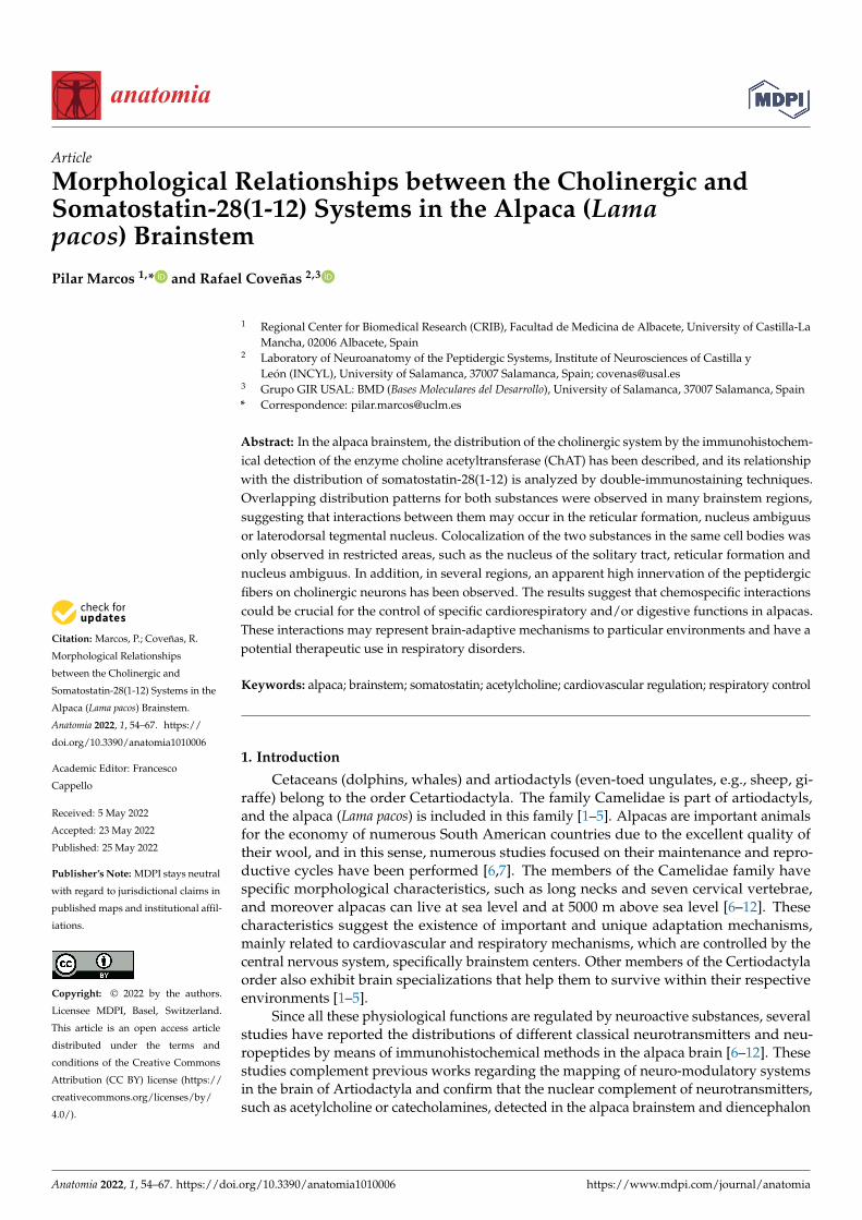
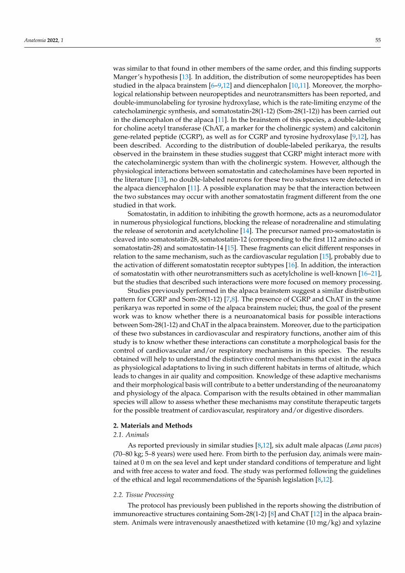
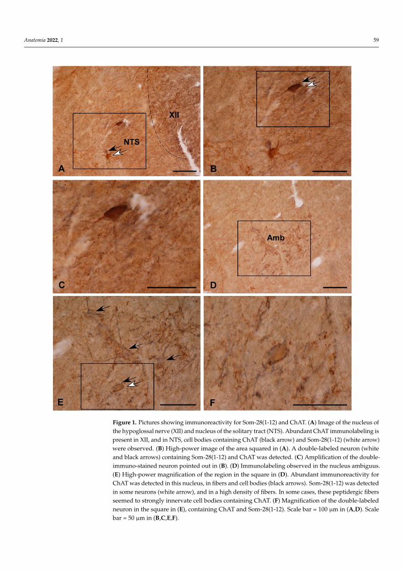
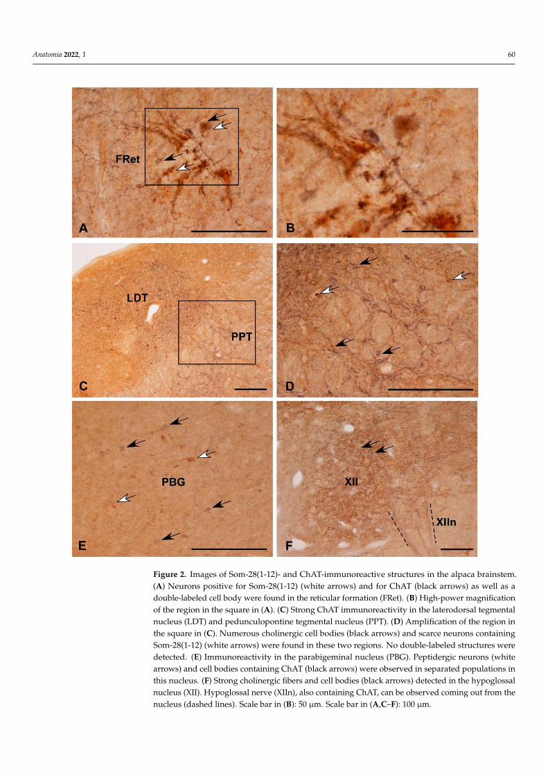
Comparing the distribution patterns of ChAT- and Som-28(1-12)-immunoreactive struc-tures in the alpaca brainstem (Table 1), perikarya containing ChAT or Som-28(1-12) weredetected in the nucleus of the solitary tract (Figure 1A–C), nucleus ambiguus (Figure 1D–F),dorsal motor nucleus of the vagus, the entire reticular formation (Figure 2A,B), laterodorsaltegmental nucleus (Ch6 cholinergic cell group) (Figure 2C,D), pedunculopontine tegmen-tal nucleus (Ch5 cholinergic cell group) (Figure 2C,D) and parabigeminal nucleus (Ch8cholinergic cell group) (Figure 2E). However, coexistence of the two neuroactive substancesinto the same cell bodies was only observed in isolated neurons located in the nucleusof the solitary tract (Figure 1A–C), nucleus ambiguus (Figure 1D–F) and reticular forma-tion (Figure 2A,B). In the remaining regions, neuronal populations containing ChAT orSom-28(1-12) displayed separate distributions.
3.3. Fibrilar Immunolabeling
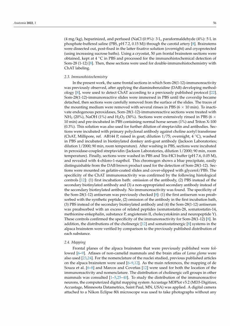
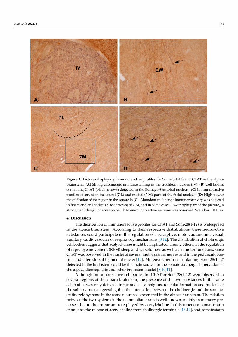
Almost all the cholinergic regions of the alpaca brainstem contained numerous im-munoreactive fibers for Som-28(1-12) (Table 1). This was especially noted in the nuclei ofmotor cranial nerves (Figures 2F and 3A) and associated regions (e.g., Edinger–Westphalnucleus) (Figure 3B). In some of these nuclei, a rich innervation of ChAT-immunoreactiveperikarya and dendrites by fibers containing Som-28(1-12) was observed, such is the caseof the nucleus ambiguus and medial division of the facial nucleus (Figure 3C,D). Theopposite pattern (peptidergic neurons innervated by ChAT-immunoreactive fibers) wasmore difficult to observe.
Anatomia 2022, 1 58
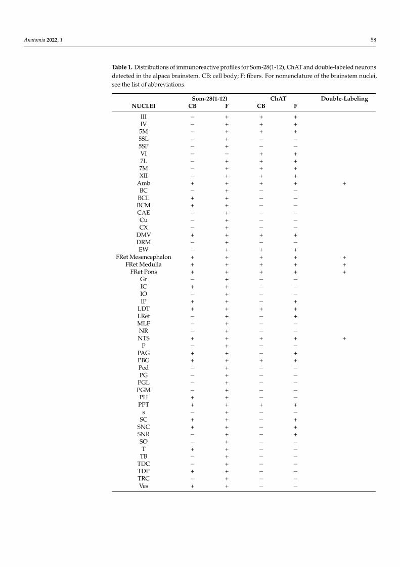
Table 1. Distributions of immunoreactive profiles for Som-28(1-12), ChAT and double-labeled neuronsdetected in the alpaca brainstem. CB: cell body; F: fibers. For nomenclature of the brainstem nuclei,see the list of abbreviations.
Som-28(1-12) ChAT Double-LabelingNUCLEI CB F CB F
III − + + +IV − + + +5M − + + +5SL − + − −5SP − + − −VI − − + +7L − + + +7M − + + +XII − + + +
Amb + + + + +BC − + − −
BCL + + − −BCM + + − −CAE − + − −Cu − + − −CX − + − −
DMV + + + +DRM − + − −EW − + + +
FRet Mesencephalon + + + + +FRet Medulla + + + + +
FRet Pons + + + + +Gr − + − −IC + + − −IO − + − −IP + + − +
LDT + + + +LRet − + − +MLF − + − −NR − + − −NTS + + + + +
P − + − −PAG + + − +PBG + + + +Ped − + − −PG − + − −
PGL − + − −PGM − + − −PH + + − −PPT + + + +
s − + − −SC + + − +
SNC + + − +SNR − + − +SO − + − −T + + − −
TB − + − −TDC − + − −TDP + + − −TRC − + − −Ves + + − −
Anatomia 2022, 1 59Anatomia 2022, 1, FOR PEER REVIEW 5
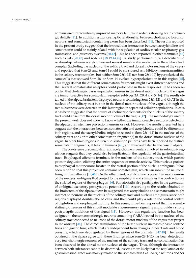
Figure 1. Pictures showing immunoreactivity for Som-28(1-12) and ChAT. (A) Image of the nucleus
of the hypoglossal nerve (XII) and nucleus of the solitary tract (NTS). Abundant ChAT immuno-
labeling is present in XII, and in NTS, cell bodies containing ChAT (black arrow) and Som-28(1-12)
(white arrow) were observed. (B) High-power image of the area squared in (A). A double-labeled
neuron (white and black arrows) containing Som-28(1-12) and ChAT was detected. (C) Amplifica-
tion of the double-immuno-stained neuron pointed out in (B). (D) Immunolabeling observed in the
nucleus ambiguus. (E) High-power magnification of the region in the square in (D). Abundant im-
munoreactivity for ChAT was detected in this nucleus, in fibers and cell bodies (black arrows). Som-
28(1-12) was detected in some neurons (white arrow), and in a high density of fibers. In some cases,
these peptidergic fibers seemed to strongly innervate cell bodies containing ChAT. (F) Magnifica-
tion of the double-labeled neuron in the square in (E), containing ChAT and Som-28(1-12). Scale bar
= 100 μm in (A,D). Scale bar = 50 μm in (B,C,E,F).
Figure 1. Pictures showing immunoreactivity for Som-28(1-12) and ChAT. (A) Image of the nucleus ofthe hypoglossal nerve (XII) and nucleus of the solitary tract (NTS). Abundant ChAT immunolabeling ispresent in XII, and in NTS, cell bodies containing ChAT (black arrow) and Som-28(1-12) (white arrow)were observed. (B) High-power image of the area squared in (A). A double-labeled neuron (whiteand black arrows) containing Som-28(1-12) and ChAT was detected. (C) Amplification of the double-immuno-stained neuron pointed out in (B). (D) Immunolabeling observed in the nucleus ambiguus.(E) High-power magnification of the region in the square in (D). Abundant immunoreactivity forChAT was detected in this nucleus, in fibers and cell bodies (black arrows). Som-28(1-12) was detectedin some neurons (white arrow), and in a high density of fibers. In some cases, these peptidergic fibersseemed to strongly innervate cell bodies containing ChAT. (F) Magnification of the double-labeledneuron in the square in (E), containing ChAT and Som-28(1-12). Scale bar = 100 µm in (A,D). Scalebar = 50 µm in (B,C,E,F).
Anatomia 2022, 1 60Anatomia 2022, 1, FOR PEER REVIEW 6
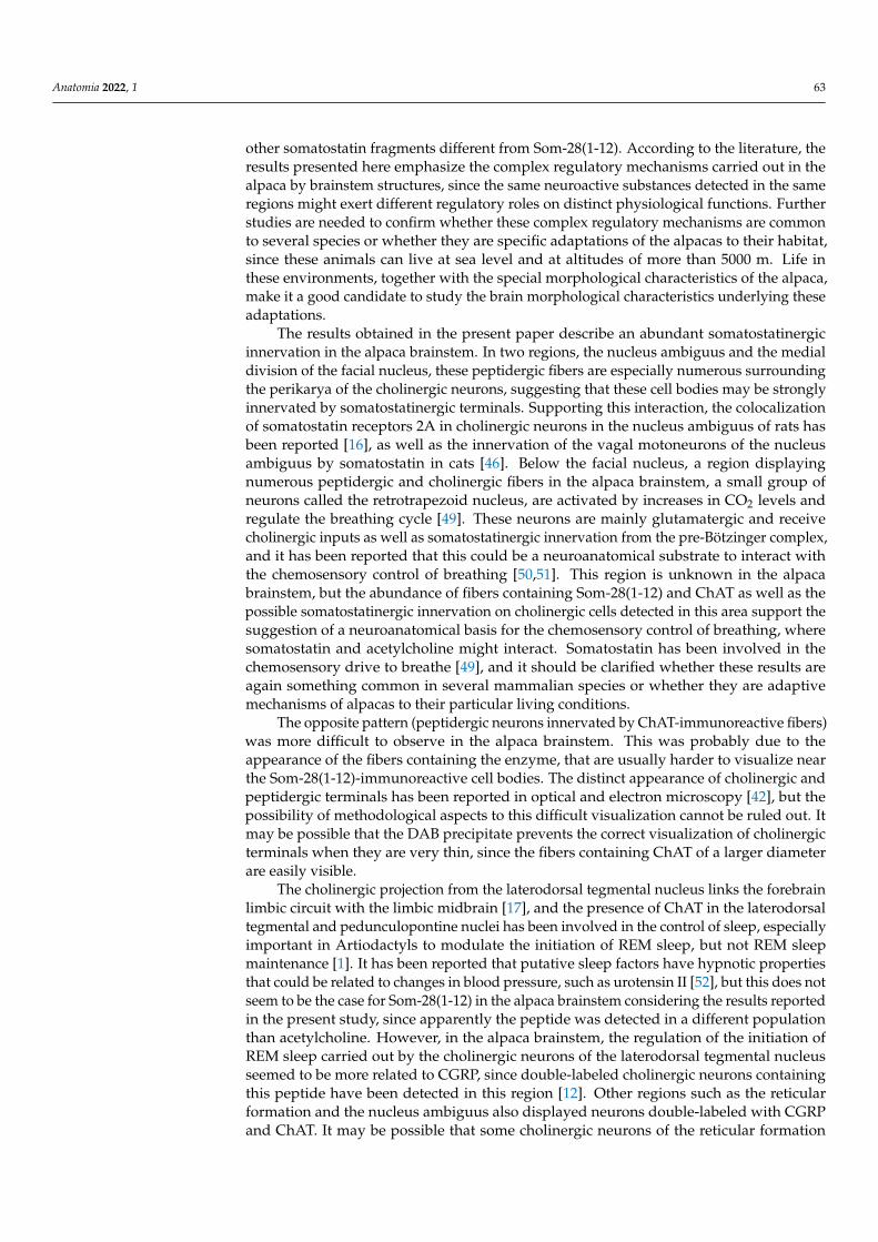
Figure 2. Images of Som-28(1-12)- and ChAT-immunoreactive structures in the alpaca brainstem.
(A) Neurons positive for Som-28(1-12) (white arrows) and for ChAT (black arrows) as well as a
double-labeled cell body were found in the reticular formation (FRet). (B) High-power magnifica-
tion of the region in the square in (A). (C) Strong ChAT immunoreactivity in the laterodorsal teg-
mental nucleus (LDT) and pedunculopontine tegmental nucleus (PPT). (D) Amplification of the re-
gion in the square in (C). Numerous cholinergic cell bodies (black arrows) and scarce neurons con-
taining Som-28(1-12) (white arrows) were found in these two regions. No double-labeled structures
were detected. (E) Immunoreactivity in the parabigeminal nucleus (PBG). Peptidergic neurons
(white arrows) and cell bodies containing ChAT (black arrows) were observed in separated popu-
lations in this nucleus. (F) Strong cholinergic fibers and cell bodies (black arrows) detected in the
hypoglossal nucleus (XII). Hypoglossal nerve (XIIn), also containing ChAT, can be observed coming
out from the nucleus (dashed lines). Scale bar in (B): 50 μm. Scale bar in (A,C–F): 100 μm.
Figure 2. Images of Som-28(1-12)- and ChAT-immunoreactive structures in the alpaca brainstem.(A) Neurons positive for Som-28(1-12) (white arrows) and for ChAT (black arrows) as well as adouble-labeled cell body were found in the reticular formation (FRet). (B) High-power magnificationof the region in the square in (A). (C) Strong ChAT immunoreactivity in the laterodorsal tegmentalnucleus (LDT) and pedunculopontine tegmental nucleus (PPT). (D) Amplification of the region inthe square in (C). Numerous cholinergic cell bodies (black arrows) and scarce neurons containingSom-28(1-12) (white arrows) were found in these two regions. No double-labeled structures weredetected. (E) Immunoreactivity in the parabigeminal nucleus (PBG). Peptidergic neurons (whitearrows) and cell bodies containing ChAT (black arrows) were observed in separated populations inthis nucleus. (F) Strong cholinergic fibers and cell bodies (black arrows) detected in the hypoglossalnucleus (XII). Hypoglossal nerve (XIIn), also containing ChAT, can be observed coming out from thenucleus (dashed lines). Scale bar in (B): 50 µm. Scale bar in (A,C–F): 100 µm.
Anatomia 2022, 1 61
Anatomia 2022, 1, FOR PEER REVIEW 8
SC + + − +
SNC + + − +
SNR − + − +
SO − + − −
T + + − −
TB − + − −
TDC − + − −
TDP + + − −
TRC − + − −
Ves + + − −
3.3. Fibrilar Immunolabeling
Almost all the cholinergic regions of the alpaca brainstem contained numerous im-
munoreactive fibers for Som-28(1-12) (Table 1). This was especially noted in the nuclei of
motor cranial nerves (Figures 2F and 3A) and associated regions (e.g., Edinger–Westphal
nucleus) (Figure 3B). In some of these nuclei, a rich innervation of ChAT-immunoreactive
perikarya and dendrites by fibers containing Som-28(1-12) was observed, such is the case
of the nucleus ambiguus and medial division of the facial nucleus (Figure 3C,D). The op-
posite pattern (peptidergic neurons innervated by ChAT-immunoreactive fibers) was
more difficult to observe.
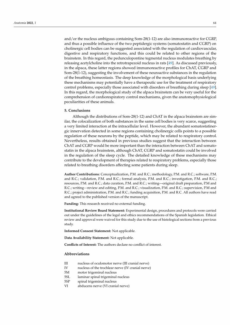
Figure 3. Pictures displaying immunoreactive profiles for Som-28(1-12) and ChAT in the alpaca
brainstem. (A) Strong cholinergic immunostaining in the trochlear nucleus (IV). (B) Cell bodies con-
taining ChAT (black arrows) detected in the Edinger–Westphal nucleus. (C) Immunoreactive pro-
files observed in the lateral (7 L) and medial (7 M) parts of the facial nucleus. (D) High-power mag-
nification of the region in the square in (C). Abundant cholinergic immunoreactivity was detected
Figure 3. Pictures displaying immunoreactive profiles for Som-28(1-12) and ChAT in the alpacabrainstem. (A) Strong cholinergic immunostaining in the trochlear nucleus (IV). (B) Cell bodiescontaining ChAT (black arrows) detected in the Edinger–Westphal nucleus. (C) Immunoreactiveprofiles observed in the lateral (7 L) and medial (7 M) parts of the facial nucleus. (D) High-powermagnification of the region in the square in (C). Abundant cholinergic immunoreactivity was detectedin fibers and cell bodies (black arrows) of 7 M, and in some cases (lower right part of the picture), astrong peptidergic innervation on ChAT-immunoreactive neurons was observed. Scale bar: 100 µm.
4. Discussion
The distribution of immunoreactive profiles for ChAT and Som-28(1-12) is widespreadin the alpaca brainstem. According to their respective distributions, these neuroactivesubstances could participate in the regulation of nociceptive, motor, autonomic, visual,auditory, cardiovascular or respiratory mechanisms [8,12]. The distribution of cholinergiccell bodies suggests that acetylcholine might be implicated, among others, in the regulationof rapid eye movement (REM) sleep and wakefulness as well as in motor functions, sinceChAT was observed in the nuclei of several motor cranial nerves and in the pedunculopon-tine and laterodorsal tegmental nuclei [12]. Moreover, neurons containing Som-28(1-12)detected in the brainstem could be the main source for the somatostatinergic innervation ofthe alpaca diencephalic and other brainstem nuclei [8,10,11].
Although immunoreactive cell bodies for ChAT or Som-28(1-12) were observed inseveral regions of the alpaca brainstem, the presence of the two substances in the samecell bodies was only detected in the nucleus ambiguus, reticular formation and nucleus ofthe solitary tract, suggesting that the interaction between the cholinergic and the somato-statinergic systems in the same neurons is restricted in the alpaca brainstem. The relationbetween the two systems in the mammalian brain is well-known, mainly in memory pro-cesses due to the important role played by acetylcholine in this function: somatostatinstimulates the release of acetylcholine from cholinergic terminals [18,19], and somatostatin
Anatomia 2022, 1 62
administered intracerebrally improved memory failures in rodents showing brain choliner-gic deficits [21]. In addition, a monosynaptic relationship between cholinergic forebrainneurons and somatostatin-containing axons has been described [41]. The results reportedin the present study suggest that the intracellular interaction between acetylcholine andsomatostatin could be mainly related with the regulation of cardiovascular, respiratory, gas-trointestinal and gustatory systems [20,42]. This has been reported in other mammals [43]such as cats [20,42] and rodents [18,19,44,45]. A study performed in rats described therelationship between acetylcholine and several somatostatin molecules in the solitary tractcomplex (including the nucleus of the solitary tract and dorsal motor nucleus of the vagus)and reported that Som-28 and Som-14 could be considered as inhibitory neurotransmittersin the solitary tract complex, but neither Som-28(1-12) nor Som-28(1-10) hyperpolarized thesame cells that showed Som-28- or Som-14-evoked hyperpolarization in this region [45].This suggests that the different somatostatin fragments might exert different actions andthat several somatostatin receptors could participate in these responses. It has been re-ported that cholinergic parasympathetic neurons in the dorsal motor nucleus of the vagusare immunoreactive for somatostatin receptor subtypes 2A, 2B, 4 and 5 [16]. The results ob-tained in the alpaca brainstem displayed neurons containing Som-28(1-12) and ChAT in thenucleus of the solitary tract but not in the dorsal motor nucleus of the vagus, although thetwo substances were detected in this later region in separated cellular populations. In cats,it has been suggested that the source of cholinergic elements for the nucleus of the solitarytract could arise from the dorsal motor nucleus of the vagus [42]. The methodology used inthe present work does not allow to know whether the immunoreactive neurons detected inthe alpaca brainstem are projection neurons or not. However, the results presented heresuggest that the interactions between somatostatin and acetylcholine could be different inboth regions, and that acetylcholine might be related to Som-28(1-12) in the nucleus of thesolitary tract and/or to other somatostatin fragment(s) in the dorsal motor nucleus of thevagus. In other brain regions, different distribution patterns have been reported for distinctsomatostatin fragments, at least in humans [43], and this could also be the case in alpaca.
The coexistence of somatostatin and acetylcholine in centers involved in autonomic reg-ulation suggests that they could also be implicated in the regulation of the gastrointestinaltract. Esophageal afferents terminate in the nucleus of the solitary tract, which partici-pates in deglution, eliciting the entire sequence of muscle activity. This nucleus projectsto esophageal motoneurons located in the rostral portion of the nucleus ambiguus. It hasbeen reported that this projection contains somatostatin, which can inhibit the neuronalfiring in this pathway [19,46]. On the other hand, acetylcholine is present in motoneuronsof the nucleus ambiguus that project to the esophagus and stimulates the contraction ofthe striated regions of the esophagus [46]. Somatostatin also participates in the generationof ambigual excitatory postsynaptic potential [19]. According to the results obtained inthe brainstem of the alpaca, it can be suggested that acetylcholine and somatostatin mightinteract on neurons of the nucleus of the solitary tract and nucleus ambiguus, since bothregions displayed double-labeled cells, and then could play a role in the central controlof deglution and esophageal motility. In this sense, it has been reported that the somato-statinergic neurons of this circuit modulate viscerosensory signaling and provide a strongpostsynaptic inhibition of this signal [44]. However, this regulatory function has beenassigned to the somatostatinergic neurons containing GABA located in the nucleus of thesolitary tract connected to neurons of the dorsal motor nucleus of the vagus that projectto the antrum [44]. The direct stimulation of the latter nucleus increases phasic contrac-tions and gastric tone, effects that are independent from changes in heart rate and bloodpressure, which are also regulated by these regions of the brainstem [47,48]. The resultsobtained in the alpaca agree with these findings, since Som-28(1-12) has been detected invery few cholinergic neurons of the nucleus of the solitary tract and no colocalization hasbeen observed in the dorsal motor nucleus of the vagus. Thus, although the interactionbetween both substances cannot be discarded, it seems more likely that the regulation of thegastrointestinal tract was mainly related to the somatostatin-GABAergic neurons and/or
Anatomia 2022, 1 63
other somatostatin fragments different from Som-28(1-12). According to the literature, theresults presented here emphasize the complex regulatory mechanisms carried out in thealpaca by brainstem structures, since the same neuroactive substances detected in the sameregions might exert different regulatory roles on distinct physiological functions. Furtherstudies are needed to confirm whether these complex regulatory mechanisms are commonto several species or whether they are specific adaptations of the alpacas to their habitat,since these animals can live at sea level and at altitudes of more than 5000 m. Life inthese environments, together with the special morphological characteristics of the alpaca,make it a good candidate to study the brain morphological characteristics underlying theseadaptations.
The results obtained in the present paper describe an abundant somatostatinergicinnervation in the alpaca brainstem. In two regions, the nucleus ambiguus and the medialdivision of the facial nucleus, these peptidergic fibers are especially numerous surroundingthe perikarya of the cholinergic neurons, suggesting that these cell bodies may be stronglyinnervated by somatostatinergic terminals. Supporting this interaction, the colocalizationof somatostatin receptors 2A in cholinergic neurons in the nucleus ambiguus of rats hasbeen reported [16], as well as the innervation of the vagal motoneurons of the nucleusambiguus by somatostatin in cats [46]. Below the facial nucleus, a region displayingnumerous peptidergic and cholinergic fibers in the alpaca brainstem, a small group ofneurons called the retrotrapezoid nucleus, are activated by increases in CO2 levels andregulate the breathing cycle [49]. These neurons are mainly glutamatergic and receivecholinergic inputs as well as somatostatinergic innervation from the pre-Bötzinger complex,and it has been reported that this could be a neuroanatomical substrate to interact withthe chemosensory control of breathing [50,51]. This region is unknown in the alpacabrainstem, but the abundance of fibers containing Som-28(1-12) and ChAT as well as thepossible somatostatinergic innervation on cholinergic cells detected in this area support thesuggestion of a neuroanatomical basis for the chemosensory control of breathing, wheresomatostatin and acetylcholine might interact. Somatostatin has been involved in thechemosensory drive to breathe [49], and it should be clarified whether these results areagain something common in several mammalian species or whether they are adaptivemechanisms of alpacas to their particular living conditions.
The opposite pattern (peptidergic neurons innervated by ChAT-immunoreactive fibers)was more difficult to observe in the alpaca brainstem. This was probably due to theappearance of the fibers containing the enzyme, that are usually harder to visualize nearthe Som-28(1-12)-immunoreactive cell bodies. The distinct appearance of cholinergic andpeptidergic terminals has been reported in optical and electron microscopy [42], but thepossibility of methodological aspects to this difficult visualization cannot be ruled out. Itmay be possible that the DAB precipitate prevents the correct visualization of cholinergicterminals when they are very thin, since the fibers containing ChAT of a larger diameterare easily visible.
The cholinergic projection from the laterodorsal tegmental nucleus links the forebrainlimbic circuit with the limbic midbrain [17], and the presence of ChAT in the laterodorsaltegmental and pedunculopontine nuclei has been involved in the control of sleep, especiallyimportant in Artiodactyls to modulate the initiation of REM sleep, but not REM sleepmaintenance [1]. It has been reported that putative sleep factors have hypnotic propertiesthat could be related to changes in blood pressure, such as urotensin II [52], but this does notseem to be the case for Som-28(1-12) in the alpaca brainstem considering the results reportedin the present study, since apparently the peptide was detected in a different populationthan acetylcholine. However, in the alpaca brainstem, the regulation of the initiation ofREM sleep carried out by the cholinergic neurons of the laterodorsal tegmental nucleusseemed to be more related to CGRP, since double-labeled cholinergic neurons containingthis peptide have been detected in this region [12]. Other regions such as the reticularformation and the nucleus ambiguus also displayed neurons double-labeled with CGRPand ChAT. It may be possible that some cholinergic neurons of the reticular formation
Anatomia 2022, 1 64
and/or the nucleus ambiguus containing Som-28(1-12) are also immunoreactive for CGRP,and thus a possible influence of the two peptidergic systems (somatostatin and CGRP) oncholinergic cell bodies can be suggested associated with the regulation of cardiovascular,digestive and respiratory functions, and this could be related to other regions of thebrainstem. In this regard, the pedunculopontine tegmental nucleus modulates breathing byreleasing acetylcholine into the retrotrapezoid nucleus in rats [49]. As discussed previously,in the alpaca, these latter regions showed immunoreactive profiles for ChAT, CGRP andSom-28(1-12), suggesting the involvement of these neuroactive substances in the regulationof the breathing homeostasis. The deep knowledge of the morphological basis underlyingthese mechanisms may potentially have a therapeutic use for the treatment of respiratorycontrol problems, especially those associated with disorders of breathing during sleep [49].In this regard, the morphological study of the alpaca brainstem can be very useful for thecomprehension of cardiorespiratory control mechanisms, given the anatomophysiologicalpeculiarities of these animals.
5. Conclusions
Although the distributions of Som-28(1-12) and ChAT in the alpaca brainstem are sim-ilar, the colocalization of both substances in the same cell bodies is very scarce, suggestinga very limited interaction at the intracellular level. However, the abundant somatostatiner-gic innervation detected in some regions containing cholinergic cells points to a possibleregulation of these neurons by the peptide, which may be related to respiratory control.Nevertheless, results obtained in previous studies suggest that the interaction betweenChAT and CGRP would be more important than the interaction between ChAT and somato-statin in the alpaca brainstem, although ChAT, CGRP and somatostatin could be involvedin the regulation of the sleep cycle. The detailed knowledge of these mechanisms maycontribute to the development of therapies related to respiratory problems, especially thoserelated to breathing disorders affecting some patients during sleep.
Author Contributions: Conceptualization, P.M. and R.C.; methodology, P.M. and R.C.; software, P.M.and R.C.; validation, P.M. and R.C.; formal analysis, P.M. and R.C.; investigation, P.M. and R.C.;resources, P.M. and R.C.; data curation, P.M. and R.C.; writing—original draft preparation, P.M andR.C.; writing—review and editing, P.M. and R.C.; visualization, P.M. and R.C.; supervision, P.M andR.C.; project administration, P.M. and R.C.; funding acquisition, P.M. and R.C. All authors have readand agreed to the published version of the manuscript.
Funding: This research received no external funding.
Institutional Review Board Statement: Experimental design, procedures and protocols were carriedout under the guidelines of the legal and ethics recommendations of the Spanish legislation. Ethicalreview and approval were waived for this study due to the use of histological sections from a previousstudy.
Informed Consent Statement: Not applicable.
Data Availability Statement: Not applicable.
Conflicts of Interest: The authors declare no conflict of interest.
Abbreviations
III nucleus of oculomotor nerve (III cranial nerve)IV nucleus of the trochlear nerve (IV cranial nerve)5M motor trigeminal nucleus5SL laminar spinal trigeminal nucleus5SP spinal trigeminal nucleusVI abducens nerve (VI cranial nerve)
Anatomia 2022, 1 65
7L facial nucleus, lateral division7M facial nucleus, medial divisionXII nucleus of the hypoglossal nerve (XII cranial nerve)Amb nucleus ambiguusBC brachium conjunctivumBCL marginal nucleus of the brachium conjunctivum, lateral divisionBCM marginal nucleus of the brachium conjunctivum, medial divisionCAE locus coeruleusChAT choline acetyl transferaseCu cuneate nucleusCX external cuneate nucleusDMV dorsal motor nucleus of the vagus nerveDRN dorsal raphe nucleusEW nucleus of Edinger–WestphalFRet reticular formationGr gracile nucleusIC inferior colliculusIO inferior oliveIP interpeduncular nucleusLDT laterodorsal tegmental nucleus (Ch6 cholinergic cell group)LRet lateral reticular nucleusMLF medial longitudinal fascicleNR red nucleusNTS nucleus of the solitary tractP pyramidal tractPAG periaqueductal grayPBG parabigeminal nucleus (Ch8 cholinergic cell group)Ped cerebral pedunclePG pontine grayPGL pontine gray, lateral divisionPGM pontine gray, medial divisionPH nucleus praepositus hypoglossiPPT pedunculopontine tegmental nucleus (Ch5 cholinergic cell group)S solitary tractSC superior colliculusSNC substantia nigra, pars compactaSNR substantia nigra, pars reticulataSO superior colliculusSom-28(1-12)
somatostatin-28 (1-12)
T nucleus of the trapezoid bodyTB trapezoid bodyTDC dorsal tegmental nucleus, central divisionTDP dorsal tegmental nucleus, pericentral divisionTRC tegmental reticular nucleus, central divisionVes vestibular nucleus
References1. Davimes, J.G.; Alagaili, A.N.; Bennett, N.C.; Mohammed, O.B.; Bhagwandin, A.; Manger, P.R.; Gravett, N. Neurochemical
organization and morphology of the sleep related nuclei in the brain of the Arabian oryx, Oryx leucoryx. J. Chem. Neuroanat. 2017,81, 53–70. [CrossRef] [PubMed]
2. Dell, L.A.; Karlsson, K.A.; Patzke, N.; Spocter, M.A.; Siegel, J.M.; Manger, P.R. Organization of the sleep-related neural systems inthe brain of the minke whale (Balaenoptera acutorostrata). J. Comp. Neurol. 2015, 524, 2018–2035. [CrossRef] [PubMed]
Anatomia 2022, 1 66
3. Dell, L.A.; Patzke, N.; Spocter, M.A.; Bertelsen, M.F.; Siegel, J.M.; Manger, P.R. Organization of the sleep-related neural systems inthe brain of the river hippopotamus (Hippopotamus amphibius): A most inusual Certiodactyl species. J. Comp. Neurol. 2015, 524,2036–2058. [CrossRef] [PubMed]
4. Dell, L.A.; Patzke, N.; Spocter, M.A.; Siegel, J.M.; Manger, P.R. Organization of the sleep-related neural systems in the brain of theharbour porpoise (Phocoena phocoena). J. Comp. Neurol. 2016, 524, 1999–2017. [CrossRef] [PubMed]
5. Malungo, I.B.; Gravett, N.; Bhagwandin, A.; Davimes, J.G.; Manger, P.R. A preliminary description of the sleep-related neuralsystems in the brain of the blue wildebeest, Connochaetes taurinus. Anat. Rec. 2020, 303, 1977–1997. [CrossRef]
6. De Souza, E.; Yi, P.; Aguilar, L.A.; Coveñas, R.; Lerma, L.; Andrade, R.; Mangas, A.; Narváez, J.A. Mapping of leucine-encephalinin the alpaca (Lama pacos) brainstem. In Focus on Neuropeptide Research; Coveñas, R., Mangas, A., Narváez, J.A., Eds.; TransworldResearch Network: Trivandrum, India, 2007; pp. 103–113.
7. De Souza, E.; Coveñas, R.; Yi, P.; Aguilar, L.A.; Lerma, L.; Andrade, R.; Mangas, A.; Díaz-Cabiale, Z.; Narváez, J.A. Mapping ofCGRP in the alpaca (Lama pacos) brainstem. J. Chem. Neuroanat. 2008, 35, 346–355. [CrossRef]
8. De Souza, E.; Sánchez, M.L.; Aguilar, L.A.; Díaz-Cabiale, Z.; Narváez, J.A.; Coveñas, R. Mapping of Somatostatin-28(1-12) in thealpaca (Lama pacos) brainstem. Microsc. Res. Tech. 2015, 78, 363–374. [CrossRef]
9. Marcos, P.; Arroyo-Jiménez, M.M.; Lozano, G.; Aguilar, L.A.; Coveñas, R. Mapping of tyrosine hydroxylase in the alpaca (Lamapacos) brainstem and colocalization with CGRP. J. Chem. Neuroanat. 2011, 41, 63–72. [CrossRef]
10. Coveñas, R.; Mangas, A.; Medina, L.E.; Sánchez, M.L.; Aguilar, L.A.; Díaz-Cabiale, Z.; Narváez, J.A. Mapping ofsomatostatin-28 (1-12) in the alpaca diencephalon. J. Chem. Neuroanat. 2011, 42, 89–98. [CrossRef]
11. Marcos, P.; Arroyo-Jiménez, M.M.; Lozano, G.; González-Fuentes, J.; Lagartos-Donate, M.J.; Aguilar, L.A.; Coveñas, R. Mappingof tyrosine hydroxylase in the diencephalon of alpaca (Lama pacos) and co-distribution with somatostatin-28 (1-12). J. Chem.Neuroanat. 2013, 50–51, 66–74. [CrossRef]
12. Marcos, P.; Coveñas, R. Immunohistochemical study of the brainstem cholinergic system in the alpaca (Lama pacos) and colocaliza-tion with CGRP. Eur. J. Histochem. 2021, 65, 3266. [CrossRef] [PubMed]
13. Manger, P.R. Establishing order at the systems level in mammalian brain evolution. Brain Res. Bull. 2005, 66, 282–289. [CrossRef][PubMed]
14. Reichlin, S. Somatostatin. In Brain Peptides; Krieger, D.T., Browstein, M.J., Martin, J.B., Eds.; John Wiley and Sons: New York, NY,USA, 1983; pp. 711–752.
15. Benoit, R.; Ling, N.; Bakhit, C.; Morrison, J.H.; Alford, B.; Guillemin, R. Somatostatin-28(1-12)-like immunoreactivity in the rat.Endocrinology 1982, 111, 2149–2151. [CrossRef] [PubMed]
16. Spary, E.J.; Maqbool, A.; Batten, T.F.C. Expression and localization of somatostatin receptor subtypes sst1-sst5 in areas of the ratmedulla oblongata involved in autonomic regulation. J. Chem. Neuroanat. 2008, 35, 49–66. [CrossRef] [PubMed]
17. Sutin, E.L.; Jacobowitz, D.M. Immunocytochemical localization of peptides and other neurochemical in the rat laterodorsaltegmental nucleus and adjacent area. J. Comp. Neurol. 1988, 270, 243–270. [CrossRef]
18. Wang, Y.T.; Neuman, R.S.; Bieger, D. Somatostatin inhibits nicotinic cholinoceptor mediated-excitation in rat ambigual motoneu-rons in vitro. Neurosci. Lett. 1991, 123, 236–239. [CrossRef]
19. Wang, Y.T.; Zhang, M.; Neuman, R.S.; Bieger, D. Somatostatin regulates excitatory amino acid receptor-mediated fast excitatorypostsynaptic potential components in vagal motoneurons. Neuroscience 1993, 53, 7–9. [CrossRef]
20. Batten, T.F.C. Immunolocalization of putative neurotransmitters innervating autonomic regulating neurons of cat ventral medulla.Brain Res. Bull. 1995, 37, 487–506. [CrossRef]
21. Matsuoka, N.; Yamazaki, M.; Yamaguchi, I. Changes in brain somatostatin in memory-deficient rats: Comparison with cholinergicmarkers. Neuroscience 1995, 66, 617–626. [CrossRef]
22. Marcos, P.; Corio, M.; Dubourg, P.; Tramu, G. Reciprocal synaptic connections between neurotensin- and tyrosine hydroxylase-immunoreactive neurons in the mediobasal hypothalamus of the guinea pig. Brain. Res. 1996, 715, 63–70. [CrossRef]
23. Jasper, A.L.; Ajmone-Marsan, C. A Stereotaxic Atlas of the Diencephalon of the Cat; National Research Council of Canada: Ottawa,ON, Canada, 1966.
24. Paxinos, G.; Watson, C. The Rat Brain in Stereotaxic Coordinates; Academic Press: San Diego, CA, USA, 1998.25. Badlangana, N.L.; Bhagwandin, A.; Fuxe, K.; Manger, P.R. Observations on the giraffe central nervous system related to the
corticospinal tract, motor cortex and spinal cord: What difference does a long neck make? Neuroscience 2007, 148, 522–534.[CrossRef] [PubMed]
26. Bux, F.; Bhagwandin, A.; Fuxe, K.; Manger, P.R. Organization of cholinergic, putative catecholaminergic and serotonergic nucleiin the diencephalon, midbrain and pons of sub-adult male giraffes. J. Chem. Neuroanat. 2010, 39, 189–203. [CrossRef] [PubMed]
27. Dell, L.A.; Gruger, J.L.; Bhagwandin, A.; Jillani, N.E.; Pettigrew, J.D.; Manger, P.R. Nuclear organization of cholinergic, putativecatecholaminergic, and serotonergic systems in the brains of two megachiropteran species. J. Chem. Neuroanat. 2010, 40, 177–195.[CrossRef] [PubMed]
28. Houser, C.R.; Crawford, G.D.; Barber, R.P.; Salvaterra, P.M.; Vaughn, J.E. Organization and morphological characteristics ofcholinergic neurons: An immunocytochemical study with a monoclonal antibody to choline acetyltransferase. Brain Res. 1983,266, 97–119. [CrossRef]
29. Kimura, H.; Maeda, T. Aminergic and cholinergic systems in the dorsolateral pontine tegmentum. Brain Res. Bull. 1982, 9, 493–499.[CrossRef]
Anatomia 2022, 1 67
30. Levey, A.I.; Wainer, B.H.; Mufson, E.J.; Mesulam, M.M. Co-localization of acetylcholinesterase and choline acetyltransferase in therat cerebrum. Neuroscience 1983, 9, 9–22. [CrossRef]
31. Manaye, K.F.; Zweig, R.; Wu, D.; Hersh, L.B.; de Lacalle, S.; Saper, C.B.; German, D.C. Quantification of cholinergic and selectnon-cholinergic mesopontine neuronal populations in the human brain. Neuroscience 1999, 89, 759–770. [CrossRef]
32. Marcos, P.; Coveñas, R. Neuroanatomical relationship between the cholinergic and tachykininergic systems in the adult humanbrainstem: An immunohistochemical study. J. Chem. Neuroanat. 2019, 102, 101701. [CrossRef]
33. Mesulam, M.M. Cholinergic pathways and the ascending reticular activating system of the human brain. Ann. N. Y. Acad. Sci.1995, 757, 169–179. [CrossRef]
34. Motts, S.D.; Slusarczyk, A.S.; Sowick, C.S.; Schofield, B.R. Distribution of cholinergic cells in guinea pig brainstem. Neuroscience2008, 154, 186–196. [CrossRef]
35. Oda, Y. Choline acetyltransferase: The structure, distribution and pathologic changes in the central nervous system. Pathol. Int.1999, 49, 921–937. [CrossRef]
36. Sakai, K.; Luppi, P.H.; Salvert, D.; Kimura, H.; Maeda, T.; Jouvet, M. Localization of cholinergic neurons in the cat lower brainstem.CR Acad. Sci. III 1986, 303, 317–324.
37. Tago, H.; McGeer, P.L.; McGeer, E.G.; Akiyama, H.; Hersh, L.B. Distribution of choline acetyltransferase immunopositivestructures in the rat brainstem. Brain Res. 1989, 495, 271–297. [CrossRef]
38. Yasuhara, O.; Aimi, Y.; Matsuo, A.; Kimura, H. Distribution of a splice variant of choline acetyltransferase in the trigeminalganglion and brainstem of the rat: Comparison with calcitonin gene-related peptide and substance P. J. Comp. Neurol. 2008, 509,436–448. [CrossRef] [PubMed]
39. Mesulam, M.M.; Geula, C.; Bothwell, M.A.; Hersh, L.B. Human reticular formation: Cholinergic neurons of the pedunculopontineand laterodorsal tegmental nuclei and some cytochemical comparisons to forebrain cholinergic neurons. J. Comp. Neurol. 1989,283, 611–633. [CrossRef] [PubMed]
40. Ichikawa, T.; Ajiki, K.; Matsuura, J.; Misawa, H. Localization of two cholinergic markers, choline acetyltransferase and vesicularacetylcholine transporter in the central nervous system of the rat: In situ hybridization histochemistry and immunohistochemistry.J. Chem. Neuroanat. 1997, 13, 23–39. [CrossRef]
41. Zaborsky, L. Afferent connections of the forebrain cholinergic projection neurons, with special reference to monoaminergic andpeptidergic fibers. Experientia 1989, 57, 12–32. [CrossRef]
42. Maley, B.E. Immunohistochemical localization of neuropeptides and neurotransmitters in the nucleus solitarius. Chem. Senses1996, 21, 367–376. [CrossRef]
43. Cebada-Sánchez, S.; Insausti, R.; González-Fuentes, J.; Arroyo-Jiménez, M.M.; Rivas-Infante, E.; Lagartos, M.J.; Martínez-Ruiz, J.;Lozano, G.; Marcos, P. Distribution of peptidergic populations in the human dentate gyrus (Somatostatin [SOM-28, SOM-12] andneuropeptide Y [NPY]) during postnatal development. Cell Tissue Res. 2014, 358, 25–41. [CrossRef]
44. Gillis, R.A.; Dezfuli, G.; Bellusci, L.; Vicini, S.; Sahibzada, N. Brainstem neuronal circuitries controlling gastric tonic and phasiccontractions: A review. Cell. Mol. Neurobiol. 2022, 42, 333–360. [CrossRef]
45. Jacquin, T.; Champagnat, J.; Madamba, S.; Denavit-Saubié, M.; Siggins, G.R. Somatostatin depresses excitability in neurons of thesolitary tract complex through hyperpolarization and augmentation of IM, a non-inactivating voltage-dependent outward currentblocked by muscarinic agonists. Proc. Natl. Acad. Sci. USA 1988, 85, 948–952. [CrossRef] [PubMed]
46. Cunningham, E.T.; Sawchenko, P.E. Central neural control of esophageal motility: A review. Disphagia 1990, 5, 35–51. [CrossRef][PubMed]
47. Pagani, F.D.; Norman, W.P.; Gillis, R.A. Medullary parasympathetic projections innervate specific sites in the feline stomach.Gastroenterology 1988, 95, 277–288. [CrossRef]
48. Norman, W.P.; Pagani, F.D.; Ormsbee, H.S., III; Kasbekar, D.K.; Gillis, R.A. Use of horseradish peroxidase to identify hindbrainsites that influence gastric motility in the cat. Gastroenterology 1985, 88, 701–705. [CrossRef]
49. Moreira, T.S.; Sobrinho, C.R.; Falquetto, B.; Oliveira, L.M.; Lima, J.D.; Mulkey, D.K.; Takakura, A.C. The retrotrapezoid nucleusand the neuromodulation of breathing. J. Neurophysiol. 2021, 125, 699–719. [CrossRef]
50. Tan, W.; Pagliardini, S.; Yang, P.; Janczewski, W.A.; Feldman, J.A. Projections of preBötzinger complex neurons in adult rats. J.Comp. Neurol. 2010, 518, 1862–1878. [CrossRef]
51. Yang, C.F.; Feldman, J.L. Efferent projections of excitatory and inhibitory preBötzinger Complex neurons. J. Comp. Neurol. 2018,526, 1389–1402. [CrossRef]
52. de Lecea, L.; Bourgin, P. Neuropeptide interactions and REM sleep: A role for urotensin II? Peptides 2008, 29, 845–851. [CrossRef]
Related Documents


