Review Acta Haematol 2019;141:232–244 Morphological and Immunophenotypic Clues to the WHO Categories of Acute Myeloid Leukaemia Barbara J. Bain a Marie C. Béné b a Department of Haematology, St Mary’s Hospital, London, UK; b Hematology Biology, Nantes University Hospital, Nantes, France Received: December 5, 2018 Accepted: December 8, 2018 Published online: April 9, 2019 Prof. B.J. Bain Department of Haematology, St Mary’s Hospital Praed Street London W2 1NY (UK) E-Mail b.bain @imperial.ac.uk © 2019 S. Karger AG, Basel E-Mail [email protected] www.karger.com/aha DOI: 10.1159/000496097 Keywords Acute myeloid leukaemia · Immunophenotyping · Morphology · World Health Organisation classification Abstract Diagnosis and classification of acute myeloid leukaemia (AML) require cytogenetic and molecular genetic investiga- tion. However, while these evaluations are pending, mor- phology supplemented by immunophenotyping can pro- vide clues to the diagnosis of specific cytogenetic/genetic categories of AML. Most importantly, acute promyelocytic leukaemia can be diagnosed with a high degree of certainty. However, provisional identification of cases associated with t(8; 21), inv(16), t(1; 22), and NPM1 mutation may also be pos- sible. In addition, transient abnormal myelopoiesis of Down’s syndrome can generally be diagnosed morphologically. © 2019 S. Karger AG, Basel Introduction The diagnosis of the World Health Organisation (WHO) categories of acute myeloid leukaemia (AML) re- quires the careful integration of clinical history, morphol- ogy, and cytogenetic/molecular genetic analysis, supple- mented by immunophenotyping in the residual category of AML, not otherwise specified [1]. However, results of cytogenetic and molecular genetic analyses may not be available immediately so that there is a role for morphol- ogy plus immunophenotyping in rapidly indicating a likely diagnosis. In the case of acute promyelocytic leu- kaemia (APL), in which the diagnosis is clinically urgent, the combination of these two modalities permits a diag- nosis with a high degree of certainty. In some other types of AML, morphological and immunophenotypic features may provide an indication as to the cytogenetic anomaly likely to be present [2]. The latter nevertheless has to be confirmed by karyotypic analysis or fluorescence in situ hybridisation (FISH). Moreover, new entities character- ised by molecular anomalies will require even more time- consuming targeted or whole-genome analyses. The WHO categories that will be discussed here are shown in Table 1. AML with t(8;21)(q22;q22.1); RUNX1-RUNX1T1 This category of core-binding factor (CBF) leukaemia usually shows differentiation to mature neutrophils. Neu- tropenia is uncommon and there may be neutrophilia. Neutrophils can be dysplastic. Sometimes there is eosino-

Welcome message from author
This document is posted to help you gain knowledge. Please leave a comment to let me know what you think about it! Share it to your friends and learn new things together.
Transcript
Review
Acta Haematol 2019;141:232–244
Morphological and Immunophenotypic Clues to the WHO Categories of Acute Myeloid Leukaemia
Barbara J. Bain
a Marie C. Béné
b a
Department of Haematology, St Mary’s Hospital, London, UK; b Hematology Biology, Nantes University Hospital, Nantes, France
Received: December 5, 2018Accepted: December 8, 2018Published online: April 9, 2019
Prof. B.J. BainDepartment of Haematology, St Mary’s HospitalPraed StreetLondon W2 1NY (UK)E-Mail b.bain @ imperial.ac.uk
© 2019 S. Karger AG, Basel
E-Mail [email protected]/aha
DOI: 10.1159/000496097
KeywordsAcute myeloid leukaemia · Immunophenotyping · Morphology · World Health Organisation classification
AbstractDiagnosis and classification of acute myeloid leukaemia (AML) require cytogenetic and molecular genetic investiga-tion. However, while these evaluations are pending, mor-phology supplemented by immunophenotyping can pro-vide clues to the diagnosis of specific cytogenetic/genetic categories of AML. Most importantly, acute promyelocytic leukaemia can be diagnosed with a high degree of certainty. However, provisional identification of cases associated with t(8; 21), inv(16), t(1; 22), and NPM1 mutation may also be pos-sible. In addition, transient abnormal myelopoiesis of Down’s syndrome can generally be diagnosed morphologically.
© 2019 S. Karger AG, Basel
Introduction
The diagnosis of the World Health Organisation (WHO) categories of acute myeloid leukaemia (AML) re-quires the careful integration of clinical history, morphol-ogy, and cytogenetic/molecular genetic analysis, supple-
mented by immunophenotyping in the residual category of AML, not otherwise specified [1]. However, results of cytogenetic and molecular genetic analyses may not be available immediately so that there is a role for morphol-ogy plus immunophenotyping in rapidly indicating a likely diagnosis. In the case of acute promyelocytic leu-kaemia (APL), in which the diagnosis is clinically urgent, the combination of these two modalities permits a diag-nosis with a high degree of certainty. In some other types of AML, morphological and immunophenotypic features may provide an indication as to the cytogenetic anomaly likely to be present [2]. The latter nevertheless has to be confirmed by karyotypic analysis or fluorescence in situ hybridisation (FISH). Moreover, new entities character-ised by molecular anomalies will require even more time-consuming targeted or whole-genome analyses. The WHO categories that will be discussed here are shown in Table 1.
AML with t(8; 21)(q22;q22.1); RUNX1-RUNX1T1
This category of core-binding factor (CBF) leukaemia usually shows differentiation to mature neutrophils. Neu-tropenia is uncommon and there may be neutrophilia. Neutrophils can be dysplastic. Sometimes there is eosino-
Morphological and Immunophenotypic Clues to the WHO Categories of AML
233Acta Haematol 2019;141:232–244DOI: 10.1159/000496097
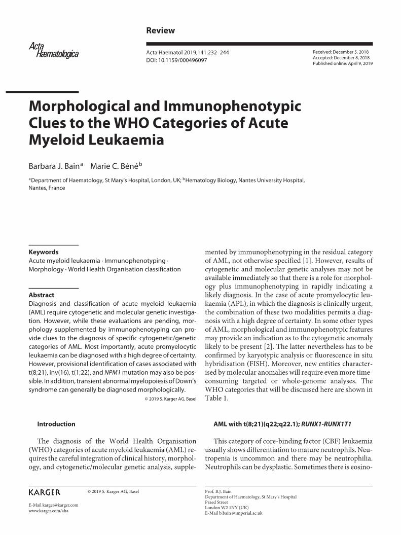
philic differentiation. Typically, some of the blast cells have an indentation in the nucleus, representing the Gol-gi zone, and often there is a long thin or fusiform Auer rod sometime within the nuclear “hof” (Fig. 1). The diag-nosis may be suspected from the cytological features.
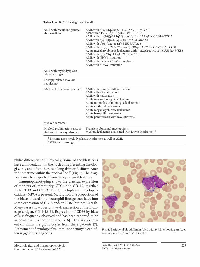
Immunophenotyping shows the classical expression of markers of immaturity, CD34 and CD117, together with CD13 and CD33 (Fig. 2). Cytoplasmic myeloper-oxidase (MPO) is present. Maturation of a proportion of the blasts towards the neutrophil lineage translates into some expression of CD15 and/or CD65 but not CD11b. Many cases show aberrant weak expression of the B-lin-eage antigen, CD19 [3–5]. Expression of CD56 by blast cells is frequently observed and has been reported to be associated with a poorer prognosis [6]. CD56 is also pres-ent on immature granulocytes from these patients [7]. Assessment of cytology plus immunophenotype can of-ten suggest this diagnosis.
Table 1. WHO 2016 categories of AML
AML with recurrent genetic abnormalities
AML with t(8;21)(q22;q22.1); RUNX1-RUNX1T1APL with t(15;17)(q24.1;q21.2); PML-RARAAML with inv(16)(p13.1q22) or t(16;16)(p13.1;q22); CBFB-MYH11AML with t(9;11)(p21.3;q23.3); KMT2A-MLLT3AML with t(6;9)(p23;q34.1); DEK-NUP214AML with inv(3)(q21.3q26.2) or t(3;3)(q21.3;q26.2); GATA2, MECOMAcute megakaryoblastic leukaemia with t(1;22)(p13.3;q13.1); RBM15-MKL1AML with t(9;22)(q34.1;q11.2); BCR-ABL1AML with NPM1 mutationAML with biallelic CEBPA mutationAML with RUNX1 mutation
AML with myelodysplasia- related changes
Therapy-related myeloid neoplasms1
AML, not otherwise specified AML with minimal differentiationAML without maturationAML with maturationAcute myelomonocytic leukaemiaAcute monoblastic/monocytic leukaemiaAcute erythroid leukaemiaAcute megakaryoblastic leukaemiaAcute basophilic leukaemiaAcute panmyelosis with myelofibrosis
Myeloid sarcoma
Myeloid proliferations associ-ated with Down syndrome2
Transient abnormal myelopoiesisMyeloid leukaemia associated with Down syndrome1, 2
1 Encompasses myelodysplastic syndromes as well as AML.2 WHO terminology.
Fig. 1. Peripheral blood film in AML with t(8; 21) showing an Auer rod in a nuclear “hof.” MGG ×100.
Bain/BénéActa Haematol 2019;141:232–244234DOI: 10.1159/000496097
APL with t(15; 17)(q24.1;q21.2); PML-RARA
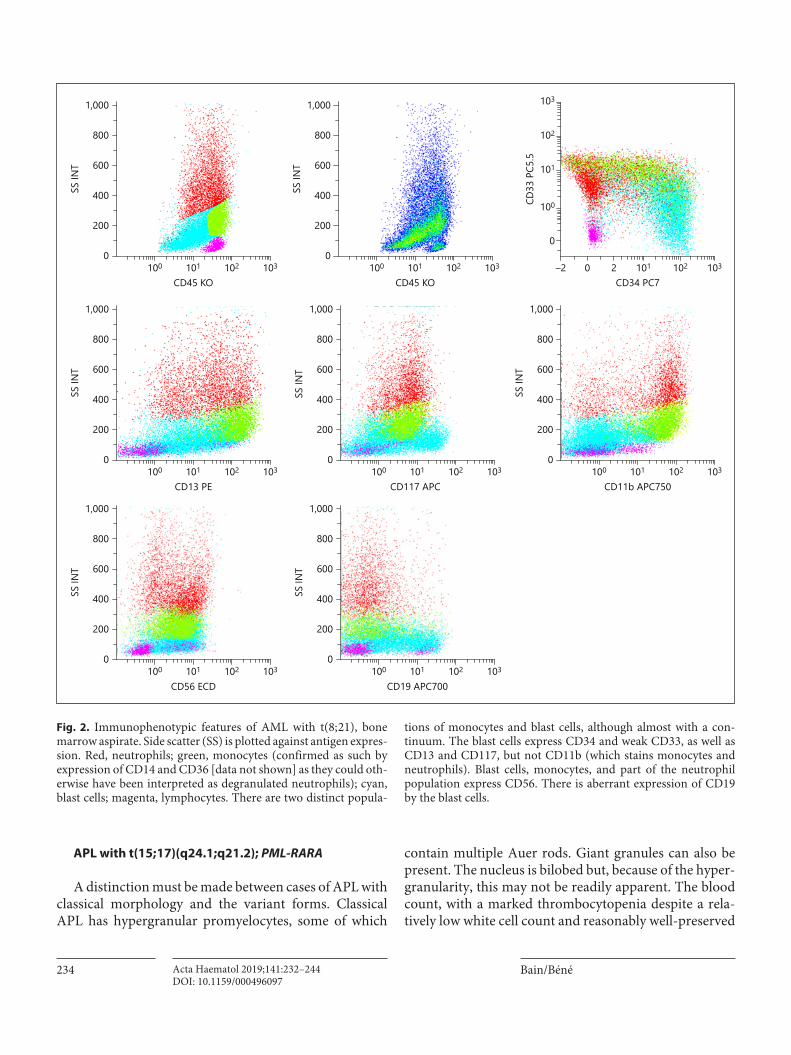
A distinction must be made between cases of APL with classical morphology and the variant forms. Classical APL has hypergranular promyelocytes, some of which
contain multiple Auer rods. Giant granules can also be present. The nucleus is bilobed but, because of the hyper-granularity, this may not be readily apparent. The blood count, with a marked thrombocytopenia despite a rela-tively low white cell count and reasonably well-preserved
CD45 KO
800
400
600
200
1,000
0102101100 103
SS IN
T
CD11b APC750
800
400
600
200
1,000
0102101100 103
SS IN
T
CD19 APC700
800
400
600
200
1,000
0102101100 103
SS IN
T
CD56 ECD
800
400
600
200
1,000
0102101100 103
SS IN
T
CD13 PE
800
400
600
200
1,000
0102101100 103
SS IN
T
CD117 APC
800
400
600
200
1,000
0102101100 103
SS IN
TCD45 KO
800
400
600
200
1,000
0102101100 103
SS IN
TCD34 PC7
1021010 2–2 103
CD33
PC5
.5
103
102
101
100
0
Fig. 2. Immunophenotypic features of AML with t(8; 21), bone marrow aspirate. Side scatter (SS) is plotted against antigen expres-sion. Red, neutrophils; green, monocytes (confirmed as such by expression of CD14 and CD36 [data not shown] as they could oth-erwise have been interpreted as degranulated neutrophils); cyan, blast cells; magenta, lymphocytes. There are two distinct popula-
tions of monocytes and blast cells, although almost with a con-tinuum. The blast cells express CD34 and weak CD33, as well as CD13 and CD117, but not CD11b (which stains monocytes and neutrophils). Blast cells, monocytes, and part of the neutrophil population express CD56. There is aberrant expression of CD19 by the blast cells.
Morphological and Immunophenotypic Clues to the WHO Categories of AML
235Acta Haematol 2019;141:232–244DOI: 10.1159/000496097
haemoglobin concentration, supports the diagnosis and should lead to further testing for disseminated intravas-cular coagulation. Leukaemic promyelocytes may be rare in the peripheral blood (or even absent) so that a careful search is needed followed by urgent examination of the bone marrow (Fig. 3). The cytological features are so characteristic that, when circumstances necessitate, treat-ment with ATRA (all-trans-retinoic aid) can be started without waiting for further supporting evidence. How-
ever, demonstration of the typical immunophenotype (see below) makes the diagnosis even more firmly based.
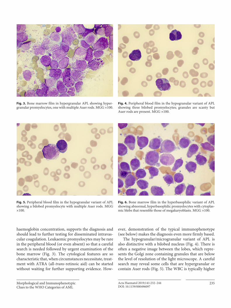
The hypogranular/microgranular variant of APL is also distinctive with a bilobed nucleus (Fig. 4). There is often a negative image between the lobes, which repre-sents the Golgi zone containing granules that are below the level of resolution of the light microscope. A careful search may reveal some cells that are hypergranular or contain Auer rods (Fig. 5). The WBC is typically higher
Fig. 3. Bone marrow film in hypergranular APL showing hyper-granular promyelocytes, one with multiple Auer rods. MGG ×100.
Fig. 4. Peripheral blood film in the hypogranular variant of APL showing three bilobed promyelocytes; granules are scanty but Auer rods are present. MGG ×100.
Fig. 5. Peripheral blood film in the hypogranular variant of APL showing a bilobed promyelocyte with multiple Auer rods. MGG ×100.
Fig. 6. Bone marrow film in the hyperbasophilic variant of APL showing abnormal, hyperbasophilic promyelocytes with cytoplas-mic blebs that resemble those of megakaryoblasts. MGG ×100.
Bain/BénéActa Haematol 2019;141:232–244236DOI: 10.1159/000496097
than in the hypergranular variant. Cytochemical staining shows strong MPO and Sudan black B positivity. The cy-tological features should arouse suspicion of this diagno-sis, often quite a strong suspicion. A coagulation screen and urgent immunophenotyping are important in sup-porting the diagnosis and permitting the initiation of
treatment while awaiting cytogenetic/molecular genetic confirmation.
The third cytological variant is the hyperbasophilic variant in which cytoplasm is more scanty, is basophilic, and shows cytoplasmic blebs (Fig. 6). Granules may or may not be visible. Confusion with the cytological fea-
CD45 KO
800
400
600
200
1,000
0105104103 106
SS IN
T (×
103 )
CD33 PC5.5
800
400
600
200
1,000
01051041030 106
SS IN
T (×
103 )
CD45 KO
800
400
600
200
1,000
0105104103 106
SS IN
T (×
103 )
CD15 PB
800
400
600
200
1,000
0105104103 106
SS IN
T (×
103 )
CD34 PC7
800
400
600
200
1,000
0105104103 106
SS IN
T (×
103 )
HL-DR PB
800
400
600
200
1,000
0105104103 106
SS IN
T (×
103 )
cMPO PE
800
400
600
200
1,000
0105104103 106
SS IN
T (×
103 )
CD117 APC
800
400
600
200
1,000
0105104103 106
SS IN
T (×
103 )
CD13 PE
800
400
600
200
1,000
0105104103 106
SS IN
T (×
103 )
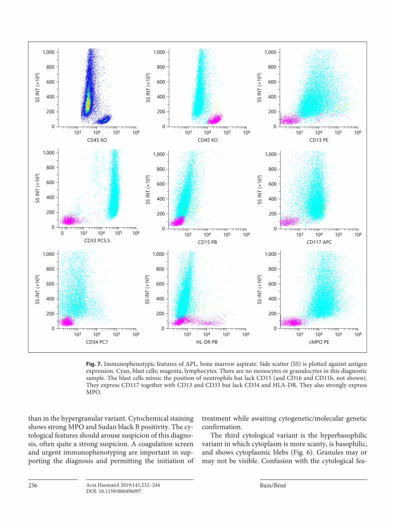
Fig. 7. Immunophenotypic features of APL, bone marrow aspirate. Side scatter (SS) is plotted against antigen expression. Cyan, blast cells; magenta, lymphocytes. There are no monocytes or granulocytes in this diagnostic sample. The blast cells mimic the position of neutrophils but lack CD15 (and CD16 and CD11b, not shown). They express CD117 together with CD13 and CD33 but lack CD34 and HLA-DR. They also strongly express MPO.
Morphological and Immunophenotypic Clues to the WHO Categories of AML
237Acta Haematol 2019;141:232–244DOI: 10.1159/000496097
tures of acute megakaryoblastic leukaemia is possible so that coagulation screening and immunophenotyping have a role in making a rapid provisional diagnosis.
The immunophenotype, considered by some as un-necessary, is however very characteristic while slightly tricky [8]. On the canonical CD45/side scatter (SSC) car-tography, the first impression may be that of an absence of progenitors/blasts with large numbers of slightly de-granulated neutrophils (Fig. 7). In fact, this “flame-like” image of cells with intermediate expression of CD45 re-flects the granularity of the leukaemic cells. These cells express typical myeloid markers such as CD13, CD33, and often CD117. However, they typically lack CD34 ex-pression. Testing for this quadruplet of fundamental markers will thus strengthen the diagnosis. Strong ex-pression of MPO is also demonstrated by flow cytometry. Of other classically tested antigens, leukaemic cells of APL fail to express CD15 and CD16, which would be present on neutrophils. Another key feature is the ab-sence of HLA-DR, which is often present on myeloid blasts of different types of AML. There is also lack expres-sion of beta2 integrins (CD18 beta-chain and CD11a, CD11b, or CD11c alpha-chains). Testing for CD41, CD42 and/or CD61 will permit exclusion of megakaryoblastic leukaemia in the hyperbasophilic variant [9].
Assessment of the cytological and immunophenotypic features enables this diagnosis to be made reliably, and within hours, in the great majority of cases, before confir-
mation of the t(15; 17) by FISH or conventional cytoge-netic analysis.
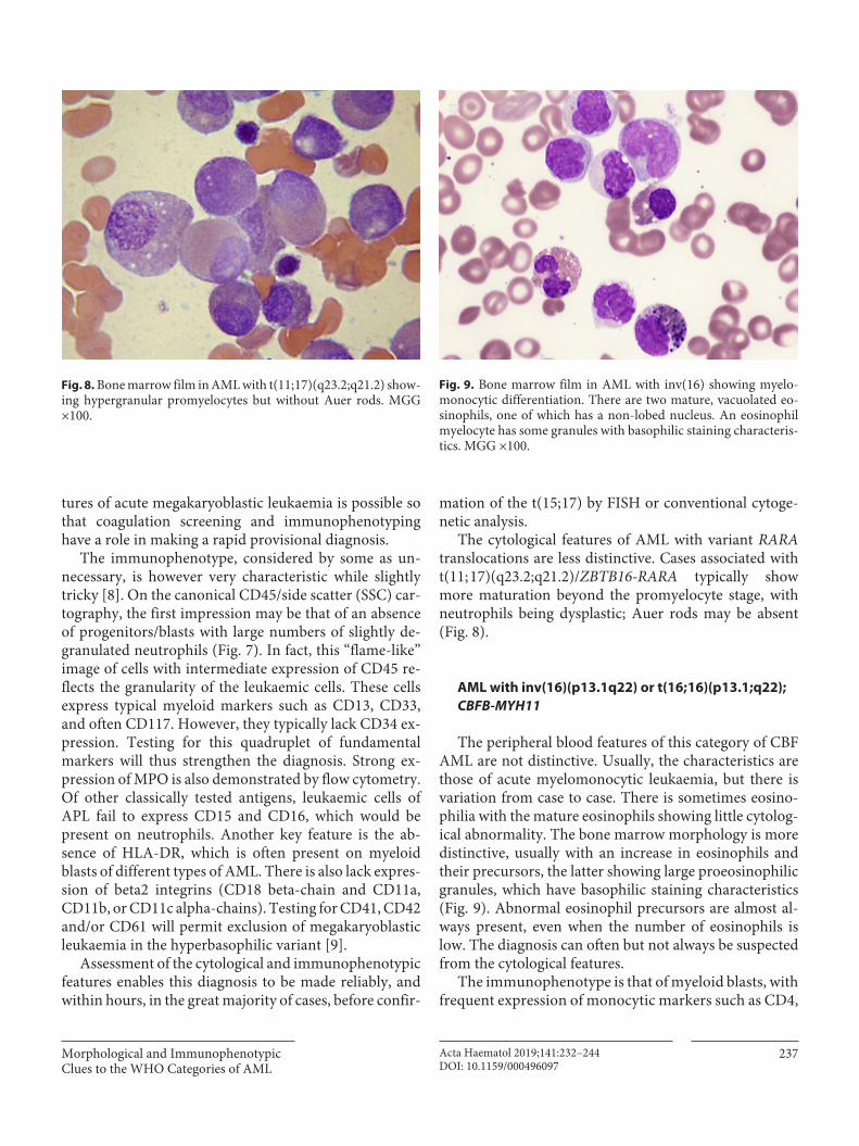
The cytological features of AML with variant RARA translocations are less distinctive. Cases associated with t(11; 17)(q23.2;q21.2)/ZBTB16-RARA typically show more maturation beyond the promyelocyte stage, with neutrophils being dysplastic; Auer rods may be absent (Fig. 8).
AML with inv(16)(p13.1q22) or t(16; 16)(p13.1;q22); CBFB-MYH11
The peripheral blood features of this category of CBF AML are not distinctive. Usually, the characteristics are those of acute myelomonocytic leukaemia, but there is variation from case to case. There is sometimes eosino-philia with the mature eosinophils showing little cytolog-ical abnormality. The bone marrow morphology is more distinctive, usually with an increase in eosinophils and their precursors, the latter showing large proeosinophilic granules, which have basophilic staining characteristics (Fig. 9). Abnormal eosinophil precursors are almost al-ways present, even when the number of eosinophils is low. The diagnosis can often but not always be suspected from the cytological features.
The immunophenotype is that of myeloid blasts, with frequent expression of monocytic markers such as CD4,
Fig. 8. Bone marrow film in AML with t(11; 17)(q23.2;q21.2) show-ing hypergranular promyelocytes but without Auer rods. MGG ×100.
Fig. 9. Bone marrow film in AML with inv(16) showing myelo-monocytic differentiation. There are two mature, vacuolated eo-sinophils, one of which has a non-lobed nucleus. An eosinophil myelocyte has some granules with basophilic staining characteris-tics. MGG ×100.
Bain/BénéActa Haematol 2019;141:232–244238DOI: 10.1159/000496097
CD36, or CD38 among those most often tested [10, 11]. Part of the blastic population may express neutrophil markers such as CD15 and/or CD65. The neutrophilic/monocytic differentiation trend of these blasts also translates into clear expression of CD11b, an antigen that is generally more strongly expressed by monocytes than by neutrophils. CD2 is often positive. Assessment of the cytological (particularly in the bone marrow) and immunophenotypic features suggests this diagnosis in the majority of cases, before FISH and/or conventional cytogenetic analysis disclose the inversion or transloca-tion.
AML with t(9; 11)(p21.3;q23.3); KMT2A-MLLT3
Cytological features are usually those of acute mono-blastic leukaemia but sometimes of acute monocytic or acute myelomonocytic leukaemia. There are no specific cytological clues to this diagnosis.
The immunophenotype shows the presence of mono-cytic markers, without expression of CD19. Some cases are CD4 positive. There may be some asynchrony in the expression of CD33 (positive) and CD13 (negative), and CD34 is often lacking. Monocytic differentiation may also be deduced from some co-expression (often weak) of CD15 and/or CD65, usually in the absence of CD14 [4]. There are no strong morphological or immunophenotyp-ic clues to this diagnosis.
AML with t(6; 9)(p23;q34.1); DEK-NUP214
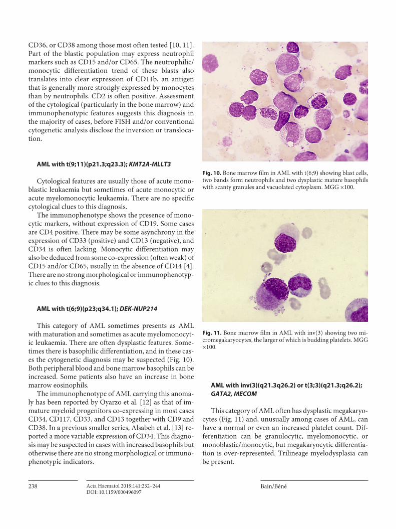
This category of AML sometimes presents as AML with maturation and sometimes as acute myelomonocyt-ic leukaemia. There are often dysplastic features. Some-times there is basophilic differentiation, and in these cas-es the cytogenetic diagnosis may be suspected (Fig. 10). Both peripheral blood and bone marrow basophils can be increased. Some patients also have an increase in bone marrow eosinophils.
The immunophenotype of AML carrying this anoma-ly has been reported by Oyarzo et al. [12] as that of im-mature myeloid progenitors co-expressing in most cases CD34, CD117, CD33, and CD13 together with CD9 and CD38. In a previous smaller series, Alsabeh et al. [13] re-ported a more variable expression of CD34. This diagno-sis may be suspected in cases with increased basophils but otherwise there are no strong morphological or immuno-phenotypic indicators.
AML with inv(3)(q21.3q26.2) or t(3; 3)(q21.3;q26.2); GATA2, MECOM
This category of AML often has dysplastic megakaryo-cytes (Fig. 11) and, unusually among cases of AML, can have a normal or even an increased platelet count. Dif-ferentiation can be granulocytic, myelomonocytic, or monoblastic/monocytic, but megakaryocytic differentia-tion is over-represented. Trilineage myelodysplasia can be present.
Fig. 10. Bone marrow film in AML with t(6; 9) showing blast cells, two bands form neutrophils and two dysplastic mature basophils with scanty granules and vacuolated cytoplasm. MGG ×100.
Fig. 11. Bone marrow film in AML with inv(3) showing two mi-cromegakaryocytes, the larger of which is budding platelets. MGG ×100.
Morphological and Immunophenotypic Clues to the WHO Categories of AML
239Acta Haematol 2019;141:232–244DOI: 10.1159/000496097
The immunophenotype is that of immature myeloid cells co-expressing CD34, CD117, CD13, and CD33 with MPO often being negative [14, 15]. Expression of CD7, CD11c, CD11b, and CD123 is frequent, but positivity for CD56 is seen less often [14]. When megakaryocytic dif-ferentiation is present, it can be confirmed by CD41, CD42, and/or CD61 positivity. Monocytic forms may ex-press CD14.
Only cases with a normal or increased platelet count and with dysplastic megakaryocytes are likely to be sus-pected on morphological and immunophenotypic grounds. Otherwise, making a distinction from AML with myelodysplasia-related changes is difficult.
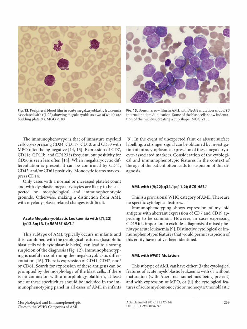
Acute Megakaryoblastic Leukaemia with t(1; 22)(p13.3;q13.1); RBM15-MKL1
This subtype of AML typically occurs in infants and this, combined with the cytological features (basophilic blast cells with cytoplasmic blebs), can lead to a strong suspicion of the diagnosis (Fig. 12). Immunophenotyp-ing is useful in confirming the megakaryoblastic differ-entiation [16]. There is expression of CD41, CD42, and/or CD61. Search for expression of these antigens can be prompted by the morphology of the blast cells. If there is no connexion with a morphology platform, at least one of these specificities should be included in the im-munophenotyping panel in all cases of AML in infants
[9]. In the event of unexpected faint or absent surface labelling, a stronger signal can be obtained by investiga-tion of intracytoplasmic expression of these megakaryo-cyte-associated markers. Consideration of the cytologi-cal and immunophenotypic features in the context of the age of the patient often leads to suspicion of this di-agnosis.
AML with t(9; 22)(q34.1;q11.2); BCR-ABL1
This is a provisional WHO category of AML. There are no specific cytological features.
Immunophenotyping shows expression of myeloid antigens with aberrant expression of CD7 and CD19 ap-pearing to be common. However, in cases expressing CD19 it is important to exclude a diagnosis of mixed phe-notype acute leukaemia [9]. Distinctive cytological or im-munophenotypic features that would permit suspicion of this entity have not yet been identified.
AML with NPM1 Mutation
This subtype of AML can have either: (i) the cytological features of acute myeloblastic leukaemia with or without maturation (with Auer rods sometimes being present) and with expression of MPO, or (ii) the cytological fea-tures of acute myelomonocytic or monocytic/monoblastic
Fig. 12. Peripheral blood film in acute megakaryoblastic leukaemia associated with t(1; 22) showing megakaryoblasts, two of which are budding platelets. MGG ×100.
Fig. 13. Bone marrow film in AML with NPM1 mutation and FLT3 internal tandem duplication. Some of the blast cells show indenta-tion of the nucleus, creating a cup shape. MGG ×100.
Bain/BénéActa Haematol 2019;141:232–244240DOI: 10.1159/000496097
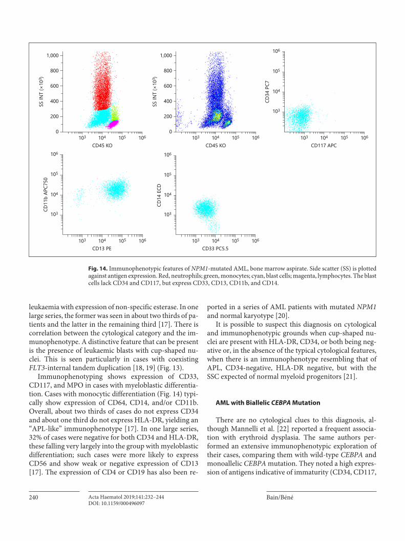
leukaemia with expression of non-specific esterase. In one large series, the former was seen in about two thirds of pa-tients and the latter in the remaining third [17]. There is correlation between the cytological category and the im-munophenotype. A distinctive feature that can be present is the presence of leukaemic blasts with cup-shaped nu-clei. This is seen particularly in cases with coexisting FLT3-internal tandem duplication [18, 19] (Fig. 13).
Immunophenotyping shows expression of CD33, CD117, and MPO in cases with myeloblastic differentia-tion. Cases with monocytic differentiation (Fig. 14) typi-cally show expression of CD64, CD14, and/or CD11b. Overall, about two thirds of cases do not express CD34 and about one third do not express HLA-DR, yielding an “APL-like” immunophenotype [17]. In one large series, 32% of cases were negative for both CD34 and HLA-DR, these falling very largely into the group with myeloblastic differentiation; such cases were more likely to express CD56 and show weak or negative expression of CD13 [17]. The expression of CD4 or CD19 has also been re-
ported in a series of AML patients with mutated NPM1 and normal karyotype [20].
It is possible to suspect this diagnosis on cytological and immunophenotypic grounds when cup-shaped nu-clei are present with HLA-DR, CD34, or both being neg-ative or, in the absence of the typical cytological features, when there is an immunophenotype resembling that of APL, CD34-negative, HLA-DR negative, but with the SSC expected of normal myeloid progenitors [21].
AML with Biallelic CEBPA Mutation
There are no cytological clues to this diagnosis, al-though Mannelli et al. [22] reported a frequent associa-tion with erythroid dysplasia. The same authors per-formed an extensive immunophenotypic exploration of their cases, comparing them with wild-type CEBPA and monoallelic CEBPA mutation. They noted a high expres-sion of antigens indicative of immaturity (CD34, CD117,
CD117 APC106105104103
CD34
PC7
106
105
104
103
CD13 PE106105104103
CD11
b AP
C750
106
105
104
103
CD33 PC5.5106105104103
CD14
ECD
106
105
104
103
CD45 KO
800
400
600
200
1,000
0105104103 106
SS IN
T (×
103 )
CD45 KO
800
400
600
200
1,000
0105104103 106
SS IN
T (×
103 )
Fig. 14. Immunophenotypic features of NPM1-mutated AML, bone marrow aspirate. Side scatter (SS) is plotted against antigen expression. Red, neutrophils; green, monocytes; cyan, blast cells; magenta, lymphocytes. The blast cells lack CD34 and CD117, but express CD33, CD13, CD11b, and CD14.
Morphological and Immunophenotypic Clues to the WHO Categories of AML
241Acta Haematol 2019;141:232–244DOI: 10.1159/000496097
and HLA-DR) together with strong MPO and asynchro-nous expression of antigens typical of more mature cells, CD15 and CD65. CD7 and CD56 were also frequently expressed. Interestingly, CD64 was found to be expressed not only by blasts, but also by neutrophils (unusual and mostly characteristic of activated neutrophils and of monocytes). The erythroid dysplasia detectable morpho-logically was reflected in flow cytometry with features of immature erythroblasts being observed, respectively CD117 and CD105 (endoglin) positivity associated with low levels of CD36 and CD71 [22]. By analysing SSC and the expression of six antigens on blast cells, neutrophils, monocytes, and erythroid cells, these authors were able to predict CEBPA biallelic mutation and thus target molecu-lar analysis [22]. The cytological features are not distinc-tive but detailed immunophenotypic analysis can permit suspicion of this diagnosis.
AML with RUNX1 Mutation
There are no cytological features suggestive of this anomaly although this genetic subtype is over-represent-ed among cases of MPO-negative AML. In a large Tai-wanese study, Tang et al. [23] found 4 out of 10 patients with French-American-British (FAB) M0 AML to have a RUNX1 mutation.
The same authors also reported that the blast cells usu-ally expressed CD34, CD13, and HLA-DR, whereas expres-
sion of CD33 and CD15 was less common than in other cases of AML (77 and 28%, respectively), suggesting imma-ture cells; CD56 was also expressed less often, being seen in only 13% of cases. CD19 was not expressed in any case.
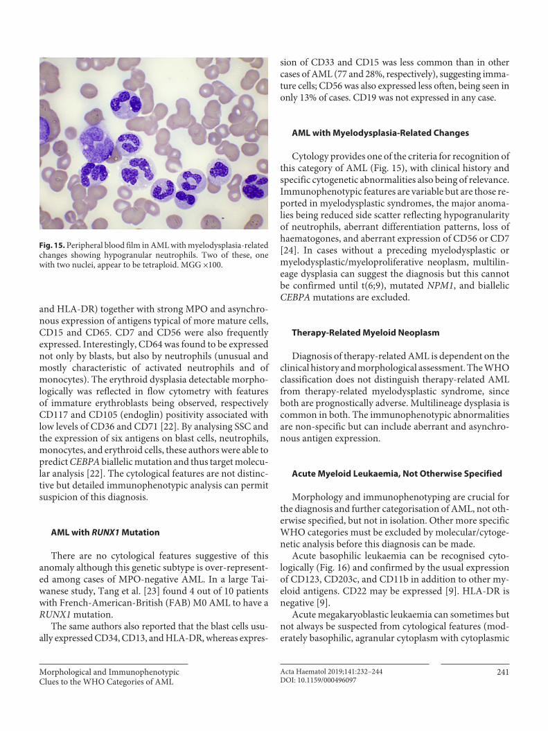
AML with Myelodysplasia-Related Changes
Cytology provides one of the criteria for recognition of this category of AML (Fig. 15), with clinical history and specific cytogenetic abnormalities also being of relevance. Immunophenotypic features are variable but are those re-ported in myelodysplastic syndromes, the major anoma-lies being reduced side scatter reflecting hypogranularity of neutrophils, aberrant differentiation patterns, loss of haematogones, and aberrant expression of CD56 or CD7 [24]. In cases without a preceding myelodysplastic or myelodysplastic/myeloproliferative neoplasm, multilin-eage dysplasia can suggest the diagnosis but this cannot be confirmed until t(6; 9), mutated NPM1, and biallelic CEBPA mutations are excluded.
Therapy-Related Myeloid Neoplasm
Diagnosis of therapy-related AML is dependent on the clinical history and morphological assessment. The WHO classification does not distinguish therapy-related AML from therapy-related myelodysplastic syndrome, since both are prognostically adverse. Multilineage dysplasia is common in both. The immunophenotypic abnormalities are non-specific but can include aberrant and asynchro-nous antigen expression.
Acute Myeloid Leukaemia, Not Otherwise Specified
Morphology and immunophenotyping are crucial for the diagnosis and further categorisation of AML, not oth-erwise specified, but not in isolation. Other more specific WHO categories must be excluded by molecular/cytoge-netic analysis before this diagnosis can be made.
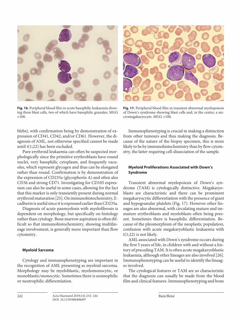
Acute basophilic leukaemia can be recognised cyto-logically (Fig. 16) and confirmed by the usual expression of CD123, CD203c, and CD11b in addition to other my-eloid antigens. CD22 may be expressed [9]. HLA-DR is negative [9].
Acute megakaryoblastic leukaemia can sometimes but not always be suspected from cytological features (mod-erately basophilic, agranular cytoplasm with cytoplasmic
Fig. 15. Peripheral blood film in AML with myelodysplasia-related changes showing hypogranular neutrophils. Two of these, one with two nuclei, appear to be tetraploid. MGG ×100.
Bain/BénéActa Haematol 2019;141:232–244242DOI: 10.1159/000496097
blebs), with confirmation being by demonstration of ex-pression of CD41, CD42, and/or CD61. However, the di-agnosis of AML, not otherwise specified cannot be made until t(1; 22) has been excluded.
Pure erythroid leukaemia can often be suspected mor-phologically since the primitive erythroblasts have round nuclei, very basophilic cytoplasm, and frequently vacu-oles, which represent glycogen and thus can be elongated rather than round. Confirmation is by demonstration of the expression of CD235a (glycophorin A) and often also CD36 and strong CD71. Investigating for CD105 expres-sion can also be useful in some cases, allowing for the fact that this marker is only transiently present during normal erythroid maturation [25]. On immunohistochemistry, E-cadherin is useful since it is expressed earlier than CD235a.
Diagnosis of acute panmyelosis with myelofibrosis is dependent on morphology, but specifically on histology rather than cytology. Bone marrow aspiration is often dif-ficult so that immunohistochemistry, showing multilin-eage involvement, is generally more important than flow cytometry.
Myeloid Sarcoma
Cytology and immunophenotyping are important in the recognition of AML presenting as myeloid sarcoma. Morphology may be myeloblastic, myelomonocytic, or monoblastic/monocytic. Sometimes there is eosinophilic or neutrophilic differentiation.
Immunophenotyping is crucial in making a distinction from other tumours and thus making the diagnosis. Be-cause of the nature of the biopsy specimen, this is more likely to be by immunohistochemistry than by flow cytom-etry, the latter requiring cell-dissociation of the sample.
Myeloid Proliferations Associated with Down’s Syndrome
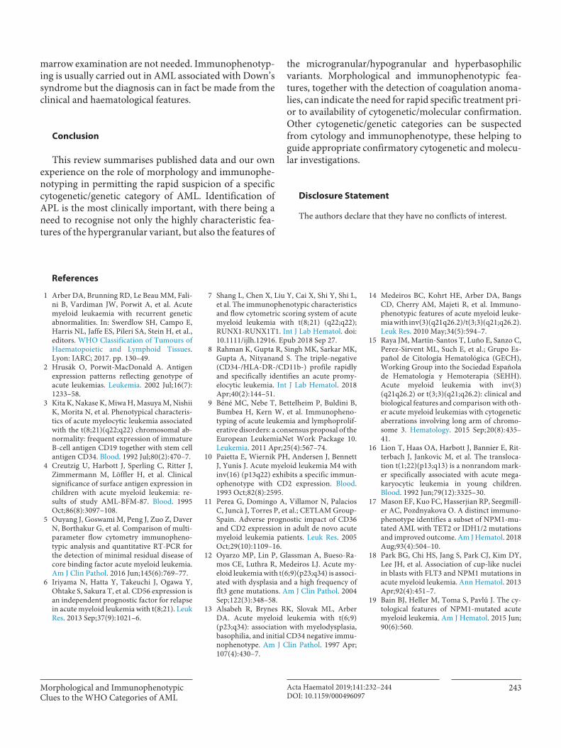
Transient abnormal myelopoiesis of Down’s syn-drome (TAM) is cytologically distinctive. Megakaryo-blasts are characteristic and there can be prominent megakaryocytic differentiation with the presence of giant and hypogranular platelets (Fig. 17). However other lin-eages are also abnormal, with circulating mature and im-mature erythroblasts and myeloblasts often being pres-ent. Sometimes there is basophilic differentiation. Be-cause of the pleomorphism of the neoplastic population, confusion with acute megakaryoblastic leukaemia with t(1; 22) is not likely.
AML associated with Down’s syndrome occurs during the first 5 years of life, in children with and without a his-tory of preceding TAM. It is often acute megakaryoblastic leukaemia, although other lineages are also involved [26]. Immunophenotyping can be useful to identify the lineag-es involved.
The cytological features or TAM are so characteristic that the diagnosis can usually be made from the blood film and clinical features. Immunophenotyping and bone
Fig. 16. Peripheral blood film in acute basophilic leukaemia show-ing three blast cells, two of which have basophilic granules. MGG ×100.
Fig. 17. Peripheral blood film in transient abnormal myelopoiesis of Down’s syndrome showing blast cells and, in the centre, a mi-cromegakaryocyte. MGG ×100.
Morphological and Immunophenotypic Clues to the WHO Categories of AML
243Acta Haematol 2019;141:232–244DOI: 10.1159/000496097
marrow examination are not needed. Immunophenotyp-ing is usually carried out in AML associated with Down’s syndrome but the diagnosis can in fact be made from the clinical and haematological features.
Conclusion
This review summarises published data and our own experience on the role of morphology and immunophe-notyping in permitting the rapid suspicion of a specific cytogenetic/genetic category of AML. Identification of APL is the most clinically important, with there being a need to recognise not only the highly characteristic fea-tures of the hypergranular variant, but also the features of
the microgranular/hypogranular and hyperbasophilic variants. Morphological and immunophenotypic fea-tures, together with the detection of coagulation anoma-lies, can indicate the need for rapid specific treatment pri-or to availability of cytogenetic/molecular confirmation. Other cytogenetic/genetic categories can be suspected from cytology and immunophenotype, these helping to guide appropriate confirmatory cytogenetic and molecu-lar investigations.
Disclosure Statement
The authors declare that they have no conflicts of interest.
References
1 Arber DA, Brunning RD, Le Beau MM, Fali-ni B, Vardiman JW, Porwit A, et al. Acute myeloid leukaemia with recurrent genetic abnormalities. In: Swerdlow SH, Campo E, Harris NL, Jaffe ES, Pileri SA, Stein H, et al., editors. WHO Classification of Tumours of Haematopoietic and Lymphoid Tissues. Lyon: IARC; 2017. pp. 130–49.
2 Hrusák O, Porwit-MacDonald A. Antigen expression patterns reflecting genotype of acute leukemias. Leukemia. 2002 Jul; 16(7):
1233–58. 3 Kita K, Nakase K, Miwa H, Masuya M, Nishii
K, Morita N, et al. Phenotypical characteris-tics of acute myelocytic leukemia associated with the t(8; 21)(q22;q22) chromosomal ab-normality: frequent expression of immature B-cell antigen CD19 together with stem cell antigen CD34. Blood. 1992 Jul; 80(2): 470–7.
4 Creutzig U, Harbott J, Sperling C, Ritter J, Zimmermann M, Löffler H, et al. Clinical significance of surface antigen expression in children with acute myeloid leukemia: re-sults of study AML-BFM-87. Blood. 1995 Oct; 86(8): 3097–108.
5 Ouyang J, Goswami M, Peng J, Zuo Z, Daver N, Borthakur G, et al. Comparison of multi-parameter flow cytometry immunopheno-typic analysis and quantitative RT-PCR for the detection of minimal residual disease of core binding factor acute myeloid leukemia. Am J Clin Pathol. 2016 Jun; 145(6): 769–77.
6 Iriyama N, Hatta Y, Takeuchi J, Ogawa Y, Ohtake S, Sakura T, et al. CD56 expression is an independent prognostic factor for relapse in acute myeloid leukemia with t(8; 21). Leuk Res. 2013 Sep; 37(9): 1021–6.
7 Shang L, Chen X, Liu Y, Cai X, Shi Y, Shi L, et al. The immunophenotypic characteristics and flow cytometric scoring system of acute myeloid leukemia with t(8; 21) (q22;q22); RUNX1-RUNX1T1. Int J Lab Hematol. doi: 10.1111/ijlh.12916. Epub 2018 Sep 27.
8 Rahman K, Gupta R, Singh MK, Sarkar MK, Gupta A, Nityanand S. The triple-negative (CD34-/HLA-DR-/CD11b-) profile rapidly and specifically identifies an acute promy-elocytic leukemia. Int J Lab Hematol. 2018 Apr; 40(2): 144–51.
9 Béné MC, Nebe T, Bettelheim P, Buldini B, Bumbea H, Kern W, et al. Immunopheno-typing of acute leukemia and lymphoprolif-erative disorders: a consensus proposal of the European LeukemiaNet Work Package 10. Leukemia. 2011 Apr; 25(4): 567–74.
10 Paietta E, Wiernik PH, Andersen J, Bennett J, Yunis J. Acute myeloid leukemia M4 with inv(16) (p13q22) exhibits a specific immun-ophenotype with CD2 expression. Blood. 1993 Oct; 82(8): 2595.
11 Perea G, Domingo A, Villamor N, Palacios C, Juncà J, Torres P, et al.; CETLAM Group-Spain. Adverse prognostic impact of CD36 and CD2 expression in adult de novo acute myeloid leukemia patients. Leuk Res. 2005 Oct; 29(10): 1109–16.
12 Oyarzo MP, Lin P, Glassman A, Bueso-Ra-mos CE, Luthra R, Medeiros LJ. Acute my-eloid leukemia with t(6; 9)(p23;q34) is associ-ated with dysplasia and a high frequency of flt3 gene mutations. Am J Clin Pathol. 2004 Sep; 122(3): 348–58.
13 Alsabeh R, Brynes RK, Slovak ML, Arber DA. Acute myeloid leukemia with t(6; 9) (p23;q34): association with myelodysplasia, basophilia, and initial CD34 negative immu-nophenotype. Am J Clin Pathol. 1997 Apr;
107(4): 430–7.
14 Medeiros BC, Kohrt HE, Arber DA, Bangs CD, Cherry AM, Majeti R, et al. Immuno-phenotypic features of acute myeloid leuke-mia with inv(3)(q21q26.2)/t(3; 3)(q21;q26.2). Leuk Res. 2010 May; 34(5): 594–7.
15 Raya JM, Martín-Santos T, Luño E, Sanzo C, Perez-Sirvent ML, Such E, et al.; Grupo Es-pañol de Citología Hematológica (GECH), Working Group into the Sociedad Española de Hematología y Hemoterapia (SEHH). Acute myeloid leukemia with inv(3)(q21q26.2) or t(3; 3)(q21;q26.2): clinical and biological features and comparison with oth-er acute myeloid leukemias with cytogenetic aberrations involving long arm of chromo-some 3. Hematology. 2015 Sep; 20(8): 435–41.
16 Lion T, Haas OA, Harbott J, Bannier E, Rit-terbach J, Jankovic M, et al. The transloca-tion t(1; 22)(p13;q13) is a nonrandom mark-er specifically associated with acute mega-karyocytic leukemia in young children. Blood. 1992 Jun; 79(12): 3325–30.
17 Mason EF, Kuo FC, Hasserjian RP, Seegmill-er AC, Pozdnyakova O. A distinct immuno-phenotype identifies a subset of NPM1-mu-tated AML with TET2 or IDH1/2 mutations and improved outcome. Am J Hematol. 2018 Aug; 93(4): 504–10.
18 Park BG, Chi HS, Jang S, Park CJ, Kim DY, Lee JH, et al. Association of cup-like nuclei in blasts with FLT3 and NPM1 mutations in acute myeloid leukemia. Ann Hematol. 2013 Apr; 92(4): 451–7.
19 Bain BJ, Heller M, Toma S, Pavlů J. The cy-tological features of NPM1-mutated acute myeloid leukemia. Am J Hematol. 2015 Jun;
90(6): 560.
Bain/BénéActa Haematol 2019;141:232–244244DOI: 10.1159/000496097
20 Dalal BI, Mansoor S, Manna M, Pi S, Sauro GD, Hogge DE. Detection of CD34, TdT, CD56, CD2, CD4, and CD14 by flow cytom-etry is associated with NPM1 and FLT3 mu-tation status in cytogenetically normal acute myeloid leukemia. Clin Lymphoma Myelo-ma Leuk. 2012 Aug; 12(4): 274–9.
21 Carluccio P, Mestice A, Pastore D, Delia M, Ricco A, Russo-Rossi A, et al. Immunophe-notypic and molecular features of ‘cuplike’ acute myeloid leukemias. Eur J Haematol. 2014 Feb; 92(2): 121–6.
22 Mannelli F, Ponziani V, Bencini S, Bonetti MI, Benelli M, Cutini I, et al. CEBPA-dou-
ble-mutated acute myeloid leukemia dis-plays a unique phenotypic profile: a reliable screening method and insight into biological features. Haematologica. 2017 Mar; 102(3):
529–40.23 Tang JL, Hou HA, Chen CY, Liu CY, Chou
WC, Tseng MH, et al. AML1/RUNX1 muta-tions in 470 adult patients with de novo acute myeloid leukemia: prognostic implication and interaction with other gene alterations. Blood. 2009 Dec; 114(26): 5352–61.
24 Porwit A, van de Loosdrecht AA, Bettelheim P, Brodersen LE, Burbury K, Cremers E, et al. Revisiting guidelines for integration of flow
cytometry results in the WHO classification of myelodysplastic syndromes-proposal from the International/European Leukemi-aNet Working Group for Flow Cytometry in MDS. Leukemia. 2014 Sep; 28(9): 1793–8.
25 Chakhachiro ZI, Zuo Z, Aladily TN, Kantar-jian HM, Cortes JE, Alayed K, et al. CD105 (endoglin) is highly overexpressed in a sub-set of cases of acute myeloid leukemias. Am J Clin Pathol. 2013 Sep; 140(3): 370–8.
26 Xavier AC, Ge Y, Taub J. Unique clinical and biological features of leukemia in Down syn-drome children. Expert Rev Hematol. 2010 Apr; 3(2): 175–86.
Related Documents