1090 Mini-Reviews in Medicinal Chemistry, 2012, 12, 1090-1106 Morphological and Functional Features of the Sex Steroid-Responsive Posterodorsal Medial Amygdala of Adult Rats A.A. Rasia-Filho* ,1-3 , D. Haas 1 , A.P. de Oliveira 2 , J. de Castilhos 2 , R. Frey 2 , D. Stein 2 , V.M. Lazzari 3 , F. Back 3 , G.N. Pires 3 , E. Pavesi 4 , E.C. Winkelmann-Duarte 4 and M. Giovenardi 2,3 1 Program in Pathology, Federal University of Health Sciences, Porto Alegre, Brazil; 2 Programs in Neuroscience and in Physiology, Federal University of Rio Grande do Sul, Porto Alegre, Brazil; 3 Laboratory of Physiology, Department of Basic Sciences, Federal University of Health Sciences, Porto Alegre, Brazil; 4 Departments of Pharmacology and Morphological Sciences, Federal University of Santa Catarina, Florianópolis, Brazil Abstract: The rat posterodorsal medial amygdala (MePD) expresses receptors for gonadal hormones and integrates sex steroid-sensitive subcortical networks. Male-female differences are found in the morphology, connectivity, and local neuropil structure of MePD. For example, dendritic spine density is sexually-dimorphic and changes with the estrous cycle and following gonadal hormones manipulations. Due to its connectivity, the MePD may affect emotionally-loaded social behaviors, according to a former Newman’s seminal proposition. Unilateral fiber-sparing ibotenic acid damage of the MePD does not impair male sexual behavior. However, microinjecting glutamate and histamine into the right MePD facilitates ejaculation. Further, MePD-lesioned rats are not different from normal rats in anxiety-like behavior as evaluated by the elevated plus maze test or innate fear test induced by a live cat. In another study, an adapted model for inducing aggressive behavior in rats by a brief period of restraint prior to the resident-intruder paradigm was used to study Fos- immunoreactivity in the MePD. Following stressful stimulation (restraint) or the restraint and fight condition, but not after aggression alone, Fos-immunoreactivity was detected in the MePD. Microinjecting the inhibitory neuropeptide somatostatin into the right MePD notably reduces fighting behavior without affecting locomotion. Overall, these data indicate that sex steroids and local neurochemical stimulatory/inhibitory transmitters modulate the MePD and reinforce the idea that this area is a node for modulating social behavior neural networks. Keywords: Anxiety, extended amygdala, Fos immunoreacitivity, ibotenic acid, innate fear, neuronal morphology, sexual dimorphism, sexual behavior. INTRODUCTION Gonadal hormones affect early sexual development, puberty, and various aspects of reproduction in many species [1]. Efforts have been devoted to finding neural areas that are sensitive to circulating levels of sex steroids, modulate gonadotrophic hormones synthesis and release or induce sexual and parental behaviors. The hypothalamic nuclei have been the primary candidates to date due to evidence of their ability to control hypophysial secretions in rodents and humans. Other mammalian brain areas that alter local neuronal or glial morphology and cause changes in physiological activities associated with reproduction in response to gonadal steroid have also been of interest. To this end, the rat amygdaloid nuclei have been investigated considerably in the last decades. Some of these studies will be mentioned here. First, the morphological features of the medial amygdaloid nucleus (MeA) and various differences between adult male and female rats will be reviewed. Then, hodology-based efforts directed at studying social behaviors *Address corresponding to this author at the UFCSPA/Physiology, R. Sarmento Leite 245, Porto Alegre 90170-050 RS, Brazil;Tel: + 55 51 91161643; Fax: + 55 51 33038752; E-mail: [email protected] modulated by the MeA subnuclei will be discussed. Finally, studies of sexual behavior, anxiety-like behavior, innate fear and aggression are reviewed and augmented by the present study in adult male rats. Here it is provided additional behavioral data to previous reports [2, and references therein]. The present work expands upon and confirms the proposition that the MeA subnuclei participate in a subcortical neural network that modulates social behavior. MORPHOLOGICAL FEATURES OF THE MEDIAL AMYGDALA Several lines of evidence indicate that the amygdaloid complex in the rat basal forebrain is neither a morphological nor a functional unit [3], based on a previous Brodal’s proposition. Rather, it is composed of different nuclei and subnuclei that have distinct intra- and extra-amygdaloid connections [4-8]. The MeA is a complex area that is considered to be a component of the “extended amygdala” [see details in 8-10] as opposed to being a ventral continuation of the striatum in rats [11]. In mice, the morphological, electrophysiological, and neurotransmitter properties of neurons in the posterior MeA resemble those of a subset of pyramidal neurons in the piriform cortex [12]. 1 -5 /12 $58.00+.00 © 2012 Bentham Science Publishers

Welcome message from author
This document is posted to help you gain knowledge. Please leave a comment to let me know what you think about it! Share it to your friends and learn new things together.
Transcript

1090 Mini-Reviews in Medicinal Chemistry, 2012, 12, 1090-1106
Morphological and Functional Features of the Sex Steroid-Responsive Posterodorsal Medial Amygdala of Adult Rats
A.A. Rasia-Filho*,1-3, D. Haas1, A.P. de Oliveira2, J. de Castilhos2, R. Frey2, D. Stein2, V.M. Lazzari3, F. Back3, G.N. Pires3, E. Pavesi4, E.C. Winkelmann-Duarte4 and M. Giovenardi2,3
1Program in Pathology, Federal University of Health Sciences, Porto Alegre, Brazil;
2Programs in Neuroscience and in
Physiology, Federal University of Rio Grande do Sul, Porto Alegre, Brazil; 3Laboratory of Physiology, Department of
Basic Sciences, Federal University of Health Sciences, Porto Alegre, Brazil; 4Departments of Pharmacology and
Morphological Sciences, Federal University of Santa Catarina, Florianópolis, Brazil
Abstract: The rat posterodorsal medial amygdala (MePD) expresses receptors for gonadal hormones and integrates sex steroid-sensitive subcortical networks. Male-female differences are found in the morphology, connectivity, and local neuropil structure of MePD. For example, dendritic spine density is sexually-dimorphic and changes with the estrous cycle and following gonadal hormones manipulations. Due to its connectivity, the MePD may affect emotionally-loaded social behaviors, according to a former Newman’s seminal proposition. Unilateral fiber-sparing ibotenic acid damage of the MePD does not impair male sexual behavior. However, microinjecting glutamate and histamine into the right MePD facilitates ejaculation. Further, MePD-lesioned rats are not different from normal rats in anxiety-like behavior as evaluated by the elevated plus maze test or innate fear test induced by a live cat. In another study, an adapted model for inducing aggressive behavior in rats by a brief period of restraint prior to the resident-intruder paradigm was used to study Fos-immunoreactivity in the MePD. Following stressful stimulation (restraint) or the restraint and fight condition, but not after aggression alone, Fos-immunoreactivity was detected in the MePD. Microinjecting the inhibitory neuropeptide somatostatin into the right MePD notably reduces fighting behavior without affecting locomotion. Overall, these data indicate that sex steroids and local neurochemical stimulatory/inhibitory transmitters modulate the MePD and reinforce the idea that this area is a node for modulating social behavior neural networks.
Keywords: Anxiety, extended amygdala, Fos immunoreacitivity, ibotenic acid, innate fear, neuronal morphology, sexual dimorphism, sexual behavior.
INTRODUCTION
Gonadal hormones affect early sexual development, puberty, and various aspects of reproduction in many species [1]. Efforts have been devoted to finding neural areas that are sensitive to circulating levels of sex steroids, modulate gonadotrophic hormones synthesis and release or induce sexual and parental behaviors. The hypothalamic nuclei have been the primary candidates to date due to evidence of their ability to control hypophysial secretions in rodents and humans. Other mammalian brain areas that alter local neuronal or glial morphology and cause changes in physiological activities associated with reproduction in response to gonadal steroid have also been of interest. To this end, the rat amygdaloid nuclei have been investigated considerably in the last decades. Some of these studies will be mentioned here. First, the morphological features of the medial amygdaloid nucleus (MeA) and various differences between adult male and female rats will be reviewed. Then, hodology-based efforts directed at studying social behaviors
*Address corresponding to this author at the UFCSPA/Physiology, R. Sarmento Leite 245, Porto Alegre 90170-050 RS, Brazil;Tel: + 55 51 91161643; Fax: + 55 51 33038752; E-mail: [email protected]
modulated by the MeA subnuclei will be discussed. Finally, studies of sexual behavior, anxiety-like behavior, innate fear and aggression are reviewed and augmented by the present study in adult male rats. Here it is provided additional behavioral data to previous reports [2, and references therein].
The present work expands upon and confirms the proposition that the MeA subnuclei participate in a subcortical neural network that modulates social behavior.
MORPHOLOGICAL FEATURES OF THE MEDIAL AMYGDALA
Several lines of evidence indicate that the amygdaloid complex in the rat basal forebrain is neither a morphological nor a functional unit [3], based on a previous Brodal’s proposition. Rather, it is composed of different nuclei and subnuclei that have distinct intra- and extra-amygdaloid connections [4-8]. The MeA is a complex area that is considered to be a component of the “extended amygdala” [see details in 8-10] as opposed to being a ventral continuation of the striatum in rats [11]. In mice, the morphological, electrophysiological, and neurotransmitter properties of neurons in the posterior MeA resemble those of a subset of pyramidal neurons in the piriform cortex [12].
1875-5607/12 $58.00+.00 © 2012 Bentham Science Publishers

Morphology and Function of the Rat Medial Amygdala Mini-Reviews in Medicinal Chemistry, 2012, Vol. 12, No. 11 1091
Embryological development and molecular properties of the mouse MeA indicate that its cells have multiple origins [13, 14]. As in other components of the extended amygdala, the MeA subnuclei are composed of cell clusters that migrate from a caudoventral pallidal subdivision, the ventral pallium, the commissural peptic area, and the supraoptopara-ventricular domain of the hypothalamus [14]. These data do not support a striatal origin of the mice MeA [13].
Histological, connectivity, neurochemical, and functional studies have divided the rat MeA into the anterodorsal (MeAD), anteroventral, posterodorsal (MePD), and posteroventral (MePV) subnuclei [8, 9, 15-18]; Fig.(1). These subnuclei are reciprocally interconnected in the same hemisphere [15]. Using Nissl staining, the MePD can be further divided in three parallel columns of aggregated cells that extend from the medial to the lateral border of this subnucleus. They are adjacent to the optic tract (OT) and ventral to the stria terminalis (ST) [8]. Moreover, efferent fibers from the accessory olfactory bulb form a cell-sparse rim that surrounds the medial part of the MePD close to the OT [8, 19]. This rim was formerly called the ventromedial “molecular” layer [20]. Among other functions, the MeA subnuclei are important for the interpretation of the social relevance of both olfactory and vomeronasal stimuli [21-25]; the response to genitosensorial stimulation, modulation of sexual behavior in males and females, and sexual satiety [9, 26-31]; maternal behavior or avoidance of the offspring [32, 33]; anxiety and conditioned or innate fear [34, 35]; and, emotional processing or neuroendocrine responses to stressful stimuli [36-39]. The finding that various social behaviors can be modulated by the same MeA subnuclei suggests that local cells integrate different demands from specific pathways. Each pathway provides inputs that are then temporally and spatially integrated within neural networks to trigger the most appropriate action according to the animal’s context [2, 9, 40-42].
Notably, the MePD neurons and glial cells are affected by normal levels of gonadal steroids in males and in females (e.g., along the estrous cycle) and experimental manipulations such as castration and hormone replacement therapies [40, 43-47]. ]. It is not clear whether hormonal effects act directly through locally mediated actions or are caused indirectly by synaptic changes. Indeed, these two possibilities are not mutually exclusive. In support of the local-mediation hypothesis, MePD neurons have a high concentration of gonadal hormone receptors, resembling the levels found in the hypothalamic nuclei involved in reproduction [48]. Most neurons expressing androgen receptors in the MeA are found within 500 m of the rostral-caudal plane [49, 50]. The MePD is located within this area. In male hamsters, the number of androgen receptor-immunoreactive cells increases in the MePD after exposure to chemosensory stimuli [51]. Fos-immuno reactivity (Fos-ir), suggestive of neuronal activation, completely overlaps androgen receptor-immunoreactivity in MePD neurons of male rats that mated to ejaculation [52]. Otherwise, only 18% of androgen receptor immunofluorescent neurons in the MePD coexpress Fos after mating [52]. Aromatase, the enzyme that converts testosterone into estradiol, is found in the anterior and posterior areas of the MeA [53, 54]. Its
activity in the MePD was higher in males than in females [53, 54]. The MePD also has high concentrations of both estrogen receptors and (ER- , ER- ) [48, 50, 55-57] and progesterone receptors [58]. These receptors show a complex local organization and dynamics during the estrous cycle and after ovarian steroids withdrawal [56, 59, 60]. For example, some MePD cells have both progesterone receptors and ER-
[56]. Additionally, replacement treatment with estradiol decreases the number of cells expressing ER- and those co expressing ER- /ER- in the MePD of adult ovariectomized females [56]. The distributions of ER- and ER- are also different in the dorsal and ventral MePD of female rats, though some co-localization is observed [56, 57]. In castrated and hormone-primed female rats, cells expressing ER- or both ER- /ER- in the dorsal MePD and cells expressing ER- in the ventral MePD show positive Fos-ir following mating [57]. Male rats also express ER- in the MePD, and increased receptor density is associated with a single ejaculation or sexual satiety [61].
Therefore, it is not surprising that plasma gonadal steroids can affect MePD cells in both sexes. As reviewed recently [2], male-female differences in the rat posterior MeA and the MePD specifically include the following: MePD volume [62], neuron and glial cells number [44, 46], neuronal somatic volumes [63], spatial orientation of the dendritic branches and dendritic length [17, 64], dendritic spine density [40, 65], synaptic connectivity and frequency of excitatory post-synaptic currents [20, 66, 67], and the concentration of neuropeptides and neurotransmitter binding sites [58, 68-76]. Investigations of astrocyte density and glial fibrillary acidic protein immunoreactivity have suggested comparable interpretations [44, 45, 77]. These findings further confirm the idea that interactions between neurons and glia in the MePD can be dynamically controlled by sex steroids and can affect local physiology [44, 45, 77].
Golgi studies of male and female adult rats show that the MePD is composed of bitufted (not “bipolar”, as classically referred to by Ramón y Cajal) [commented in 77], and stellate multipolar neurons. Representative images of these neurons can be found elsewhere [17, 18, 37, 78, 79]. There is no clear evidence for the presence of MePD medium spiny stellate neurons, similar to those found in the striatum, in adult Wistar rats [37]. Bitufted neurons are characterized by two dendritic shafts that arise from a fusiform or round soma. Stellate neurons have three or more primary dendrites, and the shape of the cell body is determined by the number and initial width of the dendrites [78]. The dendritic trees are rectilinear or sinuous, have few branch points, radiate in all directions, show preferred spatial localizations within the MePD, and extend over a wide range of path lengths [4, 17, 78, and 79].
The rat MePD assembles coexisting subpopulations of neurons [14, 28]. No single morphological characteristic is used to reliably identify these neurons [17, 18, 37, 78-85 Though gonadal hormone receptors are not present in all cells in the neural regions, the classic Golgi method cannot distinguish neurons that concentrate sex steroids from those that do not [78, 81]. However, confocal immunofluorescent microscopy has revealed that transcription factor Lhx6-immunoreactive cells constitute approximately 80% of all

1092 Mini-Reviews in Medicinal Chemistry, 2012, Vol. 12, No. 11 Rasia-Filho et al.
neurons in the MePD [82]. These neurons are connected to specific hypothalamic nuclei and appear to modulate reproduction [82]. This homogeneity among MePD neurons might explain the low variability in soma size, soma volume, and dendritic spine density density described previously [30, 37, 65, 78, 83, 84].
Recently, Carney et al. [85] classified 3 MePD neurons subtypes based on developmental histogenetic and whole- cell patch clamp recording characteristics in mice. Specifically, MePD neurons could be classified according to
the spatial and temporal expression patterns of morphogenic Sonic hedgehog-responsive genes (Nkx2-1, which is linked to Lhx6, and Gli1) coming from the embryonic medial ganglionic eminence and preoptic area progenitor domains. These expression patterns generate diversity among inhibitory neurons in the MePD, and these genes co-express with the inhibitory markers calbindin, neuronal nitric oxide synthase (nNOS, a marker of MeA projection neurons), Forkhead box transcription factor (FoxP2), and somatostatin (found in interneurons). Accordingly, the absence of Nkx2-1 caused a marked reduction in MePD GAD67 (a GABA
Fig. (1). Schematic drawings of coronal sections of the adult rat brain showing the four medial amygdala subnuclei: anterodorsal (boundaries in yellow), anteroventral (green), posterodorsal (blue), and posteroventral (red). References are in mm posterior to the bregma. Spatial coordinates: D, dorsal; L, lateral; M, medial; V, ventral. Adapted from the atlas of Paxinos and Watson [200]. Courtesy: A. Dall’Oglio and E. Quagliotto [201].
�������������
��������� ����
��������������
��������������
�������� ������
�������� � ����
�
�
��

Morphology and Function of the Rat Medial Amygdala Mini-Reviews in Medicinal Chemistry, 2012, Vol. 12, No. 11 1093
synthesizing enzyme) mRNA expression. However, mutant mice retained cells in the MePD expressing Lhx6 and somatostatin mRNAs in contrast to other telencephalic areas. The 3 subpopulations of mouse MePD neurons, sampled at approximately the same rate, were then sorted by their electrophysiological and immunohistochemical profiles. Type I and II, but not type III, neurons were immunoreactive for nNOS and FoxP2. These data and our own studies suggest that type I and III cells are similar to bitufted neurons, whereas type II cells resemble stellate neurons [see Figure 8 in 85 and compare to those in 17 and 83]. Altogether these morphological and physiological features may underlie the differences in the connectivity and functions of MePD local and output neurons [85].
Gonadal hormone influences on dendritic spines of MePD neurons have also been of experimental interest. The study of spines has provided new insights on the modulation of synaptic organization in the adult rat MePD. Neuronal changes can reflect functional adaptations to an animal’s external and internal environment and the dendritic spines can be a cellular substrate for this plasticity [2, 37]. Proximal spines of Golgi- impregnated neurons are strategically located to affect cell body voltage, neuronal function and outputs in a network [30, 37, 40, 65] and are thus excellent candidates for study. Nevertheless, spines in other locations along the dendrites have shown significant modifications as well [67, 81, 86]. Descriptive studies of this highly specialized cellular structure have shown connectional and possible functional correlations that are needed to co-ordinate the expression of complex behaviors and and timing of physiological events [40, 58, 87]. In fact, dendritic spines spacing and shape can amplify membrane potentials, associate postsynaptic potentials with other spines and adjacent dendritic shafts, activate intracellular biochemical cascades, and induce and prolong long-term changes in neuronal excitability [88-92]. Increase in spine number enhances the membrane surface area on dendrites that is available to establish new contacts [92, 93; see relevant comments in 94 and 95]. Moreover, the selectivity and functional properties of local receptors and ionic channels modulate electric potential generation [92, 96] and connection formations [97].
Pleomorphic spines of a continuum of different shapes and sizes are found in the MePD in isolation or in small clusters [2, 84]. Spines are classified morphologically as being thin, mushroom-like, stubby/wide, ramified, and other transitional aspects [84, 93, 99, see further detals in 94]. In the MePD of adult Wistar males, thin spines are the most abundant (approximately 53%), followed by mushroom-like (22%) and stubby/wide ones (21%) [84]. There is no difference in the density of proximal spines between bitufted and stellate neurons [30], percentage of the different dendritic spines in these cells [84] or spine density of the medial and lateral aspects of the MePD [30].
Moreover, there is no difference in dendritic spine density of the dorsal or ventral MePD in proestrus females, despite local heterogeneity in the expression and distribution of estrogen receptors in these areas [83]. In addition, no hemispheric difference exists in the proximal dendritic spine density in the MePD of adult Wistar males or diestrus
females [83]. This result is interesting because there are more dendritic shafts, greater dendritic branching [100], and more asymmetric excitatory synapses on dendritic spines in left MePD neurons of male than female prepubertal Sprague Dawley rats [67]. Though strain differences might exist, puberty affect dendritic spines in the rat MePD in both hemispheres. Indeed, this life event has marked influence on MePD morphology and can account for differences between young and adult animals [64, 101, 102].
Proximal dendritic spine density in the MePD was found to be sexually dimorphic (higher in males than in females in proestrus, estrus or metaestrus) [40]. It is affected by normal ovarian steroid hormone variation along the estrous cycle (spine density is reduced by approximately 35% during the transition from diestrus to proestrus) and by motherhood (spine density is reduced by 24% in postpartum diestrus rats compared to age-matched virgin diestrus females) [40]. Ninety days following orchidectomy, adult males exhibit reduced dendritic spine density and a smaller basal population of stable spines remaining in the MePD [65]. This finding reinforces the proposition that androgens affect the MePD neuropil and local synaptic organization. It is also in accordance with studies showing the reduction in substance P immunoreactivity and MePD volume [76], and dendritic atrophy after castration [81]. In adult ovariectomized females, replacement estradiol and progesterone therapy increases spine number to supra-physiological levels compared to normal proestrus phase [40, 65]. This higher spine density indicates that the influence of ovarian sex steroids on spine density under different experimental condition is modulated by complex regulatory mechanisms [65; see a parallel comment in 45].
Further methodological studies of the MePD have been performed by applying carbocyanine dye DiI extracelularly to visualize dendritic spines associated with immunolabeling for pre-synaptic protein synaptophysin under confocal microscopy [84]. Most spines showed synaptophysin puncta close to their head and neck, but some spines were not labeled or were characterized by multiple puncta [84]. Although ultrastructural data suggest that dendritic spines usually receive one asymmetrical synapse with round electron-lucent and dense core vesicles [103], inhibitory and multisynaptic contacts have also been found on the dendritic spines of adult male MePD [2].
The existence of non-synaptic spines or multisynaptic dendritic spines making contacts with two or more axonal terminals remains controversial. Studies are also needed to establish the proportion of spines that are genetically and functionally developed to be stable and those spines that developed to be labile and plastic in the MePD. In this sense, it has been hypothesized that stable spines have steady properties in neural networks, whereas unstable spines have intrinsic plastic capacities for synaptic processing and behavior modulation [2, 84].
Dendritic shafts and spines may receive inputs from circuits that are organized differently in male and female rats. More dendritic branches are oriented medially in the MePD of males compared with those of diestrus females [17]. This spatial orientation suggests that males gather part

1094 Mini-Reviews in Medicinal Chemistry, 2012, Vol. 12, No. 11 Rasia-Filho et al.
of the synaptic inputs from the superficial “molecular layer” through which vomeronasal information passes [8, 19, 23]. Males also have more dendritic shafts and spine synapses on neurons in the medial part of this “molecular layer” [66].
On the other hand, morphological (dendritic spine density modulation) and biochemical changes (e.g., through expression of different neuropeptides) suggest that the female MePD may alter the transmission of information through interconnected circuitry along the estrous cycle [40, 70, 87]. For example, fewer dendritic spines are found in the MePD of proestrus and estrus females [40]. This reduction is coincident with a decrease in synapsin reactivity, which could be associated with synaptic pruning [104]. It has also been hypothesized that labile dendritic spines in the MePD that change their number along the estrous cycle are in position to modulate phasic synaptic inputs, whereas dendritic shafts may receive more stable inputs [2]. It would be interesting to determine whether synaptic inputs to both dendritic spines and shafts are neurochemically distinct and/or come from different subpopulations of neurons of the same or connected areas.
FUNCTIONAL FEATURES OF THE MEDIAL AMY-
GDALA
Cellular morphology and function are interrelated and inextricable. The morphological findings in the rat MePD should thus have functional causes and/or consequences. For example, testes removal that causes decreases in the rat MePD spine density after 90 days also strikingly reduces ejaculatory and intromission behaviors [26, 30, 105]. Although the pieces of this intriguing mosaic are still being put together, the MeA subnuclei are in a strategic condition for gathering chemosensory and hormonal information and modulating behaviors in conjunction with other brain areas in male and female rats [9, 19, 20, 33, 106, 107]. The MeA subnuclei also form associated circuits to regulate various aspects of the social displays [5, 7, 9, 28].
As depicted recently [2], “the male MeAD receives inputs from the accessory olfactory bulb and evaluates the social relevance of vomeronasal stimuli, altering the inhibitory circuit that involves the intercalated amygdaloid nuclei and the MePD [22]. The MeAD also receives fibers indirectly from the main olfactory bulb via the anterior and posterolateral cortical nucleus, and from the infralimbic cortex, which can integrate olfactory information with non- olfactory inputs [5]. Several hypothalamic nuclei are innervated by both the MeAD and the MePV [15]. Hypothalamic areas that are more heavily innervated by the MeAD include portions of the medial preoptic area (MPOA), the lateral part of the medial preoptic nucleus, the lateral hypothalamic area adjacent to the cerebral peduncle, the lateral regions of the arcuate nucleus, the posterior hypothalamic periventricular nucleus, and the paraventricular nucleus [see additional data in 7, 15, 108]. Several lines of evidence indicate that the MeAD is part of the general behavioral arousal system and for nonspecific activation of social behaviors in rodents [9].
The MePV is one of the brain areas that integrate gonadal hormone actions with pelvic viscero-sensorial information
[29, 109]. Directly or indirectly, MePV axons reach 1) the periventricular zone in the anterior hypothalamic level, forming a ‘neuroendocrine motor zone’; 2) the subparaventricular zone of the periventricular region, which contains a ‘visceromotor pattern generator network’ most likely involved in circadian rhythms; and 3) the ventromedial hypothalamic nucleus (VMH), which regulates reproductive and defensive hypothalamic circuits [7, 15, 82, 108]… Different subpopulations of MePV neurons related to defensive and reproductive stimuli have been identified [82].
In males, the MePD projects to the hypothalamic anteroventral periventricular nucleus (AVPV), the MPOA, and the ventral premamillary nucleus (PMv) [7, 15, 110]. These hypothalamic nuclei are interconnected with the neuroendocrine, sympathetic and parasympathetic, and sexually dimorphic areas of the medial and periventricular zones of the hypothalamus [7, 15]. Furthermore, the male MePD makes indirect connections with the hypothalamus via GABAergic efferents from the bed nucleus of the ST [108, 111]. Synaptic codification of MePD activity to these hypothalamic areas can ultimately elicit a male sexual response [112]…Male MePD activation during mating most likely desinhibits brain areas involved in sexual behavior…
In the male MePD, there are clusters of neurons involved in either intromission or ejaculation [27, 28]… The MeA subnuclei of adult male mice is essential for female recognition through memory acquisition mechanisms after social contacts [74]. Projections to the entorhinal area and to the postpiriform transitional area allow pheromonal influences to act on hippocampal circuitry [7]”. Recent findings have updated and expanded upon the knowledge of this scenario [see, for example, 113-119].
The MeA subnuclei modulate male copulatory behavior by matching sexual experiences with context processing of encounters with females [120, 121]. Sex steroids can also remarkably alter the function of the MePD cells in rodents. For example, unilateral implantation of estradiol in this subnucleus restores mounting behavior in adult castrated male rats [26]. The same effect can be achieved following implantation of testosterone or estradiol, but not dihydrotestosterone, in the posterior MeA of adult castrated hamsters [122]. Copulatory activity also increases after unilateral and bilateral implants of testosterone in the MePD of adult castrated hamsters [123]. These results provide evidence for redundant, but not additive, mechanisms of androgenic effects on this area [123].
In females, the MePD is part of neural circuitry that is responsive to variations in circulating gonadal hormone levels. Estrogens reduce axonal excitability of MeA afferents during the proestrus phase in rats [124]. At this same time, dendritic spine density decreases in the MePD [40]. The female MePD also modulates hypothalamic neuroendocrine secretion and ovulation [85, 125, 126]. This modulation involves processing the pheromonal stimuli relevant for gonadotrophin releasing hormone (GnRH) secretion by the PMv [110] and prolactin release by the hypothalamic arcuate nucleus [127]. As reviewed previously [2], “some of these output connections are inhibitory [3, 82], but their functional

Morphology and Function of the Rat Medial Amygdala Mini-Reviews in Medicinal Chemistry, 2012, Vol. 12, No. 11 1095
consequence are heterogeneous. Indeed, MPOA neurons of proestrus females respond either with suppression or facilitation of firing activity following electrical stimulation of the MeA [128]. Local interneurons are also involved in the estrogen-mediated increase in MPOA GABAergic activity [see comments in 111]. Considering that excitatory afferences to dendritic spines decrease in proestrus and that female MePD neurons output can be inhibitory and can act on hypothalamic nuclei, two hypotheses can be made. 1) During the proestrus phase, MePD neurons should be activated less frequently, which lowers inhibition on hypothalamic neurons. The resulting effect would be a greater activation of the hypothalamic nuclei to induce the neuroendocrine secretion and sexual behavior typical of this estrous phase. For example, the MePD connects the VMH [7], a hypothalamic nucleus that is highly involved with lordosis behavior [1]. Additionally, electrical stimulation of the MePD induces GnRH secretion [125, 126] via connections with the AVPV [87]. 2) Decreased inhibitory influence of the MePD on GABAergic interneurons should activate MPOA neurons. The female MPOA is therefore able to inhibit lordosis but facilitate pacing and other proceptive female behaviors [29]. It is possible that some excitatory efferent fibers from the MePD [129] decrease their activity to reduce stimulation on local MPOA GABAergic cells. Both hypotheses fit well with early findings showing that MeA projections can form both facilitatory and inhibitory multi-pathway influences to the MPOA [128]”.
Altogether, these data support the seminal proposition that the interconnected, sex-steroid sensitive subcortical network of the MeA subnuclei play different roles that collectively regulates various social behaviors, as proposed by Neman [9]. Sexual behavior was first to be investigated and used a reference for other social displays. As stated [9], “…borrowing an important concept from our colleagues in cognitive neuroscience, we envision that a particular social behavior, for example, male sexual behavior, is an emergent property of the pattern of activity across the network…. It is not an action produced by the “on” or “off” state of any one of the nodes, such as the medial preoptic area, but a sequence of multiple behaviors (e.g., sniffing, mounting, ejaculation, grooming) that is initiated by and emerges from a temporal pattern, and therefore a dynamic pattern, of activity across the network. Initiation and maintenance of male sexual behavior… would require activation of the MPOA but in conjunction with particular levels of activation of other areas in the network, and in the context of a unique temporal pattern of activation across the whole network. Other similar but distinguishable patterns of activity in this same circuit…, arising as a result of changing sensory stimuli or fluctuations in the hormonal milieu, could result in a progression of behaviors blowing seamlessly from one to another, for example, territorial marking and aggressive activities interspersed with copulatory acts to produce the full spectrum of mating behavior observed in a variety of species... As a result of steroid action, the network becomes sexually dimorphic with respect to the number of cells and the specific cell types produced in each node. This, in turn, influences the baseline number and types of sex-steroid receptors produced by those cells and the strength of
connections between nodes in the network. It is these factors that ultimately regulate the predisposition, but not routinely predictable or exclusive function, of the network to produce particular timing patterns of activity in a given individual, such as male versus female sexual behavior… These same factors, sex-steroid sensitivity and neuronal connections, are of course dynamically modulated throughout life by sexual maturation, by experience or learning, by reproductive cycles and diurnal cycles, and by disease and aging…. At the very least, these stimuli produce immediate changes in synaptic activity in the nodes of the social behavior network. In some cases the effects are long-lasting changes in the strength of synaptic connections… We will have to demonstrate mini- circuits within this network, each one independently regulating a specific aspect of a particular behavior.”
Additional experiments have tested the involvement of the MePD in hierarquical and parallel circuits for neural processing in different social behaviors [9, 30, 82, 121]. Sexual behavior, anxiety-like behavior, innate fear, and aggression were studied using different approaches, including direct neurotoxic or neurotransmitters microinjections in the MePD of adult Wistar rats. Here, a note of caution is warranted. Present animal models and tests most likely provide partial data about the behavior under study. For example, different tests of anxiety-like behaviors in rodents and different methodological approaches show distinct predictive validity and non-similar effects of anxiogenic or anxiolytic pharmacological compounds [130-132]. Moreover, our first experimental approach has been applied to test the behavior of adult males to avoid confounding variations due to cyclical changes in circulating sex hormones [40, 65, 133]. However, this approach does not preclude the role of the MePD in females [32, 33, 40, 134, 135].
Experiments of male sexual behavior have identified possible correlations between brain steroid-sensitive regions and their functional organization. They have provided contrasts with previous studies employing electrolytic damage to local cells and ST-bound travelling fibers of the male posterior MeA [see a parallel discussion in 136]. These lesions lead to impaired ejaculatory capacity in rats [137]. Interestingly, unilateral right MePD microinjection of 8-OH-DPAT, a 5-HT1A receptor agonist, decreases latencies to intromission and ejaculation and shortens the postejaculatory refractory period in rats [30]. This facilitatory effect is consistent with results obtained after systemic injections of this drug and the proposed function of the MePD [30, 105]. The rat MePD is also involved in penile erection and sexual arousal [138, and relevant references therein]. Further, synaptic plasticity of the MeA subnuclei may be related to sexual behavior in male rats [121]. For example, copulatory performance impairment after extensive lesions of the MeA is distinct in sexually inexperienced or sexually experienced males [139, 140]. Stimulations of both the MePD and MePV facilitate male sexual behavior in the presence of a non-estrus female but suppress copulatory activity in the presence of a receptive female [141]. As previously mentioned, these findings suggest that MeA modulation of masculine sexual behavior involves contextual processing of sexual encounters

1096 Mini-Reviews in Medicinal Chemistry, 2012, Vol. 12, No. 11 Rasia-Filho et al.
and individual memories. The MePD is thus a part of a dynamic neural network that controls ongoing and adaptive needs of the animal at each moment [2, 9].
MALE SEXUAL BEHAVIOR
The effects of fiber-sparing lesions caused by 1% ibotenic acid solution (3 g/0.3 l) microinjected in the right MePD of sexually experienced Wistar male rats were tested. The following parameters were observed: latency and frequency of anogenital investigation of the female, mounting without pelvic thrusting, mounting with pelvic thrusting, intromission, time for the first ejaculation, and the duration of the first postejaculatory refractory period. The latency (in seconds) and frequency of these parameters were recorded in sessions that lasted: (a) for 30 min if the male showed at least one intromission; (b) until the end of the postejaculatory interval when the first ejaculation occurred during the 30-min test; or, (c) for 15 min if no intromission occurred, as described elsewhere [30, 105].
Data were tested using a within group comparison of sexual behavior of each rat before and after stereotaxic surgery and a between groups comparison of control (saline) and ibotenic acid-treated animals one week after microinjection. The within-group comparisons controls for many sources of data variability, which is advantageous for studies with small sample sizes [30, 142].
Neurotoxic damage promoted enlargement of the extracellular space, neuronal loss and glial reactivity involving a considerable part of the MePD. However, only a
slight difference between groups was found in the sexual behavior studied. Specifically, a reduction in the latency for mounting with thrusting in the MePD-lesioned rats was identified, a statistical finding that could have been caused by variability in the saline group. Other behavioral differences were considered without biological relevance for the neurotoxic data interpretation (Table 1). This result indicates that the MePD of one hemisphere is not crucial for complete sexual behavior as long as the other hemisphere is intact, which confirms previously described functional redundancy of this area [123]. On the other hand, chemical stimulation of the MePD of one hemisphere promotes a significant change in sexual activity [9, 26, 30, 123]. To further evaluate this hypothesis, the right MePD was injected with effective doses of glutamate (2 g/0.3 l), GABA (100
g/0.3 l), and histamine (30 g/0.3 l). These doses were used in a preliminary manner to query possible effects of neurotransmitters in this subnucleus.
Although the precise physiological extracellular concentrations of these neurotransmitters in the rat MePD are not currently known, these doses were chosen based on previous studies [143-151]. Notably, glutamate accounts for almost 70% of all ejaculation-activated cells and GABAergic neurons account for half of the activated cells following mating in the gerbil MePD [152]. The MeA subnuclei have high levels of the histamine synthesizing enzyme histidine decarboxylase [153]. Histamine may also be important for modulating sexual behavior in rats [154]. Moreover, as a component of neural circuits involving hypothalamic nuclei that modulate sympathetic and parasympathetic responses
Table 1. Male Sexual Behavior Following Posterodorsal Medial Amygdala Neurotoxic Lesion
Saline Ibotenic Acid
Latency
Mounting without thrusting
12 (0/155.5)
10 (3/112)
5.5 (0/50.2) (control)
9.5 (5/623.7) (test)
Mounting with thrusting 207 (11.5/874)
251 (110/525)
9 (3.7/158)
6.5 (4/150.2) a
Intromission 43 (16.5/58.5)
18 (13/21) c
11.5 (9/14.5) b
9.5 (8.2/92)
Ejaculation 694 (516/1288)
774 (393.5/1577)
873.5 (727/1174.5)
769.5 (491.5/1169.2)
Postejaculatory refractory period 298 (287/344)
290 (243/395)
328.5 (254.5/348.2)
278 (268/333.7)
Frequency
Mounting without thrusting
3 (0/6.5)
4 (1/8.5)
3 (0/6)
4.5 (1.7/12)
Mounting with thrusting 3 (2.5/6.5)
3 (2/9.5)
12 (7.7/13.7)d
6.5 (4.2/13.7)
Intromission 12 (10/13.5)
12 (8.5/14)
14 (9.7/17.2)
11 (6.7/13.5)
Median and interquartile ranges for the latencies, postejaculatory refractory period (in seconds) and the frequencies of sexual behavior parameters obtained before stereotaxic surgery (“control”, data in the first row in each behavior studied) and after microinjection (“test”, data in the second row in each behavior studied) of saline (0.3 l, n = 5) or 1% ibotenic acid (3 g/0.3 l, n = 6) in the posterodorsal medial amygdala of adult male rats. a: Mann-Whitney test, P < 0.03 when compared to the saline “test” data; b: Mann-Whitney test, P < 0.01 when compared to the saline “control” data; c: Wilcoxon test, P < 0.05 when compared to control (pre-injection) data in the same group; d: Mann-Whitney test, P < 0.05 when compared to the saline “control” data.

Morphology and Function of the Rat Medial Amygdala Mini-Reviews in Medicinal Chemistry, 2012, Vol. 12, No. 11 1097
[7], the MePD can have a complementary role in integrating concomitant, beat-to-beat, cardiovascular adjustments associated with behavior [151]. That is, cardiovascular support for behaviors required for the survival of the animal and ultimately the species is critical [41]. Histamine may serve parallel functions in the MePD because it can broaden the ranges of baroreceptor- and chemoreceptor-reflexes and adjust cardiovascular homeostatic responses for the proper execution of an intended behavior [41, 151].
To be succinct and for the ease of comparison, only clear relevant behavioral modifications of the study will be mentioned here. The within-groups comparisons showed that (1) saline does not promote any reliable modification in male sexual behavior, except for a change in the latency of the first mount that does not affect the interpretation of other data; (2) glutamate significantly reduces the latency for ejaculation; (3) GABA decreases the latencies for mounting with thrusting and to the first intromission; and (4) histamine also decreases the latency for ejaculation (Table 2). That is, both glutamate and histamine induce ejaculation, an indication of male sexual behavior stimulation. GABA also facilitates sexual behavior by reducing the latency of copulatory activity. These GABAergic effects may be due to local desinhibition. Clearly, additional experiments are necessary to characterize GABA-induced responses at physiological and pharmacological doses in the male rat MePD. The study of 5-HT1A receptor stimulation [30] and glutamate and histamine microinjections suggested that the MePD is involved in the modulation of ejaculatory behavior, which relevance is vital for species perpetuation. This proposition is consistent with Fos-ir in the rat MePD after ejaculation [28] and with previous results of impaired ejaculatory capacity in rats with electrolytic lesions of the posterior MeA [137]. However, the MePD appears to play a modulatory rather than crucial role in the sequence of sexual behavior because neurotoxic or electrolytic damage does not block sexual behavior completely. These results further support the idea of hemispheric specializations and redundancy of brain circuitry.
ANXIETY-LIKE BEHAVIOR AND INNATE FEAR
Other socially related behavioral displays are also modulated by the MePD. Anxiety-like behaviors and innate
fear have also been studied after neurotoxic damage of the right MePD of adult male Wistar rats. Kindling of the medial/basolateral amygdala in the left hemisphere decreases anxiety for at least 1 week after the last seizure whereas the same procedure in the right hemisphere increases anxiety [34]. Kindling in more anterior loci in the amygdala are associated with greater anxiety and the opposite is found with kindling in more posterior amygdaloid regions [34]. To complement these data, c-fos activity is found in the MePD of male rats subjected to the elevated plus maze [155], which is an ethologically and pharmacologically valid test of anxiety in rats [156-158]. The following parameters have been evaluated using this apparatus [159]: percentage of open arms entries and time spent in the open arms, total frequency of open and enclosed arms entries, and risk assessment behaviors (“scanning” the space outside of the closed arms and around the apparatus, which includes head “dipping”). After ibotenic acid microinjection in the MePD, no statistically significant changes were found in the parameters related to anxiety (e.g., percentages of entries in the open arms and time spent in these arms), frequency of risk assessment behaviors or locomotion (total number of arm entries) exhibited by lesioned rats compared with control rats in the maze (Table 3).
Moreover, innate fear was tested in males exposed for 15 min to a live, non-sedated cat placed in an experimental cage separated by a glass wall that allowed predator odor perception, in accordance with previously described method [160, 161]. Control and MePD ibotenic acid-lesioned rats were observed for the following behaviors: duration and frequency of “freezing”, locomotion, rearing, and sniffing of the cat compartment. No statistically significant change was found in any of the studied parameters when comparing the lesioned and control data (Table 4). These results are in agreement with the lack of Fos-ir in the MePD of rats exposed to a caged cat [160]. Plastic synaptic changes are found in the MeAD [162], and Fos-ir is clearly increased in the MePV of cat odor-exposed rats [21]. Other results indicate that neurotoxic lesions of the whole MeA reduce freezing in response to cat odor presentation or cat odor context tests, but not after cat exposure [163]. Considering that higher amounts of the predator odor induce unconditioned fear behavior, particularly freezing, it is of
Table 2. Male Sexual Behavior Following Posterodorsal Medial Amygdala Microinjection of Different Neurotransmitters
Saline Glutamate GABA Histamine
Latency
Mount with thrusting
9.5 (3/15)
13 (10/50.2) a
11 (7.5/139.5)
9 (6.5/117)
18 (11/195)
8 (5/13) b
13.5 (7/120.7) (control)
12.5 (7/19.7) (test)
Intromission 16.5 (4.7/36.2)
21.5 (12.5/117.7)
12 (8.5/19.5)
9 (8/18.5)
19 (14/134)
14 (13/15) c
17 (12/199)
16.5 (10.2/56.5)
Ejaculation 969.5 (632.5/993.2)
745.5 (386.2/1240.5)
953 (609/1356)
316 (202/734.5) d
973 (474/1113)
581 (280/992)
669.5 (490.2/917.2)
339.5 (143.7/560.2) e
Median and interquartile ranges of the latencies (in seconds) of the main sexual behavior parameters obtained before stereotaxic surgery (“control”, data in the first row in each behavior studied) and that were affected by the microinjection (“test”, data in the second row in each behavior studied) of saline to serve as control (0.3 l, n = 6), glutamate (2 μg/0.3 μl, n = 5), GABA (100 μg/0.3 μl, n = 7) or histamine (30 μg/0.3 μl, n = 8). a: Wilcoxon test, P < 0.03 when compared to “control” data in the same group; b: Wilcoxon test, P < 0.03; and, c: P < 0.05 when compared to “control” data in the same group; d: Wilcoxon test, P < 0.05; and, e: P < 0.02 when compared to “control” data in the same group.
1098 Mini-Reviews in Medicinal Chemistry, 2012, Vol. 12, No. 11 Rasia-Filho et al.
particular interest that “rats with MeA lesions were also reported to reduce their fear response in the presence of cat odor per se but not to a live cat… Complex stimuli associated with a live cat may activate multiple brain centers that facilitate unconditioned fear behavior” [164].
Rats exposed to a live neutered cat held by an experimenter for 10 min exhibited increased Fos-ir in various brain areas, including the MePD [165]. This finding indicates that different methods of cat exposure result in distinct patterns of Fos-ir in the MePD of rats [compare 160 and 165]. The implications of these studies deserve further investigation. Complementary and consistent with some of these previous reports are the present cat-exposure data in rats with lesions in a subcomponent of the MeA. It is possible that the MePD has functional redundancy for innate fear behavior and associated stimuli when rats are faced with a live caged cat or when unilateral damage is not enough to abolish behavioral display. Overall, based on data from lesion studies and behavioral tests reported here, the right MePD does not appear to be critical for anxiety-like or innate fear behavior in adult male rats.
AGGRESSIVE BEHAVIOR
Experimental data suggest that different amygdaloid nuclei are also involved in broad emotional processing [2, 9, 42, 166, 167], adrenocortical and neuroendocrine responses to stressor stimuli or inhibitory avoidance tasks [36, 37, 168], and defensive rage and attack behavior in different species [42, 169-171]. In cats, stimulation of all MeA subnuclei together, the anterior or the basomedial amygdaloid nuclei facilitates defensive rage behavior [172, 173]. Stimulation of the central and lateral amygdaloid nuclei suppresses this activity [172, 173]. The opposite is true for predatory attack behavior [172, 173]. Lesions in the whole MeA markedly decreases aggression in rats [174, 175]. Based on Newman [9, and references therein], “data from studies employing a wide variety of paradigms, including discrete lesions, electrical stimulation, localized hormonal or neuropharmacological manipulations, and
immediate early gene expression, have led… to implicate a common group of limbic areas, including nuclear groups in the medial extended amygdala, the lateral septum, the MPOA, the anterior hypothalamus, the VMH and adjacent ventrolateral hypothalamus, and the midbrain periaqueductal gray and adjacent tegmentum… not only in male and female sexual behavior and maternal behavior, but also in the reproductive-related behaviors, such as territorial marking, territorial aggression, and maternal aggression”. Furthermore, neural inputs converge from both the anterior and posterior MeA to the rat mediobasal hypothalamic “aggression area” [113].
This “aggression area” consists of the anterior nucleus, the tuber cinereum area, the dorsomedial nucleus, the ventromedial nucleus, and the lateral hypothalamus [113, 171]. These results are consistent with findings that the whole rat MeA controls hypothalamic attack release mechanisms [171] and natural aggressive attack patterns [176]. An overlapping neuroanatomical network would provide a substrate for the broad repertoire of rodent social behaviors [9, but see further critical comments in 176].
To study the involvement of the MePD in aggression, an adaptation of different procedures was used to induce attack behavior in domesticated adult male Wistar rats. It is unusual that animals of this strain, reared under constant standard laboratory conditions, show aggressive behavior comparable to wild Rattus norvegicus [177, 178]. Aggression can be reliably induced by a sequential procedure. First, the rat is isolated in a home cage for 2 weeks and then placed in gentle manual restraint for 1 minute. Subsequently, a younger intruder rat is introduced into the resident’s cage. Wistar males will reliably fight the intruder and show various aggressive postures. Four experimental groups were studied: (1) control rats, which were isolated for 14 days; rats in the (2) “restraint” and (3) “restraint + fight” groups, which were isolated in their home cages for 2 weeks and restrained prior to the intruder presentation on 3 successive experimental days, but the “restraint + fight” males were additionally exposed to the intruder on the last experimental day; and (4)
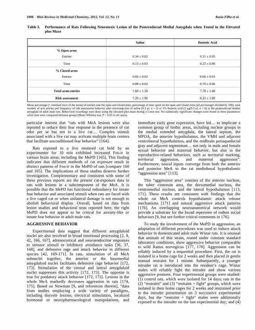
Table 3. Performance of Rats Following Neurotoxic Lesion of the Posterodorsal Medial Amygdala when Tested in the Elevated
plus Maze
Saline Ibotenic Acid
% Open arms
Entries
0.34 ± 0.02
0.31 ± 0.05
Time 0.15 ± 0.03 0.22 ± 0.06
% Closed arms
Entries
0.66 ± 0.02
0.66 ± 0.03
Time 0.88 ± 0.02 0.70 ± 0.06
Total arms entries 7.60 ± 1.30 7.78 ± 1.40
Risk assessment 7.20 ± 1.90 9.33 ± 1.90
Mean percentage (+ standard error of the mean) of entries onto the open and closed arms, percentage of time spent on the open and closed arms (all percentages divided by 100), total number of arm entries and frequency of risk assessment behavior after microinjection of saline (0.3 l, n = 5) or 1% ibotenic acid (3 g/0.3 l, n = 6) in the posterodorsal medial amygdala of adult male rats. Behavioral recordings were done using the elevated plus maze during a 5-min test. No statistically significant changes were found in these parameters when data were compared between groups (Mann Whitney test, P > 0.05 in all cases).

Morphology and Function of the Rat Medial Amygdala Mini-Reviews in Medicinal Chemistry, 2012, Vol. 12, No. 11 1099
the “fight” group, which consisted of rats that were not submitted to restraint but still exhibited aggressive behavior towards an intruder following 2 weeks of isolation. As a rule, the rats in the “restraint + fight” group showed sequential bursts of typical fighting displays, with the resident male exhibiting offensive acts and the intruder presenting defensive postures. Fos-ir cells in the MePD of the experimental groups were quantified according previously published procedures [179-182]. Notably, these cells were scarce in control rats that were subjected only to isolation (mean ± standard error of the mean, 3.3 ± 0.9/100 m2). In contrast, Fos-ir cells were clearly visible, abundant and homogeneously distributed in the MePD of males in the “restraint” (15.1 ± 4.1/100 m2) and “restraint + fight” (16 ± 3.5/100 m2) groups. Fos-ir in the MePD of the “fight” group remained low and similar to control (5.7 ± 1.3/100
m2) (Fig. 2).
In summary, MePD Fos-ir increased in response to restraint rather than fighting behavior. No statistically significant difference in the number of Fos-ir cells in the medial versus lateral aspects of the MePD or in the right versus left hemispheres MePD was found in the “restraint + fight” group (data not shown).
These results suggest that the MePD responds to non-mutually exclusive possibilities: conditioned learning paradigm for aggression, mild stressors, and to aggressive displays. In this sense, the MeA is associated with the consolidation of aversive memories [168]. It is possible that repetitive restraining and the sudden presence of an intruder into the resident’s cage lead to an associative conditioning response. That would explain Fos-ir in the MePD of the “restraint” group but not the “fight” group.
Subsequently, because the MePD appears to be involved in the processing of mild stressful stimuli (restraint) prior to an aggressive encounter with an intruder, an additional experiment was performed to further evaluate the role of MePD in aggression. Somatostatin (1 M/0.3 l), a neuropeptide with marked inhibitory actions [183-186], was microinjected into the right MePD of males that previously showed reliable aggressive behavior against intruders. Somatostatin was hypothesized to reduce the aggressive outcome in tested rats. Dosage was chosen based on previous studies of the inhibitory efficacy of somatostatin using
different experimental approaches and in various brain areas [85, 183, 184, 187, 188], including the MeA [185]. The recorded behaviors included the following: social investigation (approach of the intruder’s body and/or genitals); attack (lateral or frontally-oriented movements to strike the intruder’s back); boxing (standing on hind limbs and projection of forelimbs towards the intruder); “immobilization” or domination posture (lying over the intruder and maintaining immobility for at least 3 seconds); and, “resting” (no obvious offensive or defensive motor actions). Bites were not directly evaluated. Locomotion (i.e., movements in the cage toward the intruder or not) was interpreted as motor activity. Latency, frequency, and duration of these parameters were compared between groups.
Somatostatin drastically impaired the resident’s aggressive attacks towards an intruder, leading to significant reduction of immobilization and attack behaviors (Fig. 3). Social investigation and locomotion behavior were not affected by the microinjection of saline or somatostatin in the MePD (data not shown), which indicates that inhibition was specific to aggression rather than to locomotor modulation. The action of somatostatin on aggression did not last beyond the microinjection (Fig. 3), suggesting that the action of this neuropeptide is transient but specific.
These findings indicate that the MePD is involved in mixed responses to stressful conditions and in the modulation of aggressive behavior. Restraint induces and tests stress-related neuroendocrine and cardiovascular responses [37, 189, 190]. Accordingly, Fos expression in MeA neurons incrementally increases after exposure to 15, 30 or 60 min of restraint [189]. Acute restraint stress for 1 h on one single day reduces the MePD dendritic spine density [37]. This result does not occur with a single (6 h long) period of restraint or after repeated (6 h daily for 28 days) restraint stress [37, see parallel comments in 30]. Immobilization stress also causes a marked release of substance P, a neurotransmitter/ neuromodulator relevant for emotional states, suggesting that the MeA is also implicated in processing stressful stimuli and most likely responsible for some stress-related disorders [191, 192]. Fos-ir in the MePD following 1-min restraint confirms the participation of the MePD on this neural response. Further, because plasma levels of corticosterone exert positive feedback control over brain mechanisms of aggressive behavior, stress hormones
Table 4. Performance of Rats Following Neurotoxic Lesion of the Posterodorsal Medial Amygdala when Faced to a Live Cat
Saline Ibotenic Acid
Sniffing 35 (21.5/45.5) 27 (9/42)
Rearing 7 (5/23.5) 12 (8.25/19.5)
Frequency
Freezing
Locomotion
29 (12.5/32)
19 (8.5/41)
19 (7/30)
25 (17/27)
Freezing
Duration
437 (169/581) 294 (218/629)
Median and interquartile ranges to the frequency and duration (in seconds) of innate fear-related behavioral displays following microinjection of saline (0.3 l, n = 5) or 1% ibotenic acid (3 g/0.3 l, n = 6) in the posterodorsal medial amygdala of adult male rats. Recordings were performed during exposure of experimental groups to a live, caged cat during a 15-min test. No statistically significant change was found in these parameters between groups (Mann Whitney test, P > 0.05 in all cases).

1100 Mini-Reviews in Medicinal Chemistry, 2012, Vol. 12, No. 11 Rasia-Filho et al.
could also facilitate the display of aggression [193]. Both glucocorticoid receptor immunoreactivity and mRNA have
been found in cells of the posterior MeA [194-196]. Isolation and restraint are stressful conditions that increase stress-related hormones in the resident rat before confrontation with an unfamiliar intruder. These conditions may in fact have triggered the aggressive display to dominate the intruding rival as described here. However, corticosterone-independent pathways may also be at play because neither adrenalectomy nor corticosterone treatment affects c-Fos activation in the MeA of rats exposed to fighting [176].
As partially supported by the present data, the MePD can be functionally relevant in offensive contexts [167, 176, 197], but not in defensive reactions against natural predators [175]. The medial, but not the lateral, MePD exhibits enhanced Fos-ir following aggression in Wild-type Brown Norway male rats following exposure to a resident-intruder paradigm [177]. The caudal part of the MePD in this same rat strain also shows marked increase in Fos-ir after aggressive encounters [177]. However, this medial/lateral difference in the MePD is not detected in Wistar rats, suggesting the possibility of rat strain differences. Further efforts would verify whether the Fos-ir after the aggressive behavior colocalizes with cells expressing androgen receptors or cells that respond to olfactory stimulation in the MePD [e.g., see 198, 199].
CONCLUDING REMARKS
Among the different MeA subnuclei, the MePD is most often studied as part of integrated neural circuitries that are affected by sex steroids and that modulate different social behaviors in rats. Receptors for gonadal hormones are highly expressed in the MePD and exhibit complex functional interactions in both males and females. Current knowledge indicates that the morphology of MePD neurons is sexually dimorphic. For example, a considerable number of dendritic spines are plastic. They can change naturally or following experimental sex steroid manipulations. This neural plasticity involving gonadal hormones is relevant for synaptic processing and should be correlated with the connectivity of the MePD. In addition to its remarkable role in sexual behavior displays (specifically for the occurrence of ejaculation), the male rat MePD modulates other social behaviors, as proposed by Newman [9]. According to the studies presented here, anxiety-like behaviors and innate fear are not markedly affected by unilateral neurotoxic damage to the MePD. However, responses to stressful stimulation and the modulation of aggressive display do involve the function of this subnucleus in Wistar males. Altogether, the MePD is a nodal point within interconnected, gonadal-hormone sensitive neural networks that collectively regulate social behaviors in adult rats.
CONFLICT OF INTEREST
The authors state that there is no known conflict of interest.
ACKNOWLEDGEMENTS
All rats were manipulated according to international laws for the ethical care and use of laboratory animals. Authors are thankful to the anonymous reviewers that provided commentaries to improve this work. Grants from Brazilian
Fig. (2). A. Schematic diagram of a coronal brain section showing right and left posterodorsal medial amygdala (MePD, approximately 3.14 mm posterior to the bregma) and a rectangular overlay (the “area of interest” of 0.136 mm ) within which Fos- immunoreactive cells were counted in each brain hemisphere of the control, “restraint”, “restraint + fight” and “fight” experimental groups (n = 5 males each). Adapted from the atlas of Paxinos and Watson [200]. Scale bar = 1 mm. B-I. Digitized microscopic images showing representative Fos immunoreactivity results in the right and left MePD of rats from the control (B and C), “restraint” (D and E), “restraint + fight” (F and G), and “fight” (H and I) groups. Ot = optic tract, st = stria terminalis. Only background contrast was adjusted. Scale bar = 250 m.
�����������
����
��
��
�
���!�"���#���
$��!�"���#��%$#�&�� '
()��$#�&��
��
����
��
*��
��
�+��
��
��
�+��

Morphology and Function of the Rat Medial Amygdala Mini-Reviews in Medicinal Chemistry, 2012, Vol. 12, No. 11 1101
agencies “Fundação de Amparo à Pesquisa do Rio Grande do Sul” (FAPERGS) and “Conselho Nacional de Pesquisa e Desenvolvimento” (CNPq).
ABBREVIATIONS
AVPV = anteroventral periventricular nucleus
ER- = estrogen receptor
ER- = estrogen receptor
Fos-ir = Fos-immunoreactivity
GnRH = gonadotrophin releasing hormone
MeA = medial amygdaloid nucleus
MeAD = anterodorsal subnucleus of the medial amygdale
MePD = posterodorsal subnucleus of the medial amygdale
MePV = posteroventral subnucleus of the medial amygdale
MPOA = medial preoptic area
PMv = ventral premamillary nucleus
OT = optic tract
ST = stria terminalis
VMH = ventromedial hypothalamic nucleus.
REFERENCES
[1] Nelson, R.J. An Introduction to Behavioral Endocrinology; Sinauer Associates: Sunderland, 2000.
[2] Rasia-Filho, A.A.; Brusco, J.; Moreira, J.E. Spine plasticity in the rat medial amygdala.In: Dendritic Spines: Biochemistry, Modeling and Properties, Baylog, L.R. Ed.; Nova Science Publishers: Hauppauge, 2009; pp. 67-90.
[3] Swanson, L. W.; Petrovich, G. D. What is the amygdala? Trends. Neurosci.,1998, 21, 323-331.
[4] Alheid, G. F.; de Olmos, J. S.; Beltramino, C. A. Amygdala and extended amygdala.Paxinos, G., editor. 1995; pp. 495-578.
Fig. (3). A. Photomicrography of a representative rat brain coronal section showing the location of the cannula implanted above the posterodorsal medial amygdala (MePD) in adult male rats. Dashed lines represent the course of the microinjection needle. Scale bar = 1.5 mm. Spatial coordinates: D, dorsal; L, lateral; M, medial; V, ventral. B-D. Mean (± standard error of the mean) of the immobilization behavior frequency (B), frequency of attacks (C), and duration of the attacks (D) made by the resident against the intruder during an aggressive encounter (10-min test). These measures were taken prior to stereotaxic surgery (pre-test, control data); on the day of saline (0.3
l; n = 9) or somatostatin microinjections (1 M/0.3 l; n = 6) in the MePD; and on the day immediately after the microinjection day (post-test, additional control data). * One-way analysis of variance test followed by the Tukey test, P < 0.05, when comparing to control data in the same group and saline microinjection.
* �
��
(���,#�#-��#��
$��./��+0
*���+1*���+1
$��./��+0
�/���#����"�
��������
���������
���� �
����
��������
���������
���� �
����
��������
���������
���� �
����
����������������
��
�
�
�
�
��
��
��
�
�
��
��
��
�
2 2
2
2
�
3
�4

1102 Mini-Reviews in Medicinal Chemistry, 2012, Vol. 12, No. 11 Rasia-Filho et al.
[5] McDonald, A.J. Cortical pathways to the mammalian amygdala. Prog. Neurobiol.,1998, 55, 257-332.
[6] Pitkänen, A. Connectivity of the rat amygdaloid complex. In: The amygdala: a functional analysis, Aggleton, J.P. Ed.; Oxford University Press: Oxford, 2000; pp 31-115.
[7] Petrovich, G.D.; Canteras, N.S.; Swanson, L.W. Combinatorial amygdalar inputs to hippocampal domains and hypothalamic behavior systems. Brain Res. Rev., 2001, 38, 247-289.
[8] de Olmos, J.S.; Beltramino, C.A.; Alheid, G. Amygdala and extended amygdala of the rat: a cytoarchitectonical, fibroarchitectonical, and chemoarchitectonical survey. Paxinos, G., editor; 2004: pp. 509-603.
[9] Newman, S. W. The medial extended amygdala in male reproductive behavior. A node in the mammalian social behavior network. Ann. NY Acad. Sci., 1999, 877, 242-257.
[10] Alheid, G.F. Extended amygdala and basal forebrain. Ann. N.Y. Acad. Sci, 2003 985, 185-205.
[11] Swanson, L.W. The amygdala and its place in the cerebral hemisphere. Ann. N.Y. Acad. Sci., 2003, 985, 174-184.
[12] Bian, X.; Yanagawa, Y.; Chen, W.R.; Luo, M. Cortical-like functional organization of the pheromone-processing circuits in the medial amygdala. J. Neurophysiol.,2008, 99, 77-86.
[13] García-López, M.; Abellán, A.; Legaz, I.; Rubenstein, J.L.R.; Puelles, L.; Medina, L. Histogenetic compartments of the Mouse centromedial and extended amygdala base on gene expression patterns during development. J. Comp. Neurol., 2008, 506, 46-74.
[14] Bupesh, M.; Legaz, I.; Abellán, A.; Medina, L. Multiple telencephalic and extratelencephalic embryonic domains contribute neurons to the medial extended amygdala. J. Comp. Neurol., 2011, 519, 1505-1525.
[15] Canteras, N.S.; Simerly, R.B.; Swanson, L.W. Organization of projections from the medial nucleus of the amygdala: a PHAL study in the rat. J. Comp. Neurol., 1995, 360, 213-245.
[16] Carrillo, B.; Pinos, H.; Guillamón, A.; Panzica, G.; Collado, P. Morphometrical and neurochemical changes in the anteroventral subdivision of the rat medial amygdala during estrous cycle. Brain. Res., 2007, 1150, 83-93.
[17] Dall’Oglio, A.; Gehlen, G.; Achaval, M.; Rasia-Filho, A.A. Dendritic branching features of posterodorsal medial amygdala neurons of adult male and female rats: Further data based on the Golgi method. Neurosci. Lett., 2008, 430, 151-156.
[18] Dall’Oglio, A.; Gehlen, G.; Achaval, M.; Rasia-Filho, A.A. Dendritic branching features of Golgi-impregnated neurons from the "ventral" medial amygdala subnuclei of adult male and female rats. Neurosci Lett., 2008, 439, 287-292.
[19] Scalia, F.; Winans, S.S. The differential projections of the olfactory bulb and accessory olfactory bulb in mammals. J. Comp. Neurol., 1975, 161, 31-55.
[20] Nishizuka, M.; Arai, Y. Male-female differences in the intra- amygdaloid input to the medial amygdala. Exp. Brain Res.,1983, 52, 328-332.
[21] Dielenberg, R.A.; McGregor, I.S. Defensive behavior in rats towards predatory odors: a review. Neurosci. Biobehav. Rev., 2001, 25, 597-609.
[22] Meredith, M.; Westberry, J.M. Distinctive responses in the medial amygdala to same-species and different-species pheromones. J. Neurosci., 2004, 24, 5719-5725.
[23] Pro-Sistiaga, P.; Mohedano-Moriano, A.; Ubeda-Bañon, I.; Arroio- Jimenez, M.D.M.; Marcos, P.; Artacho-Pérula, E.; Crespo, C.; Insausti, R.; Martinez-Marcos, A. Convergence of olfactory and vomeronasal projections in the rat basal telencephalon. J. Comp.
Neurol., 2007, 504, 346-362. [24] Blake, C.B.; Meredith, M. Change in number and activation of
androgen receptor-immunoreactive cells in the medial amygdala in response to chemosensory input. Neuroscience, 2011, 190, 228- 238.
[25] Dhungel, S.; Urakawa, S.; Kondo, Y.; Sakuma, Y. Olfactory preference in the male rat depends on multiple chemosensory inputs converging on the preoptic area. Horm. Behav., 2011, 59, 193-199.
[26] Rasia-Filho, A.A.; Peres, T.M.S.; Cubilla-Gutierrez, F.H.; Lucion, A.B. Effect of estradiol implanted in the corticomedial amygdala on the sexual behavior of castrated male rats. Braz. J. Med. Biol. Res.,1991, 24, 1041-1049.
[27] Coolen, L.M.; Peters, H.J.P.W.; Veening, J.G. Distribution of Fos immunoreactivity following mating versus anogenital investigation in the male rat brain. Neuroscience, 1997, 77, 1151-1161.
[28] Coolen, L.M.; Peters, H.J.P.W.; Veening, J.G. Fos immunoreactivity in the rat brain following consummatory elements of sexual behavior: a sex comparison. Brain. Res.,1996, 738, 67-82.
[29] Pfaus, J.G.; Heeb, M.M. Implications of immediate-early gene induction in the brain following sexual stimulation of female and male rodents. Brain Res. Bull., 1997, 44, 397-407.
[30] de Castilhos, J.; Marcuzzo, S.; Forti, C.D.; Frey, R.M.; Stein, D.; Achaval, M.; Rasia-Filho, A.A. Further studies on the rat posterodorsal medial amygdala: Dendritic spine density and effect of 8-OH-DPAT microinjection on male sexual behavior. Brain Res. Bull., 2006, 69, 131-139.
[31] Cameron, N.M.; Carey, P.; Erskine, M.S. Medullary noradrenergic neurons release norepinephrine in the medial amygdala in females in response to mating stimulation sufficient for pseudopregnancy. Brain. Res., 2004, 1022, 137-147.
[32] Fleming, A.S.; Vaccarino, F.; Luebke, C. Amygdaloid inhibition of maternal behavior in the nulliparous female rat. Physiol. Behav.,1980, 25, 731-743.
[33] Sheehan, T.P.; Paul, M.; Amaral, E.; Numan, M.J.; Numan, M. Evidence that the medial amygdala projects to the anterior/ventromedial hypothalamic nuclei to inhibit maternal behavior in rats. Neuroscience, 2001, 106, 341-356.
[34] Adamec, R. E.; Morgan, H. D. The effect of kindling of different nuclei in the left and right amygdala on anxiety in the rat. Physio.l Behav., 1994, 55, 1-12.
[35] Duncan, G. E.; Knapp, D. J.; Breese, G. R. Neuroanatomical characterization of Fos induction in rat behavioral models of anxiety. Brain. Res.,1996, 713, 79-91.
[36] Dayas, C.V.; Buller, K.M.; Day, T.A. Neuroendocrine responses to an emotional stressor: evidence for the involvement of the medial but not the central amygdala. Eur. J. Neurosci., 1999, 11, 2312- 2322.
[37] Marcuzzo, S.; Dall’Oglio, A.; Ribeiro, M.F.; Achaval, M.; Rasia- Filho, A.A. Dendritic spines in the posterodorsal medial amygdala after restraint stress and ageing in rats. Neurosci. Lett., 2007, 424, 16-21.
[38] Lin, Y.; Li, X.; Lupi, M.; Kinsey-Jones, J.S.; Shao, B.; Lightman, S.L.; O’Byrne, K.T. The role of the medial and central amygdala in stress-induced suppression of pulsatile LH secretion in female rats. Endocrinology, 2011, 152, 545-555.
[39] Timmer, M.; Cordero, M.I.; Sevelinges, Y.; Sandi C. Evidence for a role of oxytocin receptors in the long-term establishment of dominance hierarchies. Neuropsychopharmacol, 2011, doi: 10.1038/npp.2011.125.
[40] Rasia-Filho, A.A.; Fabian, C.; Rigoti, K.; Achaval., M. Influence of sex, estrous cycle and motherhood in dendritic spine density in the rat medial amygdala revealed by the Golgi method. Neuroscience, 2004, 126, 839-847.
[41] Rasia-Filho, A. A. Is there anything "autonomous" in the nervous system? Adv. Physiol. Educ., 2006, 30, 9-12.
[42] Rasia-Filho, A.A.; Giovenardi, M.; de Almeida, R.M. Drugs and aggression. Recent Patents CNS Drug Discov., 2008, 3, 40-49.
[43] Mizukami, S.; Nishizuka, M.; Arai, Y. Sexual difference in nuclear volume and its ontogeny in the rat amygdala. Exp. Neurol., 1983, 79, 569-575.
[44] Johnson, R.T.; Breedlove, S.M.; Jordan, C.L. Sex differences and laterality in astrocyte number and complexity in the adult rat medial amygdala. J. Comp. Neurol., 2008, 511, 599-609.
[45] Martinez, F.G.; Hermel, E.E.; Xavier, L.L.; Viola, G.G.; Riboldi, J.; Rasia-Filho, A.A.; Achaval, M. Gonadal hormone regulation of glial fibrillary acidic protein immunoreactivity in the medial amygdala subnuclei across the estrous cycle and in castrated and treated female rats. Brain. Res., 2006, 1108, 117-126.
[46] Morris, J.A.; Jordan, C.L.; Breedlove, S.M. Sexual dimorphism in neuronal number of the posterodorsal medial amygdala is independent of circulating androgens and regional volume in adult rats. J. Comp. Neurol., 2008, 506, 851-859.
[47] Zhou, J.; Zhang, H.; Cohen, R.S.; Pandey, S.C. Effects of estrogen treatment on expression of BDNF and CREB expression and phosphorylation in rat amygdaloid and hippocampal structures. Neuroendocrinology, 2005, 81, 294-310.

Morphology and Function of the Rat Medial Amygdala Mini-Reviews in Medicinal Chemistry, 2012, Vol. 12, No. 11 1103
[48] Simerly, R.B.; Chang, C.; Muramatsu, M.; Swanson, L.W. Distribution of androgen and estrogen receptor mRNA-containing cells in the rat brain: An in situ hybridization study. J. Comp.
Neurol.,1990, 294, 76-95. [49] Sheridan, P. J. The nucleus interstitialis stria terminalis and the
nucleus amygdaloideus medialis: Prime targets for androgen in the rat forebrain. Endocrinology, 1979, 104, 130-136.
[50] Gréco, B.; Edwards, D.A.; Michael, R.P.; Clancy, N.A. Androgen receptors and estrogen receptors are colocalized in male rat hypothalamic and limbic neurons that express Fos immunoreactivity induced by mating. Neuroendocrinology, 1998, 67, 18-28.
[51] Blake, C.B.; Meredith, M. Change in number and activation of androgen receptor-immunoreactive cells in the medial amygdala in response to chemosensory input. Neuroscience, 2011, 190, 228-238.
[52] Gréco, B.; Edwards, D.A.; Michael, R.P.; Clancy, A.N. Androgen receptor immunoreactivity and mating-induced Fos expression in forebrain and midbrain structures in the male rat. Neuroscience, 1996, 75, 161-171.
[53] Wagner, C. K.; Morrell, J. I. Distribution and steroid hormone regulation of aromatase mRNA expression in the forebain of adult male and female rats: A cellular-level analysis using in situ hybridization. J. Comp. Neurol., 1996, 370, 71- 84.
[54] Roselli, C. E.; Horton, L. E.; Resko, J. A. Distribution and regulation of aromatase activity in the rat hypothalamus and limbic system. Endocrinology, 1985, 117, 2471-2477.
[55] Shughrue, P.J.; Lane, M.V.; Merchenthaler, I.J. Comparative distribution of estrogen receptor- and - mRNA in the rat central nervous system. J. Comp. Neurol., 1997, 388, 507-525.
[56] Gréco, B.; Edwards, D.A.; Michael, R.P.; Clancy, N.A. Androgen receptors and estrogen receptors are colocalized in male rat hypothalamic and limbic neurons that express Fos immunoreactivity induced by mating. Neuroendocrinol., 1998, 67, 18-28.
[57] Gréco, B.; Allegretto, E.A.; Tetel, M.J.; Blaustein, J.D. Coexpression of ER with ER and progestin receptor proteins in the female rat forebrain: effects of estradiol treatment. Endocrinology, 2001, 142, 5172-5181.
[58] Gréco, B.; Blasberg, M.E.; Kosinski, E.C.; Blaustein, J.D. Response of ER -IR and ER -IR cells in the forebrain of female rats to mating stimuli. Horm. Behav., 2003, 43, 444-453.
[59] De Vries, G.J.; Simerly, R.B. Anatomy, development, and function of sexually dimorphic neural circuits in the mammalian brain. In: Hormones, Brain and Behavio, Pfaff, D,W.; Arnold, A.P.; Etgen, A.M.; Fahrbach, S.E.; Rubin, R,.T., Eds;. Academic Press, San Diego, 2002; pp. 137-191.
[60] Isgor, C.; Huang, G.C.; Akil, H.; Watson, S.J. Correlation of estrogen -receptor messenger RNA with endogenous levels of plasma estradiol and progesterone in the female rat hypothalamus, the bed nucleus of stria terminalis and the medial amygdala. Mol. Brain Res., 2002, 106, 30-41.
[61] Phillips-Farfán, B.V.;Lemus, A. E.; Fernández-Guasti, E. Increased estrogen receptor alpha immunoreactivity in the forebrain of sexually satiated rats. Horm. Behav., 2007, 51, 328-334
[62] Hines, M.; Allen, L. S.; Gorski, R. A. Sex differences in subregions of the medial nucleus of the amygdala and the bed nucleus of the stria terminalis of the rat. Brain Res., 1992, 579, 321-326.
[63] Hermel, E.E.; Ilha, J.; Xavier, L.L.; Rasia-Filho, A.A.; Achaval, M. Influence of sex and estrous cycle, but not laterality, on the neuronal somatic volume of the posterodorsal medial amygdala of rats. Neurosci. Lett., 2006, 405, 153-158.
[64] Zehr, J.L.; Todd, B.J.; Schulz, K.M.; McCarthy, M.M.; Sisk, C.L. Dendritic pruning of the medial amygdala during pubertal development of the male Syrian hamster. J. Neurobiol., 2006, 66, 578-590.
[65] de Castilhos, J.; Forti, C.D.; Achaval, M.; Rasia-Filho, A.A. Dendritic spine density of posterodorsal medial amygdala neurons can be affected by gonadectomy and sex steroid manipulations in adult rats: A Golgi study. Brain. Res., 2008, 1240, 73-81.
[66] Nishizuka, M.; Arai, Y. Regional difference in sexually dimorphic synaptic organization of the medial amygdala. Exp. Brain Res.,1983, 49, 462-465.
[67] Cooke, B.M.; Woolley, C.S. Sexually dimorphic synaptic organization of the medial amygdala. J. Neurosci., 2005, 25, 10759-10767.
[68] Arimatsu, Y.; Seto, A.; Amano, T. Sexual dimorphism in bungarotoxin binding capacity in the mouse amygdala. Brain
Res., 1981, 213, 432-437. [69] Micevych, P.E.; Matt, D.W.; Go, V.L.W. Concentrations of
cholecystokinin, substance P, and bombesin in discrete regions of male and female rat brain: sex differences and estrogen effects. Exp. Neurol., 1988, 100, 416-425.
[70] Oro, A.E.; Simerly, R.B.; Swanson, L.W. Estrous cycle variations in levels of cholecystokinin immunoreactivity within cells of three interconnectedsexuallydimorphicforebrainnuclei. Neuroendocrinology, 1988, 47, 225-235.
[71] Wilson, M.A.; Mascagni, F.; McDonald, A.J. Sex differences in delta opioid receptor immunoreactivity in rat medial amygdala. Neurosci. Lett., 2002, 328, 160-164.
[72] Stefanova, N. -Aminobutyric acid-immunoreactivity neurons in the amygdala of the rat - sex differences and effect of early postnatal castration. Neurosci. Lett., 1998, 255, 175-177.
[73] Polston, E.K.; Heitz, M.; Barnes, W.; Cardamone, K.; Erskine, M.S. NMDA-mediated activation of the medial amygdala initiates a downstream neuroendocrine memory responsible for pseudopregnancy in the female rat. J. Neurosci., 2001, 21, 4104-4110.
[74] Fergunson, J.N.; Aldag, J.M.; Insel, T.R.; Young, L.J. Oxytocin in the medial amygdala is essential for social recognition in the mouse. J. Neurosci., 2001, 21, 8278-8285.
[75] Lehmann, M.L.; Erskine, M.S. Glutamatergic stimulation of the medial amygdala induces steroid dependent c-fos expression within forebrain nuclei responsive to mating stimulation. Neuroscience, 2005, 136, 55-64.
[76] Malsbury, C.W.; McKay, K. Neurotrophic effects of testosterone on the medial nucleus of the amygdala in adult male rats. J. Neuroendocrinol., 1994, 6, 57-69.
[77] Rasia-Filho, A.A.; Xavier, L.L.; Santos, P.; Gehlen, P.; Achaval, M. Glial fibrillary acidic protein immunodetection and immunoreactivity in the anterior and in the posterior medial amygdala of male and female rats. Brain Res. Bull., 2002, 58, 67- 75.
[78] Rasia-Filho, A.A.; Londero, R.G.; Achaval, M. Effects of gonadal hormones on the morphology of neurons from the medial amygdaloid nucleus of rats. Brain Res. Bull., 1999, 48, 173-183.
[79] McDonald, A.J. Cell types and intrinsic connections of the amygdala. In: The amygdala: neurobiological aspects of emotion, memory, and mental dysfunction, Aggleton, J.P. Ed.; Wiley-Liss; NewYork; 1992; pp. 67-96.
[80] Nabekura, J.; Oomura, Y.; Minami, T.; Mizuno, Y.; Fukuda, A. Mechanism of the rapid effect of 17a-estradiol on medial amygdala neurons. Science, 1986, 233, 226-228.
[81] Gomez, D.M.; Newman, S.W. Medial nucleus of the amygdala in the adult Syrian hamster: A quantitative Golgi analysis of gonadal hormonal regulation of neuronal morphology. Anat. Rec., 1991, 231, 498-509.
[82] Choi, G.B.; Dong, H-W.; Murphy, A.J.; Valenzuela, D.M.; Yancopoulos, G.D.; Swanson, L.W.; Anderson, D.J. Lhx6 delineates a pathway mediating innate reproductive behaviors from the amygdala to the hypothalamus. Neuron, 2005, 46, 647-660.
[83] Arpini, M.; Menezes, I.C.; Dall’Oglio, A.; Rasia-Filho, A.A. The density of Golgi-impregnated dendritic spines from adult rat posterodorsal medial amygdalaneurons displays no evidence of hemispheric or dorsal/ventral differences. Neurosci. Lett., 2010, 469, 209-213.
[84] Brusco, J.; Dall’Oglio, A.; Rocha, L.B.; Rossi, M.A.; Moreira, J.E.; Rasia-Filho, A.A. Descriptive findings on the morphology of dendritic spines in the rat medial amygdala. Neurosci. Lett., 2010, 483, 152-156.
[85] Carney, R.S.E.; Mangin, J-M.; Hayes, L.; Mansfield, K.; Sousa, V.H.; Fishell, G.; Machold, R.P.; Ahn, S.; Gallo, V.; Corbin, J.G. Sonic Hedgehog expressing and responding cells generate neuronal Diversity in the medial amygdala. Neural Develop., 2010, 5, 14.
[86] Cunningham, R.L.; Clairborne, B.J.; McGinnis, M.Y. Pubertal exposure to anabolic androgenic steroids increases spine densities on neurons in the limbic system of male rats. Neuroscience, 2007, 150, 609-615.

1104 Mini-Reviews in Medicinal Chemistry, 2012, Vol. 12, No. 11 Rasia-Filho et al.
[87] Simerly, R.B. Anatomical substrates of hypothalamic integration. In: The Rat Nervous System. Paxinos, G., Ed.; Academic Press: San Diego; 2004; pp. 335-368.
[88] Rasia- Filho, A.A.; Brusco, J.; Rocha, L.; Moreira, J. Dendritic spines observed by extracellular DiI dye and immunolabeling under confocal microscopy. Nature Protocols/Protocol Exchange (2010). doi:10.1038/nprot.2010.153
[89] Toni, N.; Buchs, P.A.; Nikonenko, I.; Bron, C.R.; Muller, D. LTP promotes formation of multiple spine synapses between a single axon terminal and a dendrite. Nature, 1999, 402, 421-425.
[90] Shepherd, G.M. The dendritic spine: a multifunctional integrative unit. J. Neurophysiol., 1996, 75, 2197-2210.
[91] Nimchinsky, E.A.; Sabatini, B.L.; Svoboda, K. Structure and function of dendritic spines. Annu. Rev. Physiol., 2002, 64, 313-353.
[92] Kasai, H.; Matsuzaki, M.; Noguchi, J.; Yasumatsu, N.; Nakahara, H. Sructure-stability-function relationships of dendritic spines. Trends Neurosci., 2003, 26, 360-368.
[93] Peters, A.; Palay, S.; Webster, H. The fine structure of the nervous system, Oxford University Press: New York, 1991.
[94] Stuart, G.; Sprutson, N.; Häusser, M. Dendrites. Oxford University Press: New York, 1999.
[95] Arellano, J.I.; Espinosa, A.; Fairén, A.; Yuste, R.; DeFelipe, J. Non-synaptic dendritic spines in neocortex. Neuroscience, 2007, 145, 464-469.
[96] Harris, K.M.; Kater, S.B. Dendritic spines: Cellular specializations imparting both stability and flexibility to synaptic function. Ann. Rev. Neurosci., 1994, 17, 341-371.
[97] Tsay, D.; Yuste, R. On the electrical function of dendritic spines. Trends Neurosci., 2004, 27, 77-83.
[98] García-López, P.; García-Marin, V.; Freire. M. Dendritic spines and development: towards a unifying model of spinogenesis. A present day review of Cajal’s histological slides and drawings. Neural Plasticity, 2010, 2010:769207.
[99] Peters, A.; Kaiserman-Abramof, I.R. The small pyramidal neuron of the rat cerebral cortex. The perikaryon, dendrites and spines. Am. J. Anat., 1970, 127, 321-356.
[100] Cooke, B.M.; Stokas, M.R.; Woolley, C.S. Morphological sex differences and laterality in the prepubertal medial amygdala. J. Comp. Neurol., 2007, 501, 904-915.
[101] Cooke, B.M. Synaptic reorganization of the medial amygdala during puberty. J. Neuroendocrinol., 2011, 23, 65-73.
[102] Krebs-Kraft, D.L.; Hill, M.N.; Hillard, C.J.; McCarthy, M.M. Sex difference in cell proliferation in developing rat amygdala mediated by endocannabinoids has implications for social behavior. Proc.
Natl. Acad. Sci., 2010, 107, 20535-20540. [103] Hermel, E.E.; Faccioni-Heuser, M.C.; Marcuzzo, S.; Rasia-Filho,
A.A.; Achaval, M. Ultrastructural features of neurons and synaptic contacts in the posterodorsal medial amygdala of adult male rats. J.
Anat., 2006, 208, 565-575. [104] Oberlander, J.G. ; Erskine, M.S. Receipt of vaginal-cervical
stimulation modifies synapsin content in limbic areas of the female rat. Neuroscience, 2008, 153, 581-593.
[105] Rasia-Filho, A.A.; Lucion, A.B. Effects of 8-OH-DPAT on sexual behavior of male rats castrated at different ages. Horm. Behav., 1996, 30, 251-258.
[106] Cooke, B.M.; Woolley, C.S. Effects of prepubertal gonadectomy on a male-typical behavior and excitatory synaptic transmission in the amygdala. Develop. Neurobiol., 2009, 69, 141-152.
[107] Pereno, G.L.; Balaszczuk, V.; Beltramino, C.A. Detection of conspecific pheromones elicits fos expression in GABA and calcium-binding cells of the rat vomeronasal system-medial extended amygdala. J. Physiol. Biochem., 2011, 67, 71-85.
[108] Dong, H.-W.; Petrovich, G.; Swanson, L.W. Topography of projections from amygdala to bed nuclei of the stria terminalis. Brain Res. Rev.,2001, 38, 192-246.
[109] Shelley, D.N.; Meisel, R.L. The effects of mating stimulation on c-fos immunoreactivity in the female hamster medial amygdala are region and context dependent. Horm. Behav.,2005, 47, 212-222.
[110] Cavalcante, J.C.; Sita, L.V.; Mascaro, M.B.; Bittencourt, J.C.; Elias, C.F. Distribution of urocortin 3 neurons innervating the ventral premammillary nucleus in the rat brain. Brain Res., 2006, 1089, 116-125.
[111] Polston, E.K.; Gu, G.; Simerly, R.B. Neurons in the principal nucleus of the bed nuclei of the stria terminalis provide a sexually
dimorphic GABAergic input to the anteroventral periventricular nucleus of the hypothalamus. Neuroscience, 2004, 123, 793-803.
[112] Dominguez, J.M.; Hull, E.M. Stimulation of the medial amygdala enhances medial preoptic dopamine release: Implications for male rat sexual behavior. Brain Res., 2001, 917, 225-229.
[113] Toth, M.; Fuzesi, T.; Halász, J.; Tulodgi, A.; Haller, J. Neural inputs of the hypothalamic “aggression area” in the rat. Behav. Brain Res., 2010, 215, 7-20.
[114] Been, L.E.; Petrulis, A. Chemosensory and hormone information are relayed directly between the medial amygdala, posterior bed nucleus of the stria terminalis, and medial preoptic area in male Syrian hamster. Horm. Behav., 2011, 59, 536-548.
[115] Kim, J.; Semaan, S.J.; Clifton, D.K.; Steiner, R.A.; Dhamija, S.; Kauffman, A.S. Regulation of Kiss1 expression by sex steroids in the amygdala of the rat and mouse. Endocrinology, 2011, 152, 2020-2030.
[116] Simmons, D.A.; Yahr, P. Nitric oxidergic cells related to ejacuilation in gerbil forebrain contain androgen receptor and respond to testosterone. J. Comp. Neurol., 2011, 519, 900-915.
[117] Samuelsen, C.L.; Meredith, M. Oxytocin antagonist disrupts male mouse medial amygdala response to chemical-communication signals. Neuroscience, 2011, 180, 96-104.
[118] Arakawa, H.; Arakawa, K.; Deak, T. Oxytocin and vasopressin in the medial amygdala differentially modulate approach and avoidance behavior toward illness-related social odor. Neuroscience, 2010, 171, 1141-1151.
[119] Veyrac, A.; Wang, G.; Baum, M.J.; Bakker, J. The main and accessory olfactory systems of female mice are activated differentially by dominant versus subordinate male urinary odors. Brain Res., 2011, 1402, 20-29.
[120] De Jonge, F.H.; Oldenburger, W.P.; Louwerse, A.L.; Van de Poll, N.E. Changes in male copulatory behavior after sexual exciting stimuli: effects of medial amygdala lesions. Physiol. Behav., 1992, 52, 327-332.
[121] Stark, C.H. Behavioral effects of stimulation of the medial amygdala in the male rat are modified by prior sexual experience. J. Gen. Psychol., 2005, 132, 207-224.
[122] Wood R Estradiol, but not dihydrotestosterone, in the medial amygdala facilitates male hamster sex behavior. Physiol. Behav., 1996, 59, 833-841.
[123] Coolen, L.M.; Wood, R.I. Testosterone stimulation of the medial preoptic area and medial amygdala in the control of male hamster sexual behavior: redundancy without mplification. Behav. Brain
Res., 1999, 98, 143-153. [124] Yoshida, M.; Suga, S.; Sakuma, Y. Estrogen reduces the
excitability of the female rat medial amygdala afferents from the medial preoptic area but not those from the lateral septum. Exp.
Brain Res., 1994, 101, 1-7. [125] Kalra, P.S.; McCann, S.M. The stimulatory effect on gonadotropin
release of implants of estradiol or progesterone in certain sites in the central nervous system. Neuroendocrinology, 1975, 19, 289-302.
[126] Beltramino, C.; Taleisnik, S. Facilitatory and inhibitory effects of electrochemical stimulation of the amydala on the release of luteinizing hormone. Brain Res., 1978, 144, 95-107.
[127] Gu, G.B.; Simerly, R.B. Projections of the sexually dimorphic anteroventral periventricular nucleus in the female rat. J. Comp. Neurol.,1997, 384, 142-164.
[128] Carrer, H.F.; Whitmoyer, D.I.; Sawyer, C.H. Effects of hippocampal and amygdaloid stimulation on the firing of preoptic neurons in the proestrous female rat. Brain Res.,1978, 142, 363-367.
[129] Bagga, N.; Chhina, G.S.; Kumar, V.M.; Singh, B. Cholinergic activation of medial preoptic area by amydala for ovulation in rat. Physiol. Behav.,1984, 32, 45-48.
[130] Paterson, N.E.; Iwunze, M.; Davis, S.F.; Malekiani, S.A.; Hanania, T. Comparison of the predictive validity of the mirror chamber and elevated plus maze tests in mice. J. Neurosci. Methods, 2010, 188, 62-70.
[131] Singh, K.; Bishnoi, M.; Kulkarni, S.K. Elevated zero-maze: a paradigm to evaluate anti-anxiety effects of drugs. Methods Find. Exp. Clin. Pharmacol., 2007, 29, 343-348.
[132] Briones-Aranda, A.; López-Rubalcava, C.; Picazo, O. Influence of forced swimming-induced stress on the -like effect of 5-HT1A agents in mice. Psychopharmacol., 2002, 162, 147-155.

Morphology and Function of the Rat Medial Amygdala Mini-Reviews in Medicinal Chemistry, 2012, Vol. 12, No. 11 1105
[133] Brusco, J.; Wittmann, R.; de Azevedo, M.S.; Lucion, A.B.; Franci, C.R.; Giovenardi, M.; Rasia-Filho, A.A. Plasma hormonal profiles and dendritic spine density and morphology in the hippocampal CA1 stratum radiatum, evidenced by light microscopy, of virgin and postpartum female rats. Neurosci. Lett., 2008, 438, 346-350.
[134] Erskine, M.S.; Lehmann, M.L.; Cameron, N.M.; Polston, E.K. Co-regulation of female sexual behavior and pregnancy induction: An exploratory synthesis. Behav. Brain Res., 2004, 153, 295-315.
[135] Lehmann, M.L.; McKellar, H.; Erskine, M.S. Coding for the iniation of pseudopregnancy by temporally patterned activation of amygdalar NMDA receptors. J. Neurosci., 2005, 25, 8696-8703.
[136] Rosa, C.B.; Goularte, J.F.; Trindade, N.A.; de Oliveira, A.P.; Rasia-Filho, A.A. Glutamate microinjected in the posterodorsal medial amydala induces subtle increase in the consumption of a three-choice macronutrient self-selection diet in male rats. Anat.
Rec., 2011, 294, 1226-1232. [137] Meisel, R.L.; Sachs, B.D. The physiology of male sexual behavior.
In: The Physiology of Reproduction, Knobil, E.; Neil, J.D., Eds., Raven Press: New York, 1994; pp. 3-105.
[138] Bialy, M.; Nikolaev-Diak, A.; Kalata, U.; Nikolaev, E. Blockade of androgen receptor in the medial amygdala inhibits noncontact erections in male rats. Physiol. Behav., 2011, 103, 295-301.
[139] Kondo, Y. Lesions of the medial amygdala produce severe impairment of copulatory behavior in sexually inexperienced male rats. Physiol. Behav., 1992, 51, 939-949.
[140] Kondo, Y.; Sachs, B.D.; Sakuma, Y. Importance of the medial amygdala in rat penile erection evoked by remote stimuli from estrous females. Behav. Brain Res., 1998, 91, 215-222.
[141] Stark, C.P.; Alpern, H.P.; Fuhrer, J.; Trowbridge, M.G.; Wimbish, H.; Smock, T. The medial amygdaloid nucleus modifies social behavior in male rats. Physiol. Behav., 1998, 63 253-259.
[142] Zolman, J.F. Biostatistics; Oxford University Press: New York, 1993.
[143] Majchrzak, M.; Brailowsky, S.; Will, B. Chronic infusion of GABA into the nucleus basalis magnocellularis or frontal cortex of rats: a behavioral and histological study. Exp. Brain Res., 1992, 88, 531-40.
[144] Fillenz, M. Physiological release of excitatory amino acids. Behav. Brain Res., 1995, 71, 51-67.
[145] Kaura, S.; Bradford, H. F.; Young, A. M.; Croucher, M. J.; Hughes, P. D. Effect of amygdaloid kindling on the content and release of amino acids from the amygdaloid complex: in vivo and in vitro studies. J. Neurochem., 1995, 65, 1240-1249.
[146] Timmerman, W.; Westerink, B. H. Brain microdialysis of GABA and glutamate: what does it signify? Synapse, 1997, 27, 242-61.
[147] Hagiwara, Y.; Kubo, T. Anterior hypothalamic neurons respond to blood pressure changes via gamma-aminobutyric acid and angiotensins in rats. Neurosci. Lett., 2005, 384, 250-253.
[148] Kubo, T.; Hagiwara, Y. Posterior hypothalamus cholinergic stimulation-induced activation of anterior hypothalamic area neurons is enhanced in spontaneously hypertensive rats. Brain Res, 2005, 1061, 36-41.
[149] Genest, S. E.; Balon, N.; Laforest, S.; Drolet, G.; Kinkead, R. Neonatal maternal separation and enhancement of the hypoxic ventilatory response in rat: the role of GABAergic modulation within the paraventricular nucleus of the hypothalamus. J. Physiol., 2007, 583, 299-314.
[150] Ferreira, T. L.; Shammah-Lagnado, S. J.; Bueno, O. F.; Moreira, K. M.; Fornari, R. V.; Oliveira, M. G. The indirect amygdala-dorsal striatum pathway mediates conditioned freezing: insights on emotional memory networks. Neuroscience, 2008, 153, 84-94.
[151] Quagliotto, E.; Neckel, H.; Riveiro, D. F.; Casali, K. R.; Mostarda, C.; Irigoyen, M. C.; Dall'Ago, P.; Rasia-Filho, A. A. Histamine in the posterodorsal medial amygdala modulates cardiovascular reflex responses in awake rats. Neuroscience, 2008, 157, 709-19.
[152] Simmons, D. A.; Yahr, P. GABA and glutamate in mating-activated cells in the preoptic area and medial amygdala of male gerbils. J. Comp. Neurol., 2003, 459, 290-300.
[153] Ben-Ari, Y. ; Le Gal La Salle, G. ; Barbin, G. ; Schwartz, J. C. ; Garbarg, M. Histamine synthesizing afferents within the amygdaloid complex and bed nucleus of the stria terminalis of the rat. Brain Res., 1977, 138, 285-294.
[154] Chiavegatto, S.; Bernardi, M.M.; de Souza-Spinosa, H. Effects of prenatal diphenhydramine administration on sexual behavior in rat. Braz. J. Med. Biol. Res., 1989, 22, 729-732.
[155] Silveira, M.C.L.; Sandner, G.; Graeff. F.G. Induction of Fos immunoreactivity in the brain by exposure to the elevated plus-maze. Behav. Brain Res., 1993, 56, 115-118.
[156] Pellow, S.; Chopin, P.; File, S.E.; Briley, M. Validation of open:closed arm entries in an elevated plus-maze as a measure of anxiety in the rat. J. Neurosci. Methods, 1985, 14, 149-167.
[157] Cruz, A.P.M.; Frei, F.; Graeff, F.G. Ethopharmacological analysis of rat behavior on the elevated plus-maze. Pharmacol. Biochem. Behav., 1994, 49, 171-176.
[158] Fernandes, C.; González, M.I.; Wilson, C.A.; File, S.E. Factor analysis shows that female rat behaviour is characterized primarily by activity, male rats are driven by sex and anxiety. Pharmacol. Biochem. Behav., 1999, 64, 731-738.
[159] Lucion, A.B.; Charchat, H.; Pereira, G.A.M.; Rasia-Filho, A.A. Influence of early postnatal gonadal hormones on anxiety in adult male rats. Physiol. Behav., 1996, 60, 1419-1423.
[160] Canteras, N.S.; Chiavegatto, S.; Ribeiro do Valle, L.E.; Swanson, L.W. Severe reduction of rat defensive behavior to a predator by discrete hypothalamic chemical lesions. Brain Res. Bull., 1997, 44, 297-305.
[161] Canteras, N.S.; Ribeiro-Barbosa, E.R.; Comoli, E. Tracing from the dorsal premammillary nucleus prosencephalic systems involved in the organization of innate fear responses. Neurosci. Biobehav.
Rev., 2001, 25, 661-668. [162] Collins, D.R. Synthetic versus natural cat odorant effects on rodent
behavior and medial amygdala plasticity. Behav. Neurosci., 2011, 125, 124-129.
[163] Blanchard, D.C.; Canteras, N.S.; Markham, C.M.; Pentkowski, N.S.; Blanchard. R.J. Lesions of structures showing Fos expression to cat presentation: effects on responsivity to a cat, cat odor, and nonpredator threat. Neurosci. Biobehav. Rev., 2005, 29, 1243-1253.
[164] Takahashi, L.K.; Nakashima, B.R.; Hong, H.; Watanabe, K. The smell of danger: a behavioral and neural analysis of predator odor-induced fear. Neurosci. Biobehav. Rev., 2005, 2, 1157-1167.
[165] Martinez, R.C.; Carvalho-Netto, E.F.; Ribeiro-Barbosa, É.R.; Baldo, M.V.C.; Canteras, N.S. Amygdalar roles during exposure to a live predator and to a predator-associated context. Neuroscience, 2011, 172, 314-328.
[166] Rasia-Filho, A.A.; Londero, R.G.; Achaval, M. On some functional activities of the amygdala: an overview. J. Psychiatry
Neurosci.,2000, 25: 14-23. [167] Nelson, R.J.; Trainor, B.C. Neural mechanisms of aggression.
Nature Rev., 2007, 8, 536-546. [168] Roozendaal, B.; McGaugh, J.L. Amygdaloid nuclei lesions
differentially affect glucocorticoid-induced memory enhancement in an inhibitory avoidance task. Neurobiol. Learn. Mem., 1996, 65, 1-8.
[169] David, J.T.; Cervantes, M.C.; Trosky, K.A.; Salinas, J.A.; Delville, Y. A neural network underlying individual differences in emotion and aggression in male golden hamsters. Neuroscience, 2004, 126, 567-578.
[170] Haller, J.; Tóth, M.; Halász, J.; De Boer, S.F. Patterns of violent aggression-induced brain c-fos expression in male mice selected for aggressiveness. Physiol. Behav., 2006, 88, 173-182.
[171] Siegel, A.; Roeling, T.A.P.; Gregg, T.R.; Kruk, M.R. Neuropharmacology of brain-stimulation-evoked aggression. Neurosci. Biobehav. Rev., 1999, 23, 359-389.
[172] Shaikh, M.B.; Siegel, A. Neuroanatomical and neurochemical mechanisms underlying amygdaloid control of defensive rage behavior in the cat. Braz. J. Med. Biol. Res., 1994, 27, 2759-2779.
[173] Smith, D.A.; Flynn, J.P. Afferent projections to affective attack sites in cat hypothalamus. Brain Res., 1980, 194, 41-51.
[174] Shibata, S.; Suwandi, D.; Yamamoto, T.; Ueki, S. Effects of medial amygdaloid lesions on the initiation and the maintenance of muricide in olfactory bulbectomized rats. Physiol. Behav., 1982, 29, 939-941.
[175] Shibata, S.; Yamamoto, T.; Ueki, S. Differential effects of medial, central and basolateral amygdaloid lesions on four models of experimentally-induced aggression in rats. Physiol. Behav., 1982, 28, 289-294.
[176] Halász, J.; Liposits, Z.; Kruk, M.R.; Haller, J. Neural background of glucocorticoid dysfunction-induced abnormal aggression in rats: involvement of fear- and stress-related structures. Eur. J. Neurosci., 2002, 15, 561-569.

1106 Mini-Reviews in Medicinal Chemistry, 2012, Vol. 12, No. 11 Rasia-Filho et al.
[177] Veening; J.G.; Coolen, L.M.; de Jong, T.R.; Joosten, H.W.; de Boer, S.F.; Koolhaas, J.M.; Olivier, B. Do similar neural systems subserve aggressive and sexual behaviour in male rats? Insights from c-Fos and pharmacological studies. Eur. J. Pharmacol., 2005, 526, 226-239.
[178] de Almeida, R.M.M.; Lucion, A.B. Role of the intruder in the aggressive behavior of colonies of wild rats (Rattus norvegicus). In: Animal Model in Psychopharmacology, Olivier, B.; Mos, J.; Slagen, J.L. Eds.; Birkhauser-Verlag: Basel, 1991; pp 347-356,
[179] Kollack, S.S.; Newman, S.W. Mating behavior induces selective expression of Fos protein within the chemosensory pathways of the male Syrian hamster brain. Neurosci. Lett., 1992, 143, 223-228.
[180] Kollack-Walker, S.; Newman, S.W. Mating and agonistic behavior produce different patterns of Fos immunolabeling in the male Syrian hamster brain. Neuroscience, 1995, 66, 721-736.
[181] Kollack-Walker, S.; Newman, S.W. Mating-induced expression of c-fos in the male Syrian hamster brain: role of experience, pheromones, and ejaculations. J. Neurobiol., 1997, 32, 481-501.
[182] Parfitt, D.B.; Newman, S.W. Fos-immunoreactivity within the extended amygdala is correlated with the onset of sexual satiety. Horm. Behav., 1998, 34, 17-29.
[183] Boehm, S.; Betz, H. Somatostatin inhibits excitatory transmission at rat hippocampal synapses via presynaptic receptors. J. Neurosci., 1997, 17, 4066-4075.
[184] Delfs, J.R.; Dichter, M.A. Effects of somatostatin on mammalian cortical neurons in culture: physiological actions and unusual dose response characteristics. J. Neurosci., 1983, 3, 1176-1188.
[185] Lu, Y.F.; Hattori, Y.; Moriwaki, A.; Hayashi, Y.; Hori, Y. Inhibition of neurons in the rat medial amygdaloid nucleus in vitro by somatostatin. Can. J. Physiol. Pharmacol., 1995, 73, 670-674.
[186] Saha, S.; Henderson, Z.; Batten, T.F. Somatostatin immunoreactivity in axon terminals in rat nucleus tractus solitarii arising from central nucleus of amygdala: coexistence with GABA and postsynaptic expression of sst2A receptor. J. Chem. Neuroanat., 2002, 24, 1-13.
[187] Rakovska, A.; Kiss, J.P.; Raichev, P.; Lazarova, M.; Kalfin, R.; Milenov, K. Somatostatin stimulates striatal acetylcholine release by glutamatergic receptors: an in vivo microdialysis study. Neurochem. Int., 2002, 40, 269-275.
[188] Meis, S.; Sosulina, L.; Schulz, S.; Höllt, V.; Pape HC. Mechanisms of somatostatin-evoked responses in neurons of the rat lateral amygdala. Eur. J. Neurosci., 2005, 21, 755-762.
[189] Crane, J.W.; French, K.R.; Buller, K.M. Patterns of neuronal activation in the rat brain and spinal cord in response to increasing durations of restraint stress. Stress, 2005, 8, 199-211.
[190] Kubo, T.; Okatani, H.; Nishigori, Y.; Hagiwara, Y.; Fukumori, R.; Goshima, Y. Involvement of the medial amygdaloid nucleus in restraint stress-induced pressor responses in rats. Neurosci. Lett., 2004, 354, 84-86.
[191] Ebner, K.; Rupniak, N.M.; Saria, A.; Singewald, N. Substance P in the medial amygdala: emotional stress-sensitive release and modulation of anxiety related behavior in rats. Proc. Natl. Acad.
Sci., USA, 2004, 101:4280-4285. [192] Singewald, N.; Chicchi, G.G.; Thurner, C.C.; Tsao, K.L.; Spetea,
M.; Schmidhammer, H.; Sreepathi, H.K.; Ferraguti, F.; Singewald, G.M.; Ebner, K. Modulation of basal and stress-induced amygdaloid substance P release by the potent and selective NK1 receptor antagonist L-822429. J. Neurochem., 2008, 106, 2476-2488.
[193] Kruk, M.R.; Westphal, K.G.; Van Erp, A.M.; van Asperen, J.; Cave, B.J.; Slater, E.; de Koning, J.; Haller, J. The hypothalamus: cross-roads of endocrine and behavioural regulation in grooming and aggression. Neurosci. Biobehav. Rev., 1998, 23, 163-177.
[194] Morimoto, M.; Morita, N.; Ozawa, H.; Yokoyama, K.; Kawata, M. Distribution of glucocorticoid receptor immunoreactivity and mRNA in the rat brain: an immunohistochemical and in situ hybridization study. Neurosci. Res., 1996, 26, 235–269.
[195] Herman, J.P.; Ostrander, M.M.; Mueller, N.K.; Figueiredo, H. Limbic system mechanisms of stress regulation: hypothalamo-pituitary-adrenocortical axis. Prog. Neuro-Psychopharmacol. Biol.
Psychiatry, 2005, 29, 1201–1213. [196] Brunton, P.J.; Donadio, M.V.; Russell, J.A. Sex differences in
prenatally programmed anxiety behavior in rats: Differential corticotrophin-releasing hormone receptor mRNA expression in the amygdaloid complex. Stress, 2011, PMID: 21854167.
[197] Delville, Y.; De Vries, G.J.; Ferris, C.F. Neural connections of the anterior hypothalamus and agonistic behavior in golden hamsters. Brain Behav. Evol., 2000, 55, 53-76.
[198] Donato Jr, J.; Cavalcante, J.C.; Silva, R.J.; Teixeira, A.S.; Bittencourt, J.C.; Elias, C.F. Male and female odors induce Fos expression in chemically defined neuronal population. Physiol. Behav., 2010, 99, 67-77.
[199] Koolhaas, J.M.; Van den Brink, T.H.C.; Roozendaal, B.; Boorsma, F. Medial amygdala and aggressive behavior: interaction between testosterone and vasopressin. Aggress. Behav., 1990, 16, 223-229.
[200] Paxinos, G.; Watson, C. The rat brain in stereotaxic coordinates. Academic Press: San Diego, 1998.
[201] Quagliotto, E. Efeito da microinjeção de histamina e metil-histamina no núcleo póstero-dorsal da amígdala medial sobre o controle da pressão arterial em ratos. M.Sc Thesis. Federal University of Rio Grande do Sul/CAPES: Porto Alegre, 2006.
Received: April 15, 2011 Revised: June 07, 2011 Accepted: June 18, 2011
Related Documents
![Activity of human hippocampal and amygdala neurons during ... · amygdala [the medial temporal lobe (MTL)] contain information about both components of memory. We analyzed a class](https://static.cupdf.com/doc/110x72/5fc309a3e781104790367c11/activity-of-human-hippocampal-and-amygdala-neurons-during-amygdala-the-medial.jpg)


