EUKARYOTIC CELL, Apr. 2008, p. 698–711 Vol. 7, No. 4 1535-9778/08/$08.000 doi:10.1128/EC.00021-08 Copyright © 2008, American Society for Microbiology. All Rights Reserved. MORN1 Has a Conserved Role in Asexual and Sexual Development across the Apicomplexa David J. P. Ferguson, 1 Nivedita Sahoo, 2 Robert A. Pinches, 3 Janene M. Bumstead, 4 Fiona M. Tomley, 4 and Marc-Jan Gubbels 2 * Nuffield Department of Pathology, University of Oxford, John Radcliffe Hospital, Oxford OX3 9DU, United Kingdom 1 ; Department of Biology, Boston College, Chestnut Hill, Massachusetts 2 ; Nuffield Department of Medicine, Molecular Parasitology, Weatherall Institute of Molecular Medicine, University of Oxford, John Radcliffe Hospital, Oxford OX3 9DS, United Kingdom 3 ; and Division of Microbiology, Institute for Animal Health, Compton, Newbury, Berkshire RG20 7NN, United Kingdom 4 Received 16 January 2008/Accepted 21 February 2008 The gene encoding the membrane occupation and recognition nexus protein MORN1 is conserved across the Apicomplexa. In Toxoplasma gondii, MORN1 is associated with the spindle poles, the anterior and posterior rings of the inner membrane complex (IMC). The present study examines the localization of MORN1 during the coccidian development of T. gondii and three Eimeria species (in the definitive host) and erythrocytic schizogony of Plasmodium falciparum. During asexual proliferation, MORN1 is associated with the posterior ring of the IMCs of the multiple daughters forming during T. gondii endopolygeny and schizogony in Eimeria and P. falciparum. Furthermore, the expression of P. falciparum MORN1 protein peaked in late schizogony. These data fit a model with a conserved role for MORN1 during IMC assembly in all variations of asexual development. An important new observation is the reactivity of MORN1 antibody with certain sexual stages in T. gondii and Eimeria species. Here MORN1 is organized as a ring-like structure where the microgametes bud from the microgametocyte while in mature microgametes it is present near the flagellar basal bodies and mitochondrion. These observations suggest a conserved role for MORN1 in both asexual and sexual develop- ment across the Apicomplexa. Protozoan parasites of the phylum Apicomplexa cause many diseases that interfere with human well-being. Several diseases affect humans directly, for example malaria (Plasmodium spe- cies), toxoplasmosis (Toxoplasma gondii), and cryptosporidi- osis (Cryptosporidium parvum), whereas various parasites (Ei- meria, Theileria, Babesia, and Sacrocystis species plus Neospora caninum and T. gondii) can affect domestic animals. The life cycles of apicomplexan parasites are extremely variable, em- ploying various methods of transmission between hosts, which can involve direct transmission (the fecal/oral route, e.g., Ei- meria species), insect vectors (e.g., Plasmodium, Theileria, and Babesia species), intermediate hosts (e.g., the cyst-forming Coccidia, such as T. gondii), or vertical transmission (mother to offspring, e.g., N. caninum and T. gondii). In all cases, the life cycle involves both asexual and sexual development and, as obligate intracellular parasites, most of this development oc- curs within the confines of host cells. Within the Apicomplexa, there have been numerous studies of both asexual and sexual development, but this has been limited to morphological studies of in vivo development with the exception of the few parasite stages that can be maintained in culture. In the case of asexual division, the start and end points are always the same: an (increased number of) invasion- competent motile “zoite” (Fig. 1). However, ultrastructural studies have identified four variations of the asexual process related to differences in the number and timing of DNA rep- lication and nuclear division and the location of daughter for- mation, which result in distinctive morphological features (11, 14, 30, 35, 38, 40) summarized in Fig. 1. The vast majority of species in all genera of the Apicomplexa undergo a process termed schizogony in which large numbers (ranging from 16 to many hundreds) of daughters are formed simultaneously from one invading zoite. The process involves repeated cycles of DNA replication and nuclear division, giving rise to a multinu- cleated stage, followed by daughter cell formation, which is associated with budding from the surface of the mother cell (Fig. 1). However, within a subgroup of the Coccidia, termed the cyst-forming Coccidia, three additional processes can be identified (Fig. 1). All three processes differ from schizogony in that daughter formation takes place within the mother cell cytoplasm, but the processes differ from each other in the number of DNA replication cycles and timing of nuclear divi- sion (Fig. 1). The terms schizogony, endodyogeny, Toxoplasma endopolygeny for the process undergone by T. gondii in the definite host, and Sarcocystis endopolygeny for that undergone by Sarcocystis species in the intermediate host were proposed previously (reviewed in reference 14). The most extensively studied division process is the simplest form, endodyogeny, undergone by the tachyzoite of T. gondii in which two daughters are formed (15, 16, 18, 19, 31, 35, 39). One of the earliest steps in endodyogeny is the duplication of the centrosome (16, 31, 39), which appears to provide the spatial nucleation cue for the development of two new daugh- ter cytoskeleton scaffolds (15, 18, 19). The new scaffolds de- velop into cone-shaped structures with the open end always directed toward the nuclear poles/centrosome (15, 22, 29, 35). These daughter cytoskeletal scaffolds are composed of a com- * Corresponding author. Mailing address: Department of Biology, Boston College, 140 Commonwealth Avenue, 355 Higgins Hall, Chest- nut Hill, MA. Phone: (617) 552-8722. Fax: (617) 552-2011. E-mail: [email protected]. Published ahead of print on 29 February 2008. 698 on November 9, 2018 by guest http://ec.asm.org/ Downloaded from

Welcome message from author
This document is posted to help you gain knowledge. Please leave a comment to let me know what you think about it! Share it to your friends and learn new things together.
Transcript
EUKARYOTIC CELL, Apr. 2008, p. 698–711 Vol. 7, No. 41535-9778/08/$08.00�0 doi:10.1128/EC.00021-08Copyright © 2008, American Society for Microbiology. All Rights Reserved.
MORN1 Has a Conserved Role in Asexual and Sexual Developmentacross the Apicomplexa�
David J. P. Ferguson,1 Nivedita Sahoo,2 Robert A. Pinches,3 Janene M. Bumstead,4Fiona M. Tomley,4 and Marc-Jan Gubbels2*
Nuffield Department of Pathology, University of Oxford, John Radcliffe Hospital, Oxford OX3 9DU, United Kingdom1; Department of Biology,Boston College, Chestnut Hill, Massachusetts2; Nuffield Department of Medicine, Molecular Parasitology, Weatherall Institute of
Molecular Medicine, University of Oxford, John Radcliffe Hospital, Oxford OX3 9DS, United Kingdom3; and Division ofMicrobiology, Institute for Animal Health, Compton, Newbury, Berkshire RG20 7NN, United Kingdom4
Received 16 January 2008/Accepted 21 February 2008
The gene encoding the membrane occupation and recognition nexus protein MORN1 is conserved across theApicomplexa. In Toxoplasma gondii, MORN1 is associated with the spindle poles, the anterior and posteriorrings of the inner membrane complex (IMC). The present study examines the localization of MORN1 duringthe coccidian development of T. gondii and three Eimeria species (in the definitive host) and erythrocyticschizogony of Plasmodium falciparum. During asexual proliferation, MORN1 is associated with the posteriorring of the IMCs of the multiple daughters forming during T. gondii endopolygeny and schizogony in Eimeriaand P. falciparum. Furthermore, the expression of P. falciparum MORN1 protein peaked in late schizogony.These data fit a model with a conserved role for MORN1 during IMC assembly in all variations of asexualdevelopment. An important new observation is the reactivity of MORN1 antibody with certain sexual stages inT. gondii and Eimeria species. Here MORN1 is organized as a ring-like structure where the microgametes budfrom the microgametocyte while in mature microgametes it is present near the flagellar basal bodies andmitochondrion. These observations suggest a conserved role for MORN1 in both asexual and sexual develop-ment across the Apicomplexa.
Protozoan parasites of the phylum Apicomplexa cause manydiseases that interfere with human well-being. Several diseasesaffect humans directly, for example malaria (Plasmodium spe-cies), toxoplasmosis (Toxoplasma gondii), and cryptosporidi-osis (Cryptosporidium parvum), whereas various parasites (Ei-meria, Theileria, Babesia, and Sacrocystis species plus Neosporacaninum and T. gondii) can affect domestic animals. The lifecycles of apicomplexan parasites are extremely variable, em-ploying various methods of transmission between hosts, whichcan involve direct transmission (the fecal/oral route, e.g., Ei-meria species), insect vectors (e.g., Plasmodium, Theileria, andBabesia species), intermediate hosts (e.g., the cyst-formingCoccidia, such as T. gondii), or vertical transmission (mother tooffspring, e.g., N. caninum and T. gondii). In all cases, the lifecycle involves both asexual and sexual development and, asobligate intracellular parasites, most of this development oc-curs within the confines of host cells.
Within the Apicomplexa, there have been numerous studiesof both asexual and sexual development, but this has beenlimited to morphological studies of in vivo development withthe exception of the few parasite stages that can be maintainedin culture. In the case of asexual division, the start and endpoints are always the same: an (increased number of) invasion-competent motile “zoite” (Fig. 1). However, ultrastructuralstudies have identified four variations of the asexual processrelated to differences in the number and timing of DNA rep-
lication and nuclear division and the location of daughter for-mation, which result in distinctive morphological features (11,14, 30, 35, 38, 40) summarized in Fig. 1. The vast majority ofspecies in all genera of the Apicomplexa undergo a processtermed schizogony in which large numbers (ranging from 16 tomany hundreds) of daughters are formed simultaneously fromone invading zoite. The process involves repeated cycles ofDNA replication and nuclear division, giving rise to a multinu-cleated stage, followed by daughter cell formation, which isassociated with budding from the surface of the mother cell(Fig. 1). However, within a subgroup of the Coccidia, termedthe cyst-forming Coccidia, three additional processes can beidentified (Fig. 1). All three processes differ from schizogony inthat daughter formation takes place within the mother cellcytoplasm, but the processes differ from each other in thenumber of DNA replication cycles and timing of nuclear divi-sion (Fig. 1). The terms schizogony, endodyogeny, Toxoplasmaendopolygeny for the process undergone by T. gondii in thedefinite host, and Sarcocystis endopolygeny for that undergoneby Sarcocystis species in the intermediate host were proposedpreviously (reviewed in reference 14).
The most extensively studied division process is the simplestform, endodyogeny, undergone by the tachyzoite of T. gondii inwhich two daughters are formed (15, 16, 18, 19, 31, 35, 39).One of the earliest steps in endodyogeny is the duplication ofthe centrosome (16, 31, 39), which appears to provide thespatial nucleation cue for the development of two new daugh-ter cytoskeleton scaffolds (15, 18, 19). The new scaffolds de-velop into cone-shaped structures with the open end alwaysdirected toward the nuclear poles/centrosome (15, 22, 29, 35).These daughter cytoskeletal scaffolds are composed of a com-
* Corresponding author. Mailing address: Department of Biology,Boston College, 140 Commonwealth Avenue, 355 Higgins Hall, Chest-nut Hill, MA. Phone: (617) 552-8722. Fax: (617) 552-2011. E-mail:[email protected].
� Published ahead of print on 29 February 2008.
698
on Novem
ber 9, 2018 by guesthttp://ec.asm
.org/D
ownloaded from
bination of flattened vesicles and intermediate filaments withunderlying microtubules, collectively known as the inner mem-brane complex (IMC). The IMC grows posteriorly and forms ascaffold for new organelle assembly and segregation (19, 40).Finally, the mother’s cytoskeleton is disassembled and the mothercell plasma membrane encloses the emerging daughters. In arecent study, it was shown that the protein TgMORN1 is associ-ated with endodyogeny (15). With the use of an antibody torecombinant TgMORN1, it has previously been shown that theprotein was located in the nuclear pole and the free ends of theIMC, with weak staining of the anterior polar ring and strongerstaining of the posterior ring of the mature tachyzoite (15).During endodyogeny, the protein was prominently associatedwith the open end of the daughter IMC as it grew posteriorly(15). Therefore, the first question investigated in the presentstudy was whether MORN1 played a similar role in Toxo-plasma endopolygeny during coccidian development. This mat-ter was examined during asexual development within the small
intestine of the cat by using immunocytochemistry. In addition,MORN1 homologues are conserved in a number of apicom-plexan species (15), which raises the question of a possibleconserved role for MORN1 during development across theApicomplexa. The present study was extended to examineasexual development during schizogony of Eimeria tenella, Ei-meria acervulina, and Eimeria maxima in the ceca and smallintestines of the chicken and Plasmodium falciparum within invitro-cultured erythrocytes.
With the exception of Plasmodium species, where the sexualstages can be observed in the insect blood meal or the processof gamete development can be stimulated in vitro, our knowl-edge of sexual development has almost exclusively been lim-ited to in vivo morphology and a few immunohistological stud-ies (23, 37). To date, the sexual stages of all coccidian parasiteshave proven refractile to cell-culturing techniques, which hasmade applying modern molecular techniques difficult. Withinthe Coccidia, sexual development involves the formation ofdistinctive micro- and macrogametes that fuse to form a zy-gote. The zygote is the only diploid stage and immediatelyundergoes meiotic division, making all the asexual stages hap-loid. Within the definitive host, after a number of asexualcycles, merozoites entering new host cells can develop intoeither microgametocytes or macrogametocytes rather than un-dergoing another round of asexual proliferation. Since theparasite is haploid and there are no sex chromosomes, thisdevelopment must represent a phenotypic change. However,the factors controlling the conversion to sexual developmentand the factors controlling whether a merozoite will developinto a micro- or macrogametocyte are unknown. The micro-gametocyte gives rise to a number (ranging from 30 to manyhundreds) of microgametes, which bud from the surface of themother cell (7, 11, 33). The motile microgametes are uniquewithin the coccidian life cycle in possessing flagella. In contrast,macrogametogony involves an increase in size, but no nucleardivision, and the synthesis of material required for oocyst wallformation (wall-forming bodies) and storage (polysaccharidegranules and lipid droplets) to allow the mature macrogameteto develop into the oocyst and undergo sporulation in the externalenvironment (8, 12, 34). In the present study, while examiningthe asexual development in the definitive host, we also exam-ined the expression of MORN1 during sexual development inT. gondii and in the three species of Eimeria. An unexpectedfinding was the observation that MORN1 was involved with thebudding process during microgamete formation and was alsopresent within the mature microgamete. Thus, MORN1 ap-pears to play a conserved, possibly structural role in bothasexual and sexual development.
MATERIALS AND METHODS
Parasites. (i) T. gondii. The coccidian stages of T. gondii were examined in thesmall intestines of cats as described previously (9).
(ii) E. tenella, E. acervulina, and E. maxima. Samples of small intestine (E.maxima and E. acervulina) or cecum (E. tenella) were taken from chickensautopsied at various intervals postinfection by using a technique described pre-viously (14).
(iii) P. falciparum. The process of schizogony was examined in the A4 strain ofP. falciparum grown in human erythrocytes by using techniques described pre-viously (42).
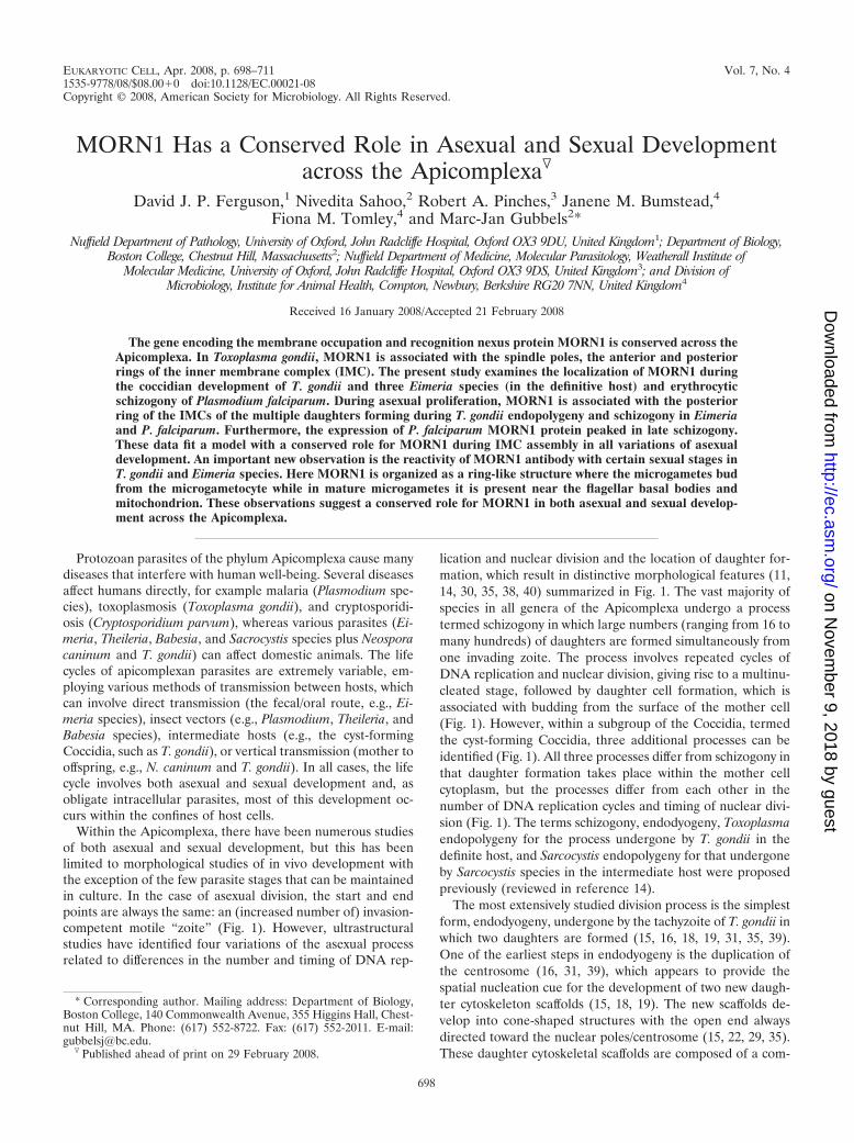
FIG. 1. Apicomplexan cell division flexibility. (A) The beginningand end product of division is the host cell invasion-competent “zoite.”Endodyogeny (A to C) is the formation of two daughters throughinternal budding. In schizogony (A and D to F), parasites first gothrough multiple rounds of DNA replication and mitosis before bud-ding and this is set apart from other strategies in that budding takesplace at the plasma membrane. Division by endopolygeny, as carriedout by T. gondii in the cat gut (A, D, G, and H), also involves multiplerounds of DNA replication and mitosis, but budding takes place in-ternally within the cytoplasm. Endopolygeny, as undergone by Sarco-cystis neurona (A and H to J), goes through several rounds of DNAreplication but omits karyokineses, and budding takes place internallywithin the cytoplasm. Daughter cell budding in schizogony and bothforms of endodyogeny is dependent on a final round of DNA replica-tion and mitosis and, in this last round, is very reminiscent ofendodyogeny. The nucleus is shown in light blue, the cytoskeleton isshown in red (IMC-positive subpellicular microtubules), and apicalrhoptry organelles are presented in green. The black “knobs” on thenucleus represent the spindle pole/centriolar complex.
VOL. 7, 2008 MORN1 IN APICOMPLEXA 699
on Novem
ber 9, 2018 by guesthttp://ec.asm
.org/D
ownloaded from

Antibodies. (i) Anti-TgMORN1. Anti-TgMORN1 is an affinity-purified rabbitantibody against the recombinant membrane occupation and recognition nexusprotein 1 (MORN1) of T. gondii (15).
(ii) Anti-TgIMC1. Anti-TgIMC1 is a monoclonal mouse antibody that recog-nizes the T. gondii IMC protein 1 (IMC1) (26) (kindly provided by Gary Ward,University of Vermont).
(iii) Anti-TgENO2. Anti-TgENO2 is a mouse antibody that recognizes theenolase 2 (ENO2) isoform of T. gondii, which weakly stains the cytoplasm andstrongly stains the nuclei in actively developing parasites (13) (kindly provided byStan Tomavo, Universite des Sciences et Technologies de Lille, France).
(iv) Anti-TgENR. Anti-TgENR is a mouse antibody that recognizes the enoylreductase (ENR) of T. gondii, which is located in the apicoplast (10).
(v) Anti-TgGRA7. Anti-TgGRA7 is a mouse monoclonal antibody to the densegranule protein 7 (GRA7) (20) that stains the parasitophorous vacuole and thedense granules within the merozoites (9) (kindly provided by D. Jacobs, Inno-genetics NV, Belgium).
(vi) Anti-EtENR. Anti-EtENR is a mouse antibody to E. tenella ENR, whichidentifies the apicoplast in Eimeria species (14).
(vii) Anti-EtAMA2. Anti-EtAMA2 is protein that recognizes the apical mem-brane antigen 2 protein (AMA2) that is located in the micronemes of themerozoites of E. tenella (F. Tomley, unpublished data).
(viii) Anti-EmAPGA. Anti-EmAPGA is a protein raised against affinity-puri-fied gametocyte antigens (APGA) of E. maxima that recognizes the wall-formingbodies within the macrogamete and the oocyst wall (5).
Histology and immunocytochemistry. Portions of the small intestine or cecum(E. tenella) were removed and fixed in 2% paraformaldehyde in 0.1 M phosphatebuffer, dehydrated, and embedded in wax. For histology, sections were stained withhematoxylin and eosin. For immunocytochemistry, sections were dewaxed and pres-sure cooked prior to immunolabeling. The sections were single labeled with anti-MORN1 or were double labeled with anti-TgMORN1 combined with one of thefollowing: anti-TgENR, anti-TgIMC1, anti-TgENO2, anti-TgGRA7, anti-EtENR,anti-EmAPGA, or anti-EtAMA2. After tissue sections were washed, the location ofthe primary antibody was visualized by using either goat anti-rabbit immunoglobulin(Ig) conjugated to fluorescein isothiocyanate (FITC) for single labeling or a mixtureof anti-rabbit Ig conjugated to FITC and goat anti-mouse Ig-conjugated Texas Red.After being washed, the sections were exposed to 4�,6�-diamindino-2-phenylindole(DAPI), washed, and mounted. Sections were examined by using a fluorescencemicroscope, and images were recorded by using Openlab software (Improvision).
Smears of sporozoites of E. tenella and P. falciparum-infected erythrocytescontaining mature parasites were fixed in 2% paraformaldehyde and then ace-tone before being air dried. The smears of P. falciparum were pretreated by anantigen-retrieval method involving heat treatment (80°C) in citrate buffer priorto staining with anti-TgMORN1. After blocking with 1% bovine serum albumin(BSA)-phosphate-buffered saline (PBS), slides of both parasites were stainedwith anti-TgMORN1, washed, and visualized with goat anti-rabbit Ig conjugatedto FITC. Slides were exposed to DAPI and mounted prior to examination.
Electron microscopy and immunoelectron microscopy. Small pieces of thesurface mucosa of the small intestine or cecum were fixed in 4% glutaraldehydein 0.1 M phosphate buffer for routine electron microscopy. For transmissionelectron microscopy, the samples were postfixed in osmium tetroxide, dehy-drated in ethanol, treated with propylene oxide, and embedded in Spurr’s epoxyresin. Sections were stained with uranyl acetate and lead citrate prior to exam-ination with a JEOL 1200EX electron microscope (9). For scanning electronmicroscopy, small slices (through the mucosa) were dehydrated, critical pointdried, mounted on stubs, sputter coated with gold, and examined under a Philips505 scanning electron microscope.
For immunoelectron microscopy, small pieces of tissue were fixed in 2% para-formaldehyde in 0.1 M phosphate buffer, dehydrated, and embedded in LR Whiteacrylic resin. Thin sections on Formvar-coated nickel grids were floated on drops of1% BSA in PBS to block nonspecific staining, followed with the use of primaryantibody, anti-TgMORN1. After being washed, sections were floated on drops ofgoat anti-rabbit Ig conjugated to 10-nm gold particles. The sections were stained withuranyl acetate prior to examination with the electron microscope (9).
P. falciparum cell fractionation. Synchronous cultures of P. falciparum para-sites were harvested at the ring stage (6 to 12 h), the trophozoite stage (18 to24 h), and the schizont stage (36 to 42 h). Cells were solubilized in 150 mM NaCl,5 mM EDTA, 50 mM Tris (pH 8.0), 1% Triton X-100 with protease inhibitors togenerate the Triton X-100-soluble fraction. Triton X-100-insoluble pellets werethen extracted in the same buffer supplemented with 2% sodium dodecyl sulfate(SDS) to produce the Triton X-100-insoluble, SDS-soluble fraction. Cocultureduninfected human red blood cells were processed as described above to providecontrol samples for subsequent analysis. The samples were subjected to SDS-polyacrylamide gel electrophoresis (10% acrylamide) and blotted onto nitrocel-
lulose membranes. Immunodetection was carried out using ECL reagents(Amersham) with anti-TgMORN1 antibody (1:1,000 dilution) and peroxidase-conjugated swine anti-rabbit secondary antibody (1:1,000 dilution; DAKO).
E. tenella-stage isolation and Western blotting. (i) Sporozoites. Sporozoiteswere recovered from cleaned oocysts by in vitro excystation and purified by anionexchange chromatography over columns of nylon wool and DE-52 cellulose (32).
(ii) In vitro first-generation schizogony. Monolayers of Madin-Darby bovinekidney cells (Flow Laboratories) were seeded into 24-well culture plates (Nunc)in Ham’s F-12 nutrient medium with 10% fetal calf serum and infected with250,000 freshly sporulated sporozoites. Cells were then cultured at 41°C in 5%CO2 until first-generation schizonts were observed. Samples were taken at 40 h(mid-stage schizonts) and 55 h (late-stage schizonts) postinfection. The cellmedium was aspirated, the monolayers were washed three times, the number ofschizonts per well was counted, and cells were scraped into 100 �l PBS contain-ing protease inhibitors.
(iii) In vivo second-generation merozoites. Second-generation merozoiteswere recovered from the cecal mucosa of chickens at 112 h after oral inoculationwith 5 � 105 sporulated oocysts (36).
(iv) Gametocytes. Chickens at 4 weeks of age were infected with 10,000oocysts. The chickens were killed at 143 to 145 h postinfection, and the ceca wereremoved and washed with cold SAC (170 mM NaCl, 10 mM Tris-HCl, pH 7, 10mM glucose, 5 mM CaCl2, 1 mM phenylmethanesulfonyl fluoride, 1 mg ml�1
BSA). The ceca were slit open and incubated at 37°C in a beaker with 0.5 mgml�1 hyaluronidase in SAC (five ceca in 50 ml). The ceca were placed on top ofa 17-�m mesh filter, and the mucosae were washed through with SAC (roomtemperature). The material left on the mesh was discarded, and the flowthroughwas filtered through a 10-�m mesh filter. The gametocytes accumulated on thisfilter and were washed off with SAC and centrifuged at 800 � g for 5 min (44).
The Western blot analysis was set up using standard procedures. Proteins fromeach sample were resolved by electrophoresis through 4 to 15% gradient SDS-polyacrylamide gel electrophoresis gels and transferred to nitrocellulose mem-branes by semidry blotting. Nonspecific binding sites were blocked by incubationfor 1 h in 5% BSA in PBS, and then filters were probed with anti-MORN1,followed by goat anti-rabbit IgG conjugated to horseradish peroxidase. Bindingwas visualized by ECL (Amersham).
RESULTS
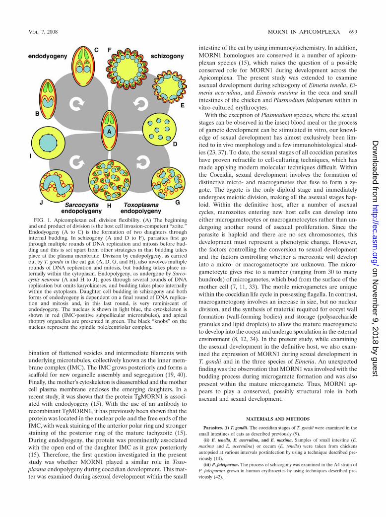
Asexual replication of T. gondii in the definite host by en-dopolygeny. The asexual development of T. gondii in the catintestine has previously been described as involving a prolifer-ative phase with repeated nuclear divisions, followed by a dif-ferentiation phase involving daughter formation. Daughterformation is initiated within the mother cell cytoplasm andprogresses internally (Fig. 1 and 2A). Because more than twodaughters are forming, this mechanism has been termed en-dopolygeny (11, 14, 30). The process was examined by double-labeled immunofluorescence using anti-TgMORN1 combinedwith anti-ENR, anti-ENO2, anti-GRA7, or anti-IMC1 to assistin identifying the individual developmental stages. It was ob-served that the early multinucleate stages, possessing elongateand branched ENR-positive apicoplasts, were unstained withMORN1 (data not shown). However, in the later stages, withmultiple fragmented ENR-positive apicoplasts and ENO2-pos-itive nuclei, there were multiple MORN1-positive ring-likestructures distributed within the mother cell cytoplasm (Fig.2C). To identify the subcellular location of MORN1, immuno-electron microscopy was employed. At an early stage in daugh-ter formation, the eccentric nuclear spindle present in each ofthe multiple nuclei was labeled for MORN1 (Fig. 2B). Inaddition, the free ends of the cone-shaped structures formedby the daughter IMC were also labeled (Fig. 2B). The labelingappeared at the ends of the IMC as small clumps of goldparticles in longitudinal sections (arrowheads, Fig. 2B) or anelongate line of gold particles in tangential sections (Fig. 2B).Two daughters formed in association with each nucleus and
700 FERGUSON ET AL. EUKARYOT. CELL
on Novem
ber 9, 2018 by guesthttp://ec.asm
.org/D
ownloaded from
FIG. 2. MORN1 distribution during endopolygeny of T. gondii in the cat intestinal epithelium. (A) Transmission electron micrograph of aparasite in mid-stage Toxoplasma endopolygeny, showing the internally located cone-shaped IMCs of the developing daughters (D) associated withthe multiple nuclei (N). Bar � 1 �m. (B) Electron micrograph of the early stage in daughter formation; immunostaining was done with TgMORN1antibody, and labeling was done with 10-nm gold particles. Note the gold particles localizing to the spindle poles (NS) of the multiple nuclei (N) andthe posterior extremities of the cup-shaped (arrowheads), newly forming, inner membrane cytoskeleton scaffolds (I). In addition, one IMC hasbeen cut tangentially through the base of the cone, showing gold particles along its length (parallel arrows in the lower right corner). Bar � 100nm. (C) Fluorescent micrograph of T. gondii (in a stage of endopolygeny similar to that shown in panel A) double labeled with anti-MORN1(green) and ENO2 (red) counterstained with DAPI (blue). A number of MORN1-positive rings (arrows), often in pairs (arrows), are observed overthe ENO2- and DAPI-positive nuclei (N) distributed throughout the cytoplasm. Bar � 1 �m. (D) Detail of part of an immunoelectron micrographof a schizont showing more advanced daughter formation with the IMCs (outlined by the dotted lines) enclosing the ends of the crescent-shapednucleus (N). Arrowheads mark MORN1-specific gold particles at the sites of posterior budding of the IMC. In addition, apical staining is observed(arrow). R, rhoptry. Bar � 100 nm. (E) Immunofluorescence section stained with anti-MORN1 (green) and ENO2 (red) showing a mature schizontwith fully formed merozoites. Note the strong MORN1 staining of the basal end of the merozoites (arrow), with weak labeling of the nuclear pole(arrowhead). N, nucleus. Bar � 1 �m. (F) Immunofluorescence section stained with anti-MORN1 (green) and anti-IMC (red), showing a matureschizont with fully formed merozoites. The merozoites are outlined in red due to the IMC staining and exhibit strong posterior staining for MORN1(arrow). I, IMC. Bar � 1 �m. (G) Immunoelectron micrograph of a mature schizont labeled with anti-MORN1, showing the fully formedmerozoites (M) still connected by their posterior ends to the residual cytoplasmic mass. Bar � 1 �m. (H) Enlargement of the enclosed area in panelG showing numerous gold particles associated with the basal end of the IMC (arrows). Bar � 100 nm.
701
on Novem
ber 9, 2018 by guesthttp://ec.asm
.org/D
ownloaded from
were located above the nuclear poles (Fig. 2B and C). As theposterior growth of the IMC continued, they started to enclosevarious organelles, with each nucleus dividing between twodaughters (Fig. 2D). During this process, the ends of the IMCremained labeled with MORN1 and there was evidence of low-level labeling of the anterior ring of the IMC (Fig. 2D). Thisgrowth continued until the daughters were fully formed and oc-
cupied all the space within the mother cell (data not shown). Thefinal stage involved invagination of the mother cell plasmalemmaaround each of the daughters from anterior to posterior to formthe characteristic pellicle with the posterior of each daughterremaining attached to a small amount of residual cytoplasm (Fig.2E to H). The posterior ring formed by the IMC was still stronglylabeled for MORN1 (Fig. 2E to G).
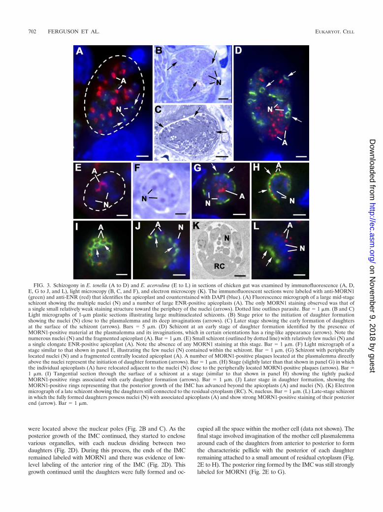
FIG. 3. Schizogony in E. tenella (A to D) and E. acervulina (E to L) in sections of chicken gut was examined by immunofluorescence (A, D,E, G to J, and L), light microscopy (B, C, and F), and electron microscopy (K). The immunofluorescent sections were labeled with anti-MORN1(green) and anti-ENR (red) that identifies the apicoplast and counterstained with DAPI (blue). (A) Fluorescence micrograph of a large mid-stageschizont showing the multiple nuclei (N) and a number of large ENR-positive apicoplasts (A). The only MORN1 staining observed was that ofa single small relatively weak staining structure toward the periphery of the nuclei (arrows). Dotted line outlines parasite. Bar � 1 �m. (B and C)Light micrographs of 1-�m plastic sections illustrating large multinucleated schizonts. (B) Stage prior to the initiation of daughter formationshowing the nuclei (N) close to the plasmalemma and its deep invaginations (arrows). (C) Later stage showing the early formation of daughtersat the surface of the schizont (arrows). Bars � 5 �m. (D) Schizont at an early stage of daughter formation identified by the presence ofMORN1-positive material at the plasmalemma and its invaginations, which in certain orientations has a ring-like appearance (arrows). Note thenumerous nuclei (N) and the fragmented apicoplast (A). Bar � 1 �m. (E) Small schizont (outlined by dotted line) with relatively few nuclei (N) anda single elongate ENR-positive apicoplast (A). Note the absence of any MORN1 staining at this stage. Bar � 1 �m. (F) Light micrograph of astage similar to that shown in panel E, illustrating the few nuclei (N) contained within the schizont. Bar � 1 �m. (G) Schizont with peripherallylocated nuclei (N) and a fragmented centrally located apicoplast (A). A number of MORN1-positive plaques located at the plasmalemma directlyabove the nuclei represent the initiation of daughter formation (arrows). Bar � 1 �m. (H) Stage (slightly later than that shown in panel G) in whichthe individual apicoplasts (A) have relocated adjacent to the nuclei (N) close to the peripherally located MORN1-positive plaques (arrows). Bar �1 �m. (I) Tangential section through the surface of a schizont at a stage (similar to that shown in panel H) showing the tightly packedMORN1-positive rings associated with early daughter formation (arrows). Bar � 1 �m. (J) Later stage in daughter formation, showing theMORN1-positive rings representing that the posterior growth of the IMC has advanced beyond the apicoplasts (A) and nuclei (N). (K) Electronmicrograph of a late schizont showing the daughters still connected to the residual cytoplasm (RC). N, nucleus. Bar � 1 �m. (L) Late-stage schizontin which the fully formed daughters possess nuclei (N) with associated apicoplasts (A) and show strong MORN1-positive staining of their posteriorend (arrow). Bar � 1 �m.
702 FERGUSON ET AL. EUKARYOT. CELL
on Novem
ber 9, 2018 by guesthttp://ec.asm
.org/D
ownloaded from
MORN1 in classical Eimeria species schizogony. In a man-ner similar to that for Toxoplasma endopolygeny, schizogonyalso involves a proliferative phase with repeated nuclear divi-sions, followed by a differentiation phase involving daughterformation by budding from the mother cell surface (Fig. 1Eand F). The number of daughters produced varies significantlybetween species and even between asexual generations withineach species. The number of daughters ranges from approxi-mately 16 (E. acervulina and E. maxima) to many hundreds (E.tenella). When schizonts produce large numbers of daughters,the surface area is increased by multiple deep invaginations ofthe plasmalemma (Fig. 3B) (14).
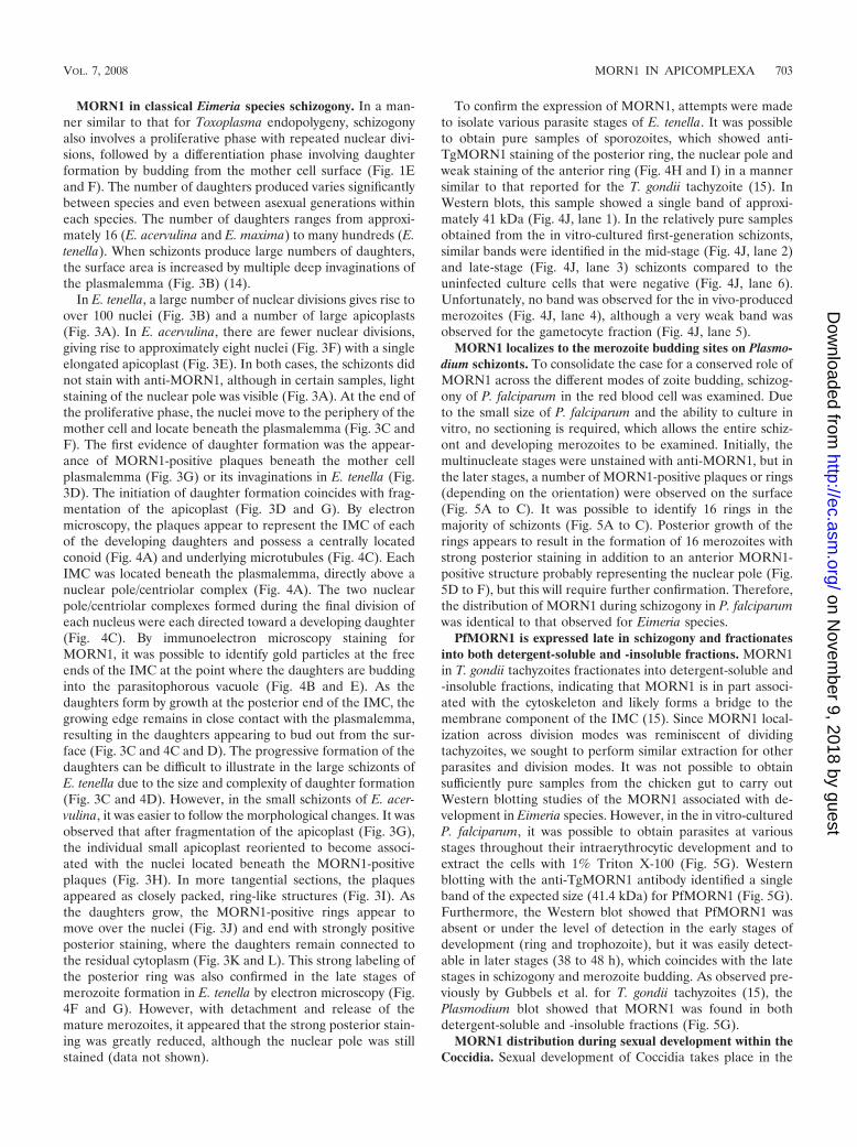
In E. tenella, a large number of nuclear divisions gives rise toover 100 nuclei (Fig. 3B) and a number of large apicoplasts(Fig. 3A). In E. acervulina, there are fewer nuclear divisions,giving rise to approximately eight nuclei (Fig. 3F) with a singleelongated apicoplast (Fig. 3E). In both cases, the schizonts didnot stain with anti-MORN1, although in certain samples, lightstaining of the nuclear pole was visible (Fig. 3A). At the end ofthe proliferative phase, the nuclei move to the periphery of themother cell and locate beneath the plasmalemma (Fig. 3C andF). The first evidence of daughter formation was the appear-ance of MORN1-positive plaques beneath the mother cellplasmalemma (Fig. 3G) or its invaginations in E. tenella (Fig.3D). The initiation of daughter formation coincides with frag-mentation of the apicoplast (Fig. 3D and G). By electronmicroscopy, the plaques appear to represent the IMC of eachof the developing daughters and possess a centrally locatedconoid (Fig. 4A) and underlying microtubules (Fig. 4C). EachIMC was located beneath the plasmalemma, directly above anuclear pole/centriolar complex (Fig. 4A). The two nuclearpole/centriolar complexes formed during the final division ofeach nucleus were each directed toward a developing daughter(Fig. 4C). By immunoelectron microscopy staining forMORN1, it was possible to identify gold particles at the freeends of the IMC at the point where the daughters are buddinginto the parasitophorous vacuole (Fig. 4B and E). As thedaughters form by growth at the posterior end of the IMC, thegrowing edge remains in close contact with the plasmalemma,resulting in the daughters appearing to bud out from the sur-face (Fig. 3C and 4C and D). The progressive formation of thedaughters can be difficult to illustrate in the large schizonts ofE. tenella due to the size and complexity of daughter formation(Fig. 3C and 4D). However, in the small schizonts of E. acer-vulina, it was easier to follow the morphological changes. It wasobserved that after fragmentation of the apicoplast (Fig. 3G),the individual small apicoplast reoriented to become associ-ated with the nuclei located beneath the MORN1-positiveplaques (Fig. 3H). In more tangential sections, the plaquesappeared as closely packed, ring-like structures (Fig. 3I). Asthe daughters grow, the MORN1-positive rings appear tomove over the nuclei (Fig. 3J) and end with strongly positiveposterior staining, where the daughters remain connected tothe residual cytoplasm (Fig. 3K and L). This strong labeling ofthe posterior ring was also confirmed in the late stages ofmerozoite formation in E. tenella by electron microscopy (Fig.4F and G). However, with detachment and release of themature merozoites, it appeared that the strong posterior stain-ing was greatly reduced, although the nuclear pole was stillstained (data not shown).
To confirm the expression of MORN1, attempts were madeto isolate various parasite stages of E. tenella. It was possibleto obtain pure samples of sporozoites, which showed anti-TgMORN1 staining of the posterior ring, the nuclear pole andweak staining of the anterior ring (Fig. 4H and I) in a mannersimilar to that reported for the T. gondii tachyzoite (15). InWestern blots, this sample showed a single band of approxi-mately 41 kDa (Fig. 4J, lane 1). In the relatively pure samplesobtained from the in vitro-cultured first-generation schizonts,similar bands were identified in the mid-stage (Fig. 4J, lane 2)and late-stage (Fig. 4J, lane 3) schizonts compared to theuninfected culture cells that were negative (Fig. 4J, lane 6).Unfortunately, no band was observed for the in vivo-producedmerozoites (Fig. 4J, lane 4), although a very weak band wasobserved for the gametocyte fraction (Fig. 4J, lane 5).
MORN1 localizes to the merozoite budding sites on Plasmo-dium schizonts. To consolidate the case for a conserved role ofMORN1 across the different modes of zoite budding, schizog-ony of P. falciparum in the red blood cell was examined. Dueto the small size of P. falciparum and the ability to culture invitro, no sectioning is required, which allows the entire schiz-ont and developing merozoites to be examined. Initially, themultinucleate stages were unstained with anti-MORN1, but inthe later stages, a number of MORN1-positive plaques or rings(depending on the orientation) were observed on the surface(Fig. 5A to C). It was possible to identify 16 rings in themajority of schizonts (Fig. 5A to C). Posterior growth of therings appears to result in the formation of 16 merozoites withstrong posterior staining in addition to an anterior MORN1-positive structure probably representing the nuclear pole (Fig.5D to F), but this will require further confirmation. Therefore,the distribution of MORN1 during schizogony in P. falciparumwas identical to that observed for Eimeria species.
PfMORN1 is expressed late in schizogony and fractionatesinto both detergent-soluble and -insoluble fractions. MORN1in T. gondii tachyzoites fractionates into detergent-soluble and-insoluble fractions, indicating that MORN1 is in part associ-ated with the cytoskeleton and likely forms a bridge to themembrane component of the IMC (15). Since MORN1 local-ization across division modes was reminiscent of dividingtachyzoites, we sought to perform similar extraction for otherparasites and division modes. It was not possible to obtainsufficiently pure samples from the chicken gut to carry outWestern blotting studies of the MORN1 associated with de-velopment in Eimeria species. However, in the in vitro-culturedP. falciparum, it was possible to obtain parasites at variousstages throughout their intraerythrocytic development and toextract the cells with 1% Triton X-100 (Fig. 5G). Westernblotting with the anti-TgMORN1 antibody identified a singleband of the expected size (41.4 kDa) for PfMORN1 (Fig. 5G).Furthermore, the Western blot showed that PfMORN1 wasabsent or under the level of detection in the early stages ofdevelopment (ring and trophozoite), but it was easily detect-able in later stages (38 to 48 h), which coincides with the latestages in schizogony and merozoite budding. As observed pre-viously by Gubbels et al. for T. gondii tachyzoites (15), thePlasmodium blot showed that MORN1 was found in bothdetergent-soluble and -insoluble fractions (Fig. 5G).
MORN1 distribution during sexual development within theCoccidia. Sexual development of Coccidia takes place in the
VOL. 7, 2008 MORN1 IN APICOMPLEXA 703
on Novem
ber 9, 2018 by guesthttp://ec.asm
.org/D
ownloaded from
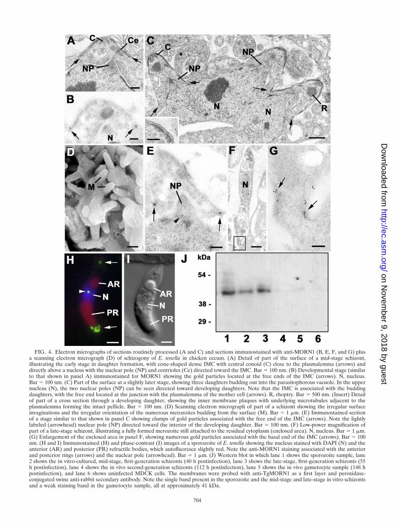
FIG. 4. Electron micrographs of sections routinely processed (A and C) and sections immunostained with anti-MORN1 (B, E, F, and G) plusa scanning electron micrograph (D) of schizogony of E. tenella in chicken cecum. (A) Detail of part of the surface of a mid-stage schizont,illustrating the early stage in daughter formation, with cone-shaped dense IMC with central conoid (C) close to the plasmalemma (arrows) anddirectly above a nucleus with the nuclear pole (NP) and centrioles (Ce) directed toward the IMC. Bar � 100 nm. (B) Developmental stage (similarto that shown in panel A) immunostained for MORN1 showing the gold particles located at the free ends of the IMC (arrows). N, nucleus.Bar � 100 nm. (C) Part of the surface at a slightly later stage, showing three daughters budding out into the parasitophorous vacuole. In the uppernucleus (N), the two nuclear poles (NP) can be seen directed toward developing daughters. Note that the IMC is associated with the buddingdaughters, with the free end located at the junction with the plasmalemma of the mother cell (arrows). R, rhoptry. Bar � 500 nm. (Insert) Detailof part of a cross section through a developing daughter, showing the inner membrane plaques with underlying microtubules adjacent to theplasmalemma forming the intact pellicle. Bar � 100 nm. (D) Scanning electron micrograph of part of a schizont showing the irregular surfaceinvaginations and the irregular orientation of the numerous merozoites budding from the surface (M). Bar � 1 �m. (E) Immunostained sectionof a stage similar to that shown in panel C showing clumps of gold particles associated with the free end of the IMC (arrows). Note the lightlylabeled (arrowhead) nuclear pole (NP) directed toward the interior of the developing daughter. Bar � 100 nm. (F) Low-power magnification ofpart of a late-stage schizont, illustrating a fully formed merozoite still attached to the residual cytoplasm (enclosed area). N, nucleus. Bar � 1 �m.(G) Enlargement of the enclosed area in panel F, showing numerous gold particles associated with the basal end of the IMC (arrows). Bar � 100nm. (H and I) Immunostained (H) and phase-contrast (I) images of a sporozoite of E. tenella showing the nucleus stained with DAPI (N) and theanterior (AR) and posterior (PR) refractile bodies, which autofluoresce slightly red. Note the anti-MORN1 staining associated with the anteriorand posterior rings (arrows) and the nuclear pole (arrowhead). Bar � 1 �m. (J) Western blot in which lane 1 shows the sporozoite sample, lane2 shows the in vitro-cultured, mid-stage, first-generation schizonts (40 h postinfection), lane 3 shows the late-stage, first-generation schizonts (55h postinfection), lane 4 shows the in vivo second-generation schizonts (112 h postinfection), lane 5 shows the in vivo gametocyte sample (148 hpostinfection), and lane 6 shows uninfected MDCK cells. The membranes were probed with anti-TgMORN1 as a first layer and peroxidase-conjugated swine anti-rabbit secondary antibody. Note the single band present in the sporozoite and the mid-stage and late-stage in vitro schizontsand a weak staining band in the gametocyte sample, all at approximately 41 kDa.
704
on Novem
ber 9, 2018 by guesthttp://ec.asm
.org/D
ownloaded from
cecum and small intestine, resulting in microgametes and mac-rogametes with morphological features reminiscent of maleand female gametes, respectively. Microgametogony gives riseto a variable number of motile microgametes, whereas mac-
rogametogony gives rise to a single macrogamete. Sexual de-velopment was studied across four coccidian species: T. gondii,E acervulina, E. tenella, and E. maxima. They developed insimilar manners and varied only in size and the number ofgametes formed. Therefore, coccidian sexual development willbe represented in a combined description without differenti-ating between the species. Various stages in the developmentof the two processes were observed located in adjacent epithe-lial cells (Fig. 6A, B, and D and 7A).
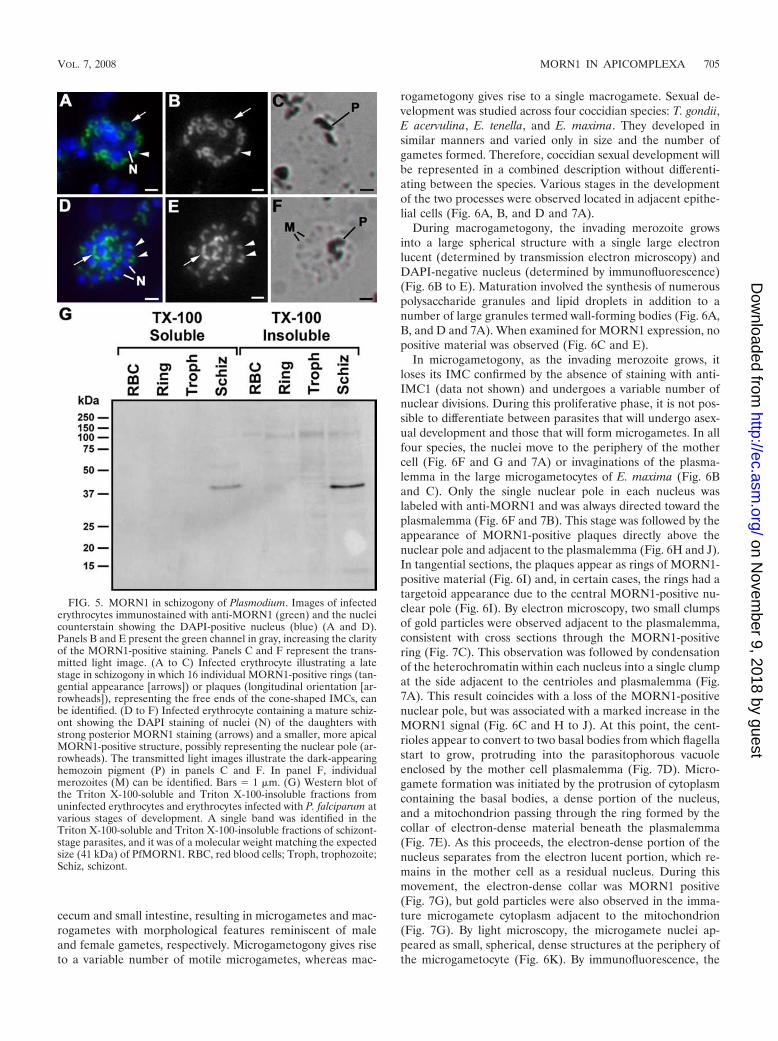
During macrogametogony, the invading merozoite growsinto a large spherical structure with a single large electronlucent (determined by transmission electron microscopy) andDAPI-negative nucleus (determined by immunofluorescence)(Fig. 6B to E). Maturation involved the synthesis of numerouspolysaccharide granules and lipid droplets in addition to anumber of large granules termed wall-forming bodies (Fig. 6A,B, and D and 7A). When examined for MORN1 expression, nopositive material was observed (Fig. 6C and E).
In microgametogony, as the invading merozoite grows, itloses its IMC confirmed by the absence of staining with anti-IMC1 (data not shown) and undergoes a variable number ofnuclear divisions. During this proliferative phase, it is not pos-sible to differentiate between parasites that will undergo asex-ual development and those that will form microgametes. In allfour species, the nuclei move to the periphery of the mothercell (Fig. 6F and G and 7A) or invaginations of the plasma-lemma in the large microgametocytes of E. maxima (Fig. 6Band C). Only the single nuclear pole in each nucleus waslabeled with anti-MORN1 and was always directed toward theplasmalemma (Fig. 6F and 7B). This stage was followed by theappearance of MORN1-positive plaques directly above thenuclear pole and adjacent to the plasmalemma (Fig. 6H and J).In tangential sections, the plaques appear as rings of MORN1-positive material (Fig. 6I) and, in certain cases, the rings had atargetoid appearance due to the central MORN1-positive nu-clear pole (Fig. 6I). By electron microscopy, two small clumpsof gold particles were observed adjacent to the plasmalemma,consistent with cross sections through the MORN1-positivering (Fig. 7C). This observation was followed by condensationof the heterochromatin within each nucleus into a single clumpat the side adjacent to the centrioles and plasmalemma (Fig.7A). This result coincides with a loss of the MORN1-positivenuclear pole, but was associated with a marked increase in theMORN1 signal (Fig. 6C and H to J). At this point, the cent-rioles appear to convert to two basal bodies from which flagellastart to grow, protruding into the parasitophorous vacuoleenclosed by the mother cell plasmalemma (Fig. 7D). Micro-gamete formation was initiated by the protrusion of cytoplasmcontaining the basal bodies, a dense portion of the nucleus,and a mitochondrion passing through the ring formed by thecollar of electron-dense material beneath the plasmalemma(Fig. 7E). As this proceeds, the electron-dense portion of thenucleus separates from the electron lucent portion, which re-mains in the mother cell as a residual nucleus. During thismovement, the electron-dense collar was MORN1 positive(Fig. 7G), but gold particles were also observed in the imma-ture microgamete cytoplasm adjacent to the mitochondrion(Fig. 7G). By light microscopy, the microgamete nuclei ap-peared as small, spherical, dense structures at the periphery ofthe microgametocyte (Fig. 6K). By immunofluorescence, the
FIG. 5. MORN1 in schizogony of Plasmodium. Images of infectederythrocytes immunostained with anti-MORN1 (green) and the nucleicounterstain showing the DAPI-positive nucleus (blue) (A and D).Panels B and E present the green channel in gray, increasing the clarityof the MORN1-positive staining. Panels C and F represent the trans-mitted light image. (A to C) Infected erythrocyte illustrating a latestage in schizogony in which 16 individual MORN1-positive rings (tan-gential appearance [arrows]) or plaques (longitudinal orientation [ar-rowheads]), representing the free ends of the cone-shaped IMCs, canbe identified. (D to F) Infected erythrocyte containing a mature schiz-ont showing the DAPI staining of nuclei (N) of the daughters withstrong posterior MORN1 staining (arrows) and a smaller, more apicalMORN1-positive structure, possibly representing the nuclear pole (ar-rowheads). The transmitted light images illustrate the dark-appearinghemozoin pigment (P) in panels C and F. In panel F, individualmerozoites (M) can be identified. Bars � 1 �m. (G) Western blot ofthe Triton X-100-soluble and Triton X-100-insoluble fractions fromuninfected erythrocytes and erythrocytes infected with P. falciparum atvarious stages of development. A single band was identified in theTriton X-100-soluble and Triton X-100-insoluble fractions of schizont-stage parasites, and it was of a molecular weight matching the expectedsize (41 kDa) of PfMORN1. RBC, red blood cells; Troph, trophozoite;Schiz, schizont.
VOL. 7, 2008 MORN1 IN APICOMPLEXA 705
on Novem
ber 9, 2018 by guesthttp://ec.asm
.org/D
ownloaded from

FIG. 6. Sections illustrating the light microscopic (A, B, D, G, K, and O) and immunolight microscopic (C, E, F, H to J, L to N, P, and Q)appearances of the sexual development of various coccidian parasites in the intestine of the definitive host. The immunostained sections werelabeled with anti-MORN1 (green) combined with anti-APGP (wall-forming bodies stained red) (C and E) or anti-ENR (apicoplasts stained red)(F, H to J, L to N, P, and Q), and all were counterstained with DAPI (blue). The light microscopy for morphology consisted of wax sections stainedwith hematoxylin and eosin (A) or 1-�m plastic sections stained with Azure A (B, C, G, K, and O). (A) Low-power magnification of a longitudinalsection through a villus of a chicken infected with E. maxima, illustrating numerous developmental stages of microgametocytes (Mi) andmacrogametocytes (Ma) and early oocyst formation (O) within the epithelial cells. Bar � 10 �m. (B and D) Moderate-power magnificationshowing sexual development of E. maxima and E. tenella, respectively, showing early-stage (EMi), mid-stage (MMi), and late-stage (LMi)microgametocytes and an adjacent macrogametocyte (Ma) in panel B characterized by the presence of densely staining granules representing thewall-forming bodies (W). Bar � 5 �m. Panels C and E show sections containing sexual stages similar to those in panels B and D, respectively, inwhich the microgametocytes were labeled with anti-MORN1 (green), while the macrogametocytes (Ma) identified by the wall-forming bodies(W) were unstained. Note that in the early microgametocyte (EMi), there was low-intensity staining limited to the nuclear poles, while there wasincreased staining intensity in the mid-stage microgametocytes (MMi) and late microgametocytes (LMi) associated with microgamete formation.Bar � 5 �m. (F) Early microgametocyte of E. tenella showing the peripherally located nuclei (N), each with a single MORN1-positive spot on theouter aspect of each nucleus (arrows), while the cytoplasm contains a few small apicoplasts (A). Bar � 1 �m. (G) Light microscopy (of a stagesimilar to that shown in panel F) showing the peripherally located nuclei (N). Bar � 1 �m. (H) Early microgametocyte of E. acervulina in whichMORN1-positive plaques were present at the surface of the parasite (arrows) directly above each nucleus (N), representing the initiation ofmicrogamete formation. A, apicoplast. Bar � 1 �m. (Insert) Enlargement of the enclosed area in panel H in which it was possible to resolve theMORN1-positive nuclear pole just beneath the MORN1-positive plaque. Bar � 500 nm. (I) Tangential section through the surface of an E.acervulina microgametocyte (similar to that shown in panel H) illustrating the ring-like appearance of the plaques in cross section (arrows). A fewhave targetoid appearances, where the centrally located MORN1-positive nuclear pole was also visible (arrowhead). Bar � 1 �m. (J) Earlymicrogametocyte of T. gondii showing relatively few MORN1-positive plaques (arrows) at the surface above the nuclei (N). Bar � 1 �m. (K) Light
706
on Novem
ber 9, 2018 by guesthttp://ec.asm
.org/D
ownloaded from

nuclei were strongly stained with DAPI and had moved frombeing internal to the MORN1-positive material (Fig. 6H to J)to being located on the external side of the MORN1-positiverings (Fig. 6L and M). The microgamete matures while stillattached by its anterior to the mother cell by an aperturedelineated by the electron-dense collar (Fig. 7F), which labelswith MORN1 (Fig. 7H). Maturation involves the continuedgrowth of the two flagella and elongation of the mitochon-drion, which is located close to the basal bodies and partiallyoverlaps with the DAPI-positive, elongated, crescent-shapednucleus (Fig. 6N and O and 7F and H). This was also associ-ated with an elongated appearance of MORN1 staining mate-rial in the anterior of the microgamete (Fig. 6M, N, and P),which correlated to the numerous gold particles observed inthe microgamete anterior around the mitochondrion (Fig. 7K,L, and M). The mature microgamete is released by detachmentfrom the ring-like collar. The anterior of each microgametepossesses two basal bodies from which long flagella protrude,an electron-dense, bar-like structure termed the perforatoriumand four microtubules that run posteriorly over the mitochon-drion and part of the nucleus (Fig. 6P and 7I and J). The freemicrogametes retain the strong anterior MORN1 staining, al-lowing individual microgametes to be identified by immuno-fluorescence within the gut lumen and close to early oocysts(Fig. 6Q). Immunoelectron microscopy showed a few goldparticles at the apex (Fig. 7M), but the largest numbers werelocated in the cytoplasm between the mitochondrion and theplasmalemma (Fig. 7K, L, and M).
DISCUSSION
This study provides evidence that the antibody to TgMORN1recognizes and stains the MORN1 homologues with the cor-rect predicted molecular weight in E. tenella (Fig. 4J) and P.falciparum (Fig. 5G). From examination of the asexual andsexual processes, we observed that MORN1 has similar dy-namic expression and localization patterns and therefore prob-ably has a conserved function in the various members of theApicomplexa. MORN1 has been identified at four distinctlocations, the nuclear poles, the edges of the IMC duringasexual development (the leading edge during formation isspecifically enriched in MORN1), the site of budding of themicrogamete, and the anterior of microgamete.
Nuclear localization. The early stages of Toxoplasma en-dopolygeny, schizogony, and microgametogony are indistin-guishable and involve repeated nuclear division, giving rise to
a multinucleate organism. At this stage, MORN1 staining wasobserved to be limited to the nuclear poles. This observationwas described in detail during endodyogeny in T. gondii, whereit was shown that MORN1 was associated with the character-istic conical-shaped structure of the nuclear pole, the centro-cone, formed by a dilation of the nuclear membranes, whichremain intact during division (15). In the present study, thislocation was confirmed across the Apicomplexa species exam-ined. The presence and specific orientation of the MORN1-positive nuclear spindle pole and associated centrioles appearto act as an organizing unit for both daughter and microgameteformation. In both situations, this control mechanism will en-sure that a nucleus will be associated with each developingzoite or microgamete, thus ensuring their viability. However,microgametogony is characterized by peripheral condensationof the nuclear chromatin and with loss of the nuclear pole,which is the first distinguishing morphological feature of mi-crogametogony (11). In contrast, the zoites formed by all formsof asexual development retain the MORN1-positive nuclearpole throughout their transmission cycle, which is consistentwith the case described previously for the tachyzoite of T.gondii (15).
Maintenance of MORN1 in the centrocone throughout theasexual cycle was also shown in the endopolygeny of Sarcocystisneurona, since here the polyploid nucleus has to be resolvedinto haploid zoites. The maintenance of the spindle and asso-ciation with the chromosomes throughout the cell cycle en-sures that each zoite is endowed with a single complete set ofchromosomes (15, 43). However, the stability of MORN1 lo-calization in the centrocone is not dependent on the presenceof microtubules, as its localization remained unchanged uponincubation with the microtubule depolymerizing agent oryzalin(15). Therefore, it appears that in addition to the proposedinvolvement of MORN1 with nucleation of daughter scaffolds,it is likely also part of the structural complex organizing thespindle pole, where the microtubules penetrate the nuclearenvelope. Both functions appear to be conserved throughoutthe different asexual division modes across the Apicomplexa.
Asexual replication. While there are distinct morphologicaldifferences in the various asexual processes undergone by api-complexan parasites concerning the number and timing ofnuclear divisions and the number and location of daughterformation (Fig. 1), the mechanism of daughter formation ap-pears to have retained many similarities across the phylum. Inall four processes, the formation of the IMC of the daughterpellicle appeared to be coordinated via the nuclear pole/cen-
micrograph of a mid-stage microgametocyte of E. tenella in which the dense staining spherical-shaped nuclei (N) are arranged around the peripheryof the parasite. Bar � 1 �m. (L) Stage (similar to that shown in panel K) showing the spherical DAPI staining nuclei (N) now located on theexternal side of the MORN1-positive plaques (arrows). Bar � 1 �m. (M) Slightly later stage than that shown in panel L, showing that the nuclei(N) have started to elongate and an elongated MORN1-positive structure is present anterior to the nuclei (arrows). Bar � 1 �m. (N) Part of alate microgametocyte of E. maxima in which a large number of microgametes with crescent-shaped DAPI-positive nuclei (N) and elongatedanterior MORN1-positive structures (arrows) are still attached to the mother cell. Bar � 1 �m. (O) Light micrograph of a late microgametocyteof E. tenella in which a number of mature microgametes with elongated nuclei (N) can be identified. Bar � 1 �m. (P) Late microgametocyte ofE. acervulina showing a number of mature microgametes each with the DAPI-positive nuclei (N) and the elongated anterior MORN1-positivestructure (arrows). Bar � 1 �m. (Q) Section through the lumen of a cecal crypt of a chicken infected with E. tenella in which two microgametescan be identified by the DAPI-positive nucleus and the undulating MORN1-positive anterior staining (arrows). Note that one is adjacent to an earlyoocyst (O) recognized by the slight autofluorescence of the oocyst wall (arrowheads) and the apicoplast (A) associated with the nucleus. Bar �1 �m.
VOL. 7, 2008 MORN1 IN APICOMPLEXA 707
on Novem
ber 9, 2018 by guesthttp://ec.asm
.org/D
ownloaded from
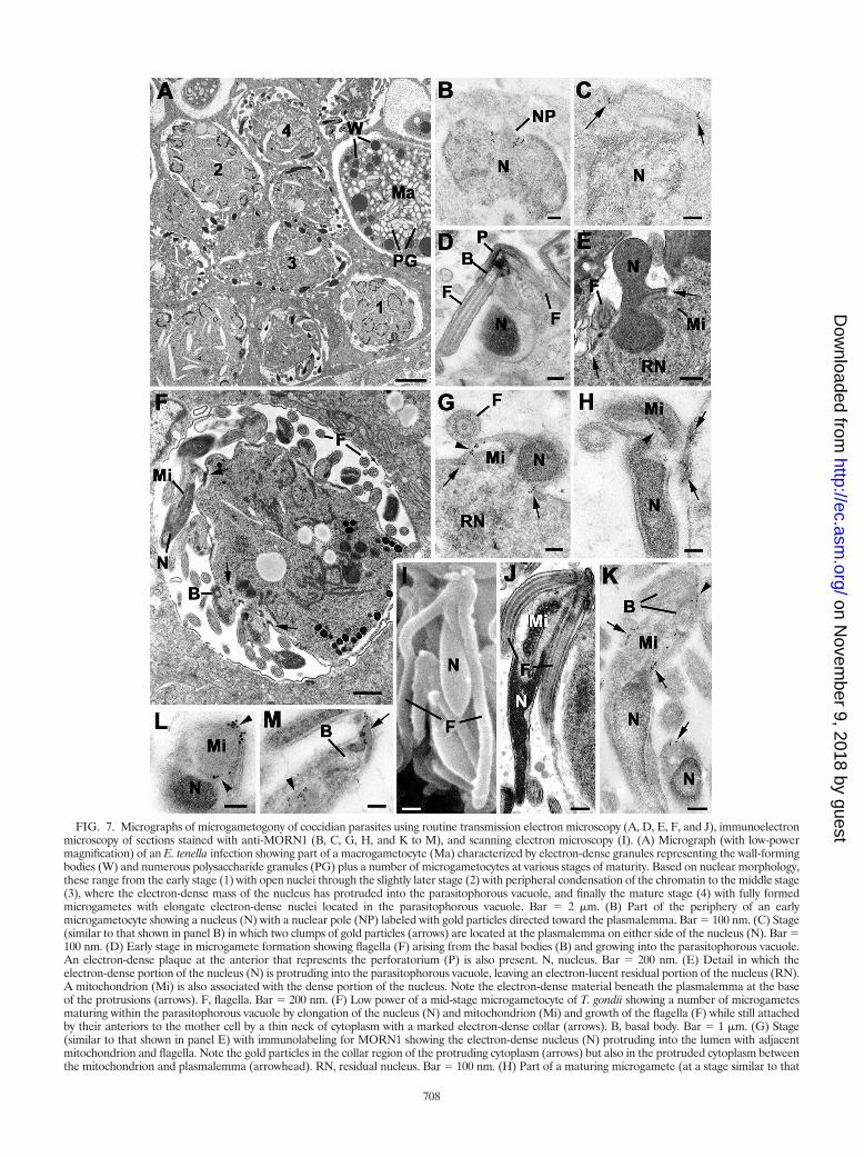
FIG. 7. Micrographs of microgametogony of coccidian parasites using routine transmission electron microscopy (A, D, E, F, and J), immunoelectronmicroscopy of sections stained with anti-MORN1 (B, C, G, H, and K to M), and scanning electron microscopy (I). (A) Micrograph (with low-powermagnification) of an E. tenella infection showing part of a macrogametocyte (Ma) characterized by electron-dense granules representing the wall-formingbodies (W) and numerous polysaccharide granules (PG) plus a number of microgametocytes at various stages of maturity. Based on nuclear morphology,these range from the early stage (1) with open nuclei through the slightly later stage (2) with peripheral condensation of the chromatin to the middle stage(3), where the electron-dense mass of the nucleus has protruded into the parasitophorous vacuole, and finally the mature stage (4) with fully formedmicrogametes with elongate electron-dense nuclei located in the parasitophorous vacuole. Bar � 2 �m. (B) Part of the periphery of an earlymicrogametocyte showing a nucleus (N) with a nuclear pole (NP) labeled with gold particles directed toward the plasmalemma. Bar � 100 nm. (C) Stage(similar to that shown in panel B) in which two clumps of gold particles (arrows) are located at the plasmalemma on either side of the nucleus (N). Bar �100 nm. (D) Early stage in microgamete formation showing flagella (F) arising from the basal bodies (B) and growing into the parasitophorous vacuole.An electron-dense plaque at the anterior that represents the perforatorium (P) is also present. N, nucleus. Bar � 200 nm. (E) Detail in which theelectron-dense portion of the nucleus (N) is protruding into the parasitophorous vacuole, leaving an electron-lucent residual portion of the nucleus (RN).A mitochondrion (Mi) is also associated with the dense portion of the nucleus. Note the electron-dense material beneath the plasmalemma at the baseof the protrusions (arrows). F, flagella. Bar � 200 nm. (F) Low power of a mid-stage microgametocyte of T. gondii showing a number of microgametesmaturing within the parasitophorous vacuole by elongation of the nucleus (N) and mitochondrion (Mi) and growth of the flagella (F) while still attachedby their anteriors to the mother cell by a thin neck of cytoplasm with a marked electron-dense collar (arrows). B, basal body. Bar � 1 �m. (G) Stage(similar to that shown in panel E) with immunolabeling for MORN1 showing the electron-dense nucleus (N) protruding into the lumen with adjacentmitochondrion and flagella. Note the gold particles in the collar region of the protruding cytoplasm (arrows) but also in the protruded cytoplasm betweenthe mitochondrion and plasmalemma (arrowhead). RN, residual nucleus. Bar � 100 nm. (H) Part of a maturing microgamete (at a stage similar to that
708
on Novem
ber 9, 2018 by guesthttp://ec.asm
.org/D
ownloaded from
triolar complex, with daughter formation always associatedwith the final round of mitosis and initiated directly above eachnuclear spindle pole. In the present study, MORN1 appearedto be a unifying feature across the various processes. In allcases, the nuclear poles are labeled with MORN1 as well as thefree and growing edge of the conical-shaped IMC, irrespectiveof whether daughter formation occurs at the surface (classicalschizogony) or within the cytoplasm (endodyogeny, Toxo-plasma endopolygeny, or Sarcocystis endopolygeny). The asso-ciation of MORN1 with the IMC during Sarcocystis endopoly-geny has previously been described (15). The IMC consists ofa double-unit membrane structure formed by flattened vacu-oles produced by the Golgi, with an underlying meshwork ofintermediate filaments (named IMC proteins) and longitudi-nally running microtubules (2–4, 28). MORN1 is present as acontinuous ring on the posterior end of the IMC, and thislocation, confirmed by immunoelectron microscopy, is consis-tent with the “free” end of the IMC, where no microtubules areobserved (15). In the final stage, the posterior ring at the pointof attachment with the residual body is strongly labeled. Incontrast, posterior labeling is reduced in free merozoites.These features are similar to those observed in the sporozoiteof E. tenella (Fig. 4H).
The loss of intensity upon maturation suggests that MORN1 isrequired in the budding process but less so to maintain the cy-toskeleton in the mature zoite. It was previously shown that in T.gondii TgMORN1 segregates in roughly equal proportions in thedetergent-soluble and detergent-resistant fractions (15). Since themajority of T. gondii TgMORN1 is found at the posterior ring inmature and dividing tachyzoites, these data are consistent withMORN1 having a bridging role between the alveolar vesiclesforming the inner membrane and the intermediate filaments ofthe IMC, specifically at the free end of the IMC. In the case for P.falciparum schizogony, Western blotting with the anti-TgMORN1antibody shows that PfMORN1 is under the level of detection inthe early stages of development (ring and trophozoite). This pat-tern is in agreement with the microarray data (www.plasmodb.org) which indicate low-transcript abundance in early stages anda strong increase, with a peak at 38 h in the 48-h intraerythrocyticcycle (1, 24), coinciding with the initiation of merozoite budding.In addition, detergent extraction showed that the signal is foundin both the detergent-soluble and -insoluble fractions, which iscomparable to the case for T. gondii (15). Therefore, it appearsthat MORN1 is present in the same functional niche in T. gondiiand P. falciparum, at the interface between alveolar membraneand intermediate filament cytoskeleton. These data support amodel wherein TgMORN1 fulfills a bridging position betweenthe alveolar membrane and the IMC-filament meshwork. How-
ever, the exact function of the MORN domain is not known. Ithas been shown that the MORN domain is able to maintain amembrane association through direct binding to phospholipids(25). This observation fits the detergent-extractable fraction ofMORN1, whereas the detergent-resistant fraction could serve asthe scaffold maintaining the budding ring. With the similarity ofexpression and location across the Apicomplexa, MORN1 ap-pears to have a conserved role in daughter bud stabilization dur-ing asexual development. Because this location is the site wherethe bud grows, MORN1 potentially serves as a scaffold for thebudding machinery. The overexpression of TgMORN1 resultedin incorrect initiation of the daughter buds and an uncoupling ofthe microtubular skeleton from the IMC skeleton and resulted inunstructured IMC formation (15). Taken together, this stronglysuggests that MORN1 is required for the assembly of the IMCskeleton and/or connection with the microtubule skeleton. Wepreviously showed that myosin C is colocalizing with the MORN1ring during T. gondii tachyzoite budding (15), but this may not bethe case for other parasites, because myosin C is not stronglyconserved across the Apicomplexa (data not shown). More re-cently, a calcium-dependent contraction of the MORN1 ring inthe maturation of the daughters was proposed to be mediated byTgCentrin2 (17). The future direction of our work is to identifythe functional components in the establishment and extension ofthe asexual bud.
Microgamete. Unfortunately there are no in vitro systemsfor the production of the sexual stages of any coccidian para-site, which has greatly limited the examination of these para-sites by molecular techniques. The current study thereforeprovides a significant extension of current molecular knowl-edge. We were able to examine the expression and localizationof MORN1 during sexual development within the definitivehost by using anti-TgMORN1 for four coccidian species (T.gondii, E. acervulina, E. tenella, and E. maxima). However, thepossibility that anti-TgMORN1 staining in microgametes re-sults from cross-reactivity of the antibody with other MORNdomain containing proteins cannot be excluded, although it isunlikely, since the weak 41-kDa band observed by Westernblotting was consistent with the predicted mass of EtMORN1.
The role of the MORN1-positive nuclear pole appears todirect to the location of microgamete formation, as discussedabove. The formation of a MORN1-positive ring-like structurebeneath the plasmalemma is shared between both sexual andasexual development, but in the case of the microgametocyte,MORN1 was associated with amorphous dense material lo-cated beneath the plasmalemma in the absence of an IMC. Thedense collar at the site of microgamete protrusion has previ-ously been described for various species of Eimeria, Isospora
shown in panel F), where gold particles can be seen associated with the dense collar underlying the plasmalemma (arrows) and also between thenucleus (N) and mitochondrion (Mi) of the microgamete (arrowhead). Bar � 100 nm. (I) Scanning electron micrograph of a mature microgameteshowing the elongated nucleus (N) and the two very long flagella (F). Bar � 500 nm. (J) Longitudinal section through a mature microgameteshowing the two flagella (F) arising from basal bodies in the anterior below which is the elongated mitochondrion (Mi) and then the electron-densenucleus (N). Bar � 500 nm. (K) An immunostained longitudinal section showing a few gold particles anterior (arrowhead) to the basal bodies (B)and a larger number of gold particles lining either side of the mitochondrion (Mi). The gold particles were located between the mitochondrionand the plasmalemma, but they did not appear to encircle the mitochondrion. Bar � 200 nm. (L) Cross section through the anterior portion ofa microgamete at the level where the mitochondrion (Mi) and nucleus (N) overlap. Note that the gold particles (arrowheads) only partiallysurround the mitochondrion. Bar � 100 nm. (M) Detail of the anterior of a microgamete showing a number of gold particles located both apical(arrow) and basal (arrowhead) to the basal body. Bar � 100 nm.
VOL. 7, 2008 MORN1 IN APICOMPLEXA 709
on Novem
ber 9, 2018 by guesthttp://ec.asm
.org/D
ownloaded from

felis, and T. gondii (6, 11, 33). It appears that MORN1 isassociated with ring formation and maintains the integrity ofthe site of microgamete budding. This observation suggeststhat the ring-like appearance of MORN1 requires neither IMCnor microtubules and is controlled at another cytoskeletallevel. MORN1 itself could, at least in part, play a role in this,as MORN1 shows a strong tendency to aggregate into multim-ers and, upon overexpression, has been observed in fiber-likestructures (15, 18; N. Sahoo and M. J. Gubbels, unpublisheddata). Interestingly, an IMC1 knockout generated in Plasmo-dium berghei resulted in sideways budding of sporozoites fromthe oocyst and mechanically unstable sporozoites (21). It isexpected that MORN1 localization is undisturbed in this con-text, and this will be interesting to investigate.
Since the developing microgamete remained enclosed by theplasmalemma of the mother cell, new membrane is required.New membrane may be formed within the developing micro-gamete, or it could be an extension of plasmalemma from themother cell, which may mean movement through the collarregion. Since the flagella form by an outgrowth into the para-sitophorous vacuole, the subunits for microtubular formationmust be transported to the free end of the flagella. Unlikemerozoite formation, where there is a budding out involvingposterior growth of the pellicle, the developing microgameteremains attached by the anterior end and undergoes changes inshape during maturation while still attached. The maintenanceof the connection during microgamete maturation will be im-portant for the rapid movement of materials synthesized in themother cell into the microgametes. This will be particularlyimportant for the growth of the flagella.
An additional observation is the finding of MORN1-positivematerial associated with the anterior end of the microgametelocated within the apex and also partially enclosing the mito-chondrion. This is the first report of a specific molecule asso-ciated with microgamete formation and the mature microga-mete. Therefore, MORN1 is the first immunocytochemicalmarker that can be used to identify individual microgametes,which, due to their small size, can be extremely difficult toidentify in normal histological sections. It can be hypothesizedthat this anterior location of MORN1 is involved in maintain-ing rigidity and structural integrity of the microgamete duringmassive physical stresses that will be associated with flagellarmovement. It may also be that it functions as the anchor of themitochondria to stay close to the flagellum, where the energyproduced in the mitochondria is consumed by the beatingflagellum (Fig. 7M). Similar close membrane oppositions havepreviously been described for eukaryotic myocytes, where aMORN1-like protein called junctophilin maintains the sarco-plasmic reticulum position underlying the plasma membrane(27, 41).
Conclusion. The exact function of the MORN1 protein isunknown, but from the distribution observed in the presentstudy across the Apicomplexa, it is possible to hypothesizeabout its role. The amount of MORN1 protein appears to varywith developmental stage: an increased amount is associatedwith daughter and microgamete formation during asexual pro-liferation and microgametogony, respectively, but it is not ob-served during macrogametogony, where no division occurs.This distribution during both asexual and sexual developmentand the location in the mature zoite and microgamete would
be consistent with MORN1 maintaining structural integrity,probably by cross-linking other structural components. Thismay also explain its possible role within the nuclear pole.Further characterization of additional components that specif-ically interact with MORN1 are under way to advance ourunderstanding of the specific and distinct localizations androles of MORN1 in the various developmental processes.
ACKNOWLEDGMENTS
This work was supported in part by a Scientist Development Grantfrom the American Heart Association (no. 0635480N) to M.-J.G. anda Knights Templar Eye Foundation Research Grant to N.S. M.-J.G. isa Smith Family Foundation New Investigator (through the LudckeFoundation). D.J.P.F. was supported by an equipment grant from theWellcome Trust.
We thank Damer Blake for access to E. maxima-infected chickens.
REFERENCES
1. Bozdech, Z., M. Llinas, B. L. Pulliam, E. D. Wong, J. Zhu, and J. L. DeRisi.2003. The transcriptome of the intraerythrocytic developmental cycle ofPlasmodium falciparum. PLoS Biol. 1:E5.
2. Dubremetz, J. F. 1975. Genesis of merozoites in the coccidia, Eimerianecatrix. Ultrastructural study. J. Protozool. 22:71–84. (In French.)
3. Dubremetz, J. F. 1973. Ultrastructural study of schizogonic mitosis in thecoccidian, Eimeria necatrix (Johnson 1930). J. Ultrastruct. Res. 42:354–376.(In French.)
4. Dubremetz, J. F., and Y. Y. Elsner. 1979. Ultrastructural study of schizogonyof Eimeria bovis in cell cultures. J. Protozool. 26:367–376.
5. Ferguson, D. J., S. I. Belli, N. C. Smith, and M. G. Wallach. 2003. Thedevelopment of the macrogamete and oocyst wall in Eimeria maxima: im-munolight and electron microscopy. Int. J. Parasitol. 33:1329–1340.
6. Ferguson, D. J., A. Birch-Andersen, W. M. Hutchison, and J. C. Siim. 1980.Ultrastructural observations on microgametogenesis and the structure of themicrogamete of Isospora felis. Acta Pathol. Microbiol. Scand. Sect. B 88:151–159.
7. Ferguson, D. J., A. Birch-Andersen, W. M. Hutchison, and J. C. Siim. 1977.Ultrastructural studies on the endogenous development of Eimeria brunetti.II. Microgametogony and the microgamete. Acta Pathol. Microbiol. Scand.Sect. B 85B:67–77.
8. Ferguson, D. J., A. Birch-Andersen, W. M. Hutchison, and J. C. Siim. 1977.Ultrastructural studies on the endogenous development of eimeria brunetti.III. Macrogametogony and the macrogamete. Acta Pathol. Microbiol. Scand.Sect. B 85B:78–88.
9. Ferguson, D. J., M. F. Cesbron-Delauw, J. F. Dubremetz, L. D. Sibley, K. A.Joiner, and S. Wright. 1999. The expression and distribution of dense gran-ule proteins in the enteric (Coccidian) forms of Toxoplasma gondii in thesmall intestine of the cat. Exp. Parasitol. 91:203–211.
10. Ferguson, D. J., F. L. Henriquez, M. J. Kirisits, S. P. Muench, S. T. Prigge,D. W. Rice, C. W. Roberts, and R. L. McLeod. 2005. Maternal inheritanceand stage-specific variation of the apicoplast in Toxoplasma gondii duringdevelopment in the intermediate and definitive host. Eukaryot. Cell 4:814–826.
11. Ferguson, D. J., W. M. Hutchison, J. F. Dunachie, and J. C. Siim. 1974.Ultrastructural study of early stages of asexual multiplication and micro-gametogony of Toxoplasma gondii in the small intestine of the cat. ActaPathol. Microbiol. Immunol. Scand. Sect. B 82:167–181.
12. Ferguson, D. J., W. M. Hutchison, and J. C. Siim. 1975. The ultrastructuraldevelopment of the macrogamete and formation of the oocyst wall of Toxo-plasma gondii. Acta Pathol. Microbiol. Scand. Sect. B 83:491–505.
13. Ferguson, D. J., S. F. Parmley, and S. Tomavo. 2002. Evidence for nuclearlocalisation of two stage-specific isoenzymes of enolase in Toxoplasma gondiicorrelates with active parasite replication. Int. J. Parasitol. 32:1399–1410.
14. Ferguson, D. J. P., S. A. Campbell, F. L. Henriquez, L. Phan, E. Mui, T. A.Richards, S. P. Muench, M. Allary, J. Z. Lu, S. T. Prigge, F. Tomley, M. W.Shirley, D. W. Rice, R. McLeod, and C. W. Roberts. 2007. Enzymes of typeII fatty acid synthesis and apicoplast differentiation and division in Eimeriatenella. Int. J. Parasitol. 37:33–51.
15. Gubbels, M. J., S. Vaishnava, N. Boot, J. F. Dubremetz, and B. Striepen.2006. A MORN-repeat protein is a dynamic component of the Toxoplasmagondii cell division apparatus. J. Cell Sci. 119:2236–2245.
16. Hartmann, J., K. Hu, C. Y. He, L. Pelletier, D. S. Roos, and G. Warren. 2006.Golgi and centrosome cycles in Toxoplasma gondii. Mol. Biochem. Parasitol.145:125–127.
17. Hu, K. 2008. Organizational changes of the daughter basal complex duringthe parasite replication of Toxoplasma gondii. PLoS Pathog. 4:e10.
18. Hu, K., J. Johnson, L. Florens, M. Fraunholz, S. Suravajjala, C. DiLullo, J.Yates, D. S. Roos, and J. M. Murray. 2006. Cytoskeletal components of an
710 FERGUSON ET AL. EUKARYOT. CELL
on Novem
ber 9, 2018 by guesthttp://ec.asm
.org/D
ownloaded from

invasion machine—the apical complex of Toxoplasma gondii. PLoS Pathog.2:121–138.
19. Hu, K., T. Mann, B. Striepen, C. J. Beckers, D. S. Roos, and J. M. Murray.2002. Daughter cell assembly in the protozoan parasite Toxoplasma gondii.Mol. Biol. Cell 13:593–606.
20. Jacobs, D., J. F. Dubremetz, A. Loyens, F. Bosman, and E. Saman. 1998.Identification and heterologous expression of a new dense granule protein(GRA7) from Toxoplasma gondii. Mol. Biochem. Parasitol. 91:237–249.
21. Khater, E. I., R. E. Sinden, and J. T. Dessens. 2004. A malaria membraneskeletal protein is essential for normal morphogenesis, motility, and infec-tivity of sporozoites. J. Cell Biol. 167:425–432.
22. Kohler, S., C. F. Delwiche, P. W. Denny, L. G. Tilney, P. Webster, R. J.Wilson, J. D. Palmer, and D. S. Roos. 1997. A plastid of probable green algalorigin in Apicomplexan parasites. Science 275:1485–1489.
23. Kooij, T. W., and K. Matuschewski. 2007. Triggers and tricks of Plasmodiumsexual development. Curr. Opin. Microbiol. 10:547–553.
24. Le Roch, K. G., Y. Zhou, P. L. Blair, M. Grainger, J. K. Moch, J. D. Haynes,P. De La Vega, A. A. Holder, S. Batalov, D. J. Carucci, and E. A. Winzeler.2003. Discovery of gene function by expression profiling of the malariaparasite life cycle. Science 301:1503–1508.
25. Ma, H., Y. Lou, W. H. Lin, and H. W. Xue. 2006. MORN motifs in plantPIPKs are involved in the regulation of subcellular localization and phos-pholipid binding. Cell Res. 16:466–478.
26. Mann, T., and C. Beckers. 2001. Characterization of the subpellicular net-work, a filamentous membrane skeletal component in the parasite Toxo-plasma gondii. Mol. Biochem. Parasitol. 115:257–268.
27. Minamisawa, S., J. Oshikawa, H. Takeshima, M. Hoshijima, Y. Wang, K. R.Chien, Y. Ishikawa, and R. Matsuoka. 2004. Junctophilin type 2 is associatedwith caveolin-3 and is down-regulated in the hypertrophic and dilatedcardiomyopathies. Biochem. Biophys. Res. Commun. 325:852–856.
28. Nichols, B. A., and M. L. Chiappino. 1987. Cytoskeleton of Toxoplasmagondii. J. Protozool. 34:217–226.
29. Ogino, N., and C. Yoneda. 1966. The fine structure and mode of division ofToxoplasma gondii. Arch. Ophthalmol. 75:218–227.
30. Piekarski, G., B. Pelster, and H. M. Witte. 1971. Endopolygeny in Toxo-plasma gondii. Z. Parasitenkd. 36:122–130. (In German.)
31. Radke, J. R., B. Striepen, M. N. Guerini, M. E. Jerome, D. S. Roos, and
M. W. White. 2001. Defining the cell cycle for the tachyzoite stage of Toxo-plasma gondii. Mol. Biochem. Parasitol. 115:165–175.
32. Schmatz, D. M., M. S. Crane, and P. K. Murray. 1984. Purification ofEimeria sporozoites by DE-52 anion exchange chromatography. J. Proto-zool. 31:181–183.
33. Scholtyseck, E., H. Mehlhorn, and D. M. Hammond. 1972. Electron micro-scope studies of microgametogenesis in Coccidia and related groups. Z.Parasitenkd. 38:95–131.
34. Scholtyseck, E., H. Mehlhorn, and D. M. Hammond. 1971. Fine structureof macrogametes and oocysts of Coccidia and related organisms. Z.Parasitenkd. 37:1–43.
35. Sheffield, H. G., and M. L. Melton. 1968. The fine structure and reproductionof Toxoplasma gondii. J. Parasitol. 54:209–226.
36. Shirley, M. W. 1995. Eimeria species and strains of chickens, p. 1–24. In J.Eckert, R. Braun, M. W. Shirley, and P. Coudert (ed.), Biotechnology—guidelines on techniques in coccidiosis research. The European CommissionDGXII, Luxembourg, Luxembourg.
37. Sinden, R. E. 1983. The cell biology of sexual development in plasmodium.Parasitology 86:7–28.
38. Speer, C. A., and J. P. Dubey. 1981. An ultrastructural study of first- andsecond-generation merogony in the coccidian Sarcocystis tenella. J. Proto-zool. 28:424–431.
39. Striepen, B., M. J. Crawford, M. K. Shaw, L. G. Tilney, F. Seeber, and D. S.Roos. 2000. The plastid of Toxoplasma gondii is divided by association withthe centrosomes. J. Cell Biol. 151:1423–1434.
40. Striepen, B., C. N. Jordan, S. Reiff, and G. G. van Dooren. 2007. Building theperfect parasite: cell division in apicomplexa. PLoS Pathog. 3:e78.
41. Takeshima, H., S. Komazaki, M. Nishi, M. Iino, and K. Kangawa. 2000.Junctophilins: a novel family of junctional membrane complex proteins. Mol.Cell 6:11–22.
42. Trager, W., and J. B. Jensen. 2005. Human malaria parasites in continuousculture. 1976. J. Parasitol. 91:484–486.
43. Vaishnava, S., D. P. Morrison, R. Y. Gaji, J. M. Murray, R. Entzeroth, D. K.Howe, and B. Striepen. 2005. Plastid segregation and cell division in theapicomplexan parasite Sarcocystis neurona. J. Cell Sci. 118:3397–3407.
44. Wallach, M., N. C. Smith, M. Petracca, C. M. Miller, J. Eckert, and R.Braun. 1995. Eimeria maxima gametocyte antigens: potential use in a sub-unit maternal vaccine against coccidiosis in chickens. Vaccine 13:347–354.
VOL. 7, 2008 MORN1 IN APICOMPLEXA 711
on Novem
ber 9, 2018 by guesthttp://ec.asm
.org/D
ownloaded from
Related Documents