Monocrotophos Induces the Expression and Activity of Xenobiotic Metabolizing Enzymes in Pre-Sensitized Cultured Human Brain Cells Vinay K. Tripathi 1,3 , Vivek Kumar 1,3 , Abhishek K. Singh 1 , Mahendra P. Kashyap 1 , Sadaf Jahan 1 , Ankita Pandey 1 , Sarfaraz Alam 2 , Feroz Khan 2 , Vinay K. Khanna 1 , Sanjay Yadav 1 , Mohtshim Lohani 3 , Aditya B. Pant 1 * 1 In Vitro Toxicology Laboratory, CSIR-Indian Institute of Toxicology Research, Lucknow, India, 2 Metabolic and Structural Biology Department, CSIR-Central Institute of Medicinal and Aromatic Plants, Lucknow, India, 3 Department of Biotechnology, Integral University, Lucknow, India Abstract The expression and metabolic profile of cytochrome P450s (CYPs) is largely missing in human brain due to non-availability of brain tissue. We attempted to address the issue by using human brain neuronal (SH-SY5Y) and glial (U373-MG) cells. The expression and activity of CYP1A1, 2B6 and 2E1 were carried out in the cells exposed to CYP inducers viz., 3- methylcholanthrene (3-MC), cyclophosphamide (CPA), ethanol and known neurotoxicant- monocrotophos (MCP), a widely used organophosphorous pesticide. Both the cells show significant induction in the expression and CYP-specific activity against classical inducers and MCP. The induction level of CYPs was comparatively lower in MCP exposed cells than cells exposed to classical inducers. Pre-exposure (12 h) of cells to classical inducers significantly added the MCP induced CYPs expression and activity. The findings were concurrent with protein ligand docking studies, which show a significant modulatory capacity of MCP by strong interaction with CYP regulators-CAR, PXR and AHR. Similarly, the known CYP inducers- 3-MC, CPA and ethanol have also shown significantly high docking scores with all the three studied CYP regulators. The expression of CYPs in neuronal and glial cells has suggested their possible association with the endogenous physiology of the brain. The findings also suggest the xenobiotic metabolizing capabilities of these cells against MCP, if received a pre-sensitization to trigger the xenobiotic metabolizing machinery. MCP induced CYP-specific activity in neuronal cells could help in explaining its effect on neurotransmission, as these CYPs are known to involve in the synthesis/transport of the neurotransmitters. The induction of CYPs in glial cells is also of significance as these cells are thought to be involved in protecting the neurons from environmental insults and safeguard them from toxicity. The data provide better understanding of the metabolizing capability of the human brain cells against xenobiotics. Citation: Tripathi VK, Kumar V, Singh AK, Kashyap MP, Jahan S, et al. (2014) Monocrotophos Induces the Expression and Activity of Xenobiotic Metabolizing Enzymes in Pre-Sensitized Cultured Human Brain Cells. PLoS ONE 9(3): e91946. doi:10.1371/journal.pone.0091946 Editor: Aamir Nazir, CSIR-Central Drug Research Institute, India Received November 17, 2013; Accepted February 16, 2014; Published March 24, 2014 Copyright: ß 2014 Tripathi et al. This is an open-access article distributed under the terms of the Creative Commons Attribution License, which permits unrestricted use, distribution, and reproduction in any medium, provided the original author and source are credited. Funding: Financial support from Department of Biotechnology, Ministry of Science & Technology, Government of India, New Delhi, India [Grant No. 102/IFD/ SAN/PR1524/2010–2011]; Department of Science and Technology, Ministry of Science & Technology, Government of India, New Delhi, India [Grant No. SR/SO/Z 36/2007/91/10]; and Council of Scientific & Industrial Research, Government of India, New Delhi, India [Grant No. BSC0111/INDEPTH/CSIR Network Project] is acknowledged. The funders had no role in the study design, data collection and analysis, decision to publish, or preparation of the manuscript. Competing Interests: The corresponding author Dr. Aditya B Pant is currently acting as Academic Editor for PLoS ONE. This does not alter the authors’ adherence to PLOS ONE Editorial Policies and criteria. * E-mail: [email protected] Introduction The key role of cytochrome P450s (CYPs) super-family in endogenous and xenobiotic metabolism is well established [1,2]. Though the liver has been reported as the major CYPs mediated metabolic site [3], but the significant expression and activity of selected CYPs has also been reported in brain tissues [4,5,6]. In general, the brain has a comparatively lower level of expression and activity of CYPs than liver, but due to tissue heterogeneity few specific regions and cells of the brain have been reported to have significantly higher expression and activity of CYPs than that of liver [7,8]. The regional specific expression and inducibility of several members of the CYP gene family involved in metabolism, toxicity and detoxification have already been documented in the brain of experimental animals receiving exposure to environmen- tal chemicals and drugs [4,9]. Brain cells have shown high inducibility of CYPs, and quite often in a different fashion from their hepatic forms [10,11]. Our group has also shown the constitutive and inducible expression of CYPs in human and rat brain primary culture of neuronal and glial cells [12,13,14,15]. Immortalized human-derived brain endothelial cell line has also reported to express CYP enzymes [16,17]. CYPs in families 1 to 3 are primarily involved in the detoxification of various xenobiotic compounds and drugs [18], whereas the remaining groups are broadly play the role in the metabolism of endogenous compound such as steroids, fatty acids, hormones, neurotransmitters, cholesterol, bile acids and vitamins, etc. [19]. Khokhar and Tyndale [20] provided strong evidences supporting the role of brain CYPs in local drug metabolism, and subsequent alterations in the pharmacological actions of drugs. CYP1A1 is well known for its role in the bioactivation of carcinogens such as aromatic amines and polycyclic aromatic PLOS ONE | www.plosone.org 1 March 2014 | Volume 9 | Issue 3 | e91946

Welcome message from author
This document is posted to help you gain knowledge. Please leave a comment to let me know what you think about it! Share it to your friends and learn new things together.
Transcript
Monocrotophos Induces the Expression and Activity ofXenobiotic Metabolizing Enzymes in Pre-SensitizedCultured Human Brain CellsVinay K. Tripathi1,3, Vivek Kumar1,3, Abhishek K. Singh1, Mahendra P. Kashyap1, Sadaf Jahan1,
Ankita Pandey1, Sarfaraz Alam2, Feroz Khan2, Vinay K. Khanna1, Sanjay Yadav1, Mohtshim Lohani3,
Aditya B. Pant1*
1 In Vitro Toxicology Laboratory, CSIR-Indian Institute of Toxicology Research, Lucknow, India, 2 Metabolic and Structural Biology Department, CSIR-Central Institute of
Medicinal and Aromatic Plants, Lucknow, India, 3 Department of Biotechnology, Integral University, Lucknow, India
Abstract
The expression and metabolic profile of cytochrome P450s (CYPs) is largely missing in human brain due to non-availabilityof brain tissue. We attempted to address the issue by using human brain neuronal (SH-SY5Y) and glial (U373-MG) cells. Theexpression and activity of CYP1A1, 2B6 and 2E1 were carried out in the cells exposed to CYP inducers viz., 3-methylcholanthrene (3-MC), cyclophosphamide (CPA), ethanol and known neurotoxicant- monocrotophos (MCP), a widelyused organophosphorous pesticide. Both the cells show significant induction in the expression and CYP-specific activityagainst classical inducers and MCP. The induction level of CYPs was comparatively lower in MCP exposed cells than cellsexposed to classical inducers. Pre-exposure (12 h) of cells to classical inducers significantly added the MCP induced CYPsexpression and activity. The findings were concurrent with protein ligand docking studies, which show a significantmodulatory capacity of MCP by strong interaction with CYP regulators-CAR, PXR and AHR. Similarly, the known CYPinducers- 3-MC, CPA and ethanol have also shown significantly high docking scores with all the three studied CYPregulators. The expression of CYPs in neuronal and glial cells has suggested their possible association with the endogenousphysiology of the brain. The findings also suggest the xenobiotic metabolizing capabilities of these cells against MCP, ifreceived a pre-sensitization to trigger the xenobiotic metabolizing machinery. MCP induced CYP-specific activity in neuronalcells could help in explaining its effect on neurotransmission, as these CYPs are known to involve in the synthesis/transportof the neurotransmitters. The induction of CYPs in glial cells is also of significance as these cells are thought to be involvedin protecting the neurons from environmental insults and safeguard them from toxicity. The data provide betterunderstanding of the metabolizing capability of the human brain cells against xenobiotics.
Citation: Tripathi VK, Kumar V, Singh AK, Kashyap MP, Jahan S, et al. (2014) Monocrotophos Induces the Expression and Activity of Xenobiotic MetabolizingEnzymes in Pre-Sensitized Cultured Human Brain Cells. PLoS ONE 9(3): e91946. doi:10.1371/journal.pone.0091946
Editor: Aamir Nazir, CSIR-Central Drug Research Institute, India
Received November 17, 2013; Accepted February 16, 2014; Published March 24, 2014
Copyright: � 2014 Tripathi et al. This is an open-access article distributed under the terms of the Creative Commons Attribution License, which permitsunrestricted use, distribution, and reproduction in any medium, provided the original author and source are credited.
Funding: Financial support from Department of Biotechnology, Ministry of Science & Technology, Government of India, New Delhi, India [Grant No. 102/IFD/SAN/PR1524/2010–2011]; Department of Science and Technology, Ministry of Science & Technology, Government of India, New Delhi, India [Grant No. SR/SO/Z36/2007/91/10]; and Council of Scientific & Industrial Research, Government of India, New Delhi, India [Grant No. BSC0111/INDEPTH/CSIR Network Project] isacknowledged. The funders had no role in the study design, data collection and analysis, decision to publish, or preparation of the manuscript.
Competing Interests: The corresponding author Dr. Aditya B Pant is currently acting as Academic Editor for PLoS ONE. This does not alter the authors’adherence to PLOS ONE Editorial Policies and criteria.
* E-mail: [email protected]
Introduction
The key role of cytochrome P450s (CYPs) super-family in
endogenous and xenobiotic metabolism is well established [1,2].
Though the liver has been reported as the major CYPs mediated
metabolic site [3], but the significant expression and activity of
selected CYPs has also been reported in brain tissues [4,5,6]. In
general, the brain has a comparatively lower level of expression
and activity of CYPs than liver, but due to tissue heterogeneity few
specific regions and cells of the brain have been reported to have
significantly higher expression and activity of CYPs than that of
liver [7,8]. The regional specific expression and inducibility of
several members of the CYP gene family involved in metabolism,
toxicity and detoxification have already been documented in the
brain of experimental animals receiving exposure to environmen-
tal chemicals and drugs [4,9]. Brain cells have shown high
inducibility of CYPs, and quite often in a different fashion from
their hepatic forms [10,11]. Our group has also shown the
constitutive and inducible expression of CYPs in human and rat
brain primary culture of neuronal and glial cells [12,13,14,15].
Immortalized human-derived brain endothelial cell line has also
reported to express CYP enzymes [16,17].
CYPs in families 1 to 3 are primarily involved in the
detoxification of various xenobiotic compounds and drugs [18],
whereas the remaining groups are broadly play the role in the
metabolism of endogenous compound such as steroids, fatty acids,
hormones, neurotransmitters, cholesterol, bile acids and vitamins,
etc. [19]. Khokhar and Tyndale [20] provided strong evidences
supporting the role of brain CYPs in local drug metabolism, and
subsequent alterations in the pharmacological actions of drugs.
CYP1A1 is well known for its role in the bioactivation of
carcinogens such as aromatic amines and polycyclic aromatic
PLOS ONE | www.plosone.org 1 March 2014 | Volume 9 | Issue 3 | e91946

hydrocarbons (PAHs) [21,22]. Induction and a high activity of the
CYP1A1 have been associated with increased toxicity and cancer
risk [23,24]. CYP2B6 is also expressed in the brain, highly
expressed in specific cells such as cortical pyramidal cells and
astrocytes [25] and may be an important factor in the metabolism
of drugs acting on the central nervous system (CNS). The
concentration of CYP2B6 in the brain regions of smokers and
alcoholics has been reported abnormally high, particularly in the
cerebellar Purkinje cells, granular cell layer and hippocampal
pyramidal neurons [26]. CYP2B6 metabolizes a wide range of
substances including endogenous compounds such as arachidonic
acid, 17 b-estradiol, testosterone [27] and the neurotransmitter
serotonin [28], dopamine [29] and neurotoxins [30]. It metabo-
lizes therapeutically important drugs as well as drugs of abuse
[31,32]. The expression of CYP2E1 has also been reported in
cultured rat [13,33] and human brain cells [34]. The mechanism
of CYP2E1 induction is complex and depends on the substrate,
species, tissue, or cell type [35,36] and in the brain regions and cell
type specific expression [37]. CYP2E1 enzymes are involved in the
metabolism of alcohol other low-molecular-weight xenobiotics,
including drugs, organic solvents, and pro-carcinogens [38,39,40].
CYP2E1 have potential impact on brain function by metabolizing
ethanol resulting in generation of reactive oxygen species that have
direct concern in the neurodegenerative disorders such as
Parkinson’s disease, minor dendritic and synaptic changes to
neuronal cell death [41,42] through lipid peroxidation, protein
inactivation, and DNA damage [43]. The protein level of CYP2E1
in smokers has significantly more in comparison to non-smokers in
several brain regions [7,44].
Inspite of several investigations pertaining to the metabolizing
capabilities of CYPs in brain tissues and primary cultures of brain
cells of experimental animals, the status of these CYPs in human
brain cells is poorly understood. The reason being is non-
availability of human brain due to ethical dubious. Thus, to
address the issue, the expression and inducibility of CYP1A1,
CYP2B6 and CYP2E1 were investigated in cultured human brain
neuronal (SH-SY5Y) and glial (U373-MG) cell lines. Xenobiotic
metabolizing capability of cells was studied using known inducers
of CYP1A1 (3-methylcholanthrene), CYP2B6 (cyclophosphamide)
and CYP2E1 (ethanol). The studies were further extended to
investigate the responsiveness of CYPs in the cells against
monocrotophos, a widely used organophosphate pesticide used
in many parts of the world, including India for more than forty
years and known for its systemic toxicity including neurotoxicity
[45,46,47].
Materials and Methods
Reagents and ConsumablesAll the specified chemicals and reagents viz., monocrotophos
(MCP), 3-methylcholanthrene and cyclophosphamide (CPA) were
purchased from Sigma (Sigma St Louis, MO, USA) unless
otherwise stated. Culture medium DMEM/F-12, antibiotics, fetal
bovine serum and Trypsin-EDTA were purchased from Gibco
BRL, USA. All the antibodies used in this study were procured
from Chemicon International, USA. Culture wares and other
plastic wares used in the study were procured commercially from
Nunc, Denmark. Milli Q water (double distilled, deionized water)
was used in all the experiments.
Cell CultureHuman neuroblastoma cell line SH-SY5Y and glioblastoma
U373-MG cell lines used in the study were initially procured from
National Centre for Cell Sciences, Pune, India and since then have
been maintained at In Vitro Toxicology Laboratory, Indian
Institute of Toxicology Research, Lucknow, India, as per the
standard protocols. In brief, the cells were cultured in DMEM/F-
12, supplemented with 10% fetal bovine serum (FBS), 0.2%
sodium bicarbonate, 100 units/ml penicillin G sodium, 100 mg/ml
streptomycin sulfate and 0.25 mg/ml amphotericin B. Cultures
were maintained at 370C, in 5% CO2-95% atmosphere under
high humid conditions. Medium was changed twice weekly and
cultures were passaged at a ratio of 1:6 once in a week. Prior to use
in the experiments, cell viability was ascertained by Trypan blue
dye exclusion assay. The culture showing viability more than 95%
were used in all the experiments. All the experiments were done on
the cells with passage 18–25 only.
Identification of non-cytotoxic doses of MCP, 3-MC, CPAand Ethanol
In the present investigations, known inducers of CYP1A1 [3-
methylcholanthrene (3-MC)], CYP2B6 [cyclophosphamide
(CPA)], CYP2E1 [ethanol] and known neurotoxin - monocroto-
phos (MCP), an organophosphate pesticide, were used. 3-MC was
dissolved in culture grade dimethylsulphoxide (DMSO), while
CPA, ethanol and MCP were dissolved in DMEM/F-12 medium.
Prior using in the expression studies, non-cytotoxic doses of 3-MC,
CPA, ethanol and MCP were identified in SH-SY5Y (human
neuronal cell line) and U373-MG (human glial cell line).
Cytotoxicity assessment was done using standard endpoint i.e.,
tetrazolium bromide MTT (3-(4, 5-dimethylthiazol-2-yl) -2, 5-
diphenyl tetrazolium bromide) assay as described earlier by us
[48]. In brief, neuronal - SH-SY5Y cells and glial - U373-MG cells
(16104 cells/well) were seeded in 96-well tissue culture plates and
incubated in the CO2 incubator for 24 h at 37uC. Then the
medium was aspirated and cells were exposed to medium
containing either of 3-MC (1 mM–10 mM), CPA (0.5 mM–
8 mM), ethanol (25 mM–400 mM) and MCP (1027 M–1023 M)
for 24–96 h at 37uC in 5% CO2-95% atmosphere under high
humid conditions. Tetrazolium salt (10 ml/well; 5 mg/ml of stock
in PBS) was added 4 h prior to completion of respective
incubation periods. At the completion of incubation period, the
reaction mixture was carefully taken out and 200 ml of culture
grade DMSO was added to each well. The content was mixed well
by pipetting up and down several times until dissolved completely.
Plates were then incubated for 10 minutes at room temperature
and color was read at 550 nm using Multiwell Microplate Reader
(Synergy HT, Bio-Tek, USA). The unexposed sets, and sets
exposed to MnCl2 (1024 M) were also run parallel under identical
conditions that served as a basal and positive control respectively.
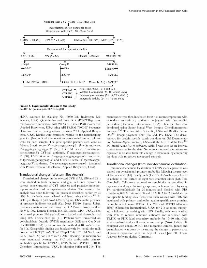
Experimental DesignThe flow map of experimental design is depicted in Figure 1.
Transcriptional ChangesTranscriptional changes in the selected CYPs (1A1, 2B6 and
2E1) were studied in both neuronal and glial cell lines exposed to
various concentrations of CYP inducers and pesticide-monocro-
tophos as described in experimental design. Xenobiotics induced
alterations in the mRNA expression level were expressed in
relative quantification by comparing the data obtained from
unexposed cells. The quantitative Real Time PCR analysis was
done following the protocol described earlier by us [49]. In brief,
total RNA was isolated from both experimental and unexposed
control sets using Gene Elute mammalian total RNA Miniprep Kit
(Catalog No. RTN-70, Sigma, USA). Total RNA (1 mg) was
reverse transcribed into cDNA by Super Script III first strand
Xenobiotic Metabolism in MCP Exposed Brain Cells
PLOS ONE | www.plosone.org 2 March 2014 | Volume 9 | Issue 3 | e91946
cDNA synthesis kit (Catalog No. 18080-051, Invitrogen Life
Science, USA). Quantitative real time PCR (RT-PCRq) assay
reactions were carried out with 26SYBR Green PCR master mix
(Applied Biosystems, USA) using ABI PRISM 7900HT Sequence
Detection System having software version 2.2.1 (Applied Biosys-
tems, USA). Results were expressed relative to the housekeeping
gene i.e., b-actin. Real time reactions were carried out in triplicate
wells for each sample. The gene specific primers used were as
follows: b-actin sense, 59-aaccccaaggccaaccg-39; b-actin antisense,
59-agggatagcacagcctgga-39 [50]; CYP1A1 sense, 59-accttccga-
cactcttccttcg-39; CYP1A1 antisense, 59-cagatgggttgacccatagcttct-
39 [50]; CYP2B6 sense, 59-tggaggatggtggtgaagaag-39; antisense,
59-tgccatcaaggataggcaag-39 and CYP2E1 sense, 59-tgccatcaagga-
taggcaag-39; antisense, 59-caacaaaagaaacaactccatga-39 (designed
with Primer Express 3.0 software; Applied Biosystems, USA).
Translational changes (Western Blot Analysis)Translational changes in the selected CYPs (1A1, 2B6 and 2E1)
were studied in both neuronal and glial cell lines exposed to
various concentrations of CYP inducers and pesticide-monocro-
tophos as described in experimental design. The western blot
analysis was done following the protocol described earlier by us
[49]. In brief,cells were pelleted and lysed using CelLyticTM M
Cell Lysis Reagent (Cat No# C2978, Sigma, USA) in the presence
of protease inhibitor cocktail (Cat No# P8340, Sigma, USA).
Protein estimation was done using the BCA Protein Assay Kit (Cat
No# G1002, Lamda Biotech, Inc., St. Louise, MO, USA). Then
denatured proteins (100 mg/well) were loaded and electrophorsed
using 10% Tricine-SDS gel [51]. Proteins were transferred on
polyvinylidene fluoride (PVDF) membrane (Millipore Cat No#IPVH00010, USA) by the wet transfer method at 180 mA current
for 3 h. Nonspecific binding was blocked with 5% nonfat dry milk
powder in TBST [20 mM Tris-HCl (pH 7.4), 137 mM NaCl, and
0.1% Tween-20] for 2 h at 37uC. After blocking, the membranes
were incubated overnight at 4uC with anti-protein primary
antibodies specific for CYP1A1, CYP2B6 and CYP2E1 (1:1000,
Chemicon International, USA), in blocking buffer (pH 7.5). The
membranes were then incubated for 2 h at room temperature with
secondary anti-primary antibody conjugated with horseradish
peroxidase (Chemicon International, USA). Then the blots were
developed using Super Signal West Fempto Chemiluminescent
SubstrateTM (Thermo Fisher Scientific, USA) and Bio-Rad Versa
DocTM Imaging System 4000 (Bio-Rad, PA, USA). The densi-
tometry for protein specific bands was done on Gel Documenta-
tion System (Alpha Innotech, USA) with the help of Alpha EaseTM
FC Stand Alone V.4.0 software. Actin-b was used as an internal
control to normalize the data. Xenobiotics induced alterations are
expressed in relative term fold change in expression by comparing
the data with respective unexposed controls.
Translational changes (Immunocytochemical localization)Immunocytochemical localization of CYPs specific proteins was
carried out by using anti-primary antibodies following the protocol
of Kapoor et al. [14]. Briefly, cells (16104 cells/well) were allowed
to adhere to the surface of eight well chamber slides (Lab Tek,
Campbell). Cells were exposed to xenobiotics as described in
experimental design. Following exposure, cells were fixed by using
4% paraformaldehyde for 20 minutes and blocked with PBS
containing 0.02% Triton-6100 and 0.1% BSA for 2 h to block the
non-specific binding sites. Cells were then washed with PBS and
incubated with primary antibodies against specific gene proteins,
i.e. rabbit anti human CYP1A1, CYP2B6 and CYP2E1 (dilution-
1:500) (Chemicon International, USA) for 2 h at room temper-
ature followed by washing with PBS. Finally, cells were washed
with PBS to remove unbound antibody and incubated with
TRITC or FITC label secondary antibody for 15–30 min. Cells
were visualized under a fluorescent microscope (Nikon Eclipse 80i
equipped with Nikon DS-Ri1 12.7 megapixel camera, Japan) and
quantification was done by measuring the change in percent area
of protein expression with the help of Leica Qwin 500 Image
Analysis Software (Leica, Germany).
Figure 1. Experimental design of the study.doi:10.1371/journal.pone.0091946.g001
Xenobiotic Metabolism in MCP Exposed Brain Cells
PLOS ONE | www.plosone.org 3 March 2014 | Volume 9 | Issue 3 | e91946
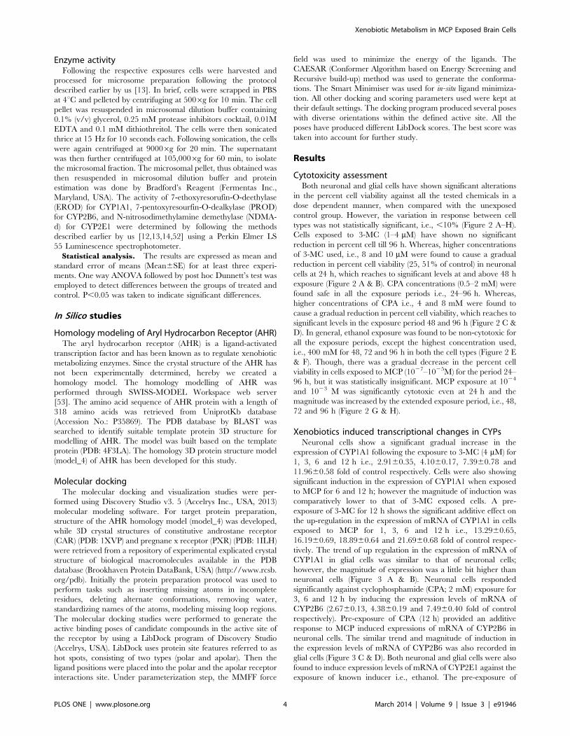
Enzyme activityFollowing the respective exposures cells were harvested and
processed for microsome preparation following the protocol
described earlier by us [13]. In brief, cells were scrapped in PBS
at 4uC and pelleted by centrifuging at 5006g for 10 min. The cell
pellet was resuspended in microsomal dilution buffer containing
0.1% (v/v) glycerol, 0.25 mM protease inhibitors cocktail, 0.01M
EDTA and 0.1 mM dithiothreitol. The cells were then sonicated
thrice at 15 Hz for 10 seconds each. Following sonication, the cells
were again centrifuged at 90006g for 20 min. The supernatant
was then further centrifuged at 105,0006g for 60 min, to isolate
the microsomal fraction. The microsomal pellet, thus obtained was
then resuspended in microsomal dilution buffer and protein
estimation was done by Bradford’s Reagent (Fermentas Inc.,
Maryland, USA). The activity of 7-ethoxyresorufin-O-deethylase
(EROD) for CYP1A1, 7-pentoxyresourfin-O-dealkylase (PROD)
for CYP2B6, and N-nitrosodimethylamine demethylase (NDMA-
d) for CYP2E1 were determined by following the methods
described earlier by us [12,13,14,52] using a Perkin Elmer LS
55 Luminescence spectrophotometer.
Statistical analysis. The results are expressed as mean and
standard error of means (Mean6SE) for at least three experi-
ments. One way ANOVA followed by post hoc Dunnett’s test was
employed to detect differences between the groups of treated and
control. P,0.05 was taken to indicate significant differences.
In Silico studies
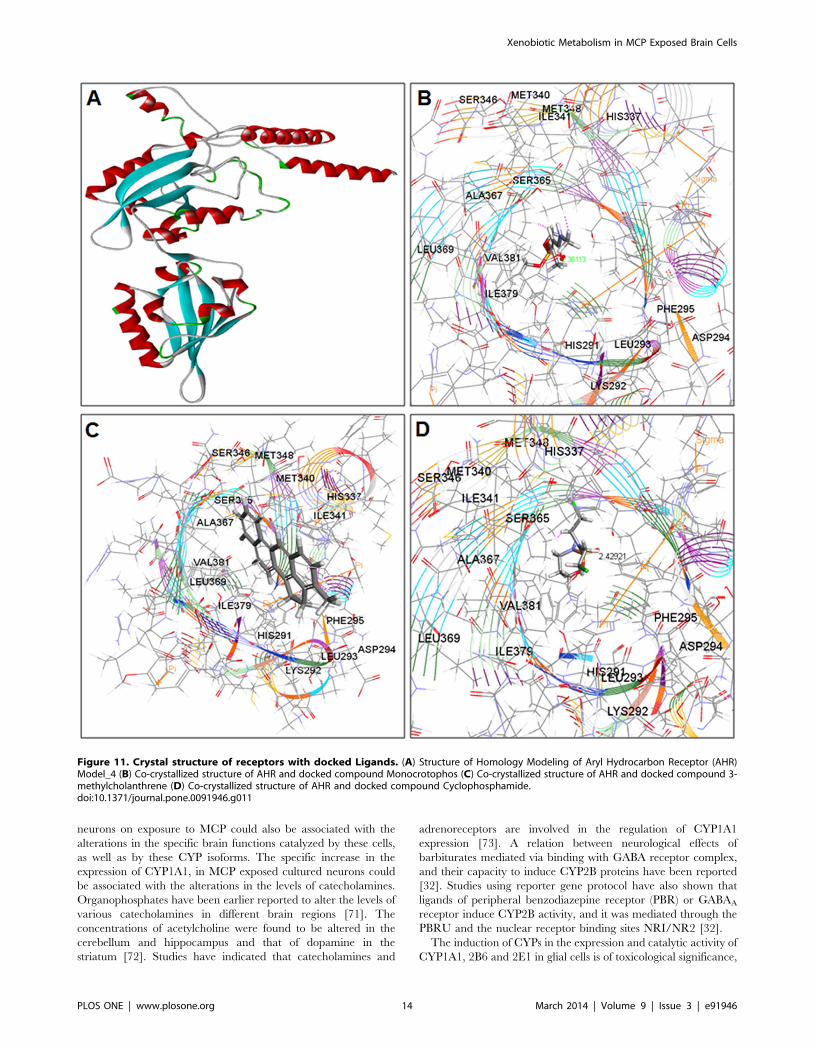
Homology modeling of Aryl Hydrocarbon Receptor (AHR)The aryl hydrocarbon receptor (AHR) is a ligand-activated
transcription factor and has been known as to regulate xenobiotic
metabolizing enzymes. Since the crystal structure of the AHR has
not been experimentally determined, hereby we created a
homology model. The homology modelling of AHR was
performed through SWISS-MODEL Workspace web server
[53]. The amino acid sequence of AHR protein with a length of
318 amino acids was retrieved from UniprotKb database
(Accession No.: P35869). The PDB database by BLAST was
searched to identify suitable template protein 3D structure for
modelling of AHR. The model was built based on the template
protein (PDB: 4F3LA). The homology 3D protein structure model
(model_4) of AHR has been developed for this study.
Molecular dockingThe molecular docking and visualization studies were per-
formed using Discovery Studio v3. 5 (Accelrys Inc., USA, 2013)
molecular modeling software. For target protein preparation,
structure of the AHR homology model (model_4) was developed,
while 3D crystal structures of constitutive androstane receptor
(CAR) (PDB: 1XVP) and pregnane x receptor (PXR) (PDB: 1ILH)
were retrieved from a repository of experimental explicated crystal
structure of biological macromolecules available in the PDB
database (Brookhaven Protein DataBank, USA) (http://www.rcsb.
org/pdb). Initially the protein preparation protocol was used to
perform tasks such as inserting missing atoms in incomplete
residues, deleting alternate conformations, removing water,
standardizing names of the atoms, modeling missing loop regions.
The molecular docking studies were performed to generate the
active binding poses of candidate compounds in the active site of
the receptor by using a LibDock program of Discovery Studio
(Accelrys, USA). LibDock uses protein site features referred to as
hot spots, consisting of two types (polar and apolar). Then the
ligand positions were placed into the polar and the apolar receptor
interactions site. Under parameterization step, the MMFF force
field was used to minimize the energy of the ligands. The
CAESAR (Conformer Algorithm based on Energy Screening and
Recursive build-up) method was used to generate the conforma-
tions. The Smart Minimiser was used for in-situ ligand minimiza-
tion. All other docking and scoring parameters used were kept at
their default settings. The docking program produced several poses
with diverse orientations within the defined active site. All the
poses have produced different LibDock scores. The best score was
taken into account for further study.
Results
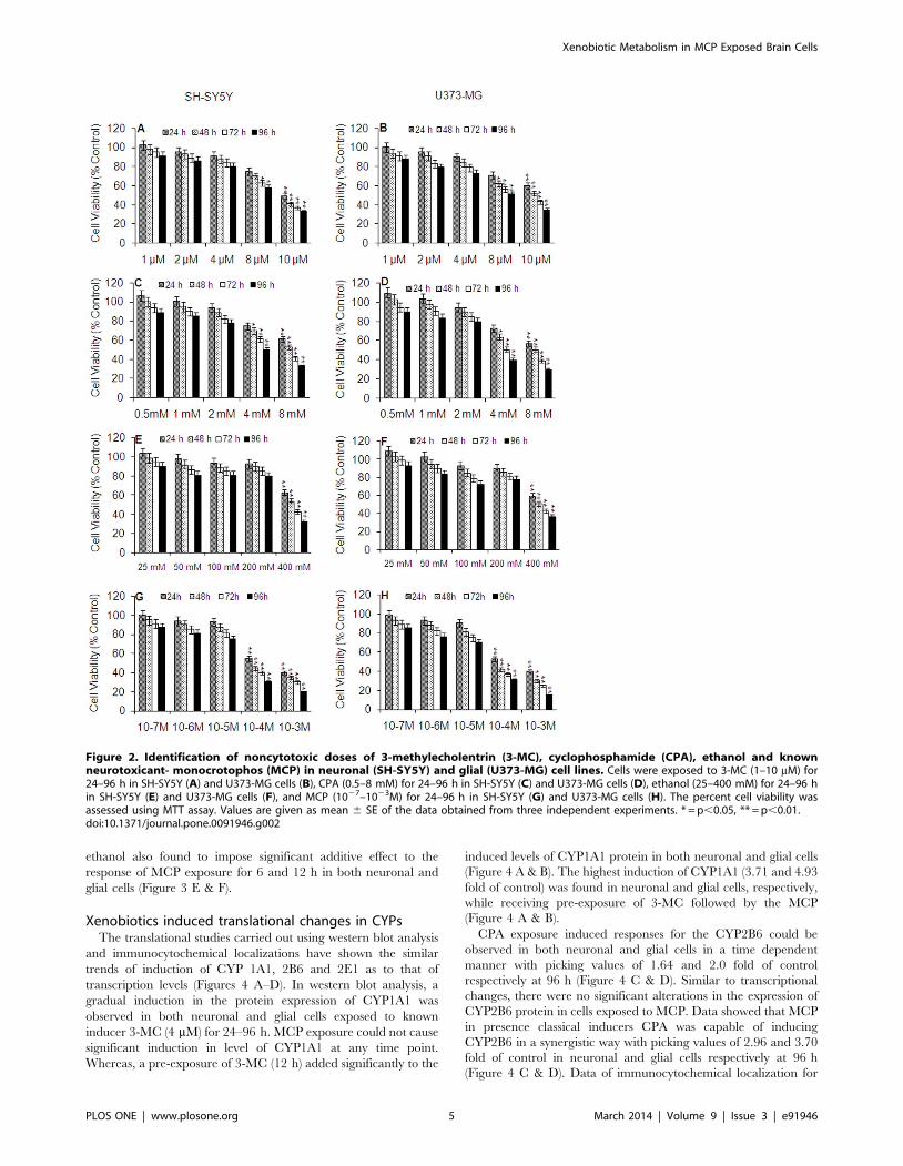
Cytotoxicity assessmentBoth neuronal and glial cells have shown significant alterations
in the percent cell viability against all the tested chemicals in a
dose dependent manner, when compared with the unexposed
control group. However, the variation in response between cell
types was not statistically significant, i.e., ,10% (Figure 2 A–H).
Cells exposed to 3-MC (1–4 mM) have shown no significant
reduction in percent cell till 96 h. Whereas, higher concentrations
of 3-MC used, i.e., 8 and 10 mM were found to cause a gradual
reduction in percent cell viability (25, 51% of control) in neuronal
cells at 24 h, which reaches to significant levels at and above 48 h
exposure (Figure 2 A & B). CPA concentrations (0.5–2 mM) were
found safe in all the exposure periods i.e., 24–96 h. Whereas,
higher concentrations of CPA i.e., 4 and 8 mM were found to
cause a gradual reduction in percent cell viability, which reaches to
significant levels in the exposure period 48 and 96 h (Figure 2 C &
D). In general, ethanol exposure was found to be non-cytotoxic for
all the exposure periods, except the highest concentration used,
i.e., 400 mM for 48, 72 and 96 h in both the cell types (Figure 2 E
& F). Though, there was a gradual decrease in the percent cell
viability in cells exposed to MCP (1027–1025M) for the period 24–
96 h, but it was statistically insignificant. MCP exposure at 1024
and 1023 M was significantly cytotoxic even at 24 h and the
magnitude was increased by the extended exposure period, i.e., 48,
72 and 96 h (Figure 2 G & H).
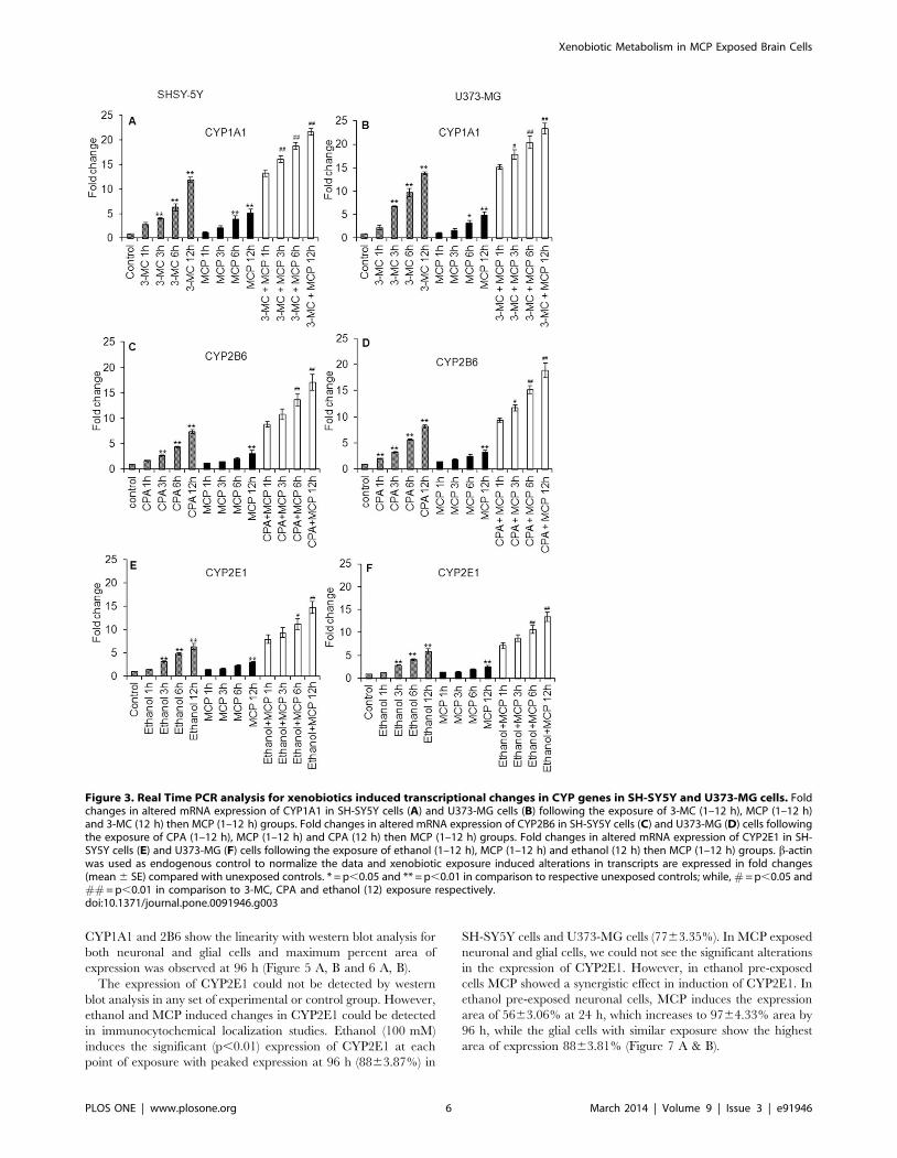
Xenobiotics induced transcriptional changes in CYPsNeuronal cells show a significant gradual increase in the
expression of CYP1A1 following the exposure to 3-MC (4 mM) for
1, 3, 6 and 12 h i.e., 2.9160.35, 4.1060.17, 7.3960.78 and
11.9660.58 fold of control respectively. Cells were also showing
significant induction in the expression of CYP1A1 when exposed
to MCP for 6 and 12 h; however the magnitude of induction was
comparatively lower to that of 3-MC exposed cells. A pre-
exposure of 3-MC for 12 h shows the significant additive effect on
the up-regulation in the expression of mRNA of CYP1A1 in cells
exposed to MCP for 1, 3, 6 and 12 h i.e., 13.2960.65,
16.1960.69, 18.8960.64 and 21.6960.68 fold of control respec-
tively. The trend of up regulation in the expression of mRNA of
CYP1A1 in glial cells was similar to that of neuronal cells;
however, the magnitude of expression was a little bit higher than
neuronal cells (Figure 3 A & B). Neuronal cells responded
significantly against cyclophosphamide (CPA; 2 mM) exposure for
3, 6 and 12 h by inducing the expression levels of mRNA of
CYP2B6 (2.6760.13, 4.3860.19 and 7.4960.40 fold of control
respectively). Pre-exposure of CPA (12 h) provided an additive
response to MCP induced expressions of mRNA of CYP2B6 in
neuronal cells. The similar trend and magnitude of induction in
the expression levels of mRNA of CYP2B6 was also recorded in
glial cells (Figure 3 C & D). Both neuronal and glial cells were also
found to induce expression levels of mRNA of CYP2E1 against the
exposure of known inducer i.e., ethanol. The pre-exposure of
Xenobiotic Metabolism in MCP Exposed Brain Cells
PLOS ONE | www.plosone.org 4 March 2014 | Volume 9 | Issue 3 | e91946
ethanol also found to impose significant additive effect to the
response of MCP exposure for 6 and 12 h in both neuronal and
glial cells (Figure 3 E & F).
Xenobiotics induced translational changes in CYPsThe translational studies carried out using western blot analysis
and immunocytochemical localizations have shown the similar
trends of induction of CYP 1A1, 2B6 and 2E1 as to that of
transcription levels (Figures 4 A–D). In western blot analysis, a
gradual induction in the protein expression of CYP1A1 was
observed in both neuronal and glial cells exposed to known
inducer 3-MC (4 mM) for 24–96 h. MCP exposure could not cause
significant induction in level of CYP1A1 at any time point.
Whereas, a pre-exposure of 3-MC (12 h) added significantly to the
induced levels of CYP1A1 protein in both neuronal and glial cells
(Figure 4 A & B). The highest induction of CYP1A1 (3.71 and 4.93
fold of control) was found in neuronal and glial cells, respectively,
while receiving pre-exposure of 3-MC followed by the MCP
(Figure 4 A & B).
CPA exposure induced responses for the CYP2B6 could be
observed in both neuronal and glial cells in a time dependent
manner with picking values of 1.64 and 2.0 fold of control
respectively at 96 h (Figure 4 C & D). Similar to transcriptional
changes, there were no significant alterations in the expression of
CYP2B6 protein in cells exposed to MCP. Data showed that MCP
in presence classical inducers CPA was capable of inducing
CYP2B6 in a synergistic way with picking values of 2.96 and 3.70
fold of control in neuronal and glial cells respectively at 96 h
(Figure 4 C & D). Data of immunocytochemical localization for
Figure 2. Identification of noncytotoxic doses of 3-methylecholentrin (3-MC), cyclophosphamide (CPA), ethanol and knownneurotoxicant- monocrotophos (MCP) in neuronal (SH-SY5Y) and glial (U373-MG) cell lines. Cells were exposed to 3-MC (1–10 mM) for24–96 h in SH-SY5Y (A) and U373-MG cells (B), CPA (0.5–8 mM) for 24–96 h in SH-SY5Y (C) and U373-MG cells (D), ethanol (25–400 mM) for 24–96 hin SH-SY5Y (E) and U373-MG cells (F), and MCP (1027–1023M) for 24–96 h in SH-SY5Y (G) and U373-MG cells (H). The percent cell viability wasassessed using MTT assay. Values are given as mean 6 SE of the data obtained from three independent experiments. * = p,0.05, ** = p,0.01.doi:10.1371/journal.pone.0091946.g002
Xenobiotic Metabolism in MCP Exposed Brain Cells
PLOS ONE | www.plosone.org 5 March 2014 | Volume 9 | Issue 3 | e91946
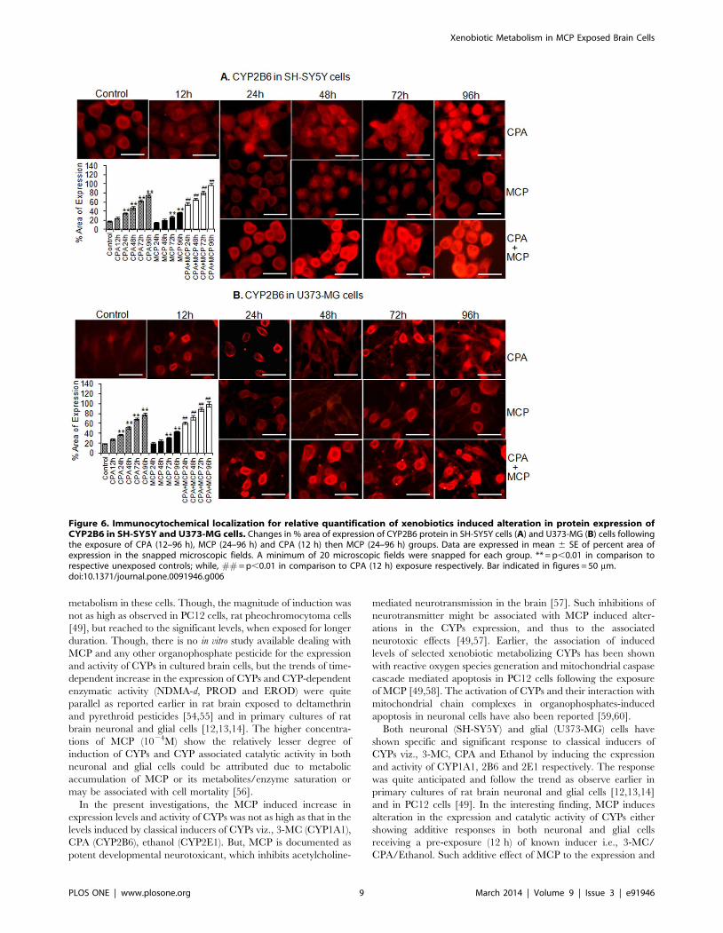
CYP1A1 and 2B6 show the linearity with western blot analysis for
both neuronal and glial cells and maximum percent area of
expression was observed at 96 h (Figure 5 A, B and 6 A, B).
The expression of CYP2E1 could not be detected by western
blot analysis in any set of experimental or control group. However,
ethanol and MCP induced changes in CYP2E1 could be detected
in immunocytochemical localization studies. Ethanol (100 mM)
induces the significant (p,0.01) expression of CYP2E1 at each
point of exposure with peaked expression at 96 h (8863.87%) in
SH-SY5Y cells and U373-MG cells (7763.35%). In MCP exposed
neuronal and glial cells, we could not see the significant alterations
in the expression of CYP2E1. However, in ethanol pre-exposed
cells MCP showed a synergistic effect in induction of CYP2E1. In
ethanol pre-exposed neuronal cells, MCP induces the expression
area of 5663.06% at 24 h, which increases to 9764.33% area by
96 h, while the glial cells with similar exposure show the highest
area of expression 8863.81% (Figure 7 A & B).
Figure 3. Real Time PCR analysis for xenobiotics induced transcriptional changes in CYP genes in SH-SY5Y and U373-MG cells. Foldchanges in altered mRNA expression of CYP1A1 in SH-SY5Y cells (A) and U373-MG cells (B) following the exposure of 3-MC (1–12 h), MCP (1–12 h)and 3-MC (12 h) then MCP (1–12 h) groups. Fold changes in altered mRNA expression of CYP2B6 in SH-SY5Y cells (C) and U373-MG (D) cells followingthe exposure of CPA (1–12 h), MCP (1–12 h) and CPA (12 h) then MCP (1–12 h) groups. Fold changes in altered mRNA expression of CYP2E1 in SH-SY5Y cells (E) and U373-MG (F) cells following the exposure of ethanol (1–12 h), MCP (1–12 h) and ethanol (12 h) then MCP (1–12 h) groups. b-actinwas used as endogenous control to normalize the data and xenobiotic exposure induced alterations in transcripts are expressed in fold changes(mean 6 SE) compared with unexposed controls. * = p,0.05 and ** = p,0.01 in comparison to respective unexposed controls; while, # = p,0.05 and## = p,0.01 in comparison to 3-MC, CPA and ethanol (12) exposure respectively.doi:10.1371/journal.pone.0091946.g003
Xenobiotic Metabolism in MCP Exposed Brain Cells
PLOS ONE | www.plosone.org 6 March 2014 | Volume 9 | Issue 3 | e91946
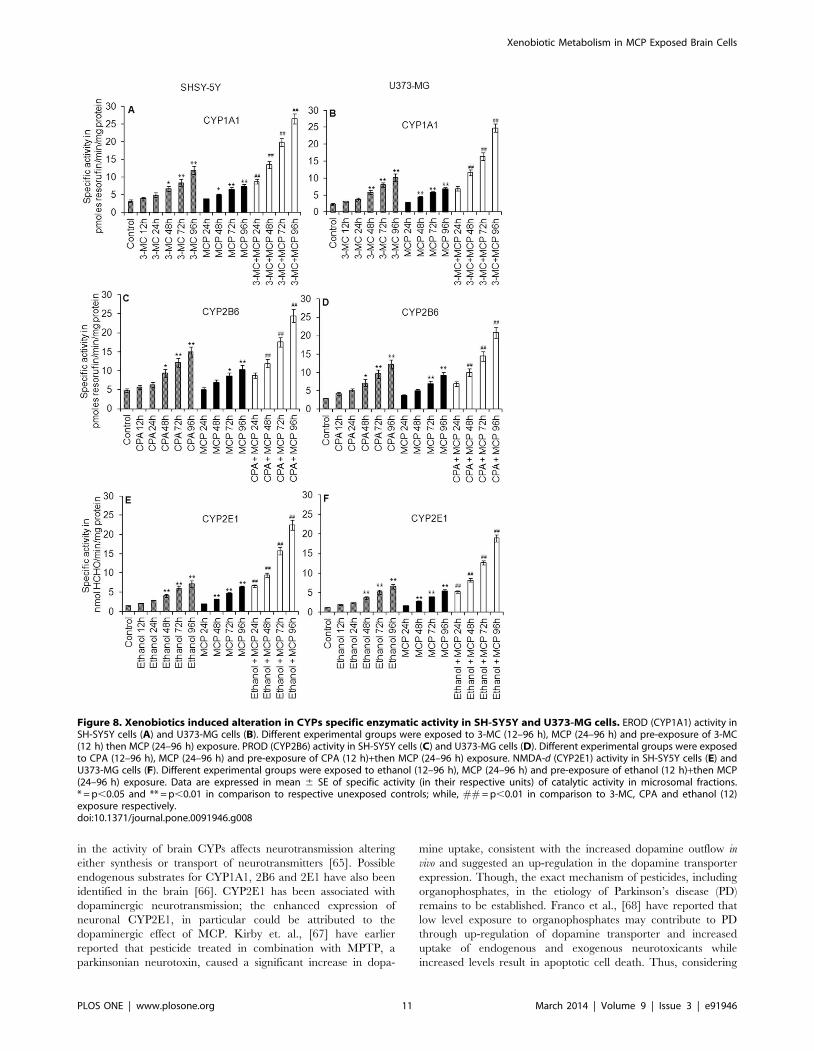
Xenobiotics induced alterations in the catalytic activity ofCYPs
In general, the microsomal fractions from both neuronal and
glial cells show significant induction in CYP dependent EROD
(CYP1A1), PROD (CYP2B6) and NDMA-d (CYP2E1) activity.
The trend in the induction of CYPs activity was fairly correlated
with the data of western blot analysis and real time analysis
(Figure 8 A–F).
3-MC, a known inducer of CYP1A1 causes significant (p,0.01)
induction in the catalytic activity of CYP1A1 (4.1260.19,
4.9660.62, 6.7360.73, 8.4961.03 and 11.8961.18 pmoles of
resorufin/min/mg protein at 12 h, 24 h, 48 h, 72 h and 96 h,
respectively) in neuronal cells while, (3.160.16, 3.6960.39,
5.8360.59, 7.9760.68 and 10.1561.09 pmoles of resorufin/
min/mg protein at 12 h, 24 h, 48 h, 72 h and 96 h respectively)
in glial cells. MCP alone again failed to activate the CYP1A1
enzyme to catalyze. 3-MC pre-exposure significantly (p,0.01)
induces the EROD induction capacity of MCP in both neuronal
(Figure 8 A) and glial cells (Figure 8 B).
In case of PROD, the specific activity of CYP2B6 was increased
from (5.6360.53) at 12 h to (14.9261.34) at 96 h in neuronal cells
and from (4.1260.39) at 12 h to (12.2161.24) at 96 h in glial cells
following the exposure of CPAs. MCP did not induce the activity
of CYP2B6 at any time point. CPA pre-exposed neuronal cells
show significant induction in the PROD activity upon the
exposure of MCP i.e., 8.7660.74 pmoles of resorufin/min/mg
protein at 24 h to 24.4861.67 pmoles of resorufin/min/mg
protein at 96 h. In glial cells, MCP after the pre-exposure of
CPA induces CYP2B6 activity at all points of exposure with peak
expression at 96 h (20.8261.40) (Figure 8 C & D).
Ethanol, known inducer of CYP2E1, induces the NDMA-d
activity in neuronal cells at each point of exposure, such as at 12 h
(2.1260.10) to 96 h (7.1960.88). Likewise, in glial cells, ethanol
induced increase in specific activity of CYP2E1 has been recorded,
i.e., at 12 h (1.860.08) and at 96 h (6.4560.72). MCP could not
induce the specific activity of CYP2E1 significant in any cell type
used even at any time point of exposure. However, MCP
significantly enhances the specific activity of CYP2E1 (NDMA-d)
in both neuronal and glial cells pre-exposed to ethanol. The peak
induction was observed at 96 h in both neuronal (22.4661.26) and
glial (18.9460.96) cells respectively (Figure 8 E & F).
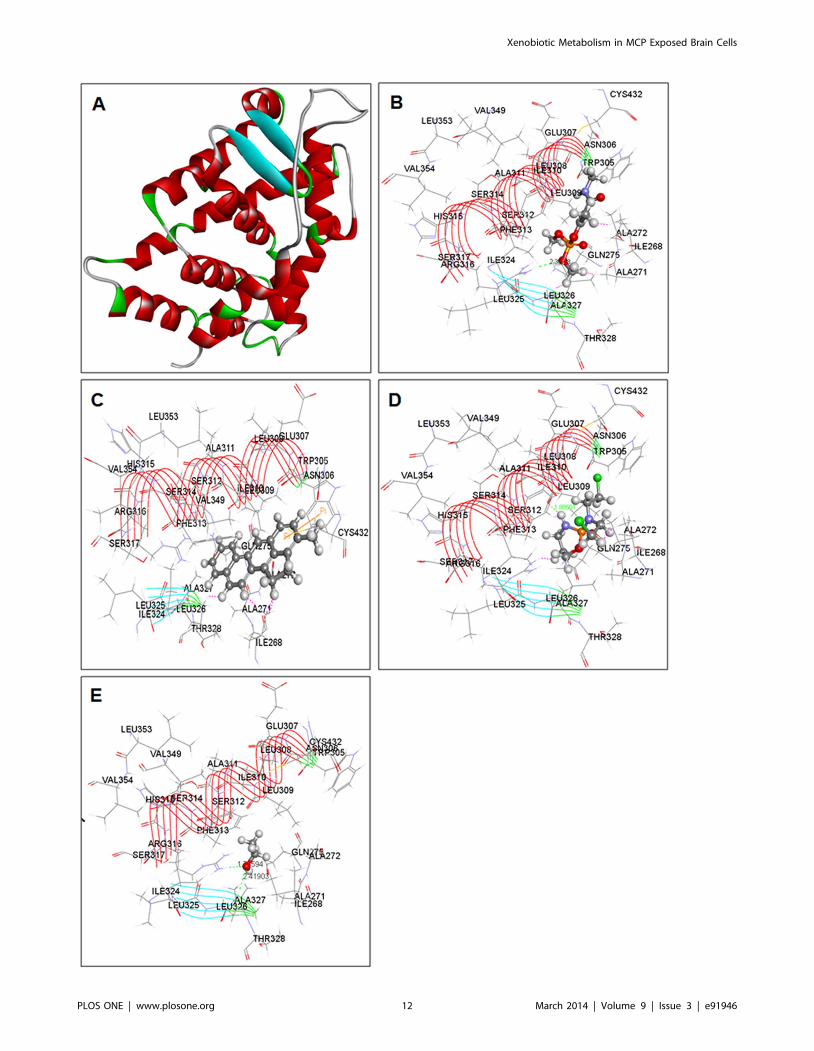
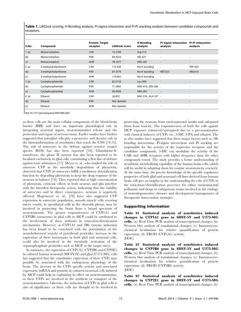
Protein-ligand docking studiesThe results of possible mechanisms of action of MCP in relation
to human CYP regulators (CAR, PXR and AHR) revealed
through molecular docking studies are summarized in Figure 9 (A–
E), 10 (A–D), 11 (A–D) and Table 1. The finding demonstrates a
significant modulatory activity of MCP to the respective receptors
of CYP. This compound exhibited single hydrogen (-H) bonds
with each of the studied receptor, i.e., CAR, PXR and AHR. The
Figure 4. Western blot analysis for xenobiotic induced translational changes in CYP proteins in SH-SY5Y and U373-MG cells. (A)Western blot analysis of expression of CYP1A1 protein in SH-SY5Y cells following the exposure of 3-MC (24–96 h), MCP (24–96 h) and 3-MC (12 h)followed by post-exposure of MCP (24–96 h). (B) Western blot analysis of expression of CYP1A1 protein in U373-MG cells following the exposure of 3-MC (24–96 h), MCP (24–96 h) and 3-MC (12 h) followed by post-exposure of MCP (24–96 h). (C) Western blot analysis of expression of CYP2B6 proteinin SH-SY5Y cells following the exposure of CPA (24–96 h), MCP (24–96 h) and CPA (12 h) followed by post-exposure of MCP (24–96 h). (D) Westernblot analysis of expression of CYP2B6 protein in U373-MG cells following the exposure of CPA (24–96 h), MCP (24–96 h) and CPA (12 h) followed bypost-exposure of MCP (24–96 h). The values obtained in unexposed cells were considered basal, i.e., relative quantification in expression at differentpoint of various exposures was done comparing the values of unexposed controls. b-actin was used as endogenous control to normalize the data.doi:10.1371/journal.pone.0091946.g004
Xenobiotic Metabolism in MCP Exposed Brain Cells
PLOS ONE | www.plosone.org 7 March 2014 | Volume 9 | Issue 3 | e91946

amino acid residue interacted with MCP through (-H) bond was
histidine (HIS-327) of PXR and arginine (ARG-281) & (Arg-316)
of AHR and CAR respectively (Table 1). Similarly, the result
shows that 3-MC, a highly carcinogenic polycyclic aromatic
hydrocarbon, have positive positive interactions with all the three
CYP receptors. 3-MC has shown comparatively better docking
(LibDock) score than that of other compounds used in this study,
i.e., MCP, CPA and ethanol (Table 1). It’s LibDock score with
CAR receptor CAR was 112.426 and has shown an exposing (-H)
bond interaction with tryptophan (TRP-305). MC represents a
score of 114.962 through AHR receptor. These scoring are
considered as significantly good interaction between compound
and receptors. The compound 3-MC has shown a LibDock score
of 87.5779 with PXR and presenting Pi-Pi interaction and Pi-
sigma interaction with amino acid residue arginine (ARG-410) and
methionine (MET-323). This score is also considered to be a good
docking score, implies to more effective binding with the receptors
and may be a more toxic than other tested compounds.
Correspondingly the result for CPA revealed that it can bind
more effectively with AHR receptor. This binding represents a
LibDock (docking) score of 90.4056 and a single (-H) bond
interaction with residue arginine (ARG-281). On the other hand,
this compound illustrates a LibDock score of 85.5133 with CAR
and 71.5965 with PXR. The amino acid residues involved in the
hydrogen bond interactions were leucine (LEU-309) of CAR and
arginine (ARG-410) & serine (SER-208) of PXR. Apart this the
molecular docking result of ethanol, known inducer of CYP2E1,
specifies that it can partially interact with only one of the receptor
i.e., CAR. With this receptor its represent a low docking score
(LibDock) of 26.835 (Table 1). The amino acid residue involved in
these interactions was arginine (ARG-316) and alanine (ALA-327).
On the other hand, results indicate that there is no interaction
between ethanol and receptors PXR & AHR.
Discussion
The data of alterations in the expression (mRNA and protein)
and marker CYPs enzyme activity in cultured human brain
neuronal and glial cells have demonstrated that even at lower
doses (non-cytotoxic dose), monocrotophos (MCP) is an inducer of
these CYPs. These MCP induced alterations in the expression and
activity of different CYP isoforms could be indicative of their
Figure 5. Immunocytochemical localization for relative quantification of xenobiotics induced alteration in protein expression ofCYP1A1 in SH-SY5Y and U373-MG cells. Changes in % area of expression of CYP1A1 protein in SH-SY5Y cells (A) and U373-MG cells (B)following the exposure of 3-MC (12–96 h), MCP (24–96 h) and 3-MC (12 h) then MCP (24–96 h) groups. Data are expressed in mean 6 SE of percentarea of expression in the snapped microscopic fields. A minimum of 20 microscopic fields were snapped for each group. ** = p,0.01 in comparison torespective unexposed controls; while, ## = p,0.01 in comparison to 3-MC (12 h) exposure respectively. Bar indicated in figures = 50 mm.doi:10.1371/journal.pone.0091946.g005
Xenobiotic Metabolism in MCP Exposed Brain Cells
PLOS ONE | www.plosone.org 8 March 2014 | Volume 9 | Issue 3 | e91946
metabolism in these cells. Though, the magnitude of induction was
not as high as observed in PC12 cells, rat pheochromocytoma cells
[49], but reached to the significant levels, when exposed for longer
duration. Though, there is no in vitro study available dealing with
MCP and any other organophosphate pesticide for the expression
and activity of CYPs in cultured brain cells, but the trends of time-
dependent increase in the expression of CYPs and CYP-dependent
enzymatic activity (NDMA-d, PROD and EROD) were quite
parallel as reported earlier in rat brain exposed to deltamethrin
and pyrethroid pesticides [54,55] and in primary cultures of rat
brain neuronal and glial cells [12,13,14]. The higher concentra-
tions of MCP (1024M) show the relatively lesser degree of
induction of CYPs and CYP associated catalytic activity in both
neuronal and glial cells could be attributed due to metabolic
accumulation of MCP or its metabolites/enzyme saturation or
may be associated with cell mortality [56].
In the present investigations, the MCP induced increase in
expression levels and activity of CYPs was not as high as that in the
levels induced by classical inducers of CYPs viz., 3-MC (CYP1A1),
CPA (CYP2B6), ethanol (CYP2E1). But, MCP is documented as
potent developmental neurotoxicant, which inhibits acetylcholine-
mediated neurotransmission in the brain [57]. Such inhibitions of
neurotransmitter might be associated with MCP induced alter-
ations in the CYPs expression, and thus to the associated
neurotoxic effects [49,57]. Earlier, the association of induced
levels of selected xenobiotic metabolizing CYPs has been shown
with reactive oxygen species generation and mitochondrial caspase
cascade mediated apoptosis in PC12 cells following the exposure
of MCP [49,58]. The activation of CYPs and their interaction with
mitochondrial chain complexes in organophosphates-induced
apoptosis in neuronal cells have also been reported [59,60].
Both neuronal (SH-SY5Y) and glial (U373-MG) cells have
shown specific and significant response to classical inducers of
CYPs viz., 3-MC, CPA and Ethanol by inducing the expression
and activity of CYP1A1, 2B6 and 2E1 respectively. The response
was quite anticipated and follow the trend as observe earlier in
primary cultures of rat brain neuronal and glial cells [12,13,14]
and in PC12 cells [49]. In the interesting finding, MCP induces
alteration in the expression and catalytic activity of CYPs either
showing additive responses in both neuronal and glial cells
receiving a pre-exposure (12 h) of known inducer i.e., 3-MC/
CPA/Ethanol. Such additive effect of MCP to the expression and
Figure 6. Immunocytochemical localization for relative quantification of xenobiotics induced alteration in protein expression ofCYP2B6 in SH-SY5Y and U373-MG cells. Changes in % area of expression of CYP2B6 protein in SH-SY5Y cells (A) and U373-MG (B) cells followingthe exposure of CPA (12–96 h), MCP (24–96 h) and CPA (12 h) then MCP (24–96 h) groups. Data are expressed in mean 6 SE of percent area ofexpression in the snapped microscopic fields. A minimum of 20 microscopic fields were snapped for each group. ** = p,0.01 in comparison torespective unexposed controls; while, ## = p,0.01 in comparison to CPA (12 h) exposure respectively. Bar indicated in figures = 50 mm.doi:10.1371/journal.pone.0091946.g006
Xenobiotic Metabolism in MCP Exposed Brain Cells
PLOS ONE | www.plosone.org 9 March 2014 | Volume 9 | Issue 3 | e91946

activity of all the three CYPs studied in pre-sensitized cells indicate
the involvement of all these CYPs in the metabolism of MCP. The
findings also indicate that non-sensitized cells were not having
enough triggering signals to induce the expression and activity like
primary cultures of rat brain neuronal and glial cells [14] and in
PC12 cells [49]. Thus, the findings suggest the xenobiotic
metabolizing capabilities of these cells against MCP or may be
to any other organophosphate pesticide, provided they received a
pre-sensitization stimulus to trigger the xenobiotic metabolizing
machinery in them. It is widely documented that the specific
chemicals contributes significantly to induce the expression of
specific xenobiotic metabolizing enzymes by inducing the
increased expression of the master regulators of CYPs genes viz.,
aryl hydrocarbon receptor (AHR), constitutive androstane recep-
tor (CAR) and pregnane-X-receptor (PXR) [61]. In the present
investigation, it seems that MCP profoundly adds/synergizes
classical CYP-inducers viz., 3-MC, CPA and ethanol (ligands of
AHR, CAR and PXR) in inducing the expression of CYP1A1,
2B6 and 2E1 respectively. Thus, MCP might be trans-activating
these master regulators of CYPs (AHR, CAR and PXR).
The linearity in the expression and inducibility between mRNA
and protein level (Western blot analysis) for CYP2E1 could not be
established. While, the trends were similar for mRNA and
immunocytochemical localization and catalytic activity in case of
CYP2E1. Such inconsistency might be due to the post-transla-
tional regulation of CYP2E1, which involves various cellular
factors viz., insulin, growth hormones, epidermal growth factor,
etc. [35]. Besides that, the mechanism of CYP2E1 induction is
complex, depends on the substrate, species, tissue, or cell type
[35,36,37]. Several levels of gene regulation like transcription,
translation, and post-translational modification, play an important
role in maintaining the proper function of CYPs. Post-translational
modifications (like phosphorylation, glycosylation, nitration),
ubiquitination for proteasome mediated protein degradation,
targeting to specific cellular compartments are well reported for
non-consistency between the expression of mRNA and the protein
of CYPs [62]. Recently, CYPs and their nuclear receptor
regulators have been found to be post-transcriptionally regulated
by miRNAs. Mohri et al., [63] reported that the molecular
mechanism of CYP2E1 regulation by miR-378 to clarify the non-
consistency between mRNA and protein expression of CYP2E1.
The ability of MCP to induce the expression of neuronal CYPs
is of significance as studies have indicated a role of CYPs in
neurotransmission [64]. Studies have also shown that modulation
Figure 7. Immunocytochemical localization for relative quantification of xenobiotics induced alteration in protein expression ofCYP2E1 in SH-SY5Y and U373-MG cells. Changes in % area of expression of CYP2E1 protein in SH-SY5Y cells (A) and U373-MG (B) cells followingthe exposure of ethanol (12–96 h), MCP (24–96 h) and ethanol (12 h) then MCP (24–96 h) groups. Data are expressed in mean 6 SE of percent area ofexpression in the snapped microscopic fields. A minimum of 20 microscopic fields were snapped for each group. ** = p,0.01 in comparison torespective unexposed controls; while, ## = p,0.01 in comparison to ethanol (12 h) exposure respectively. Bar indicated in figures = 50 mm.doi:10.1371/journal.pone.0091946.g007
Xenobiotic Metabolism in MCP Exposed Brain Cells
PLOS ONE | www.plosone.org 10 March 2014 | Volume 9 | Issue 3 | e91946
in the activity of brain CYPs affects neurotransmission altering
either synthesis or transport of neurotransmitters [65]. Possible
endogenous substrates for CYP1A1, 2B6 and 2E1 have also been
identified in the brain [66]. CYP2E1 has been associated with
dopaminergic neurotransmission; the enhanced expression of
neuronal CYP2E1, in particular could be attributed to the
dopaminergic effect of MCP. Kirby et. al., [67] have earlier
reported that pesticide treated in combination with MPTP, a
parkinsonian neurotoxin, caused a significant increase in dopa-
mine uptake, consistent with the increased dopamine outflow in
vivo and suggested an up-regulation in the dopamine transporter
expression. Though, the exact mechanism of pesticides, including
organophosphates, in the etiology of Parkinson’s disease (PD)
remains to be established. Franco et al., [68] have reported that
low level exposure to organophosphates may contribute to PD
through up-regulation of dopamine transporter and increased
uptake of endogenous and exogenous neurotoxicants while
increased levels result in apoptotic cell death. Thus, considering
Figure 8. Xenobiotics induced alteration in CYPs specific enzymatic activity in SH-SY5Y and U373-MG cells. EROD (CYP1A1) activity inSH-SY5Y cells (A) and U373-MG cells (B). Different experimental groups were exposed to 3-MC (12–96 h), MCP (24–96 h) and pre-exposure of 3-MC(12 h) then MCP (24–96 h) exposure. PROD (CYP2B6) activity in SH-SY5Y cells (C) and U373-MG cells (D). Different experimental groups were exposedto CPA (12–96 h), MCP (24–96 h) and pre-exposure of CPA (12 h)+then MCP (24–96 h) exposure. NMDA-d (CYP2E1) activity in SH-SY5Y cells (E) andU373-MG cells (F). Different experimental groups were exposed to ethanol (12–96 h), MCP (24–96 h) and pre-exposure of ethanol (12 h)+then MCP(24–96 h) exposure. Data are expressed in mean 6 SE of specific activity (in their respective units) of catalytic activity in microsomal fractions.* = p,0.05 and ** = p,0.01 in comparison to respective unexposed controls; while, ## = p,0.01 in comparison to 3-MC, CPA and ethanol (12)exposure respectively.doi:10.1371/journal.pone.0091946.g008
Xenobiotic Metabolism in MCP Exposed Brain Cells
PLOS ONE | www.plosone.org 11 March 2014 | Volume 9 | Issue 3 | e91946
Xenobiotic Metabolism in MCP Exposed Brain Cells
PLOS ONE | www.plosone.org 12 March 2014 | Volume 9 | Issue 3 | e91946

that CYP2E1 has a role in dopamine metabolism [65], and the
fact that CYP2E1 has been found to be co-localized with tyrosine
hydroxylase [69], one could speculate that the MCP induced
alterations in neuronal CYP2E1 could, in turn, be associated with
alterations in the levels of dopamine induced by the MCP.
The increase in the activity of CYP1A1 and CYP2B6 isoforms,
in cultured neurons following MCP exposure could also be of
significance, as earlier studies from our laboratory have shown the
involvement of these xenobiotic metabolizing CYPs in the
neurobehavioral toxicity of pyrethroid pesticide-deltamethrin
[70]. The increase in the expression of these CYPs in cultured
Figure 9. Crystal structure of receptors with docked Ligands. (A) Crystal structure of CAR (PDB ID: 1XVP) (B) Co-crystallized structure of CARand docked compound monocrotophos (C) Co-crystallized structure of CAR and docked compound 3-methylcholanthrene (D) Co-crystallizedstructure of CAR and docked compound Cyclophosphamide (E) Co-crystallized structure of CAR and docked compound Ethanol.doi:10.1371/journal.pone.0091946.g009
Figure 10. Crystal structure of receptors with docked Ligands. (A) Crystal structure of PXR (PDB ID: 1ILH) (B) Co-crystallized structure of PXRand docked compound Monocrotophos (C) Co-crystallized structure of PXR and docked compound 3-methylcholanthrene (D) Co-crystallizedstructure of PXR and docked compound Cyclophosphamide.doi:10.1371/journal.pone.0091946.g010
Xenobiotic Metabolism in MCP Exposed Brain Cells
PLOS ONE | www.plosone.org 13 March 2014 | Volume 9 | Issue 3 | e91946
neurons on exposure to MCP could also be associated with the
alterations in the specific brain functions catalyzed by these cells,
as well as by these CYP isoforms. The specific increase in the
expression of CYP1A1, in MCP exposed cultured neurons could
be associated with the alterations in the levels of catecholamines.
Organophosphates have been earlier reported to alter the levels of
various catecholamines in different brain regions [71]. The
concentrations of acetylcholine were found to be altered in the
cerebellum and hippocampus and that of dopamine in the
striatum [72]. Studies have indicated that catecholamines and
adrenoreceptors are involved in the regulation of CYP1A1
expression [73]. A relation between neurological effects of
barbiturates mediated via binding with GABA receptor complex,
and their capacity to induce CYP2B proteins have been reported
[32]. Studies using reporter gene protocol have also shown that
ligands of peripheral benzodiazepine receptor (PBR) or GABAA
receptor induce CYP2B activity, and it was mediated through the
PBRU and the nuclear receptor binding sites NRI/NR2 [32].
The induction of CYPs in the expression and catalytic activity of
CYP1A1, 2B6 and 2E1 in glial cells is of toxicological significance,
Figure 11. Crystal structure of receptors with docked Ligands. (A) Structure of Homology Modeling of Aryl Hydrocarbon Receptor (AHR)Model_4 (B) Co-crystallized structure of AHR and docked compound Monocrotophos (C) Co-crystallized structure of AHR and docked compound 3-methylcholanthrene (D) Co-crystallized structure of AHR and docked compound Cyclophosphamide.doi:10.1371/journal.pone.0091946.g011
Xenobiotic Metabolism in MCP Exposed Brain Cells
PLOS ONE | www.plosone.org 14 March 2014 | Volume 9 | Issue 3 | e91946
as these cells are the main cellular components of the blood-brain
barrier (BBB) and have an important physiological role in
integrating neuronal inputs, neurotransmitter release and the
protection and repair of nervous tissue. Earlier studies have further
suggested that astroglial cells play a protective and decisive role in
the biotransformation of xenobiotics that reach the CNS [74,75].
The role of astrocytes in the defense against reactive oxygen
species (ROS) has also been reported [76]. Glutathione-S-
transferase, the phase II enzyme has also been reported to be
localized exclusively in glial cells, constituting a first line of defense
against toxic substances [77]. Meyer et. al., who studied the role of
astrocyte CYP in the metabolic degradation of phenytoin,
observed that CYPs in astrocytes fulfill a mediatory detoxification
function by degrading phenytoin to keep the drug response of the
neurons in balance [74]. They reported that at high concentration
of phenytoin, cytotoxic effects in both neurons and glia interfere
with the intended therapeutic action, indicating that the viability
of astrocytes and in direct consequence, neurons is negatively
affected. Hagemeyer et. al., [78] have also suggested CYP
expression in astrocytic population, smooth muscle cells covering
micro vessels, in ependymal cells in the choroids plexus, may be
involved in protecting the brain from a broad spectrum of
neurotoxicants. The greater responsiveness of CYP1A1 and
CYP2B6 isoenzymes in glial cells to MCP could be attributed to
the involvement of these isoforms in toxication-detoxication
mechanisms. However, as CYP1A1 and 2B6 enzyme induction
has been found to be correlated with the potentiation of the
neurobehavioral toxicity of pyrethroid pesticides, increase in the
expression of these isoenzymes in both glial and neuronal cells,
could also be involved in the metabolic activation of the
organophosphate pesticides such as MCP at the target site(s).
In summary, the expression of CYP1A1, CYP2B6 and CYP2E1
in cultured human neuronal (SH-SY5Y) and glial (U373-MG) cells
has suggested that the constitutive expression of these CYPs may
possibly be associated with the endogenous physiology of the
brain. The increase in the CYPs specific activity and associated
expression (mRNA and protein) in cultured neuronal cells induced
by MCP could help in explaining its effect on neurotransmission,
as these CYPs are involved in the synthesis or transport of the
neurotransmitters. Likewise, the induction of CYPs in glial cells is
also of significance as these cells are thought to be involved in
protecting the neurons from environmental insults and safeguard
them from toxicity. The responsiveness of both the cells against
MCP exposure enhanced/synergized due to a pre-sensitization
with classical inducers of CYPs viz., 3-MC, CPA and ethanol. The
in silico studies have suggested that three major factors such as (-H)
bonding interactions, Pi-sigma interactions and Pi stacking are
responsible for the activity of the respective receptors and the
candidate compounds. 3-MC can modulate the activity of the
CAR and AHR receptors with higher frequency than the other
compounds tested. The study provides a better understanding of
xenobiotic metabolizing capability of the human brain cells, which
will be useful in adopting them for routine neurotoxicity research.
At the same time, the precise knowledge of the specific regulatory
properties of both glial and neuronal cell lines derived from human
brain will give us insights in the understanding the role of CYPs in
the toxication/detoxification processes for either environmental
pollutants and drugs or endogenous toxins involved in the etiology
of neurodegenerative diseases and development/management of
therapeutic intervention strategies.
Supporting Information
Table S1 Statistical analysis of xenobiotics inducedchanges in CYP1A1 gene in SHSY-5Y and U373-MGcells. (a): Real Time PCR analysis of transcriptional changes; (b):
Western blot analysis of translational changes; (c): Immunocyto-
chemical localization for relative quantification of protein
expression; (d): EROD (CYP1A1) activity.
(DOC)
Table S2 Statistical analysis of xenobiotics inducedchanges in CYP2B6 gene in SHSY-5Y and U373-MGcells. (a): Real Time PCR analysis of transcriptional changes; (b):
Western blot analysis of translational changes; (c): Immunocyto-
chemical localization for relative quantification of protein
expression; (d): PROD (CYP2B6) activity.
(DOC)
Table S3 Statistical analysis of xenobiotics inducedchanges in CYP2E1 gene in SHSY-5Y and U373-MGcells. (a): Real Time PCR analysis of transcriptional changes; (b):
Table 1. LibDock scoring, H-Bonding analysis, Pi-sigma interaction and Pi-Pi stacking analysis between candidate compounds andreceptors.
S.No. CompoundProtein Targetreceptor LibDock score
H-Bondinganalysis
Pi-sigma interactionanalysis
Pi-Pi interactionanalysis
1 (a) Monocrotophos CAR 75.2769 Arg-316 - -
(b) Monocrotophos PXR 64.4324 HIS-327 - -
(c) Monocrotophos AHR 78.1077 ARG-281 - -
2 (a) 3-methylcholanthrene CAR 112.426 No-H bonding - TRP-305
(b) 3-methylcholanthrene PXR 87.5779 No-H bonding MET323 ARG410
(c) 3-methylcholanthrene AHR 114.962 No-H bonding - -
3 (a) Cyclophosphamide CAR 85.5133 Leu-309 - -
(b) Cyclophosphamide PXR 71.5965 ARG-410, SER-208 - -
(c) Cyclophosphamide AHR 90.4056 ARG-281 - -
4 (a) Ethanol CAR 26.835 ARG-316, ALA-327 - -
(b) Ethanol PXR Not docked - - -
(c) Ethanol AHR Not docked - - -
doi:10.1371/journal.pone.0091946.t001
Xenobiotic Metabolism in MCP Exposed Brain Cells
PLOS ONE | www.plosone.org 15 March 2014 | Volume 9 | Issue 3 | e91946
Immunocytochemical localization for relative quantification of
protein expression; (c): NDMA-d (CYP2E1) activity.
(DOC)
Acknowledgments
The authors are grateful to the Director, Indian Institute of Toxicology
Research, Lucknow, India, for his keen interest in the study. The technical
support of Mr. Puneet Khare is acknowledged.
Author Contributions
Conceived and designed the experiments: ABP. Performed the experi-
ments: VKT VK AKS MPK SJ SA FK AP. Analyzed the data: VKT VK
AKS MPK SJ SA FK VKK SY ML AP ABP. Contributed reagents/
materials/analysis tools: ABP FK VKK SY ML. Wrote the paper: VKT
ABP FK.
References
1. Johansson I, Ingelman-Sundberg M (2011) Genetic polymorphism and
toxicology–with emphasis on cytochrome p450. Toxicol Sci 120: 1–13.
2. Turesky RJ, Le Marchand L (2011) Metabolism and biomarkers of heterocyclic
aromatic amines in molecular epidemiology studies: lessons learned fromaromatic amines. Chem Res Toxicol 24: 1169–1214.
3. Antherieu S, Chesne C, Li R, Camus S, Lahoz A, et al. (2010) Stable expression,
activity, and inducibility of cytochromes P450 in differentiated HepaRG cells.
Drug Metab Dispos 38: 516–525.
4. Ferguson CS, Tyndale RF (2011) Cytochrome P450 enzymes in the brain:emerging evidence of biological significance. Trends Pharmacol Sci 32: 708–
714.
5. Dutheil F, Dauchy S, Diry M, Sazdovitch V, Cloarec O, et al. (2009)
Xenobiotic-metabolizing enzymes and transporters in the normal human brain:regional and cellular mapping as a basis for putative roles in cerebral function.
Drug Metab Dispos 37: 1528–1538.
6. Ravindranath V, Strobel HW (2013) Cytochrome P450-mediated metabolism inbrain: functional roles and their implications. Expert Opin Drug Metab Toxicol
9: 551–558.
7. Miksys S, Tyndale RF (2004) The unique regulation of brain cytochrome P450 2
(CYP2) family enzymes by drugs and genetics. Drug Metab Rev 36: 313–333.
8. Miksys S, Tyndale RF (2009) Brain drug-metabolizing cytochrome P450enzymes are active in vivo, demonstrated by mechanism-based enzyme
inhibition. Neuropsychopharmacology 34: 634–640.
9. Johri A, Yadav S, Dhawan A, Parmar D (2008) Responsiveness of cerebral and
hepatic cytochrome P450s in rat offspring prenatally exposed to lindane. ToxicolAppl Pharmacol 231: 10–16.
10. Miksys S, Tyndale RF (2006) Nicotine induces brain CYP enzymes: relevance to
Parkinson’s disease. J Neural Transm Suppl: 177–180.
11. Meyer RP, Gehlhaus M, Knoth R, Volk B (2007) Expression and function of
cytochrome p450 in brain drug metabolism. Curr Drug Metab 8: 297–306.
12. Kapoor N, Pant AB, Dhawan A, Dwievedi UN, Seth PK, et al. (2006)Cytochrome P450 1A isoenzymes in brain cells: Expression and inducibility in
cultured rat brain neuronal and glial cells. Life Sci 79: 2387–2394.
13. Kapoor N, Pant AB, Dhawan A, Dwievedi UN, Gupta YK, et al. (2006)
Differences in sensitivity of cultured rat brain neuronal and glial cytochromeP450 2E1 to ethanol. Life Sci 79: 1514–1522.
14. Kapoor N, Pant AB, Dhawan A, Dwievedi UN, Seth PK, et al. (2007)
Differences in the expression and inducibility of cytochrome P450 2B
isoenzymes in cultured rat brain neuronal and glial cells. Mol Cell Biochem305: 199–207.
15. Singh AK, Kashyap MP, Jahan S, Kumar V, Tripathi VK, et al. (2012)
Expression and inducibility of cytochrome P450s (CYP1A1, 2B6, 2E1, 3A4) in
human cord blood CD34(+) stem cell-derived differentiating neuronal cells.Toxicol Sci 129: 392–410.
16. Dauchy S, Dutheil F, Weaver RJ, Chassoux F, Daumas-Duport C, et al. (2008)
ABC transporters, cytochromes P450 and their main transcription factors:expression at the human blood-brain barrier. J Neurochem 107: 1518–1528.
17. Ghosh C, Gonzalez-Martinez J, Hossain M, Cucullo L, Fazio V, et al. (2010)Pattern of P450 expression at the human blood-brain barrier: roles of epileptic
condition and laminar flow. Epilepsia 51: 1408–1417.
18. Kumar S (2010) Engineering cytochrome P450 biocatalysts for biotechnology,medicine and bioremediation. Expert Opin Drug Metab Toxicol 6: 115–131.
19. Ingelman-Sundberg M, Sim SC (2010) Intronic polymorphisms of cytochromesP450. Hum Genomics 4: 402–405.
20. Khokhar JY, Tyndale RF (2011) Drug metabolism within the brain changes
drug response: selective manipulation of brain CYP2B alters propofol effects.Neuropsychopharmacology 36: 692–700.
21. Gilot D, Le Meur N, Giudicelli F, Le Vee M, Lagadic-Gossmann D, et al. (2011)RNAi-based screening identifies kinases interfering with dioxin-mediated up-
regulation of CYP1A1 activity. PLoS One 6: e18261.
22. Niestroy J, Barbara A, Herbst K, Rode S, van Liempt M, et al. (2011) Single andconcerted effects of benzo[a]pyrene and flavonoids on the AhR and Nrf2-
pathway in the human colon carcinoma cell line Caco-2. Toxicol In Vitro 25:
671–683.
23. Gu J, Horikawa Y, Chen M, Dinney CP, Wu X (2008) Benzo(a)pyrene diolepoxide-induced chromosome 9p21 aberrations are associated with increased
risk of bladder cancer. Cancer Epidemiol Biomarkers Prev 17: 2445–2450.
24. Stejskalova L, Dvorak Z, Pavek P (2011) Endogenous and exogenous ligands of
aryl hydrocarbon receptor: current state of art. Curr Drug Metab 12: 198–212.
25. Miksys S, Lerman C, Shields PG, Mash DC, Tyndale RF (2003) Smoking,
alcoholism and genetic polymorphisms alter CYP2B6 levels in human brain.
Neuropharmacology 45: 122–132.
26. Howard LA, Miksys S, Hoffmann E, Mash D, Tyndale RF (2003) BrainCYP2E1 is induced by nicotine and ethanol in rat and is higher in smokers and
alcoholics. Br J Pharmacol 138: 1376–1386.
27. Mo SL, Liu YH, Duan W, Wei MQ, Kanwar JR, et al. (2009) Substratespecificity, regulation, and polymorphism of human cytochrome P450 2B6. Curr
Drug Metab 10: 730–753.
28. Seliskar M, Rozman D (2007) Mammalian cytochromes P450–importance of
tissue specificity. Biochim Biophys Acta 1770: 458–466.
29. Bromek E, Haduch A, Daniel WA (2010) The ability of cytochrome P450 2Disoforms to synthesize dopamine in the brain: An in vitro study. Eur J Pharmacol
626: 171–178.
30. Ekins S, Iyer M, Krasowski MD, Kharasch ED (2008) Molecular characteriza-
tion of CYP2B6 substrates. Curr Drug Metab 9: 363–373.
31. Zanger UM, Turpeinen M, Klein K, Schwab M (2008) Functionalpharmacogenetics/genomics of human cytochromes P450 involved in drug
biotransformation. Anal Bioanal Chem 392: 1093–1108.
32. Rodriguez-Antona C, Gomez A, Karlgren M, Sim SC, Ingelman-Sundberg M(2010) Molecular genetics and epigenetics of the cytochrome P450 gene family
and its relevance for cancer risk and treatment. Hum Genet 127: 1–17.
33. Sanchez-Catalan MJ, Hipolito L, Guerri C, Granero L, Polache A (2008)
Distribution and differential induction of CYP2E1 by ethanol and acetone in themesocorticolimbic system of rat. Alcohol Alcohol 43: 401–407.
34. Meyer RP, Gehlhaus M (2010) A role for CYP in the drug-hormone crosstalk of
the brain. Expert Opin Drug Metab Toxicol 6: 675–687.
35. Novak RF, Woodcroft KJ (2000) The alcohol-inducible form of cytochrome
P450 (CYP 2E1): role in toxicology and regulation of expression. Arch PharmRes 23: 267–282.
36. Kessova I, Cederbaum AI (2003) CYP2E1: biochemistry, toxicology, regulation
and function in ethanol-induced liver injury. Curr Mol Med 3: 509–518.
37. Hipolito L, Sanchez MJ, Polache A, Granero L (2007) Brain metabolism of
ethanol and alcoholism: an update. Curr Drug Metab 8: 716–727.
38. Chandrasekaran K, Swaminathan K, Chatterjee S, Dey A (2010) Apoptosis inHepG2 cells exposed to high glucose. Toxicol In Vitro 24: 387–396.
39. Trafalis DT, Panteli ES, Grivas A, Tsigris C, Karamanakos PN (2010) CYP2E1
and risk of chemically mediated cancers. Expert Opin Drug Metab Toxicol 6:307–319.
40. Pal D, Kwatra D, Minocha M, Paturi DK, Budda B, et al. (2011) Effluxtransporters- and cytochrome P-450-mediated interactions between drugs of
abuse and antiretrovirals. Life Sci 88: 959–971.
41. Zimatkin SM, Pronko SP, Vasiliou V, Gonzalez FJ, Deitrich RA (2006)Enzymatic mechanisms of ethanol oxidation in the brain. Alcohol Clin Exp Res
30: 1500–1505.
42. Baillie TA, Rettie AE (2011) Role of biotransformation in drug-induced toxicity:
influence of intra- and inter-species differences in drug metabolism. Drug MetabPharmacokinet 26: 15–29.
43. Cederbaum AI, Wu D, Mari M, Bai J (2001) CYP2E1-dependent toxicity and
oxidative stress in HepG2 cells. Free Radic Biol Med 31: 1539–1543.
44. Benowitz NL (2009) Pharmacology of nicotine: addiction, smoking-induceddisease, and therapeutics. Annu Rev Pharmacol Toxicol 49: 57–71.
45. Singh S, Ranjit A, Parthasarathy S, Sharma N, Bambery P (2004) Organo-phosphate induced delayed neuropathy: report of two cases. Neurol India 52:
525–526.
46. Singh M, Sandhir R, Kiran R (2004) In vitro effects of organophosphatepesticides on rat erythrocytes. Indian J Exp Biol 42: 292–296.
47. Masoud A, Kiran R, Sandhir R (2009) Impaired mitochondrial functions in
organophosphate induced delayed neuropathy in rats. Cell Mol Neurobiol 29:
1245–1255.
48. Kashyap MP, Singh AK, Siddiqui MA, Kumar V, Tripathi VK, et al. (2010)Caspase cascade regulated mitochondria mediated apoptosis in monocrotophos
exposed PC12 cells. Chem Res Toxicol 23: 1663–1672.
49. Kashyap MP, Singh AK, Kumar V, Tripathi VK, Srivastava RK, et al. (2011)
Monocrotophos induced apoptosis in PC12 cells: role of xenobiotic metabolizingcytochrome P450s. PLoS One 6: e17757.
50. Shao J, Stapleton PL, Lin YS, Gallagher EP (2007) Cytochrome p450 and
glutathione s-transferase mRNA expression in human fetal liver hematopoieticstem cells. Drug Metab Dispos 35: 168–175.
51. Schagger H (2006) Tricine-SDS-PAGE. Nat Protoc 1: 16–22.
Xenobiotic Metabolism in MCP Exposed Brain Cells
PLOS ONE | www.plosone.org 16 March 2014 | Volume 9 | Issue 3 | e91946

52. Nash T (1953) The colorimetric estimation of formaldehyde by means of the
Hantzsch reaction. Biochem J 55: 416–421.53. Arnold K, Bordoli L, Kopp J, Schwede T (2006) The SWISS-MODEL
workspace: a web-based environment for protein structure homology modelling.
Bioinformatics 22: 195–201.54. Patel S, Bajpayee M, Pandey AK, Parmar D, Dhawan A (2007) In vitro
induction of cytotoxicity and DNA strand breaks in CHO cells exposed tocypermethrin, pendimethalin and dichlorvos. Toxicol In Vitro 21: 1409–1418.
55. Johri A, Yadav S, Singh RL, Dhawan A, Ali M, et al. (2006) Long lasting effects
of prenatal exposure to deltamethrin on cerebral and hepatic cytochrome P450sand behavioral activity in rat offspring. Eur J Pharmacol 544: 58–68.
56. Sjogren E, Svanberg P, Kanebratt KP (2012) Optimized experimental design forthe estimation of enzyme kinetic parameters: an experimental evaluation. Drug
Metab Dispos 40: 2273–2279.57. Kazi AI, Oommen A (2012) The effect of acute severe monocrotophos poisoning
on inhibition, expression and activity of acetylcholinesterase in different rat brain
regions. Neurotoxicology 33: 1284–1290.58. Junior HV, de Franca Fonteles MM, Mendes de Freitas R (2009) Acute seizure
activity promotes lipid peroxidation, increased nitrite levels and adaptivepathways against oxidative stress in the frontal cortex and striatum. Oxid Med
Cell Longev 2: 130–137.
59. Kaur P, Radotra B, Minz RW, Gill KD (2007) Impaired mitochondrial energymetabolism and neuronal apoptotic cell death after chronic dichlorvos (OP)
exposure in rat brain. Neurotoxicology 28: 1208–1219.60. Galluzzi L, Blomgren K, Kroemer G (2009) Mitochondrial membrane
permeabilization in neuronal injury. Nat Rev Neurosci 10: 481–494.61. Aleksunes LM, Klaassen CD (2012) Coordinated regulation of hepatic phase I
and II drug-metabolizing genes and transporters using AhR-, CAR-, PXR-,
PPARalpha-, and Nrf2-null mice. Drug Metab Dispos 40: 1366–1379.62. Aguiar M, Masse R, Gibbs BF (2005) Regulation of cytochrome P450 by
posttranslational modification. Drug Metab Rev 37: 379–404.63. Mohri T, Nakajima M, Fukami T, Takamiya M, Aoki Y, et al. (2010) Human
CYP2E1 is regulated by miR-378. Biochem Pharmacol 79: 1045–1052.
64. Shahabi HN, Andersson DR, Nissbrandt H (2008) Cytochrome P450 2E1 in thesubstantia nigra: relevance for dopaminergic neurotransmission and free radical
production. Synapse 62: 379–388.65. Nissbrandt H, Bergquist F, Jonason J, Engberg G (2001) Inhibition of
cytochrome P450 2E1 induces an increase in extracellular dopamine in ratsubstantia nigra: a new metabolic pathway? Synapse 40: 294–301.
66. Miksys S, Tyndale RF Cytochrome P450-mediated drug metabolism in the
brain. J Psychiatry Neurosci 37: 120133.
67. Kirby, M. L.; Castagnoli, K.; Bloomquist, J. R., In vivo effects of deltamethrin
on dopamine neurochemistry and the role of augmented neurotransmitter
release. Pesticide Biochemistry and Physiology 1999, 65, 160–168.
68. Franco R, Li S, Rodriguez-Rocha H, Burns M, Panayiotidis MI (2010)
Molecular mechanisms of pesticide-induced neurotoxicity: Relevance to
Parkinson’s disease. Chem Biol Interact 188: 289–300.
69. Vaglini F, Pardini C, Viaggi C, Bartoli C, Dinucci D, et al. (2004) Involvement
of cytochrome P450 2E1 in the 1-methyl-4-phenyl-1,2,3,6-tetrahydropyridine-
induced mouse model of Parkinson’s disease. J Neurochem 91: 285–298.
70. Dayal M, Parmar D, Dhawan A, Ali M, Dwivedi UN, et al. (2003) Effect of
pretreatment of cytochrome P450 (P450) modifiers on neurobehavioral toxicity
induced by deltamethrin. Food Chem Toxicol 41: 431–437.
71. Slotkin TA, Tate CA, Cousins MM, Seidler FJ (2002) Functional alterations in
CNS catecholamine systems in adolescence and adulthood after neonatal
chlorpyrifos exposure. Brain Res Dev Brain Res 133: 163–173.
72. Jeffrey P, Summerfield S Assessment of the blood-brain barrier in CNS drug
discovery. Neurobiol Dis 37: 33–37.
73. Konstandi M, Kostakis D, Harkitis P, Marselos M, Johnson EO, et al. (2005)
Role of adrenoceptor-linked signaling pathways in the regulation of CYP1A1
gene expression. Biochem Pharmacol 69: 277–287.
74. Meyer RP, Knoth R, Schiltz E, Volk B (2001) Possible function of astrocyte
cytochrome P450 in control of xenobiotic phenytoin in the brain: in vitro studies
on murine astrocyte primary cultures. Exp Neurol 167: 376–384.
75. Malaplate-Armand C, Leininger-Muller B, Batt AM (2004) [Astrocytic
cytochromes p450: an enzyme subfamily critical for brain metabolism and
neuroprotection]. Rev Neurol (Paris) 160: 651–658.
76. Takahashi S, Izawa Y, Suzuki N (2012) Astrogliopathy as a loss of astroglial
protective function against glycoxidative stress under hyperglycemia. Rinsho
Shinkeigaku 52: 41–51.
77. Murphy TH, Yu J, Ng R, Johnson DA, Shen H, et al. (2001) Preferential
expression of antioxidant response element mediated gene expression in
astrocytes. J Neurochem 76: 1670–1678.
78. Hagemeyer CE, Rosenbrock H, Ditter M, Knoth R, Volk B (2003)
Predominantly neuronal expression of cytochrome P450 isoforms CYP3A11
and CYP3A13 in mouse brain. Neuroscience 117: 521–529.
Xenobiotic Metabolism in MCP Exposed Brain Cells
PLOS ONE | www.plosone.org 17 March 2014 | Volume 9 | Issue 3 | e91946
Related Documents


