Toxins 2014, 6, 869-891; doi:10.3390/toxins6030869 toxins ISSN 2072-6651 www.mdpi.com/journal/toxins Review Monoclonal Antibody Therapy and Renal Transplantation: Focus on Adverse Effects Gianluigi Zaza 1, *, Paola Tomei 1 , Simona Granata 1 , Luigino Boschiero 2 and Antonio Lupo 1 1 Renal Unit, Department of Medicine, University-Hospital of Verona, Piazzale A. Stefani 1, Verona 37126, Italy; E-Mails: [email protected] (P.T.); [email protected] (S.G.); [email protected] (A.L.) 2 First Surgical Clinic, Kidney Transplantation Center, University-Hospital of Verona, Piazzale A. Stefani 1, Verona 37126, Italy; E-Mail: [email protected] * Author to whom correspondence should be addressed; E-Mail: [email protected]; Tel.: +045-812-2528; Fax: +045-802-7311. Received: 29 November 2013; in revised form: 7 February 2014 / Accepted: 21 February 2014 / Published: 28 February 2014 Abstract: A series of monoclonal antibodies (mAbs) are commonly utilized in renal transplantation as induction therapy (a period of intense immunosuppression immediately before and following the implant of the allograft), to treat steroid-resistant acute rejections, to decrease the incidence and mitigate effects of delayed graft function, and to allow immunosuppressive minimization. Additionally, in the last few years, their use has been proposed for the treatment of chronic antibody-mediated rejection, a major cause of late renal allograft loss. Although the exact mechanism of immunosuppression and allograft tolerance with any of the currently used induction agents is not completely defined, the majority of these medications are targeted against specific CD proteins on the T or B cells surface (e.g., CD3, CD25, CD52). Moreover, some of them have different mechanisms of action. In particular, eculizumab, interrupting the complement pathway, is a new promising treatment tool for acute graft complications and for post-transplant hemolytic uremic syndrome. While it is clear their utility in renal transplantation, it is also unquestionable that by using these highly potent immunosuppressive agents, the body loses much of its innate ability to mount an adequate immune response, thereby increasing the risk of severe adverse effects (e.g., infections, malignancies, haematological complications). Therefore, it is extremely important for clinicians involved in renal transplantation to know the potential side effects of monoclonal antibodies in order to plan a correct therapeutic strategy minimizing/avoiding the onset and development of severe clinical complications. OPEN ACCESS

Welcome message from author
This document is posted to help you gain knowledge. Please leave a comment to let me know what you think about it! Share it to your friends and learn new things together.
Transcript
Toxins 2014, 6, 869-891; doi:10.3390/toxins6030869
toxins ISSN 2072-6651
www.mdpi.com/journal/toxins
Review
Monoclonal Antibody Therapy and Renal Transplantation: Focus on Adverse Effects
Gianluigi Zaza 1,*, Paola Tomei 1, Simona Granata 1, Luigino Boschiero 2 and Antonio Lupo 1
1 Renal Unit, Department of Medicine, University-Hospital of Verona, Piazzale A. Stefani 1,
Verona 37126, Italy; E-Mails: [email protected] (P.T.); [email protected] (S.G.);
[email protected] (A.L.) 2 First Surgical Clinic, Kidney Transplantation Center, University-Hospital of Verona, Piazzale A.
Stefani 1, Verona 37126, Italy; E-Mail: [email protected]
* Author to whom correspondence should be addressed; E-Mail: [email protected];
Tel.: +045-812-2528; Fax: +045-802-7311.
Received: 29 November 2013; in revised form: 7 February 2014 / Accepted: 21 February 2014 /
Published: 28 February 2014
Abstract: A series of monoclonal antibodies (mAbs) are commonly utilized in renal
transplantation as induction therapy (a period of intense immunosuppression immediately
before and following the implant of the allograft), to treat steroid-resistant acute rejections,
to decrease the incidence and mitigate effects of delayed graft function, and to allow
immunosuppressive minimization. Additionally, in the last few years, their use has been
proposed for the treatment of chronic antibody-mediated rejection, a major cause of late
renal allograft loss. Although the exact mechanism of immunosuppression and allograft
tolerance with any of the currently used induction agents is not completely defined, the
majority of these medications are targeted against specific CD proteins on the T or B cells
surface (e.g., CD3, CD25, CD52). Moreover, some of them have different mechanisms of
action. In particular, eculizumab, interrupting the complement pathway, is a new promising
treatment tool for acute graft complications and for post-transplant hemolytic uremic
syndrome. While it is clear their utility in renal transplantation, it is also unquestionable
that by using these highly potent immunosuppressive agents, the body loses much of its
innate ability to mount an adequate immune response, thereby increasing the risk of severe
adverse effects (e.g., infections, malignancies, haematological complications). Therefore, it
is extremely important for clinicians involved in renal transplantation to know the potential
side effects of monoclonal antibodies in order to plan a correct therapeutic strategy
minimizing/avoiding the onset and development of severe clinical complications.
OPEN ACCESS
Toxins 2014, 6 870
Keywords: renal transplantation; adverse effects; toxicity; Basiliximab; Rituximab;
Eculizumab; malignancy; infection; toxicity
1. Role and Biological Functions of Monoclonal Antibody Therapy in Renal Transplantation
Renal transplantation has been a major breakthrough in the treatment of end-stage renal disease
(ESRD) by improving quality of life and reducing the mortality risk for most patients, when compared
with maintenance dialysis [1]. However, renal allograft recipients still have a high mortality rate
compared with the general population.
In addition, in the last years, there has been a significant improvement in short-time graft survival
by ameliorating organ preservation, surgical techniques, postoperative care, and, in particular, by
introducing more effective immunosuppressive drugs [2]. Recent literature evidence shows that
one-year renal allograft survival has increased from 50% to nearly 90% when cadaveric donors and to
95% when living donors are used [3–5]. This success has been also achieved by providing a high degree
of immunosuppression at the time of transplantation utilizing several induction therapy protocols.
The use of antibody induction therapy has increased dramatically over the last 20 years [6]. Prior to
1993, fewer than 30% of renal transplantations were performed with induction therapy and, currently,
it is utilized in over 80% of renal transplantations [7].
This therapeutic strategy, initiated intraoperatively or immediately postoperatively, has the main
objective to reduce the incidence of early acute rejections [8], historically known to predict early
graft loss [9] in particular in renal transplant recipients at high risk for poor short-term outcomes,
such as patients with preformed antibodies, history of previous organ transplants, multiple human
leukocyte antigen mismatches, or transplanted organs with a prolonged cold-ischemic time or from
expanded-criteria donors [10].
Induction therapy in renal transplant recipients has also the aim to decrease the incidence of delayed
graft function (DGF) [11], mitigate the impact of DGF by reducing the incidence of acute rejection,
and allow immunosuppressive minimization preventing calcineurin inhibitor (CNI)-induced
nephrotoxicity immediately after transplant surgery [12]. By using induction therapy, initiation of CNI
therapy can often be delayed until the graft regains some degree of function [12–14].
The majority of these medications, targeted against specific CD proteins on the T or B cells surface
(e.g., CD3, CD25, CD52) (Figure 1), have a primary role in the control of cellular and humoral
immune system activation that provides a significant barrier to solid organ transplantation through a
direct effects of cytotoxic/effector cells or indirectly by an antibody-mediated recognition of non-self
proteins and carbohydrates expressed on transplanted organs [15,16].
Then, in the last few years, the use of monoclonal antibodies has been proposed for the treatment of
chronic antibody-mediated rejection (CAMR), the major cause of late renal allograft loss [17].
Toxins 2014, 6 871
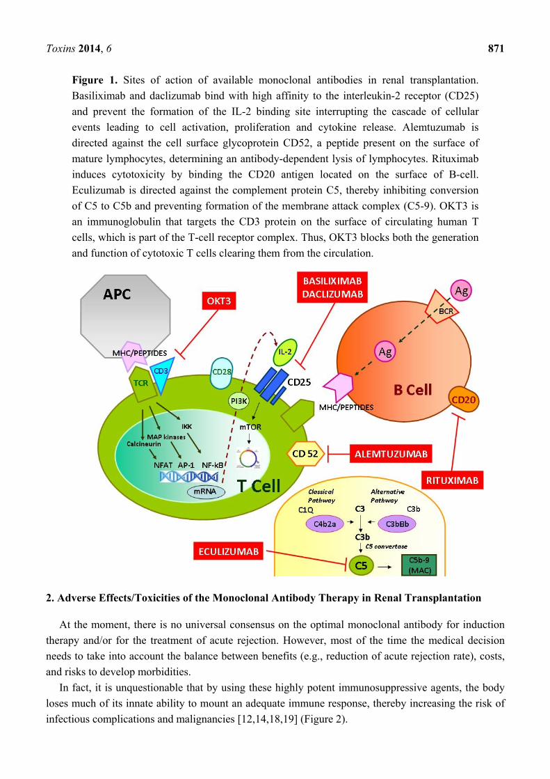
Figure 1. Sites of action of available monoclonal antibodies in renal transplantation.
Basiliximab and daclizumab bind with high affinity to the interleukin-2 receptor (CD25)
and prevent the formation of the IL-2 binding site interrupting the cascade of cellular
events leading to cell activation, proliferation and cytokine release. Alemtuzumab is
directed against the cell surface glycoprotein CD52, a peptide present on the surface of
mature lymphocytes, determining an antibody-dependent lysis of lymphocytes. Rituximab
induces cytotoxicity by binding the CD20 antigen located on the surface of B-cell.
Eculizumab is directed against the complement protein C5, thereby inhibiting conversion
of C5 to C5b and preventing formation of the membrane attack complex (C5-9). OKT3 is
an immunoglobulin that targets the CD3 protein on the surface of circulating human T
cells, which is part of the T-cell receptor complex. Thus, OKT3 blocks both the generation
and function of cytotoxic T cells clearing them from the circulation.
2. Adverse Effects/Toxicities of the Monoclonal Antibody Therapy in Renal Transplantation
At the moment, there is no universal consensus on the optimal monoclonal antibody for induction
therapy and/or for the treatment of acute rejection. However, most of the time the medical decision
needs to take into account the balance between benefits (e.g., reduction of acute rejection rate), costs,
and risks to develop morbidities.
In fact, it is unquestionable that by using these highly potent immunosuppressive agents, the body
loses much of its innate ability to mount an adequate immune response, thereby increasing the risk of
infectious complications and malignancies [12,14,18,19] (Figure 2).
Toxins 2014, 6 872
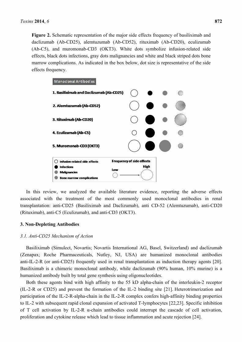
Figure 2. Schematic representation of the major side effects frequency of basiliximab and
daclizumab (Ab-CD25), alemtuzumab (Ab-CD52), rituximab (Ab-CD20), eculizumab
(Ab-C5), and muromonab-CD3 (OKT3). White dots symbolize infusion-related side
effects, black dots infections, gray dots malignancies and white and black striped dots bone
marrow complications. As indicated in the box below, dot size is representative of the side
effects frequency.
In this review, we analyzed the available literature evidence, reporting the adverse effects
associated with the treatment of the most commonly used monoclonal antibodies in renal
transplantation: anti-CD25 (Basiliximab and Daclizumab), anti CD-52 (Alemtuzumab), anti-CD20
(Rituximab), anti-C5 (Eculizumab), and anti-CD3 (OKT3).
3. Non-Depleting Antibodies
3.1. Anti-CD25 Mechanism of Action
Basiliximab (Simulect, Novartis; Novartis International AG, Basel, Switzerland) and daclizumab
(Zenapax; Roche Pharmaceuticals, Nutley, NJ, USA) are humanized monoclonal antibodies
anti-IL-2-R (or anti-CD25) frequently used in renal transplantation as induction therapy agents [20].
Basiliximab is a chimeric monoclonal antibody, while daclizumab (90% human, 10% murine) is a
humanized antibody built by total gene synthesis using oligonucleotides.
Both these agents bind with high affinity to the 55 kD alpha-chain of the interleukin-2 receptor
(IL-2-R or CD25) and prevent the formation of the IL-2 binding site [21]. Heterotrimerization and
participation of the IL-2-R-alpha-chain in the IL-2-R complex confers high-affinity binding properties
to IL-2 with subsequent rapid clonal expansion of activated T-lymphocytes [22,23]. Specific inhibition
of T cell activation by IL-2-R α-chain antibodies could interrupt the cascade of cell activation,
proliferation and cytokine release which lead to tissue inflammation and acute rejection [24].
Toxins 2014, 6 873
Based on their peculiar biological/pharmacological characteristics, they have been largely studied
and used in renal transplantation causing a significant reduction in the acute rejection rate and allowing
a CNI and steroids minimization in the early postoperative period after renal transplantation [25–27].
In particular, basiliximab has been approved by the FDA only for transplant induction therapy
in 1998 and is currently used in renal transplant patients with low immunological risk [28–30]. In fact,
as reported by KDIGO (Kidney Disease: Improving Global Outcomes), although rabbit anti-thymocyte
globulin (rATG) is superior to anti-CD25 in high-risk transplant recipients, the potential risks of
infection and malignancy outweigh these benefits in standard-risk recipients [30].
Additionally, as extensively demonstrated, these drugs have been successfully used in high risk
patients with other medical comorbidities that preclude usage of lymphocyte-depleting antibody safely.
In fact several clinical reports have showed that basiliximab induction is safe and adequate for renal
transplant, also including the high risk transplants, such as deceased donor kidney transplants in highly
sensitized African Americans [31], particularly in simultaneous kidney pancreas transplant [32], and
splitting single pediatric donor kidney transplant [33], as long as the conventional triple regimen
consisting of calcineurin inhibitors, mycophenolic acid, and steroids.
Although having similar efficacy, the dosing regimen for basiliximab consists of two 20-mg doses,
one administered two hours before transplantation and the second on day four posttransplant, whereas
daclizumab is usually administered as five doses over eight weeks [34]. This simple and convenient
regimen provides suppression of the IL-2 receptor for 30 to 45 days, when the risk of acute rejection is
the greatest [35].
During time, this difference in administration regimen has led to a more frequent use of basiliximab
than daclizumab. For this reason, daclizumab has been withdrawn from the market.
3.2. Anti-CD25 Side Effects
Although, anti-CD25 has the best safety profile compared to other available induction
antibodies [34,36,37], they can still enhance the risk of developing important adverse effects/toxicities.
Hypersensitivity reactions: These rare complications (occurring in less than 1% of treated patients),
may determine several clinical effects: hypotension, tachycardia, cardiac failure, bronchospasms,
pulmonary edema, and respiratory failure [34–36]. The exact biological mechanism involved is not
completely defined.
To avoid hypersensitivity reactions, sometimes life-threatening, extreme caution should be used in
all patients previously treated with these drugs.
Infectious complications: Perioperative induction therapy with anti-CD25 may induce higher risk of
bacterial, viral, and fungal infections as compared with placebo [38]. However, the cumulative
infection risk is significantly lower in anti-CD25-treated patients compared to those undergoing a more
aggressive anti-thymocyte globulin (ATG) induction therapy [38–41].
In particular, Brennan et al. have reported that the incidence of overall infection was higher in the
ATG group than in the basiliximab group (85.8% vs. 75.2%, p = 0.03). This difference appeared to be
attributable to a greater frequency of urinary tract infections (39.0%, vs. 27.0%; p = 0.04) and
non-cytomegalovirus (CMV) viral infections (21.3% vs. 11.7%, p = 0.04) in the ATG group. On the
Toxins 2014, 6 874
contrary, the incidence of CMV infection resulted lower in the ATG group as compared with
anti-CD25 (7.8% vs. 17.5%, p = 0.02) [38].
Malignancies: Malignancies or post-transplant lymphoproliferative disorders are rare in anti-CD25
treated renal transplant patients [42] and less frequent as compared with ATG-treated patients [38,43].
Myelosuppression: It has been reported that, basiliximab-treated renal transplant patients may
experience leukopenia (approximately 10%–15%) and thrombocytopenia (5%). Anyway, the incidence
of these adverse effects resulted most of the time lower as compared with that evaluated in
ATG-treated patients [38,44].
3.3. Rabbit Antithymocyte Globulin (rATG) versus BASILIXIMAB: Just a Few Words
It is largely reported that rabbit antithymocyte globulin (rATG, Thymoglobulin™, Genzyme
Canada Inc., Mississauga, ON, Canada), together with basiliximab, are the most widely used
antibodies in renal transplantation.
This agent is a polyclonal gamma immunoglobulin derived from the immunization of rabbits with
human thymocytes and indicated for the prevention and treatment of acute renal transplant rejection [45].
Induction with rATG, together with maintenance immunosuppression, has been shown to be more
effective than maintenance immunosuppression alone in preventing episodes of acute rejection in adult
renal transplant recipients [46,47].
Additionally, results of a large clinical trial, using moderate to high-risk deceased donor recipients,
demonstrated an improved composite endpoint of the incidence of rejection, graft loss, and patient
death that favored rATG versus basiliximab [38,48]. At a mean of 10 months of follow-up, the
estimate of combined endpoint was 19.1% in the rATG arm and 31.6% in the basiliximab arm
(p = 0.01), with acute rejection being the driving factor (14.2% rATG vs. 25% basiliximab, p = 0.013).
Five-year follow-up demonstrates that the incidence of acute rejection requiring antibody rescue
remained lower in the rATG group (3% vs. 12%, p = 0.05).
However, adverse events, such as fever, chills, and gastrointestinal distress, are more frequent with
rATG than with other induction agents [38,39]. Serious reactions, such as cytokine release syndrome
with hemodynamic instability, can also occur and are most commonly associated with the first dose
and rapid infusion rates [45]. Finally, rATG may induce severe CMV infections [39,46,49–52].
4. Depleting Antibodies
4.1. Mechanism of Action of Alemtuzumab (Anti-CD52 Antibody)
Alemtuzumab (MabCampath or Campath; Genzyme, Cambridge, MA, USA) is a novel recombinant
DNA-derived humanized rat monoclonal antibody directed against the 21–28 kDa cell surface
glycoprotein CD52 [53]. Alemtuzumab was never approved for use in transplantation and no Phase II/III
trials have been performed. Furthermore, this drug has been withdrawn from general marketing, not out
of safety or efficacy concerns, but rather to reposition the drug for use in multiple sclerosis.
Human CD52 is a peptide of 12 amino acids, anchored to glycosylphosphatidylinositol, present on the
surface of mature lymphocytes (including T and B lymphocytes, macrophages, monocytes, and natural
killer cells), but not on on lymphoid progenitors [54]. It is also found on monocytes and dendritic cells.
Toxins 2014, 6 875
Although the exact underlying mechanism remains unclear, alemtuzumab has been shown to
produce significant leukopenia by means of antibody-dependent lysis of lymphocytes, leading to
depletion of T and B cells in the peripheral circulation for several months after administration [54,55].
Alemtuzumab was initially used as an anti-cancer therapy for the treatment of B-cell chronic
lymphocytic leukaemia, but, in the last few years, its use as an induction therapy following solid organ
transplantation (off-label) is increasing world-wide [56].
Accordingly, a number of clinical trials have been undertaken to evaluate its efficacy and safety in
kidney transplantation with contrasting results that make difficult to reach a consensus on its
application [57–61].
4.2. Alemtuzumab Side Effects
Results from clinical reports and trials focusing on renal transplantation have demonstrated that,
although effective, alemtuzumab may induce severe adverse effects.
Infusion related side effects: Because of the xenogenic nature of the antibody, adverse reactions can
occur during or shortly after alemtuzumab infusion (generally first week of treatment). They include:
pyrexia, chills/rigors, nausea, hypotension, urticaria, dyspnea, rash, emesis, and bronchospasm [62].
In some cases, serious, including fatal, infusion reactions have also been identified in post-marketing
reports: syncope, pulmonary infiltrates, acute respiratory distress syndrome, respiratory arrest, cardiac
arrhythmias, myocardial infarction, acute cardiac insufficiency, cardiac arrest, angioedema, and
anaphylactic shock [63].
Most of the time, alemtuzumab-associated cytokine release syndrome could be prevented by
co-administration of steroids and/or antihistamines.
Malignancy: Although present, clinical studies do not report a higher incidence of neoplasia in
alemtuzumab-treated patients in particular when compared to other induction regimens.
As described by Puttarajappa C et al. the incidence of malignancies observed during a median follow
up of four years was 2.8%. However, alemtuzumab induction was not associated with a significant
increased post-renal transplant cancer risk when compared to no induction therapy and it was associated
with lower cancer incidence when compared to thymoglobulin. Authors of this paper underlined that the
relatively low observed predisposition to induce cancer could be mainly due to CNI minimization [64].
Additionally, the group of Hanaway described a very low incidence of B-cell lymphoma (<1%) and
skin cancer (4%) in patients treated with alemtuzumab during a follow-up period of three years [65].
Moreover an analysis of the data collected by the Organ Procurement and Transplantation
Network/United Network for Organ Sharing (OPTN/UNOS) demonstrated no association between
alemtuzumab treatment and risk for post-transplant lymphoproliferative disorder (PTLD) [66].
Infection: It is indisputable that almetuzumab, as other immunosuppressive induction agents, may
increase the incidence of infections in renal transplant recipients.
A recent study has reported a frequency of CMV infection of 13%, BK virus infection of 11%,
Herpes simplex of 3%, and Herpes Zoster of 5% in anti-CD52 treated patients [65].
However, unlike bone marrow and liver transplant recipients, and hemo-oncological patients [67],
no strong evidence supports the association between infection risk and almetuzumab treatment in renal
transplant recipients. In fact, only one randomized controlled trial clearly reported a significant
Toxins 2014, 6 876
difference in the risk of CMV infection in alemtuzumab treated patients as compared with controls no
receiving induction immunosuppression (28% vs. 12%, p = 0.03) [58].
The relatively low incidence of CMV infections in renal transplant patients treated with anti-CD52
could be mainly due to the use of a well standardized CMV viral prophylaxis (using ganciclovir or
valganciclovir) in high risk patients (recipients of CMV-positive donors) and in an effective
world-wide adopted pre-emptive treatment strategy.
Bone marrow disease: In a phase 2 clinical trial the treatment of relapsing-remitting multiple
sclerosis with alemetuzumab, six of 216 patients (2.8%) developed immune thrombocytopenia (ITP),
strikingly higher than the incidence rate of ITP reported for the general adult population. They describe
an unique form of ITP associated with alemtuzumab treatment and characterized by delayed
presentation after drug exposure, responsiveness to conventional ITP therapies, and prolonged
remission. The pathogenesis of alemtuzumab-associated ITP is incompletely understood. The authors
suggested that alemtuzumab-associated ITP may arise as a consequence of defects in central tolerance
checkpoints during lymphocyte reconstitution. With immune recovery, resolution of these defects may
contribute to the favorable natural history of the disorder [68].
In addition, Reda G et al. reported an increase incidence of ITP in patients affected by chronic
lymphocytic leukemia treated with low-dose alemtuzumab. ITP was diagnosed in 12 (18.7%) patients: nine
(14%) patients developed ITP during or after treatment with alemtuzumab (median observation time
30 months, range 9–23), whereas, in the remaining three patients, it preceded the treatment [69]. This data,
associated with the report of Cuker et al., suggest an important role of alemtuzumab in the pathogenesis of
ITP, which could be related to its ability to induce dysregulation of T-lymphocyte activity.
Hanaway et al. describe among low-risk patients a mean total lymphocyte count significantly lower
in patients treated with alemtuzumab than basiliximab. Among high-risk patients, the use of
lymphocyte-depleting antibodies (alemtuzumab and ATG) in both treatment groups resulted in a low
mean total lymphocyte count within the first week after transplantation to less than 10% of the
baseline value [65].
4.3. Rituximab (Anti-CD20/ Anti-B-Cell) Mechanism of Action
Rituximab is a chimeric anti-CD20 monoclonal antibody licensed for use in non-Hodgkin’s
lymphoma (NHL), chronic lymphocytic leukaemia, and rheumatoid arthritis [70]. At the moment,
rituximab is not approved for use in renal transplantation and it is used on an off label basis. No phase
II or phase III studies have been performed yet and the published data are still controversial. The CD20
antigen is a transmembrane nonglycosylated phosphoprotein, expressed on both immature and mature
B cells, involved in calcium conductance and regulation of cell proliferation/differentiation [71].
Once rituximab has bound to the CD20 antigen, it affects B cells by inducing [72]: (1) activation of
the complement cascade, leading to complement-mediated cytotoxicity; (2) macrophage recognition,
inducing phagocytosis and antibody-dependent cell-mediated cytotoxicity (ADCC); and (3) natural
killer cell interaction, also leading to ADCC. It does not have a direct effect on plasma cells (which do
not express the CD20 antigen).
Toxins 2014, 6 877
Additionally, rituximab causes a decrement of the number of B cells present in the peripheral blood
circulation within one to three days of administration, and complete B cell depletion in most of the
patients within one to six weeks [73].
Although, never licensed for use in renal transplantation, this agent has been frequently used in
several induction protocols for HLA antibody incompatible transplantation and for the treatment of
acute renal allograft rejections, CAMR and PTLD [74–78].
4.4. Rituximab Side Effects
Even if generally considered a safe agent, rituximab may induce several dose- and/or time-related
adverse effects. Early recognition of them could help clinicians to plan a prophylaxis strategy and/or
start a treatment in order to avoid the development of severe clinical complications.
The most frequent adverse effects associated with rituximab treatment are the following:
Infectious complications: Although debated, several studies have suggested that rituximab may
slightly increase the risk of infection (including Pneumocystis pneumonia, Hepatitis B, and CMV
disease and fungal infection) [79,80].
Additionally, during a three-year follow-up of ABO-incompatible renal transplant recipients treated
with antigen-specific immunoadsorption and rituximab reported an incidence rate of sepsis (6.7%),
urinary tract infection (13%), and surgical wound infection (13%) [81]. In the same population, the
incidence of CMV infection was 6.7% and none of the patients developed EBV, BKV associated
nephropathy, invasive fungal infections, and Clostridium difficile colitis.
Additionally, however, as with other immunomodulators, the current label for rituximab contains a
black box warning about the risk of re-activation of hepatitis B virus (HBV) infection, although the
exact impact of rituximab on the incidence of HBV re-activation in patients with current or past HBV
infection remains largely unknown [82].
At present, it is well known that the above mentioned risk of HBV re-activation is quite high among
HBV carriers, particularly if rituximab is given alone or in combination with steroids [83,84]. The
issue is much more complex in patients with serological markers of past HBV infection [HBsAg
negative with positive antibodies against the HBV core antigen (anti-HBc positive)] undergoing either
conventional chemotherapy or rituximab-based therapy [85].
For instance, the EASL clinical practice guidelines for the management of chronic HBV infection in
HBsAg-negative patients with positive anti-HBc antibodies who receive chemotherapy and/or
immunosuppression suggest HBV-DNA determination in the serum and if undetectable, strict
follow-up by means of alanine aminotransferase (ALT) and HBV-DNA testing [86].
Treatment with potent antivirals having a high barrier to resistance (e.g., entecavir or tenofovir) is
recommended upon confirmation of HBV re-activation before ALT elevation [86].
However, there is no clear evidence for when and how often this “strict” follow-up should be
performed as surrogate or prognostic markers related to the development of HBV re-activation in these
subjects are obscure and the cost effectiveness particularly of HBV-DNA serial testing is unknown.
However, further studies, designed explicitly to assess the impact of rituximab on infection rates,
are required.
Toxins 2014, 6 878
Non-infectious pulmonary toxicity: A systematic literature review reported several cases of
rituximab-associated interstitial lung disease (RTX-ILD) in non-renal transplant patients [87,88]. A
total of 121 cases of potential RTX-ILD were identified from 21 clinical studies/trials. In 30 (24.7%)
cases it was given as monotherapy. The mean time of onset, from the last infusion until symptoms
development or relevant abnormal radiological change was 30 days.
Radiological findings were characterized by diffuse bilateral lung infiltrates apparent on chest
radiographs and/or thoracic computed tomography (CT).
In all cases, rituximab was discontinued and corticosteroids were administered immediately. In
patients who improved in association with corticosteroids treatment, symptomatic recovery was
achieved within several days, with radiological resolution lagging behind (weeks to months). Lung
infiltrates on CT disappeared within a maximum of five months from initial onset. RTX-ILD was fatal
in more than 10% of cases.
Moreover, cases of bronchiolitis obliterans with organizing pneumonia (BOOP) have been reported
in patients who received rituximab-based therapy for NHL [89,90].
The mechanisms leading to RTX-induced interstitial lung disease are unknown.
Cytokine release syndrome: Similarly to OKT3, also rituximab may induce cytokine release
syndrome [91] particularly in patients with B cell malignancies where the number of CD20+ cells
susceptible to this therapy are much greater than in patients with renal failure, who tend to have lower
numbers of B cells [92]. The effects of a cytokine release syndrome can be preempted by prophylactic
administration of paracetamol, steroid, and antihistaminic drugs.
Haematological adverse effects: Neutropenia and thrombocytopenia are the main haematological
complications occurring in rituximab-treated patients [93,94].
The mechanisms underlying the development of neutropenia are not clear, and it has only rarely
been seen in solid organ transplantation.
Rituximab treatment depletes the normal B-lymphocyte population, which recovers within three
to nine months. During recovery, the acquisition of a new immune repertoire may determine the
transient production of autoantibodies, some of which may be directed against neutrophils or
hematopoietic precursors [95].
Another hypothesized mechanism includes perturbation of stromal-derived factor 1 levels at the time of
B-cell recovery that may interfere with neutrophil egress from the bone marrow and cause neutropenia [96].
Rituximab related late-onset neutropenia has been reported [95] in single patients or small case
series of patients receiving rituximab for acute antibody mediated rejection. Ishida et al. reported that a
significant number of ABO-incompatible and HLA-incompatible transplant patients (approximately
42%) developed late-onset neutropenia (grades III to IV) after the last administration of rituximab [97].
Acute thrombocytopenia is a rare, self-limiting complication following rituximab administration,
which is unlikely to lead to bleeding [98]. It may be due to the number of pre-treatment circulating B
cells and the onset of cytokine release syndrome [98–100].
Progressive multifocal leukoencephalopathy: Progressive multifocal leukoencephalopathy (PML) is
a rare, serious, and usually fatal demyelinating disease that occurs predominantly in severely
immunosuppressed patient populations [101]. Rituximab appears to be responsible of PML, but the
causal relationship is unclear.
Toxins 2014, 6 879
The etiologic agent is the JC virus (JCV), a polyomavirus that is widely distributed as a latent
infection in the general population but becomes activated, leading to destruction of myelin-producing
oligodendrocytes. Patients that develop this complication may present devastating neurologic sequelae.
However, to our knowledge, no cases of PML in renal transplant patients have been reported so far.
Rituximab-induced coagulopathy: Disseminated intravascular coagulation (DIC) appears to be an
extremely rare side effect of rituximab. It is a self-limiting event occurring early after the beginning of
rituximab treatment.
To date, this phenomenon has been observed only in patients with WM (Waldenström
macroglobulinemia) or HCL (hairy cell leukemia).
Coagulopathy occurred early after the administration of rituximab and was associated with
clinically significant bleeding in half of the cases. The platelet count may drop to approximately 20%
and D-dimer values could result frequently elevated. The treatment of this complication is the
substitution of either fresh frozen plasma or platelets, or both [102].
Rituximab and cardiovascular complications: An increased cardiovascular risk has been reported in
randomized controlled trials examining the use of rituximab as induction therapy in renal
transplantation. In particular, in a recent trial after a three-year follow-up, six of 44 patients died from
myocardial infarction. None of 47 patients in the placebo group died. When examined on an
intention-to-treat basis (i.e., using death rates from the original cohorts of 68) the difference in
mortality was statistically significant (p = 0.006) [103].
Therefore, even whether formal guidelines are missed, rituximab should probably be avoided in
patients with important cardiovascular comorbidities. Additionally, cardiac tests should be routinely
performed in all renal transplant patients prior to rituximab administration.
4.5. Muromonab-CD3 (OKT3) Mechanism of Action
Muromonab CD3 (Orthoclone OKT3, Janssen-Cilag, Beerse, Belgium) [104], has been the first
monoclonal antibody approved for the treatment of acute rejection and used in several induction
protocols for renal transplantation although it has never been formally approved for this indication.
However, this agent has largely been replaced in transplant medicine by modern induction protocols
using ATG characterized by a similar effectiveness and more favourable side-effect profile [105,106].
OKT3 is an Ig2a immunoglobulin produced by hybridoma technique in pathogen-free standard-bred
mice that targets the CD3 protein on the surface of circulating human T cells, which is part of the
T-cell receptor complex. Thus, OKT3 blocks both the generation and function of cytotoxic T cells
clearing them from the circulation. Within 48 hours after OKT3 discontinuation, the normal array of
surface molecules including CD3 is found again on all T cells [107].
4.6. OKT3 Side Effects
Although effective, this drug is characterized by several adverse effects [108,109]:
Cytokine release syndrome: It is an infusion reaction, typically occurring during the first infusion of
a new drug, leading to a large range of systemic effects such as flu-like symptoms and, rarely, severe
hypotension, bronchospasm, tachycardia, and even death [110].
Toxins 2014, 6 880
The main biological mechanism leading to this condition is an excessive systemic immune response
that involves the release of many different inflammatory mediators, predominantly cytokines.
Additionally, in most cases this syndrome may be, also, associated with: nephropathy as
consequence of the enhanced cytokines synthesis and a decrement in the intra-renal prostaglandin
synthesis [111]; pulmonary edema, due to the concomitant complement-related vascular endothelium
damage [112]; central nervous system (CNS) complications associated to an activation of T-cells that
directly attack specific CNS elements; activation of coagulation and fibrinolysis due to complement
activation and cytokine release itself leading to graft thrombosis [113–115].
Rare life-threatening complications have been also described such as: pulmonary edema, aseptic
meningitis, and encephalopathy, opportunistic infections, particularly with cytomegalovirus and
post-transplant lymphoproliferative disorders [114,116–119].
Cancer induction: OKT3 treatment implies an increased risk of post-transplant malignancies. In
particular, because of its specific action against T-cells, it can induce the development of severe
lymphoproliferative disorders [108,120,121].
Infectious complications: The occurrence of CMV and Herpes infections seem to be enhanced in
transplanted patients treated with OKT3, especially when this therapy is used in the early phase after
transplantation [122].
OKT3 immunization: A substantial number (3%–11%) [123] of OKT3-treated patients develop
antibodies against the xenogeneic epitope of the agent that are responsible for decreased
pharmacological/therapeutic efficacy and increased risk of AMR. In particular, these antibodies can
inhibit the binding of OKT3 on CD3 receptor [124].
5. Monoclonal Antibody Directed against Human Complement Protein
5.1. Mechanism of Action of Eculizumab (Anti-C5)
Eculizumab (Soliris®, Alexion Pharmaceuticals Inc., Cheshire, CT, USA) is a humanized
monoclonal antibody directed against complement protein C5. It binds to the C5 protein with high
affinity, thereby inhibiting conversion of C5 to C5b and preventing formation of the membrane attack
complex (C5-9) [125].
Initially approved for use in paroxysmal nocturnal hemoglobinuria [126,127] and atypical
hemolytic-uremic syndrome (aHUS) [128], this agent has been recently used in the treatment of renal
allografts recipients with recurrent dense deposit disease and C3 glomerulonephritis [129,130],
antibody mediate acute rejections [131–133] and prophylaxis of post-transplant recurrence of aHUS [134].
However, no clinical trials involving renal transplant recipients have been performed and the
existing data are mainly based on small series and off label use of this agent for the treatment of
humoral rejections.
Several authors have reported their experience in the use of eculizumab to prevent acute
antibody-mediated rejection after transplantation in a series of HLA-sensitized pre-transplant
positive-flow cytometric crossmatch patients [132]. Authors reported that the incidence of this
complication at 3 months was significantly lower compared to an historical control group (7.7% versus
41.2%), although the presence of C4d in patients with donor-specific antibodies did not differ between
Toxins 2014, 6 881
the study and control group, thus providing evidence for the downstream activity of eculizumab in
blocking the complement pathway.
Nevertheless, even if this drug is considered a great promising in renal transplantation, at the
present, it is rarely used because of the extremely high cost.
5.2. Eculizumab Side Effects
At present, there is still limited but convincing evidence that eculizumab is efficient and well tolerated,
but, unfortunately, because of the underutilization of this agent, data are incomplete and not exhaustive.
As reported in the FDA report on the use of eculizumab for the treatment of patients with
paroxysmal nocturnal hemoglobinuria [135,136], the more common adverse events associated with
eculizumab are: hypertension, diarrhea, headache, anemia, vomiting, nausea, leucopenia, upper
respiratory and urinary tract infections, and viral infections (e.g., Herpes simplex virus).
However, as well described, patients treated with eculizumab may develop severe and
life-threatening Neisseria meningitides infections [137,138]. For this reason is strongly recommended
a vaccine therapy against N. meningitides before eculizumab administration.
In this context, Struijk et al. have described that one transplant patient developed meningococcal
sepsis after eculizumab administration despite receiving a polysaccharide meningitis vaccine two weeks
beforehand [139]. The patient recovered and showed a humoral response only to N. meningitidis
serotype C, not to the other constituents of the vaccine (serotypes A, Y, and W135). Moreover, this
same group of clinicians has raised concern that the unconjugated polysaccharide vaccine is
insufficient as it does not provide protection against serogroup B [140].
Based on the above observations, it is currently recommended that all eculizumab-treated patients
should receive a tetravalent (preferably conjugated) vaccine against N. meningitidis serotypes A, C, Y,
and W135 [104]. Whether it will be sufficient remains to be seen.
6. Conclusions
The last decade have witnessed a large employment of monoclonal antibodies in renal
transplant clinical practice. They have been utilized as induction therapy, for treatment of
steroid-resistant/antibody-mediated acute rejections and chronic antibody mediated-rejections.
Among them basiliximab has demonstrated a great safety profile and high effectiveness in
preventing acute rejection in low risk patients, while the new depleting anti-lymphocyte antibodies
rituximab and eculizumab have been useful to prevent/treat transplant-related acute complications
particularly in the high risk renal transplant patients.
Additionally, rituximab has demonstrated promising tool to use in the preoperative regimen for the
blood group ABO-incompatible transplantation, in desensitization protocols, and for treatment of
antibody-mediated rejections.
Then, eculizumab, interrupting the complement pathway, seems to be highly effective in protecting
renal allografts when post-transplant aHUS or AMR occur. Nevertheless, it is unclear how long
treatment should continue (a particularly important issue given the high cost of the drug), or whether
eculizumab contributes to the development of accommodation in humans.
Toxins 2014, 6 882
However, despite the above described encouraging clinical reports, at the state of art, only few
research studies and clinical trials have been undertaken in renal transplantation to really assess the
effectiveness of monoclonal antibodies and to measure the clinical impact of these agents on long-term
graft and patient survival.
None of the immunosuppressive regimens using these agents is capable of inducing graft
acceptance as one would expect in case of donor-specific tolerance.
Furthermore, no universal consensus exists on the optimal monoclonal antibody dosage and timing.
Most of the time the medical decision need to take into account the balance between benefits, costs and
risks to develop morbidities.
Therefore, it’s unquestionable that phase II and phase III clinical trials and collaborative research
projects should be undertaken to better address the pharmacological potentialities and reduce risks of
these agents. In fact, while it is clear their utility, it is also unquestionable that by using these highly
potent immunosuppressive agents, the body loses much of its innate ability to mount an adequate
immune response, thereby increasing the risk of severe adverse effects (e.g., infections, malignancies,
haematological complications).
A step forward could be also to employ the off-label use of monoclonal antibodies that have
originally been developed for other indications.
Finally, we believe that innovative biomolecular or “omics” research strategies could be undertaken
to personalize monoclonal antibodies administration in renal transplant recipients [141].
Conflict of Interest
The authors have no conflicts of interest to disclosure.
References
1. Van Der Woude, F.J. Impact of renal cadaveric transplantation on survival in end-stage renal
failure: evidence for reduced mortality risk compared with hemodialysis during long-term
follow-up. J. Am. Soc. Nephrol. 1998, 9, 2135–2141.
2. Safinia, N.; Afzali, B.; Atalar, K.; Lombardi, G.; Lechler, R.I. T-cell alloimmunity and chronic
allograft dysfunction. Kidney Int. Suppl. 2010, 119, S2–S12.
3. Cecka, J.M. The UNOS renal transplant registry. Clin. Transpl. 2002, 16, 1–20.
4. Starzl, T.E.; Klintmalm, G.B.; Weil, R., 3rd; Porter, K.A.; Iwatsuki, S.; Schroter, G.P.;
Fernandez-Bueno, C.; MacHugh, N. Cyclosporin A and steroid therapy in sixty-six cadaver
kidney recipients. Surg. Gynecol. Obstet. 1981, 153, 486–494.
5. Wolfe, R.A.; Roys, E.C.; Merion, R.M. Trends in organ donation and transplantation in the
United States, 1999–2008. Am. J. Transplant. 2010, 10, 961–972.
6. Ciancio, G.; Burke, G.W.; Miller, J. Induction therapy in renal transplantation: An overview of
current developments. Drugs 2007, 67, 2667–2680.
7. Organ Procurement and Transplantation Network and the Scientific Registry of Transplant
Recipients: Transplant Data 1998–2010; Annual Report of the U.S.; Department of Health and
Human Services, Health Resources and Services Administration, Healthcare Systems Bureau,
Division of Transplantation: Rockville, MD, USA, 2011.
Toxins 2014, 6 883
8. Bunnapradist, S.; Takemoto, S.K. Multivariate analyses of antibody induction therapies. Clin.
Transpl. 2003, 17, 405–417.
9. Hariharan, S.; Johnson, C.P.; Bresnahan, B.A.; Taranto, S.E.; McIntosh, M.J.; Stablein, D.
Improved graft survival after renal transplantation in the United States, 1988 to 1996. N. Engl. J.
Med. 2000, 342, 605–612.
10. Gabardi, S.; Martin, S.T.; Roberts, K.L.; Grafals, M. Induction immunosuppressive therapies in
renal transplantation. Am. J. Health Syst. Pharm. 2011, 68, 211–218.
11. Goggins, W.C.; Pascual, M.A.; Powelson, J.A.; Magee, C.; Tolkoff-Rubin, N.; Farrell, M.L.;
Ko, D.S.; Williams, W.W.; Chandraker, A.; Delmonico, F.L.; et al. A prospective, randomized,
clinical trial of intraoperative versus postoperative Thymoglobulin in adult cadaveric renal
transplant recipients. Transplantation 2003, 76, 798– 802.
12. Nashan, B. Antibody induction therapy in renal transplant patients receiving calcineurin-inhibitor
immunosuppressive regimens: A comparative review. BioDrugs 2005, 19, 39–46.
13. Berard, J.L.; Velez, R.L.; Freeman, R.B.; Tsunoda, S.M. A review of interleukin-2 receptor
antagonists in solid organ transplantation. Pharmacotherapy 1999, 19, 1127–1137.
14. Hardinger, K.L.; Koch, M.J.; Brennan, D.C. Current and future immunosuppressive strategies in
renal transplantation. Pharmacotherapy 2004, 24, 1159–1176.
15. Kissmeyer-Nielsen, F.; Olsen, S.; Petersen, V.P.; Fjeldborg, O. Hyperacute rejection of kidney
allografts, associated with pre-existing humoral antibodies against donor cells. Lancet 1966, 288,
662–665.
16. Lefaucheur, C.; Loupy, A.; Hill, G.S.; Andrade, J.; Nochy, D.; Antoine, C.; Gautreau, C.;
Charron, D.; Glotz, D.; Suberbielle-Boissel, C. Preexisting donor-specific HLA antibodies
predict outcome in kidney transplantation. J. Am. Soc. Nephrol. 2010, 21, 1398–1406.
17. Fehr, T.; Rüsi, B.; Fischer, A.; Hopfer, H.; Wüthrich, R.P.; Gaspert, A. Rituximab and
intravenous immunoglobulin treatment of chronic antibody-mediated kidney allograft rejection.
Transplantation 2009, 87, 1837–1841.
18. Zaza, G.; Tomei, P.; Ria, P.; Granata, S.; Boschiero, L.; Lupo, A. Systemic and nonrenal adverse
effects occurring in renal transplant patients treated with mTOR inhibitors. Clin. Dev. Immunol.
2013, 2013, doi: 10.1155/2013/403280.
19. Casey, M.J.; Meier-Kriesche, H.U. Calcineurin inhibitors in kidney transplantation: Friend or foe?
Curr. Opin. Nephrol. Hypertens. 2011, 20, 610–615
20. Pascual, J.; Marcén, R.; Ortuño, J. Anti-interleukin-2 receptor antibodies: Basiliximab and
daclizumab. Nephrol. Dial. Transplant. 2001, 16, 1756–1760.
21. Leonard, W.J.; Depper, J.M.; Uchiyama, T.; Smith, K.A.; Waldmann, T.A.; Greene, W.C. A
monoclonal antibody that appears to recognize the receptor for human T-cell growth factor:
Partial characterization of the receptor. Nature 1982, 300, 267–269.
22. Ringheim, G.E.; Freimark, B.D.; Robb, R.J. Quantitative characterization of the intrinsic
ligand-binding affinity of the interleukin 2 receptor beta chain and its modulation by the alpha
chain and a second affinity-modulating element. Lymphokine. Cytokine Res. 1991, 10, 219–224.
23. Robb, R.J.; Greene, W.C.; Rusk, C.M. Low and high affinity cellular receptors for interleukin 2.
Implications for the level of Tac antigen. J. Exp. Med. 1984, 160, 1126–1146.
Toxins 2014, 6 884
24. Ferrara, J.L. Pathogenesis of acute graft-versus-host disease: Cytokines and cellular effectors.
J. Hematother. Stem Cell. Res. 2000, 9, 299–306.
25. Kahan, B.D.; Rajagopalan, P.R.; Hall, M. Reduction of the occurrence of acute cellular rejection
among renal allograft recipients treated with basiliximab, a chimeric anti-interleukin-2-receptor
monoclonal antibody. United States Simulect Renal Study Group. Transplantation 1999, 67,
276–284.
26. Nashan, B.; Moore, R.; Amlot, P.; Schmidt, A.G.; Abeywickrama, K.; Soulillou, J.P.
Randomised trial of basiliximab versus placebo for control of acute cellular rejection in renal
allograft recipients. CHIB 201 International Study Group. Lancet 1997, 350, 1193–1198.
27. Vincenti, F.; de Andrés, A.; Becker, T.; Choukroun, G.; Cole, E.; González-Posada, J.M.;
Kumar, M.A.; Moore, R.; Nadalin, S.; Nashan, B.; et al. Interleukin-2 receptor antagonist
induction in modern immunosuppression regimens for renal transplant recipients. Transpl. Int.
2006, 19, 446–457.
28. Cai, J.; Terasaki, P.I. Induction immunosuppression improves long-term graft and patient
outcome in organ transplantation: An analysis of United Network for Organ Sharing registry data.
Transplantation 2010, 90, 1511–1515.
29. Waldmann, T.A. Immunotherapy: Past, present and future. Nat. Med. 2003, 9, 269–277
30. Kidney Disease Improving Global Outcomes (KDIGO). KDIGO clinical practice guideline for
the care of kidney transplant recipients. Am. J. Transplant. 2009, 9, S1–S155
31. Ren, Q.; Paramesh, A.; Yau, C.L.; Killackey, M.; Slakey, D.; Florman, S.; Buell, J.; Alper, B.;
Simon, E.; Hamm, L.L. Long-term outcome of highly sensitized African American patients
transplanted with deceased donor kidneys. Transpl. Int. 2011, 24, 259–265.
32. Zhang, R.; Florman, S.; Devidoss, S.; Zarifian, A.; Yau, C.L.; Paramesh, A.; Killackey, M.;
Alper, B.; Fonseca, V.; Slakey, D. A comparison of long-term survivals of simultaneous
pancreas-kidney transplant between African American and Caucasian recipients with basiliximab
induction therapy. Am. J. Transpl. 2007, 7, 1815–1821.
33. Zhang, R.; Paramesh, A.; Florman, S.; Yau, C.L.; Balamuthusamy, S.; Krane, N.K.; Slakey, D.
Long-term outcome of adults who undergo transplantation with single pediatric kidneys: How
young is too young. Clin. J. Am. Soc. Nephrol. 2009, 4, 1500–1506.
34. Ramirez, C.B.; Marino, I.R. The role of basiliximab induction therapy in organ transplantation.
Expert Opin. Biol. Ther. 2007, 7, 137–148.
35. Novartis Pharmaceutical Corporation. Simulect® (basiliximab): Summary of product
characteristics. Available online: www.pharma.us.novartis.com (accessed on 11 November 2013).
36. Mottershead, M.; Neuberger, J. Daclizumab. Expert Opin. Biol. Ther. 2007, 7, 1583–1596.
37. Penfornis, A.; Kury-Paulin, S. Immunosuppressive drug-induced diabetes. Diabetes MeTable
2006, 32, 539–546.
38. Brennan, D.C.; Daller, J.A.; Lake, K.D.; Cibrik, D.; Del Castillo, D.; Thymoglobulin Induction
Study Group. Rabbit antithymocyte globulin versus basiliximab in renal transplantation. N. Engl.
J. Med. 2006, 355, 1967–1977.
39. Lebranchu, Y.; Bridoux, F.; Büchler, M.; Le Meur, Y.; Etienne, I.; Toupance, O.;
Hurault de Ligny, B.; Touchard, G.; Moulin, B.; Le Pogamp, P.; et al. Immunoprophylaxis with
Toxins 2014, 6 885
basiliximab compared with antithymocyte globulin in renal transplant patients receiving
MMF-containing triple therapy. Am. J. Transpl. 2002, 2, 48–56.
40. Mourad, G.; Rostaing, L.; Legendre, C.; Garrigue, V.; Thervet, E.; Durand, D. Sequential
protocols using basiliximab versus antithymocyte globulins in renal-transplant patients receiving
mycophenolate mofetil and steroids. Transplantation 2004, 78, 584–590.
41. Kim, J.M.; Jang, H.R.; Kwon, C.H.; Huh, W.S.; Kim, G.S.; Kim, S.J.; Joh, J.W.; Oh, H.Y. Rabbit
antithymocyte globulin compared with basiliximab in kidney transplantation: A single-center study.
Transpl. Proc. 2012, 44, 167–170.
42. Chapman, T.M.; Keating, G.M. Basiliximab: A review of its use as induction therapy in renal
transplantation. Drugs 2003, 63, 2803–2835.
43. Sollinger, H.; Kaplan, B.; Pescovitz, M.D.; Philosophe, B.; Roza, A.; Brayman, K.; Somberg, K.
Basiliximab versus antithymocyte globulin for prevention of acute renal allograft rejection.
Transplantation 2001, 72, 1915–1919.
44. Wang, W.; Yin, H.; Li, X.B.; Hu, X.P.; Yang, X.Y.; Liu, H.; Ren, L.; Wang, Y.; Zhang, X.D. A
retrospective comparison of the efficacy and safety in kidney transplant recipients with
basiliximab and anti-thymocyte globulin. Chin. Med. J. 2012, 125, 1135–1140.
45. Deeks, E.D.; Keating, G.M. Rabbit antithymocyte globulin (thymoglobulin): A review of its use
in the prevention and treatment of acute renal allograft rejection. Drugs 2009, 69, 1483–1512.
46. Mourad, G.; Garrigue, V.; Squifflet, J.P.; Besse, T.; Berthoux, F.; Alamartine, E.; Durand, D.;
Rostaing, L.; Lang, P.; Baron, C.; et al. Induction versus noninduction in renal transplant
recipients with tacrolimus based immunosuppression. Transplantation 2001, 72, 1050–1055.
47. Charpentier, B.; Rostaing, L.; Berthoux, F.; Lang, P.; Civati, G.; Touraine, J.L.; Squifflet, J.P.;
Vialtel, P.; Abramowicz, D.; Mourad, G.; et al. A three-arm study comparing immediate
tacrolimus therapy with antithymocyte globulin induction therapy followed by tacrolimus or
cyclosporine A in adult renal transplant recipients. Transplantation 2003, 75, 844–851.
48. Brennan, D.C.; Schnitzler, M.A. Long-term results of rabbit antithymocyte globulin and
basiliximab induction. N. Engl. J. Med. 2008, 359, 1736–1738.
49. Jamil, B.; Nicholls, K.M.; Becker, G.J.; Walker, R.G. Influence of anti-rejection therapy on the
timing of cytomegalovirus disease and other infections in renal transplant recipients. Clin.
Transpl. 2000, 14, 14–18.
50. Zamora, M.R. Controversies in lung transplantation: Management of cytomegalovirus infections.
J. Heart Lung Transpl. 2002, 21, 841–849.
51. Huurman, V.A.; Kalpoe, J.S.; van de Linde, P.; Vaessen, N.; Ringers, J.; Kroes, A.C.;
Roep, B.O.; De Fijter, J.W. Choice of antibody immunotherapy influences cytomegalovirus
viremia in simultaneous pancreas-kidney transplant recipients. Diabetes Care 2006, 29, 842–847.
52. Ozaki, K.S.; Pestana, J.O.; Granato, C.F.; Pacheco-Silva, A.; Camargo, L.F. Sequential
cytomegalovirus antigenemia monitoring in kidney transplant patients treated with
antilymphocyte antibodies. Transpl. Infect. Dis. 2004, 6, 63–68.
53. Hale, G.; Xia, M.Q.; Tighe, H.P.; Dyer, M.J.; Waldmann, H. The CAMPATH-1 antigen
(CDw52). Tissue Antigens. 1990, 35, 118–127.
54. Weaver, T.A.; Kirk, A.D. Alemtuzumab. Transplantation 2007, 84, 1545–1547.
Toxins 2014, 6 886
55. Xia, M.Q.; Hale, G.; Lifely, M.R.; Ferguson, M.A.; Campbell, D.; Packman, L.; Waldmann, H.
Structure of the CAMPATH-1 antigen, a GPI-anchored glycoprotein which is an exceptionally
good target for complement lysis. Biochem. J. 1993, 29, 633–640.
56. Magliocca, J.F.; Knechtle, S.J. The evolving role of alemtuzumab (Campath-1H) for
immunosuppressive therapy in organ transplantation. Transpl. Int. 2006, 19, 705–714.
57. Huang, E.; Cho, Y.W.; Hayashi, R.; Bunnapradist, S. Alemtuzumab induction in deceased donor
kidney transplantation. Transplantation 2007, 84, 821–828.
58. Margreiter, R.; Klempnauer, J.; Neuhaus, P.; Muehlbacher, F.; Boesmueller, C.; Calne, R.Y.
Alemtuzumab (Campath-1H) and tacrolimus monotherapy after renal transplantation: Results of
a prospective randomized trial. Am. J. Transplant. 2008, 8, 1480–1485.
59. Zhang, X.; Huang, H.; Han, S.; Fu, S.; Wang, L. Alemtuzumab induction in renal transplantation:
A meta-analysis and systemic review. Transpl. Immunol. 2012, 27, 63–68.
60. Ortiz, J.; Palma-Vargas, J.; Wright, F.; Bingaman, A.; Agha, I.; Rosenblatt, S.; Foster, P.
Campath induction for kidney transplantation: Report of 297 cases. Transplantation 2008, 85,
1550–1556.
61. Tan, H.P.; Donaldson, J.; Basu, A.; Unruh, M.; Randhawa, P.; Sharma, V.; Morgan, C.;
McCauley, J.; Wu, C.; Shah, N.; et al. Two hundred living donor kidney transplantations under
alemtuzumab induction and tacrolimus monotherapy: 3-year follow-up. Am. J. Transpl. 2009, 9,
355–366.
62. Hillmen, P.; Skotnicki, A.B.; Robak, T.; Jaksic, B.; Dmoszynska, A.; Wu, J.; Sirard, C.; Mayer, J.
Alemtuzumab compared with chlorambucil as first-line therapy for chronic lymphocytic
leukemia. J. Clin. Oncol. 2007, 25, 5616–5623.
63. Ferrajoli, A.; O’Brien, S.; Keating, M.J. Alemtuzumab: A novel monoclonal antibody. Expert
Opin. Biol. Ther. 2001, 1, 1059–1065.
64. Puttarajappa, C.; Yabes, J.; Bei, L.; Shah, N.; Bernardo, J.; McCauley, J.; Basu, A.; Tan, H.;
Shapiro, R.; Unruh, M.; et al. Cancer risk with alemtuzumab following kidney transplantation.
Clin. Transpl. 2013, 27, E264–E271.
65. Hanaway, M.J.; Woodle, E.S.; Mulgaonkar, S.; Peddi, V.R.; Kaufman, D.B.; First, M.R.;
Croy, R.; Holman, J. Alemtuzumab induction in renal transplantation. N. Engl. J. Med. 2011, 364,
1909–1919.
66. Kirk, A.D.; Cherikh, W.S.; Ring, M.; Burke, G.; Kaufman, D.; Knechtle, S.J.; Potdar, S.; Shapiro,
R.; Dharnidharka, V.R.; Kauffman, H.M. Dissociation of depletional induction and
posttransplant lymphoproliferative disease in kidney recipients treated with alemtuzumab. Am. J.
Transpl. 2007, 7, 2619–2625.
67. Peleg, A.Y.; Husain, S.; Kwak, E.J.; Silveira, F.P.; Tran, M.N.J.; Shutt, K.A.; Shapiro, R.;
Thai, N.; Abu-Elmagd, K.; McCurry, K.R.; et al. Opportunistic infections in 547 organ transplant
recipients receiving Alemtuzumab, a humanized monoclonal CD-52 antibody. Clin. Infect. Dis.
2007, 44, 204–212
68. Cuker, A.; Coles, A.J.; Sullivan, H.; Fox, E.; Goldberg, M.; Oyuela, P.; Purvis, A.;
Beardsley, D.S.; Margolin, D.H. A distinctive form of immune thrombocytopenia in a phase 2
study of alemtuzumab for the treatment of relapsing-remitting multiple sclerosis. Blood 2011,
118, 6299–6305.
Toxins 2014, 6 887
69. Reda, G.; Maura, F.; Gritti, G.; Gregorini, A.; Binda, F.; Guidotti, F.; Piciocchi, A.; Visco, C.;
Rodeghiero, F.; Cortelezzi, A. Low-dose alemtuzumab-associated immune thrombocytopenia in
chronic lymphocytic leukemia. Am. J. Hematol. 2012, 87, 936–937.
70. Barnett, A.N.; Hadjianastassiou, V.G.; Mamode, N. Rituximab in renal transplantation. Transpl.
Int. 2013, 26, 563–575.
71. Nadler, L.M.; Ritz, J.; Hardy, R.; Pesando, J.M.; Schlossman, S.F.; Stashenko, P. A unique cell
surface antigen identifying lymphoid malignancies of B cell origin. J. Clin Invest. 1981, 67, 134–140.
72. Markasz, L.; Vanherberghen, B.; Flaberg, E.; Otvös, R.; Stuber, G.; Gustafsson, A.J.; Olah, E.;
Skribek, H.; Szekely, L. NK cell-mediated lysis is essential to kill Epstein-Barr virus transformed
lymphoblastoid B cells when using rituximab. Biomed. Pharmacother. 2009, 63, 413–420.
73. Di Gaetano, N.; Cittera, E.; Nota, R.; Vecchi, A.; Grieco, V.; Scanziani, E.; Botto, M.; Introna, M.;
Golay, J. Complement activation determines the therapeutic activity of rituximab in vivo. J.
Immunol. 2003, 171, 1581–1587.
74. Vo, A.A.; Lukovsky, M.; Toyoda, M.; Wang, J.; Reinsmoen, N.L.; Lai, C.H.; Peng, A.; Villicana, R.;
Jordan, S.C. Rituximab and intravenous immune globulin for desensitization during renal
transplantation. N. Engl. J. Med. 2008, 359, 242–251.
75. Takagi, T.; Ishida, H.; Shirakawa, H.; Shimizu, T.; Tanabe, K. Evaluation of low-dose rituximab
induction therapy in living related kidney transplantation. Transplantation 2010, 89, 1466–1470.
76. Kaposztas, Z.; Podder, H.; Mauiyyedi, S.; Illoh, O.; Kerman, R.; Reyes, M.; Pollard, V.;
Kahan, B.D. Impact of rituximab therapy for treatment of acute humoral rejection. Clin. Transpl.
2009, 23, 63–73.
77. Hong, Y.A.; Kim, H.G.; Choi, S.R.; Sun, I.O.; Park, H.S.; Chung, B.H.; Choi, B.S.; Park, C.W.;
Kim, Y.S.; Yang, C.W. Effectiveness of rituximab and intravenous immunoglobulin therapy in
renal transplant recipients with chronic active antibody-mediated rejection. Transpl. Proc. 2012,
44, 182–184.
78. Elstrom, R.L.; Andreadis, C.; Aqui, N.A.; Ahya, V.N.; Bloom, R.D.; Brozena, S.C.;
Olthoff, K.M.; Schuster, S.J.; Nasta, S.D.; Stadtmauer, E.A.; et al. Treatment of PTLD with
rituximab or chemotherapy. Am. J. Transpl. 2006, 6, 569–576.
79. Gea-Banacloche, J.C. Rituximab-associated infections. Semin. Hematol. 2010, 47, 187–198.
80. Kamar, N.; Milioto, O.; Puissant-Lubrano, B.; Esposito, L.; Pierre, M.C.; Mohamed, A.O.;
Lavayssière, L.; Cointault, O.; Ribes, D.; Cardeau, I.; et al. Incidence and predictive factors for
infectious disease after rituximab therapy in kidney-transplant patients. Am. J. Transpl. 2010, 10,
89–98.
81. Genberg, H.; Kumlien, G.; Wennberg, L.; Berg, U.; Tydén, G. ABO-incompatible kidney
transplantation using antigen-specific immunoadsorption and rituximab: A 3-year follow-up.
Transplantation 2008, 85, 1745–1754.
82. Mitka, M. FDA: Increased HBV reactivation risk with ofatumumab or rituximab. JAMA 2013,
310, 1664.
83. Dong, H.J.; Ni, L.N.; Sheng, G.F.; Song, H.L.; Xu, J.Z.; Ling, Y. Risk of hepatitis B virus
(HBV) reactivation in non-Hodgkin lymphoma patients receiving rituximab-chemotherapy: A
meta-analysis. J. Clin. Virol. 2013, 57, 209–214.
Toxins 2014, 6 888
84. Kusumoto, S.; Tanaka, Y.; Ueda, R.; Mizokami, M. Reactivation of hepatitis B virus following
rituximab-plus-steroid combination chemotherapy. J. Gastroenterol. 2011, 46, 9–16.
85. Hui, C.K.; Cheung, W.W.; Zhang, H.; Au, W.Y.; Yueng, Y.H.; Leung, A.Y.H.; Leung, N.;
Luk, J.M.C.; Lie, A.K.W.; Kwong, Y.L.; et al. Rituximab increases the risk of de novo hepatitis
B infection in hepatitis B surface antigen negative patients undergoing cytotoxic chemotherapy. J.
Gastroenterol. Hepatol. 2006, 21, A73–A74.
86. European Association for the Study of the Liver. EASL clinical practice guidelines: Management
of chronic hepatitis B. J. Hepatol. 2012, 57, 167–185.
87. Hadjinicolaou, A.V.; Nisar, M.K.; Parfrey, H.; Chilvers, E.R.; Ostor, A.J.K. Non-infectious
pulmonary toxicity of rituximab: A systematic review. Rheumatology 2012, 51, 653–662.
88. Wu, Y.; Jia, Y.; Xu, J.; Shuai, X.; Wu, Y. Fatal interstitial lung disease induced by
rituximab-containing chemotherapy, treatment with TNF-a antagonist and cytokine profiling: A
case-report and review of the literature. J. Clin. Pharm. Ther. 2013, 38, 249–253.
89. Biehn, S.E.; Kirk, D.; Rivera, M.P.; Martinez, A.E.; Khandani, A.H.; Orlowski, R.Z.
Bronchiolitis obliterans with organizing pneumonia after rituximab therapy for non-Hodgkin’s
lymphoma. Hematol. Oncol. 2006, 24, 234–237.
90. Macartney, C.; Burke, E.; Elborn, S.; Magee, N.; Noone, P.; Gleadhill, I.; Allen, D.; Kettle, P.;
Drake, M. Bronchiolitis obliterans organizing pneumonia in a patient with non-Hodgkin’s
lymphoma following R-CHOP and pegylated filgrastim. Leuk. Lymphoma 2005, 46, 1523–1526.
91. Agarwal, A.; Vieira, C.A.; Book, B.K.; Sidner, R.A.; Fineberg, N.S.; Pescovitz, M.D. Rituximab,
anti-CD20, induces in vivo cytokine release but does not impair ex vivo T-cell responses. Am. J.
Transpl. 2004, 4, 1357–1360.
92. Pahl, M.V.; Gollapudi, S.; Sepassi, L.; Gollapudi, P.; Elahimehr, R.; Vaziri, N.D. Effect of
end-stage renal disease on B-lymphocyte subpopulations, IL-7, BAFF and BAFF receptor
expression. Nephrol. Dial. Transpl. 2010, 25, 205–212.
93. Wolach, O.; Shpilberg, O.; Lahav, M. Neutropenia after rituximab treatment: New insights on a
late complication. Curr. Opin. Hematol. 2012, 19, 32–38.
94. Giezen, T.J.; Mantel-Teeuwisse, A.K.; ten Berg, M.J.; Straus, S.M.; Leufkens, H.G.;
van Solinge, W.W.; Egberts, T.C. Rituximab-induced thrombocytopenia: A cohort study. Eur. J.
Haematol. 2012, 89, 256–266.
95. Mitsuhata, N.; Fujita, R.; Ito, S.; Mannami, M.; Keimei, K. Delayed onset neutropenia in a
patient receiving rituximab as treatment for refractory kidney transplantation. Transplantation
2005, 80, 1355.
96. Grant, C.; Wilson, W.H.; Dunleavy, K. Neutropenia associated with rituximab therapy. Curr.
Opin. Hematol. 2011, 18, 49–54.
97. Ishida, H.; Inui, M.; Furusawa, M.; Tanabe, K. Late-onset neutropenia (LON) after low-dose
rituximab treatment in living related kidney transplantation--single-center study. Transpl.
Immunol. 2013, 28, 93–99.
98. Ram, R.; Bonstein, L.; Gafter-Gvili, A.; Ben-Bassat, I.; Shpilberg, O.; Raanani, P.
Rituximab-associated acute thrombocytopenia: An under-diagnosed phenomenon. Am. J.
Hematol. 2009, 84, 247–250.
Toxins 2014, 6 889
99. Winkler, U.; Jensen, M.; Manzke, O.; Schulz, H.; Diehl, V.; Engert, A. Cytokine-release
syndrome in patients with B-cell chronic lymphocytic leukemia and high lymphocyte counts
after treatment with an Anti-CD20 monoclonal antibody (Rituximab, IDEC-C2B8). Blood 1999,
94, 2217–2224.
100. Yi, J.H.; Kim, S.J.; Ahn, H.K.; Lee, S.J.; Chang, M.H.; Kim, W.S. Rituximab-induced acute
thrombocytopenia: A case report and review of the literature. Med. Oncol. 2009, 26, 45–48.
101. Carson, K.R.; Evens, A.M.; Richey, E.A.; Habermann, T.M.; Focosi, D.; Seymour, J.F.;
Laubach, J.; Bawn, S.D.; Gordon, L.I.; Winter, J.N.; et al. Progressive multifocal
leukoencephalopathy after rituximab therapy in HIV-negative patients: A report of 57 cases from
the research on adverse drug events and reports project. Blood 2009, 113, 4834–4840.
102. Novak, J.; Mocikova, H.; Pavlicek, P.; Gaherova, L.; Kozak, T. Rituximab-induced coagulopathy.
Leuk. Lymphoma. 2012, 53, 2299–2301.
103. Tydén, G.; Ekberg, H.; Tufveson, G.; Mjörnstedt, L. A randomized, double-blind, placebo-controlled
study of single dose rituximab as induction in renal transplantation: A 3-year follow-up.
Transplantation 2012, 94, e21–e22.
104. Parlevliet, K.J.; Schellekens, P.T. Monoclonal antibodies in renal transplantation: A review.
Transpl. Int. 1992, 5, 234–246.
105. Jaffers, G.J.; Fuller, T.C.; Cosimi, A.B.; Russell, P.S.; Winn, H.J.; Colvin, R.B. Monoclonal
antibody therapy. Anti-idiotypic and non-anti-idiotypic antibodies to OKT3 arising despite
intense immunosuppression. Transplantation 1986, 41, 572–578.
106. Chatenoud, L.; Baudrihaye, M.F.; Chkoff, N.; Kreis, H.; Goldstein, G.; Bach, J.F. Restriction of
the human in vivo immune response against the mouse monoclonal antibody OKT3. J. Immunol.
1986, 137, 830–838.
107. Norman, D.J. Mechanisms of action and overview of OKT3. Ther. Drug Monit. 1995, 17, 615–620.
108. Sgro, C. Side-effects of a monoclonal antibody, muromonab CD3/orthoclone OKT3:
Bibliographic review. Toxicology 1995, 105, 23–29.
109. Kuypers, D.R.; Vanrenterghem, Y.F. Monoclonal antibodies in renal transplantation: Old and
new. Nephrol. Dial. Transpl. 2004, 19, 297–300.
110. Breslin, S. Cytokine-release syndrome: Overview and nursing implications. Clin. J. Oncol. Nurs.
2007, 11, 37–42.
111. First, M.R.; Schroeder, T.J.; Hariharan, S. OKT3-induced cytokine-release syndrome: Renal
effects (cytokine nephropathy). Transpl. Proc. 1993, 25, 25–26.
112. Raasveld, M.H.; Bemelman, F.J.; Schellekens, P.T.; van Diepen, F.N.; van Dongen, A.;
van Royen, E.A.; Hack, C.E.; ten Berge, I.J. Complement activation during OKT3 treatment: A
possible explanation for respiratory side effects. Kidney Int. 1993, 43, 1140–1149.
113. Ten Berge, R.J.; Raasveld, M.H.; van Diepen, F.N.; Hack, C.E. Activation of coagulation and
fibrinolysis during treatment with OKT3. Transpl. Proc. 1993, 25, 566–567.
114. Abramowicz, D.; Pradier, O.; Marchant, A.; Florquin, S.; De Pauw, L.; Vereerstraeten, P.;
Kinnaert, P.; Vanherweghem, J.L.; Goldman, M. Induction of thromboses within renal grafts by
high-dose prophylactic OKT3. Lancet 1992, 339, 777–778.
Toxins 2014, 6 890
115. Raasveld, M.H.; Hack, C.E.; ten Berge, I.J. Activation of coagulation and fibrinolysis following
OKT3 administration to renal transplant recipients: Association with distinct mediators. Thromb.
Haemost. 1992, 68, 264–267.
116. Martin, M.A.; Massanari, R.M.; Nghiem, D.D.; Smith, J.L.; Corry, R.J. Nosocomial aseptic
meningitis associated with administration of OKT3. JAMA 1988, 259, 2002–2005.
117. Shihab, F.S.; Barry, J.M.; Norman, D.J. Encephalopathy following the use of OKT3 in renal
allograft transplantation. Transpl. Proc. 1993, 25, 31–34.
118. Morgan, J.D.; Horsburgh, T.; Simpson, A.; Donnelly, P.K.; Veitch, P.S.; Bell, P.R.
Cytomegalovirus infection during OKT3 treatment for renal allograft rejection. Transpl. Proc.
1992, 24, 2634–2635.
119. Swinnen, L.J.; Costanzo-Nordin, M.R.; Fisher, S.G.; O’Sullivan, E.J.; Johnson, M.R.;
Heroux, A.L.; Dizikes, G.J.; Pifarre, R.; Fisher, R.I. Increased incidence of lymphoproliferative
disorder after immunosuppression with the monoclonal antibody OKT3 in cardiac-transplant
recipients. N. Engl. J. Med. 1990, 323, 1723–1728.
120. Melosky, B.; Karim, M.; Chui, A.; McBride, M.; Cameron, E.C.; Yeung, C.K.; Landsberg, D.;
Shackleton, C.; Keown, P.A. Lymphoproliferative disorders after renal transplantation in patients
receiving triple or quadruple immunosuppression. J. Am. Soc. Nephrol. 1992, 2, S290–S294.
121. Opelz, G.; Henderson, R. Incidence of non-Hodgkin’s lymphoma in kidney and heart transplant
recipients. Lancet 1993, 342, 1514–1516.
122. Stratta, R.J.; Shaefer, M.S.; Cushing, K.A.; Markin, R.S.; Reed, E.C.; Langnas, A.N.;
Pillen, T.J.; Shaw, B.W., Jr. A randomized prospective trial of acyclovir and immune globulin
prophylaxis in liver transplant recipients receiving OKT3 therapy. Arch. Surg. 1992, 127, 55–63.
123. Abramowicz, D.; Crusiaux, A.; Goldman, M. Anaphylactic shock after retreatment with OKT3
monoclonal antibody. N. Engl. J. Med. 1992, 327, 736.
124. Legendre, C.; Kreis, H.; Bach, J.F.; Chatenoud, L. Prediction of successful allograft rejection
retreatment with OKT3. Transplantation 1992, 53, 87–90.
125. Rother, R.P.; Rollins, S.A.; Mojcik, C.F.; Brodsky, R.A.; Bell, L. Discovery and development of
the complement inhibitor eculizumab for the treatment of paroxysmal nocturnal hemoglobinuria.
Nat. Biotechnol. 2007, 25, 1256–1264.
126. Hillmen, P.; Young, N.S.; Schubert, J.; Brodsky, R.A.; Socié, G.; Muus, P.; Röth, A.; Szer, J.;
Elebute, M.O.; Nakamura, R.; et al. The complement inhibitor eculizumab in paroxysmal
nocturnal hemoglobinuria. N. Engl. J. Med. 2006, 355, 1233–1243.
127. McKeage, K. Eculizumab: A review of its use in paroxysmal nocturnal haemoglobinuria. Drugs
2011, 71, 2327–2345.
128. Mache, C.J.; Acham-Roschitz, B.; Fre´meaux-Bacchi, V.; Kirschfink, M.; Zipfel, P.F.; Roedl, S.;
Vester, U.; Ring, E. Complement inhibitor Eculizumab in atypical hemolytic uremic syndrome.
Clin. J. Am. Soc. Nephrol. 2009, 4, 1312–1316.
129. McCaughan, J.A.; O’Rourke, D.M.; Courtney, A.E. Recurrent dense deposit disease after renal
transplantation: An emerging role for complementary therapies. Am. J. Transpl. 2012, 12, 1046–1051.
130. Bomback, A.S.; Smith, R.J.; Barile, G.R.; Zhang, Y.; Heher, E.C.; Herlitz, H.L.; Stokes, M.B.;
Markowitz, G.S.; D’Agati, V.D.; Canetta, P.A.; et al. Eculizumab for dense deposit disease and
C3 Glomerulonephritis. Clin. J. Am. Soc. Nephrol. 2012, 7, 748–756.
Toxins 2014, 6 891
131. Legendre, C.; Sberro-Soussan, R.; Zuber, J.; Loupy, M.R.A.; Timsit, M.; Anglicheau, D.
Eculizumab in renal transplantation. Transpl. Rev. 2013, 27, 90–92.
132. Stegall, M.D.; Diwan, T.; Raghavaiah, S.; Cornell, L.D.; Burns, J.; Dean, P.G.; Cosio, F.G.;
Gandhi, M.J.; Kremers, W.; Gloor, J.M. Terminal complement inhibition decreases
antibody-mediated rejection in sensitized renal transplant recipients. Am. J. Transpl. 2011, 11,
2405–2413.
133. Stegall, M.D.; Chedid, M.F.; Cornell, L.D. The role of complement in antibody-mediated
rejection in kidney transplantation. Nat. Rev. Nephrol. 2012, 8, 670–678.
134. Zuber, J.; Le Quintrec, M.; Morris, H.; Frémeaux-Bacchi, V.; Loirat, C.; Legendre, C. Targeted
strategies in the prevention and management of atypical HUS recurrence after kidney
transplantation. Transpl. Rev. 2013, 27, 117–125.
135. Dmytrijuk, A.; Robie-Suh, K.; Cohen, M.H.; Rieves, D.; Weiss, K.; Pazdur, R. FDA report:
Eculizumab (Soliris) for the treatment of patients with paroxysmal nocturnal hemoglobinuria.
Oncologist 2008, 13, 993–1000.
136. Barnett, A.N.R.; Asgari, E.; Chowdhury, P.; Sacks, S.H.; Dorling, A.; Mamode, N. The use of
eculizumab in renal transplantation. Clin. Transpl. 2013, 27, E216–E229.
137. Haeney, M.R.; Thompson, R.A.; Faulkner, J.; Mackintosh, P.; Ball, A.P. Recurrent bacterial
meningitis in patients with genetic defects of terminal complement components. Clin. Exp.
Immunol. 1980, 40, 16–24.
138. Platonov, A.E.; Vershinina, I.V.; Kuijper, E.J.; Borrow, R.; Kayhty, H. Long term effects of
vaccination of patients deficient in a late complement component with a tetravalent
meningococcal polysaccharide vaccine. Vaccine 2003, 21, 4437–4447.
139. Struijk, G.H.; Bouts, A.H.; Rijkers, G.T.; Kuin, E.A.; ten Berge, I.J.; Bemelman, F.J.
Meningococcal sepsis complicating eculizumab treatment despite prior vaccination. Am. J.
Transpl. 2013, 13, 819–820.
140. Bouts, A.; Monnens, L.; Davin, J.C.; Struijk, G.; Spanjaard, L. Insufficient protection by Neisseria
meningitidis vaccination alone during eculizumab therapy. Pediatr. Nephrol. 2011, 26, 1919–1920.
141. Zaza, G.; Granata, S.; Sallustio, F.; Grandaliano, G.; Schena, F.P. Pharmacogenomics: A new
paradigm to personalize treatments in nephrology patients. Clin. Exp. Immunol. 2010, 159, 268–280.
© 2014 by the authors; licensee MDPI, Basel, Switzerland. This article is an open access article
distributed under the terms and conditions of the Creative Commons Attribution license
(http://creativecommons.org/licenses/by/3.0/).
Related Documents