Mono-Uridylation of Pre-MicroRNA as a Key Step in the Biogenesis of Group II let-7 MicroRNAs Inha Heo, 1,2,3 Minju Ha, 1,2,3 Jaechul Lim, 1,2 Mi-Jeong Yoon, 2 Jong-Eun Park, 1,2 S. Chul Kwon, 1,2 Hyeshik Chang, 1,2 and V. Narry Kim 1,2, * 1 Institute for Basic Science 2 School of Biological Sciences Seoul National University, Seoul 151-742, Korea 3 These authors contributed equally to this work *Correspondence: [email protected] http://dx.doi.org/10.1016/j.cell.2012.09.022 SUMMARY RNase III Drosha initiates microRNA (miRNA) matu- ration by cleaving a primary miRNA transcript and releasing a pre-miRNA with a 2 nt 3 0 overhang. Dicer recognizes the 2 nt 3 0 overhang structure to selec- tively process pre-miRNAs. Here, we find that, unlike prototypic pre-miRNAs (group I), group II pre- miRNAs acquire a shorter (1 nt) 3 0 overhang from Drosha processing and therefore require a 3 0 -end mono-uridylation for Dicer processing. The majority of let-7 and miR-105 belong to group II. We identify TUT7/ZCCHC6, TUT4/ZCCHC11, and TUT2/PAPD4/ GLD2 as the terminal uridylyl transferases respon- sible for pre-miRNA mono-uridylation. The TUTs act specifically on dsRNAs with a 1 nt 3 0 overhang, thereby creating a 2 nt 3 0 overhang. Depletion of TUTs reduces let-7 levels and disrupts let-7 function. Although the let-7 suppressor, Lin28, induces inhi- bitory oligo-uridylation in embryonic stem cells, mono-uridylation occurs in somatic cells lacking Lin28 to promote let-7 biogenesis. Our study reveals functional duality of uridylation and introduces TUT7/4/2 as components of the miRNA biogenesis pathway. INTRODUCTION Biogenesis of microRNA (miRNA) involves multiple maturation steps (Kim et al., 2009). As miRNA sequences are embedded in the stem of a local hairpin in a nascent transcript (primary miRNA [pri-miRNA]), a couple of endonucleolytic reactions are needed to yield a functional miRNA. The nuclear RNase III Dro- sha initiates the maturation process by cleaving a pri-miRNA to release an 70 nt hairpin-shaped RNA (pre-miRNA) (Lee et al., 2003). Together with its cofactor DGCR8 (also known as Pasha), Drosha cuts the hairpin at 11 bp away from the base of the hairpin (Denli et al., 2004; Gregory et al., 2004; Han et al., 2004, 2006; Landthaler et al., 2004). Like other RNase-III-type endonucleases, Drosha introduces a staggered cut such that the product acquires a characteristic 2 nt overhang at the 3 0 terminus. After cleavage, the pre-miRNA is exported to the cytoplasm by exportin 5 in a complex with Ran-GTP (Bohnsack et al., 2004; Lund et al., 2004; Yi et al., 2003). The cytoplasmic RNase III Dicer processes the pre-miRNA further to liberate a small RNA duplex (Bernstein et al., 2001; Grishok et al., 2001; Hutva ´ gner et al., 2001; Ketting et al., 2001; Knight and Bass, 2001). Human Dicer binds to the pre-miRNA with a pre- ference for the 2 nt 3 0 overhang (Zhang et al., 2004). The 5 0 and 3 0 ends of pre-miRNA are accommodated in two basic pockets (5 0 and 3 0 pockets, respectively) located in the PAZ domain of Dicer (Park et al., 2011). Dicer measures 22 nt from the 5 0 phos- phorylated end of pre-miRNA and cleaves near the terminal loop (Park et al., 2011; Vermeulen et al., 2005; Zhang et al., 2002, 2004). The resulting small RNA duplex is loaded on to Argonaute and one of the strands is selected to form an active RNA-induced silencing complex (RISC) (Hammond et al., 2001; Mourelatos et al., 2002; Tabara et al., 1999). The let-7 miRNA family is highly conserved throughout bilaterian animals (Pasquinelli et al., 2000; Reinhart et al., 2000; Roush and Slack, 2008). Let-7 miRNAs suppress cell prolife- ration and promote cell differentiation by targeting multiple genes including HMGA2, RAS, and Lin28 (Bu ¨ ssing et al., 2008). At the organismal level, let-7 has been implicated in multiple processes such as larval development in Caenorhabditis elegans and growth and glucose metabolism in mammals (Grosshans et al., 2005; Meneely and Herman, 1979; Pasquinelli et al., 2000; Reinhart et al., 2000; Zhu et al., 2010, 2011). Biogenesis of let-7 is suppressed in embryonic stage and in certain cancer cells (Bu ¨ ssing et al., 2008). We and other groups have previously shown that let-7 maturation is inhibited by an RNA-binding protein Lin28 (Heo et al., 2008; Newman et al., 2008; Rybak et al., 2008; Viswanathan et al., 2008). There are two paralogues of Lin28 (Lin28A and Lin28B) in mammals that are biochemically similar but are distinct in expression patterns and subcellular localization (Balzer and Moss, 2007; Guo et al., 2006; Piskou- nova et al., 2011; Polesskaya et al., 2007; Richards et al., Cell 151, 521–532, October 26, 2012 ª2012 Elsevier Inc. 521

Welcome message from author
This document is posted to help you gain knowledge. Please leave a comment to let me know what you think about it! Share it to your friends and learn new things together.
Transcript

Mono-Uridylation of Pre-MicroRNAas a Key Step in the Biogenesisof Group II let-7 MicroRNAsInha Heo,1,2,3 Minju Ha,1,2,3 Jaechul Lim,1,2 Mi-Jeong Yoon,2 Jong-Eun Park,1,2 S. Chul Kwon,1,2 Hyeshik Chang,1,2
and V. Narry Kim1,2,*1Institute for Basic Science2School of Biological Sciences
Seoul National University, Seoul 151-742, Korea3These authors contributed equally to this work
*Correspondence: [email protected]
http://dx.doi.org/10.1016/j.cell.2012.09.022
SUMMARY
RNase III Drosha initiates microRNA (miRNA) matu-ration by cleaving a primary miRNA transcript andreleasing a pre-miRNA with a 2 nt 30 overhang. Dicerrecognizes the 2 nt 30 overhang structure to selec-tively process pre-miRNAs. Here, we find that, unlikeprototypic pre-miRNAs (group I), group II pre-miRNAs acquire a shorter (1 nt) 30 overhang fromDrosha processing and therefore require a 30-endmono-uridylation for Dicer processing. The majorityof let-7 and miR-105 belong to group II. We identifyTUT7/ZCCHC6, TUT4/ZCCHC11, and TUT2/PAPD4/GLD2 as the terminal uridylyl transferases respon-sible for pre-miRNA mono-uridylation. The TUTsact specifically on dsRNAs with a 1 nt 30 overhang,thereby creating a 2 nt 30 overhang. Depletion ofTUTs reduces let-7 levels and disrupts let-7 function.Although the let-7 suppressor, Lin28, induces inhi-bitory oligo-uridylation in embryonic stem cells,mono-uridylation occurs in somatic cells lackingLin28 to promote let-7 biogenesis. Our study revealsfunctional duality of uridylation and introducesTUT7/4/2 as components of the miRNA biogenesispathway.
INTRODUCTION
Biogenesis of microRNA (miRNA) involves multiple maturation
steps (Kim et al., 2009). As miRNA sequences are embedded
in the stem of a local hairpin in a nascent transcript (primary
miRNA [pri-miRNA]), a couple of endonucleolytic reactions are
needed to yield a functional miRNA. The nuclear RNase III Dro-
sha initiates the maturation process by cleaving a pri-miRNA to
release an �70 nt hairpin-shaped RNA (pre-miRNA) (Lee et al.,
2003). Together with its cofactor DGCR8 (also known as Pasha),
Drosha cuts the hairpin at 11 bp away from the base of the
hairpin (Denli et al., 2004; Gregory et al., 2004; Han et al.,
2004, 2006; Landthaler et al., 2004). Like other RNase-III-type
endonucleases, Drosha introduces a staggered cut such that
the product acquires a characteristic 2 nt overhang at the 30
terminus. After cleavage, the pre-miRNA is exported to the
cytoplasm by exportin 5 in a complex with Ran-GTP (Bohnsack
et al., 2004; Lund et al., 2004; Yi et al., 2003). The cytoplasmic
RNase III Dicer processes the pre-miRNA further to liberate
a small RNA duplex (Bernstein et al., 2001; Grishok et al.,
2001; Hutvagner et al., 2001; Ketting et al., 2001; Knight and
Bass, 2001). Human Dicer binds to the pre-miRNA with a pre-
ference for the 2 nt 30 overhang (Zhang et al., 2004). The 50 and30 ends of pre-miRNA are accommodated in two basic pockets
(50 and 30 pockets, respectively) located in the PAZ domain of
Dicer (Park et al., 2011). Dicer measures 22 nt from the 50 phos-phorylated end of pre-miRNA and cleaves near the terminal
loop (Park et al., 2011; Vermeulen et al., 2005; Zhang et al.,
2002, 2004). The resulting small RNA duplex is loaded on to
Argonaute and one of the strands is selected to form an active
RNA-induced silencing complex (RISC) (Hammond et al., 2001;
Mourelatos et al., 2002; Tabara et al., 1999).
The let-7 miRNA family is highly conserved throughout
bilaterian animals (Pasquinelli et al., 2000; Reinhart et al., 2000;
Roush and Slack, 2008). Let-7 miRNAs suppress cell prolife-
ration and promote cell differentiation by targeting multiple
genes including HMGA2, RAS, and Lin28 (Bussing et al., 2008).
At the organismal level, let-7 has been implicated in multiple
processes such as larval development inCaenorhabditis elegans
and growth and glucose metabolism in mammals (Grosshans
et al., 2005; Meneely and Herman, 1979; Pasquinelli et al.,
2000; Reinhart et al., 2000; Zhu et al., 2010, 2011). Biogenesis
of let-7 is suppressed in embryonic stage and in certain cancer
cells (Bussing et al., 2008). We and other groups have previously
shown that let-7 maturation is inhibited by an RNA-binding
protein Lin28 (Heo et al., 2008; Newman et al., 2008; Rybak
et al., 2008; Viswanathan et al., 2008). There are two paralogues
of Lin28 (Lin28A and Lin28B) in mammals that are biochemically
similar but are distinct in expression patterns and subcellular
localization (Balzer and Moss, 2007; Guo et al., 2006; Piskou-
nova et al., 2011; Polesskaya et al., 2007; Richards et al.,
Cell 151, 521–532, October 26, 2012 ª2012 Elsevier Inc. 521

Western blottingB
HeLa
A3-1
(mES
)Hu
h7
293T
1 2 3 4
Lin28A
Lin28B
Tubulin
TUT4
In vitro uridylation by TUT4 without Lin28C
In vitro uridylation by TUTasesD
72nt73nt
pre-let-7a-1
pre-let-7a-1
Moc
k
TUT2
TUT3
TUT4
TUT5
TUT1
TUT6
72nt73nt
Flag-TUT:
TUTase4 (ZCCHC11/ PAPD3/ Hs3) 1640 a.a
TUTase7 (ZCCHC6/ PAPD6/ Hs2) 1495 a.a
TUTase1 (mtPAP/ PAPD1/ Hs4) 582 a.a
TUTase2 (GLD2/ PAPD4) 484 a.a
TUTase3 (PAPD5/ TRF4-2) 489 a.a
TUTase5 (POLS/ TRF4-1) 542 a.a
TUTase6 (TUT1/ PAPD2/ Hs5) 874 a.a
Catalytic motif
Moc
kTU
T4TU
T7
1 2 3 4 5 6 7 8 9 10
Moc
kW
T
D10
11A
4 5 6
Western blotting (anti-Flag Ab)
Flag-TUT4:
Moc
k
WT
D10
11A
Flag-TUT4:
1 2 3
Sequencing of pre-let-7 in HeLaA
Rea
ds (%
) 60
80
40
20
0
20%
60%
Unm
odifi
edM
ono-
UM
ono-
ATr
imm
edO
ther
s
1%14%
5%
Figure 1. Pre-let-7 Is Mono-Uridylated in the Absence of Lin28
(A) Significant amount of pre-let-7 carries an untemplated uridine at its 30 end(Mono-U). A total of 145 pre-let-7 clones were obtained from two independent
experiments by using HeLa cells (see Figure S1A for details). ‘‘Trimmed’’ reads
are shorter than pre-let-7 and ‘‘others’’ reads do not belong to any other
categories (see Table S1). Error bars indicate SD.
(B) Expression pattern of Lin28A, Lin28B, and TUT4 in HeLa (human cervical
adenocarcinoma), 293T (human embryonic kidney), A3-1 (mouse embryonic
stem cell), and Huh7 (human hepatocellular carcinoma). Tubulin was detected
as a loading control.
(C) TUT4 catalyzed mono-uridylation of pre-let-7a-1 in the absence of Lin28
(left). Immunopurified wild-type or catalytically dead mutant (D1011A) TUT4
was incubated with 50 end-labeled pre-let-7a-1 and 0.25 mM UTP. (right)
Comparable amounts of proteins were used in the reaction.
522 Cell 151, 521–532, October 26, 2012 ª2012 Elsevier Inc.
2004; Yang and Moss, 2003). Lin28A is expressed in embryonic
cells and cancer cells and localized mainly in the cytoplasm,
whereas Lin28B is induced in cancer cells that do not express
Lin28A and is predominantly located in the nucleolus. The
Lin28 proteins bind to the terminal loop of let-7 precursors
through a conserved motif GGAG to interfere with Drosha and
Dicer processing (Heo et al., 2009; Loughlin et al., 2012;
Nam et al., 2011). Lin28 also interacts with terminal uridylyl
transferase 4 (TUT4, also known as ZCCHC11, PAPD3, and
Hs3) to induce oligo-uridylation (10–30 nt) of pre-let-7 by TUT4
(Hagan et al., 2009; Heo et al., 2009). TUT7 (also known as
ZCCHC6, PAPD6, and Hs2) has a similar but weaker activity
compared to that of TUT4 (Heo et al., 2009). Because Dicer
disfavors a substrate with such a long single-stranded RNA
tail, oligo-uridylation provides an effective way of suppressing
let-7 maturation. Uridylation has also been associated with
RNA degradation and the U tails are thought to facilitate the
recruitment of exonucleases (Ji and Chen, 2012; Kim et al.,
2010; Ren et al., 2012; Wickens and Kwak, 2008; Wilusz and
Wilusz, 2008; Zhao et al., 2012).
Recent high-throughput studies of miRNA population in
various cell types suggested that miRNAs and their precursors
may undergo multiple types of posttranscriptional modifica-
tions (Burroughs et al., 2010; Chiang et al., 2010; Jones et al.,
2009; Newman et al., 2011; Wu et al., 2009; Wyman et al.,
2011). However, most modification events appear to be rare
and their functional relevance remains unclear. In the current
study, we find that certain pre-miRNAs are frequently mono-
uridylated and that the uridylating enzymes play critical roles in
the maturation and function of miRNAs.
RESULTS
Mono-Uridylation of Pre-let-7 in the Absence of Lin28To investigate posttranscriptional regulation of miRNAs at the
pre-miRNA level, we cloned and sequenced pre-let-7 from
HeLa cells (Figure S1A available online). To our surprise, a
considerable portion of the pre-let-7 clones (20%) carried one
untemplated uridyl residue at the 30 end (Figure 1A). Because
HeLa cells do not express Lin28A or Lin28B (Figure 1B), this
result indicates that pre-let-7 may undergo uridylation even in
the absence of Lin28. Other modifications appear to be rare
events: only 1% carried an untemplated adenosine and there
was no clone with extra guanosine or cytidine (Figure 1A and
Table S1). Most of the remaining clones were shorter than pre-
let-7, likely representing degradation intermediates.
We refer to the nontemplated addition of a single uridine as
‘‘mono-uridylation’’ because the length of the U tail is clearly
distinct from that in ‘‘oligo-uridylation’’ (10–30 nt) observed in
(D) Not only TUT4 but also TUT7 and TUT2 mono-uridylate pre-let-7a-1 (top).
In vitro uridylation was performed by using immunopurified human TUTases
(TUTs). The levels of immunoprecipitated TUTs are shown in Figure S1B.
Bottom: domain organization of human TUTs. (red, nucleotidyl transferase
domain; orange, PAP-associated domain; hatched red, inactive nucleotidyl
transferase domain due to sequence variations; light green, C2H2 zinc finger
domain; green, CCHC zinc finger domain; yellow, RNA recognition motif).
Dashed line indicates discontinuous lanes from the same gel.

Lin28-expressing cells (Hagan et al., 2009; Heo et al., 2008,
2009). In a recent report, Hammond and colleagues made
a similar observation of mono-uridylation in various cell types
(Newman et al., 2011). A caveat of ours and Hammond group’s
findings, however, is that only the steady-state levels were
examined. Thus, it is unclear from these data whether the
mono-uridylated pre-miRNAs are active precursors to be pro-
cessed or they are nonfunctional dead-end products facing
degradation.
TUT7, TUT4, and TUT2 Mono-Uridylate Pre-let-7 In VitroTo understand the functional significance of mono-uridylation,
it was critical for us to identify the factors responsible for the
modification. Interestingly, from in vitro uridylation assay, we
noticed that TUT4 has a mono-uridylating activity in the absence
of Lin28 (Figure 1C), whereas the same enzyme catalyzes oligo-
uridylation when Lin28 is bound (Heo et al., 2009). We detected
pre-let-7a-1 extended by 1 nt when 50 end-labeled pre-let-7a-1
was incubated with wild-type TUT4 but not with a catalytically
dead mutant (D1011A) (Figure 1C). Consistent with this result,
TUT4 interacts with pre-let-7 even in the absence of Lin28,
albeit transiently, as recently shown by single-molecule detec-
tion technique called SIMPlex (Yeom et al., 2011). The duration
of the interaction between TUT4 and pre-let-7 is very short
(1.1 ± 0.2 s), explaining why we previously failed to detect the
interaction in bulk binding assays (Heo et al., 2009).
We further examined other terminal uridylyl transferases
(TUTases or TUTs, also called poly [U] polymerases) for their
activity toward pre-let-7 (Martin and Keller, 2007; Stevenson
and Norbury, 2006; Wilusz and Wilusz, 2008). We immuno-
precipitated seven TUTases (TUT1, TUT2, TUT3, TUT4, TUT5,
TUT6, and TUT7) for in vitro uridylation experiments (Figures
1D and S1B). Interestingly, TUT7 and TUT2 as well as TUT4
can catalyze mono-uridylation of pre-let-7a-1 (Figure 1D).
Such mono-uridylation activity was not detected when point
mutants of the conserved catalytic site in TUT7 (D1060A) or
TUT2 (D215A) were used (Figures S1C and S1D), excluding the
possibility that the observed activity is due to contamination
of another enzyme. When we substituted UTP with other NTPs,
TUT7 and TUT4 failed to utilize other NTPs, whereas TUT2
incorporated ATP and GTP as well as UTP (Figure S1E). Thus,
TUT7 and TUT4 are specific to uridylyl transfer, whereas TUT2
has a broader nucleotide usage.
TUT7 is closely related to TUT4 (Figure 1D), but its oligo-uridy-
lation activity for pre-let-7 is lower compared to that of TUT4
(Heo et al., 2009). It was unexpected that TUT2 (also known as
GLD2 and PAPD4) has uridylating activity and acts on a pre-
miRNA. TUT2 is known to induce translation by poly-adenylating
mRNAs at synapses (Rouhana et al., 2005) and during oogenesis
(Nakanishi et al., 2006). In addition, TUT2 mono-adenylates
and stabilizes mature miR-122 in mammalian liver cells and
fibroblasts (Burns et al., 2011; Katoh et al., 2009).
TUT7, TUT4, and TUT2 Promote let-7 Biogenesis in CellsWe next asked whether TUT7, TUT4, and TUT2 (TUT7/4/2) have
any effect on let-7 biogenesis by transfecting siRNAs against
the TUTs into HeLa cells. Western blotting confirmed the deple-
tion of each protein (Figure 2A). An individual knockdown of
TUTs induced marginal alteration at the levels of mature and
pre-let-7 (Figure 2B, lanes 3-5). Because our uridylation assay
by using immunoprecipitates indicated that TUT7/4/2 have
similar activities (Figure 1D), we reasoned that the three TUTs
may function redundantly in cells. Consistent with this notion,
when all the three TUTs were depleted simultaneously with
a mixture of three siRNAs (siTUT mix), pre-let-7 increased sig-
nificantly, whereas mature let-7 decreased (Figures 2B, lane 2,
and 2C). Of note, the let-7a probe may cross-hybridize to other
let-7 members because of sequence similarities. Our result
indicates that let-7 maturation was blocked at the pre-let-7 level
after depletion of TUTs. Unlike let-7, the change in miR-16 level
was insignificant in TUT-depleted cells (Figures 2B lane 8, and
2C), implicating that TUT7/4/2 may contribute specifically to
let-7 maturation. Similar observations were made with two
additional sets of siRNA mixtures against TUT7/4/2, ruling out
a possibility of off-target effects (Figures S2A–S2C). Notably,
the impact of TUT knockdown on let-7 was as strong as that of
Dicer (Figures 2B, 2C, S2E and S2F), which indicates that
TUT7/4/2 may play an integral role in let-7 biogenesis.
Combinatorial knockdown of two TUTs (siTUT7&4, siTUT7&2,
and siTUT4&2) resulted in similar but less prominent effects on
let-7 maturation compared to that of TUT7/4/2 (siTUT mix)
(Figures S2D–S2F). Thus, all the three TUTs may act redundantly
in let-7 biogenesis. It is noted that TUT7 depletion in single- or
double-knockdown experiments affected let-7 maturation
modestly but significantly, whereas that of TUT4 and TUT2 had
less obvious effects (Figures 2C, S2E and S2F). These data
implicate that TUT7 may be the major enzyme for pre-let-7
mono-uridylation although we cannot rule out the possibility
that TUT4 or TUT2 may function dominantly in other cell types.
TUT7, TUT4, and TUT2 Are Required for Pre-let-7Mono-Uridylation in CellsTo investigate the uridylation status of pre-let-7 in HeLa cells
depleted of TUT7/4/2, we performed sequencing of pre-let-7
(Figure 2D). The portion of mono-uridylated pre-let-7 (let-7a-1,
d, f-1, f-2, and g) decreased markedly (from 20% to 3%) in
siTUT mix-treated cells. This result clearly demonstrates that
TUT7/4/2 are indeed required for mono-uridylation of pre-let-7
in cells.
Interestingly, the trimmed forms of pre-let-7 (mostly 1 nt
shorter at the 30 end than unmodified pre-let-7) increased con-
siderably upon TUT knockdown (Figure 2D and Table S1),
suggesting that mono-uridylation may protect pre-miRNA from
30-exonuclease-mediated trimming. Because the 30 trimm-
ing enzyme for mammalian miRNA is unknown, it is currently
unclear by which mechanism pre-miRNAs are degraded and
how mono-uridylation influences trimming.
Mono-Uridylation of Pre-let-7 Enhances DicerProcessingHow does mono-uridylation promote let-7 biogenesis? We
found that pre-let-7a-1 has an unusual end structure: a 1 nt 50
overhang and a 2 nt 30 overhang (Figure 3A, unmodified).
Because this structure is equivalent to a 1 nt 30 overhang as far
as Dicer processing is concerned (Park et al., 2011), it is ex-
pected that pre-let-7a-1 is a suboptimal substrate for Dicer.
Cell 151, 521–532, October 26, 2012 ª2012 Elsevier Inc. 523

siLu
c
siTU
T2
siTU
T m
ix†
siTU
T4
siTU
T7
siD
icer
siLu
c
siTU
T2
siTU
T m
ix†
siTU
T4
siTU
T7
siD
icer
*let-7a
0
1.0
2.0
3.0
4.0
5.0
miR
NA
leve
l(n
orm
aliz
ed to
tRN
A)
***
**
miR-16
Western blotting following TUT knock-down
A Northern blotting following TUT knock-downB
Quantification miRNA levels from northern dataC Sequencing of pre-let-7 following TUT knock-down(pre-let-7a-1, a-3, d, f-1, f-2, g)
D
let-7a probed
1 2 3 4 5 6
miR-16 probed
siLu
c
siTU
T2
siTU
T m
ix†
siTU
T4si
TUT7
siD
icer
7 8 9 10 11 12
siLu
c
siTU
T2
siTU
T m
ix†
siTU
T4si
TUT7
siD
icer
1 2 3 4 5 6
*TUT2
TUT7
TUT4
GAPDH
Dicer
maturemiR-16
pre-miR-16
tRNA
Rea
ds (%
)
UnmodifiedMono-UTrimmed
0
40
60
20
10
30
70
50
siLu
c
siTU
T m
ix†
mature miRNApre-miRNA
***
***
pre-let-7a
maturelet-7a
siLu
c
siTU
T2
siTU
T m
ix†
siTU
T4si
TUT7
siD
icer
Figure 2. TUT7, TUT4, and TUT2 Redundantly Promote Biogenesis of let-7(A) TUT7, TUT4, and TUT2 proteins were depleted in HeLa cells. GAPDH was detected as a loading control. An asterisk indicates a nonspecific band.
(B) Concurrent knockdown of TUT7, TUT4, and TUT2 increased pre-let-7a levels, whereas decreasingmature let-7a levels (left). The samemembranewas probed
for miR-16 (right). tRNA-lys was detected as a loading control.
(C) The levels of mature and precursor of let-7a (left) and miR-16 (right) were quantified from two independent northern blot experiments that include the data
shown in (B) and normalized against tRNA levels. Error bars indicate SDs. Paired one-tailed t test was used to calculate the statistical significance of decrease
in the ratio of mature to pre-let-7a level (*p < 0.05, **p < 0.01). See also Figure S2.
(D) Pre-let-7 was sequenced following the knockdown of TUTs (Figure S1A). A proportion of mono-uridylated pre-let-7 significantly decreased in TUT-depleted
HeLa cells (***p < 0.001, Fisher’s exact test). Percentages of each let-7 population were calculated from biological duplicates (Table S1). Error bars indicate
SDs. y: siTUT mix represents a mixture of equal amounts of siTUT7, siTUT4, and siTUT2, which applies for all figures.
This unusual end structure is generated by Drosha cleavage
(not by trimming), which we confirmed by performing in vitro
Drosha processing of pri-let-7 and cloning the products from
the reaction (Figure S3). Given that a 2 nt 30 overhang of pre-
miRNA is favored by Dicer (Park et al., 2011; Zhang et al.,
2004), we expected that mono-uridylation of pre-let-7 would
create an optimal substrate for Dicer processing (Figure 3A,
mono-U). Consistent with our prediction, in vitro assay with
immunopurified human Dicer demonstrated clearly that mono-
524 Cell 151, 521–532, October 26, 2012 ª2012 Elsevier Inc.
uridylated pre-let-7a-1 is processed more efficiently than the
unmodified counterpart (Figure 3B). Another family member,
pre-let-7b, gave a similar but more dramatic result (Figures 3C
and D). Mono-uridylated pre-let-7b was cleaved by Dicer
efficiently, whereas unmodified counterpart was barely pro-
cessed in our assay, which clearly shows that mono-uridylation
is necessary for efficient Dicer processing (Figure 3D). Taken
together with the results from the knockdown experiments
(Figure 2), our data indicate that mono-uridylation of pre-let-7

Structure of pre-let-7a-1A
Net length of3′ overhang : 1 nt
(Optimal end for Dicer cleavage)
2 nt
Unmodified Mono-U
UU
U
U
UU
UU
UU
U
UU
U
GG
GG
G
GG
A
A
AA
AA
A
A
A
AA
A
C
AC
C
C
C
C
CCACACUGGGAUU
CCACUGGGAGAU
UG UG
U UC5′
3′ U5′3′
UU
U
U
UU
UU
UU
U
UU
U
GG
GG
G
GG
A
A
AA
AA
A
A
A
AA
A
C
AC
C
C
C
C
CCACACUGGGAUU
CCACUGGGAGAU
UG UG
U UC
* *
Structure of pre-let-7bC
1 nt (Optimal end
for Dicer cleavage)
2 nt
Unmodified Mono-U
GGAUG
U
UAC
GGG
ACGUAGUGU UGCCCCUCGGA
GAAUUU
U
U
UC
UU
UU
UU
GG
GG
GA
A
AA
AA
A
A
GG
A
C
C
C
C
UUCCG
CC5′3′ U5′
3′
GGAUG
U
UAC
GGG
ACGUAGUGU UGCCCCUCGGA
GAAUUU
U
U
UC
UU
UU
UU
GG
GG
GA
A
AA
AA
A
A
GG
A
C
C
C
C
UUCCG
CC
* *Net length of3′ overhang :
BIn vitro Dicer processing of pre-let-7a-1
0 10 30 60120Time(min):
Unmodified Mono-U
pre-let-7a-1
1 2 3 4 5 6 7 8 9 10 1211
5 0 10 30 601205
Pro
cess
ing
effic
ienc
y (%
)
Time (min)200 40 60 80 100 120
80
100
60
20
0
40
UnmodifiedMono-U
maturelet-7a-1
DIn vitro Dicer processing of pre-let-7b
pre-let-7b
maturelet-7b
Pro
cess
ing
effic
ienc
y (%
)
Time (min)200 40 60 80 100 120
40
50
60
30
10
0
20UnmodifiedMono-U
0 10 30 60120Time(min):
Unmodified Mono-U
5 0 10 30 601205
1 2 3 4 5 6 7 8 9 10 1211
Figure 3. Mono-Uridylation of Pre-let-7
Enhances Dicer Processing
(A and C) Shown are the structures of human
pre-let-7a-1 and pre-let-7b. Mono-uridylation
makes pre-let-7 an optimal substrate for Dicer
cleavage by elongating the overhang from 1 nt to
2 nt. Arrows indicate Dicer processing sites and
untemplated uridine addition is represented in red.
See also Figure S3.
(B and D) Mono-uridylated pre-let-7a-1 or pre-let-
7b was processed more efficiently by purified
Dicer than their unmodified counterparts. Pro-
cessing efficiency was measured from two inde-
pendent experiments. Error bars indicate SDs.
by TUT7/4/2 promotes let-7 biogenesis by enhancing Dicer
processing.
The let-7 Family Is Subdivided into Two Groups Basedon the End Structure of the PrecursorIn humans, nine distinct let-7 members are generated from 12
different precursors. To see whether all let-7 members are
regulated by the same mechanism, we examined the end
structure of let-7 precursors (Figures 4A and S4A). We inferred
Cell 151, 521–532,
the 30 end of pre-let-7 based on the 30
end of mature let-7-3p sequences from
multiple small RNA deep sequencing
data (See Experimental Procedures for
details). In the case of let-7a-1 and let-
7d, we performed in vitro Drosha pro-
cessing and cloned the products in
order to annotate the exact Drosha
cleavage sites (Figure S3). Based on
these analyses, we redetermined the 30
end of several let-7 precursors (let-7b,
c, d, f-1, f-2, i, and miR-98) that appear
to be misannotated in miRBase data-
base. By analyzing the end structure of
precursor, we found that three let-7
sisters (let-7a-2, c, and e) are predicted
to carry a typical end structure (2 nt 30
overhang) as seen in most other pre-
miRNAs outside the let-7 family (Figures
4A and S4A). We refer to this prototypic
subset as ‘‘group I.’’ On the other hand,
the precursors of nine let-7 miRNAs (let-
7a-1, a-3, b, d, f-1, f-2, g, i, and miR-98)
have a 1 nt 30 overhang. We name this
unusual class as ‘‘group II’’ (Figures 4A
and S4A). The pri-miRNAs of group II
let-7 contain a bulged uridine (adeno-
sine in the case of let-7d) next to Drosha
processing site. It is likely that Drosha
does not recognize this bulged nucleo-
tide, which is expected to loop out
without disrupting the stem, as often
found in structural studies on small
bulges in dsRNA (Tian et al., 2004).
Thus, Drosha processing of a group II pri-miRNA would result
in a 1 nt 50 overhang (the bulged uridine) and a 2 nt 30 over-hang, which, together, is equivalent to a 1 nt 30 overhang
structure.
When we examined the let-7-3p reads from small RNA
deep sequencing libraries from various human tissues (listed
in Table S2), in the case of group II let-7, mono-uridylated
let-7-3p was more abundant than the unmodified let-7-3p
(Figures 4B and S4B). For group I let-7, however, the unmodified
October 26, 2012 ª2012 Elsevier Inc. 525

Q-PCR or northern blottingC
Structures of human pri-let-7A
Group II let-7: pre-miRNA with a 1-nt 3′ overhang(includes let-7a-1, a-3, b, d, f-1, f-2, g, i, miR-98)
5′
3′
U GU uuagggucacacucac GAG AGUAGGUUGUAUAGUU ccUUC UCAUCUAACAUAUCaa a- UG uagagggucaccCUagug
ug
ca
ugggaauc
cu
au
hsa-let-7a-1
Group I let-7: pre-miRNA with a 2-nt 3′ overhang(includes let-7a-2, c, e)
5′
3′
a uU G U ua g uaugugc uccggg GAG UAG AGGUUGUAUGGUU ga ccacacg agguuC c- CU -- g gg u
cauu gu
UUC AUC UCCAACAUGUCaaG U
ahsa-let-7c§
1.50
1.25
1.00
0.75
0.50
0.25
0
** *
let-7
ble
t-7f
let-7
ale
t-7c
miR
-21
miR
-20a
miR
-151
a-3p
miR
-148
bm
iR-9
3
(siTUT mix† / siLuc) (siDicer / siLuc)
let-7
ble
t-7f
let-7
ale
t-7c
miR
-21
miR
-20a
miR
-151
a-3p
miR
-148
bm
iR-9
3mat
ure
miR
NA
leve
l cha
nge
Reads of let-7c and let-7a-1 in deep sequencing librariesB
hsa-let-7a-1 (group II)
hsa-let-7c (group I)
gguUGAGGUAGUAGGUUGUAUGGUUuagaguuacacccugggaguuaaCUGUACAACCUUCUAGCUUUCCuugga
15927
678 (Unmodified)27 (Mono-U or templated)
Terminal Loop5p strand 3p strandread counts read counts
((..(((.(((.(((((((((((((...(.(..(.....)..).).))))))))))))).))).))).)).....
ugagguaguagguuguaugguu607,447
3,549,729277,656259,340
ugagguaguagguuguauggu
ugagguaguagguuguaugguuuugagguaguagguuguaugguuA
cuguacaaccuucuagcuuuccuguacaaccuucuagcuuucUcuguacaaccuucuagcuuucccuguacaaccuucuagcuuuccu
read counts
ggaUGAGGUAGUAGGUUGUAUAGUUuuagggucacacccaccacugggagauaaCUAUACAAUCUACUGUCUUUCcuaac
Terminal Loop5p strand 3p strandread counts
(((.(((..((((((((((((((((...(((.....))).((....))....))))))))))))))))..))))))....
cuauacaaucuacugucuuucuauacaaucuacugucuuuccuauacaaucuacugucuuucU
1,3631,529 (Unmodified)4,024 (Mono-U)
4,388,85931,937,0061,561,6562,361,574
ugagguaguagguuguauaguugagguaguagguuguauaguuugagguaguagguuguauaguuuugagguaguagguuguauaguuA
Group IGroup II Group IGroup II
Figure 4. TUT7, TUT4, and TUT2 Are
Required for the Biogenesis of Group II let-7
(A) Shown are the pri-miRNA structures of repre-
sentative group I (let-7c) and group II (let-7a-1)
let-7 in humans. Mature miRNA sequences are
shown in uppercase with blue (group I let-7-5p),
red (group II let-7-5p), or black (let-7-3p). Arrows
indicate Drosha processing sites. Structures
of other let-7 members in humans are illustrated
in Figure S4A. xThe 30 end of pre-let-7c (mis-
annotated in miRBase) was redetermined based
on our analysis.
(B) Reads of let-7c (group I) and let-7a-1 (group II)
from 100 different deep sequencing libraries
from human cells (Table S2) were aligned to
their reference sequences. Reads of mono-
uridylated let-7a-1* (let-7a-1-3p) are even more
abundant than those of unmodified counter-
parts, whereas mono-uridylated let-7c* (let-7c-
3p) is rarely detected. Reference sequences
of 5p and 3p (gray box) were annotated ac-
cording to the most frequent read among the
reads perfectly mapped to genomic sequences
in total libraries. Total read numbers of each
sequence combined from all libraries are pre-
sented on the left and right side. Mono-
uridylation on 3p sequence is shown in red
inside a yellow box. Uppercase in sequences
indicates untemplated modification. See also
Figure S4B.
(C) The levels of mature miRNAs were deter-
mined by quantitative RT-PCR (Q-PCR) for let-7,
miR-151a-3p, and miR-148b, and northern blot-
ting for miR-21, miR-20a, and miR-93. The levels
of group II let-7 (let-7b, f, and a) were reduced
significantly upon knockdown of TUTs, whereas
levels of other miRNAs (group I) remained
largely unaltered (*p < 0.05, Paired one-tailed
t test). Note that all detected miRNAs were
reduced upon knockdown of Dicer (right side).
SDs are from biological duplicates. See also
Figure S4C.
let-7-3p was dominant (Figures 4B and S4B). These data
suggest that mono-uridylation may be required specifically for
group II let-7 members.
526 Cell 151, 521–532, October 26, 2012 ª2012 Elsevier Inc.
Group II miRNAs, but Not Group ImiRNAs, Depend onMono-UridylationTo test whether group I and group II
miRNAs are controlled differentially by
TUTs, we performed quantitative RT-
PCR or northern blotting of miRNAs
from HeLa cells depleted of TUT7/4/2.
Group II let-7 (let-7a, b, and f) decreased
significantly upon TUT knockdown
(Figure 4C, red bars), whereas the
group I let-7 (let-7c) was unaltered (Fig-
ure 4C, blue bar). The levels of other
group I miRNAs (miR-21, miR-20a, miR-
151a-3p, miR-148b, and miR-93) did
not change in the TUT knockdown sample (Figure 4C, gray
bars). Our data indicate that TUT7/4/2 play a critical role in
the biogenesis of group II let-7.

0
40
60
20
10
30
70
50
80
Rea
ds (%
)
UnmodifiedMono-UTrimmed
siLu
c
siTU
T m
ix†
Sequencing of group I pre-let-7 following TUT knock-down(pre-let-7a-2, c)
C
In vitro uridylation of pre-let-7A
In vitro uridylation of dsRNA (35-nt perfect match)B
Flag-TUT7:
ds-35
CAU
AG
U
U U U UU U UU U5′ 5′ 5′ 5′ 5′3′
3′3′
3′3′
CAU
AG
UCAU
AG
UCAU
AG
UCAU
AG
U
1 2 3 4 5 6 7 8 9 10
35nt
ds-35
CC
U
A
GU
U
G
C
A
A
GG
C
U
U
A
G
A
GA
U
G
U
G
CC
G
A
U
CC
A
A
U
A
C
UAU AU
UUG
U
GU
A
G
UC
GA
U
GA
A
CC
C
GA
U
C
C
A
U A
A
*5′
5′
3′
3′
U3′ overhang:
End structure:
UUG
GA
C
U UC5′
3′ U
UUG
GA
C
U UC5′
3′* *
UUG
GA
C
U UC
UUG
GA
C
U UCC5′ 5′
3′C
3′* * U
pre-let-7a-1
Flag-TUT7:
pre-let-7c
Group II Group I
+13′ overhang:
1 2 3 4 5 6 7 8
End structure:
67nt68nt
72nt73nt
- ++2
- ++2
- ++3
- +
+0
- ++1
- ++2
- ++3
- ++4
- +
Figure 5. TUTs Act Specifically on dsRNAs with a 1 nt 30 Overhang
(A) Unmodified pre-let-7a-1 bearing a 1 nt 30 overhang (group II) underwent
mono-uridylation more efficiently than pre-let-7c (group I) and their mono-
uridylated variants. In vitro uridylation assay was performed by incubating
immunopurified TUT7 with 50-end labeled RNA. See also Figure S5.
(B) The optimal substrate for mono-uridylation of TUT are dsRNAs with a 1 nt 30
overhang. Immunopurified TUT7 was incubated with dsRNAs (ds-35) carrying
an extra 30 tail of different lengths. An asterisk indicates a radio-labeled
phosphate.
(C) Cloning of group I pre-let-7 shows that only a small portion of group I
pre-let-7 carries an extra uridine in HeLa cells (2%, 2 out of 99) (see also
Figure S1A). Percentages of each let-7 population were calculated from
biological duplicates (Table S4). Error bars indicate SDs.
We also performed small RNA deep sequencing to investigate
the global changes of miRNA upon knockdown of TUTs
(Figure S4C and Table S3). Consistent with the quantitative RT-
PCR result, all of the group II let-7 decreased after depletion of
TUT7/4/2, whereas group I let-7 remained unchanged or
modestly increased. It is noted that some of the group I miRNAs
appeared to have been affected by TUT depletion in deep
sequencingdata (e.g.,miR-21andmiR-93 inFigureS4C)although
we could not confirm such changes by quantitative RT-PCR
and northern blotting (Figure 4C), indicating that our sequencing
data may not have been highly quantitative. Nevertheless, our
results collectively demonstrate that TUT7/4/2 are required
specifically for the biogenesis of group II let-7.
Note that we classified let-7a (which are produced from three
loci, let-7a-1, a-2, and a-3) as group II miRNA in Figures 4C and
S4C because the contribution of let-7a-2 (group I) to total let-7a
population is expected to be trivial (�1%), judging from the rela-
tive abundance of let-7a-2* compared to let-7a-1*/3* from small
RNA sequencing data in HeLa cells (data not shown).
TUTs Specifically Mono-Uridylate Group II miRNAIn order to understand the mechanism underlying the specificity
of uridylation, we compared a group II precursor (let-7a-1) and
a group I precursor (let-7c) in uridylation assay. Interestingly,
pre-let-7a-1 was mono-uridylated more efficiently than pre-let-
7c (Figure 5A, lanes 1 and 2 and lanes 5 and 6). Another group
II miRNA, pre-let-7b, was also mono-uridylated with higher effi-
ciencycompared topre-let-7c (FigureS5A, lane1and2and lanes
7 and 8). Thus, TUT7 acts selectively on group II pre-miRNAs.
We hypothesized that TUT7 may discriminate the substrates
based on the 30 overhang structure. Consistent with this notion,
mono-uridylated pre-let-7a-1 was not uridylated efficiently
by TUT7 (Figure 5A, lanes 3 and 4), indicating that TUT7
recognizes the 30 overhang and transfers one uridylyl residue
only when the pre-existing overhang is 1 nt in net length.
Mono-uridylated pre-let-7a-1 may not be extended further
because a 2 nt 30 overhang is disfavored by TUT7. TUT4
and TUT2 also showed similar preference for unmodified pre-
let-7a-1 (with 1 nt 30 overhang) compared to its mono-uridylated
counterpart (with 2 nt 30 overhang) (Figures S5B and S5C). To
further validate our hypothesis, we incubated immunopurified
TUT7 with dsRNAs containing 30 overhang of variable length
(0 to 4 nt). Interestingly, only the substrate with 1 nt 30 overhangwas significantly mono-uridylated (Figure 5B, lane 4). Our results
demonstrate that TUT7 specifically recognizes dsRNAs with 1 nt
30 overhang to generate an optimal substrate for Dicer.
Consistently, when we cloned precursors of group I miRNAs
(pre-let-7c and a-2), we found that only 2% of the precursors
have an extra U at the 30 end and that the U residue is genome
templated (Figure 5C and Table S4). Furthermore, the proportion
of mono-uridylated group I let-7 was not reduced in TUT-
depleted cells (Figure 5C), indicating that mono-uridylation of
group I is a rare event, if any.
Functional Significance and Evolutionary Conservationof Group II let-7Group II let-7 is expressed more abundantly than group I let-7 in
HeLa cells (accounting for 98% of total let-7 reads) (Table S3).
Cell 151, 521–532, October 26, 2012 ª2012 Elsevier Inc. 527

Vertebrates
Tunicates
Cephalochordates
Chordates
HemichordatesEchinoderms
Ecdysozoa
Lophotrochozoa
Bilaterians
CnidariansPoriferans
ArthropodsNematods
AnnelidsMolluscsPlatyhelminthes
D. melanogasterC. elegans
H. sapiens (Human)M. musculus (Mouse)G. gallus (Chicken)X. tropicalis (Frog)D. rerio (Zebrafish)
C. intestinalis (Sea squirt)
B. floridae (Lancelet)
S. kowalevskii (Acorn worm)S. purpuratus (Sea urchin)
C. teleta (Polychaete)L. gigantea (Owl limpet)E. granulosus (Flatworm)
N. vectensis (Sea anemone)
O. tenuis (Sponge)
Let-7
No let-7
Group II let-7
Metazoans
Group II let-7
Group I let-7
Phylogenetic distribution of the let-7 familyD
Group I let-7Group II let-7
Percentage of group II let-7 (%)40 8060 1000 20
M. mulattaB. taurusS. scrofa
M. musculus
G. gallusX. tropicalis
H. sapiensP. troglodytes
R. norvegicus
Relative expression of group I and II let-7AHuman Vertebrates
mRNA sequencing following TUT knock-downB
Log2 Median derepression (siTUT mix† / siLuc)
Targ
et d
erep
ress
ion
(p-v
alue
)
Log2 mRNA fold change (siTUT mix† / siLuc)
Cum
ulat
ive
fract
ion
Q-PCR of let-7 targetsupon TUT knock-down
C
mRNA level (normalized to GAPDH)
siLu
c
siTU
T m
ix†
2.5
2.0
0
1.5
1.0
0.5
****
HMGA2NRAS
−0.2 −0.1 0.0 0.1 0.21
0.1
0.01
0.001let-7
miR-17
miR-16
−1.5 −1.0 −0.5 0.0 0.5 1.0 1.50.0
0.2
0.4
0.6
0.8
1.0
40 8060 1000 20
BloodColon
LungSkin
BreastCervixBoneBrainLiver
Percentage of group II let-7 (%)
1 site (p=3x10⁻⁴)non-targets
> 2 sites (p=1x10⁻³)let-7 target sites
Figure 6. Mono-Uridylation Is Critical for
Functionality of Group II let-7
(A) Group II let-7 miRNAs are expressed
predominantly in most human cells as well as in
other vertebrates. Let-7 reads were analyzed in
sequencing libraries from 104 different human
cells (from nine distinct organs) and from various
tissues of vertebrate species (listed in Table S2).
Average percentages are presented with error
bars corresponding to SDs. Number of libraries
used: blood, 37; colon, 2; lung, 2; cervix, 14;
skin, 25; breast, 3; bone, 2; brain, 4; liver, 15;
H. sapiens, 104; Pan troglodytes, 3; Macaca
mulatta, 3; Bos taurus, 1; Sus scrofa, 7;
M. musculus, 75; Rattus norvegicus, 16; G. gallus,
2; X. tropicalis, 2.
(B) Let-7 target genes were specifically dere-
pressed in mRNA deep sequencing from
TUT-depleted HeLa cells. (left) Shown is the
cumulative distribution of mRNA changes. Two-
sided Kolmogorov-Smirnov test was used to
calculate the statistical significance of mRNA
derepression. (right) The x axis represents
a median derepression of mRNAs that contain
target sites of each miRNA. The y axis repre-
sents a significance of mRNA derepression
calculated by using two-sided Kolmogorov-
Smirnov test. Target genes with 7-mer and
8-mer sites were predicted from TargetScan
human release 6.1.
(C) Q-PCR showed that mRNA levels of HMGA2
and NRAS, targets of let-7, accumulated signifi-
cantly upon knockdown of TUT7/4/2 (**p < 0.01,
paired one-tailed t test). SDs are from four inde-
pendent experiments.
(D) Phylogenetic distribution of the let-7 miRNAs
in metazoans. Group II let-7 is conserved in
chordates (gray box), whereas group I let-7 is
present in all bilaterians, suggesting that group II
let-7 may have duplicated and diverged from
ancient group I let-7. Precursor structures of
all let-7 miRNAs expressed in metazoans were
analyzed. See also Figure S6.
According to the deep sequencing data from 104 different
human cell lines that had originated from nine different organs
(Table S2), group II let-7 accounts for more than 90% of total
let-7 population in most human cell types (Figure 6A, left). We
also found that in other vertebrate species, group II let-7 is ex-
pressed dominantly over group I (Figure 6A, right). Therefore,
mono-uridylation-dependent control of group II let-7 is expected
to have a substantial impact on the overall activity of the let-7
family. Consistently, in the mRNA sequencing from TUT7/4/2-
depleted HeLa cells, the let-7 target genes (with either one or
multiple target sites) were upregulated, whereas nontarget
genes remained unaffected (Figure 6B, left). In addition, only
the let-7 target genes were derepressed, whereas targets of
other miRNAs did not change significantly (Figure 6B, right).
We also validated the increase of known let-7 targets (HMGA2
and NRAS) by quantitative RT-PCR upon TUT knockdown (Fig-
528 Cell 151, 521–532, October 26, 2012 ª2012 Elsevier Inc.
ure 6C). Thus, TUT7/4/2 are necessary to maintain the function-
ality of let-7.
The structure and sequences of group II let-7 precursors are
conserved in chordates (including vertebrates, tunicates, and
cephalochordates), suggesting that group II may have evolved
in the common ancestor of chordates (Figures 6D and S6A).
Group I let-7 loci are found in all bilaterian animals and seem
more ancient than group II. However, there are more group II
let-7 loci than group I loci in vertebrates, implying that group II
may have expanded more rapidly than group I in vertebrates
(Bompfunewerer et al., 2005; Roush and Slack, 2008). Thus,
group II is dominant over group I in vertebrates, not only in
terms of expression levels but also in gene numbers, suggesting
that group II let-7 may have played an important role during
vertebrate evolution. Of note, TUT7/4/2 homologs are found in
all examined vertebrate species (Figure S6B).
Structures of other group II pri-miRNAs (pre-miRNAs with a 1-nt 3′ overhang)
A Changes in the miR-105 level after TUT knock-down
B
hsa-miR-105-1§
ugu U A UC g uggcaucgugg CA AUGCUCAGAC CUGUGGug c cuguggcaUC GU UACGAGUUUG GGCAccac g cauc - G uaUA -
u5′
3′
hsa-miR-449b§
ugacc gaa caggu GGCAG GUA UUGU GCUG gg
gg cuu guuCA CCGUC CAU AACA CGAC gaacuaua - C C
u u
u c
UA
u
-5′
3′
- UA
C-
CUgac-
G cuugg
0204060
10080
Rel
ativ
e re
ads
(%)
siLuc
siTUT
mix
†
C Group IIGroup I
(2-nt 3′ overhang) (1-nt 3′ overhang)
TUT7, TUT4 or TUT2
Mono-uridylation
(2-nt 3′ overhang)
(1
U
Incorporation into RISCand gene regulation
U
Group II
Drosha processing
Dicer processing
Drosha cleavage siteDrosha cleavage site
(2
Group I
DD
DroDr
osha cleDroD
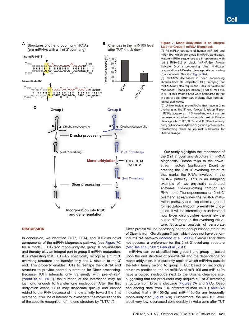
Figure 7. Mono-Uridylation is an Integral
Step for Group II miRNA Biogenesis
(A) Pri-miRNA structure of human miR-105 and
miR-449b, which are group II miRNA candidates.
Mature miRNA sequences are in uppercase with
red (miRNA-5p) or black (miRNA-3p). Arrows
indicate Drosha processing sites. xIndicatesreannotation of Drosha cleavage site according
to our analysis. See also Figure S7A.
(B) miR-105 decreased in deep sequencing
libraries from TUT-depleted HeLa, implying that
miR-105 may also require the TUTs for its efficient
maturation. Reads per million (RPM) of miR-105
in siTUT mix-treated cells were compared to that
in control cells. Error bars indicate SDs from bio-
logical duplicates.
(C) Unlike typical pre-miRNAs that have a 2 nt
overhang at the 30 end (group I), group II pre-
miRNAs acquire a 1 nt 30 overhang end structure
because of a bulged nucleotide next to Drosha
cleavage site. TUT7, TUT4, and TUT2 redundantly
carry outmono-uridylation of group II pre-miRNAs,
transforming them to optimal substrates for
Dicer cleavage.
DISCUSSION
In conclusion, we identified TUT7, TUT4, and TUT2 as novel
components of the miRNA biogenesis pathway (see Figure 7C
for a model). TUT7/4/2 mono-uridylate group II pre-miRNAs
and thereby play an integral part in group II miRNA maturation.
It is interesting that TUT7/4/2 specifically recognize a 1 nt 30
overhang structure and transfer only one U residue to the 30
end. This property enables TUTs to reshape the dsRNA end
structure to provide optimal substrates for Dicer processing.
Because TUT4 interacts only transiently with pre-let-7a-1
(Yeom et al., 2011), the duration of the interaction may be
just long enough to transfer one nucleotide. After the first
uridylation event, TUTs may dissociate quickly and cannot
rebind to the RNA because of the low affinity toward the 2 nt
overhang. It will be of interest to investigate the molecular basis
of the specific recognition of the end structure by TUT7/4/2.
Cell 151, 521–532,
Our study highlights the importance of
the 2 nt 30 overhang structure in miRNA
biogenesis. Drosha talks to the down-
stream factors (particularly Dicer) by
creating the 2 nt 30 overhang structure
that marks the RNAs involved in the
miRNA pathway. This is an intriguing
example of two physically separated
enzymes communicating through an
RNA motif. The dependence on 2 nt 30
overhang streamlines the miRNA matu-
ration pathway and also offers a ground
for regulation through pre-miRNA uridy-
lation. It will be interesting to understand
how Dicer distinguishes exquisitely the
subtle difference in the overhang struc-
ture. Structural analysis of vertebrate
Dicer protein will be necessary as the only published structure
of Dicer is from Giardia intestinalis, which does not have canon-
ical miRNA pathway (Macrae et al., 2006). Giardia Dicer does
not possess a preference for the 2 nt 30 overhang structure
(MacRae et al., 2007; Park et al., 2011).
miRNAs can be classified into group I and group II, based
upon the end structure of pre-miRNA and the dependence on
mono-uridylation. It is currently unclear which miRNAs outside
the let-7 family belong to group II. But based on secondary
structure prediction, the pri-miRNAs of miR-105 and miR-449b
have a bulged nucleotide next to the Drosha cleavage site,
suggesting that the precursors may acquire a 1 nt 30 overhangstructure from Drosha cleavage (Figures 7A and S7A). Deep
sequencing data from 104 different human cells (Table S2)
indicated that miR-105-3p and miR-449b-3p are frequently
mono-uridylated (Figure S7A). Furthermore, the miR-105 level,
albeit very low, decreased considerably in HeLa cells after TUT
October 26, 2012 ª2012 Elsevier Inc. 529

knockdown, suggesting that miR-105 is a bona fide member of
group II (Figure 7B). The level of miR-449b was below detection
limit in HeLa cells. Further investigation will be needed to identify
additional group II members.
Our data suggest that the group I pre-let-7 is rarely uridylated
both in HeLa cells and in vitro assays. In addition, although
mono-uridylation has been observed on other group I pre-
miRNAs (Newman et al., 2011), the frequency of such uridylation
was low and the functional significance of the modification is
unclear. Group I miRNAs remained largely unaffected upon
TUT7/4/2 depletion in our experiments (Figures 4C and S4C).
Thus, although it is possible that other TUTases may control
group I miRNAs in certain conditions or cell types, mono-uridyla-
tion of group I pre-miRNAs may not be frequent enough to exert
a meaningful effect in HeLa cells.
TUT7/4/2 mediate mono-uridylation of pre-let-7 in cells lack-
ing Lin28 and thereby facilitate Dicer processing and compete
with exonucleases. This mechanism would effectively shift the
balance in favor of biogenesis in the absence of Lin28. Because
uridylation is generally associated with RNA degradation (Ji and
Chen, 2012; Kim et al., 2010; Ren et al., 2012; Wickens and
Kwak, 2008; Wilusz and Wilusz, 2008; Zhao et al., 2012), it was
unexpected that TUT knockdown resulted in an increase of
trimmed pre-let-7 (Figure 2D), providing an intriguing case where
uridylation may have a protective effect.
We have recently reported that Lin28 increases the dwelling
time of TUT4 on pre-let-7 (Yeom et al., 2011). Thus, Lin28
enhances the processivity of TUT4 to induce oligo-uridylation,
which in turn blocks Dicer processing and facilitates pre-let-7
decay (Hagan et al., 2009; Heo et al., 2008, 2009). Therefore,
Lin28 serves as a molecular switch that converts TUT4 (and
TUT7) from key biogenesis factors into negative regulators
(Figure S7B). Our study unveils two opposing functions of
TUTs in miRNA biogenesis. The functional duality of uridylation
may contribute to the tight control of let-7 expression during
developmental transition and tumorigenesis.
EXPERIMENTAL PROCEDURES
Pre-let-7 Sequencing
Twenty to fifty micrograms of HeLa total RNA were resolved on 15% urea-
polyacrylamide gel and RNAs of 50–100 nt were gel purified. Size-fractionated
RNAs were ligated to 30 adaptor by using T4 RNA ligase 2, truncated K227Q
(NEB). The ligated RNAs were reverse transcribed with a RT primer that is
complementary to the 30 adaptor by using superscript II (Life Technologies),
followed by PCR amplification with the RT primer and a let-7a-specific (or
let-7c-specific) forward primer that contains BanI restriction enzyme sites.
Because of sequence similarities, the forward primers can hybridize to other
let-7 members. The PCR products were cleaved by using BanI (NEB) and
then concatamerized by using T4 DNA ligase (NEB). The concatamerized
DNAs were cloned for Sanger-sequencing. The sequences of 30 adaptor andprimers are listed in Table S5.
Immunoprecipitation and In Vitro Uridylation
For immunoprecipitation of Flag-TUTases, HEK293T cells were collected
48 hr after transfection with Flag-TUTase expression plasmids. The cells
were incubated in Buffer D (200 mM KCl, 10 mM Tris-HCl [pH8.0], 0.2 mM
EDTA) for 20 min followed by sonication on ice and centrifugation twice
for 15 min at 4�C. The supernatant was incubated with 5 ml of anti-Flag
antibody-conjugated agarose beads (anti-Flag M2 affinity gel, Sigma) with
constant rotation for 1 hr at 4�C. The beads were washed six times with
530 Cell 151, 521–532, October 26, 2012 ª2012 Elsevier Inc.
Buffer D. In vitro uridylation reaction was performed in a total volume of
30 ml in 3.2 mM MgCl2, 1 mM DTT, 0.25 mM UTP, 50 end labeled pre-miRNA
or dsRNA of 1 3 104 to 1 3 105 cpm, and 15 ml of immunopurified proteins
on beads (Figures 1 and S1) or 33 Flag-peptide (Sigma) eluted proteins
(Figures 5 and S5) in Buffer D. The reaction mixture was incubated at 37�Cfor 10 (Figures 5 and S5) or 20 min (Figures 1 and S1). The RNA was purified
from the reaction mixture by phenol extraction and run on 6% urea polyacryl-
amide sequencing gel (20 3 40 cm, 0.4 mm thick) at constant 1500 V for
2 hr. Unmodified and mono-uridylated form of pre-let-7 (let-7a-1, b, and c)
and dsRNAs were synthesized by ST Pharm. The pre-miRNAs or dsRNAs
were radio-labeled at the 50 end with T4 polynucleotide kinase (Takara)
and (g-32P) ATP. The sequences of pre-miRNAs and dsRNAs are listed in
Table S5.
mRNA Library Preparation
mRNAs were purified from total RNA by using Dynabeads mRNA Purification
Kit (Life Technologies, 61011). Purified mRNAs were fragmented by RNA
Fragmentation Reagents (Life Technologies, AM8740). After fragmentation,
phosphate group at 30 end was removed by Antarctic phosphatase (NEB,
M0289L), and RNAs were 50 phosphorylated by T4 PNK (NEB). Directional
and multiplexed mRNA libraries were generated by using TruSeq Small RNA
Sample Preparation Kit (Illumina, RS-200–0012) and sequenced by using
Illumina HiSeq 2000.
Phylogenetic Analysis of the Structures of let-7 Precursor
All sequences and structures of the let-7 stem-loop in bilaterians were down-
loaded from miRBase release 18 and UCSC genome browser. The end struc-
tures of each pre-let-7 were manually classified into group I and group II.
The phylogenetic tree illustrated in Figure 6D was modified from the earlier
studies (Niwa and Slack, 2007; Pasquinelli et al., 2003, 2000) and miRBase
18. The full genus and species names are as follows: Homo sapiens, Mus
musculus, Gallus gallus, Xenopus tropicalis, Danio rerio, Ciona intestinalis,
Branchiostoma floridae, Saccoglossus kowalevskii, Strongylocentrotus
purpuratus, Drosophila melanogaster, Caenorhabditis elegans, Capitella
teleta, Lottia gigantean, Echinococcus granulosus, Nematostella vectensis,
and Ophlitaspongia tenuis.
ACCESSION NUMBERS
The GEO accession number for the small RNA sequencing and mRNA
sequencing data is GSE40236.
SUPPLEMENTAL INFORMATION
Supplemental Information includes Extended Experimental Procedures, seven
figures, and five tables and can be found with this article online at http://dx.doi.
org/10.1016/j.cell.2012.09.022.
ACKNOWLEDGMENTS
We are grateful to the members of our laboratory, particularly Joha Park,
Hyerim Yi, Dr. Kyu-Hyeon Yeom, Dr. Yoosik Kim, Ahyoung Cho, and Yun
Cheng Chang for discussion and technical help. We thank Dr. Yun-jeong
Kim and Dr. Daehyun Baek for help in preparation of mRNA sequencing
libraries. This work was supported by the Research Center Program
(EM1202) of IBS (Institute for Basic Science); the BK21 Research Fellowships
(J.L., J.-E.P., and H.C.) from the Ministry of Education, Science and Tech-
nology of Korea; and the National Honor Scientist Program (20100020415)
through the National Research Foundation of Korea (NRF).
Received: March 27, 2012
Revised: June 26, 2012
Accepted: August 15, 2012
Published online: October 11, 2012

REFERENCES
Balzer, E., and Moss, E.G. (2007). Localization of the developmental timing
regulator Lin28 to mRNP complexes, P-bodies and stress granules. RNA
Biol. 4, 16–25.
Bernstein, E., Caudy, A.A., Hammond, S.M., and Hannon, G.J. (2001). Role for
a bidentate ribonuclease in the initiation step of RNA interference. Nature 409,
363–366.
Bohnsack, M.T., Czaplinski, K., andGorlich, D. (2004). Exportin 5 is a RanGTP-
dependent dsRNA-binding protein that mediates nuclear export of pre-
miRNAs. RNA 10, 185–191.
Bompfunewerer, A.F., Flamm, C., Fried, C., Fritzsch, G., Hofacker, I.L.,
Lehmann, J., Missal, K., Mosig, A., Muller, B., Prohaska, S.J., et al. (2005).
Evolutionary patterns of non-coding RNAs. Theory Biosci. 123, 301–369.
Burns, D.M., D’Ambrogio, A., Nottrott, S., and Richter, J.D. (2011). CPEB and
two poly(A) polymerases control miR-122 stability and p53 mRNA translation.
Nature 473, 105–108.
Burroughs, A.M., Ando, Y., de Hoon, M.J., Tomaru, Y., Nishibu, T., Ukekawa,
R., Funakoshi, T., Kurokawa, T., Suzuki, H., Hayashizaki, Y., and Daub, C.O.
(2010). A comprehensive survey of 30 animal miRNA modification events and
a possible role for 30 adenylation in modulating miRNA targeting effectiveness.
Genome Res. 20, 1398–1410.
Bussing, I., Slack, F.J., and Grosshans, H. (2008). let-7 microRNAs in develop-
ment, stem cells and cancer. Trends Mol. Med. 14, 400–409.
Chiang, H.R., Schoenfeld, L.W., Ruby, J.G., Auyeung, V.C., Spies, N., Baek,
D., Johnston, W.K., Russ, C., Luo, S., Babiarz, J.E., et al. (2010). Mammalian
microRNAs: experimental evaluation of novel and previously annotated genes.
Genes Dev. 24, 992–1009.
Denli, A.M., Tops, B.B., Plasterk, R.H., Ketting, R.F., and Hannon, G.J. (2004).
Processing of primary microRNAs by the Microprocessor complex. Nature
432, 231–235.
Gregory, R.I., Yan, K.P., Amuthan, G., Chendrimada, T., Doratotaj, B., Cooch,
N., and Shiekhattar, R. (2004). The Microprocessor complex mediates the
genesis of microRNAs. Nature 432, 235–240.
Grishok, A., Pasquinelli, A.E., Conte, D., Li, N., Parrish, S., Ha, I., Baillie, D.L.,
Fire, A., Ruvkun, G., and Mello, C.C. (2001). Genes and mechanisms related
to RNA interference regulate expression of the small temporal RNAs that
control C. elegans developmental timing. Cell 106, 23–34.
Grosshans, H., Johnson, T., Reinert, K.L., Gerstein, M., and Slack, F.J. (2005).
The temporal patterningmicroRNA let-7 regulates several transcription factors
at the larval to adult transition in C. elegans. Dev. Cell 8, 321–330.
Guo, Y., Chen, Y., Ito, H., Watanabe, A., Ge, X., Kodama, T., and Aburatani, H.
(2006). Identification and characterization of lin-28 homolog B (LIN28B) in
human hepatocellular carcinoma. Gene 384, 51–61.
Hagan, J.P., Piskounova, E., and Gregory, R.I. (2009). Lin28 recruits the TU-
Tase Zcchc11 to inhibit let-7 maturation in mouse embryonic stem cells.
Nat. Struct. Mol. Biol. 16, 1021–1025.
Hammond, S.M., Boettcher, S., Caudy, A.A., Kobayashi, R., and Hannon, G.J.
(2001). Argonaute2, a link between genetic and biochemical analyses of RNAi.
Science 293, 1146–1150.
Han, J., Lee, Y., Yeom, K.H., Kim, Y.K., Jin, H., and Kim, V.N. (2004). The
Drosha-DGCR8 complex in primary microRNA processing. Genes Dev. 18,
3016–3027.
Han, J., Lee, Y., Yeom, K.H., Nam, J.W., Heo, I., Rhee, J.K., Sohn, S.Y., Cho,
Y., Zhang, B.T., and Kim, V.N. (2006). Molecular basis for the recognition of
primary microRNAs by the Drosha-DGCR8 complex. Cell 125, 887–901.
Heo, I., Joo, C., Cho, J., Ha, M., Han, J., and Kim, V.N. (2008). Lin28 mediates
the terminal uridylation of let-7 precursor MicroRNA. Mol. Cell 32, 276–284.
Heo, I., Joo, C., Kim, Y.K., Ha, M., Yoon, M.J., Cho, J., Yeom, K.H., Han, J.,
and Kim, V.N. (2009). TUT4 in concert with Lin28 suppresses microRNA
biogenesis through pre-microRNA uridylation. Cell 138, 696–708.
Hutvagner, G., McLachlan, J., Pasquinelli, A.E., Balint, E., Tuschl, T., and
Zamore, P.D. (2001). A cellular function for the RNA-interference enzyme Dicer
in the maturation of the let-7 small temporal RNA. Science 293, 834–838.
Ji, L., and Chen, X. (2012). Regulation of small RNA stability: methylation and
beyond. Cell Res. 22, 624–636.
Jones, M.R., Quinton, L.J., Blahna, M.T., Neilson, J.R., Fu, S., Ivanov, A.R.,
Wolf, D.A., and Mizgerd, J.P. (2009). Zcchc11-dependent uridylation of
microRNA directs cytokine expression. Nat. Cell Biol. 11, 1157–1163.
Katoh, T., Sakaguchi, Y., Miyauchi, K., Suzuki, T., Kashiwabara, S., Baba, T.,
and Suzuki, T. (2009). Selective stabilization of mammalian microRNAs by 30
adenylation mediated by the cytoplasmic poly(A) polymerase GLD-2. Genes
Dev. 23, 433–438.
Ketting, R.F., Fischer, S.E., Bernstein, E., Sijen, T., Hannon, G.J., and Plasterk,
R.H. (2001). Dicer functions in RNA interference and in synthesis of small RNA
involved in developmental timing in C. elegans. Genes Dev. 15, 2654–2659.
Kim, V.N., Han, J., and Siomi, M.C. (2009). Biogenesis of small RNAs in
animals. Nat. Rev. Mol. Cell Biol. 10, 126–139.
Kim, Y.K., Heo, I., and Kim, V.N. (2010). Modifications of small RNAs and
their associated proteins. Cell 143, 703–709.
Knight, S.W., and Bass, B.L. (2001). A role for the RNase III enzyme DCR-1 in
RNA interference and germ line development in Caenorhabditis elegans.
Science 293, 2269–2271.
Landthaler, M., Yalcin, A., and Tuschl, T. (2004). The human DiGeorge
syndrome critical region gene 8 and Its D. melanogaster homolog are required
for miRNA biogenesis. Curr. Biol. 14, 2162–2167.
Lee, Y., Ahn, C., Han, J., Choi, H., Kim, J., Yim, J., Lee, J., Provost, P., Rad-
mark, O., Kim, S., and Kim, V.N. (2003). The nuclear RNase III Drosha initiates
microRNA processing. Nature 425, 415–419.
Loughlin, F.E., Gebert, L.F., Towbin, H., Brunschweiger, A., Hall, J., and Allain,
F.H. (2012). Structural basis of pre-let-7 miRNA recognition by the zinc
knuckles of pluripotency factor Lin28. Nat. Struct. Mol. Biol. 19, 84–89.
Lund, E., Guttinger, S., Calado, A., Dahlberg, J.E., and Kutay, U. (2004).
Nuclear export of microRNA precursors. Science 303, 95–98.
Macrae, I.J., Zhou, K., Li, F., Repic, A., Brooks, A.N., Cande, W.Z., Adams,
P.D., and Doudna, J.A. (2006). Structural basis for double-stranded RNA
processing by Dicer. Science 311, 195–198.
MacRae, I.J., Zhou, K., and Doudna, J.A. (2007). Structural determinants of
RNA recognition and cleavage by Dicer. Nat. Struct. Mol. Biol. 14, 934–940.
Martin, G., and Keller, W. (2007). RNA-specific ribonucleotidyl transferases.
RNA 13, 1834–1849.
Meneely, P.M., and Herman, R.K. (1979). Lethals, steriles and deficiencies
in a region of the X chromosome of Caenorhabditis elegans. Genetics 92,
99–115.
Mourelatos, Z., Dostie, J., Paushkin, S., Sharma, A., Charroux, B., Abel, L.,
Rappsilber, J., Mann, M., and Dreyfuss, G. (2002). miRNPs: a novel class
of ribonucleoproteins containing numerous microRNAs. Genes Dev. 16,
720–728.
Nakanishi, T., Kubota, H., Ishibashi, N., Kumagai, S., Watanabe, H., Yama-
shita, M., Kashiwabara, S., Miyado, K., and Baba, T. (2006). Possible role of
mouse poly(A) polymerase mGLD-2 during oocyte maturation. Dev. Biol.
289, 115–126.
Nam, Y., Chen, C., Gregory, R.I., Chou, J.J., and Sliz, P. (2011). Molecular
basis for interaction of let-7 microRNAs with Lin28. Cell 147, 1080–1091.
Newman,M.A., Thomson, J.M., and Hammond, S.M. (2008). Lin-28 interaction
with the Let-7 precursor loop mediates regulated microRNA processing. RNA
14, 1539–1549.
Newman, M.A., Mani, V., and Hammond, S.M. (2011). Deep sequencing of
microRNA precursors reveals extensive 30 end modification. RNA 17, 1795–
1803.
Niwa, R., and Slack, F.J. (2007). The evolution of animal microRNA function.
Curr. Opin. Genet. Dev. 17, 145–150.
Cell 151, 521–532, October 26, 2012 ª2012 Elsevier Inc. 531

Park, J.E., Heo, I., Tian, Y., Simanshu, D.K., Chang, H., Jee, D., Patel, D.J., and
Kim, V.N. (2011). Dicer recognizes the 50 end of RNA for efficient and accurate
processing. Nature 475, 201–205.
Pasquinelli, A.E., Reinhart, B.J., Slack, F., Martindale, M.Q., Kuroda, M.I.,
Maller, B., Hayward, D.C., Ball, E.E., Degnan, B., Muller, P., et al. (2000).
Conservation of the sequence and temporal expression of let-7 heterochronic
regulatory RNA. Nature 408, 86–89.
Pasquinelli, A.E., McCoy, A., Jimenez, E., Salo, E., Ruvkun, G., Martindale,
M.Q., and Baguna, J. (2003). Expression of the 22 nucleotide let-7 hetero-
chronic RNA throughout the Metazoa: a role in life history evolution? Evol.
Dev. 5, 372–378.
Piskounova, E., Polytarchou, C., Thornton, J.E., LaPierre, R.J., Pothoulakis,
C., Hagan, J.P., Iliopoulos, D., and Gregory, R.I. (2011). Lin28A and Lin28B
inhibit let-7 microRNA biogenesis by distinct mechanisms. Cell 147, 1066–
1079.
Polesskaya, A., Cuvellier, S., Naguibneva, I., Duquet, A., Moss, E.G., and
Harel-Bellan, A. (2007). Lin-28 binds IGF-2 mRNA and participates in skeletal
myogenesis by increasing translation efficiency. Genes Dev. 21, 1125–1138.
Reinhart, B.J., Slack, F.J., Basson, M., Pasquinelli, A.E., Bettinger, J.C.,
Rougvie, A.E., Horvitz, H.R., and Ruvkun, G. (2000). The 21-nucleotide let-7
RNA regulates developmental timing in Caenorhabditis elegans. Nature 403,
901–906.
Ren, G., Chen, X., and Yu, B. (2012). Uridylation of miRNAs by hen1
suppressor1 in Arabidopsis. Curr. Biol. 22, 695–700.
Richards, M., Tan, S.P., Tan, J.H., Chan, W.K., and Bongso, A. (2004). The
transcriptome profile of human embryonic stem cells as defined by SAGE.
Stem Cells 22, 51–64.
Rouhana, L., Wang, L., Buter, N., Kwak, J.E., Schiltz, C.A., Gonzalez, T.,
Kelley, A.E., Landry, C.F., and Wickens, M. (2005). Vertebrate GLD2 poly(A)
polymerases in the germline and the brain. RNA 11, 1117–1130.
Roush, S., and Slack, F.J. (2008). The let-7 family of microRNAs. Trends Cell
Biol. 18, 505–516.
Rybak, A., Fuchs, H., Smirnova, L., Brandt, C., Pohl, E.E., Nitsch, R., and
Wulczyn, F.G. (2008). A feedback loop comprising lin-28 and let-7 controls
pre-let-7 maturation during neural stem-cell commitment. Nat. Cell Biol. 10,
987–993.
Stevenson, A.L., and Norbury, C.J. (2006). The Cid1 family of non-canonical
poly(A) polymerases. Yeast 23, 991–1000.
Tabara, H., Sarkissian, M., Kelly, W.G., Fleenor, J., Grishok, A., Timmons, L.,
Fire, A., and Mello, C.C. (1999). The rde-1 gene, RNA interference, and trans-
poson silencing in C. elegans. Cell 99, 123–132.
Tian, B., Bevilacqua, P.C., Diegelman-Parente, A., and Mathews, M.B. (2004).
The double-stranded-RNA-binding motif: interference and much more. Nat.
Rev. Mol. Cell Biol. 5, 1013–1023.
532 Cell 151, 521–532, October 26, 2012 ª2012 Elsevier Inc.
Vermeulen, A., Behlen, L., Reynolds, A., Wolfson, A., Marshall, W.S., Karpilow,
J., and Khvorova, A. (2005). The contributions of dsRNA structure to Dicer
specificity and efficiency. RNA 11, 674–682.
Viswanathan, S.R., Daley, G.Q., and Gregory, R.I. (2008). Selective blockade
of microRNA processing by Lin28. Science 320, 97–100.
Wickens, M., and Kwak, J.E. (2008). Molecular biology. A tail tale for U.
Science 319, 1344–1345.
Wilusz, C.J., and Wilusz, J. (2008). New ways to meet your (30) end oligouridy-
lation as a step on the path to destruction. Genes Dev. 22, 1–7.
Wu, H., Ye, C., Ramirez, D., and Manjunath, N. (2009). Alternative processing
of primary microRNA transcripts by Drosha generates 50 end variation of
mature microRNA. PLoS ONE 4, e7566.
Wyman, S.K., Knouf, E.C., Parkin, R.K., Fritz, B.R., Lin, D.W., Dennis, L.M.,
Krouse, M.A., Webster, P.J., and Tewari, M. (2011). Post-transcriptional
generation of miRNA variants by multiple nucleotidyl transferases contributes
to miRNA transcriptome complexity. Genome Res. 21, 1450–1461.
Yang, D.H., and Moss, E.G. (2003). Temporally regulated expression of Lin-28
in diverse tissues of the developing mouse. Gene Expr. Patterns 3, 719–726.
Yeom, K.H., Heo, I., Lee, J., Hohng, S., Kim, V.N., and Joo, C. (2011). Single-
molecule approach to immunoprecipitated protein complexes: insights into
miRNA uridylation. EMBO Rep. 12, 690–696.
Yi, R., Qin, Y., Macara, I.G., and Cullen, B.R. (2003). Exportin-5 mediates the
nuclear export of pre-microRNAs and short hairpin RNAs. Genes Dev. 17,
3011–3016.
Zhang, H., Kolb, F.A., Brondani, V., Billy, E., and Filipowicz, W. (2002). Human
Dicer preferentially cleaves dsRNAs at their termini without a requirement for
ATP. EMBO J. 21, 5875–5885.
Zhang, H., Kolb, F.A., Jaskiewicz, L., Westhof, E., and Filipowicz, W. (2004).
Single processing center models for human Dicer and bacterial RNase III.
Cell 118, 57–68.
Zhao, Y., Yu, Y., Zhai, J., Ramachandran, V., Dinh, T.T., Meyers, B.C., Mo, B.,
and Chen, X. (2012). The Arabidopsis nucleotidyl transferase HESO1 uridy-
lates unmethylated small RNAs to trigger their degradation. Curr. Biol. 22,
689–694.
Zhu, H., Shah, S., Shyh-Chang, N., Shinoda, G., Einhorn, W.S., Viswanathan,
S.R., Takeuchi, A., Grasemann, C., Rinn, J.L., Lopez, M.F., et al. (2010). Lin28a
transgenic mice manifest size and puberty phenotypes identified in human
genetic association studies. Nat. Genet. 42, 626–630.
Zhu, H., Shyh-Chang, N., Segre, A.V., Shinoda, G., Shah, S.P., Einhorn, W.S.,
Takeuchi, A., Engreitz, J.M., Hagan, J.P., Kharas, M.G., et al; DIAGRAM
Consortium; MAGIC Investigators. (2011). The Lin28/let-7 axis regulates
glucose metabolism. Cell 147, 81–94.

本文献由“学霸图书馆-文献云下载”收集自网络,仅供学习交流使用。
学霸图书馆(www.xuebalib.com)是一个“整合众多图书馆数据库资源,
提供一站式文献检索和下载服务”的24 小时在线不限IP
图书馆。
图书馆致力于便利、促进学习与科研,提供最强文献下载服务。
图书馆导航:
图书馆首页 文献云下载 图书馆入口 外文数据库大全 疑难文献辅助工具
Related Documents
![The Molecular Basis and Therapeutic Potential of Let-7 ...downloads.hindawi.com/journals/cjgh/2018/5769591.pdf · microRNA Cancer microRNA- Lung[] microRNA-Neuroblastoma[ ] ... Recent](https://static.cupdf.com/doc/110x72/604147fde9c3331b744ecb0e/the-molecular-basis-and-therapeutic-potential-of-let-7-microrna-cancer-microrna-.jpg)