Universidade de Lisboa 2011 Faculdade de Ciências Departamento de Biologia Animal Monitoring Rates of Particulate Matter Pollution Using Genetic Biomarkers in Small Mammals Ana Sofia Ribeiro da Costa Boa-Alma Mestrado em Biologia Humana e Ambiente

Welcome message from author
This document is posted to help you gain knowledge. Please leave a comment to let me know what you think about it! Share it to your friends and learn new things together.
Transcript

Universidade de Lisboa
2011
Faculdade de Ciências
Departamento de Biologia Animal
Monitoring Rates of Particulate Matter
Pollution Using Genetic Biomarkers in Small
Mammals
Ana Sofia Ribeiro da Costa Boa-Alma
Mestrado em Biologia Humana e Ambiente

Universidade de Lisboa
2011
Faculdade de Ciências
Departamento de Biologia Animal
Monitoring Rates of Particulate Matter
Pollution Using Genetic Biomarkers in Small
Mammals
Ana Sofia Ribeiro da Costa Boa-Alma
Dissertação orientada por:
Doutora Graça Ramalhinho (Museu Nacional de História Natural/Faculdade de Ciências da
Universidade de Lisboa)
Professora Doutora Deodália Dias (Departamento de Biologia Animal/ Faculdade de Ciências
da Universidade de Lisboa)
Mestrado em Biologia Humana e Ambiente

i
Foreword
Part of the work, including the observation of particles in light and electron
microscopy was done with the collaboration of Dr. António Pedro Matos,
Department of Pathological Anatomy, Curry Cabral Hospital.
The entire thesis is written in English to facilitate a possible publication. The
bibliography was made according to the criteria of Mutation Research, since it is
a journal that addresses issues experienced here.
Lisbon, October 2011
Ana Sofia Boa-Alma

ii
Acknowledgments
Firstly I would like to thank my supervisors, in particular, Doutora Graça
Ramalhinho for the friendship, dedication and for “getting their hands dirty”
when necessary and Prof. Doutora Deodália Dias also for the friendship,
understanding and help in finding solutions for some of the problems that
occurred. Without them this work would never be possible.
To Doutor António Pedro Matos, for the work with the transmission electron
microscopy.
To the technicians at the Curry Cabral Hospital, especially to Cristina Correia
for the help and time spent teaching and to Bruno Matos by answering
questions that had to be answered.
To Vânia Gaio, for the enormous help and cooperation, without her all this
work would have been more difficult.
To Rita Oliveira Dario, for the teachings in the area of genetics and for her
invaluable friendship and to Catarina Dourado for helping in deciphering the
genetic sequences.
To Sofia Gabriel, for the helpful hints in terms of genetics, which helped to
successfully overcome some of the problems that came up.
To Joaquim Tapisso, Margarida Duarte, Patrícia Sardinha and Susana
Ferreira for the companionship and tips.
To Flávio Júnior, for his assistance in capturing some of the mice at Paio
Pires.
Last but not least, to my parents, for their support, understanding and
assistance especially in the field.
iii
Abstract
Particulate matter is a complex mixture of ultrafine, fine and coarse particles
from a variety of sources. Important compounds associated with airborne
particles are polycyclic aromatic hydrocarbons, among which several are
established carcinogens. Reactive intermediates, which can cause direct DNA
damage, are generated by their degradation. Moreover, they can enter a
reaction with other pollutants, including nitrogen oxides and ozone, and
generate other potential carcinogens. The aim of the present study was to
analyze the genotoxic and cytotoxic effects of particulate matter and its
consequences for the environment and human health, using the Algerian
mouse (Mus spretus) as a bioindicator. For this purpose we used genetic
biomarkers - sperm shape abnormalities assay, micronucleus test, comet assay
and determination and characterization of the genetic polymorphisms of
CYP1A1 gene - in addition to registration of body and internal organs weights
and evaluation of the levels of particulate matter in the respiratory tract of the
animals captured at sites with different concentrations of particulate matter in
the districts of Lisbon and Setubal. The results showed a significant increase in
liver weight with the level of pollution, as well as an increase in the number of
micronuclei and comet score. Relatively to the sperm shape abnormalities
assay, there were no differences between the three groups. Through the
analysis of semi-thin and electron microscopy lung cuts it was observed that
some of the animals from Paio Pires had apparently macrophages with
phagocytized particles, which was not observed in the animals from Lourinhã,
but it is not possible to draw objective conclusions since the work was not
completed. For CYP1A1 gene, it was only possible its amplification and
therefore also can not draw conclusions. Then, this work can provide important
information on how particles affect our health in a real environment.
Key words: Particulate matter; Comet assay; Micronucleus test; Sperm shape
abnormalities assay; Algerian mouse (Mus spretus).

iv
Resumo
O ar que respiramos está poluído com os subprodutos da combustão da
indústria, geração de energia e transporte, bem como o fabrico e utilização de
produtos químicos. Os principais poluentes atmosféricos na Europa e América
do Norte são o dióxido de enxofre, óxidos de azoto, partículas inaláveis e
ozono. Os poluentes do ar podem aparecer na forma de partículas sólidas,
gotículas ou gases. Além disso, podem ser naturais ou fabricados pelo homem.
As fontes de poluição do ar referem-se aos vários locais, actividades ou
factores que são responsáveis pela liberação de poluentes para a atmosfera.
Em concentrações suficientes, estes gases e partículas podem prejudicar a
saúde humana a curto (ardor nos olhos e garganta, dificuldade em respirar) e a
longo prazo (cancro e danos a longo prazo nos sistemas imunológico,
neurológico, reprodutivo e respiratório).
Os mecanismos pelos quais a poluição do ar influencia negativamente
algumas comorbidades não são bem compreendidos. Mecanismos possíveis,
como o influxo de cálcio aumentado quando em contacto com macrófagos,
activação de mediadores pró-inflamatórios, viscosidade do sangue aumentada,
aumento do fibrinogénio e dos níveis de proteína C-reactiva e alterações na
reologia do sangue favorecendo a coagulação, têm sido sugeridos. O stress
oxidativo gerado pela poluição do ar também tem sido proposto como um
mecanismo importante de lesão tecidual que leva à inflamação pulmonar e
sistémica.
Segundo dados científicos de 2004, o excesso de partículas inaláveis provoca
em Portugal quase 4000 mortes prematuras e uma redução de 6 meses na
esperança média de vida dos habitantes do Porto e Lisboa (estudo da Agência
Europeia do Ambiente). A Organização Mundial da Saúde estima que as
doenças associadas à poluição do ar causadas por partículas inaláveis podem
ser consideradas dentro das dez principais causas de morte nos países
desenvolvidos.
"Partículas inaláveis" é, então, um termo geral que engloba as partículas de
poeira, fuligem queimada, partículas de exaustão diesel e hidrocarbonetos
v
aromáticos policíclicos. As partículas inaláveis têm contribuições tanto de
fontes primárias (ou seja, emitidas directamente na atmosfera) como de
processos secundários (ou seja, formadas na atmosfera a partir de emissões
de substâncias precursoras). Tanto as emissões primárias como as emissões
precursoras secundárias podem ser originadas a partir de qualquer origem
antropogénica ou natural.
Enquanto que as partículas maiores que as PM10 não são muito susceptíveis
de atingir o tracto respiratório inferior, as partículas grossas (PM10) podem
chegar tão longe quanto os brônquios. As partículas finas (PM2.5) podem
penetrar mais profundamente e atingir os alvéolos, assim como as partículas
de exaustão diesel, que estão na faixa de tamanho das partículas finas (0.1-2.5
µm) e ultrafinas (<0.1 µm).
Compostos importantes associados às partículas em suspensão são os, já
referidos, hidrocarbonetos aromáticos policíclicos, entre os quais vários são
estabelecidos carcinogéneos. Os hidrocarbonetos aromáticos policíclicos
carcinogénicos tornaram-se recentemente o centro da atenção devido ao seu
efeito potencial na etiologia do cancro, doenças respiratórias e
cardiovasculares e mortalidade. Os hidrocarbonetos aromáticos policíclicos
carcinogénicos são componentes da matéria orgânica ligada às partículas
aerosóis inaláveis (<2.5 µm). Intermediários reactivos, que podem causar
danos directos no DNA, são gerados pela sua degradação. Além disso, eles
podem induzir uma reacção com outros poluentes, nomeadamente óxidos de
azoto e ozono, além de gerar outros potenciais cancerígenos.
A biomonitorização ambiental pode fazer uso de organismos “sentinela” que
vivem no seu habitat natural e reflectir uma exposição contínua, a longo prazo.
Estudos de pequenos mamíferos, principalmente roedores selvagens, têm
demonstrado uma capacidade de acumular um amplo espectro de poluentes.
Assim, eles são adequados para a monitorização da poluição ambiental e risco
de exposição de pessoas que vivem numa área contaminada, além de serem
geralmente abundantes em áreas facilmente identificadas e rapidamente
capturados.
Quando se trata da monitorização da genotoxicidade no ambiente, uma
questão de suma importância, para além da selecção adequada de organismos

vi
representativos como sentinelas, é a realização de testes sensíveis e
confiáveis, tais como aqueles projectados para a avaliação de danos no DNA.
Independentemente das características particulares do evento de
contaminação como um todo, é também muito importante que o ensaio de
escolha tenha sido devidamente validado por laboratórios no mundo inteiro e
que possa ser usado para monitorizar praticamente qualquer espécie selvagem
potencialmente ameaçada.
Desta forma, o objectivo do presente estudo foi analisar os efeitos
genotóxicos e citotóxicos das partículas inaláveis e as suas consequências
para o meio ambiente e para a saúde humana, utilizando o rato argelino (Mus
spretus) como bioindicador. Para este efeito, foram utilizados biomarcadores
genéticos em animais de locais com diferentes concentrações de partículas
inaláveis nos distritos de Lisboa e Setúbal. Objectivos específicos podem ser
apontados: avaliação do papel do rato argelino (Mus spretus) como um
indicador de poluição ambiental; identificação de efeitos adversos, ou seja,
mudanças de peso nos órgãos dos ratinhos e comparação com os níveis de
exposição; avaliação dos níveis de partículas inaláveis no tracto respiratório
dos animais capturados; avaliação dos efeitos genotóxicos e citotóxicos das
partículas inaláveis através do teste das anomalias dos espermatozóides, teste
do micronúcleo e ensaio do cometa; determinação e caracterização dos
polimorfismos genéticos do gene CYP1A1, envolvidos no metabolismo de
xenobióticos, incluindo hidrocarbonetos aromáticos policíclicos; confirmação do
risco para a saúde pública e ambiental causado pela poluição por partículas
inaláveis.
Assim, os animais foram capturados nas três zonas seleccionadas de acordo
com os níveis de poluição por partículas inaláveis. Depois de sacrificados,
foram feitas medições morfológicas, ou seja, tirados os pesos e medidas
corporais e dos órgãos internos e feito o teste das anomalias dos
espermatozóides, o teste do micronúcleo e o ensaio do cometa. Para além
disso, foi também retirado o músculo para análise genética, nomeadamente, a
determinação dos polimorfismos do gene CYP1A1 e feita a observação em
microscopia electrónica de transmissão de macrófagos presentes nos pulmões
dos animais capturados, com o intuito de encontrar partículas fagocitadas.
vii
Como resultado, pôde-se observar medidas corporais superiores nos animais
de Paio Pires, assim como o peso relativo do fígado, baço, rins e testículos. Tal
seria de esperar visto o fígado ser o principal órgão essencialmente exposto a
substâncias tóxicas e essenciais que entram no organismo através de inalação
ou ingestão e o baço ser bastante sensível a infecções. Ao contrário do teste
das anomalias dos espermatozóides que não revelou diferenças significativas
entre os grupos, o que poderá ser devido em parte a artefactos técnicos, o
teste do micronúcleo e o ensaio do cometa mostraram valores superiores com
o aumento do nível de poluição por partículas inaláveis. Para além disto,
também foi possível amplificar o gene CYP1A1, mas a determinação dos seus
polimorfismos não pôde ser concluída, assim como a microanálise das
possíveis partículas observadas em microscopia electrónica.
Através deste estudo pode-se concluir que a inalação de partículas em altas
concentrações tem um risco genotóxico significativo que pode induzir danos no
DNA e, consequentemente, levar a sérios problemas de saúde.
Embora este trabalho esteja dirigido para o estudo dos efeitos que as
partículas inaláveis têm sobre a saúde dos seres vivos, é natural que outros
xenobióticos possam ser responsáveis por alguns dos efeitos adversos
registados. No entanto, embora não possamos manipular e controlar muitas
das variáveis, este tipo de estudo é muito útil para analisar os efeitos da
poluição como um todo e exactamente da maneira como ela nos afecta. No
laboratório podemos controlar essas variáveis, mas apenas observamos os
efeitos que cada poluente pode ter e não os efeitos da interacção entre todos
eles, além do facto de que as doses de poluentes podem por vezes ser
administradas em quantidades muito diferentes do registado num ambiente
real.
Os nossos resultados podem fornecer informações valiosas sobre os riscos
para a saúde decorrentes de partículas urbanas e industriais, assim, alertando-
nos para a necessidade da utilização de biomarcadores de efeitos, exposição e
susceptibilidade e estabelecer medidas de prevenção antes de sermos
confrontados com a carga social e o custo económico das doenças causadas
pela poluição atmosférica por partículas inaláveis. Assim, neste trabalho foi
utilizado o Mus spretus como espécie bioindicadora dos efeitos que a poluição

viii
ambiental, especificamente por partículas inaláveis, pode ter sobre a saúde,
sendo possível, dentro de certos limites e sempre com cautela, transferir estes
dados para os seres humanos.
Palavras-chave: Partículas inaláveis; Ensaio do cometa; Teste do
micronúcleo; Teste das anomalias dos espermatozóides; Rato argelino (Mus
spretus).
ix
Contents
Foreword ........................................................................................................................................ i
Acknowledgments .......................................................................................................................... ii
Abstract ......................................................................................................................................... iii
Resumo ......................................................................................................................................... iv
Contents ........................................................................................................................................ ix
Figures Index ................................................................................................................................. xi
Tables Index ................................................................................................................................ xiii
List of Abbreviations .................................................................................................................... xiv
1. Introduction ................................................................................................................................ 1
1.1. Framework of the present study ......................................................................................... 1
1.2. Environmental pollution ...................................................................................................... 2
1.2.1. Inhalable particles and their toxicity ............................................................................ 4
1.3. Bioindicators of environmental pollution ............................................................................. 7
1.3.1. The Algerian mouse (Mus spretus) as a model of in situ exposure ............................ 8
1.4. Biomarkers of environmental toxicity ................................................................................. 9
1.4.1. Sperm shape abnormalities assay ............................................................................ 10
1.4.2. Micronucleus test ...................................................................................................... 11
1.4.3. Comet assay ............................................................................................................. 13
1.4.4. CYP1A1 gene polymorphisms .................................................................................. 15
1.5. Objectives of the work ...................................................................................................... 17
1.5.1. General objective ...................................................................................................... 17
1.5.2. Specific objectives ..................................................................................................... 17
2. Materials and Methods ............................................................................................................ 19
2.1. Areas of study according to the concentrations of particulate matter (PM10) ................... 19
2.2. Sampling........................................................................................................................... 21
2.3. Sacrifice of the animals and collection of the samples .................................................... 21
2.4. Morphological analysis ..................................................................................................... 22
2.5. Sperm shape abnormalities assay ................................................................................... 22
2.6. Micronucleus test ............................................................................................................. 23
2.7. Comet assay..................................................................................................................... 23
2.8. CYP1A1 gene polymorphisms ......................................................................................... 24
2.9. Whole lung lavage and lung tissue sampling ................................................................... 26
2.10. Statistical analysis .......................................................................................................... 26
3. Results ..................................................................................................................................... 28
3.1. Morphological analysis ..................................................................................................... 28
3.2. Sperm shape abnormalities assay ................................................................................... 32

x
3.3. Micronucleus test ............................................................................................................. 33
3.4. Comet assay..................................................................................................................... 34
3.5. CYP1A1 gene polymorphisms ......................................................................................... 35
3.6. Observation of particles in the respiratory system ........................................................... 36
4. Discussion ............................................................................................................................... 38
5. Conclusions ............................................................................................................................. 42
6. References .............................................................................................................................. 44

xi
Figures Index
Fig. 1 - Linking genotoxicity with ecotoxicological responses . . . . . . . . . . . . . . . . . . . . . . . . . . . 3
Fig. 2 - Proposed mechanism of hydroxyl radical-mediated DNA damage by particulate matter
(PM) in lung epithelial cells . . . . . . . . . . . . . . . . . . . . . . . . . . . . . . . . . . . . . . . . . . . . . . . . . . . . . .5
Fig. 3 - Mechanism of micronucleus (MN) formation in polychromatic erythrocytes (PCE) and
normochromatic erythrocytes (NCE) . . . . . . . . . . . . . . . . . . . . . . . . . . . . . . . . . . . . . . . . . . . . . 12
Fig. 4 - General principles of the comet assay . . . . . . . . . . . . . . . . . . . . . . . . . . . . . . . . . . . . . .14
Fig. 5 - Relationships among genetic risk factors (genetic polymorphisms), environmental and
others exposures to genotoxic agents and induction of DNA mutations . . . . . . . . . . . . . . . . . . 16
Fig. 6 - Major pathway of metabolic activation and DNA adduct formation of benzo[a]pyrene
(B[a]P) . . . . . . . . . . . . . . . . . . . . . . . . . . . . . . . . . . . . . . . . . . . . . . . . . . . . . . . . . . . . . . . . . . . . 17
Fig. 7 - Schematic of a multiple-endpoint assay combining quantification and/or qualification of
biologically external or internal PM with cytogenetic tests and genetic polymorphisms . . . . . . .18
Fig. 8 - Map of Lisbon and Setubal regions showing the air monitoring stations near the
sampling sites . . . . . . . . . . . . . . . . . . . . . . . . . . . . . . . . . . . . . . . . . . . . . . . . . . . . . . . . . . . . . . .19
Fig. 9 - Variation of concentrations of particulate matter (PM10) in Lourinhã, Reboleira and Paio
Pires stations between July and December 2010 . . . . . . . . . . . . . . . . . . . . . . . . . . . . . . . . . . . 20
Fig. 10 - The shape of A is normal whereas B to E are abnormal murine sperm . . . . . . . . . . . 22
Fig. 11 - Scores assigned by visual scoring (0-4) . . . . . . . . . . . . . . . . . . . . . . . . . . . . . . . . . . . 24
Fig. 12 - Diagram with the location of the amplified fragment and of exons 4–7 (rectangles) and
introns 4–6 (lines) in the mouse CYP1A1 gene . . . . . . . . . . . . . . . . . . . . . . . . . . . . . . . . . . . . . 25
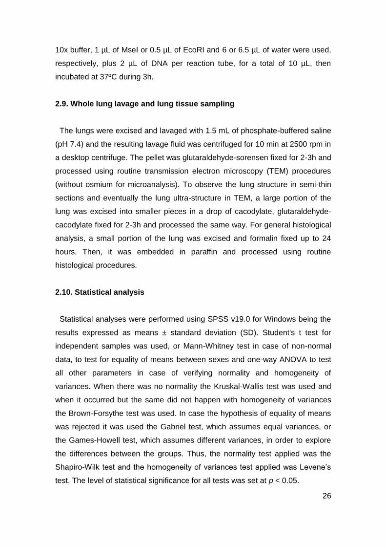
Fig. 13 - Box plots of body (A) and relative tail (B), paw (C) and ear (D) lengths data according
to capture site . . . . . . . . . . . . . . . . . . . . . . . . . . . . . . . . . . . . . . . . . . . . . . . . . . . . . . . . . . . . . . .29
Fig. 14 - Box plots of body (A) and relative liver (B), spleen (C) and left (D) and right kidneys (E)
weights data according to capture site . . . . . . . . . . . . . . . . . . . . . . . . . . . . . . . . . . . . . . . . . . . .30
Fig. 15 - Box plots of relative left (A) and right testis (B) weights and number of scars of the left
(C) and right oviducts (D) data according to capture site . . . . . . . . . . . . . . . . . . . . . . . . . . . . . .32
Fig. 16 - Box plot of total abnormal sperm (%) according to capture site . . . . . . . . . . . . . . . . . 33
Fig. 17 - Box plot of the number of micronucleated polychromatic erythrocytes
(MNPCE/1000PCE) according to capture site . . . . . . . . . . . . . . . . . . . . . . . . . . . . . . . . . . . . . .34
Fig. 18 - Box plot of the comet score obtained through the classification in four classes of 200
comets per animal depending on the size of the comet tail according to capture site . . . . . . . .35
Fig. 19 - PCR amplification of the CYP1A1 gene fragment on agarose gel . . . . . . . . . . . . . . . 36
Fig. 20 - Fragments resulting from the reaction of the amplified fragment of CYP1A1 gene with
MseI and EcoRI restriction enzymes from samples of the contaminated and control area,
arranged alternately . . . . . . . . . . . . . . . . . . . . . . . . . . . . . . . . . . . . . . . . . . . . . . . . . . . . . . . . . . 36

xii
Fig. 21 - Semi-thin lung cuts, observing (A) macrophages without phagocytosed material
(arrows) from an individual from Lourinhã and (B) macrophages with phagocytosed material
(arrows), probably particles, from an individual from Paio Pires . . . . . . . . . . . . . . . . . . . . . . . . 37
Fig. 22 - Preliminary transmission electron microscopy observations of lung tissue (A) reveals
the presence of lisossome-filled (arrows) macrophages (B and C) that may contain ingested
particulate matter . . . . . . . . . . . . . . . . . . . . . . . . . . . . . . . . . . . . . . . . . . . . . . . . . . . . . . . . . . . . 37
xiii
Tables Index
Table 1 - PM10 data for the three study sites between 1 July and 31 December 2010 (µg/m3) .20
Table 2 - Number of animals captured between January and March 2011 in the three sampling
sites . . . . . . . . . . . . . . . . . . . . . . . . . . . . . . . . . . . . . . . . . . . . . . . . . . . . . . . . . . . . . . . . . . . . . . 21
Table 3 - PCR conditions for CYP1A1 . . . . . . . . . . . . . . . . . . . . . . . . . . . . . . . . . . . . . . . . . . . .25
Table 4 - External and internal biometric measurements in the three populations of Mus spretus
. . . . . . . . . . . . . . . . . . . . . . . . . . . . . . . . . . . . . . . . . . . . . . . . . . . . . . . . . . . . . . . . . . . . . . . . . . .28
Table 5 - Different types of sperm shape abnormalities and frequency of total abnormal sperm
in 1000 sperm cells per animal . . . . . . . . . . . . . . . . . . . . . . . . . . . . . . . . . . . . . . . . . . . . . . . . . .32
Table 6 - Mean frequency ± SD of micronucleated polychromatic erythrocytes
(MNPCE/1000PCE) of bone marrow . . . . . . . . . . . . . . . . . . . . . . . . . . . . . . . . . . . . . . . . . . . . . 33
Table 7 - Results of comets classified according to comet categories (0-4) in 200 cells per
animal (arbitrary units, AU) . . . . . . . . . . . . . . . . . . . . . . . . . . . . . . . . . . . . . . . . . . . . . . . . . . . . .34
Table 8 - Comparison of the phases, pace and other features of testis development and
spermatogenesis in rodents and humans relevant to sperm count/quality in adulthood and which
may affect predisposition to lifestyle/environmental effects and limit usefulness of rodents as a
model for humans . . . . . . . . . . . . . . . . . . . . . . . . . . . . . . . . . . . . . . . . . . . . . . . . . . . . . . . . . . . .41
xiv
List of Abbreviations
AhR - Aryl hydrocarbon receptor
AO - Acridine orange
AU - Arbitrary units
bp - Base pairs
CA - Chromosomal aberrations
c-PAH - Carcinogenic polycyclic aromatic hydrocarbons
CYP - Cytochrome P450
EH - Epoxide hydrolase
MN - Micronucleus/micronuclei
MNPCE - Micronucleated polychromatic erythrocytes
NOx - Nitrogen oxides
O3 - Ozone
PAH - Polycyclic aromatic hydrocarbons
PCE - Polychromatic erythrocytes
PCR - Polymerase chain reaction
PM - Particulate matter
PM10 - Particles smaller than 10 μm
PM2.5 - Particles smaller than 2.5 μm
ROS - Reactive oxygen species
SCE - Sister chromatid exchanges
SD - Standard deviation
SN - Siderurgia Nacional
SO2 - Sulphur dioxide
TEM - Transmission electron microscopy
1
1. Introduction
1.1. Framework of the present study
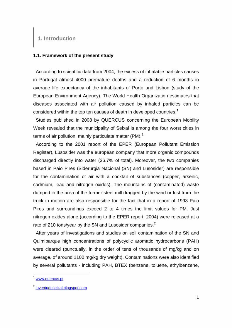
According to scientific data from 2004, the excess of inhalable particles causes
in Portugal almost 4000 premature deaths and a reduction of 6 months in
average life expectancy of the inhabitants of Porto and Lisbon (study of the
European Environment Agency). The World Health Organization estimates that
diseases associated with air pollution caused by inhaled particles can be
considered within the top ten causes of death in developed countries.1
Studies published in 2008 by QUERCUS concerning the European Mobility
Week revealed that the municipality of Seixal is among the four worst cities in
terms of air pollution, mainly particulate matter (PM).1
According to the 2001 report of the EPER (European Pollutant Emission
Register), Lusosider was the european company that more organic compounds
discharged directly into water (36.7% of total). Moreover, the two companies
based in Paio Pires (Siderurgia Nacional (SN) and Lusosider) are responsible
for the contamination of air with a cocktail of substances (copper, arsenic,
cadmium, lead and nitrogen oxides). The mountains of (contaminated) waste
dumped in the area of the former steel mill dragged by the wind or lost from the
truck in motion are also responsible for the fact that in a report of 1993 Paio
Pires and surroundings exceed 2 to 4 times the limit values for PM. Just
nitrogen oxides alone (according to the EPER report, 2004) were released at a
rate of 210 tons/year by the SN and Lusosider companies.2
After years of investigations and studies on soil contamination of the SN and
Quimiparque high concentrations of polycyclic aromatic hydrocarbons (PAH)
were cleared (punctually, in the order of tens of thousands of mg/kg and on
average, of around 1100 mg/kg dry weight). Contaminations were also identified
by several pollutants - including PAH, BTEX (benzene, toluene, ethylbenzene,
1 www.quercus.pt
2 juventudeseixal.blogspot.com

2
xylene), heavy metals, ammonia, phenols and cyanides.3 Also car traffic is
responsible for emission of several pollutants (nitrogen oxides, carbon
monoxide, sulfur, heavy metals and PM).4
Speaking now of very different cases, the municipality of Amadora, where
more than 175 thousand inhabitants live, was awarded by the third consecutive
year the Green Flag. This award resulting from the ECOXXI Project aims to
distinguish good sustainability practices developed locally, with special
emphasis on aspects of environmental quality. The city has achieved top marks
in four of the 23 classification criteria, with emphasis on the quality of air and
water, for which have contributed the municipality policies of rehabilitation and
construction of parks, such as the recently opened central park.5
Finally, Lourinhã is one of the regions with better air quality, at least in the
district of Lisbon and its surroundings, promoting, among other initiatives, more
sustainable modes of mobility, raising awareness for the environmental impacts
of fossil fuel vehicles.6
1.2. Environmental pollution
The air we breathe is polluted with the byproducts of combustion from industry,
power generation, and transportation, as well as the manufacture and use of
chemicals [1]. The major air pollutants in Europe and North America are sulphur
dioxide (SO2), nitrogen oxides (NOx), PM and ozone (O3). Air pollutants can
appear in the form of solid particles, liquid droplets, or gases. In addition, they
may be natural or man-made. Sources of air pollution refer to the various
locations, activities or factors which are responsible for the releasing of
pollutants into the atmosphere [2]. In sufficient concentrations, these gases and
particles can harm human health in the short (burning of eyes and throat,
difficulty breathing) and the long term (cancer and long-term damage to the
3 ambientequalvida.blogs.sapo.pt
4 www.iambiente.pt
5 www.publico.pt
6 www.alvorada.pt

3
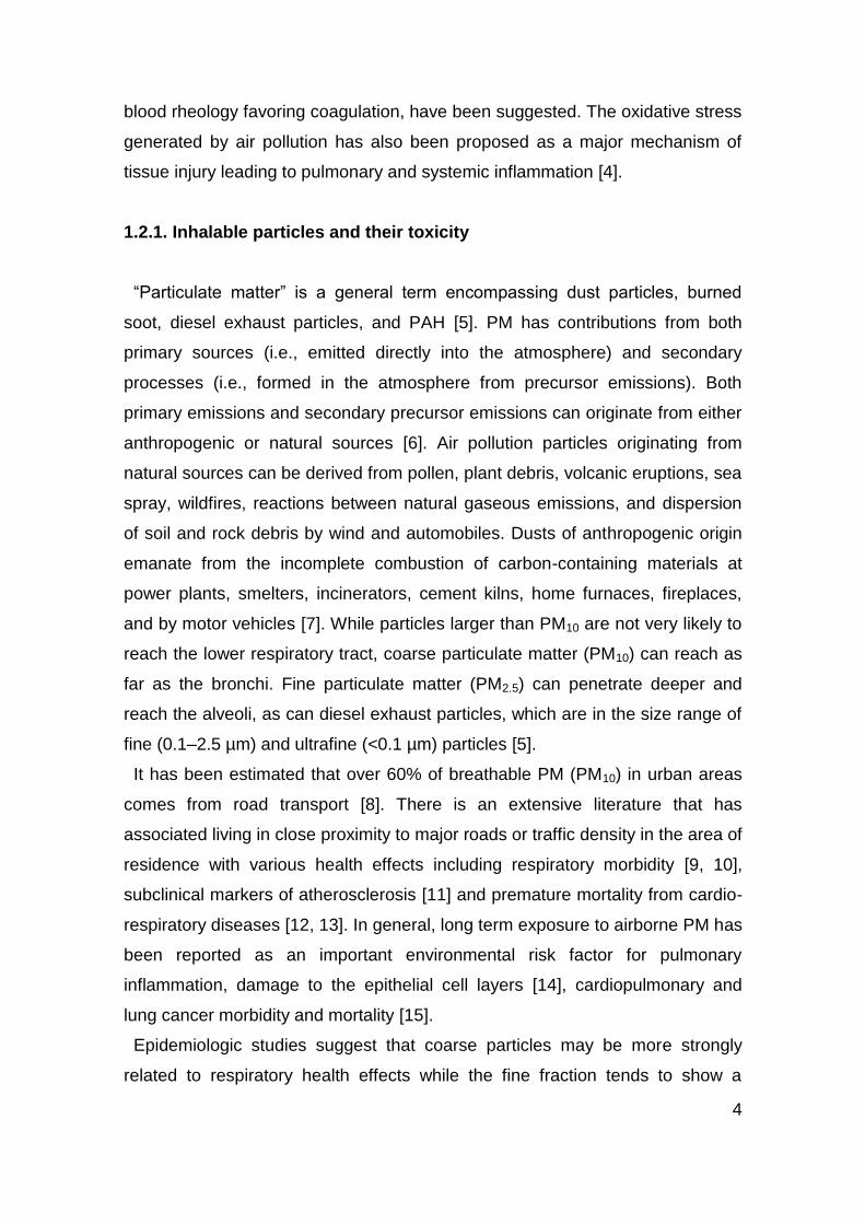
immune, neurological, reproductive, and respiratory systems) [1]. In Figure 1 it
can be seen the link between genotoxicity and ecotoxicological responses
resulting from exposure to contaminants/pollutants.
Fig. 1 - Linking genotoxicity with ecotoxicological responses. MN, micronuclei; CA,
chromosomal aberrations; SCE, sister chromatid exchanges [3].
According to Damaceno-Rodrigues et al. [4], the mechanisms by which air
pollution negatively influences some comorbidities are not well understood.
Possible mechanisms, such as increased calcium influx upon contact with
macrophages, upregulation of proinflammatory mediators, increased blood
viscosity, increased fibrinogen and C-reactive protein levels and alterations in
Exposure to Contaminants/Pollutants
Genotoxicological Responses Ecotoxicological Responses
Biochemical & Molecular Responses (DNA strand breaks, DNA adducts, comet assay point mutations, dimers, gene expressions)
Cytogenetical Responses (MN, CA, SCE)
Population level effects (Decreased population abundance & genotypic diversity)
Reproductive toxicity (Fertility & fecundity)
Developmental toxicity (Morphological, delayed growth & sexual maturity, tumours?)
Specific toxicity (Immunotoxicity, neurotoxicity, endocrine disruption)
Behavioural effects (Avoidance, locomotion, feeding)
Physiological effects (Osmoregulation, circulation, respiration, ventilation)
Biochemical, cellular & histological effects (Enzymes, lysosomes, stress proteins, ultrastructure)
4
blood rheology favoring coagulation, have been suggested. The oxidative stress
generated by air pollution has also been proposed as a major mechanism of
tissue injury leading to pulmonary and systemic inflammation [4].
1.2.1. Inhalable particles and their toxicity
“Particulate matter” is a general term encompassing dust particles, burned
soot, diesel exhaust particles, and PAH [5]. PM has contributions from both
primary sources (i.e., emitted directly into the atmosphere) and secondary
processes (i.e., formed in the atmosphere from precursor emissions). Both
primary emissions and secondary precursor emissions can originate from either
anthropogenic or natural sources [6]. Air pollution particles originating from
natural sources can be derived from pollen, plant debris, volcanic eruptions, sea
spray, wildfires, reactions between natural gaseous emissions, and dispersion
of soil and rock debris by wind and automobiles. Dusts of anthropogenic origin
emanate from the incomplete combustion of carbon-containing materials at
power plants, smelters, incinerators, cement kilns, home furnaces, fireplaces,
and by motor vehicles [7]. While particles larger than PM10 are not very likely to
reach the lower respiratory tract, coarse particulate matter (PM10) can reach as
far as the bronchi. Fine particulate matter (PM2.5) can penetrate deeper and
reach the alveoli, as can diesel exhaust particles, which are in the size range of
fine (0.1–2.5 µm) and ultrafine (<0.1 µm) particles [5].
It has been estimated that over 60% of breathable PM (PM10) in urban areas
comes from road transport [8]. There is an extensive literature that has
associated living in close proximity to major roads or traffic density in the area of
residence with various health effects including respiratory morbidity [9, 10],
subclinical markers of atherosclerosis [11] and premature mortality from cardio-
respiratory diseases [12, 13]. In general, long term exposure to airborne PM has
been reported as an important environmental risk factor for pulmonary
inflammation, damage to the epithelial cell layers [14], cardiopulmonary and
lung cancer morbidity and mortality [15].
Epidemiologic studies suggest that coarse particles may be more strongly
related to respiratory health effects while the fine fraction tends to show a
5
stronger association with cardiovascular disease and mortality while instillation
of PM size fractions shows that PM10 has more inflammatory and cytotoxicity
potential than the fine PM [8]. However, the picture is not clear and the literature
describing morbidity and mortality in response to specific size fractions varies
regionally [16]. Concentrations and properties of PM also vary with time,
season, and climate. Current research suggests that the possible toxic and pro-
inflammatory effects of PM are strongly influenced by its constituents, which
include biological agents [17], metals [16], and organic compounds [18]. Other
physico-chemical properties besides size and composition, such as mass,
number, and available surface area, are also important properties that may
determine the health impact of particulate pollutants [19, 20].
PM is also composed of a heterogeneous mixture of both inorganic and
carbonaceous materials, rich in transition metals, PAH and other compounds as
previously reported [21-23].
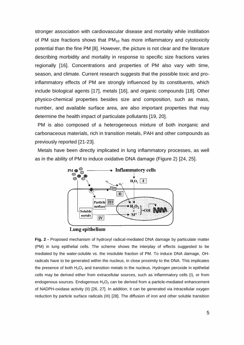
Metals have been directly implicated in lung inflammatory processes, as well
as in the ability of PM to induce oxidative DNA damage (Figure 2) [24, 25].
Fig. 2 - Proposed mechanism of hydroxyl radical-mediated DNA damage by particulate matter
(PM) in lung epithelial cells. The scheme shows the interplay of effects suggested to be
mediated by the water-soluble vs. the insoluble fraction of PM. To induce DNA damage, OH-
radicals have to be generated within the nucleus, in close proximity to the DNA. This implicates
the presence of both H2O2 and transition metals in the nucleus. Hydrogen peroxide in epithelial
cells may be derived either from extracellular sources, such as inflammatory cells (I), or from
endogenous sources. Endogenous H2O2 can be derived from a particle-mediated enhancement
of NADPH-oxidase activity (II) [26, 27]. In addition, it can be generated via intracellular oxygen
reduction by particle surface radicals (III) [28]. The diffusion of iron and other soluble transition

6
metals (M+), abundantly available from PM (IV), will then provide an optimal condition for
enhanced Fenton reaction-mediated •OH generation within the nucleus [25].
DNA oxidation is known to be one of the most common kinds of damage to
human DNA. It is mainly induced by reactive oxygen species (ROS), which
include free radicals and other highly reactive forms of oxygen (e.g. hydrogen
peroxide, superoxide anion radical, singlet oxygen, hydroxyl radical, nitric oxide
and peroxynitrite). ROS are produced in cells during normal metabolic
processes involving oxygen. They are released during cellular respiration,
processes of biosynthesis and biodegradation, biotransformation of xenobiotics
and phagocyte activation. There are about 60 enzymatic reactions that use O2
as a substrate where ROS are formed. However, the presence of ROS may be
significantly increased by exposure to different environmental toxins produced
from industry, agriculture, tobacco smoke, or pollution accidents [29].
In case of high toxin exposure, elevated levels of ROS or depressed
antioxidant defences, the excess of ROS may result in an increase in the
steady-state level of unrepaired cellular DNA damage. For example, exposure
to the environmental toxicant arsenic induces oxidative stress in the form of
increased levels of ROS. In excess, they can overwhelm the normal antioxidant
buffering capacity of the cell, leading to significant damage to cellular
components, including proteins, lipids and DNA [30]. Thus ROS contribute to
formation of mutations and, subsequently, to the etiology of degenerative
diseases such as cancer [29].
Some metals like cadmium, chromium, and nickel salts have been shown to
have aneugenic and/or clastogenic properties, inducing micronuclei (MN) in
human lung derived fibroblasts [31].
Other important and earlier referred compounds associated with airborne
particles are PAH, among which several are established carcinogens [32].
Carcinogenic polycyclic aromatic hydrocarbons (c-PAH) have recently become
the center of attention owing to their potential effect on the etiology of cancer,
respiratory and cardiovascular diseases and mortality [15]. c-PAH are
components of organic matter bound to inhalable aerosol particles (<2.5 µm).
Reactive intermediates, which can cause direct DNA damage, are generated by
7
their degradation. Moreover, they can induce a reaction with other pollutants,
including nitrogen oxides and ozone, and generate other potential carcinogens
[33].
In order to assess the genotoxicity of urban air pollution, several studies have
been conducted and the majority of them have used the Salmonella
mutagenicity assay or Ames test (reviewed in [34, 35]). Most of the mutagenicity
of PM in the Ames test has been associated mainly with PAH, nitro-PAH, and
polar compounds, such as aromatic amines and aromatic ketones [36, 37]. The
genotoxic effects of ambient PAH and their association with air PM or with
organic extracts have been also reported in mammalian cells by measuring
DNA adducts, DNA damage, MN, or chromosomal aberrations (CA) [38-42].
1.3. Bioindicators of environmental pollution
Monitoring the environment for genotoxic contaminants can have two
purposes: either assessing the risk of those contaminants for the animals and
plants normally inhabiting the environment and for the biological integrity of the
environment; or assessing the risk to humans, for example from food crops
grown on contaminated soil, or from consumption of contaminated water.
Essentially different criteria apply. When discussing the environment and its
inhabitants, we are interested in populations, and effects on reproductive
success, on the food chain, and on relative numbers of individuals of different
species. In contrast, when considering risk to humans, the health of the
individual is seen as of paramount importance [29].
Environmental biomonitoring – measuring biomarkers such as DNA damage
and repair with the comet assay – can make use of “sentinel” organisms living
in their natural habitat and reflecting long-term, continuous exposure.
Alternatively, animals or plants raised in a “clean” site or in the laboratory can
be exposed to the contaminated environment for limited periods of time, and the
change in biomarkers monitored. Organisms used in these tests have included
worms, molluscs, insects, fish, small mammals, and higher and lower plants
[29].
8
Studies of small mammals, mainly free-living wild rodents, have demonstrated
an ability to accumulate a wide spectrum of pollutants [43, 44]. Thus, they are
suitable for monitoring environmental pollution and exposure risk for people
living in a contaminated area [45, 46] in addition to being usually abundant over
easily identified areas and rapidly trapped [47].
1.3.1. The Algerian mouse (Mus spretus) as a model of in situ exposure
By obvious reasons, field conditions are always difficult to mimic in lab
experiments, especially when wild organisms are exposed to complex mixtures
of contaminants where synergistic and/or antagonistic effects are to be
expected. Additionally, available models for exposure and bioaccumulation
prediction can introduce considerable uncertainty into screening-level ecological
risk assessment [48]. Thus, in order to overcome these constraints, the Algerian
mouse (Mus spretus) could be the chosen specie because it satisfies the
criteria to be considered a good indicator, namely: (i) it has a large geographic
distribution and can be found in both contaminated and non-contaminated
areas; (ii) it may cohabit with humans which makes it a potential indicator of
human exposure; (iii) it is a component of some terrestrial ecosystems and
occupies a middle position in many food chains; (iv) it has small home ranges,
typically less than 90 m, which makes it an appropriate site-specific indicator of
contamination; and finally, (v) its population is usually large enough to support
harvesting without a major adverse effect at the population level [49, 50].
Mus spretus is a common rodent specie widely distributed in Portugal [51] and
is the best characterized aboriginal specie [52]. It is a non protected rodent that
lives in marshlands as the predominant small mammal [43], and feeds on
plants, seeds and insects around its burrow [53]. It has been demonstrated that
Mus spretus is genetically related to the sequenced mouse Mus musculus
(classical inbred laboratory specie) [54].

9
1.4. Biomarkers of environmental toxicity
When it comes to monitoring for genotoxicity in the environment, an issue of
paramount importance is the proper selection of representative organisms as
sentinels, as well as the performance of sensitive and reliable tests such as
those designed for the evaluation of DNA damage. Regardless of the particular
features of the contamination event as a whole, it is also very important that the
assay(s) of choice has been fully validated by laboratories worldwide and can
be used to monitor virtually any potentially endangered wild species [55].
Cytogenetic biomarkers have been a valuable tool for studying the most
important occupational and environmental hazards to public health occurring in
the past few decades. The use of valid biomarkers of risk in populations
exposed to agents inducing genetic damage is the most suitable approach for
studying many modern exposures, where the low doses or the complexity of
mixtures make traditional epidemiologic studies poorly informative [56]. Their
sensitivity for measuring exposure to genotoxic agents and their role as early
predictors of cancer risk have contributed to their success. As a result, several
markers have been identified to monitor the exposure of living beings to
mutagens and carcinogens [57].
Classically genotoxicity endpoints evaluate CA, MN, sister chromatid
exchanges (SCE) and DNA damage (e.g., strand breaks, crosslinking, alkali-
labile sites) [57]. Many drugs and environmental agents can damage DNA, and
understanding and predicting the outcomes of exposure to DNA-damaging
agents remains an important challenge in pharmaceutical development and in
environmental health sciences [58].
These endpoints can be considered biomarkers of effects and include
alterations of physiology, biochemistry, cell structure or function directly
attributable to exposure to a xenobiotic substance (e.g., CA, MN, DNA
damage), which are the hallmarks of molecular epidemiology [57]. MN
represent permanent damage, in which a chromosome breakage or
malsegregation is implicated, while comet assay, which detects DNA damage,
can be repaired. These genotoxic bioassays are used to assess different, but
complementary genotoxic effects [59].
10
Exposure biomarkers include determination of xenobiotic agents or associated
metabolites in biological fluids that can reflect the extent of internal exposure
(e.g., DNA-adducts, etc.) [57].
Closely related to these biomarkers are biomarkers of susceptibility, which
indicate increased vulnerability of individuals to diseases such as cancer (e.g.,
GSTM1 polymorphisms) [57]. Genetic polymorphisms are possible modifiers for
health impacts of air pollution. Mutations in exons can produce genes that are
inactive or whose substrate specificity is altered. Mutations in adjacent regions
or introns can affect the regulation of mRNA transcription or splicing and result
in hyperinducibility or protein stability alterations [60].
1.4.1. Sperm shape abnormalities assay
Ambient air pollution has been associated with a variety of health effects,
ranging from subclinical outcomes to death [61]. More recently, the effects of air
pollution on reproductive and birth outcomes have garnered increased interest
[62-64]. However, a limited amount of research has been conducted to examine
the association between air pollution and male reproductive outcomes,
specifically semen quality, which includes sperm count and concentration along
with morphologic and chromatin abnormalities [65].
Current studies, as reviewed by Sheiner et al. [66] and Jensen et al. [67] show
that a variety of environmental and occupational exposures may impair male
fertility. During past years the male reproductive function has been addressed in
relation to a number of environmental exposures that have only to a very limited
extent been investigated or reviewed earlier. These exposures and conditions
include air pollution and drinking water pollutants, biopersistent
organochlorines, trihalomethanes, phthalates and high frequency
electromagnetic radiation related to use of mobile phones [2].
A limited number of animal toxicologic studies have provided preliminary
evidence of associations between exposure to air pollutants and semen quality
outcomes. Associations have been observed between total air pollution and
reduced daily sperm production in mice and rats receiving in utero or prenatal
exposure to total diesel exhaust and filtered exhaust [68, 69]. These

11
observations are not limited to exposure durations timed to occur before or after
birth, but also have been observed in adult mice exposed to diesel exhaust for
up to 6 months [70].
It has also been shown that sperm morphology is correlated with sperm DNA
damage since morphological abnormalities of sperm indicate elevated levels of
sperm DNA fragmentation [71-73]. Even though we cannot precisely identify all
the specific components of air pollution responsible for sperm DNA damage, the
most likely biological agents that cause the damage are c-PAH present in the
PM fraction. Studies performed on animals indicated that c-PAH and their
metabolites can accumulate in the testes and epididymis and cause impaired
spermiogenesis [74, 75].
The biological mechanisms linking ambient air pollution to decreased sperm
quality [65], like reduced percentage of sperm with normal morphology [2], have
yet to be determined. Sokol et al. [76] identified several possible mechanisms,
including O3-induced oxidative stress, inflammatory reactions, and the induction
of the formation of circulating toxic species.
1.4.2. Micronucleus test
Currently, one of the most robust tests for genotoxicity is the in vivo MN assay,
which is conducted by using an improved version of the method first described
by Schmid [77].
The hematopoietic system is highly sensitive to genotoxic agents, in part
because hematopoietic cells undergo rapid division. That means that close to
70% of known human carcinogens are detected by the in vivo MN test [58].
Due to clastogenicity (chromosomal breaking) or aneugenicity (mitotic spindle
dysfunction) [78] cells undergo enucleation but form immature or polychromatic
erythrocytes (PCE) that contain “micronuclei”, additional small nuclei derived
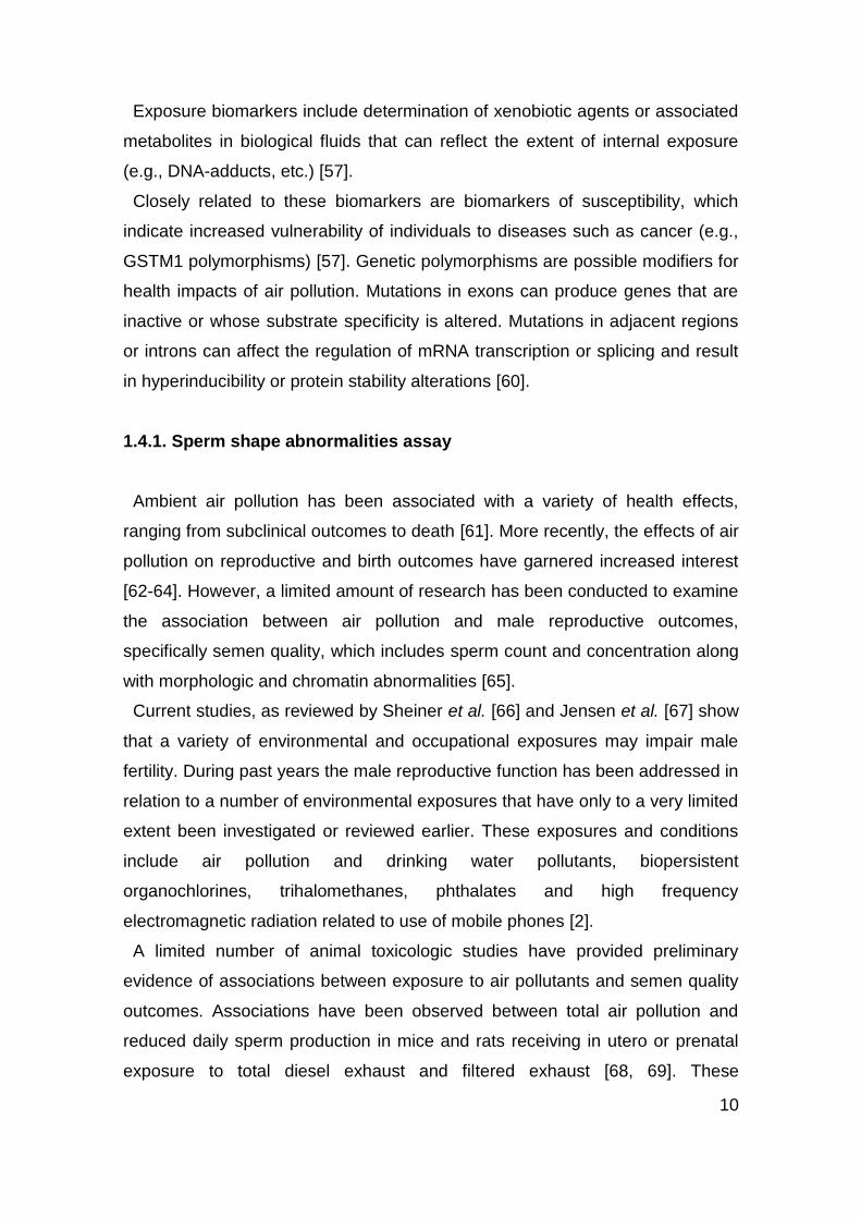
from acentric chromosomal fragments or whole chromosomes (Figure 3) [79].
The spleen clears damaged erythrocytes, so normally <1% of circulating
erythrocytes contain spontaneous MN arising from background levels of DNA
damage [58].
12
Fig. 3 - Mechanism of micronucleus (MN) formation in polychromatic erythrocytes (PCE) and
normochromatic erythrocytes (NCE). N, nucleus; PEB, proerythroblast [80].
It has been demonstrated to respond to a large number of experimental and
environmental carcinogenic pollutants, such as PAH [81, 82], heavy metals [81],
and pesticides [83]. MN induction is also an early biomarker of the health
impairment that long-term PM exposure could produce [59].
Hayashi et al. [84] introduced a new method for the observation of
micronucleated young erythrocytes using supravital staining with acridine
orange (AO).
AO is a nucleic-acid-selective fluorescent cationic dye, which emits green light
(525 nm) when exited (502 nm) only if bound to DNA. Using AO, MN can be
scored selectively in immature erythrocytes [85, 86]. AO maximum excitation
shifts to 460 nm when bound with RNA and the maximum emission shifts to the
red (650 nm). A red-stained cytoplasm is then characteristic of an immature
RNA-containing cell. However, immature erythrocyte frequency depends on the
erythropoiesis intensity and is low if the organism exhibits little mitosis. DNA-
specific staining reduces false-positive MN scoring due to artefacts. This
provides higher reliability and sensitivity for the assay. In addition, the scoring
process is faster. The AO staining procedure is now routinely used in rodent MN
assay [87, 88].
13
1.4.3. Comet assay
Primary DNA damage is considered to be an important initial event in
carcinogenesis [89]. The comet assay is one of the most commonly used
methods in environmental toxicology for assessing DNA damage [29].
It was first introduced by Ostling and Johanson in 1984. This was a neutral
version of the comet assay, and interestingly, they used quite sophisticated
techniques of image analysis for quantification of the comets, using AO as the
DNA binding dye [90]. Singh et al. [91] developed the alkaline version of the
comet assay in which they used the length of DNA migration (tail length) to
quantify the extent of damage.
In this assay, a suspension of cells is mixed with low melting point agarose
and spread onto a microscope glass slide [90]. After lysis of cells with a solution
containing Triton X-100, to break down membranes, and high salt, to remove
histones and other soluble proteins [92], DNA unwinding and electrophoresis is
carried out at a specific pH. Unwinding of the DNA and electrophoresis at
neutral pH (7-8) predominantly facilitates the detection of double strand breaks
and cross links; unwinding and electrophoresis at pH 12.1-12.4 facilitates the
detection of single and double strand breaks, incomplete excision repair sites
and cross-links [93]; whereas unwinding and electrophoresis at a pH greater
than 12.6 expresses alkali labile lesions (i.e., apurinic/apirimidinic sites) that are
converted to strand breaks under alkaline conditions [89], in addition to all types
of lesions listed above [93]. When subjected to an electric field, the DNA
migrates out of the cell, in the direction of the anode, appearing like a “comet”
[90], with a concentration of DNA at the “head” and a diffused trailing migration
of DNA referred to as the “tail” [57], when viewed by fluorescence microscopy
with a suitable stain, and the proportion of DNA in the tail indicates the
frequency of breaks (Figure 4) [92].
The comet assay is now a well-established, simple, versatile, rapid, visual, and
a sensitive, extensively used tool to assess DNA damage and repair
quantitatively as well as qualitatively in individual cell populations [94].

14
Fig. 4 - General principles of the comet assay [92].
A limitation of the comet assay is that aneugenic effects, which may be a
possible mechanism for carcinogenicity, and epigenetic mechanisms (indirect)
of DNA damage such as effects on cell-cycle checkpoints, base oxidation and
DNA adduct formation are not detected. The specific and sensitive detection of
these lesions requires the use of lesion-specific enzymes [95]. The other
drawbacks such as single cell data (which may be rate limiting), small cell
sample (leading to sample bias), technical variability, and interpretation are
some of its disadvantages. However, its advantages far outnumber the
disadvantages, and hence, it has been widely used in fields ranging from
molecular epidemiology to genetic toxicology [96].
The comet assay is widely considered to be a powerful technique for
investigating effects of environmental mutagens on humans, animals or cells
[29].
Human lymphocytes are easily collected and are assumed to be
representative of the overall status of the body. Therefore they are widely used
in the comet assay to monitor human exposure to genotoxic agents as a result
of occupation, drug treatment, diseases or environmental pollution [29].

15
In vivo comet assay in rodents is an important test model for genotoxicity
studies, as many rodent carcinogens are also human carcinogens, and hence,
this model not only provides an insight into the genotoxicity of human
carcinogens but also is suited for studying their underlying mechanisms [96].
Different routes of exposure in rodents have been used, e.g., intraperitoneal
[97, 98], oral [99, 100], and inhalation [101, 102], to study the genotoxicity of
different chemicals. The route of exposure is an important determinant of the
genotoxicity of a chemical due to its mode of action [103]. The in vivo comet
assay helps in hazard identification and assessment of dose–response
relationships as well as mechanistic understanding of a substance’s mode of
action. Besides being used for testing the genotoxicity of chemicals in
laboratory-reared animals, comet assay in wild mice can be used as a valuable
test in pollution monitoring and environmental conservation [55].
1.4.4. CYP1A1 gene polymorphisms
In everyday life, human body is exposed to a vast number of xenobiotics
including drugs, dietary compounds, or environmental carcinogens, which are
metabolized by a variety of enzymes through phase I (oxidative) and phase II
(conjugative) reactions, in addition to a small number of endogenous
substrates. The major enzymes of phase I metabolism are heme thiolate
proteins of the cytochrome P450 superfamily (CYPs). These enzymes
participate mainly in the conversion of xenobiotics to more polar and water-
soluble metabolites which are readily excreted from the body. During
metabolism of certain xenobiotics, a variety of unstable and reactive
intermediates can be formed, such as epoxide intermediates, which attack
DNA, causing cell toxicity and transformation [104, 105]. Individuals differ in
levels of expression and catalytic activities of metabolic enzymes that activate
and/or detoxify xenobiotics in various organs, and these phenomena are
thought to be critical in understanding the background of interindividual
differences in response to xenobiotics. Factors affecting these variations include
induction and inhibition of enzymes by diverse chemicals and by genetic
polymorphisms. Inherited DNA sequence variations in genes coding for
16
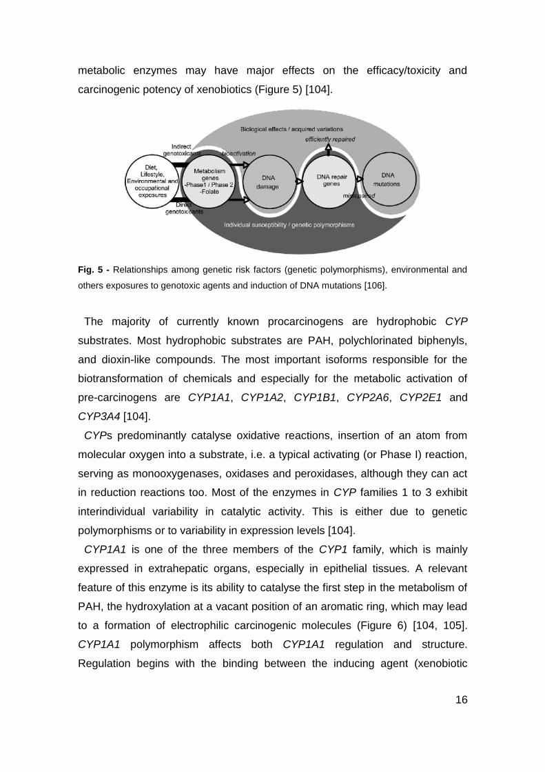
metabolic enzymes may have major effects on the efficacy/toxicity and
carcinogenic potency of xenobiotics (Figure 5) [104].
Fig. 5 - Relationships among genetic risk factors (genetic polymorphisms), environmental and
others exposures to genotoxic agents and induction of DNA mutations [106].
The majority of currently known procarcinogens are hydrophobic CYP
substrates. Most hydrophobic substrates are PAH, polychlorinated biphenyls,
and dioxin-like compounds. The most important isoforms responsible for the
biotransformation of chemicals and especially for the metabolic activation of
pre-carcinogens are CYP1A1, CYP1A2, CYP1B1, CYP2A6, CYP2E1 and
CYP3A4 [104].
CYPs predominantly catalyse oxidative reactions, insertion of an atom from
molecular oxygen into a substrate, i.e. a typical activating (or Phase I) reaction,
serving as monooxygenases, oxidases and peroxidases, although they can act
in reduction reactions too. Most of the enzymes in CYP families 1 to 3 exhibit
interindividual variability in catalytic activity. This is either due to genetic
polymorphisms or to variability in expression levels [104].
CYP1A1 is one of the three members of the CYP1 family, which is mainly
expressed in extrahepatic organs, especially in epithelial tissues. A relevant
feature of this enzyme is its ability to catalyse the first step in the metabolism of
PAH, the hydroxylation at a vacant position of an aromatic ring, which may lead
to a formation of electrophilic carcinogenic molecules (Figure 6) [104, 105].
CYP1A1 polymorphism affects both CYP1A1 regulation and structure.
Regulation begins with the binding between the inducing agent (xenobiotic
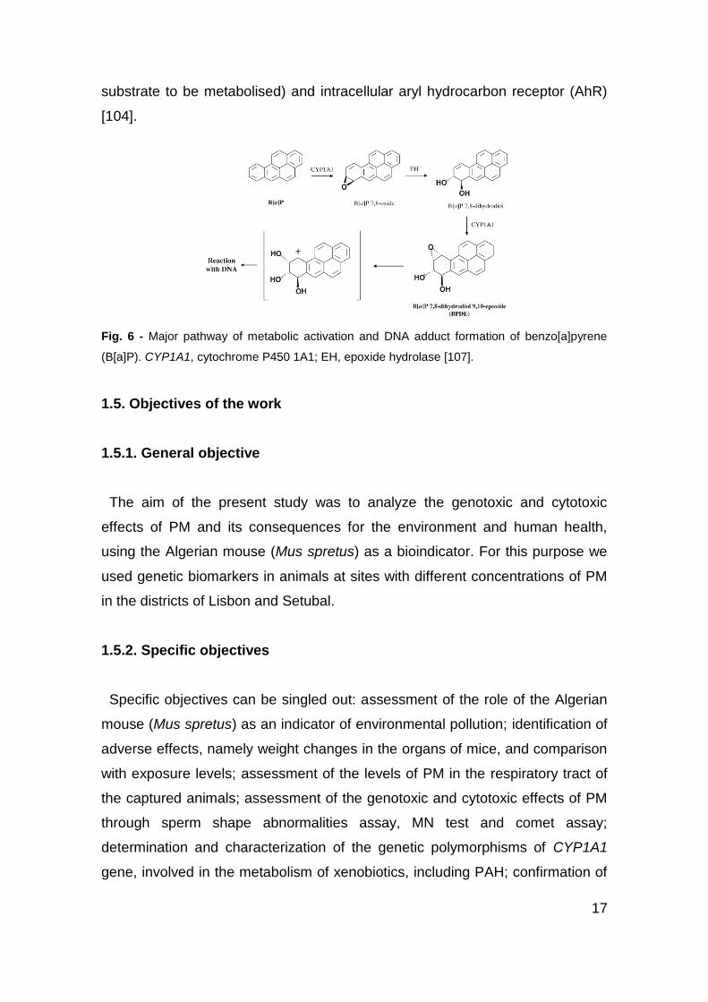
17
substrate to be metabolised) and intracellular aryl hydrocarbon receptor (AhR)
[104].
Fig. 6 - Major pathway of metabolic activation and DNA adduct formation of benzo[a]pyrene
(B[a]P). CYP1A1, cytochrome P450 1A1; EH, epoxide hydrolase [107].
1.5. Objectives of the work
1.5.1. General objective
The aim of the present study was to analyze the genotoxic and cytotoxic
effects of PM and its consequences for the environment and human health,
using the Algerian mouse (Mus spretus) as a bioindicator. For this purpose we
used genetic biomarkers in animals at sites with different concentrations of PM
in the districts of Lisbon and Setubal.
1.5.2. Specific objectives
Specific objectives can be singled out: assessment of the role of the Algerian
mouse (Mus spretus) as an indicator of environmental pollution; identification of
adverse effects, namely weight changes in the organs of mice, and comparison
with exposure levels; assessment of the levels of PM in the respiratory tract of
the captured animals; assessment of the genotoxic and cytotoxic effects of PM
through sperm shape abnormalities assay, MN test and comet assay;
determination and characterization of the genetic polymorphisms of CYP1A1
gene, involved in the metabolism of xenobiotics, including PAH; confirmation of

18
risk to public and environmental health caused by PM pollution. Figure 7
outlines the multiple-endpoint assay planned for this work.
Fig. 7 - Schematic of a multiple-endpoint assay combining quantification and/or qualification of
biologically external or internal PM with cytogenetic tests and genetic polymorphisms. PM,
particulate matter; EM, electron microscopy.
Air monitoring
data
EM observation
and microanalysis
Sperm shape
abnormalities assay
Micronucleus test
Comet assay CYP1A1
polymorphisms
PM
19
2. Materials and Methods
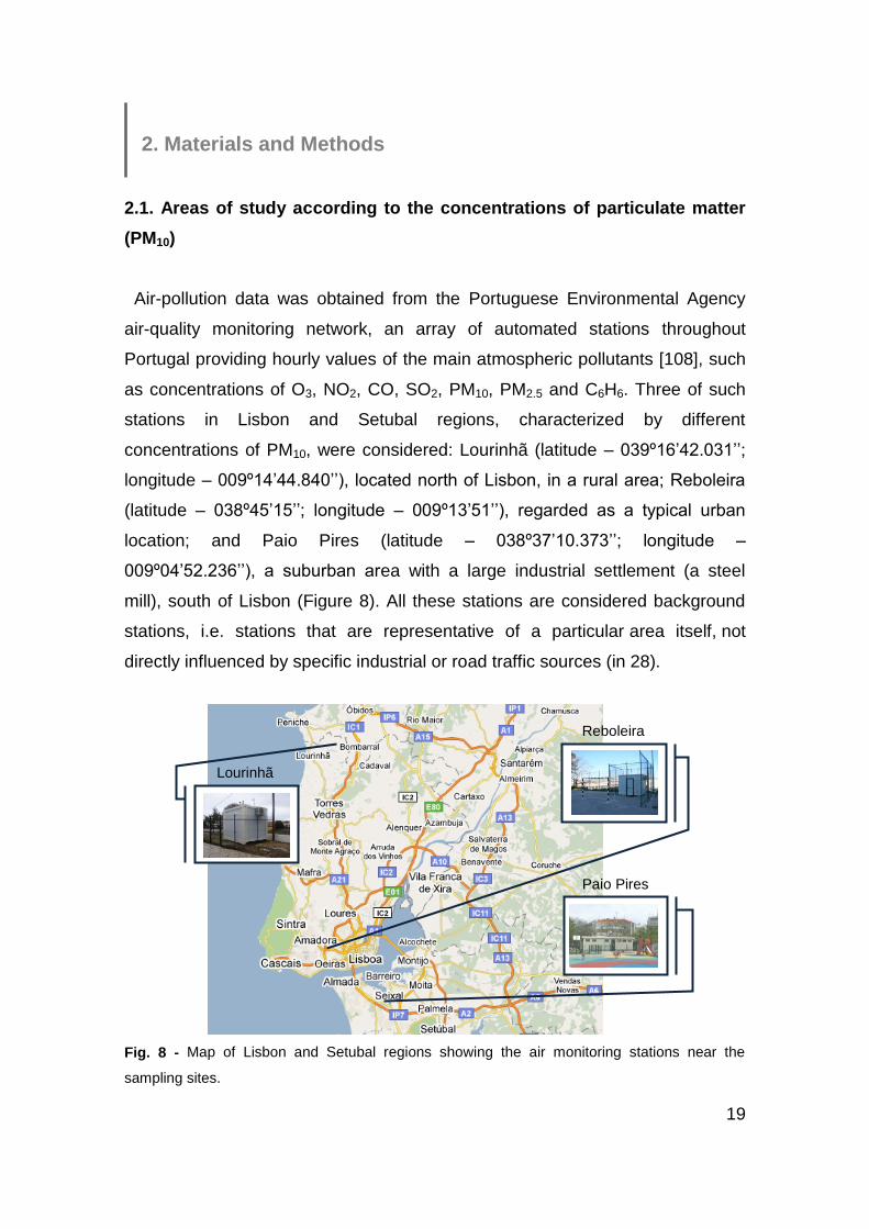
2.1. Areas of study according to the concentrations of particulate matter
(PM10)
Air-pollution data was obtained from the Portuguese Environmental Agency
air-quality monitoring network, an array of automated stations throughout
Portugal providing hourly values of the main atmospheric pollutants [108], such
as concentrations of O3, NO2, CO, SO2, PM10, PM2.5 and C6H6. Three of such
stations in Lisbon and Setubal regions, characterized by different
concentrations of PM10, were considered: Lourinhã (latitude – 039º16’42.031’’;
longitude – 009º14’44.840’’), located north of Lisbon, in a rural area; Reboleira
(latitude – 038º45’15’’; longitude – 009º13’51’’), regarded as a typical urban
location; and Paio Pires (latitude – 038º37’10.373’’; longitude –
009º04’52.236’’), a suburban area with a large industrial settlement (a steel
mill), south of Lisbon (Figure 8). All these stations are considered background
stations, i.e. stations that are representative of a particular area itself, not
directly influenced by specific industrial or road traffic sources (in 28).
Fig. 8 - Map of Lisbon and Setubal regions showing the air monitoring stations near the
sampling sites.
Lourinhã
Reboleira
Paio Pires

20
The average concentrations (μg/m3) of PM10 were obtained between July and
December 20107 (Table 1). As, for these six months, the average concentration
of PM10 in Lourinhã was 17.6 μg/m3, in Reboleira was 19.9 μg/m3 and in Paio
Pires was 40.5 μg/m3, the first will represent the control area, the second the
moderately polluted area and the third the polluted area in question. In Figure 9
we can see a graphical and more detailed representation of this data.
Table 1 - PM10 data for the three study sites between 1 July and 31 December 2010 (µg/m3).
Lourinhã Reboleira Paio Pires
Mean ± SD 17.6 ± 9.4 19.9 ± 11.9 40.5 ± 15.9
Range 3–60 4–92 12–107
SD, standard deviation.
Fig. 9 - Variation of concentrations of particulate matter (PM10) in Lourinhã, Reboleira and Paio
Pires stations between July and December 2010.
7 www.qualar.org/
21
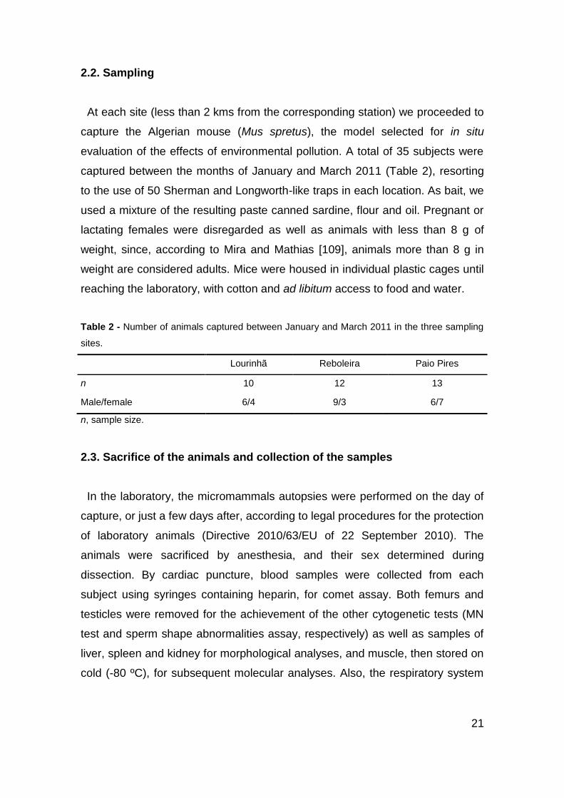
2.2. Sampling
At each site (less than 2 kms from the corresponding station) we proceeded to
capture the Algerian mouse (Mus spretus), the model selected for in situ
evaluation of the effects of environmental pollution. A total of 35 subjects were
captured between the months of January and March 2011 (Table 2), resorting
to the use of 50 Sherman and Longworth-like traps in each location. As bait, we
used a mixture of the resulting paste canned sardine, flour and oil. Pregnant or
lactating females were disregarded as well as animals with less than 8 g of
weight, since, according to Mira and Mathias [109], animals more than 8 g in
weight are considered adults. Mice were housed in individual plastic cages until
reaching the laboratory, with cotton and ad libitum access to food and water.
Table 2 - Number of animals captured between January and March 2011 in the three sampling
sites.
Lourinhã Reboleira Paio Pires
n 10 12 13
Male/female 6/4 9/3 6/7
n, sample size.
2.3. Sacrifice of the animals and collection of the samples
In the laboratory, the micromammals autopsies were performed on the day of
capture, or just a few days after, according to legal procedures for the protection
of laboratory animals (Directive 2010/63/EU of 22 September 2010). The
animals were sacrificed by anesthesia, and their sex determined during
dissection. By cardiac puncture, blood samples were collected from each
subject using syringes containing heparin, for comet assay. Both femurs and
testicles were removed for the achievement of the other cytogenetic tests (MN
test and sperm shape abnormalities assay, respectively) as well as samples of
liver, spleen and kidney for morphological analyses, and muscle, then stored on
cold (-80 ºC), for subsequent molecular analyses. Also, the respiratory system

22
was removed for observing, in light and electron microscopy, the particles and
their deleterious effects in this organ.
2.4. Morphological analysis
The animals were weighed and total body, tail, ear and hind limb lengths were
determined. The organs (liver, spleen, kidneys and testicles) were removed,
weighed and their relative weight to body weight was calculated. It was also
measured the length and breadth of the spleen and in the case of females, the
number of scars in the oviducts was counted, indicating the number of pups
already procreated.
2.5. Sperm shape abnormalities assay
The cauda epididymis of the mice was dissected out, placed in 2 mL of
Sorensen buffer (pH 7.0) and gently centrifuged (800 rpm, 10 min) to obtain a
pellet of undamaged cells. After removal of the supernatant, the pellet was re-
suspended in 1 mL of Sorensen buffer. A drop of the suspension was placed on
a clean slide and a smear was made, air-dried and fixed in absolute methanol
for 10 min. After drying overnight the slides were stained with 10% Giemsa for 1
h [110] and observed under the microscope with a magnification of 100x.
According to Wyrobek and Bruce [111], 1000 sperm per animal were assessed
for morphological abnormalities, which included lack of the usual hook, banana-
like form, amorphous and two tails (Figure 10).
Fig. 10 - The shape of A is normal whereas B to E are abnormal murine sperm. Sperm in B lack
the usual hook, C have a banana-like form, D are amorphous and E possess two tails [111].
A B C D E
23
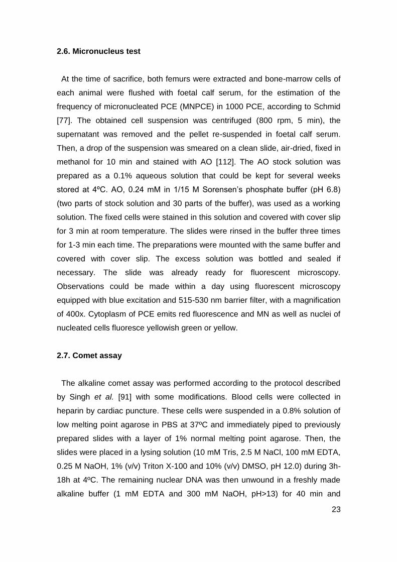
2.6. Micronucleus test
At the time of sacrifice, both femurs were extracted and bone-marrow cells of
each animal were flushed with foetal calf serum, for the estimation of the
frequency of micronucleated PCE (MNPCE) in 1000 PCE, according to Schmid
[77]. The obtained cell suspension was centrifuged (800 rpm, 5 min), the
supernatant was removed and the pellet re-suspended in foetal calf serum.
Then, a drop of the suspension was smeared on a clean slide, air-dried, fixed in
methanol for 10 min and stained with AO [112]. The AO stock solution was
prepared as a 0.1% aqueous solution that could be kept for several weeks
stored at 4ºC. AO, 0.24 mM in 1/15 M Sorensen’s phosphate buffer (pH 6.8)
(two parts of stock solution and 30 parts of the buffer), was used as a working
solution. The fixed cells were stained in this solution and covered with cover slip
for 3 min at room temperature. The slides were rinsed in the buffer three times
for 1-3 min each time. The preparations were mounted with the same buffer and
covered with cover slip. The excess solution was bottled and sealed if
necessary. The slide was already ready for fluorescent microscopy.
Observations could be made within a day using fluorescent microscopy
equipped with blue excitation and 515-530 nm barrier filter, with a magnification
of 400x. Cytoplasm of PCE emits red fluorescence and MN as well as nuclei of
nucleated cells fluoresce yellowish green or yellow.
2.7. Comet assay
The alkaline comet assay was performed according to the protocol described
by Singh et al. [91] with some modifications. Blood cells were collected in
heparin by cardiac puncture. These cells were suspended in a 0.8% solution of
low melting point agarose in PBS at 37ºC and immediately piped to previously
prepared slides with a layer of 1% normal melting point agarose. Then, the
slides were placed in a lysing solution (10 mM Tris, 2.5 M NaCl, 100 mM EDTA,
0.25 M NaOH, 1% (v/v) Triton X-100 and 10% (v/v) DMSO, pH 12.0) during 3h-
18h at 4ºC. The remaining nuclear DNA was then unwound in a freshly made
alkaline buffer (1 mM EDTA and 300 mM NaOH, pH>13) for 40 min and

24
electrophoresis occurred in the same buffer for 30 min at 25V and 300mA while
the electrophoresis tank was packed with ice packs to maintain cold conditions.
After neutralization with 3 x 5 min washes with 0.4 mM Tris (pH 7.5), in order to
make way for the removal of salts and detergent, cells were fixed in absolute
alcohol for 10 min. Slides were stained with a SYBR Safe solution (4 μL/mL).
The stained nuclei were counted visually with the support of a fluorescence
microscope (Olympus BX41) equipped with a video camera (Leica). 200 comets
were photographed per animal and visually scored into four classes, depending
on the size of the comet tail (Figure 11).
Fig. 11 - Scores assigned by visual scoring (0-4) [113].
2.8. CYP1A1 gene polymorphisms
The commercial Tissue DNA Kit (Omega bio-tek) was used to extract DNA
from twelve samples of muscle (six from Lourinhã and six from Paio Pires).
After extraction, the optimization of this technique to the samples in question
began, including the variation of some parameters, namely, regulation of
temperature used, duration of the different cycles and the amount of DNA in the
final solution. Subsequently, the samples were subjected to Polymerase Chain
Reaction (PCR), used to amplify DNA fragments using specific primers flanking
the region of interest. This study used the following primers and conditions
(Figure 12 and Table 3):

25
Fig. 12 - Diagram with the location of the amplified fragment and of exons 4–7 (rectangles) and
introns 4–6 (lines) in the mouse CYP1A1 gene. The 5’ and 3’ primers used for PCR analysis are
illustrated, which are designed to produce a genomic DNA PCR fragment of 702/705 bp.
Numbers above denote bp of the exons and introns that are included in the final PCR product
(adapted from [114]).
Table 3 - PCR conditions for CYP1A1.
PCR step Temperature (ºC) Duration (min)
Initial denaturation 94 5
Denaturation 94 1
Annealing 66 2
Extension 72 1
Final extension 72 5
Number of cycles 30
15.5 µL of water, 5 µL of buffer (1x), 1 µL of dNTPs (2.5 mM), 1.5 µL of
magnesium (1.5 mM), 0.2 µL of primers F and R (25 µM), 0.4 µL of BSA (0.16
µg/µL), 0.2 µL of Taq polymerase (5U/µL) and 1 µL of DNA were used, for a
total reaction volume of 25 µL per tube.
In addition two samples were sequenced (one from Lourinhã and one from
Paio Pires) to confirm the amplified fragment and fragments from other samples
of the same regions were then cut with two restriction enzymes, MseI and
EcoRI, which recognition sites are 5’ T|TAA 3’ and 5’ G|AATTC 3’, respectively.
Thus, the first should cut the fragment at two sites, forming three fragments and
the second should cut the fragment at one site, forming two fragments. 1 µL of
26
10x buffer, 1 µL of MseI or 0.5 µL of EcoRI and 6 or 6.5 µL of water were used,
respectively, plus 2 µL of DNA per reaction tube, for a total of 10 µL, then
incubated at 37ºC during 3h.
2.9. Whole lung lavage and lung tissue sampling
The lungs were excised and lavaged with 1.5 mL of phosphate-buffered saline
(pH 7.4) and the resulting lavage fluid was centrifuged for 10 min at 2500 rpm in
a desktop centrifuge. The pellet was glutaraldehyde-sorensen fixed for 2-3h and
processed using routine transmission electron microscopy (TEM) procedures
(without osmium for microanalysis). To observe the lung structure in semi-thin
sections and eventually the lung ultra-structure in TEM, a large portion of the
lung was excised into smaller pieces in a drop of cacodylate, glutaraldehyde-
cacodylate fixed for 2-3h and processed the same way. For general histological
analysis, a small portion of the lung was excised and formalin fixed up to 24
hours. Then, it was embedded in paraffin and processed using routine
histological procedures.
2.10. Statistical analysis
Statistical analyses were performed using SPSS v19.0 for Windows being the
results expressed as means ± standard deviation (SD). Student's t test for
independent samples was used, or Mann-Whitney test in case of non-normal
data, to test for equality of means between sexes and one-way ANOVA to test
all other parameters in case of verifying normality and homogeneity of
variances. When there was no normality the Kruskal-Wallis test was used and
when it occurred but the same did not happen with homogeneity of variances
the Brown-Forsythe test was used. In case the hypothesis of equality of means
was rejected it was used the Gabriel test, which assumes equal variances, or
the Games-Howell test, which assumes different variances, in order to explore
the differences between the groups. Thus, the normality test applied was the
Shapiro-Wilk test and the homogeneity of variances test applied was Levene’s
test. The level of statistical significance for all tests was set at p < 0.05.

27
For sequence edition, alignment and analysis the Sequencher 4.8 and BioEdit
programs were used.

28
3. Results
After completion of the Student's t test for independent samples, or Mann-
Whitney test in case of non-normal data, it was found that there are no
significant differences between sexes for all measurements and tests, so the
results were not analyzed separately (data not shown).
3.1. Morphological analysis
Statistics of the external and internal biometric measurements in the three
populations of Mus spretus studied, Lourinhã, Reboleira and Paio Pires, are
represented in Table 4 and the box plots of this data can be seen in Figures 13,
14 and 15.
Table 4 - External and internal biometric measurements in the three populations
of Mus spretus.
Lourinhã Reboleira Paio Pires
n 10 12 13
Body length (cm) 7.22 ± 0.28 7.12 ± 0.40 7.50 ± 0.38b
Relative tail length (mm/cm body length)d
7.93 ± 0.40h 8.17 ± 0.43 7.76 ± 0.20
bi
Relative paw length (mm/cm body length) 2.07 ± 0.11 2.23 ± 0.15 2.01 ± 0.24b
Relative ear length (mm/cm body length) 1.62 ± 0.10 1.72 ± 0.14 1.65 ± 0.15
Body weight (g) 11.95 ± 1.34 12.08 ± 1.24 13.64 ± 1.35ab
Relative liver weight (mg/g body weight) 57.19 ± 7.35 69.17 ± 10.39a
74.38 ± 9.99a
Relative spleen weight (mg/g body weight)c
2.96 ± 0.89 2.78 ± 1.21 4.04 ± 1.79
Relative left kidney weight (mg/g body weight) 8.39 ± 0.73 9.14 ± 1.48 9.45 ± 1.65
Relative right kidney weight (mg/g body weight) 8.53 ± 0.77 9.43 ± 1.59 9.59 ± 1.24
Relative left testis weight (mg/g body weight) 5.65 ± 0.40g
5.99 ± 1.40h
7.47 ± 1.00ag
Relative right testis weight (mg/g body weight) 5.66 ± 0.66g
6.04 ± 1.25h
7.35 ± 1.04ag
Number of scars of the left oviduct 2.25 ± 2.63f
1.33 ± 1.53e
2.50 ± 1.52g
Number of scars of the right oviduct 1.75 ± 2.36f
2.33 ± 2.08e
2.00 ± 1.41g
n, sample size; a
Statistically different from Lourinhã (p < 0.05); b
Statistically different from
Reboleira (p < 0.05); c p-value determined by Kruskal-Wallis test (otherwise by one-way ANOVA
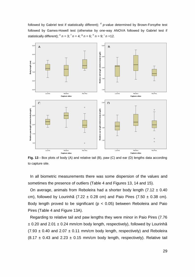
29
followed by Gabriel test if statistically different); d p-value determined by Brown-Forsythe test
followed by Games-Howell test (otherwise by one-way ANOVA followed by Gabriel test if
statistically different); e n = 3;
f n = 4;
g n = 6;
h n = 9;
i n =12.
Fig. 13 - Box plots of body (A) and relative tail (B), paw (C) and ear (D) lengths data according
to capture site.
In all biometric measurements there was some dispersion of the values and
sometimes the presence of outliers (Table 4 and Figures 13, 14 and 15).
On average, animals from Reboleira had a shorter body length (7.12 ± 0.40
cm), followed by Lourinhã (7.22 ± 0.28 cm) and Paio Pires (7.50 ± 0.38 cm).
Body length proved to be significant (p < 0.05) between Reboleira and Paio
Pires (Table 4 and Figure 13A).
Regarding to relative tail and paw lengths they were minor in Paio Pires (7.76
± 0.20 and 2.01 ± 0.24 mm/cm body length, respectively), followed by Lourinhã
(7.93 ± 0.40 and 2.07 ± 0.11 mm/cm body length, respectively) and Reboleira
(8.17 ± 0.43 and 2.23 ± 0.15 mm/cm body length, respectively). Relative tail
A B
C D
30
length also proved to be significant (p < 0.05) as well as relative paw length
between Reboleira and Paio Pires (Table 4 and Figures 13B and C).
Relative ear length showed no significant differences between the three
groups, Lourinhã having however lower values (1.62 ± 0.10 mm/cm body
length), followed by Paio Pires (1.65 ± 0.15 mm/cm body length) and Reboleira
(1.72 ± 0.14 mm/cm body length; Table 4 and Figure 13D).
Fig. 14 - Box plots of body (A) and relative liver (B), spleen (C) and left (D) and right kidneys (E)
weights data according to capture site.
A B
C D
E
31
On average, the body weight from Lourinhã was lower (11.95 ± 1.34 g) than
Reboleira (12.08 ± 1.24 g) and the later was lower than Paio Pires (13.64 ± 1.35
g), and there were significant differences (p < 0.05) between Lourinhã and Paio
Pires and Reboleira and Paio Pires (Table 4 and Figure 14A).
The relative liver weight is lower in Lourinhã (57.19 ± 7.35 mg/g body weight)
and successively larger in Reboleira (69.17 ± 10.39 mg/g body weight) and Paio
Pires (74.38 ± 9.99 mg/g body weight). It appears that there are significant
differences (p < 0.05) between Lourinhã and Reboleira and Lourinhã and Paio
Pires (Table 4 and Figure 14B).
The relative spleen weight in Reboleira is lower (2.78 ± 1.21 mg/g body
weight), followed by Lourinhã (2.96 ± 0.89 mg/g body weight) and Paio Pires
(4.04 ± 1.79 mg/g body weight). There were no significant differences between
the three groups (Table 4 and Figure 14C).
Regarding to relative left and right kidneys weights, these were lower in
Lourinhã (8.39 ± 0.73 and 8.53 ± 0.77 mg/g body weight, respectively) and
successively higher in Reboleira (9.14 ± 1.48 and 9.43 ± 1.59 mg/g body
weight, respectively) and in Paio Pires (9.45 ± 1.65 and 9.59 ± 1.24 mg/g body
weight, respectively), however, no significant differences were found between
the three groups (Table 4 and Figures 14D and E).
Finally, with regard to reproductive organs, the relative left and right testis
weights were lower in Lourinhã (5.65 ± 0.40 and 5.66 ± 0.66 mg/g body weight)
and higher in Reboleira (5.99 ± 1.40 and 6.04 ± 1.25mg/g body weight) and
Paio Pires (7.47 ± 1.00 and 7.35 ± 1.04 mg/g body weight), and there were
significant differences (p < 0.05) between Lourinhã and Paio Pires (Table 4 and
Figures 15A and B).
The number of scars of the left oviduct was lower in Reboleira (1.33 ± 1.53)
and higher in Lourinhã (2.25 ± 2.63) and Paio Pires (2.50 ± 1.52) and the
number of scars of the right oviduct was lower in Lourinhã (1.75 ± 2.36) and
higher in Paio Pires (2.00 ± 1.41) and Reboleira (2.33 ± 2.08), and no significant
differences were observed between groups in both cases (Table 4 and Figures
15C and D).

32
Fig. 15 - Box plots of relative left (A) and right testis (B) weights and number of scars of the left
(C) and right oviducts (D) data according to capture site.
3.2. Sperm shape abnormalities assay
Table 5 - Different types of sperm shape abnormalities and frequency of total abnormal sperm
in 1000 sperm cells per animal.
Lourinhã Reboleira Paio Pires
n 6 9 6
Hook less 13.83 ± 5.31 20.67 ± 9.85 16.50 ± 9.65
Bananaa 1.33 ± 2.42 11.44 ± 12.36 2.33 ± 3.67
Amorphous 15.83 ± 9.22 8.44 ± 6.27 10.33 ± 6.47
Two tailsa 0.33 ± 0.82 0.00 ± 0.00 0.17 ± 0.41
Total abnormal sperm (%) 3.13 ± 1.12 4.06 ± 2.15 2.93 ± 1.62
n, sample size; a p-value determined by Kruskal-Wallis test (otherwise by one-way ANOVA).
A B
C
D
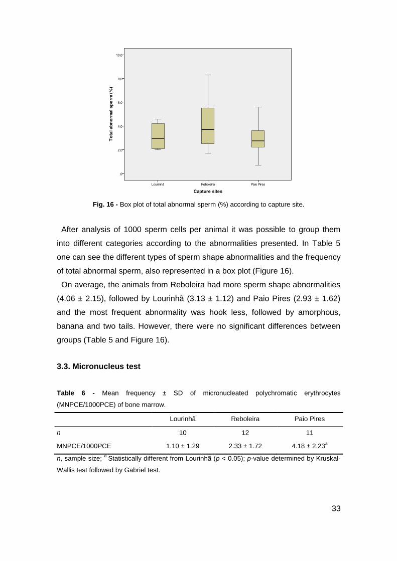
33
Fig. 16 - Box plot of total abnormal sperm (%) according to capture site.
After analysis of 1000 sperm cells per animal it was possible to group them
into different categories according to the abnormalities presented. In Table 5
one can see the different types of sperm shape abnormalities and the frequency
of total abnormal sperm, also represented in a box plot (Figure 16).
On average, the animals from Reboleira had more sperm shape abnormalities
(4.06 ± 2.15), followed by Lourinhã (3.13 ± 1.12) and Paio Pires (2.93 ± 1.62)
and the most frequent abnormality was hook less, followed by amorphous,
banana and two tails. However, there were no significant differences between
groups (Table 5 and Figure 16).
3.3. Micronucleus test
Table 6 - Mean frequency ± SD of micronucleated polychromatic erythrocytes
(MNPCE/1000PCE) of bone marrow.
Lourinhã Reboleira Paio Pires
n 10 12 11
MNPCE/1000PCE 1.10 ± 1.29 2.33 ± 1.72 4.18 ± 2.23a
n, sample size; a
Statistically different from Lourinhã (p < 0.05); p-value determined by Kruskal-
Wallis test followed by Gabriel test.
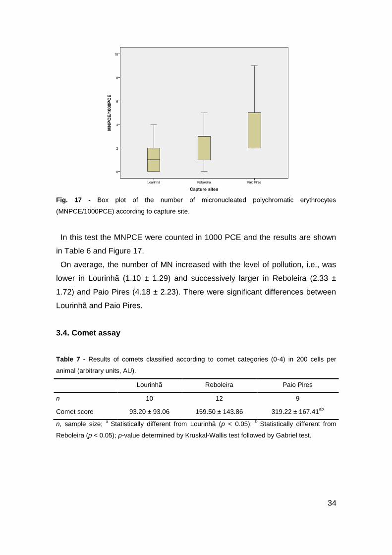
34
Fig. 17 - Box plot of the number of micronucleated polychromatic erythrocytes
(MNPCE/1000PCE) according to capture site.
In this test the MNPCE were counted in 1000 PCE and the results are shown
in Table 6 and Figure 17.
On average, the number of MN increased with the level of pollution, i.e., was
lower in Lourinhã (1.10 ± 1.29) and successively larger in Reboleira (2.33 ±
1.72) and Paio Pires (4.18 ± 2.23). There were significant differences between
Lourinhã and Paio Pires.
3.4. Comet assay
Table 7 - Results of comets classified according to comet categories (0-4) in 200 cells per
animal (arbitrary units, AU).
Lourinhã Reboleira Paio Pires
n 10 12 9
Comet score 93.20 ± 93.06 159.50 ± 143.86 319.22 ± 167.41ab
n, sample size; a
Statistically different from Lourinhã (p < 0.05); b
Statistically different from
Reboleira (p < 0.05); p-value determined by Kruskal-Wallis test followed by Gabriel test.

35
Fig. 18 - Box plot of the comet score obtained through the classification in four classes of 200
comets per animal depending on the size of the comet tail according to capture site.
In Table 7 and Figure 18 one can observe the results of the comet score
obtained through the classification in four classes of 200 comets per animal
depending on the size of the comet tail for the three capture sites.
It was found that the comet score also increased with the level of pollution,
being lower in Lourinhã (93.20 ± 93.06), followed by Reboleira (159.50 ±
143.86) and Paio Pires (319.22 ± 167.41). Results showed to be significantly
different between Lourinhã and Paio Pires and Reboleira and Paio Pires.
3.5. CYP1A1 gene polymorphisms
After the optimization of the PCR technique the CYP1A1 gene fragment could
be amplified, and bands of about 700 bp were obtained (Figure 19).
Two of these fragments were then sequenced (one from Lourinhã and another
from Paio Pires), and the amplified fragment could then be confirmed. It was
also found that both DNA samples had a frameshift mutation caused by an indel
of 2 bp in the second intron, at positions 382 and 383.
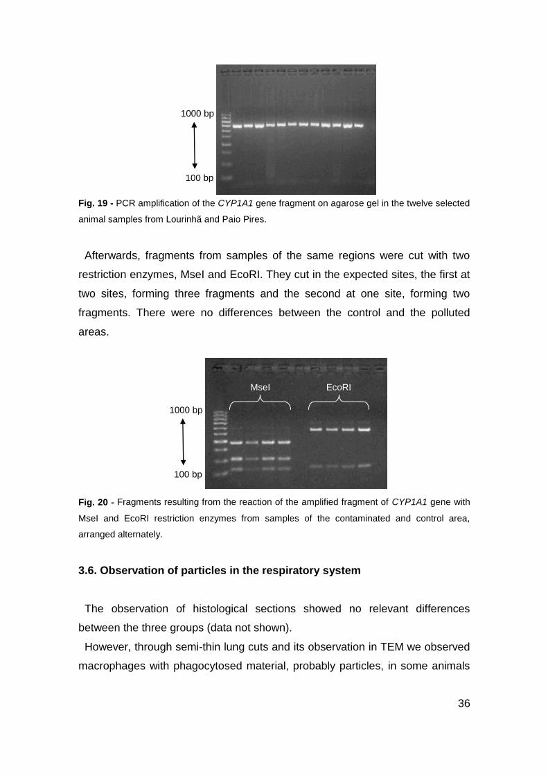
36
Fig. 19 - PCR amplification of the CYP1A1 gene fragment on agarose gel in the twelve selected
animal samples from Lourinhã and Paio Pires.
Afterwards, fragments from samples of the same regions were cut with two
restriction enzymes, MseI and EcoRI. They cut in the expected sites, the first at
two sites, forming three fragments and the second at one site, forming two
fragments. There were no differences between the control and the polluted
areas.
Fig. 20 - Fragments resulting from the reaction of the amplified fragment of CYP1A1 gene with
MseI and EcoRI restriction enzymes from samples of the contaminated and control area,
arranged alternately.
3.6. Observation of particles in the respiratory system
The observation of histological sections showed no relevant differences
between the three groups (data not shown).
However, through semi-thin lung cuts and its observation in TEM we observed
macrophages with phagocytosed material, probably particles, in some animals
1000 bp
100 bp
MseI EcoRI
1000 bp
100 bp

37
from Paio Pires and eventually also in animals from Reboleira, which did not
happen in those from Lourinhã (Figures 21 and 22).
Fig. 21 - Semi-thin lung cuts, observing (A) macrophages without phagocytosed material
(arrows) from an individual from Lourinhã and (B) macrophages with phagocytosed material
(arrows), probably particles, from an individual from Paio Pires.
Fig. 22 - Preliminary transmission electron microscopy observations of lung tissue (A) reveals
the presence of lisossome-filled (arrows) macrophages (B and C) that may contain ingested
particulate matter.
A B
38
4. Discussion
Since, in most cases, the general population is exposed to many potentially
dangerous agents simultaneously, predicting health and environmental risk is a
very complicated problem, because a mixture of genotoxic chemicals may
undergo a variety of interactions, which can affect the transport, metabolism or
molecular binding of the components. Thus, the biological effects of complex
environmental mixtures, especially genotoxicity, seem to be very informative for
risk assessment [55].
Relatively few studies have investigated the effects of different PM sources on
adverse effects under “real-world” exposure condition. These studies indicate
that PM of different origins may be associated with differential biological effects
in different organs and adverse outcomes, but how such source factors may
affect different organ systems in humans is unknown [115].
When comparing the concentrations of PM10, obtained from Lourinhã,
Reboleira and Paio Pires stations, with the classes of concentration associated
with the air quality index, we can state that the concentrations of the first are
usually low, unlike the last one that has very high values (see Table 1). For a
time interval of 6 months, it was recorded from Paio Pires station concentrations
that exceeded the average daily value of 50 μg/m3, a value that, according to
Decree-Law nr 102/2010 of 23 September 2010, should not be exceeded more
than 35 days a year, with an average annual value of 40 μg/m3, which did not
come to pass. On the other hand, Lourinhã and Reboleira stations only
exceeded the set values two to three times with a reasonable semi-annual
average.
So, this study aimed at evaluating the risk of environmental contamination,
particularly by PM, to contribute to the understanding of the effects of these
pollutants on the ecosystem and, more specifically, on human health. For this
purpose, it was used as a bioindicator of environmental pollution the Algerian
mouse (Mus spretus), which, besides giving us indications about the quality and
quantity of pollutants inhaled, gives us information about the impact of pollution
on the physiology of living beings. In addition to morphological analysis we used
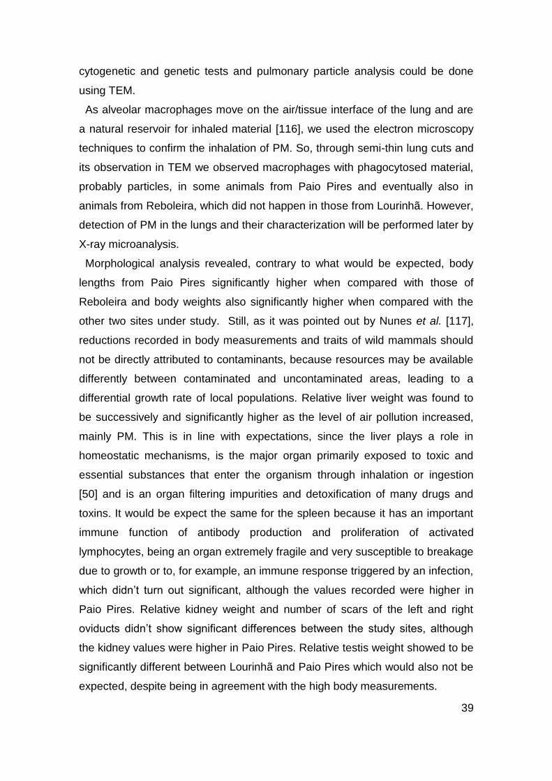
39
cytogenetic and genetic tests and pulmonary particle analysis could be done
using TEM.
As alveolar macrophages move on the air/tissue interface of the lung and are
a natural reservoir for inhaled material [116], we used the electron microscopy
techniques to confirm the inhalation of PM. So, through semi-thin lung cuts and
its observation in TEM we observed macrophages with phagocytosed material,
probably particles, in some animals from Paio Pires and eventually also in
animals from Reboleira, which did not happen in those from Lourinhã. However,
detection of PM in the lungs and their characterization will be performed later by
X-ray microanalysis.
Morphological analysis revealed, contrary to what would be expected, body
lengths from Paio Pires significantly higher when compared with those of
Reboleira and body weights also significantly higher when compared with the
other two sites under study. Still, as it was pointed out by Nunes et al. [117],
reductions recorded in body measurements and traits of wild mammals should
not be directly attributed to contaminants, because resources may be available
differently between contaminated and uncontaminated areas, leading to a
differential growth rate of local populations. Relative liver weight was found to
be successively and significantly higher as the level of air pollution increased,
mainly PM. This is in line with expectations, since the liver plays a role in
homeostatic mechanisms, is the major organ primarily exposed to toxic and
essential substances that enter the organism through inhalation or ingestion
[50] and is an organ filtering impurities and detoxification of many drugs and
toxins. It would be expect the same for the spleen because it has an important
immune function of antibody production and proliferation of activated
lymphocytes, being an organ extremely fragile and very susceptible to breakage
due to growth or to, for example, an immune response triggered by an infection,
which didn’t turn out significant, although the values recorded were higher in
Paio Pires. Relative kidney weight and number of scars of the left and right
oviducts didn’t show significant differences between the study sites, although
the kidney values were higher in Paio Pires. Relative testis weight showed to be
significantly different between Lourinhã and Paio Pires which would also not be
expected, despite being in agreement with the high body measurements.

40
Relatively to genotoxic tests, the results obtained were not always the
expected. In the sperm shape abnormalities assay there was no correlation
between the level of pollution recorded and the number of abnormal sperm, and
all results were not significant. This was unexpected although the results of
Hansen et al. [65] also show that, in studying men, there was generally no
consistent pattern of increased abnormal sperm quality with elevated exposure
to PM. These findings may reinforce the idea that this test should be optimized
or, ultimately, be replaced by others more reliable, since it is very subjective and
has many technical artefacts. In addition, the usefulness of rodents as a model
for humans is limited because of major differences between testis development
and spermatogenesis in these two species, as shown in Table 8. In addition,
only a low percentage of the sperm that is actually produced in normal young
men can be classified as normal (5–15% depending on how strict the criteria of
normality used is), which is remarkably lower than in domestic (bull, ram) or
laboratory (rat, mouse) animals where more than 90 per cent of sperm can
usually be classified as normal [118]. On the contrary, the frequency of MN and
the comet score were successively larger as the level of pollution increased,
which is in agreement with Soares et al. [119] that showed that the exposure of
rodents to urban air pollution elicited a significant increase of MN frequency,
and with Zhang et al. [120] that showed that the intratracheal instillation of PM
in rats can cause DNA damage of cells, i.e. DNA strand breakage. So, there
were significant differences between Lourinhã and Paio Pires in the number of
MN and in relation to the comet score there were significant differences
between Lourinhã and Paio Pires and Reboleira and Paio Pires. From these
results we can confirm that both the MN test, primarily through the use of AO as
a staining method, and the comet assay are sensitive enough to detect the
possible effects of genotoxic agents.
41
Table 8 - Comparison of the phases, pace and other features of testis development and
spermatogenesis in rodents and humans relevant to sperm count/quality in adulthood and which
may affect predisposition to lifestyle/environmental effects and limit usefulness of rodents as a
model for humans [118].
To determination and characterization of CYP1A1 polymorphisms we
proceeded to optimize the PCR technique, which was successfully achieved
using the mouse primers available for use. Two of the 700 bp fragments
obtained were then sequenced (one from Lourinhã and another from Paio
Pires), being confirmed the amplified fragment. It was also found that both DNA
samples had a frameshift mutation caused by an indel of 2 bp in the second
intron, at positions 382 and 383, not presenting significant differences between
the two locations. Then, fragments from samples of the same regions were cut
with two restriction enzymes, MseI and EcoRI, to search for any possible
polymorphism. They cut at the expected sites, the first at two sites, forming
three fragments and the second at one site, forming two fragments. There were
also no differences between the control and the polluted areas. In this test we
used a small number of samples due to economic constraint, so one cannot
draw conclusions. In the future, one must use a much larger number of samples
in order to try finding polymorphisms that may be associated with polluted and
unpolluted areas and possibly perform also gene expression tests, since most
CYPs involved in the biotransformation of xenobiotics are inducible [121].
Activation of CYPs as well as phase II and phase III proteins is stimulated by
increased cellular amounts of environmental xenobiotics, which may result in
higher protein expression and subsequently in lower amounts of xenobiotics
[104].

42
5. Conclusions
Through this study we can conclude that inhalation of PM at high
concentrations has a significant genotoxic risk which can induce DNA damage
and consequently lead to serious health problems.
Although this work is directed at studying the effects that PM has on the health
of living beings, it is natural that other xenobiotics may be responsible for some
of the adverse effects recorded. However, although we cannot manipulate and
control many variables, this type of study is very useful in analyzing the effects
of pollution as a whole and exactly the way it affects us. In the laboratory we
can control the variables, but we only observe the effects that each pollutant
can have and not the effects of the interaction between all of them, in addition to
the fact that the doses of pollutants administered can sometimes be in very
different amounts of those registered in a real environment.
The analysis by X-ray microanalysis of the possible particles present in
macrophages from the lungs of the animals caught in the polluted area can
provide more information about the composition of the particles in question and
their association with the genotoxic effects recorded.
The genotoxicity tests used proved to be effective in detecting genotoxic
effects at the level of air pollution, with the exception of the sperm shape
abnormalities assay. Analysis of CYP1A1 gene polymorphisms should be
developed through the use of more animals and different primers and enzymes
or expression studies could also be undertaken.
Our results may provide valuable insight into the health risks posed by urban
and industrial particles, thus, alerting us of the need to use biomarkers of
effects, exposure and susceptibility and to establish preventing measures
before we are faced with the social burden and economic cost of PM pollution
induced disease. In this work we used Mus spretus as bioindicator specie of the
effects that environmental pollution, specifically PM, may have on health, being
possible, within certain limits and always with caution, to transfer this data to
humans.

43
I hope that this work, like so many others that go in the same direction, can
show us how necessary it is to change ideas and actions, since pollution today
will not only have its influences in a distant future but is already having
consequences in human health and also in the health of all other living beings
that inhabit our ecosystem.
44
6. References
[1] L.M. Schell, M. Denham, Environmental pollution in urban environments and human biology,
Annual Review of Anthropology, 32 (2003) 111-134.
[2] J. Jurewicz, W. Hanke, M. Radwan, J.P. Bonde, Environmental factors and semen quality,
International Journal of Occupational Medicine and Environmental Health, 22 (2009) 305-329.
[3] A.N. Jha, Ecotoxicological applications and significance of the comet assay, Mutagenesis,
23 (2008) 207-221.
[4] N.R. Damaceno-Rodrigues, M.M. Veras, E.M. Negri, A.C. Tedesco Zanchi, C.R. Rhoden,
P.H. Nascimento Saldiva, M. Dolhnikoff, E.G. Caldini, Effect of pre- and postnatal exposure to
urban air pollution on myocardial lipid peroxidation levels in adult mice, Inhalation Toxicology,
21 (2009) 1129-1137.
[5] C. Minelli, I. Wei, G. Sagoo, D. Jarvis, S. Shaheen, P. Burney, Interactive Effects of
Antioxidant Genes and Air Pollution on Respiratory Function and Airway Disease: A HuGE
Review, American Journal of Epidemiology, 173 (2011) 603-620.
[6] L.W. Stanek, J.S. Brown, J. Stanek, J. Gift, D.L. Costa, Air Pollution Toxicology-A Brief
Review of the Role of the Science in Shaping the Current Understanding of Air Pollution Health
Risks, Toxicological Sciences, 120 (2011) S8-S27.
[7] A.J. Ghio, A. Hall, M.A. Bassett, W.E. Cascio, R.B. Devlin, Exposure to concentrated
ambient air particles alters hematologic indices in humans, Inhalation Toxicology, 15 (2003)
1465-1478.
[8] P. Mantecca, F. Farina, E. Moschini, D. Gallinotti, M. Gualtieri, A. Rohr, G. Sancini, P.
Palestini, M. Camatini, Comparative acute lung inflammation induced by atmospheric PM and
size-fractionated tire particles, Toxicology Letters, 198 (2010) 244-254.
[9] W.J. Gauderman, H. Vora, R. McConnell, K. Berhane, F. Gilliland, D. Thomas, F. Lurmann,
E. Avoli, N. Kunzli, M. Jerrett, J. Peters, Effect of exposure to traffic on lung development from
10 to 18 years of age: a cohort study, Lancet, 369 (2007) 571-577.
[10] N.A.H. Janssen, B. Brunekreef, P. van Vliet, F. Aarts, K. Meliefste, H. Harssema, P.
Fischer, The relationship between air pollution from heavy traffic and allergic sensitization,
bronchial hyperresponsiveness, and respiratory symptoms in Dutch schoolchildren,
Environmental Health Perspectives, 111 (2003) 1512-1518.
[11] B. Hoffmann, S. Moebus, S. Mohlenkamp, A. Stang, N. Lehmann, N. Dragano, A.
Schmermund, M. Memmesheimer, K. Mann, R. Erbel, K.H. Jockel, S. Heinz Nixdorf Recall,
Residential exposure to traffic is associated with coronary atherosclerosis, Circulation, 116
(2007) 489-496.
[12] R. Beelen, G. Hoek, P.A. van den Brandt, R.A. Goldbohm, P. Fischer, L.J. Schouten, M.
Jerrett, E. Hughes, B. Armstrong, B. Brunekreef, Long-term effects of traffic-related air pollution

45
on mortality in a Dutch cohort (NLCS-AIR study), Environmental Health Perspectives, 116
(2008) 196-202.
[13] M.M. Finkelstein, M. Jerrett, M.R. Sears, Traffic air pollution and mortality rate
advancement periods, American Journal of Epidemiology, 160 (2004) 173-177.
[14] M.R. Riley, D.E. Boesewetter, R.A. Turner, A.M. Kim, J.M. Collier, A. Hamilton, Comparison
of the sensitivity of three lung derived cell lines to metals from combustion derived particulate
matter, Toxicology in Vitro, 19 (2005) 411-419.
[15] C.A. Pope, R.T. Burnett, M.J. Thun, E.E. Calle, D. Krewski, K. Ito, G.D. Thurston, Lung
cancer, cardiopulmonary mortality, and long-term exposure to fine particulate air pollution,
Jama-Journal of the American Medical Association, 287 (2002) 1132-1141.
[16] K. Donaldson, N. Mills, W. MacNee, S. Robinson, D. Newby, Role of inflammation in
cardiopulmonary health effects of PM, Toxicol Appl Pharmacol, 207 (2005) 483-488.
[17] S. Becker, J.M. Soukup, M.I. Gilmour, R.B. Devlin, Stimulation of human and rat alveolar
macrophages by urban air particulates: Effects on oxidant radical generation and cytokine
production, Toxicology and Applied Pharmacology, 141 (1996) 637-648.
[18] N. Li, M.Y. Wang, T.D. Oberley, J.M. Sempf, A.E. Nel, Comparison of the pro-oxidative and
proinflammatory effects of organic diesel exhaust particle chemicals in bronchial epithelial cells
and macrophages, Journal of Immunology, 169 (2002) 4531-4541.
[19] K.L. Dreher, Particulate matter physicochemistry and toxicology: In search of causality - A
critical perspective, Inhalation Toxicology, 12 (2000) 45-57.
[20] K.E. Driscoll, D.L. Costa, G. Hatch, R. Henderson, G. Oberdorster, H. Salem, R.B.
Schlesinger, Intratracheal instillation as an exposure technique for the evaluation of respiratory
tract toxicity: Uses and limitations, Toxicological Sciences, 55 (2000) 24-35.
[21] M. Gualtieri, P. Mantecca, V. Corvaja, E. Longhin, M.G. Perrone, E. Bolzacchini, M.
Camatini, Winter fine particulate matter from Milan induces morphological and functional
alterations in human pulmonary epithelial cells (A549), Toxicology Letters, 188 (2009) 52-62.
[22] M. Gualtieri, J. Ovrevik, J.A. Holme, M.G. Perrone, E. Bolzacchini, P.E. Schwarze, M.
Camatini, Differences in cytotoxicity versus pro-inflammatory potency of different PM fractions in
human epithelial lung cells, Toxicology in Vitro, 24 (2010) 29-39.
[23] M.G. Perrone, M. Gualtieri, L. Ferrero, C. Lo Porto, R. Udisti, E. Bolzacchini, M. Camatini,
Seasonal variations in chemical composition and in vitro biological effects of fine PM from Milan,
Chemosphere, 78 (2010) 1368-1377.
[24] D.L. Costa, K.L. Dreher, Bioavailable transition metals in particulate matter mediate
cardiopulmonary injury in healthy and compromised animal models, Environmental Health
Perspectives, 105 (1997) 1053-1060.
[25] A.M. Knaapen, T.M. Shi, P.J.A. Borm, R.P.F. Schins, Soluble metals as well as the
insoluble particle fraction are involved in cellular DNA damage induced by particulate matter,
Molecular and Cellular Biochemistry, 234 (2002) 317-326.
[26] M. Perderiset, J.P. Marsh, B.T. Mossman, Activation of protein-kinase-c by crocidolite
asbestos in hamster tracheal epithelial-cells, Carcinogenesis, 12 (1991) 1499-1502.

46
[27] G. Gercken, I. Berg, M. Dorger, T. Schluter, Mechanisms of particle-induced activation of
alveolar macrophages, Toxicology Letters, 88 (1996) 121-129.
[28] B. Dellinger, W.A. Pryor, R. Cueto, G.L. Squadrito, V. Hegde, W.A. Deutsch, Role of free
radicals in the toxicity of airborne fine particulate matter, Chemical Research in Toxicology, 14
(2001) 1371-1377.
[29] A. Azqueta, S. Shaposhnikov, A.R. Collins, DNA oxidation: Investigating its key role in
environmental mutagenesis with the comet assay, Mutation Research-Genetic Toxicology and
Environmental Mutagenesis, 674 (2009) 101-108.
[30] L.H. Ma, C.L. Takanishi, M.J. Wood, Molecular mechanism of oxidative stress perception
by the Orp1 protein, Journal of Biological Chemistry, 282 (2007) 31429-31436.
[31] A.I. Seoane, F.N. Dulout, Genotoxic ability of cadmium, chromium and nickel salts studied
by kinetochore staining in the cytokinesis-blocked micronucleus assay, Mutation Research-
Genetic Toxicology and Environmental Mutagenesis, 490 (2001) 99-106.
[32] C.E. Bostrom, P. Gerde, A. Hanberg, B. Jernstrom, C. Johansson, T. Kyrklund, A. Rannug,
M. Tornqvist, K. Victorin, R. Westerholm, Cancer risk assessment, indicators, and guidelines for
polycyclic aromatic hydrocarbons in the ambient air, Environmental Health Perspectives, 110
(2002) 451-488.
[33] P. Georgiadis, J. Topinka, M. Stoikidou, S. Kaila, M. Gioka, K. Katsouyanni, R. Sram, H.
Autrup, S.A. Kyrtopoulos, A. Network, Biomarkers of genotoxicity of air pollution (the AULIS
project): bulky DNA adducts in subjects with moderate to low exposures to airborne polycyclic
aromatic hydrocarbons and their relationship to environmental tobacco smoke and other
parameters, Carcinogenesis, 22 (2001) 1447-1457.
[34] L.D. Claxton, P.P. Matthews, S.H. Warren, The genotoxicity of ambient outdoor air, a
review: Salmonella mutagenicity, Mutation Research-Reviews in Mutation Research, 567 (2004)
347-399.
[35] L.D. Claxton, G.M. Woodall, A review of the mutagenicity and rodent carcinogenicity of
ambient air, Mutation Research-Reviews in Mutation Research, 636 (2007) 36-94.
[36] M. Cerna, D. Pochmanova, A. Pastorkova, I. Benes, J. Lenicek, J. Topinka, B. Binkova,
Genotoxicity of urban air pollutants in the Czech Republic Part I. Bacterial mutagenic potencies
of organic compounds adsorbed on PM10 particulates, Mutation Research-Genetic Toxicology
and Environmental Mutagenesis, 469 (2000) 71-82.
[37] V.A. Du Four, N. Van Larebeke, C.R. Janssen, Genotoxic and mutagenic activity of
environmental air samples in Flanders, Belgium, Mutation Research-Genetic Toxicology and
Environmental Mutagenesis, 558 (2004) 155-167.
[38] J. Topinka, L.R. Schwarz, F.J. Wiebel, M. Cerna, T. Wolff, Genotoxicity of urban air
pollutants in the Czech Republic Part II. DNA adduct formation in mammalian cells by
extractable organic matter, Mutation Research-Genetic Toxicology and Environmental
Mutagenesis, 469 (2000) 83-93.

47
[39] X.S. Zhao, Z. Wan, G. Chen, H.G. Zhu, S.H. Jiang, J.Q. Yao, Genotoxic activity of
extractable organic matter from urban airborne particles in Shanghai, China, Mutation
Research-Genetic Toxicology and Environmental Mutagenesis, 514 (2002) 177-192.
[40] A. Gabelova, Z. Valovicova, E. Horvathova, D. Slamenova, B. Binkova, R.J. Sram, P.B.
Farmer, Genotoxicity of environmental air pollution in three European cities: Prague, Kosice and
Sofia, Mutation Research-Genetic Toxicology and Environmental Mutagenesis, 563 (2004) 49-
59.
[41] S. Motta, C. Federico, S. Saccone, V. Librando, P. Mosesso, Cytogenetic evaluation of
extractable agents from airborne particulate matter generated in the city of Catania (Italy),
Mutation Research-Genetic Toxicology and Environmental Mutagenesis, 561 (2004) 45-52.
[42] T.M. de Kok, J.G. Hogervorst, J.J. Briede, M.H. van Herwijnen, L.M. Maas, E.J. Moonen,
H.A. Driece, J.C. Kleinjans, Genotoxicity and physicochemical characteristics of traffic-related
ambient particulate matter, Environmental and Molecular Mutagenesis, 46 (2005) 71-80.
[43] L.A. Ieradi, S. Moreno, J.P. Bolivar, A. Cappai, A. Di Benedetto, M. Cristaldi, Free-living
rodents as bioindicators of genetic risk in natural protected areas, Environmental Pollution, 102
(1998) 265-268.
[44] S. Wijnhoven, R.S.E.W. Leuven, G. van der Velde, G. Jungheim, E.I. Koelemij, F.T. de
Vries, H.J.P. Eijsackers, A.J.M. Smits, Heavy-metal concentrations in small mammals from a
diffusely polluted floodplain: Importance of species- and location-specific characteristics,
Archives of Environmental Contamination and Toxicology, 52 (2007) 603-613.
[45] D.J. Obrien, J.B. Kaneene, R.H. Poppenga, The use of mammals as sentinels for human
exposure to toxic contaminants in the environment, Environmental Health Perspectives, 99
(1993) 351-368.
[46] M. Damek-Poprawa, K. Sawicka-Kapusta, Damage to the liver, kidney, and testis with
reference to burden of heavy metals in yellow-necked mice from areas around steelworks and
zinc smelters in Poland, Toxicology, 186 (2003) 1-10.
[47] J.T. Tapisso, C.C. Marques, M.D. Mathias, M.D. Ramalhinho, Induction of micronuclei and
sister chromatid exchange in bone-marrow cells and abnormalities in sperm of Algerian mice
(Mus spretus) exposed to cadmium, lead and zinc, Mutation Research-Genetic Toxicology and
Environmental Mutagenesis, 678 (2009) 59-64.
[48] K.C. Torres, M.L. Johnson, Testing of metal bioaccumulation models with measured body
burdens in mice, Environmental Toxicology and Chemistry, 20 (2001) 2627-2638.
[49] C. Natl Res, Animals as sentinels of environmental health hazards, 1991.
[50] R. Pereira, M.L. Pereira, R. Ribeiro, F. Goncalves, Tissues and hair residues and
histopathology in wild rats (Rattus rattus L.) and Algerian mice (Mus spretus Lataste) from an
abandoned mine area (Southeast Portugal), Environmental Pollution, 139 (2006) 561-575.
[51] C.C. Marques, S.I. Gabriel, T. Pinheiro, A.M. Viegas-Crespo, M.d.L. Mathias, M.J.
Bebianno, Metallothionein levels in Algerian mice (Mus spretus) exposed to elemental pollution:
An ecophysiological approach, Chemosphere, 71 (2008) 1340-1347.

48
[52] R. Montes-Nieto, C.A. Fuentes-Almagro, D. Bonilla-Valverde, M.-J. Prieto-Alamo, J. Jurado,
M. Carrascal, J. Gomez-Ariza, J. Lopez-Barea, C. Pueyo, Proteomics in free-living Mus spretus
to monitor terrestrial ecosystems, Proteomics, 7 (2007) 4376-4387.
[53] M. Gonzalez-Fernandez, M.A. Garcia-Sevillano, R. Jara-Biedma, T. Garcia-Barrera, A.
Vioque, J. Lopez-Barea, C. Pueyo, J.L. Gomez-Ariza, Size characterization of metal species in
liver and brain from free-living (Mus spretus) and laboratory (Mus Musculus) mice by SEC-ICP-
MS: Application to environmental contamination assessment, Journal of Analytical Atomic
Spectrometry, 26 (2011) 141-149.
[54] J. Ruiz-Laguna, N. Abril, T. Garcia-Barrera, J.L. Gomez-Ariza, J. Lopez-Barea, C. Pueyo,
Absolute transcript expression signatures of Cyp and Gst genes in Mus spretus to detect
environmental contamination, Environmental Science & Technology, 40 (2006) 3646-3652.
[55] S. Mateos, P. Daza, I. Dominguez, J.A. Cardenas, F. Cortes, Genotoxicity detected in wild
mice living in a highly polluted wetland area in south western Spain, Environmental Pollution,
153 (2008) 590-593.
[56] S. Bonassi, D. Ugolini, M. Kirsch-Volders, U. Stromberg, R. Vermeulen, J.D. Tucker,
Human population studies with cytogenetic biomarkers: Review of the literature and future
prospectives, Environmental and Molecular Mutagenesis, 45 (2005) 258-270.
[57] M. Valverde, E. Rojas, Environmental and occupational biomonitoring using the Comet
assay, Mutation Research-Reviews in Mutation Research, 681 (2009) 93-109.
[58] J. Shuga, J. Zhang, L.D. Samson, H.F. Lodish, L.G. Griffith, In vitro erythropoiesis from
bone marrow-derived progenitors provides a physiological assay for toxic and mutagenic
compounds, Proceedings of the National Academy of Sciences of the United States of America,
104 (2007) 8737-8742.
[59] D.A. Roubicek, M.E. Gutierrez-Castillo, M. Sordo, M.E. Cebrian-Garcia, P. Ostrosky-
Wegman, Micronuclei induced by airborne particulate matter from Mexico City, Mutation
Research-Genetic Toxicology and Environmental Mutagenesis, 631 (2007) 9-15.
[60] J. Rubes, R. Rybar, P. Prinosilova, Z. Venik, I. Chvatalova, I. Solansky, R.J. Sram, Genetic
polymorphisms influence the susceptibility of men to sperm DNA damage associated with
exposure to air pollution, Mutation Research-Fundamental and Molecular Mechanisms of
Mutagenesis, 683 (2010) 9-15.
[61] C.A. Pope, Mortality effects of longer term exposures to fine particulate air pollution:
Review of recent epidemiological evidence, Inhalation Toxicology, 19 (2007) 33-38.
[62] S.V. Glinianaia, J. Rankin, R. Bell, T. Pless-Mulloli, D. Howel, Particulate air pollution and
fetal health a systematic review of the epidemiologic evidence, Epidemiology, 15 (2004) 36-45.
[63] R.J. Sram, B.B. Binkova, J. Dejmek, M. Bobak, Ambient air pollution and pregnancy
outcomes: A review of the literature, Environmental Health Perspectives, 113 (2005) 375-382.
[64] L. Wang, K.E. Pinkerton, Air pollutant effects on fetal and early postnatal development,
Birth Defects Research, 81 (2007) 144-154.
49
[65] C. Hansen, T.J. Luben, J.D. Sacks, A. Olshan, S. Jeffay, L. Strader, S.D. Perreault, The
Effect of Ambient Air Pollution on Sperm Quality, Environmental Health Perspectives, 118
(2010) 203-209.
[66] E.K. Sheiner, E. Sheiner, R.D. Hammel, G. Potashnik, R. Carel, Effect of occupational
exposures on male fertility: Literature review, Industrial Health, 41 (2003) 55-62.
[67] T.K. Jensen, J.P. Bonde, M. Joffe, The influence of occupational exposure on male
reproductive function, Occupational Medicine-Oxford, 56 (2006) 544-553.
[68] N. Ono, S. Oshio, Y. Niwata, S. Yoshida, N. Tsukue, I. Sugawara, H. Takano, K. Takeda,
Prenatal exposure to diesel exhaust impairs mouse spermatogenesis, Inhalation Toxicology, 19
(2007) 275-281.
[69] N. Watanabe, Decreased number of sperms and Sertoli cells in mature rats exposed to
diesel exhaust as fetuses, Toxicology Letters, 155 (2005) 51-58.
[70] S. Yoshida, M. Sagai, S. Oshio, T. Umeda, T. Ihara, M. Sugamata, I. Sugawara, K. Takeda,
Exposure to diesel exhaust affects the male reproductive system of mice, International Journal
of Andrology, 22 (1999) 307-315.
[71] H.M. Shen, J. Dai, S.E. Chia, A. Lim, C.N. Ong, Detection of apoptotic alterations in sperm
in subfertile patients and their correlations with sperm quality, Human Reproduction, 17 (2002)
1266-1273.
[72] T.M. Said, A. Agarwal, R.K. Sharma, A.J. Thomas, S.C. Sikka, Impact of sperm morphology
on DNA damage caused by oxidative stress induced by ss-nicotinamide adenine dinucleotide
phosphate, Fertility and Sterility, 83 (2005) 95-103.
[73] M. Benchaib, J. Lornage, C. Mazoyer, H. Lejeune, B. Salle, J.F. Guerin, Sperm
deoxyribonucleic acid fragmentation as a prognostic indicator of assisted reproductive
technology outcome, Fertility and Sterility, 87 (2007) 93-100.
[74] A. Revel, H. Raanani, E. Younglai, J. Xu, R. Han, J.F. Savouret, R.F. Casper, Resveratrol,
a natural aryl hydrocarbon receptor antagonist, protects sperm from DNA damage and
apoptosis caused by benzo(a)pyrene, Reproductive Toxicology, 15 (2001) 479-486.
[75] F. Inyang, A. Ramesh, P. Kopsombut, M.S. Niaz, D.B. Hood, A.M. Nyanda, A.E. Archibong,
Disruption of testicular steroidogenesis and epididymal function by inhaled benzo(a)pyrene,
Reproductive Toxicology, 17 (2003) 527-537.
[76] R.Z. Sokol, P. Kraft, I.M. Fowler, R. Mamet, E. Kim, K.T. Berhane, Exposure to
environmental ozone alters semen quality, Environmental Health Perspectives, 114 (2006) 360-
365.
[77] W. Schmid, Micronucleus test, Mutation Research, 31 (1975) 9-15.
[78] T. Polard, S. Jean, G. Merlina, C. Laplanche, E. Pinelli, L. Gauthier, Giemsa versus acridine
orange staining in the fish micronucleus assay and validation for use in water quality monitoring,
Ecotoxicology and Environmental Safety, 74 (2011) 144-149.
[79] H. Norppa, G.C.M. Falck, What do human micronuclei contain?, Mutagenesis, 18 (2003)
221-233.

50
[80] G. Krishna, M. Hayashi, In vivo rodent micronucleus assay: protocol, conduct and data
interpretation, Mutation Research-Fundamental and Molecular Mechanisms of Mutagenesis,
455 (2000) 155-166.
[81] K. Alsabti, C.D. Metcalfe, Fish micronuclei for assessing genotoxicity in water, Mutation
Research-Genetic Toxicology, 343 (1995) 121-135.
[82] M. Pacheco, M.A. Santos, Induction of EROD activity and genotoxic effects by polycyclic
aromatic hydrocarbons and resin acids on the juvenile eel (Anguilla anguilla L.), Ecotoxicology
and Environmental Safety, 38 (1997) 252-259.
[83] C.K. Grisolia, A comparison between mouse and fish micronucleus test using
cyclophosphamide, mitomycin C and various pesticides, Mutation Research-Genetic Toxicology
and Environmental Mutagenesis, 518 (2002) 145-150.
[84] M. Hayashi, T. Morita, Y. Kodama, T. Sofuni, M. Ishidate, The micronucleus assay with
mouse peripheral-blood reticulocytes using acridine orange-coated slides, Mutation Research,
245 (1990) 245-249.
[85] T. Cavas, S. Ergene-Gozukara, Genotoxicity evaluation of metronidazole using the piscine
micronucleus test by acridine orange fluorescent staining, Environmental Toxicology and
Pharmacology, 19 (2005) 107-111.
[86] T. Cavas, In vivo genotoxicity of mercury chloride and lead acetate: Micronucleus test on
acridine orange stained fish cells, Food and Chemical Toxicology, 46 (2008) 352-358.
[87] S. Hamada, S. Sutou, T. Morita, A. Wakata, S. Asanami, S. Hosoya, S. Ozawa, K. Kondo,
M. Nakajima, H. Shimada, K. Osawa, Y. Kondo, N. Asano, S. Sato, H. Tamura, N. Yajima, R.
Marshall, C. Moore, D.H. Blakey, L.M. Schechtman, J.L. Weaver, D.K. Torous, R. Proudlock, S.
Ito, C. Namiki, M. Hayashi, Evaluation of the rodent micronucleus assay by a 28-day treatment
protocol: Summary of the 13th Collaborative Study by the Collaborative Study Group for the
Micronucleus Test (CSGMT)/Environmental Mutagen Society of Japan (JEMS)-Mammalian
Mutagenicity Study Group (MMS), Environmental and Molecular Mutagenesis, 37 (2001) 93-
110.
[88] T. Nishikawa, M. Haresaku, A. Fukushima, T. Nakamura, K. Adachi, M. Masuda, M.
Hayashi, Further evaluation of an in vivo micronucleus test on rat and mouse skin: results with
five skin carcinogens, Mutation Research-Genetic Toxicology and Environmental Mutagenesis,
513 (2002) 93-102.
[89] M. Villarini, M. Moretti, C. Fatigoni, E. Agea, L. Dominici, A. Mattioli, R. Volpi, R. Pasquini,
Evaluation of primary DNA damage, cytogenetic biomarkers and genetic polymorphisms for
CYP1A1 and GSTM1 in road tunnel construction workers, Journal of Toxicology and
Environmental Health-Part a-Current Issues, 71 (2008) 1430-1439.
[90] T.S. Kumaravel, B. Vilhar, S.P. Faux, A.N. Jha, Comet Assay measurements: a
perspective, Cell Biology and Toxicology, 25 (2009) 53-64.
[91] N.P. Singh, M.T. McCoy, R.R. Tice, E.L. Schneider, A simple technique for quantitation of
low-levels of dna damage in individual cells, Experimental Cell Research, 175 (1988) 184-191.

51
[92] S. Shaposhnikov, E. Frengen, A.R. Collins, Increasing the resolution of the comet assay
using fluorescent in situ hybridization-a review, Mutagenesis, 24 (2009) 383-389.
[93] Y. Miyamae, K. Iwasaki, N. Kinae, S. Tsuda, M. Murakami, M. Tanaka, Y.F. Sasaki,
Detection of DNA lesions induced by chemical mutagens using the single-cell gel
electrophoresis (Comet) assay .2. Relationship between DNA migration and alkaline condition,
Mutation Research-Genetic Toxicology and Environmental Mutagenesis, 393 (1997) 107-113.
[94] P.L. Olive, J.P. Banath, The comet assay: a method to measure DNA damage in individual
cells, Nature Protocols, 1 (2006) 23-29.
[95] A.R. Collins, The comet assay for DNA damage and repair - Principles, applications, and
limitations, Molecular Biotechnology, 26 (2004) 249-261.
[96] A. Dhawan, M. Bajpayee, D. Parmar, Comet assay: a reliable tool for the assessment of
DNA damage in different models, Cell Biology and Toxicology, 25 (2009) 5-32.
[97] K.M. Ansari, L.K.S. Chauhan, A. Dhawan, S.K. Khanna, M. Das, Unequivocal evidence of
genotoxic potential of argemone oil in mice, International Journal of Cancer, 112 (2004) 890-
895.
[98] S. Patel, A.K. Pandey, M. Bajpayee, D. Parmar, A. Dhawan, Cypermethrin-induced DNA
damage in organs and tissues of the mouse: Evidence from the comet assay, Mutation
Research-Genetic Toxicology and Environmental Mutagenesis, 607 (2006) 176-183.
[99] L. Risom, P. Moller, M. Dybdahl, U. Vogel, H. Wallin, S. Loft, Dietary exposure to diesel
exhaust particles and oxidatively damaged DNA in young oxoguanine DNA glycosylase 1
deficient mice, Toxicology Letters, 175 (2007) 16-23.
[100] X.F. Wang, M.L. Xing, Y. Shen, X. Zhu, L.H. Xu, Oral administration of Cr(VI) induced
oxidative stress, DNA damage and apoptotic cell death in mice, Toxicology, 228 (2006) 16-23.
[101] Z.Q. Meng, G.H. Qin, B. Zhang, DNA damage in mice treated with sulfur dioxide by
inhalation, Environmental and Molecular Mutagenesis, 46 (2005) 150-155.
[102] M. Valverde, T.I. Fortoul, F. Diaz-Barriga, J. Mejia, E.R. del Castillo, Genotoxicity induced
in CD-1 mice by inhaled lead: differential organ response, Mutagenesis, 17 (2002) 55-61.
[103] K. Sekihashi, A. Yamamoto, Y. Matsumura, S. Ueno, M. Watanabe-Akanuma, F. Kassie,
S. Knasmuller, S. Tsuda, Y.F. Sasaki, Comparative investigation of multiple organs of mice and
rats in the comet assay, Mutation Research-Genetic Toxicology and Environmental
Mutagenesis, 517 (2002) 53-75.
[104] N. Bozina, V. Bradamante, M. Lovric, Genetic polymorphism of metabolic enzymes p450
(cyp) as a susceptibility factor for drug response, toxicity, and cancer risk, Arhiv Za Higijenu
Rada I Toksikologiju, 60 (2009) 217-242.
[105] V.P. Androutsopoulos, A.M. Tsatsakis, D.A. Spandidos, Cytochrome P450 CYP1A1: wider
roles in cancer progression and prevention, Bmc Cancer, 9 (2009).
[106] G. Iarmarcovai, S. Bonassi, A. Botta, R.A. Baan, T. Orsiere, Genetic polymorphisms and
micronucleus formation: A review of the literature, Mutation Research-Reviews in Mutation
Research, 658 (2008) 215-233.
52
[107] D.H. Phillips, V.M. Arlt, Genotoxicity: damage to DNA and its consequences, Experientia
Supplementum, (2009) 87-110.
[108] M.C. Freitas, A.M.G. Pacheco, T.G. Verburg, H.T. Wolterbeek, Effect of particulate matter,
atmospheric gases, temperature, and humidity on respiratory and circulatory diseases' trends in
Lisbon, Portugal, Environmental Monitoring and Assessment, 162 (2010) 113-121.
[109] A. Mira, M.L. Mathias, Padrões de reprodução em ratinhos caseiros (Mus musculus
domesticus) e ratinhos ruivos (Mus spretus), (1996). In: R. Pereira, M.L. Pereira, R. Ribeiro, F.
Goncalves, Tissues and hair residues and histopathology in wild rats (Rattus rattus L.) and
Algerian mice (Mus spretus Lataste) from an abandoned mine area (Southeast Portugal),
Environmental Pollution, 139 (2006) 561-575.
[110] U.R. Acharya, R.M. Rathore, M. Mishra, Role of vitamin C on lead acetate induced
spermatogenesis in swiss mice, Environmental Toxicology and Pharmacology, 13 (2003) 9-14.
[111] A.J. Wyrobek, W.R. Bruce, Chemical induction of sperm abnormalities in mice,
Proceedings of the National Academy of Sciences of the United States of America, 72 (1975)
4425-4429.
[112] M. Hayashi, T. Sofuni, M. Ishidate, An application of acridine-orange fluorescent staining
to the micronucleus test, Mutation Research, 120 (1983) 241-247.
[113] A.R. Collins, A.A. Oscoz, G. Brunborg, I. Gaivao, L. Giovannelli, M. Kruszewski, C.C.
Smith, R. Stetina, The comet assay: topical issues, Mutagenesis, 23 (2008) 143-151.
[114] A. Dey, D.W. Nebert, Markedly increased constitutive CYP1A1 mRNA levels in the
fertilized ovum of the mouse, Biochemical and Biophysical Research Communications, 251
(1998) 657-661.
[115] Y.C.T. Huang, A.J. Ghio, J. Stonehuerner, J. McGee, J.D. Carter, S.C. Grambow, R.B.
Devlin, The role of soluble components in ambient fine particles-induced changes in human
lungs and blood, Inhalation Toxicology, 15 (2003) 327-342.
[116] H.J. Bunn, D. Dinsdale, T. Smith, J. Grigg, Ultrafine particles in alveolar macrophages
from normal children, Thorax, 56 (2001) 932-934.
[117] A.C. Nunes, J.C. Auffray, M.L. Mathias, Developmental instability in a riparian population
of the Algerian mouse (Mus spretus) associated with a heavy metal-polluted area in central
Portugal, Archives of Environmental Contamination and Toxicology, 41 (2001) 515-521.
[118] R.M. Sharpe, Environmental/lifestyle effects on spermatogenesis, Philosophical
Transactions of the Royal Society B-Biological Sciences, 365 (2010) 1697-1712.
[119] S.R.C. Soares, H.M. Bueno-Guimaraes, C.M. Ferreira, D. Rivero, I. De Castro, M.L.B.
Garcia, P.H.N. Saldiva, Urban air pollution induces micronuclei in peripheral erythrocytes of
mice in vivo, Environmental Research, 92 (2003) 191-196.
[120] W. Zhang, T. Lei, Z.-Q. Lin, H.-S. Zhang, D.-F. Yang, Z.-G. Xi, J.-H. Chen, W. Wang,
Pulmonary toxicity study in rats with PM(10) and PM(2.5): Differential responses related to scale
and composition, Atmospheric Environment, 45 (2011) 1034-1041.
[121] X.H. Yang, S. Solomon, L.R. Fraser, A.F. Trombino, D.H. Liu, G.E. Sonenshein, E.V.
Hestermann, D.H. Sherr, Constitutive regulation of CYP1B1 by the aryl hydrocarbon receptor

53
(AhR) in pre-malignant and malignant mammary tissue, Journal of Cellular Biochemistry, 104
(2008) 402-417.
Related Documents