Elazomi European Journal of Pharmaceutical and Medical Research www.ejpmr.com │ ISO 9001:2015 Certified Journal │ . Vol 8, Issue 4, 2021 │ 223 MONITORING OF FUNGI GENERA IN THE FLOOR DUST AND THE INDOOR AIR OF ELEMENTARY AND PREPARATORY SCHOOLS IN THE CITY OF ZAWIA, LIBYA Abdel-Kareem Mohamed El-Basheer 1 , Altayeb Elazomi* 2 , Abdurraouf Zaet 3 , Azab Elsayed Azab 4 , A. Dhawi 5 and Fathi Abdallah Shakurfow 6 1 Department of Nursing, Faculty of Medical Technology, University of Zawia, Libya. 2 Department of Medical Laboratories, Faculty of Medical Technology, University of Zawia, Libya. 3 Department of Dental Technology, Faculty of Medical Technology, University of Zawia, Libya. 4 Department of Physiology, Faculty of Medicine, University of Sabratha, Libya. 5 Department of Microbiology, Faculty of Veterinary Medicine, University of Tripoli, Libya. 6 Department of Medical Laboratories, Faculty of Medical Technology, University of Elmegib, Libya. Article Received on 08/02/2021 Article Revised on 01/03/2021 Article Accepted on 21/03/2021 SJIF Impact Factor 6.222 Research Article ISSN 2394-3211 EJPMR EUROPEAN JOURNAL OF PHARMACEUTICAL AND MEDICAL RESEARCH www.ejpmr.com ejpmr, 2021,8(4), 223-238 ABSTRACT Background: Spores of fungi, bacteria and actinomycetes are always present in large number in dusts and indoor air of schools. Some of these fungi are recognized to play a role in causing human and animal diseases. Studying the different groups of fungi such as spores and mycelium fragments of mesophilic (glucophilic, pathogenic and keratinophilic) fungi is of considerable important. Objectives: The present investigation is aimed to study the occurrence, the distribution, the seasonal variations, and the fluctuations of the fungi inhabiting dusts and indoor air of elementary and preparatory schools at Zawia governorate. Methods: Fifty floor dust samples were collected during January to April 2006 from different Elementary and Preparatory schools at Zawia city. Also, twenty-four floor dust samples were collected fortnightly during January-December 2006 from classrooms of Al-Shabbani Bin Nasart School at Zawia city. All samples were stored at a refrigerator (2-5 °C) till use. Modified-Czapek's agar medium used for isolation ofglucophilic fungi and Sabourauddextrose agar medium was used for isolation of pathogenic fungi. Theisolation of keratinophilic fungi was achieved by hair baiting technique. The developing colonies were counted, examined, identified and the total number of eachgenuswas calculated. Results: 18 genera of Glucophilic fungi, 11 Pathogenic fungi genera, and 13 Keratinophilic fungi genera were isolated from 50 floor dust samples. The most common Glucophilic fungi genera were: Alternaria, Aspergillus, Cladosporium, Emericella, Fusarium, Mucor, Penicillium, and Ulocladium were isolated in high frequencies of occurrence. Pathogenic fungal genera; Alternaria, Aphanoascus, Aspergillus, Mucor and Penicillium were recovered in high frequencies of occurrence as well on Sabouraud dextrose agar. The most common Keratinophilic fungi genera were: Alternaria, Aphanoascus, Aspergillus and Trichophyton. The average maximum and minimum temperature of the air of Al-Sabbani Bin Nazart elementary school during the experimental period ranged between 20° - 36°C and l0°C-26 °C, respectively. The average maximum and minimum relative humidity fluctuated between 60 -95 % and 8-24%, respectively. The monthly counts of Glucophilic fungi genera irregularly fluctuated and the highest count was estimated during spring and the lowest in summer. The monthly counts of pathogenic and keratinophilic fungi were irregularly fluctuated and varied giving peaks in January and December and minima during August and June, respectively. The monthly counts of airborne fungi fluctuated irregularly and the peak was found in winter. The monthly counts of Pathogenic and keratinophilic fungal genera irregularly fluctuated giving peaks during December and April, respectively. Conclusion: It can be concluded that a various fungal genera were isolated from floor dust, and indoor air samples. The most prevailed fungal genera isolated were Alternaria, Aspergillus, Penicillium, Mucor, Ulocladium, Emericella and Rhizopus. Overall, the highest number of fungal genera was obtained from floor dust samples. Our results obtained of pathogenic and non-pathogenic fungi in the floor dust and indoor air of Elementary and Preparatory schools were almost basically similar to those fungi in many parts of the world but with different numbers, frequencies and months of fungi. Further studies on mycoflora taxa frequently isolated from floor dust and indoor air environment of schools in different governorates of Libya would be interesting. A large number of fungal species still waiting proper identification. Different modern sampling techniques can be used to investigate culture ability and total fungal spores and to estimate Colony Forming Units (CFU). KEYWORDS: Fungi genera, Floor dust, Indoor air, Elementary schools, Preparatory schools, Zawia governorate . *Corresponding Author: Altayeb Elazomi Department of Medical Laboratories, Faculty of Medical Technology, University of Zawia, Libya.

Welcome message from author
This document is posted to help you gain knowledge. Please leave a comment to let me know what you think about it! Share it to your friends and learn new things together.
Transcript

Elazomi et al. European Journal of Pharmaceutical and Medical Research
www.ejpmr.com│ISO 9001:2015 Certified Journal │ . Vol 8, Issue 4, 2021 │
223
MONITORING OF FUNGI GENERA IN THE FLOOR DUST AND THE INDOOR AIR OF
ELEMENTARY AND PREPARATORY SCHOOLS IN THE CITY OF ZAWIA, LIBYA
Abdel-Kareem Mohamed El-Basheer1, Altayeb Elazomi*
2, Abdurraouf Zaet
3, Azab Elsayed Azab
4, A. Dhawi
5 and
Fathi Abdallah Shakurfow6
1Department of Nursing, Faculty of Medical Technology, University of Zawia, Libya.
2Department of Medical Laboratories, Faculty of Medical Technology, University of Zawia, Libya. 3Department of Dental Technology, Faculty of Medical Technology, University of Zawia, Libya.
4Department of Physiology, Faculty of Medicine, University of Sabratha, Libya.
5Department of Microbiology, Faculty of Veterinary Medicine, University of Tripoli, Libya.
6Department of Medical Laboratories, Faculty of Medical Technology, University of Elmegib, Libya.
Article Received on 08/02/2021 Article Revised on 01/03/2021 Article Accepted on 21/03/2021
SJIF Impact Factor 6.222
Research Article
ISSN 2394-3211
EJPMR
EUROPEAN JOURNAL OF PHARMACEUTICAL
AND MEDICAL RESEARCH www.ejpmr.com
ejpmr, 2021,8(4), 223-238
ABSTRACT
Background: Spores of fungi, bacteria and actinomycetes are always present in large number in dusts and indoor
air of schools. Some of these fungi are recognized to play a role in causing human and animal diseases. Studying
the different groups of fungi such as spores and mycelium fragments of mesophilic (glucophilic, pathogenic and
keratinophilic) fungi is of considerable important. Objectives: The present investigation is aimed to study the
occurrence, the distribution, the seasonal variations, and the fluctuations of the fungi inhabiting dusts and indoor air
of elementary and preparatory schools at Zawia governorate. Methods: Fifty floor dust samples were collected
during January to April 2006 from different Elementary and Preparatory schools at Zawia city. Also, twenty-four
floor dust samples were collected fortnightly during January-December 2006 from classrooms of Al-Shabbani Bin
Nasart School at Zawia city. All samples were stored at a refrigerator (2-5 °C) till use. Modified-Czapek's agar
medium used for isolation ofglucophilic fungi and Sabourauddextrose agar medium was used for isolation of
pathogenic fungi. Theisolation of keratinophilic fungi was achieved by hair baiting technique. The developing
colonies were counted, examined, identified and the total number of eachgenuswas calculated. Results: 18 genera
of Glucophilic fungi, 11 Pathogenic fungi genera, and 13 Keratinophilic fungi genera were isolated from 50 floor
dust samples. The most common Glucophilic fungi genera were: Alternaria, Aspergillus, Cladosporium,
Emericella, Fusarium, Mucor, Penicillium, and Ulocladium were isolated in high frequencies of occurrence.
Pathogenic fungal genera; Alternaria, Aphanoascus, Aspergillus, Mucor and Penicillium were recovered in high
frequencies of occurrence as well on Sabouraud dextrose agar. The most common Keratinophilic fungi genera
were: Alternaria, Aphanoascus, Aspergillus and Trichophyton. The average maximum and minimum temperature
of the air of Al-Sabbani Bin Nazart elementary school during the experimental period ranged between 20° - 36°C
and l0°C-26 °C, respectively. The average maximum and minimum relative humidity fluctuated between 60 -95 %
and 8-24%, respectively. The monthly counts of Glucophilic fungi genera irregularly fluctuated and the highest
count was estimated during spring and the lowest in summer. The monthly counts of pathogenic and keratinophilic
fungi were irregularly fluctuated and varied giving peaks in January and December and minima during August and
June, respectively. The monthly counts of airborne fungi fluctuated irregularly and the peak was found in winter.
The monthly counts of Pathogenic and keratinophilic fungal genera irregularly fluctuated giving peaks during
December and April, respectively. Conclusion: It can be concluded that a various fungal genera were isolated from
floor dust, and indoor air samples. The most prevailed fungal genera isolated were Alternaria, Aspergillus,
Penicillium, Mucor, Ulocladium, Emericella and Rhizopus. Overall, the highest number of fungal genera was
obtained from floor dust samples. Our results obtained of pathogenic and non-pathogenic fungi in the floor dust
and indoor air of Elementary and Preparatory schools were almost basically similar to those fungi in many parts of
the world but with different numbers, frequencies and months of fungi. Further studies on mycoflora taxa
frequently isolated from floor dust and indoor air environment of schools in different governorates of Libya would
be interesting. A large number of fungal species still waiting proper identification. Different modern sampling
techniques can be used to investigate culture ability and total fungal spores and to estimate Colony Forming Units
(CFU).
KEYWORDS: Fungi genera, Floor dust, Indoor air, Elementary schools, Preparatory schools, Zawia governorate.
*Corresponding Author: Altayeb Elazomi
Department of Medical Laboratories, Faculty of Medical Technology, University of Zawia, Libya.

Elazomi et al. European Journal of Pharmaceutical and Medical Research
www.ejpmr.com│ISO 9001:2015 Certified Journal │ . Vol 8, Issue 4, 2021 │
224
INTRODUCTION
Spores of fungi, bacteria and actinomycetes are always
present in large number in dusts and indoor air of
schools. These spores are proved to be associated with
human diseases such as chronic bronchitis, emphysema,
asthma, allergies, poisoning, and infection, thus hygienic
and ecologic interests have led us to study the mycoflora
of school environment.
Fungi that could degrade hairs are generally termed as
keratinophilic fungi. These fungi have the biological
ability to metabolize keratinaceous substance from
animals such as hairs that constitute the external surfaces
of the animal body. Although some of these fungi
metabolize the keratin in a saprobiontic activity therefore
only utilize the inert keratinic fragments while, in
contrast, others have developed a biochemical activity
and become parasites. The later types of fungi are known
as dermatophytes. Recent research tend to give more
concentration on the keratinolytic capacity of other
fungi, although with less frequency, and to recognize
their role in causing human and animal diseases.
Epidemiological studies that carried out earlier were
aimed to define the relationships between the above
mentioned fungi and the environment like from soil, air
dust, schools, public parks and also from household
environments.
Few studies were focused on the mycoflora of
sedimented dust particles whereas the components of
dust particles are sources of most potent allergens. It is
well know that microfungi can provoke allergy
(Maunsell 1971, Gravesen 1979 and Salvaggio&Aukrust
1981). Allergic reactions may be immediate or delayed
for several hours after exposure to the allergen (Pepys
1969). The type of allergy caused by inhalation of spores
depends on the constitution of the subject, the nature of
the inhaled particle (Woodfolk JA et al 2015), and the
degree of exposure (Lacey 1975). In infection (mycosis),
living tissue is invaded by fungal mycelium
(kovats&Bugyi 1968, Austwick 1977). Since studying
the different groups of fungi such as spores and
mycelium fragments of mesophilic (glucophilic,
pathogenic and keratinophilic) fungi is of considerable
importance.
Numerous investigations have been made on the
distribution of fungi in different types of soils. Studies on
Keratinophilic fungi are of considerable significance and
have been reported from soil in many countries all over
the world (Verhoeff et al.1994; Ulfig et al. 1995, 1996,
997 a, b, 1998 a, b, 1999; Leese et al 1997; Deshmukh
1999; Deshmukh 2000 et al.; Allerman et al. 2003, 2006;
Mayer et al 2004; Rao et al.2005; Wurtz et al.2005;
Quesada et al 2007; Bing &Ying 2008, and John et al
2008).
Due to the spread of the fungi in the floor dust and air of
Elementary and Preparatory schools at Zawia
Governorate, which may cause different diseases to
students, our research team intend to study this problem,
with a view to identify the types of these fungi. This
study play an important role in characterizing the
different types of pathogenic and non-pathogenic fungi
existing in floor dust and air of Elementary and
Preparatory schools at ZawiaGovernorate , Libya. Since
these fungi cause various diseases, so this study drives at
fighting the fungi for protecting the students from
infection.
Objectives
The present investigation is aimed to study the
occurrence, the distribution, the seasonal variations, and
the fluctuations of the fungi inhabiting dusts and indoor
air of elementary and preparatory schools at Zawia
governorate.
MATERIALS AND METHODS
Fifty floor dust samples were collected from different
Elementary and Preparatory schools at Zawia city. The
number of samples was as follows: 32 and 18 samples of
Elementary (E) and Preparatory (P) schools, respectively
(Table A). The floor dust samples were collected during
January to April 2006. Each sample was put in a
polyethylene bag, sealed and put in other bags, which
also sealed to minimize the loss of water content and
give sufficient aeration. Samples were transferred
immediately to the at EL-Igd EL-Fareed Private Medical
laboratory and sifted through a mesh screen which has
opening measuring 120 µm to remove large dust
particles. All samples were stored at a refrigerator (2-5
°C) till use.
Table A: Different Elementary (E) and Preparatory (P) schools at Zawia city from which dust samples were
collected.
Sample No School Name Sample No School Name
1-3 Gamal Abdl El-Nasser (E) 28-30 Fattima Al-Zahraa (P)
4-9 Al-Shaheed Helmy Saqleila (P) 31-33 Al-Fassy (P)
10-12 Al-Deyaa (E) 34-36 Omer Bin Eigee (P)
13-15 Khalid Bin Al-Waleed (P) 37-39 Mohammad Al-Zeheiwi (P)
16-18 Al-Ketaab Al-Akhdar (P) 40-42 Asmaa Bent Abi-Bakr (P)
19-21 7th
of October (E) 43-45 Haie El-Wehda (P)
22-24 Daiee El-Helal (E) 46-48 Al-Sabbani Bin Nazart (E)
25-27 Seidy Abd El-Wahed (P) 49-50 Emhammad Al-Ujeili (P)

Elazomi et al. European Journal of Pharmaceutical and Medical Research
www.ejpmr.com│ISO 9001:2015 Certified Journal │ . Vol 8, Issue 4, 2021 │
225
During January to December 2006
Twenty-four floor dust samples were collected fort-
nightly during January-December 2006 from classrooms
of Al-Shabbani Bin Nasart School at Zawia city. Ten
plates of agar medium were used for each sample (5
plates for glucophilic and the other 5 plates for
pathogenic fungi). The hair baiting technique was
employed. Five plates were used for each dust sample.
Airborne Fungi samples
Glucophilic and pathogenic fungi
Ten plates (5 plates for each type of medium) of 9 cm
diameter were used for each sample. Glucose-Czapek's
agar and Sabouraud dextrose agar media were used for
isolation of saprophytic and pathogenic fungi, respective-
ly. The plates (bottom-side) were exposed at 11 a.m.
fortnightly, about 1 m above floor level, for 15 min
(saprophytic fungi) or 60 min (pathogenic fungi) in the
classrooms.
Keratinophilic fungi
Plates of 9 cm diameter containing each 40 g dust were
moistened with sterilized water to about 25-30 %. Goat
hair fragments were scattered on the dust surface. The
plates were autoclaved (three times) at 121°C for 30 min.
Five plates were exposed fortnightly to the indoor air of
the classrooms, about 1 m above floor level, at 11 a.m.
for 1 h. Plates were incubated at 25°C for 10-12 weeks
and remoistened whenever necessary. Twenty-five hair
fragments (5 fragments/dish) for each exposure were
transferred to the surface of Sabouraud dextrose agar
medium which was supplemented with chloramphenicol
(0.5 mg/ml medium) and cycloheximide (0.5 mg/ml
medium) to supress bacterial growth.
Laboratory studies
Modified-Czapek's agar medium:(g/L; sodium nitrate,
3.0; potassium dihydrogen phosphate, 1.0; magnesium
sulphate, 0.5; potassium chloride, 0.5; ferrous sulphate,
0.01; glucose, 10; agar, 15) were used for isolation of
glucophilic fungi. Rose Bengal (1/30000) and
chloramphenicol (0.5 mg/ml medium) were used as
bacteriostatic agents (Smith and Dawson 1944; Al-Doory
1980).
Sabouraud dextrose agar medium for isolation of
pathogenic fungi
Sabouraud dextrose agar medium (Moss and McQuown
1969; El-Said, A. H et al, 2009): (g/L; Peptone from
meat, 10; glucose, 40; agar, 15) was used for isolation of
pathogenic fungi. Two antibiotics were added to this
medium to inhibit the growth of bacteria:
chloramphenicol (0.5 mg/ml medium) and
cycloheximide (actidione) (0.5 mg/ml medium). Before
adding to the agar medium, the first antibiotic was
dissolved separately in sterile distilled water while the
second was dissolved in methanol.
Isolation of keratinophilic fungi by hair baiting
technique
Isolation of keratinophilic fungi was carried out by hair
baiting technique (Vanbreuseghem, 1952; Larone DH
1987; Altayyar et al, 2016). One hundred grams from
each of dust sample (based on dry weight) were put in a
sterile plate containing a sufficient quantity of sterile
distilled water (about 25-30% moisture content) was
added and mixed thoroughly. Pieces of sterile goat hair
fragments were sprinkled on the surface of the moistened
dust. Two plates were used for each sample. The plates
were incubated at 25°C for 10-12 weeks, and the dust in
the plates was remoistened with sterile distilled water
whenever necessary. Pure cultures of fungi were noticed
on Sabouraud dextrose agar media containing
chloramphenicol (0.5 mg/ml) and cycloheximide (0.5
mg/ml). The plates were incubated at 25°C for 2-3
weeks, and the developing fungi were identified based on
macro-and microscopical characteristics and the total
numbers were calculated per 10 hair fragments for each
sample.
Mycoflora Analysis
Methods used for isolation of fungal genera
Fungi were isolated using dilution and settle Plate
methods described by Johnson and Curl, 1972; R.
Rathish et al, 2017).
Fungal genera frequency during the year 2006 Floor dust fungi: Twenty-four floor dust samples were
collected fortnightly during January-December 2006
from classrooms of Al-Shabbani Bin NasartSchool at
Zawia city.
The fungal analysis was studied by using the dilution
plate method as described previously. Ten plates of agar
medium were used for each sample (5 plates for
glucophilic and the other 5 plates for pathogenic fungi).
Plates were incubated at 25°C for 1-2 (saprophytic fungi)
or 2-3 weeks (pathogenic fungi). The developing
colonies were counted, examined, identified and the
numbers were calculated. The hair baiting technique was
employed for estimation of keratinophilic fungi as
previously mentioned. Five plates were used for each
dust sample. The developing fungi on hair fragments
were identified and the numbers were calculated per 50
goat-hair fragments for each sample.
Indoor air fungi: Treated Plates as mentioned above
were incubated at 25°C for 2-3 weeks and the developing
colonies were encountered, examined, identified and the
numbers were calculated per 50 goat-hair fragments in 2
exposures of 1 h each. (Ali-Shtayeh&Asa'd Al-sheikh
1988; Hiromi OHARA et al 2014).
Identification of Fungal genera
The identification of fungal genera was used based on
macro and microscopical characteristics according to the
following references (Table B).

Elazomi et al. European Journal of Pharmaceutical and Medical Research
www.ejpmr.com│ISO 9001:2015 Certified Journal │ . Vol 8, Issue 4, 2021 │
226
Table B: The identification of fungal genera based on macro and microscopical characteristics according to
references.
No. Fungal Taxonomy References No. Fungal Taxonomy References
1 Dermatophytes and their
imperfect and perfect states Ajello (1977) 17 Imperfect fungi Kendrick (1971)
2 Chaetomium species Ames (1969) 18 Scopulariopsis species Morton and Smith (1963)
3 The genera imperfect fungi Barnett (1972) 19 Medical mycology Moss and Mc Quown (1969)
4 The genera of
hyphomycetes Barron (1977) 20 Industrial mycology Onions et al. (1981)
5 Medical mycology in
general Beneke (1957) 21
Penicillium and its teleomorphic
state Talaromyces Pitt (1979)
6 Fusarium species Booth (1977 ) 22 Common Penicillium species Pitt (1985)
7 Paecilomyces Brown and Smith
(1957 ) 23 manual and atlas of Penicillium Ramirez (1982)
8
Chrysosporiumand some
other aleuriosporic
Hyphomycetes
Carmichael
(1962) 24 Aspergillus species Raperand Fennell (1965).
9
Synoptic key to Aspergillus
nidulans group species and
related Emericella species
Christensen and
Raper (1978) 25
Penicillium species and related
genera Raperand Thom (1949)
10 Cladosporium species De Vries (1952) 26 Trichoderma species Rifai (1969)
11 Fungi in general DomschandGams
(1972) 27
Aspergillus described since
1965 Samson (1979)
12 Soil fungi Domschet al .
(1980) 28 Alternaria and Ulocladium Simmons (1967)
13 Dematiaceous
Hyphomycetes Ellis (1971) 29
The bitunicate Ascomycetes and
their anamorphs Sivanesan (1984)
14 More Dematiaceous
Hyphomycetes Ells (1976) 30 Chaetomium species Skolko and Groves (1953)
15 Identification of pathogenic
fungi
Frey et al .
(1979) 31
Chrysosporium and allied
genera Van Oorshot (1980)
16 Genera of Ascomycetes Hanlin (1990) 32 Mucorales species Zycha (1963)
RESULTS AND DISCUSSION
The total number of fungal colonies in dust collected
from schools at different sites ofZawia area is presented
in Table 1. Glucophilic fungi were represented by 18
genera, isolated from 50 floor dust samples tested on
glucose - Czapek’s agar at 25°C (Table 1). The most
common genera were: Alternaria, Aspergillus,
Cladosporium, Emericella, Fusarium, Mucor,
Penicillium, and Ulocladium, which were isolated in
highfrequencies of occurrence. Similar results were
found, but with different numbers and frequencies, from
sediment dust in some Egyptian governorates on plates
of glucose – Czapek’s – Dextrose agar at 28°C (Abdel-
Hafez et al. 1990, 1993 and Abdel-Raouf 2000).
Table 1: The total number of fungal colonies in Floor dust on Glucose Czapek's agar.
Fungal genera Average Total Number Alternaria 438.36
Aspergillus 683.26
Chladosporium 300
Cunninghamella 67.5
Drechslera 128
Emericella 452
Fusarium 423
Mucor 364.5
Mycosaharella 290
Penicillum 544
Phoma 87.05
Rhizopus 144
Stachybotrys 141.14
Sterile mycelia 69.09
Torula 146.08
Trichoderma 110.90

Elazomi et al. European Journal of Pharmaceutical and Medical Research
www.ejpmr.com│ISO 9001:2015 Certified Journal │ . Vol 8, Issue 4, 2021 │
227
Ulocladium 471.06
Chaetomium 108.33
Pathogenic fungi belonging to 11 genera were isolated
from 50 floor dust samples on Sabouraud dextrose agar
(Table 2). Alternaria, Aphanoascus, Aspergillus, Mucor
and Penicillium genera were recovered in high frequency
of occurrence on Sabouraud dextrose agar. Abdel-Raouf
(2000) was isolated different species of these genera in
high frequency of occurrence on Sabouraud dextrose
agar from schools at Qena and Red Sea region and
Sohage region, Egypt.
Keratinophilic fungi belonging to 13 genera were
isolated from 50 dust samples using Goat Hair
Fragments as bait at 25 C (Table 3). Few numbers of
keratinophilic fungi had been encountered. The most
common genera were: Alternaria, Aphanoascus,
Aspergillus and Trichophyton.These fungal genera were
also prevalent in sedimented dust from Egypt (Abdel-
Raouf 2000), and in Palestine (Ali-Shtayehet al 1998).
Members of Aspergillus and Penicillium were isolated
previously, but with different frequencies, from various
types of soils in many parts of the world (El-Said 1995;
Anbuet al 2004; Ali-Shtayehet al. 2000; Hedayatiet al
2004; Vidyasagar et al.2005). Most of the above genera
were previously encountered in different types of soil
around the world (El-Said 1995; Abdel-Raouf 2000).
Seasonal variations in the mycoflora of indoor air of
classrooms of Al-Shabbani Bin Nasart school at Zawia
city were studied over a period of one year (January-
December 2006) The average maximum and minimum
temperature of the air of Al-Sabbani Bin Nazart
elementary school during the experimental period ranged
between 20°-36°C and l0°C-26 °C, respectively. The
maximum temperature was recorded in summer (June-
August) and the minimum in winter (January and
December). The average maximum and minimum
relative humidity fluctuated between 60 -95 % and 8-
24%, respectively. The maximum relative humidity was
obtained during October and the minimum during June
(Meteorological Station ,Zawia, Libya). The airborne
fungi in Al-Sabbani Bin Nasart school at Zawia city was
studied over a year (January - December 2006).
Glucophilic fungi genera were isolated from 24 floor
dust samples gathered fortnightly during January-
December 2006 from classrooms of Al-Sabbani Bin
NasartSchool at Zawia city on glucose - Czapek’s agar at
25°C. The monthly counts of these fungi irregularly
fluctuated (Figure 1) and the highest count was estimated
during Spring season, while the lowest count was in
Summer season. The most common glucophilic fungal
genera in sediment dust were: Alternaria, Aspergillus,
Cladosporium, Cochliobolus, Fusarium, Mucor,
Penicillium, Rhizopus and sterile mycelia. The monthly
counts of these genera irregularly fluctuated and their
peaks were estimated during various months.
Pathogenic and keratinophilic fungi represented by 16
genera were characterized from 24 floor dust samples on
Sabouraud dextrose agar and using goat hair fragments
as bait at 25°C. The monthly counts of these fungi were
irregularly fluctuated and varied giving peaks in January
and December and minima during August and June,
respectively. The most common pathogenic genera in
floor dust (Figure 2) were: Alternaria, Arthroderma,
Aspergillus, Aphanoascus, Candida, Scopulariopsis,
Penicillium, Sterile mycelia and Syncephalastrum.
Keratinophilic fungi belonging to six genera were
characterized from floor dust (Figure 3). These fungi
were: Emericella, Aphanoascus, Mucor, Penicillium,
Aspergillus and Cunninghamella.
Table 2: The total number of fungal colonies in Floor dust on Sabouraud dextrose agar.
Fungal Genera Average Total Number (n = 50)
Acremonium 74.28
Alternaria 373.33
Aphanoascus 169.78
Aspergillus 412.24
Cladosporium 48
Cochliobolus 69
Drechslera 42
Mucor 158.75
Penicillium 220.48
Rhizopus 80.71
Sterile mycelia 70

Elazomi et al. European Journal of Pharmaceutical and Medical Research
www.ejpmr.com│ISO 9001:2015 Certified Journal │ . Vol 8, Issue 4, 2021 │
228
Table 3: The total number of fungal colonies in Floor dust using Goat Baiting Technique.
Fungal Genera Average Total Number(n = 50 )
Alternaria 4.33
Aphanoascus 4.70
Aspergillus 4.06
Chaetomium 3.25
Cunninhamalla 1.5
Emericella 2
Fusarium 2
Mucor 2
Mycospharella 2.5
Penicillium 2.37
Rhizopus 3.33
Trichophyton 3.4
Ulocladium 2.5
Figure:(A). Figure(B).
Other opportunistic pathogens were characterized from
floor dust such as members of Aspergillus, Candida,
Emericella, Nectria, Penicillium, Scopulariopsisand
others. Their monthly counts were irregularly fluctuated
giving peaks during various months. This is almost in
accordance with the results obtained previously from
sedimented dusts of Egypt by Abdel-Raouf (2000).Also,
the above keratinophilic fungi were recovered
previously, but with different frequencies, from various
types of soils in many parts of the world using animal or
human hair fragments as baits (Anbuet al.2004;Hedayati
et al.2004; Kellogg et al. 2004; Wu et al. 2004; Ho et
al.2005; Prospero et al.2005; Vidyasagar et al. 2005;
Griffin et al.,2006 ; Dale et al.2007; Quesada et al.2007;
John et al. 2008).
The airborne fungi in Al-Shabbani Bin Nasart school at
Zawia city studied over a year (January–December,
2006) (Figure 3).Glucophilic fungi belonging to 12
genera were isolated on glucose - Czapek’s agar at 25°C.
The monthly counts of airborne fungi fluctuated
irregularly and the peak was found in winter. Previously
in Egypt, Moubasheretal. (1981) had found maximum
numbers of fungal spores present in air at Qena city in
autumn, while Moubasher and Moustafa (1974) recorded
peaks of Assiut in Spring and Autumn. Recently, Ismail
(1990) found that the monthly counts of glucophilic
fungi in the atmosphere of Hibis temple (El-Kharga
Oasis) were irregularly fluctuated giving peak during
October 1988. On the other hand, Abdel-Hafez etal.
(l993) observed peaks of outdoor airborne fungi at Assiut
in April and December 1985. In other areas of the world,
peak numbers of airborne fungi have been recorded at
different times of the years. For instance, in India,
Srivastava et al. (1990) found peaks in Winter or
Autumn. The most frequently encountered genera were:
Aspergillus, Cochliobolus, Mucor, Mycosphaerella,
Penicillium, Phoma, Rhizopus, Stachybotrys and sterile
mycelia. Their counts irregularly fluctuated giving peaks
at various months.

Elazomi et al. European Journal of Pharmaceutical and Medical Research
www.ejpmr.com│ISO 9001:2015 Certified Journal │ . Vol 8, Issue 4, 2021 │
229
Fig 3: Monthly catches (per 10 plates) of common airborne glucophilicfungi during the period from January
December 2006.
Elazomi et al. European Journal of Pharmaceutical and Medical Research
www.ejpmr.com│ISO 9001:2015 Certified Journal │ . Vol 8, Issue 4, 2021 │
230
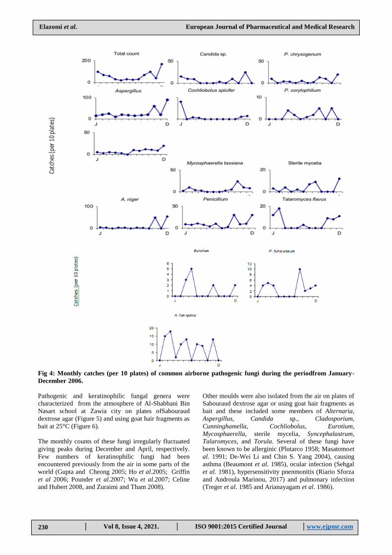
Fig 4: Monthly catches (per 10 plates) of common airborne pathogenic fungi during the periodfrom January-
December 2006.
Pathogenic and keratinophilic fungal genera were
characterized from the atmosphere of Al-Shabbani Bin
Nasart school at Zawia city on plates ofSabouraud
dextrose agar (Figure 5) and using goat hair fragments as
bait at 25°C (Figure 6).
The monthly counts of these fungi irregularly fluctuated
giving peaks during December and April, respectively.
Few numbers of keratinophilic fungi had been
encountered previously from the air in some parts of the
world (Gupta and Cheong 2005; Ho et al.2005; Griffin
et al 2006; Pounder et al.2007; Wu et al.2007; Celine
and Hubert 2008, and Zuraimi and Tham 2008).
Other moulds were also isolated from the air on plates of
Sabouraud dextrose agar or using goat hair fragments as
bait and these included some members of Alternaria,
Aspergillus, Candida sp., Cladosporium,
Cunninghamella, Cochliobolus, Eurotium,
Mycosphaerella, sterile mycelia, Syncephalastrum,
Talaromyces, and Torula. Several of these fungi have
been known to be allerginic (Plutarco 1958; Masatomoet
al. 1991; De-Wei Li and Chin S. Yang 2004), causing
asthma (Beaumont et al. 1985), ocular infection (Sehgal
et al. 1981), hypersensitivity pnenmonitis (Riario Sforza
and Androula Marinou, 2017) and pulmonary infection
(Treger et al. 1985 and Arianayagam et al. 1986).

Elazomi et al. European Journal of Pharmaceutical and Medical Research
www.ejpmr.com│ISO 9001:2015 Certified Journal │ . Vol 8, Issue 4, 2021 │
231
Dermatophytes and closely related fungi were
represented by Alternaria, Aphanoascus, Aspergillus,
Chaetomium, Cunninhamalla, Emericella, Fusarium,
Mucor, Mycospharella, Penicillium, Rhizopus,
Trichophyton Ulocladiumgenera of which Aphanoascus
was the most reported common genus. The monthly
counts of Aphanoascus irregularly fluctuated giving
peaks during April or December.
Fig. 5: Monthly catches (per 50 goat hair fragments) of common airborne keratinophilic fungi during the period
from January-December 2006.
Elazomi et al. European Journal of Pharmaceutical and Medical Research
www.ejpmr.com│ISO 9001:2015 Certified Journal │ . Vol 8, Issue 4, 2021 │
232
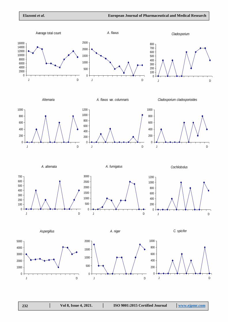
Average total count
0
2000
4000
6000
8000
10000
12000
14000
16000
J D
Alternaria
0
200
400
600
800
1000
J D
A. alternata
0
100
200
300
400
500
600
700
J D
Aspergillus
0
1000
2000
3000
4000
5000
J D
A. flavus
0
500
1000
1500
2000
2500
J D
A. flavus var. columnaris
0
200
400
600
800
1000
1200
J D
A. fumigatus
0
500
1000
1500
2000
2500
3000
J D
A. niger
0
500
1000
1500
2000
J D
Cladosporium cladosporioides
0
200
400
600
800
1000
J D
Cochliobolus
0
200
400
600
800
1000
1200
J D
C. spicifer
0
200
400
600
800
1000
J D
Cladosporium
0
100
200
300
400
500
600
700
800
J D

Elazomi et al. European Journal of Pharmaceutical and Medical Research
www.ejpmr.com│ISO 9001:2015 Certified Journal │ . Vol 8, Issue 4, 2021 │
233
Fusarium
0
200
400
600
800
1000
1200
J D
F. sambicinum
0
200
400
600
800
1000
1200
J D
Mucor hiemalis
0
200
400
600
800
1000
1200
J D
Penicillium
0
2000
4000
6000
8000
10000
12000
J D
P. chrysogenum
0
2000
4000
6000
8000
10000
12000
14000
J D
P. citrinum
0
500
1000
1500
2000
2500
J D
P. oxalicum
0
100
200
300
400
500
J D
Rhizopus stolonifer
0
500
1000
1500
2000
2500
J D
Sterile mycelia
0
500
1000
1500
2000
2500
3000
J D
Fig. 6: Monthly total counts 9calculated per g dry dust) of common glucophilic fungi in sedimented dust during
the period from January - December 2006.
CONCLUSION
It can be concluded that a various fungal genera were
isolated from floor dust, and indoor air samples. The
most prevailed fungal genera isolated were Alternaria,
Aspergillus, Penicillium, Mucor, Ulocladium,
Emericellaand Rhizopus. Overall, the highest number of
fungal genera was obtained from floor dust samples.
Our results obtained of pathogenic and non-pathogenic
fungi in the floor dust and indoor air of Elementary and
Preparatory schools were almost basically similar to
those fungi reported in many parts of the world but with
different numbers, frequencies and months of fungi.
Further studies on mycoflora taxa frequently isolated
from floor dust and indoor air environment of schools in
different governorates of Libya would be interesting. A
large number of fungal species still wait proper
identification. Different modern sampling techniques can
be used to investigate culture ability and total fungal
spores and to estimate Colony Forming Units (CFU).
REFERENCES
1. Abdel-Hafez A.I.I., Mazen M.B. and Gala] A.A.
Glycophi1ic and cellulose decomposing fungi from
soils of Sinai Peninsula, Egypt. Arab Gulf J. Scient.
Res, 1999; 8(1): 153-168.
2. Abdel-Hafez S.I.I. and Shoreit A.A.M. Mycotoxins
producing fungi and mycoflora of air-dust from Taif,
Saudi Arabia. Mycopathologia, 1985; 92: 65-71.
3. Abdel-Hafez S. I.I., Moharram A. M. and Abdel-Sater M.
A. Soil fungi of the new valley area, Western desert,
Months

Elazomi et al. European Journal of Pharmaceutical and Medical Research
www.ejpmr.com│ISO 9001:2015 Certified Journal │ . Vol 8, Issue 4, 2021 │
234
Egypt. Bull. Fac. Sci., Assiut Univ, 2000; 29(2): 255-
271.
4. Abdel-Hafez S.I.I., Moubasher A.H. and Barakat A.
Keratinophilicfungi and other moulds associated
with air-dust particles from Egypt. Folia Microbiol,
1990; 35: 311-325.
5. Abdel-Hafez S.I.I., Maharram A.M. and Ismail M.A.
Mycoflora of sedimented dust in the Oases of
western desert, Egypt. Abhath Al Yarmouk (in
press), 1993.
6. Abdel-Hafez S.I.I., Moubasher A.A.H. and Barakat A.
Seasonal variations of fungi of outdoor air and
sedimented dust at Assiut region, Upper Egypt.
Grana, 1993; 32: 115-121.
7. Abdel-Mallek A.Y., Bagy M.M.K. and Moharram A.M.
Fungiof the floor dust in students residential halls of
Assiut University, Egypt. J. Bot, 1988; 31(1-3): 69-
80.
8. Abdel-Raouf N. M. Studies on keratinophilic fungi of
schools in Sohage region. M. Sc., Thesis, Bot. Dept.,
Fac. Sci., Qena. South Valley Univ., Egypt, 2000.
9. Abu El-Souod S.M. and MoubasherA.H. Mycological
comparative study of English and Egyptian soils
using different substrates. J. of Ac.Sc, 1981; 1(1):87-
108.
10. Acosta F. J. and Roberstad G.W. Chrysosporiumspecies
as fungal air pollutants. Ann, Allergy, 1979; 42: 11-
13.
11. Ajello L. Milestones in the history of medical
mycology. The dermatophytes, 1977; 3-11. In
Recent Advances, in Medical and Veterinary
Mycology, by Kazuo Iwata. Univ. Tokyo Pess.
12. Al-Doory Y. Laboratory medical mycology (P. 410),
Lea and FebigerPhiladephiaKimpton Publisher,
London, 1980.
13. Ali-Shtayeh M.S. Keratinophilic fungi isolated from
children's sandpits in the Nablus area, West Bank of
Jordan. Mycopathologia, 1988; 103: 141-146.
14. Ali-Shtayeh M.S. Keratinophilic fungi of school
playgrounds in the Nablus area, West Bank of
Jordan. Mycopathologia, 1989; 106: 103-108.
15. Ali-Shtayeh M.S. and Asa'd Al-Sheikh B.S. Keratino-
philic fungi in floor dust of Arab kindergarten
schools in the West Bank of Jordan.
Mycopathologia, 1988; 103: 69-73.
16. Ali-Shtayeh M.S. and Arda H.M. Isolation of
keratinophilic fungi from floor dust in Arab
elementary and preparatory schools in the West
Bank of Jordan, Mycopathologia, 1989; 106: 5-11.
17. Ali-Shtayeh M. S., Salameh A.A. M., Abu-Ghdeib and
Jamous Rana M. Hair and scalp mycobiota in
school children in Nablus area. Mycopathologia,
2000; 150: 127-135.
18. Allermann L., Meyer H.W., Poulsen O.M., Nielsen J.B. and
Gyntelberg F. Inflammatory potential of dust from
schools and building related symptoms.
Occupational and Environmental Medicine, 2003;
60: 5. (about5p).
19. Alteras I. and Lehrer N. Fungal flora in the air of a
large hospital. Catellania, 1977; 5(11): 217-219.
20. Altayyar et al, Isolation and Identification of Soil
Keratinophilic Fungi from Different Area in South
of LibyaI. J. of Appl. Med. And Bio. Res, 2016;
1(1): p 27-32.
21. Ames L.M. A monograph of the
chaetomiaceae. Whelon. and Wesley. L.T.D.
New York, 1969; PP. 565
22. Anbu P., Hilda A. and Gopinath S. C. B. Keratinophilic
fungi of poultry farm and feather dumping soil in
Tamil Nadu, India, 2004; 158: 303-309.
23. Andersen A. .Microfungi in beds and their relation to
house dust mites. Grana, 1985; 24: 55-59.
24. Arianayagam S., Jayalakshmi P. and Soo-Hoo T. S.
Pulmonary aspergilloma. Case reports
frormMalaysis.Mycopathologia, 1986 ; 93: 151-153.
25. Asan A., Sen B. and Sarica S. Airborne fungi in urban
air of Edirne city (Turkey). Biologia, 2002 ; 7(1): 59
- 68.
26. Asan A., kirgiz T., Sen B., Camur- Elipek B., Guner U. and
Guher H. Isolation, identification and seasonal
distribution of airborne and waterborne fungi in
Terk Lake (Istanbul - Turkey). Journal of Basic
Microbiology, 2003 ; 43(2): 83 - 95.
27. Bagy M.M.K. Fungi on the hair of large mammals in
Egypt. Mycopathologia, 1986 ; 93: 73-75.
28. Barnett H.L. Illustrated genera of Imperfect fungi,
1972; (PP. 241). Minneapolis: Burgess Publishing.
29. Barron G.L. The genera of Hyphomycetes from soil.
Krieger R.E. Publishing company Huntington, New
York, 1977; (PP. 364).
30. Beaumont F., Kauffman H.F., Sluiter H.J. and DE Vries K.
Sequential sampling of fungal air spores inside and
outside the homes of mould-sensitive, asthmatic
patients: A search for a relationship to obstructive
reactions. Annals of Allergy, 1985; 55(5): 740-746.
31. Beneke E.S. Medical mycology. Burgess pub. Co.
Bhatt G.C: The soil microfungi white cedar forests
in Ontairo. Can. J. Hot, 1957 ; 38: 333-339.
32. Booth C..Fusarium laboratory guide to the identi-
fication of the major species, 1977 ; (p. 58).
Commonwealth Mycological Institute, Kew, Surrey,
England.
33. Brown A.H.S. and Smith G. The genus Paecilomyces
Bainierand its perfect stage Byssocheamys Westting. Trans.
Br. Mycol. Sco, 1957 ; 40: 17-89.
34. Boreson J., Dillner A.M. and Peccia J. Correlation
bioaerosol Load with pM 2.5 and
PMlocfconcentrations : a compaison between
natural desrt and urban – fringe aerosols.
Atmospheric Environment, 2004 ; 38: 6029 – 6041.
35. Cano J., Guarro J. and Figueras H.J. Some keratinophi1ic
fungi from Spain. Mycopathologia, 1987; 100: 163-
167.
36. Carmichael J.W. Chrysosporium and some others
AleruiosporicHypomycetes. Canad. J. Hot, 1962; 40:
1137-1172.
37. Celine M.O. and Hudert T.F. Prevalence of culturable
airborne spores of selected allergenic and pathogenic
fungi outdoor air Atmospheric Environment, 2008;
42: 4355-4368.

Elazomi et al. European Journal of Pharmaceutical and Medical Research
www.ejpmr.com│ISO 9001:2015 Certified Journal │ . Vol 8, Issue 4, 2021 │
235
38. Christensen M. and Raper K.B. Synoptic key to Asper-
gillusnidulans group species and related
Emericellaspecies. Trans. Br. Mycol. Soc, 1978; 71:
177-191.
39. Curtis L., Rea W., Smith W.P., Fenyves E. and Pan Y.
Adverse health effects of outdoor air pollutants
.Evironment International, 2006; 32: 815 –830.
40. Czarnecki B. &Bialasiewicz D. Fungi as a component
of the aerosphere in the H. Arctowski Polar Station
and its vicinity (King George Island, South
Shetland, Islands). Polish Polar Research, 1987; 8(2):
153-158.
41. Dale W.G., NilgunK.,MustafaK.,MikeA.G.,Timothy C.B.
and Eugene A.S. Airborne desert dust and
aeromicrobiology over the Turkish Mediterranean
coastline. Atmospheric Environment, 2007; 41:
4050-4062.
42. Della Franca P. and Caretta G. Keratinophilic fungi
isolated from the air at Pavia.
Mycopathologia, 1984; 85: 65-68.
43. Deshmukh S. K. .Keratinophilic fungi isolated from
soils of Mumbai, India. Mycopathologia, 1999; 146:
115-116.
44. Deshmukh S. K. , Agrawal S.C. and Jain P.C. Isolation of
dermatophytes and other keratinophilic fungi from
soils of Mysore (India). Mycoses, 2000; 43(1-2): 55-
57.
45. De-Vries G.A. Contribution to the knowledge of the
genus Cladosporium Link ex. Fr. Vitgeveriy and
Druk- kerij, Hollandia Press: Baarn, 1952.
46. De-Wei Li and Chin S. Yang Fungal Contamination
as a Major Contributor to Sick Building Syndrome,
Advances In Applied Microbiology, 2004. DOI:
10.1016/S0065-2164(04)55002-5
47. Domsch K.H. and Gams W. Fungi in agricultural soils,
1972 ; (p. 290). Puplished by Longman.
48. Domsch K.H., Gams W. and Anderson T. Compendium
of soil fungi, 1980 ; (PP. 859). Acad. Press, London.
49. Douwes J., Van S.B., Doekes G., Van L. F ., Wijnands L. ,
Van S.R., Verhoeff A. and Brunekreef B. Fungal
extracellular polysaccharides in house dust as a
marker for exposure to fungi: relations with
culturable fungi, reported home dampness, and
respiratory symptoms. J.Allergy Chin. Immun, 1999;
103: 494-500.
50. Ellis M.B. Dematiaceous Hyphomycetes, 1971; (PP.
608). Commonwealth Mycological Institute, Kew,
Surrey, England.
51. Ellis M.B. More Dematiaceous Hyphomycetes, 1976;
(pp.507). Commonwealth mycological Institute,
Kew, Surrey-England.
52. EL – Said A.H.M. Studies on soil mycoflora of
Bahreen. Microbiol, 1994; 149: 263 – 269.
53. El-Said A. H. M. Keratinophilic fungi in soils of
Yemen Arab Republic. J. Islam. Acad. Sci, 1995;
8(4): 151-154.
54. El-Said, A. H., Sohair, T. H., & El-Hadi, A. G.
Fungi associated with the hairs of goat and sheep in
libya. Mycobiology, 2009; 37(2): 82–88.
https://doi.org/10.4489/MYCO.2009.37.2.082
55. Euan P.B., Martin K. and Geoffrey M.G.
Geomycology: Fungi in mineral substrata.
Mycologist, 2003; 17: 98-107.
56. Frey D., Oldfield R.J. and Bridger R.C. A colour atlas of
pathogenic fungi. Wolfe Medical Publications
LTd.Smeets-Weert, Holland, 1979; (pp. 168).
57. Gherbawy Y. A. M. H. Keratinolytic and
keratinophilic fungi of graveyard's soil and air in the
city of Qena and their response to garlic extract and
onion oil treatments. Egypt. J. Microbiol, 1999 ; 34(1):
1-21.
58. Gravesen S. Identification and prevalence of
culturable mesophilic microfungi in house dust from
100 Danish homes: Comparison between airborne and
dust-bound fungi. Allergy, 1978 ; 33: 268-272.
59. Griffin D.W., Garrison V.H., Herman J.R. and Shinn E.A.
African desert dust in the Caribbean atomosphere
microbiology and pudlichealth .Aerobiologia, 2001;
14(3): 203-213.
60. Griffin D.W., kellogg C.A., Garrison V.H., Lisle J.T.,
Borden T.C and Shinn E.A. African dust in the
Caribbean atmosphere .Aerobiologia, 2003 ; 19 (3-
4): 143-157.
61. Griffin D.W., Westphal D.L. and Gray M.A. Airborne
microorganisms in the African desert dust corridor
over the mid-Atlantic ridge, Ocean Drilling
Program.Leg 209. Aerobiologia, 2006; 22(3): 211-
226.
62. Grozier W. J. The prevalence of geophilic dermato-
phytes in soils of the Illawarra area of South Wales.
Australian Journal of Dermatology, 1980.
63. Guinea J.,PelaezT.,Alcala L. and Bouza E. Outdoor
environmental level of Aspergillus spp. Conidia over
a wide geographical area.Medical Mycology, 2006 ;
44: 349-356.
64. Gupta A. and Cheong K.W.D. Airborne particulate
matter concentrations at different heights in a
residential building in Singapore In: Proceedings of
Indoor Air Beijing, China, 2005; .5: 1748 –1753.
65. HanlinR.T. Illustrated Genera of Ascomycetes. The
American Phytopathological Society (APS) press,
1990; (PP. 263).
66. Hedayati M.T., Mohseni-Bandpi A. and Moradi S. A
survey on the pathogenic fungi in soil samples of
potted plants fron Sari hospitals, Iran. Journal of
Hospital In fection, 2004; 58: 59-62.
67. Hemida S. K. Thermophilic and thermotolerant fungi
isolated from cultivated and desert soils, exposed
continuously to cement dust particles in Egypt.
Zentralbl. Mikrobiol, 1992; 147: 277-281.
68. Hollins P.D., kettewell P.S., Atkinson M.D., Stephenson
D.B., Corden J.M., Millington W.M. and Mullins J.
Relationships between airborne fungal spore
concentrations of Cladosporium and the summer
climate at two sites in Britain. International Journal
of Biometeorology, 2004; 48: 137-141.
69. Hiromi OHARA et al. Effects of Exposure to Male
Goat Hair Extracts on Luteinizing Hormone
Secretion and Neuronal Activation in Seasonally
Elazomi et al. European Journal of Pharmaceutical and Medical Research
www.ejpmr.com│ISO 9001:2015 Certified Journal │ . Vol 8, Issue 4, 2021 │
236
Anestrous Ewes. J. Vet. Med. Sci, 2014; 76(10):
1329-1337.
70. Ismail M.A. Studies on the mycoflora of air, air-dust
and pollen grains in the Oases of western desert,
Egypt. Ph. D. Thesis, Bot. Dept., Fac. Sci., Assiut
Univ.Egypt, 1990; (PP. 351).
71. Jayne L.J., Gailw T.W., Paul M. W. and Anthony J.
Consumption of mycorhizal and saprophytic fungi
by Collembola in grassland soils. Soil Biology
&Biochemistry, 2007; 39: 2594 -2602.
72. Jackson M. L. Soil chemical analysis constable and
Co. London, 1958.
73. John L., Anders J., Soren E.L., Lars H.H., Iver J. and Paul
H.k. Population performance of collembolans
feeding on soil fungi from different ecological
niches :Soil Biology & Biochemistry, 2008 ; 40: 360-
369 .
74. Javier J.R., Isabel I. and Victoria J. Variation assessment
of airborne Alternaria and Cladolisporiumspores at
different bioclimatical conditions. Mycol. Res, 2005 ;
109(4): 497 – 207.
75. Johnson L.F. and Curl E.A. Method for research on
ecology of soil-borne pathogens, Burgress publ. Co.
Minneapolis, 1972; 247.
76. Jussila J., komulainen H., kosma V.M., Nevalainen A.,
Pelkonen J. and Hirvonen M.R. Spores of Aspergillus
versicolor isolated from indoor lungs Inhal .Toxicol,
2002 ; 14: 1216 – 1277.
77. Karl R. and Iain M.Y. Interactions between soil
structure and fungi. Mycologist, 2004; 18: 52-59.
78. Kendrick B. Taxonomy of fungi imperfecti University
of Toronto, 1971.
79. Kellogg C.A., Griffin D.W., Garrison V.H., Peak K.K.,
Royall N., Smith R.R. and Shinn E.A. Characterization
of aerosolized bacteria and fungi from desert dust
events in Mali, West Africa. Aerobiologia, 2004;
20(2): 99-110.
80. Kuhn D.M. and Ghannoum M.A. Indoor mold,
toxigenic fungi and Stachybotryschartarum :
infections disease per spective. Clinical
Microbiology Reviews, 2003; 16: 144-172.
81. Kushwala R. K.S. The genus Chrysosporium, its
physiology and biotechnological potential in
Biology of dermatophytes and other
keratinophilic fungi. RevistaIberoamericana de
Micologia, Bilbao, 2000; (pp. 66-76).
82. Lalley J.S. and Viles H.A. Terricolous lichens in the
northern Namib desert of Namibia: distribution and
community composition. The Lichenologist, 2005;
37(1): 77-91.
83. Larone DH Medically Important Fungi (A Guide to
Identification). Am. Soc. of Mic. Washington, D.C,
1987.
84. Leese K.E., Cole E.C., Mall R.M. and Berry M.A.
Measurement of airborne and floor dusts in a non
problem building. Am. Ind. Hyg. Assoc, 1997; 58:
432-438.
85. Lentner C. Geigy Scientific Tables. Vol. 6. CIBA-
GEIGY Limited, Basel, Medical and Pharmaceutical
Information. New Jersey. USA, 1991.
86. Liao C.M., Luow C., Chen S.C., Chen J.W. and Liang
H.M., Temporal / seasonal variations of size.
dependent airhorne fungi indooor / outdoor
relatinships for a wind on induced naturally
ventilated airspace. Atmos Environ, 2004; 38 (26):
4415 – 4419.
87. Maria C.,Capetti E. and Bonescu O. The relationship
between air-borne fungal spores and
Dermatophagoidespeteronyssinum in house dust.
Rev Roum Med. Interne, 1981; 19(1): 73-78.
88. Masatomo M., Murakami G., Adachi Y., Kayahara M.,
Okada T.,Arakawa R., Kaawai K. and Igaroshi T.
Immunochemical quantification of the air borne
chironomid allergens.JPNJ.Allergol, 1991; 40(1): 51-
59.
89. Mercantini R., Marsella R., Lambiase L. and Fulvi F.
Isolation of keratinophilic fungi from floors in
Roman primary schools. Mycopathologia, 1983; 82:
115-120.
90. MercantiniR. ,Marsella R. , Lambiase L. and Belardi M.
Isolation ofkeratinophilic fungi from floors in
Roman kindergarten and secondary schools.
Mycopathologia, 1986; 94: 109-115.
91. Mercantini R., Marsella R. and Cervellati M.C.
.Keratinophilic fungi isolated from antarctic soil.
Mycopathologia, 1989; 106: 47-52.
92. Meyling N. and Eilenberg J. Occurrence and
distribution of soil borne entomopathogenic fungi
within a single organic agroecosystem .Agric
.Ecosyst .Environ, 2006; 113: 336-341.
93. Morton F.J. and Smith G. The genera
ScopulariopsisBainier, Microascuszukal and
Doratomyces Corda. Mycological papers, 1963; 8: 1-
96. Commonwealth Mycological Institute, Kew,
Surrey, England.
94. Moss E.S. and McquownA.L. Atlas of medical
mycology.3 rd edition. The Williams and Wikins
Company. Baltimore, 1969; (pp.366).
95. Moubasher A.H. and Moustafa A.F. Air-borne fungi at
Assiut, Egypt. J. Bot, 1974; 17: 135-149.
96. Moubasher A.H., Abdel-Fattah H.A. and Swelim M.A.
Studies on air-borne fungi at Qena. I-Seasonal fluc-
tuations. Z. Allg. Mikrobiol, 1981; 21(3): 247-253.
97. MoubasherA.M., Abdel-Hafez S.I.I. and El-Maghraby
O.M.O. Studies on soil mycoflora of Wadi Birn El-
Ain, Eastern Desert, Egypt. Cryptogamie,
Mycologie, 1985; 6: 129-143.
98. Moubasher A.H., Abdel-Hafez S.I.I., Shoreit A.A.M. and
Ismael M.A. Keratinophilic and other fungi isolated
from combine harvester wheat and sorghum dusts
and from the atmosphere of winnow sites in Egypt.
Folia Microbiol, 1990; 35: 298-310.
99. Murray P.R., Rosenthal K.S. and Pfaller M.A.
Medical Microbiology. Elsevier Mosby, 2005.
100. Onions A.H.S., Allsop D. and EgginsH.O.W.(1981).
Smith's introduction to industrial mycology.Edward
Arnold (Publisher) Ltd., London (pp.398).
Papavassilion J.T. and Bartzokas C.A. The atmospheric
fungal flora of the Athens metropolitan area.
Mycopathologia, 1975; 57: 31-34.

Elazomi et al. European Journal of Pharmaceutical and Medical Research
www.ejpmr.com│ISO 9001:2015 Certified Journal │ . Vol 8, Issue 4, 2021 │
237
101. Piper C.S.: Soil and plant analysis. A laboratory
manual of method for the examination of soil and
determination of the inorganic substituents of plants.
Inter. Pub. Inc. New York, 1955.
102. Picco A.M. and Rodolfi M. Airborne fungi as
biocontaminants at wo Milan underground stations.
International Biodeterioration and Biodegradation,
2000; 45: 43-47.
103. Pitt J.I. The genus Penicillium and its
teleomorphicstates Eupenicillium and
Talaromyces, 1979; (PP. 634). Acad. Press.
104. Pitt J.I. A laboratory guide to common Penicillium
species, 1985; (PP. 184). Commonwealth Scientific
And Industrial Research Organization Division
of Food Research.
105. Plutarco N. Etiological agents of respiratory allergy in
tropical countries of Central and South America. J.
Allergy, 1958 ; 29: 362-374.
106. Pounder J.I., Simmon K. E., Barton C.A., Hohmann S.L.,
Brandt M .E. and petti C.A. Discovering potentoal
pathogens among fungi identified as nonsporulating
molds. Journal of Clinical Microbiology, 2007; 45:
568 –571 .
107. Prospero J.M., Blades E., Mathison G. and Naidu R.
Interhemisheric transport of viable fungi and
bacteria from Africa to the caribbean with soil dust
.Aerobiologia, 2005; 21: 1-19 .
108. Quesada M., Navas C.J.A., Maranhao E.A.A., Ortiz U.A.
and Santiago A. C. Factors affecting the occurrence
and distribution of entomopathogenic fungi in
natural and cultivated soils.Mycol.Res, 2007; 111:
947-966.
109. Ramirez C. Manual and Atlas Penicillia. Elsevier
Biomedical Press, Amsterdam, The Netherlands,
1982 ; (pps. 874).
110. Raper K.B. and Fennell D.J. The genus Aspergillus,
1965; (p.686). Williams and Wilkins Baltimore,
U.S.A.
111. R. Rathish, P. Madhanraj and G.S.Senthilkumar.
DETERMINATION OF PHYSICOCHEMIAL
PARAMETERS AND DIVERSITY PATTERN OF
THREE DIFFERENT PLACES OF
KANYAKUMARI DISTRICT. Int.J.Adv. Res,
2017; 5(1): 1036-1039.
112. Raper K.B. and Thom C. A manual of the Penicillia,
1949; (p.875). Wilkins, Baltmore, U.S.A.
113. Rao C.K., Cox-Ganser J.M., Chew G.L., Doekes G. and
White S. Use of surrogate markers of biological
agents in air and settled dust samples to evaluate a
water – damaged hospital indoor Air, 2005; 15
Suppl9: 89-97.
114. Rifai M.A. A revision of the genus Trichoderma.
Mycological papers, 1969; 116: 1-55.
115. Rijckaert G.F., Van Bronswijk J.E.M.H. and Linskens H.F.
House-dust community (fungi-mites) in different
climatic regions. Oecologia, 1981; 84(2): 183-185.
116. Riario Sforza and AndroulaMarinou
Hypersensitivity Pneumonitis: a complex lung
disease. Clin Mol Allergy, 2017; 15: 6. DOI
10.1186/s12948-017-0062-7.
117. Samson R.A. A compilation of the Aspergilli de-
scribed since, 1965. Studies in Mycology 18 (PP.
40).
118. Sehgal S.C., Dhawan S., Chhiber S., Sharma M. and
Talwar P. Frequency and significance of fungal isola-
tions from conjunctival sac and their role in oculor
infections. Mycopathologia, 1981; 73: 17-19.
119. Shukia P., Shukla C. B., Kango N. and Shukla A. Isolation
and characterization of a dermatophyte,
Microsporiumgypseum from poultry farm soils of
Rewa (Madhya Pradesh), India .J. Bio. Sci, 2003;
6(6): 622-625.
120. Shelton B.G., Kirkand K.H. , Flanders W.D. and Morris
G.K. Profiles of airborne fungi in buildings and
outdoor environments in the United States . Applied
and Environmental Microbiology, 2002; 68: 1743-
1753.
121. Simmons E.G. Typification of
Alternaria,Stemphliumand Ulocladium. Mycologia,
1967; 59: 67-92.
122. Sivanesan A. The bitunicate Ascomycetes and their
anamorphs, 1984 ; (PP 701). Strauss and Cramer
GmbH, Geymany.
123. Skolko A.J. and Groves J.W. VII-Chaetomium.
Canad.J. Bot, 1953; 31: 779-322.
124. Smith N.R. and Dawson V.I. The bacteriostatic action
of rose-bengal in media used for the plate count of soil
fungi.Soil Sci, 1944; 58: 467-471.
125. Srivastava L., Srivastava J. and Srivastava N. Studies on
fungal air-spora at Gorakhpur (Uttar Pradesh,
India):I .District hospital compus. Vegetos, 1990;
3(2): 202-211.
126. Snezana J., Andrea F.K., Thomas G., Bijan K., Bernhardt
L., Valentina M., Isolde P., Karl H.S., Monika S., Ursla W.,
Lris Z. and Michael S. Indoor fungi levels in homes of
children with and without allergy history .Int .J.hgy
.Environment Health, 2004; 207: 369- 378.
127. Treger T.R., Visscher D.W., Bartlett M.S. and Smith J.W.
Diagnosis of pulmonary infection caused by
Aspergillus:Jsefulness of respiratory cultures. J. Inf,
1985; 152: 572-576.
128. Ulfig K., Gene J. and GuarroJ. Studies on
keratinophilicfungi. VI. A new Arthropsis(
fungiimperfecti) from marine sediments. Volume
LIV, 1995; pp.281-286.
129. UlfigK.,Terakowski M., Plaza G.G. and Kosarewicz O.
Keratinolytic fungi in sewage sludge.
Mycopathologia, 1996; 136: 41-46.
130. Ulfig K., Guarro J., Gano J., Gene J, Vidal P. and Figueras
MJ: General assessment of the occurrence of
keratinolytic fungi in river and marine beach
sediments of Catalonian water (Spain). Water Air, &
Soil Pollution, 1997; 99(3-4): 275-287.
131. Ulfig K., Plaza G., Terakowski M. and Staszewski T. A
study of keratinolytic in mountain sediments. I.
Hair-baiting data. Roczn. PZH, 1998 ; 49: 469-479.
132. Ulfig K., Guarro J., Cano J., Vidal P., Figueras M. J. and
Lukasik W. The occurrence of keratinolytic fungi is
sediments of the river. Tordera (Spain). FEMS Microbiology
Ecology, 1999; 22: 111-117.
Elazomi et al. European Journal of Pharmaceutical and Medical Research
www.ejpmr.com│ISO 9001:2015 Certified Journal │ . Vol 8, Issue 4, 2021 │
238
133. Vanbreuseghem R. Biological technique for the
isolation of dermatophytes from soil. Ann. Soc.
Bellge. Med. Trop, 1952; 32: 173.
134. Van Oorschot C.A.N. A revision of Chrysosporium
and allied genera. Studies in Mycology, 1980; 20: 1-
87.
135. Verhoeff A.P., Van W.J.H., Reenen H.E.S., Samson R.A.,
Van S.R.T. and BrunekreefB. (Fungel propagules in
house dust. II. Relation with residential
characteristics and respiratory symptoms. Allergy,
1994; 49: 540-547.
136. Veronika A., Joachim S., Renate R. and Christian S. In
vitro degradation of equine keratin by
dermatophytes and other kerainophilicfungi.
Veterinary Microbiology, 2006 ; 114: 352-358.
137. Vidyasagar G. M., Narayan H. and Shivkumar
D. .Keratinophilic fungi isolated from hospital dust
and soils of Puplic places at Gulbarga, India.
Mycopathologia, 2005; 159: 13-21.
138. WongN.H. and Huang B. Comparative study of the
indoor air quality of naturally ventilated and air-
conditioned bed-rooms of residential building in
Singapore. Building and Environment, 2004; 39: 1115-
1123.
139. Woodfolk JA, Commins SP, Schuyler AJ, Erwin
EA, Platts-Mills TA. Allergens, sources, particles,
and molecules: Why do we make IgE responses?
Allergol Int, 2015; 64(4): 295-303. doi:
10.1016/j.alit.2015.06.001. Epub 2015 Jul 15.
PMID: 26433525; PMCID: PMC5406225.
140. Wu P.C., Su H.J. and Lon C.K. Characteristics of in
door an outdoor airborne fungi at suburban and
urban homes in two seasons. Sci Total Environ,
2000; 253(1-3): 111-118.
141. Wu P.C., Tsai J.C., Li F.C., Lung S.C. and Su H.J.
Increased levels of ambient fungal spores in Taiwan
are associated with dust events from China.
Atmospheric Environment, 2004; 38: 4849-4886.
142. Wu Y.H., Chan C.C., Rao C.Y.,LeeC.T.,HsuH.H.,Chiu
Y.H. and Chao H.J. Characteristics, determinants and
spatial variations of ambient fungal levels in the
subtropical Taipei metropolis. Atmospheric
Environment, 2007 ; 41: 2500-2509.
143. Wurtz H., Sigsgaard T., Valbjon O., Doekes G. and Meyer
H.W. Dustfall collector – a simple passive tool for
Long – term collection of airborne dust : a project
under the Danish mould in buil ding, program
(DAMIB) In door Air, 2005; 15 (Suppl.9 ): 33-40.
144. Yeo H.G. and Kim J.H. SPM and fungal spores in the
ambient air of West Korea during the Asian dust (
yellow sand) period. Atmospheric Environment,
2002; 36(35): 5437-5442
145. Youssef K. H. A., Youssef Y.A. and Youssef H.H. Fungi
and thermophilic actinomycetes in the air of the
Cancer Institute hospital. Ain shams Sci. Bull, 1980;
22(A,B): 75-88.
146. ZuraimiM.S. and Tham K.W. Indoor air quality and its
determinants is tropical child care centers. Atmospheric
Environment, 2008; 42 : 2225 –2239.
147. Zycha H. Mucorineae VonKryptogamenflora Der Mark
Brandenburg. Band VI a: Pilze II Verlag Von.
J.Cramer, Weinheim. Johnson reprint corporation,
NewYork, 1963; (PP. 264).
Related Documents