Monitoring and modeling of pupillary dynamics: Study of the autonomous nervous system Christophe Tilmant Mathilde Charavel Matthieu Ponrouch ERIM Department of Reanimation Department of Reanimation Faculty of Medicine Gabriel Montpied Hospital Gabriel Montpied Hospital 28, place Henri Dunant BP 38 58, rue Montalembert BP 69 58, rue Montalembert BP 69 63001 Clermont-Ferrand France 63003 Clermont-Ferrand France 63003 Clermont-Ferrand France (+33) 473 178 123 (+33) 473 751 590 (+33) 473 751 590 [email protected] [email protected] [email protected] Guillaume Gindre Laurent Sarry Jean-Yves Boire Department of Reanimation ERIM ERIM Gabriel Montpied Hospital Faculty of Medicine Faculty of Medicine 58, rue Montalembert BP 69 28, place Henri Dunant BP 38 28, place Henri Dunant BP 38 63003 Clermont-Ferrand France 63001 Clermont-Ferrand France 63001 Clermont-Ferrand France (+33) 473 751 590 (+33) 473 178 123 (+33) 473 178 123 [email protected] [email protected] [email protected] ABSTRACT This study deals with the analysis of pupillary dynamics. The evaluation of pupil motion provides functional information about the autonomous nervous system. A non-invasive tool has been designed to help the understanding of physiology by modeling the reflex loop of the pupil. The monitoring is made up of three parts. First, a digital camera is chosen for image acquisition because of its simple use at the patient’s bed. Then a software estimates the pupillogram, i.e. the plot of the pupil area versus time, from the digital video sequence using deformable templates. And finally, the pupil light reflex is modeled by a second order linear system to compute characteristic parameters. They can be used to diagnose various affections from a statistical analysis. KEYWORDS Deformable template, physiological modeling, pupil light reflex, pupillogram. 1. Introduction The humoral human system is regulated by the association of the autonomic nervous system (ANS) and the endocrine system. The integration of these two systems is done by the hypothalamus. The ANS provides involuntary control and organization of both maintenance and stress responses. The ANS has two divisions: the sympathetic nervous system (SNS) and the parasympathetic nervous system (PNS). Many but not all muscles and glands that distribute nervous impulses to larger interior organs possess a double nerve supply; in such cases the two divisions may exert opposing effects. There are several tests of the ANS: clinic (arterial blood pressure and cardiac frequency), blood biochemistry, and pupil tests. In fact, one of the interesting properties of the eye is the pupil response to light, when the pupil muscles are activated and lead to a miosis. The pupil dilation depends on the SNS and its contraction on the PNS. So the diameter and kinetics of the pupil are a consequence of the balance between these two antagonistic forces. Pupillometry is the recording of the pupillary diameter according to time. In order to make the recording independent from the variations due to the ambient light, the reference method is based on the infra-red (IR) acquisition. Pupillometry was initially used in ophthalmology: the pupillary size is significant for the interpretation of perimetry of the visual field and electroretinography and for the preoperative assessment of a refractive surgery [Ambrosio et al. 2002; Duffey Leaming 2004], in neuro ophthalmology for the description of lesion of the afferent or efferent nervous ways [Loewensfeld 1993]. The modeling of pupillogram, acquired during a light stimulation, makes it possible to obtain the static and dynamic parameters driving pupil light reflex (PLR). The PLR has already been studied to detect a PNS defect [Kawasaki Kardon 2001] in various affections: alcoholism, diabetes, AIDS, Down’s syndrome, depression, anxiety, drug addiction, Alzheimer diseases, schizophrenia. Blaise Pascal University, France 441 © OICMS 2005

Welcome message from author
This document is posted to help you gain knowledge. Please leave a comment to let me know what you think about it! Share it to your friends and learn new things together.
Transcript

Monitoring and modeling of pupillary dynamics: Study of the autonomous nervous system
Christophe Tilmant Mathilde Charavel Matthieu Ponrouch ERIM Department of Reanimation Department of Reanimation Faculty of Medicine Gabriel Montpied Hospital Gabriel Montpied Hospital 28, place Henri Dunant BP 38 58, rue Montalembert BP 69 58, rue Montalembert BP 69 63001 Clermont-Ferrand France 63003 Clermont-Ferrand France 63003 Clermont-Ferrand France (+33) 473 178 123 (+33) 473 751 590 (+33) 473 751 590 [email protected] [email protected] [email protected] Guillaume Gindre Laurent Sarry Jean-Yves Boire Department of Reanimation ERIM ERIM Gabriel Montpied Hospital Faculty of Medicine Faculty of Medicine 58, rue Montalembert BP 69 28, place Henri Dunant BP 38 28, place Henri Dunant BP 38 63003 Clermont-Ferrand France 63001 Clermont-Ferrand France 63001 Clermont-Ferrand France (+33) 473 751 590 (+33) 473 178 123 (+33) 473 178 123 [email protected] [email protected] [email protected] ABSTRACT This study deals with the analysis of pupillary dynamics. The evaluation of pupil motion provides functional information about the autonomous nervous system. A non-invasive tool has been designed to help the understanding of physiology by modeling the reflex loop of the pupil. The monitoring is made up of three parts. First, a digital camera is chosen for image acquisition because of its simple use at the patient’s bed. Then a software estimates the pupillogram, i.e. the plot of the pupil area versus time, from the digital video sequence using deformable templates. And finally, the pupil light reflex is modeled by a second order linear system to compute characteristic parameters. They can be used to diagnose various affections from a statistical analysis.
KEYWORDS Deformable template, physiological modeling, pupil light reflex, pupillogram.
1. Introduction The humoral human system is regulated by the association of the autonomic nervous system (ANS) and the endocrine system. The integration of these two systems is done by the hypothalamus. The ANS provides involuntary control and organization of both maintenance and stress responses. The ANS has two divisions: the sympathetic nervous system (SNS) and the parasympathetic nervous system (PNS). Many but not all muscles and glands that distribute nervous impulses to larger interior organs possess a double nerve supply; in such cases the two divisions may exert opposing effects. There are several tests of the ANS: clinic (arterial blood pressure and cardiac frequency), blood biochemistry, and pupil tests. In fact, one of the interesting properties of the eye is the pupil response to light, when the pupil muscles are activated and lead to a miosis. The pupil dilation depends on the SNS and its contraction on the PNS. So the diameter and kinetics of the pupil are a consequence of the balance between these two antagonistic forces.
Pupillometry is the recording of the pupillary diameter according to time. In order to make the recording independent from the variations due to the ambient light, the reference method is based on the infra-red (IR) acquisition. Pupillometry was initially used in ophthalmology: the pupillary size is significant for the interpretation of perimetry of the visual field and electroretinography and for the preoperative assessment of a refractive surgery [Ambrosio et al. 2002; Duffey Leaming 2004], in neuro ophthalmology for the description of lesion of the afferent or efferent nervous ways [Loewensfeld 1993]. The modeling of pupillogram, acquired during a light stimulation, makes it possible to obtain the static and dynamic parameters driving pupil light reflex (PLR). The PLR has already been studied to detect a PNS defect [Kawasaki Kardon 2001] in various affections: alcoholism, diabetes, AIDS, Down’s syndrome, depression, anxiety, drug addiction, Alzheimer diseases, schizophrenia.
Blaise Pascal University, France 441 © OICMS 2005

In much pathology, the ANS is reached with a different prevalence. Pupillometry makes it possible to evaluate the degree of autonomic dysfunction in the migraine [Mylius et al. 2003]. Indeed, the final values of the pupillogram parameters are reached 48 hours after a migraine. The way the ANS is affected indicates whether the sympathetic or parasympathetic systems prevail [Bertinotti et al. 2002]. This evaluation is also interesting for the diabetic disease to assess the severity of the nervous attack. Other pupillogram abnormalities are observed in the Alzheimer disease.
Pupillometry is also of interest as a monitoring tool in the field of neurosurgery anesthesiology [Manley Larson 2002]. Indeed abnormalities of the different parameters of PLR are observed during the rise in intracranial pressure, the latter being either related to the effect of localized mass or a generalized oedema. They are unilateral or bilateral according to the attack mechanism [Taylor et al. 2003]. They last longer than intracranial hypertension. An asymmetry is found for only 1% of the subjects unharmed of all cerebral pathology. Pupillometry is used for the initial assessment of the serious brain trauma. The pupillogram was much altered for the most serious patients; it secondarily was nonreactive for patients for whom diagnosis of brain death was made afterwards.
One of the other applications of pupillometry is the monitoring of pain for conscious or deadened patients. Indeed any painful stimulation involves a pupillary dilation [Tassorelli et al. 1995]. Only indirect parameters enable to appreciate pain for unconscious patients, these parameters (blood pressure and rate of heartbeat) are dependent on many factors and seem modified lately in case of analgesia defect [Barvais et al. 2003; Onal Tuglular 1999]. The opioids, used in anesthesia for the treatment of the pain, inhibits pupillary dilation related to pain when suitably treated. Pupillometry could be therefore interesting to guide the use of analgesics in anesthesia. Moreover, when regional anesthesia is combined to a global one, monitoring of the PLR makes it possible to locate the level of the anesthetic block induced by regional anesthesia [Larson et al. 1993].
Many molecules modify PLR [Knaggs et al. 2004]. So pupillometry is used for the tracking of medicamentous drug-addiction. An evaluation of this technique was made in California and this method appeared profitable and efficient. [Richman Noriega 2002; Murillo et al. 2004]
Pupillometry is a non-invasive technique that seems to be promising as a tool to follow-up numerous medical problems, but it requires a more complete evaluation that will be made possible by the automated system described below. The aim of this work is to build a new tool integrating knowledge about dynamic PLR and to use image analysis, in order to track pupil motion, and physiological modeling to quantify disorders. This tool has to be easy to use at the patient’s bed and to provide reproducible measures.
In section 2, the material used for this study is described. In section 3, the methodology of image analysis and physiological modeling is exposed. Finally, section 4 focuses on the results and discusses the performances and limits of this tool.
2. Materials 2.1. Video Acquisition The choice of materials is above all justified by clinical constraints. Indeed, in order to be efficient in the clinical practice of a department of anesthesia and reanimation, image acquisition must be performed at the patient’s bed (cf. figure 1b). Furthermore, the following conditions should be fulfilled:
an initial light level as low as possible to have the most important mydriasis,
a light edge as rapid as possible to be in the conditions of the PLR,
and an amplitude high enough to obtain a significant modification of the pupil diameter.
Nevertheless, there is a compromise to find between the previous clinical constraints, the patient comfort (non painful light) and a good image quality in term of pupil-to-iris contrast. The selected device (cf. figure 1a) is an IR video camera equipped with two IR light emitting diodes (LED) for the acquisition, one visible light LED for the light excitation, and an opaque mask in order to isolate the patient from the external lighting. This tool is commercialized by the Synapsys society (ULMER SYNAPSYS S.A SY.VNS.VAGS/N; 58 Rue Paul Langevin 13013 Marseille, FRANCE). Its characteristics are:
a temporal sampling of 25 fps (frames per second),
an image size of 320*240 pixels.
It is linked to a laptop computer with an USB connection, where the data are stored. The visible light LED, that stimulates retina, emits a white light power of 200 lumens. The IR LEDs appear as two white spots in the video film. These reflections are taken into account in the image analysis method for pupil tracking.
Blaise Pascal University, France 442 © OICMS 2005
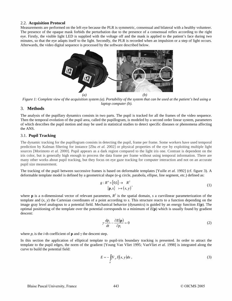
2.2. Acquisition Protocol Measurements are performed on the left eye because the PLR is symmetric, consensual and bilateral with a healthy volunteer. The presence of the opaque mask forbids the perturbation due to the presence of a consensual reflex according to the right eye. Firstly, the visible light LED is supplied with the voltage off and the mask is applied to the patient’s face during two minutes, so that the eye adapts itself to the light. Secondly, the PLR is recorded when an impulsion or a step of light occurs. Afterwards, the video digital sequence is processed by the software described below.
3. Methods
(a) (b) Figure 1: Complete view of the acquisition system (a). Portability of the system that can be used at the patient’s bed using a
laptop computer (b).
The analysis of the pupillary dynamics consists in two parts. The pupil is tracked for all the frames of the video sequence. Then the temporal evolution of the pupil area, called the pupillogram, is modeled by a second order linear system, parameters of which describes the pupil motion and may be used in statistical studies to detect specific diseases or phenomena affecting the ANS.
3.1. Pupil Tracking The dynamic tracking for the pupillogram consists in detecting the pupil, frame per frame. Some workers have used temporal prediction by Kalman filtering for instance [Zhu et al. 2002] or physical properties of the eye by exploiting multiple light sources [Morimoto et al. 2000]. Pupil appears as a dark region compared to the light iris one. Contrast is dependent on the iris color, but is generally high enough to process the data frame per frame without using temporal information. There are many other works about pupil tracking, but they focus on eye gaze tracking for computer interaction and not on an accurate pupil size measurement.
The tracking of the pupil between successive frames is based on deformable templates [Yuille et al. 1992] (cf. figure 3). A deformable template model is defined by a geometrical shape (e-g circle, parabola, ellipse, line segment, etc.) defined as:
[ ]{ } ( )yxs
RRg n
,,1;0: 2
ap→× , (1)
where p is a n-dimensional vector of relevant parameters, R2 is the spatial domain, s a curvilinear parameterization of the template and (x, y) the Cartesian coordinates of a point according to s. This structure reacts to a function depending on the image gray level analogous to a potential field. Mechanical behavior (dynamics) is guided by an energy function E(p). The optimal positioning of the template over the potential corresponds to a minimum of E(p) which is usually found by gradient descent:
( ) 0=∂∂
−i
i
pE
dtdp pγ (2)
where pi is the i-th coefficient of p and γ the descent step.
In this section the application of elliptical template to pupil-iris boundary tracking is presented. In order to attract the template to the pupil edges, the norm of the gradient [Young Van Vliet 1995; VanVliet et al. 1998] is integrated along the curve to build the potential field:
( )∫ ∇−=g
dsyxIE ,σ , (3)
Blaise Pascal University, France 443 © OICMS 2005

where is a smoothed version of the image gradient, obtained by convolving the image with the first derivative of a Gaussian kernel with standard deviation σ. More robust fields could be obtained by applying, for example, a region based strategy.
Iσ∇
The prior knowledge about the elliptical shape is exploited in the energy minimizing scheme. The ellipse is determined by a set of five parameters (cf. figure 2):
( ) ( ) ( ) ( )( ) ( ) ( ) ( ) πθ
αθαθαθαθ
≤≤⎩⎨⎧
++=−+=
0cossinsincossinsincoscos
210
210
llyyllxx
, (4)
where, (x0,y0) is the coordinates of the ellipse center, l1 and l2 are the major and minor ellipse axes, and α is the orientation angle with respect to the horizontal axis.
The advantages of this approach are:
Figure 2: Parameterization of the ellipse used for the deformable template method
y
α
(x0,y0)
l2 l1
x
no need for post-processing to obtain a closed curve;
the use of prior knowledge about the pupil geometry;
the choice of a parameterized template constrains the model and therefore increases the robustness and speed of convergence, contrary to other active contours;
the subpixel precision;
and the simple estimation of the pupil area, denoted S, by the formula:
(5) 21.. llS π=
3.2. Deformable Model Initialization
Next iteration i ←i+1
Processing of the ith frame1 video
sequence:i: 1..N frames
Elliptic deformable template [4]
Initialization Convergence
S(t)
t
Next iteration i ←i+1
Processing of the ith frame1 video
sequence:i: 1..N frames
Elliptic deformable template [4]
Initialization Convergence
S(t)
t
S(t)
t
Figure 3: Summary of the method: the video film is considered frame per frame and for each one, an elliptic deformable
template permit to estimate the pupil area.
As for all methods based on active contours, initialization is crucial. Indeed, deformable template converges to the closest local minimum, so initialization acts on the final result. Therefore, a method based on thresholding and binary mathematical morphology [Matheron 1975; Serra 1982] is proposed in order to extract an initial approximation for the pupil location.
A pre-processing step is necessary because of the spots due to the reflection of the IR LEDs. These spots look like two reflection circles in the image, so a gray level mathematical morphology closing is applied by using a circular structuring
Blaise Pascal University, France 444 © OICMS 2005

element. They are removed with hardly any change in the pupil geometry because of its smooth shape. The contrast of the pupil leads to a bimodal histogram that makes easy the determination of a global threshold by the use of the classical Otsu criterion.
The object corresponding to the pupil is extracted from the binary image by mathematical morphology operators. A binary closing is applied and all resulting connected components are labeled. The biggest one is used to initialize the ellipse parameters. Its center is given by the center of gravity and its axes are computed by identifying the surface of the object with the one of an approximating circle.
3.3. Mathematical Modeling of Pupillar Dynamics There have been several models to attempt to simulate pupillary dynamics. Longtin and Milton [Longtin Milton 1988] have developed a model of the PLR using nonlinear delay-differential equations. This technique from dynamical systems theory is used to analyze complex behaviors such as the onset of oscillations under high-gain negative feedback.
In the literature [Bressloff Wood 1998; Longtin Milton 1988], pupillary control system is modeled as a linear feedback control system. The pupil contraction system can be considered as a closed-loop system with the afferent activity that acts as the amplifier and the efferent activity as the feedback (cf. figure 4a). The proposed model of the PLR, motivated by physiological knowledge and empirical observation, is the delayed step response of a second order linear system (cf. figure 4b) that can be expressed by a second order linear ordinary differential equation:
( ) ( ) ( ) (teKtsdttdsm
dttsd
⋅=−+−
⋅⋅
+−
⋅ ττω
τω
212
2
2 ) (6)
where s(t) is the output temporal signal, e(t) the excitation signal, m the damping coefficient, K the static gain, ω the natural frequency and τ the latency time. It operates essentially in the underdamped case (m<1). This modeling enables to reduce the information into a set of parameters that have a physiological meaning. In fact, the latency time is the reaction delay of the system, the natural frequency is homogeneous to the rapidity and the static gain describes the area difference between the mydriasis and the miosis.
Parameters are identified by a Marquardt-Levenberg [Marquardt 1963; Press et al. 1986] least-square fit of the model onto the experimental pupillogram. Indeed, the solution of second order linear ordinary differential equation (6) is not linear according to parameters, so the use of a non linear method is mandatory to solve the estimation problem.
4. Results and Discussion
afferent activity
light iris
efferent activity(parasympathetic)
Midbrain
afferent activity
light iris
efferent activity(parasympathetic)
Midbrain
s(0)
s(∞)
τ0 t
s(%)
m
∝ω
s(0)
s(∞)
τ0 t
s(%)
m
∝ω
(a) (b) Figure 4: Modeling of the reflex loop based on physiologic knowledge (a) by the response of second order linear system (b).
4.1. Methodological Tests The sensor of the camera is a CCD matrix, so the image is embedded with an additive Gaussian noise. The precision of the measure versus signal to noise ratio (SNR) is plotted on figure 5a. The study was done with a synthetic image of a dark ellipse on a light background with noise added. The use of a deformable model enables to increase the stability of the measure because of the sub pixel precision coupled to a parametric shape that constrains the solution.
An interesting test is the validation of the model of PLR itself. Figure 5b shows the normalized average error and the standard deviation between the model and the pupillogram for 52 recordings on different people stored during a previous study done on voluntaries under morphine injection [Tilmant et al. 2003a]. The quadratic distance, normalized to the pupil area variation, is used as a measure of the error and is plotted against the number of samples of the pupillogram, i.e. the duration of acquisition after the light step. An optimal time of 1.13 s was found with model (6), corresponding to a minimal normalized average quadratic error of 0.07 % in area. This increase of the difference between the model and the experimental data can be explained by the adaptation of the retina sensitivity to light that causes the pupil to compensate. It permits to set the phenomena duration to modeling.
Blaise Pascal University, France 445 © OICMS 2005

The use of more realistic physiological models is envisaged in order to analyze more accurately pupillary dynamics. For example the use of the Longtin-Milton model [Longtin Milton 1988] could be considered.
4.2. Clinical applications
(a) (b)
Figure 5: Standard deviation of the measure with the signal to noise ratio (SNR)(a). Average and standard deviation of the normalized error between the dataset and the models for different numbers of temporal samples (b) [Tilmant et al. 2003a].
The purpose of clinical work was to determine the physiological characteristics of the loop reflex of the PLR for patients in basal state and in particular situations: patients under medication (opioïds, hypnotic...), or particular neurological states: brain trauma or intracerebral hemorrhage.
The purpose of the first study, based on 95 voluntaries, was to check the influence of the color of the iris on the PLR. By using the previous modeling method, it was not discovered significant difference over the latency time: 0.190 +/- 0.03 second in the group "dark eyes" (DE) versus 0.189 +/- 0.04 second in the group "clear eyes" (CE). In the same way, no difference was found for the damping coefficient: 1.17 (DE) versus 1.2 (CE), neither for the natural frequency, nor for the maximum speed of contraction (Appendix A). The studied population comprised young adults (mean age = 36 +/- 8 years). Conversely, the study confirms that the size of the pupil in population CE is lower (0.26 % of the size of the iris), with that of population DE (0.30 % of the size of the iris); this phenomenon is described in the literature.
Latency Time (s)
00.05
0.10.15
0.20.25
0.3
t0 t1 t2 t3
s(0) (% of iris area)
0%5%
10%15%20%25%30%
t0 t1 t2 t3
s(infini) (% of iris area)
0%
4%
8%
12%
16%
20%
t0 t1 t2 t3 (a) (b) (c)
Pupil Area Ratio
0%
20%
40%
60%
80%
100%
t0 t1 t2 t3
Damping Coefficient
0.40.50.60.70.80.9
11.1
t0 t1 t2 t3
Natural Frequency (rd/s)
8
9
10
11
12
13
t0 t1 t2 t3 (d) (e) (f)
Figure 6: Values of the modeling parameters in case of electrical cardioversion. Four acquisitions were done: before the anesthesia (t0), after injection of propofol (t1), after electrical cardioversion (t2) and finally when after product elimination
(t3). Latency time (a), initial pupil area (b), final pupil area (c), pupil area ratio (d), damping coefficient (e) and natural frequency (f) are given.
The second study relates to the influence of hypnotics on the loop reflex of the PLR. The studied population concerns 35 patients having to be anaesthetized without opioïds for electrical cardioversion. The hypnotic used was the propofol. The eye was filmed before anesthesia, under anesthesia (with a cerebral concentration of 3 µg/ml of propofol) before and one minute
Blaise Pascal University, France 446 © OICMS 2005

after electrical cardioversion (so the sympathic stimulation is finished), and finally at the waking up after complete elimination of the drug.
Statistical analysis acts on each parameter of the model independently from the others. The standard level of significance used to justify a claim of a statistically significant effect is 0.05. The differences at each time are computed according to parameters estimated before anesthesia (t0).
The latency time and the pupil area ratio do not appear to be influenced by propofol or by shock (cf. figures 6a&d). Conversely, pupil sizes (mydriasis and miosis) are decreased under anesthesia (cf. figure 6b&c). The fact that the opioids lead to a decreasing size of the pupil is a well know result, but the effects of hypnotics are not well described in the literature. The damping coefficient is decreased under anesthesia but it does not present a significant difference after the shock. Finally, the natural frequency shows a significant variation after the shock but not before. The forthcoming of this work concerns the link between these parameters and their changes with a possible defect of anesthesia (cf. figure 6).
The studies on the opioids and the head injuries are in hand.
5. Conclusions A tool to analyze pupillary dynamics was presented. An image analysis based on deformable template followed by a mathematical modeling of the PLR was proposed. For now, a first clinical study using this tool was performed to quantify the consequences of morphine on the PLR [Tilmant et al. 2003b]. On the whole, these studies allow a better knowledge of physiology and pharmacological effects on the PLR that is at the junction of the neurovegetative systems. ACKNOWLEDGMENTS The authors want to thank the Synapsys society for the gratuitous loan of the acquisition system and are grateful to Dr. V. Fontvieille and Dr. A. Virat for valuable assistance in the clinical validation and tool conception. This study was partially supported by a grant from the Center of Clinical Pharmacology (Pr. C. Dubray), Clermont-Ferrand, France.
APPENDIX A: PARAMETERS EXTRACTED FROM THE MODEL One interest of this modeling is the use of parameters deriving from the analytic representation of the phenomenon, giving access to physiological parameters in a less noisy and more robust way. The maximal velocity and its apparition time are two parameters usually used in the study of pupillary dynamics. They can be expressed in an analytical way for the three conditions relative to the dumping coefficient:
⎟⎟
⎠
⎞
⎜⎜
⎝
⎛
−−
−+
−+=
1
1ln12
12
2
2maxmm
mm
mtV
ωτ
m>1
( ) ( )( ) ( ) ( ) ⎥⎦⎤
⎢⎣⎡
⎟⎠⎞⎜
⎝⎛ −⎟
⎠⎞⎜
⎝⎛ −−−−⎟
⎠⎞⎜
⎝⎛ −⎟
⎠⎞⎜
⎝⎛ −+−⎟
⎟⎠
⎞⎜⎜⎝
⎛
−∞−= τωτωω
max2
max2
2max 1exp1exp12
0 VV tmmtmmm
ssv
ωτ 1
max +=Vt m=1
( ) ( )( )e
ssv ω0max −∞=
⎥⎥
⎦
⎤
⎢⎢
⎣
⎡
⎟⎟
⎠
⎞
⎜⎜
⎝
⎛
⎟⎟⎠
⎞⎜⎜⎝
⎛
−−⎟
⎟⎠
⎞⎜⎜⎝
⎛
−
−
−+=
22
2
2max1
arctan12
12arctan1
1
m
m
mm
m
mtV
ωτ
m<1
( ) ( )( ) ( )( )( )
( )⎥⎥⎥⎥⎥⎥
⎦
⎤
⎢⎢⎢⎢⎢⎢
⎣
⎡
⎟⎟
⎠
⎞
⎜⎜
⎝
⎛
⎟⎟⎠
⎞⎜⎜⎝
⎛
−+−−−
⎟⎟
⎠
⎞
⎜⎜
⎝
⎛
⎟⎟⎠
⎞⎜⎜⎝
⎛
−+−−
−−−−∞=
2max2
2max2
2
maxmax
1arctan1cos
1arctan1sin
1exp0
m
mtm
m
mtmm
m
tmssv
V
V
V
τω
τω
τωω
Blaise Pascal University, France 447 © OICMS 2005

REFERENCES [Ambrosio et al. 2002] Ambrosio, R., Schallhorn, S.C., and Wilson., S.E. The importance of pupil size in refractive
surgery. Refractive Surgery Outlook, American academy of ophtalmology, fall 2002. (http://www.aao.org/aao/member/sig/upload/Fall-2003-Outlook.pdf)
[Duffey Leaming 2004] Duffey, R., and Leaming, D. Trends in refractive surgery in the United States. Journal of Cataract & Refractive Surgery, 30-8: 1781-1785, 2004.
[Loewensfeld 1993] Loewensfeld, I.E. The pupil : Anatomy, physiology, and clinical applications. Iowa City: Iowa State University, 1993.
[Kawasaki Kardon 2001] Kawasaki, A., and Kardon, R. Disorders of the pupil. Neuroophthalmology, 14- 1: 149-168, 2001.
[Mylius et al. 2003] Mylius, V., Braune, H.J., and Schepelmann, K. Dysfunction of the pupillary light reflex following migraine headache. Clinical Autonomic Research, 13- 1: 16-21, 2003.
[Bertinotti et al. 2002] Bertinotti, L., Pietrini, U., Del Rosso, A., Casale, R., Colangelo, N., Zoppi, M., and Matucci-Cerinic, M. The use of pupillometry in joint and connective tissue diseases. Annals of the New York Academy of Sciences, 966: 446-455, 2002.
[Manley Larson 2002] Manley, G.T., and Larson, M.D. Infrared pupillometry during uncal herniation. Journal of Neurosurgical Anesthesiology, 14- 3: 223-228, 2002.
[Taylor et al. 2003] Taylor, W.R., Chen, J.W., Meltzer, H., Gennarelli, T.A., Kelbch, C., Knowlton, S., Richardson, J., Lutch, M.J., Farin, A., Hults, K.N., and Marshall, L.F. Quantitative pupillometry, a new technology: Normative data and preliminary observations in patients with acute head injury. Technical note. Journal of Neurosurgery, 98- 1: 205-213, 2003.
[Tassorelli et al. 1995] Tassorelli, C., Micieli, G., Osipova, V., Rossi, F., and Nappi, G. Pupillary and cardiovascular responses to the cold-pressor test. Journal of the Autonomic Nervous System, 55- (1-2): 45-49, 1995.
[Barvais et al. 2003] Barvais, L., Engelman, E., Eba, J.M., Coussaert, E., Cantraine, F., and Kenny, G. Effect site concentrations of remifentanil and pupil response to noxious stimulation. British Journal of Anaesthesia, 91- 3: 347-352, 2003.
[Onal Tuglular 1999] Onal, A., and Tuglular, I. The relationship between pupil diameter and pain by the administration of morphine and antidepressant drugs in mice. General Pharmacology, 33- 1: 83-89, 1999.
[Larson et al. 1993] Larson, M.D., Sessler, D.I., Ozaki, M., McGuire, J., and Schroeder, M. Pupillary assessment of sensory block level during combined epidural/general anesthesia. Anesthesiology, 79-1: 42-48, 1993.
[Knaggs et al. 2004] Knaggs, R., Crighton, I., Cobby, T., Fletcher, A., and Hobbs, G. The pupillary effects of intravenous morphine, codeine, and tramadol in volunteers. Anesthesia & Analgesia, 99: 108-112, 2004.
[Richman Noriega 2002] Richman, J., and Noriega, R. The sensitivity and specificity of infrared pupillometry measurements in identifying drug impairment in a country probation program. Forensics Drug Study, Scientific Program American Academy of Optometry, 2002. (http://www.mcjeyecheck.com/text/studies/Forensiscs Drug Study 2002.html )
[Murillo et al. 2004] Murillo, R., Crucilla, C., Schmittner, J., Hotchkiss, E., and Pickworth, W.B. Pupillometry in the detection of concomitant drug use in opioid-maintained patients. Methods and Findings in Experimental and Clinical Pharmacology, 26- 4: 271-275, 2004.
[Zhu et al. 2002] Zhu, Z., Ji, Q., Fujimura, K., and Lee, K. Combining kalman filtering and mean shift for real time eye tracking under active IR illumination. 16th International Conference on Pattern Recognition (Quebec City, Canada), 11-15 August, 2002.
[Morimoto et al. 2000] Morimoto, C., Koons, D., Amir, A., and Flickner, M. Pupil detection and tracking using multiple light sources. Image and Vision Computing, Special issue on Advances in Facial Image Analysis and Recognition Technology,18- 4: 331-335, 2000.
[Yuille et al. 1992] Yuille, A.L., Hallinan, P.W., and Cohen, D.S. Feature extraction from faces using deformable templates. International Journal of Computer Vision, 8: 99-111, 1992.
[Young Van Vliet 1995] Young, I.T., and Van Vliet, L. J. Recursive implementation of the Gaussian filter. Signal Processing, 44- 2: 139-151, 1995.
Blaise Pascal University, France 448 © OICMS 2005

[VanVliet et al. 1998] Van Vliet, L. J., Young, I. T., and Verbeek, P. W. Recursive Gaussian derivative filters. 14th International Conference on Pattern Recognition (Brisbane, Australia), 16-20 August, 1998.
[Matheron 1975] Matheron, G. Random sets and integral geometry. New-York: John Wiley & Sons, 1975.
[Serra 1982] Serra, J. Image analysis and mathematical morphology. London: Academic Press, 1982.
[Longtin Milton 1988] Longtin, A., and Milton, J. Complex oscillations in the human pupil light reflex with mixed and delayed feedback. Mathematical Biosciences, 90: 193-199, 1988.
[Bressloff Wood 1998] Bressloff, P.C., and Wood, C.V. Spontaneous oscillations in a nonlinear delayed-feedback shunting model of the pupil light reflex. Physical review E, 58- 3:. 3597-3605, 1998.
[Marquardt 1963] Marquardt, D.W. An algorithm for least-squares estimation of nonlinear parameters. Journal of the Society for Industrial and Applied Mathematics, 11- 2:431-441, 1963
[Press et al. 1986] Press, W.H., Flannery, B.P., Teukolsky, S.A., and Vetterling, W.T. Numerical Recipes. Cambridge: Cambridge University Press, 1986.
[Tilmant et al. 2003a] Tilmant, C., Sarry, L., Gindre, G., Boire, J.-Y. Monitoring and modeling of pupillary dynamics. IEEE Engineering in Medicine and Biology Society 25th Annual International Conference (Cancun, Mexico), 17-21 September 2003.
[Tilmant et al. 2003b] Tilmant, C., Ponrouch, M., Virat, A., Gindre, G., Dubray, C., and Schoeffler, P. Monitoring of pupillary dynamics: study of the pupil light reflex under morphine (in French). 10th Meeting of the French Society for Informatics and Monitoring in Anesthesia and Reanimation (Nantes, France), 4-5 April 2003.
Blaise Pascal University, France 449 © OICMS 2005
Related Documents